synaptic transmission synapse – specialized junction where an axon terminal contacts another...
TRANSCRIPT
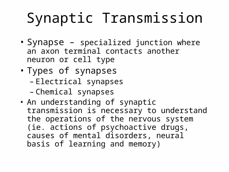
Synaptic Transmission
• Synapse – specialized junction where an axon terminal contacts another neuron or cell type
• Types of synapses– Electrical synapses – Chemical synapses
• An understanding of synaptic transmission is necessary to understand the operations of the nervous system (ie. actions of psychoactive drugs, causes of mental disorders, neural basis of learning and memory)
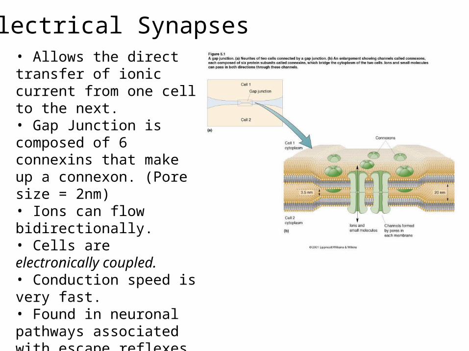
Electrical Synapses• Allows the direct transfer of ionic current from one cell to the next.• Gap Junction is composed of 6 connexins that make up a connexon. (Pore size = 2nm)• Ions can flow bidirectionally.• Cells are electronically coupled.• Conduction speed is very fast.• Found in neuronal pathways associated with escape reflexes or in neurons that need to be synchronized.• Common in non neuronal cells.•Important in development
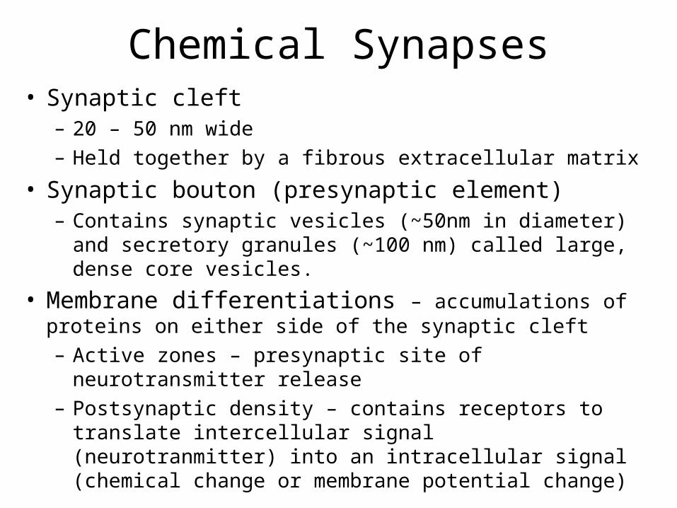
Chemical Synapses• Synaptic cleft
– 20 – 50 nm wide
– Held together by a fibrous extracellular matrix
• Synaptic bouton (presynaptic element)– Contains synaptic vesicles (~50nm in diameter) and secretory
granules (~100 nm) called large, dense core vesicles.
• Membrane differentiations – accumulations of proteins on either side of the synaptic cleft
– Active zones – presynaptic site of neurotransmitter release
– Postsynaptic density – contains receptors to translate intercellular signal (neurotranmitter) into an intracellular signal (chemical change or membrane potential change)
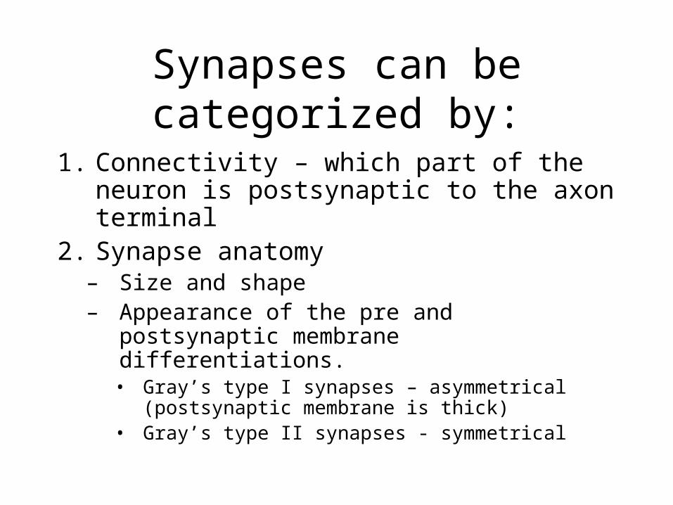
Synapses can be categorized by:
1. Connectivity – which part of the neuron is postsynaptic to the axon terminal
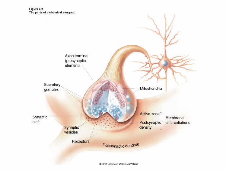
2. Synapse anatomy– Size and shape– Appearance of the pre and postsynaptic
membrane differentiations.• Gray’s type I synapses – asymmetrical
(postsynaptic membrane is thick)• Gray’s type II synapses - symmetrical
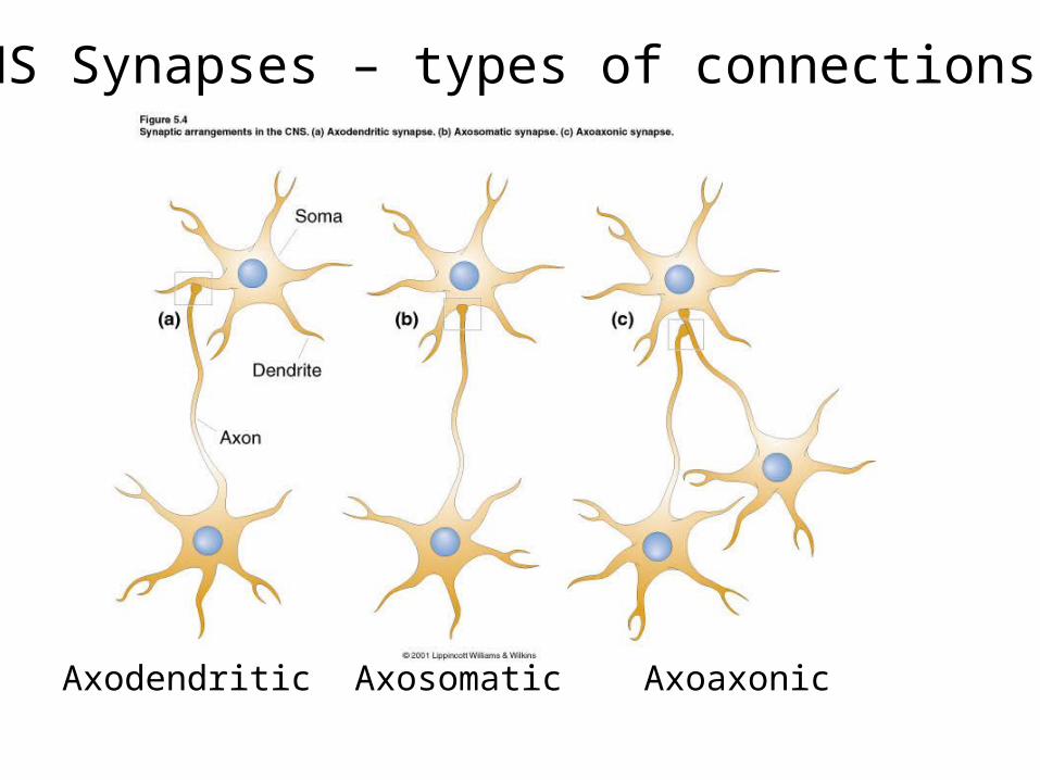
CNS Synapses – types of connections
Axodendritic Axosomatic Axoaxonic
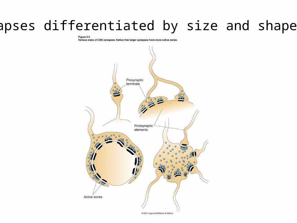
Synapses differentiated by size and shape.
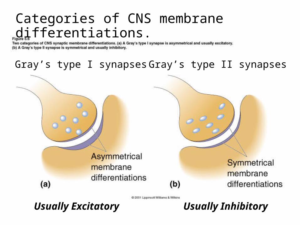
Categories of CNS membrane differentiations.
Gray’s type I synapses Gray’s type II synapses
Usually Excitatory Usually Inhibitory
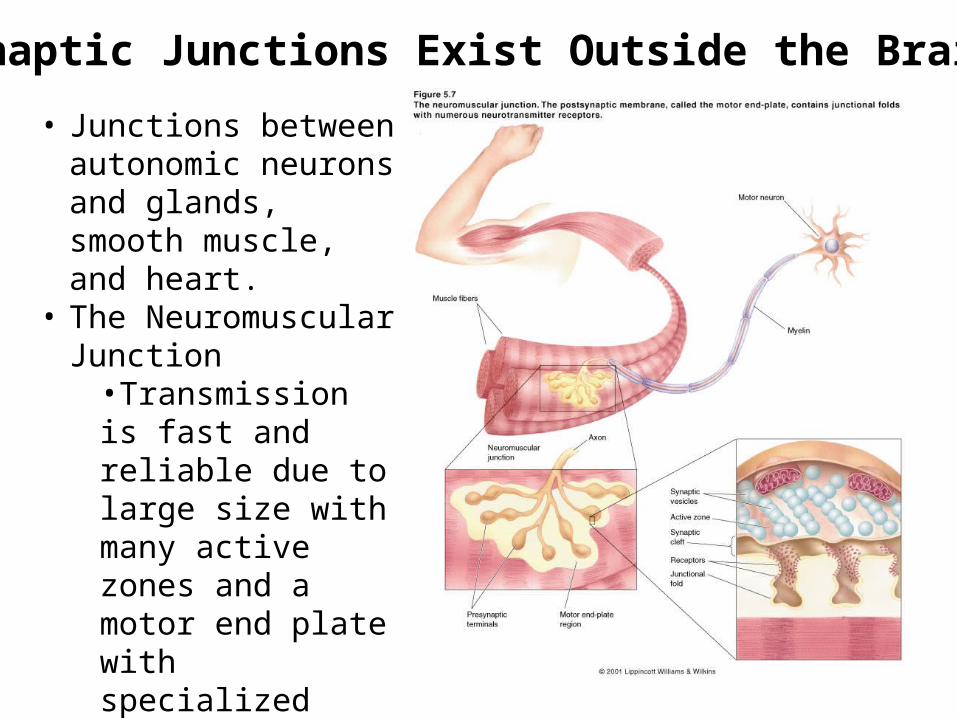
•Synaptic Junctions Exist Outside the Brain
• Junctions between autonomic neurons and glands, smooth muscle, and heart.
• The Neuromuscular Junction
•Transmission is fast and reliable due to large size with many active zones and a motor end plate with specialized folds for more receptors.
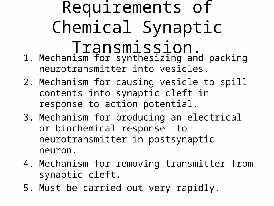
Requirements of Chemical Synaptic Transmission.
1. Mechanism for synthesizing and packing neurotransmitter into vesicles.
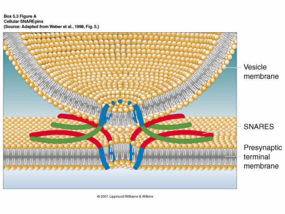
2. Mechanism for causing vesicle to spill contents into synaptic cleft in response to action potential.
3. Mechanism for producing an electrical or biochemical response to neurotransmitter in postsynaptic neuron.
4. Mechanism for removing transmitter from synaptic cleft.
5. Must be carried out very rapidly.
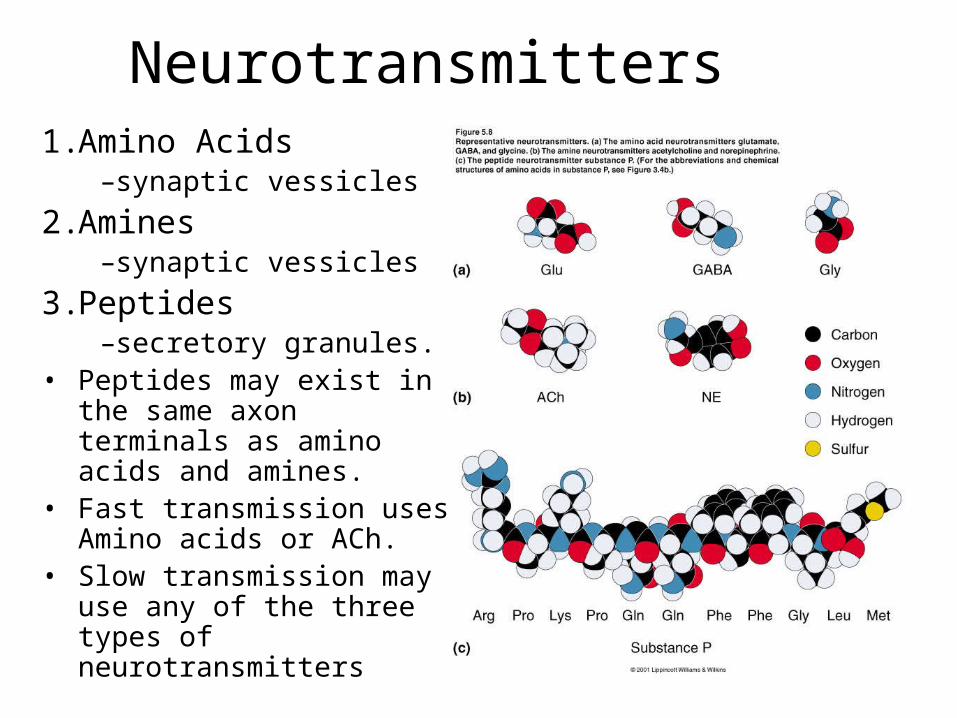
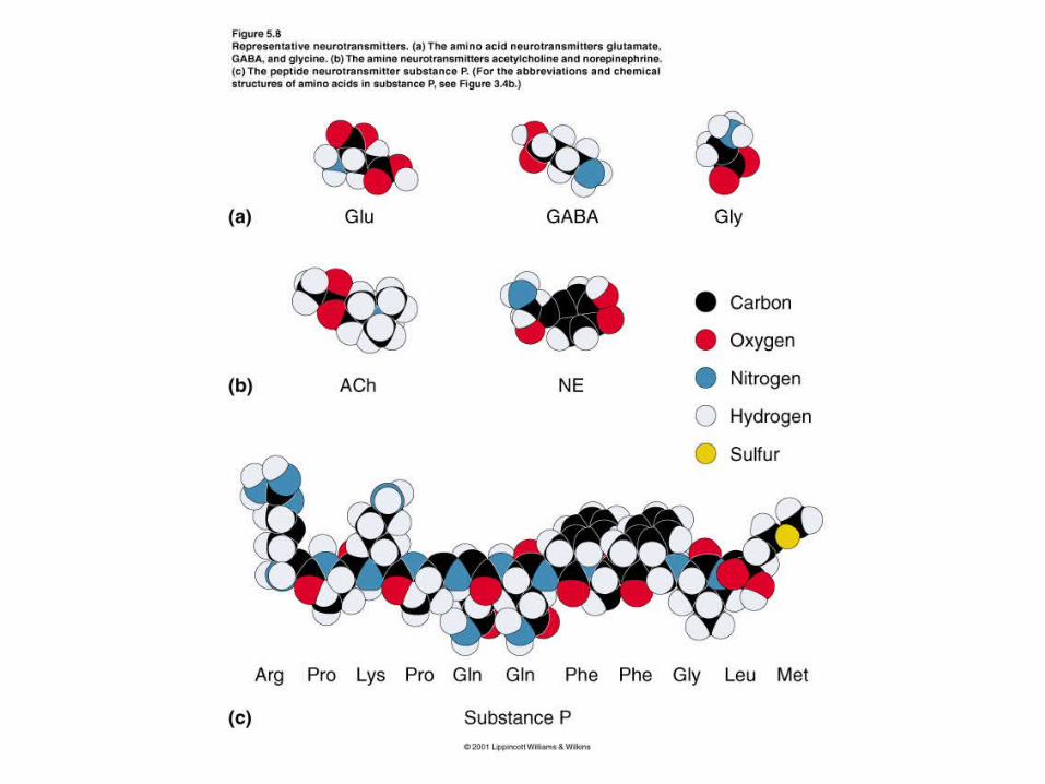
Neurotransmitters1. Amino Acids
–synaptic vessicles
2. Amines–synaptic vessicles
3. Peptides–secretory granules.
• Peptides may exist in the same axon terminals as amino acids and amines.
• Fast transmission uses Amino acids or ACh.
• Slow transmission may use any of the three types of neurotransmitters
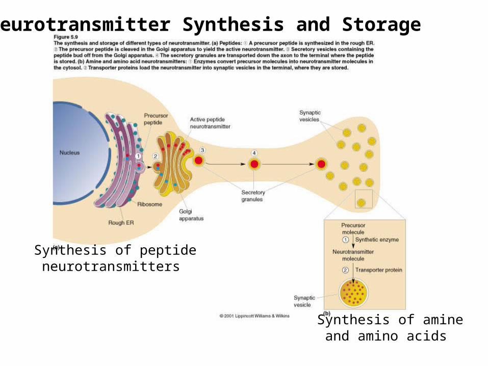
Synthesis of peptide neurotransmitters
Synthesis of amine and amino acids
Neurotransmitter Synthesis and Storage
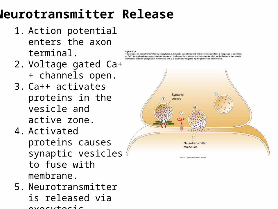
Neurotransmitter Release1. Action potential enters the
axon terminal.2. Voltage gated Ca++
channels open.3. Ca++ activates proteins in
the vesicle and active zone.
4. Activated proteins causes synaptic vesicles to fuse with membrane.
5. Neurotransmitter is released via exocytosis.
Note: Peptide release requires high frequency action potentials and is slower (50 msec vs. 0.2 msec).
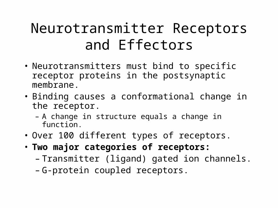
Neurotransmitter Receptors and Effectors
• Neurotransmitters must bind to specific receptor proteins in the postsynaptic membrane.
• Binding causes a conformational change in the receptor.– A change in structure equals a change in function.
• Over 100 different types of receptors.• Two major categories of receptors:
– Transmitter (ligand) gated ion channels.– G-protein coupled receptors.
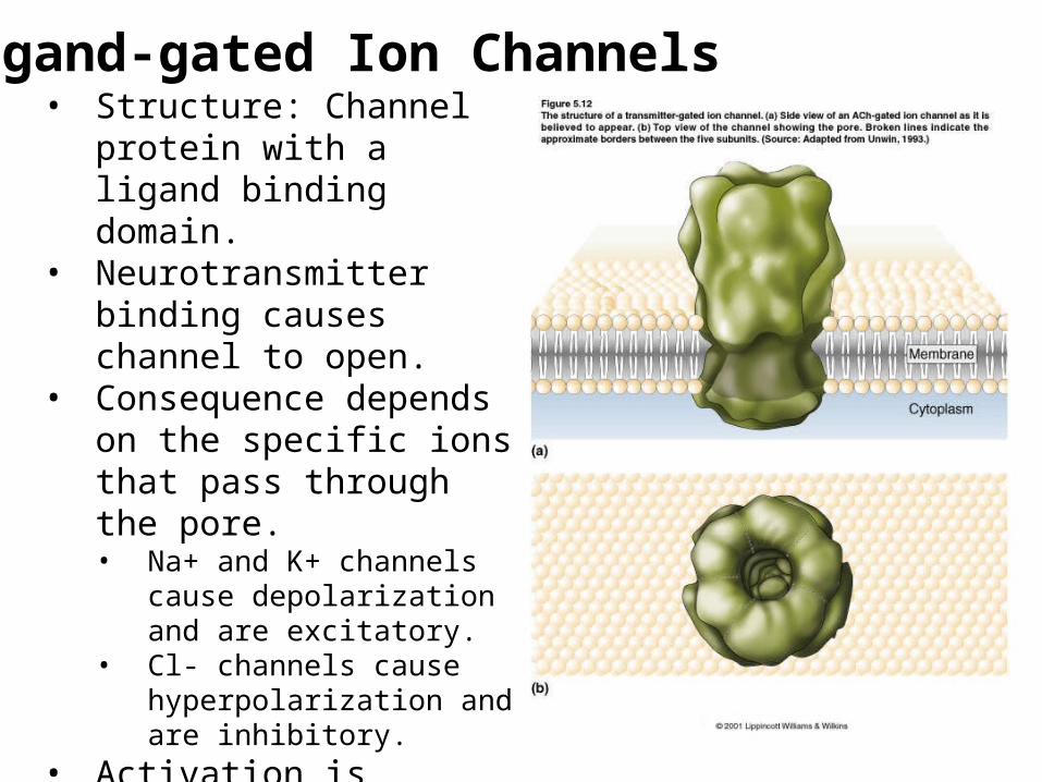
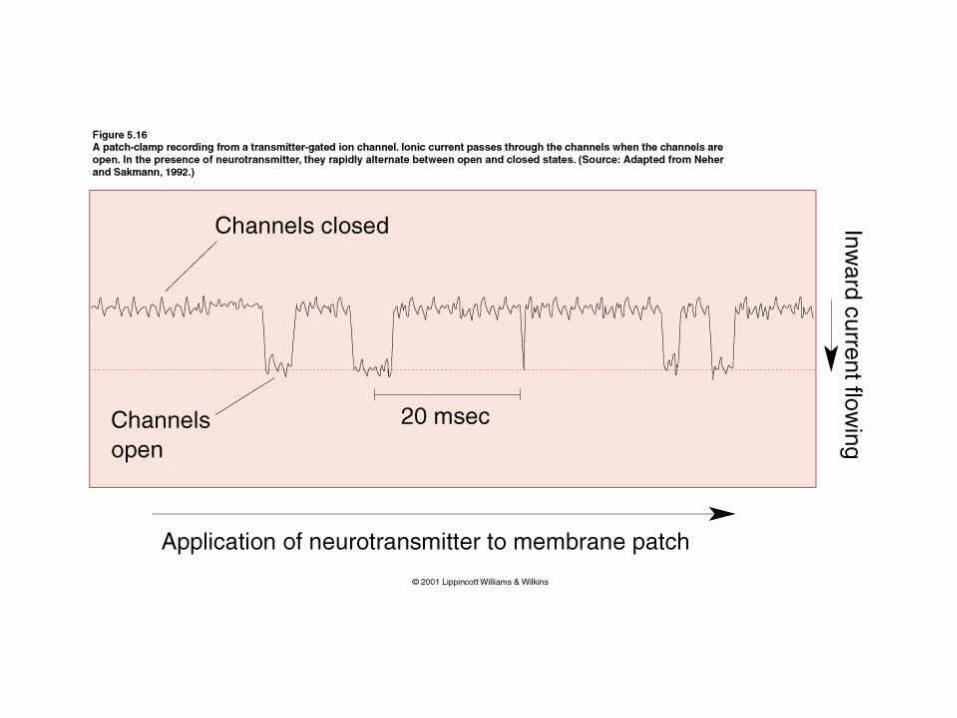
Ligand-gated Ion Channels• Structure: Channel protein
with a ligand binding domain.• Neurotransmitter binding
causes channel to open.• Consequence depends on the
specific ions that pass through the pore.• Na+ and K+ channels cause
depolarization and are excitatory.
• Cl- channels cause hyperpolarization and are inhibitory.
• Activation is generally rapid and is mediated by amino acids and amines.
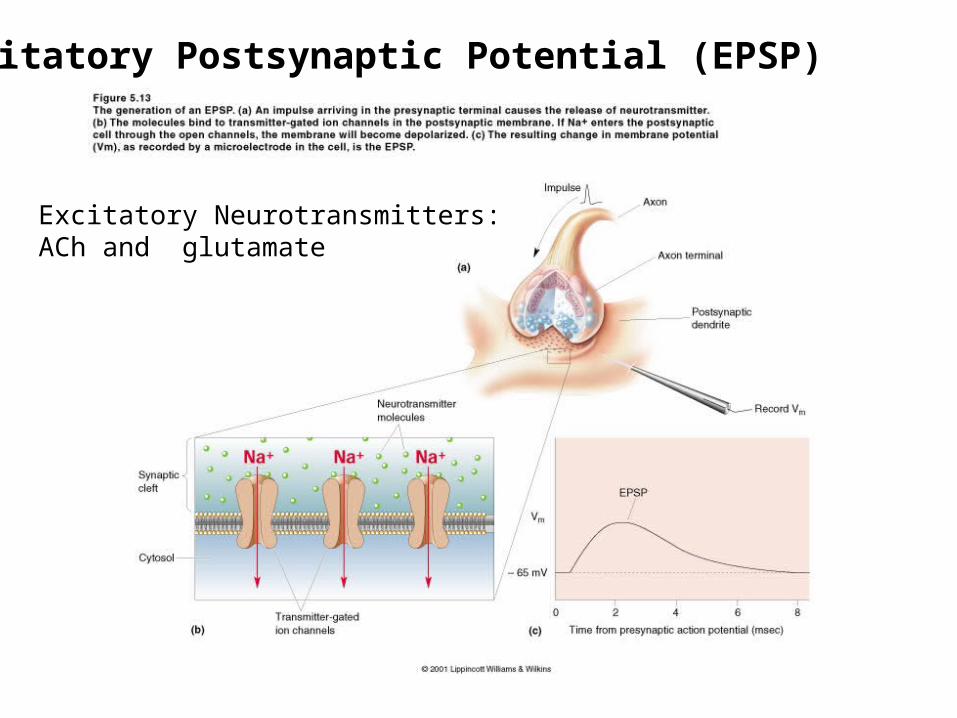
Excitatory Postsynaptic Potential (EPSP)
Excitatory Neurotransmitters:ACh and glutamate
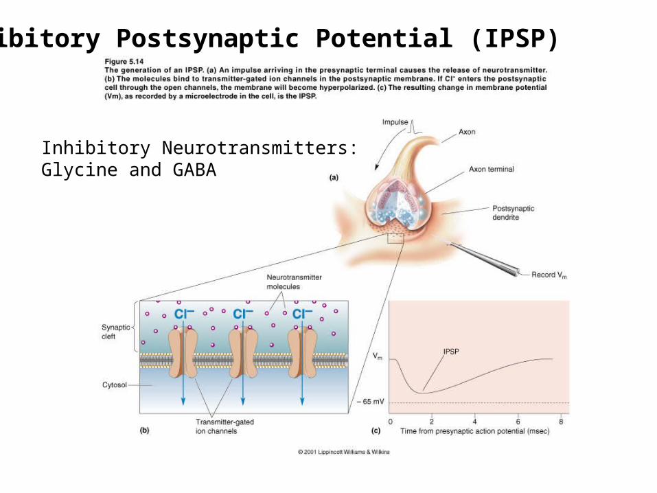
Inhibitory Postsynaptic Potential (IPSP)
Inhibitory Neurotransmitters:Glycine and GABA
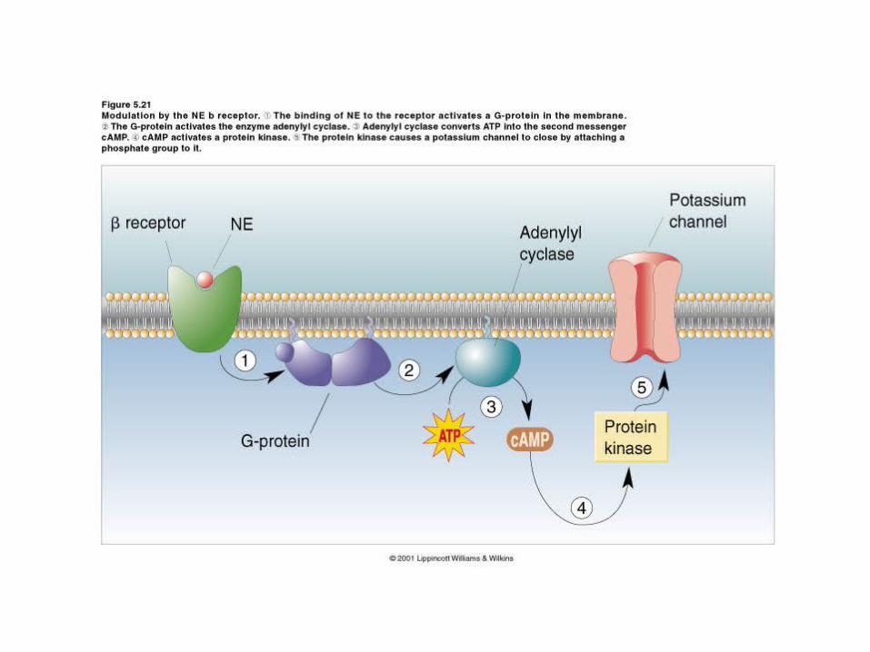
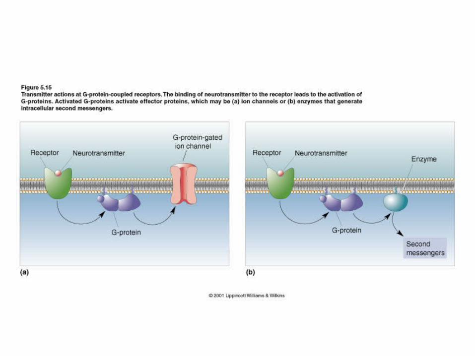
G Protein-Coupled Receptors• Structure: Receptor protein with a ligand binding
domain and connected to G –protein consisting of an alpha, beta and gama subunit.
• Activation: 1) Ligand binds to receptor; 2) Receptor activates G-protein; 3) G-protein dissociated; 4) alpha subunit activates an effector protein.
• Effectors …G-proteins act in one of two ways:– By opening ion channels– By activating enzymes that synthesize second-
messenger molecules.• Tend to be slower, longer lasting and have greater
diversity than ligand gated ion channels.• Ligand may bind to a family of receptors with different
effects due to specific receptor type.
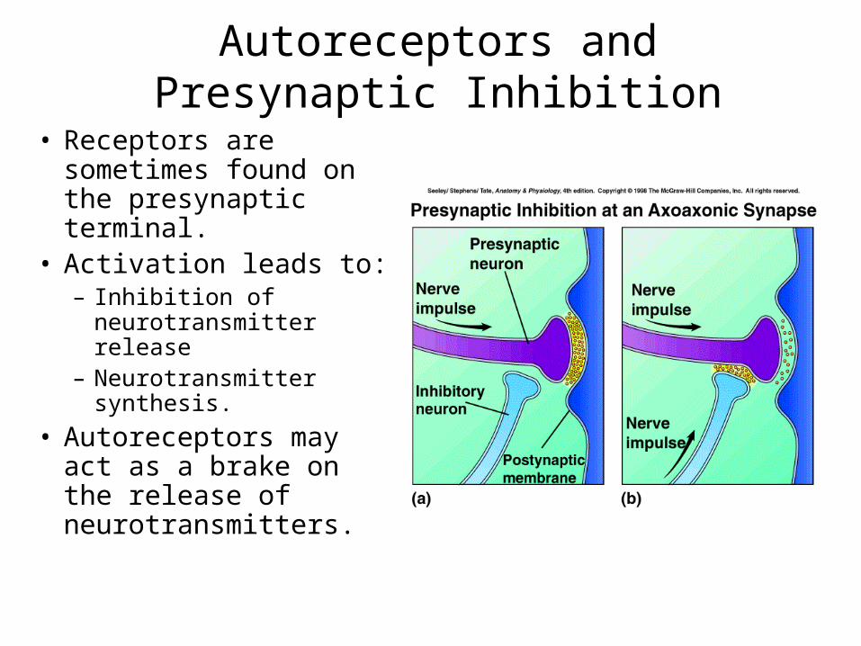
Autoreceptors and Presynaptic Inhibition
• Receptors are sometimes found on the presynaptic terminal.
• Activation leads to:– Inhibition of
neurotransmitter release– Neurotransmitter
synthesis.
• Autoreceptors may act as a brake on the release of neurotransmitters.
Neurotransmitter Recovery and Degradation
• Neurotransmitters must be cleared from the synapse to permit another round of synaptic transmission.
• Methods:– Diffusion– Enzymatic degradation in the synapse.– Presynaptic reuptake followed by degradation
or recycling.– Uptake by glia– Uptake by the postsynaptic neuron and
desensitization.
Neuropharmacology• Synaptic transmission is a chemical process and
therefore can be affected by drugs and toxins.• Neuropharmacology is the study of the effects of
drugs on the nervous system• Receptor Antagonists – inhibit the normal action of
a neurotransmitter.– Curare blocks the action of ACh at the neuromuscular
junction.
• Receptor Agonists – mimic the action of a neurotransmitter.– Morphine activates Mu-opiate receptors in the brain.
• Nervous system malfunctions are often related to neurotransmission errors.
Synaptic Integration• Each neuron may receive thousands of inputs in the
form of ion channel and G-coupled protein activation.
• These complex inputs give rise to simple output in the form of action potentials.– Neural computation
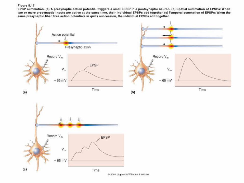
• Neurotransmitters are released in quanta.• EPSP Summation
– Neurons do sophisticated computations by adding together EPSPs to produce a significant postsynaptic depolarization.
– Types of Summation: Spatial and Temporal Summation.
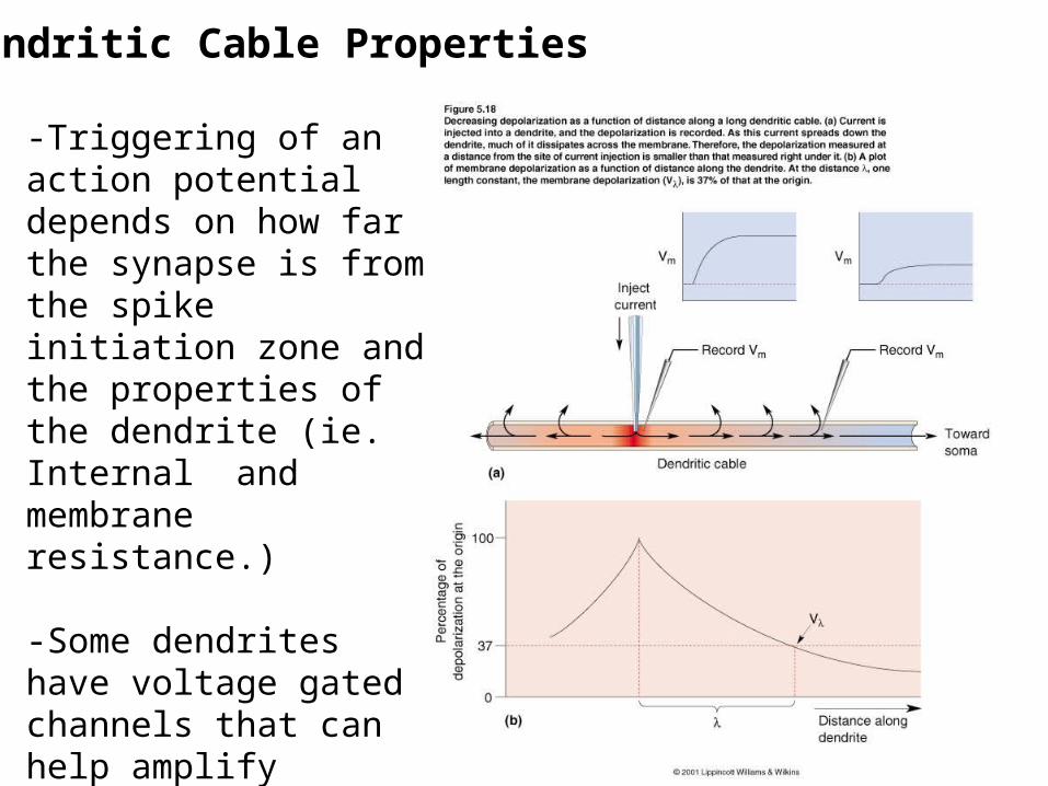
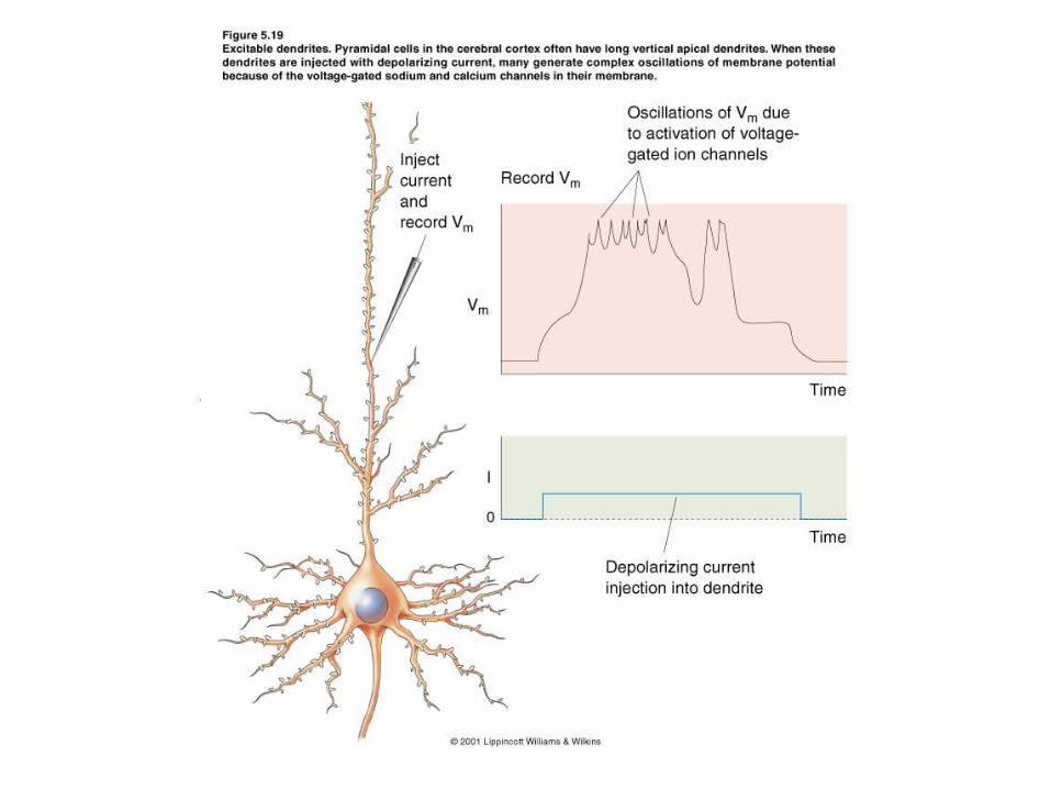
Dendritic Cable Properties
-Triggering of an action potential depends on how far the synapse is from the spike initiation zone and the properties of the dendrite (ie. Internal and membrane resistance.)
-Some dendrites have voltage gated channels that can help amplify signals along dendrites.
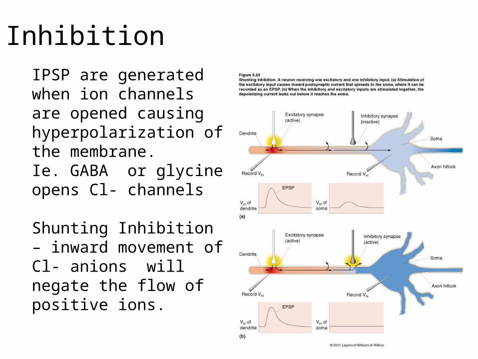
InhibitionIPSP are generated when ion channels are opened causing hyperpolarization of the membrane.Ie. GABA or glycine opens Cl- channels
Shunting Inhibition – inward movement of Cl- anions will negate the flow of positive ions.