synapse formation and preferential distribution in … journal of neuroscience, april 1992, 12(4):...
TRANSCRIPT
The Journal of Neuroscience, April 1992, 12(4): 1144-I 1.59
Synapse Formation and Preferential Distribution in the Granule Cell Layer by Regenerating Retinal Ganglion Cell Axons Guided to the Cerebellum of Adult Hamsters
T. J. Zwimpfer, A. J. Aguayo, and G. M. Bray
Centre for Research in Neuroscience, The Montreal General Hospital Research Institute and McGill University, Montreal, Quebec, Canada H3G lA4
To investigate constraints and preferences for synaptogene- sis in the injured mammalian CNS, regenerating retinal gan- glion cell (RGC) axons of adult hamsters were guided through a peripheral nerve (PN) graft to a target they do not usually innervate: the cerebellum (Cb). When identified by the pres- ence of HRP anterogradely transported from the retina 2-9 months later, such RGC axons were found to have extended into the cerebellar cortex for up to 650 pm. Most of this growth was in the granule cell layer (GCL) and only a few axons entered the molecular layer. The preference for the GCL could not be explained by the position of the PN graft in the Cb, a selective denervation of the GCL, local damage to other neurons, or the distribution of reactive gliosis in the vicinity of the graft. Furthermore, by EM, more than 95% of the labeled retinocerebellar terminals and synapses were in the GCL. Retinocerebellar terminals were larger and con- tained more synapses than the regenerated RGC terminals previously studied in the superior colliculus. These results indicate that regenerating axons of CNS neurons can form persistent synapses with novel targets. The preferential syn- aptogenesis in the GCL suggests that such unusual con- nections are not formed randomly in the CNS of these adult mammals.
After optic nerve (ON) transection in adult rats (Vidal-Sanz et al., 1987, 1991) or hamsters (Carter et al., 1989; Keirstead et al., 1989; Sauve et al., 199 I), retinal ganglion cell (RGC) axons regenerate through a peripheral nerve (PN) graft that joins the eye and the superior colliculus (SC) and establish functional synapses that persist in the SC, a normal target of retinofugal axons. A prominent feature of this regenerated retinocollicular (RGC-SC) projection is the selective reinnervation of the su- perficial, retinorecipient layers of the SC (Vidal-Sanz et al., 1987;
Received June 17, 1991; revised Oct. 22, 1991; accepted Oct. 25, 1991. The technical assistance of M. David J. Laganiere, S. Shinn, J. Trecarten, and
W. Wilcox is gratefully acknowledged. We thank S. Wang and Dr. M. Abraha- mowicz for statistical assistance, and Drs. D. Lawrence, M. Rasminsky, and C. Sotelo for reviewing earlier versions of the manuscript. T.J.Z. was supported by a Medical Research Council fellowshia. The Medical Research Council ofCanada. the Spinal Cord Research Foundation, the Multiple Sclerosis Society of Canada: and the Daniel Heumann Fund for Spinal Cord Research provided financial support. The laboratory in which these studies were carried out is part of the Canadian Network for the Study of Neural Regeneration and Functional Recovery.
Correspondence should be addressed to Dr. A. J. Aguayo, Centre for Research in Neuroscience, Montreal General Hospital, 1650 Cedar Avenue, Montreal, Que- bec, Canada, H3G 1A4. Copyright 0 1992 Society for Neuroscience 0270-6474/92/121144-16$05.00/O
Carter et al., 1989) rather than the deeper layers, which normally receive nonretinal inputs. While this distribution suggests that regenerating retinal axons are capable of recognizing certain normal targets in adult mammals, the selective denervation of the superficial layers of the SC, caused in these experiments by the transection of the ON, could have determined the distri- bution of these regenerated connections. Indeed, denervation of CNS targets can induce adjacent uninjured neurons to sprout collaterals into deafferented regions (Raisman, 1969, 1985; Cot- man et al., 1981; Chen and Hillman, 1982; Rossi et al., 1989) and may result in the expression of molecules that promote selective axonal ingrowth (Needels et al., 1986; Crutcher, 1987). Furthermore, in neonatal hamsters, developing retinal axons form anomalous connections in alternate nuclei that have lost their afferents (Kalil and Schneider, 1975; Frost, 198 1; Sur et al., 1988).
The use of PN grafts to facilitate and guide the lengthy re- growth of interrupted CNS axons permits the investigation of the synaptic predilections of axons that regenerate into different regions of the CNS of adult mammals. In the present studies, the regenerating axons of hamster RGCs were guided from the eye to the cerebellum (Cb), a structure that is not normally innervated by retinal neurons during development (Frost, 1984) or in the adult (see below). In addition, the effects of partial denervation of the Cb on the extension and terminal differen- tiation of the RGC axons were assessed by interrupting the cerebellar peduncles in one group of animals.
Short accounts of this work have been published previously (Zwimpfer et al., 1989, 1990; Aguayo et al., 1990).
Materials and Methods Grafting procedures. Adult female Syrian hamsters (Mesocricetus au- ratus), 90-120 d old, were anesthetized with intraperitoneal sodium pentobarbital(35 mg/kg). The left optic nerve was exposed intraorbitally and completely transected immediately behind the globe, avoiding dam- age to blood vessels that enter the eye. A segment of autologous peroneal nerve, 3.5 cm in length, was excised from the leg and sutured to the ocular stump of the transected ON (Vidal-Sanz et al., 1987; Carter et al., 1989) (Fig. 1, top). The rest of the PN graft was placed under the scalp with the free end buried within the posterior neck muscles.
Six to 8 weeks later, when RGC axons had grown along the graft (Vidal-Sanz et al., 1987; Carter et al., 1989), the free end of the graft was exposed, desheathed, and teased into smaller branches. Each branch was inserted 2-3 mm deep into one of the folia of lobules V, VI, or VII of the cerebellar vermis (Larsell, 1952) to the right of the midline (Fig. 1, bottom). To denervate the cerebellar cortex as extensively as possible, mossy fiber (MF) and climbing fiber a&rents were interrupted by tran- secting the right inferior and middle cerebellar peduncles in one group

The Journal of Neuroscience, April 1992. Q(4) 1145
PN
GROUP A
of animals (group A). In other hamsters, the cerebellar peduncles were left intact (group B).
Labeling techniques. From 2 to 9 months after the graft was inserted into the Cb, 4 ~1 of 30% HRP (Boehringer-Mannheim) were injected into the vitreous body of the left eye to label regenerating RGC axons and their terminals. Although there is no indication of a direct retino- cerebellar connection in mammals, the possibility was nevertheless in- vestigated in three intact adult hamsters by injecting HRP into one eye and examining the Cb for labeled axonal profiles.
To characterize normal mossy fiber (MF) terminals in the cerebellar vermis, HRP injections were performed in four control hamsters, matched for age with the experimental animals, to label terminals of cerebellar afferents by anterograde transport of the tracer. In three of these animals, five bilateral injections of 0.2 ~1 of 30% HRP were made into the lateral aspect of the cuneate tubercles (to label the external cuneate nucleus) and into the dorsal horns of the spinal cord from T12 to Ll (to label neurons in Clarke’s column). Neurons in these nuclei project as mossy fibers to the Cb through the cuneocerebellar and dorsal spinocerebellar tracts, respectively. In one animal, injections were made bilaterally into the pons and rostra1 medulla, l-2 mm deep to the floor of the fourth ventricle and extending from 1 to 5 mm rostra1 to the obex to label pontine and other brainstem nuclei that are sources of MF input to the Cb; such injections are also likely to label neurons in the inferior olivary nucleus, the major source of climbing fiber afferents to the Cb.
Anatomical analyses. Two days after injection of HRP, PN-grafted and normal animals were perfused with 0.9% NaCl followed by a so- lution of 2.5% glutaraldehyde and 0.5Oh formaldehyde prepared from paraformaldehyde. The cerebellar vermis was removed together with the caudal end of the PN graft. Serial sagittal sections of the right cer- ebellar vermis, 50-70 pm thick, were cut on a vibrating microtome and processed for HRP histochemistry. The sections were first incubated at pH 4.0 with tetramethylbenzidine (TMB); the reaction product was then stabilized with diaminobenzidine-cobalt (DAB-Co) at pH 7.4 (Lemann et al., 1985). Sections were stained with osmium tetroxide, flat embed- ded in epoxy resin, and examined by light microscopy. Camera lucida drawings of HRP-labeled axons within the Cb were prepared from serial sections. Due to the discontinuity of the HRP labeling obtained with these techniques, it was not possible to estimate accurately the number of axons that entered the Ch from the grafts or to trace the entire course of most arbors. Despite these limitations, the maximum distance that RGC axons extended into the Cb could be measured in each animal. Bv reviewina all the 50-70-urn-thick sections in which RGC axons were seen to enter the Cb, growth of labeled axons into the granular and/or molecular layers (ML) was estimated qualitatively for each PN graft according to the following scale: 0, none; + , sparse; 2 + , moderate; 3 + , large; 4+, very extensive (examples are illustrated in Figs. 2, 3).
From the 17 experimental animals in which retinal axons grew into the Cb, 13 animals were selected at random, and sections of their cer-
Figure 1. Grafting procedures. Top, An autologous peripheral nerve graft (PNG) is sutured to the ON stump and placed under the scalp (5). Sk, skull. Bottom, Six weeks later, the distal end of the PNG is divided into branches and inserted into the right side of the cerebellar vermis. Group A, The right inferior and middle cerebellar pedun- cles are transected (CUT). Group B, Cerebellar peduncles remain intact.
ebellar vermis were processed for EM. Areas of the Cb showing HRP- labeled fibers were sectioned for EM in the four control animals. Of the 202 HRP-labeled retinocerebellar terminals and 5 16 MF terminals, 97% had maximum diameters of less than 8.6 pm and 8.9 pm, respectively. Therefore, to minimize the chance of a repeated sampling of the same terminal, only terminals found in sections separated by at least 9 pm were selected for morphometric and statistical analysis. The group of normal MF terminals comprised only labeled terminals in the granule cell layer (GCL) of lobules V, VI, and VII of the cerebellar vermis.
The perimeters of HRP-labeled terminals in electron micrographs printed at 35,000 x were outlined using an IBAS-I tablet digitizer, and terminal areas, perimeters, and maximum diameters were calculated. Portions of the parent axon that contained visible microtubules and neurofilaments were not included as part of the terminal. Synapses were identified by the presence of pre- and postsynaptic densities, vesicles adjacent to the presynaptic density, and a visible synaptic cleft. The number of synapses110 pm of terminal perimeter was calculated for each individual terminal.
Statistical methods. Maximum growth of RGC axons into the Cb of each experimental animal was compared between RGC-Cb animals in groups A and B by a two-tailed Student’s t test for independent samples. Correlation coefficients for maximum axonal ingrowth and survival time were calculated with the GBSTATL@ program. The proportions of terminals and synapses in the granule cell layer of the Cb were also compared between animals in groups A and B by a Mann-Whitney U test.
The SAS procedure GLM (SAS Institute, Inc., Cary, NC) was used for statistical analysis of the retinocerebellar (RGC-Cb) terminals and for comparisons with the MF (mossy fiber) terminals. Areas, perimeters, and synapses/l0 pm perimeter of RGC-Cb and MF terminals were compared by an analysis of variance (ANOVA) for unbalanced nested design with the animal as the nested random factor. In this method, degrees of freedom are based on numbers of animals rather than num- bers of terminals or synapses, and the error variance is estimated by the pooled within-group between-animal variance. Using this approach, the following analyses were performed. (1) To assess the effect of de- nervation, groups A (ipsilateral middle and inferior cerebellar peduncles transected) and B (peduncles intact) were compared at each of the three survival times (2, 4-5, and 7-9 months; Table 1). Because data were only available for one group A animal at 7-9 months (Table l), the within-group variance of group B at 7-9 months was used to estimate the error variance for this analysis. (2) Groups A and B were also compared by pooling the data for the three survival times. (3) Age effects were tested by pooling the data from groups A and B. (4) We also carried out a two-way ANOVA with age and experimental condition (dener- vation by interrupting the cerebral peduncles) as crossed factors. In addition to adjusting the estimated effect of one factor for that of the other, this analysis also provided a test of the interaction between the

1146 Zwimpfer et al. - Regenerative Synaptogenesis in Novel CNS Targets
Table 1. Growth of RGC axons from PN grafts connecting the eye and the Cb
Group A Group B
Survival’ lngrowthb SurvivaP Ingrowthb Animal (months) 6.W Animal (months) (m)
41 2 350 38 2 400 62 2 650 67 2 190 75 2 500 54 3 280
48 4 450 53 4 260 50 5 300 II 4 200
103 5 300 -
82 6 650 - -
65 7 400 102 8 150 106 I 250 64 9 140
- - 80 9 500 Mean ? SEM 428 + 49 265 + 44*
Group A, ipsilateral middle and inferior cerebellar peduncles transected; group B, cerebellar peduncles intact. L1 Time after insertion of the graft into the Cb. b Maximum axonal extension beyond the end of the graft into the Cb. * Significantly less than group A, t test, p < 0.03.
two factors. (5) The data for groups A and B, pooled for survival time, were also used to compare RGC-Cb and MF terminals.
The possibility that a loss of Purkinje cells (PCs) contributed to the growth of RGC axons into the cerebcllar cortex was also investigated. To assess PC loss caused by the insertion of the PN graft into the Cb, the number of PC somata in a given area of cerebellar cortex was determined in 16 of the 17 RGC-Cb animals in which RGC axons grew into the Cb and in four normal animals of similar age and sex. PCs were identified by (1) their location at the interface between the granule cell and molecular layers, (2) the presence of visible nuclei and nucleoli, and (3) the characteristic shape and large size of their cell bodies. Pur- kinje cell somata were counted under light microscopy (LM) in every third 3-pm-thick section of the cerebellar cortex. In the 16 RGC-Cb animals, counts were made in the region of the cerebellar cortex into which HRP-labeled RGC axons had grown from 21 individual PN grafts. The number of PC somata/lOOO pm length of PC layer was calculated for each 3 pm section with an average of 13 sections examined for each of the PN grafts. The mean number of PC somata/lOOO pm was estimated for each of the 2 1 grafts. For normal animals, PC somata were counted in comparable regions of lobules V, VI, and VII of the vermis and the mean was determined from an average of 34 sections examined per animal.
Because the course of RGC axonal growth into the Cb may have been influenced by the distribution of reactive gliosis induced by insertion ofthe graft into the Cb, immunoreactivity to glial librillaty acidic protein (GFAP), a marker of reactive astrocytes (Bignami and Dahl, 1976), was investigated in the cerebellar regions surrounding the graft tip in three hamsters. A peripheral nerve segment was inserted into the Cb, and 2- 3 months later the animals were perfused with 4% parafonnaldehyde. Sections 20 pm thick were incubated overnight with a rabbit polyclonal anti-GFAP IgG (Sigma, St. Louis, MO) at a dilution of 1:200, followed by a fluorescein-labeled goat anti-rabbit IgG, diluted 1:50. Sections of the cerebellar vermis were examined with the fluorescence microscope.
Three intact hamsters received intravitreal injections of HRP. No direct retinal projection to the cerebellar vermis was found on LM examination. The superficial layers of the contralateral superior collic- ulus of these hamsters were extensively labeled; patchy labeling was also found in the ipsilateral SC (Carter et al., 199 1 b).
Results
In 25 hamsters with RGC-Cb grafts, 15 in group A (ipsilateral middle and inferior cerebellar peduncles transected) and 10 in group B (intact peduncles), HRP-labeled axons extended to the end of the PN graft inserted into the cerebellar vennis. Such regenerated RGC axons grew beyond the end of the graft and
into the Cb in 17 of these animals, 9 in group A and 8 in group B (Table 1). These labeled axons originated from a total of 22 different branches of the PN grafts, 11 in each of group A and B. The RGC axons grew from two branches in each of 5 animals and from single branches in the remaining 12 hamsters. No HRP-labeled cell bodies were seen in the Cb of any of the experimental animals studied.
In all 17 animals, labeled RGC axons grew more than 100 pm (range, 150-6.50 pm) into the gray matter of the cerebellar cortex (Table 1; Figs. 2, 3); growth into the white matter was observed in one animal but only for 50 Mm from the end of the graft. The longest distance covered by RGC axons within the Cb cortex of each animal was significantly greater (Student’s t test, p < 0.03) in animals of group A (428 & 49 pm, mean -t SEM) than group B (265 f 44 pm) (Table 1). However, there was no significant correlation between the extent of axonal in- growth and survival time after graft insertion into the Cb wheth- er analyzed separately for group A (correlation coefficient, -0.55; p = 0.13) and group B (correlation coefficient, 0.06; p = 0.88), or pooled for the two groups (correlation coefficient, -0.29; p = 0.25).
The RGC axons grew a maximum of 650 pm into the GCL (Fig. 36) but only up to 200 pm in the ML (Fig. 3h). In four animals, the regenerating axons formed distinct, multibranched arbors (Figs. 2, 3b,d,e); such branching was only observed in the GCL. Detailed comparisons of the incidence and extent of retinal arborizations in the Cb and those obtained previously when RGCs regenerated into the SC (Carter et al., 199 1 a) were not possible because the tracing techniques used in the present experiments cannot define the entire course of retinal axons in either of these targets. Similarly, it has not been possible to determine unequivocally how many of the labeled profiles are branches of a single axon rather than separate individual axons.
Distribution of RGC axons and synapses in the cerebellar cortex
In 14 of the 17 animals in which RGC axons entered the Cb, axons grew either exclusively or predominantly into the GCL, Growth into the ML was always sparse (Figs. 2, 3), even in the
The Journal of Neuroscience, April 1992. 12(4) 1147
WM
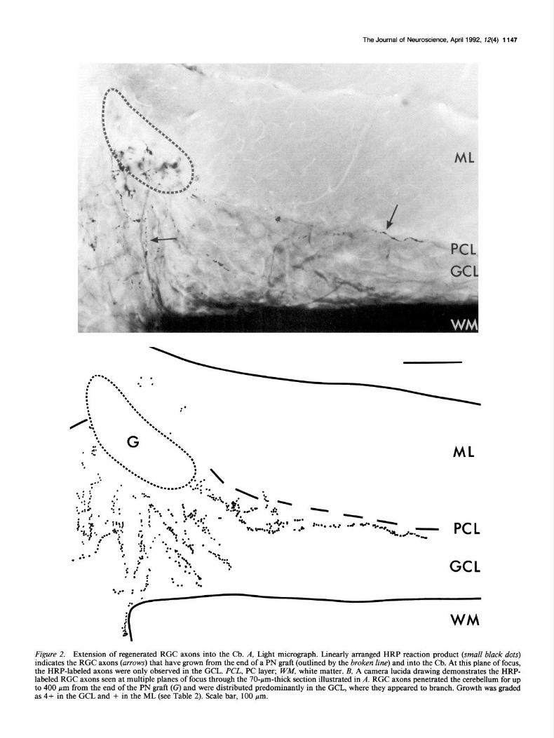
Figure 2. Extension of regenerated RGC axons into the Cb. A, Light micrograph. Linearly arranged HRP reaction product (small black dots) indicates the RGC axons (arrows) that have grown from the end of a PN graft (outlined by the broken line) and into the Cb. At this plane of focus, the HRP-labeled axons were only observed in the CCL. PCL, PC layer; W&I, white matter. B, A camera lucida drawing demonstrates the HRP- labeled RGC axons seen at multiple planes of focus through the 70-pm-thick section illustrated in A. RGC axons penetrated the cerebellum for up to 400 pm from the end of the PN graft (G) and were distributed predominantly in the GCL, where they appeared to branch. Growth was graded as 4+ in the GCL and + in the ML (see Table 2). Scale bar, 100 pm.

ML
PCL- - - - :
d
f
- ML -
- “.r$.....*”
. ..- . . . . . . . pcL ,;c -*,.... . -
. . . .‘. GCL
‘I G 8.3’ i
I
i
ML L
i ~,~~~....._............ -.--..*
;’
\ :
‘. G ::’
ML ‘**‘* ..snea
** . . .._..... .-.
. /. **’
. : . . . . . .
h PCL - :-; ‘/
; ;I $CL
: .i . : ,I@’ > :5
. . *..* -............~ ..” . . . .
%. :;
G ***- . ...!. ;* /
W-ii.*-**.. . . . . . . . . . . . ...... -J.
PCL \ :
GCL
WM
Figure 3. Distribution of RGC axon growth into the Cb. These camera lucida drawings of individual 50-70-pm-thick sections of the Cb demonstrate that HRP-labeled RGC axons (dots) penetrated the Cb for up to 650 pm (d) from the end of the PN graft (G) and, in all but one example 0, were distributed predominantly within the GCL. Growth into the ML was sparse and only up to 200 pm (h) even when the graft was in the ML Q or near the CCL-ML border (a, b, d, 1; and h). Retinal axons formed distinct arbors only in the CCL (b. d, and e). Brdcm line, PC layer (XL); WM. white matter. Scale bars, 100 rm.

Table 2. Growth of RGCs axons in the Cb in relation to the position of the PN graft and partial denervation
Position of the Cere- bellar RGC axon ingrowth*
Illus- trated in
PN graft peduncles GCL ML figure
ML
ML-GCL border
GCL
GCL-WM border
Unknown
intact intact cut cut cut intact intact cut cut cut cut cut intact intact intact intact intact intact cut cut cut intact
0 0 + +++ ++++
0 ++ ++ +++ +++ ++++ ++++ + + +++ ++++ ++++ ++++ ++ ++++ ++++ ++
+ + 0 0 + + + 0 0 0 + + 0 0 + ++ + 0 + 0 + +
3
3d 2
3b
3a 3e
k 3h 3f
3c 3i
* Qualitative assessment (see Materials and Methods for definitions) ofthe amount of axonal growth from 22 individual branches of PN grafts in the 17 experimental animals. WM, white matter.
three animals in which the RGC axons extended primarily into this layer (Table 2).
The HRP-labeled RGC terminals were found in each of the 13 animals examined by EM (Figs. 4, 5). A total of 202 labeled RGC-Cb terminals were identified in animals of groups A and
The Journal of Neuroscience, April 1992, f2(4) 1149
B, of which 190 (94%) were located in the GCL and 12 in the ML (Table 3). These 202 terminals formed 365 synapses, 353 (97%) with neurons in the GCL and 12 in the ML (Table 3). The GCL was easily identified on EM by the characteristic ul- trastructure ofgranule cell (GC) somata (Fig. 4, top): large nuclei, clumps of condensed nuclear chromatin, and a thin rim of cy- toplasm (Palay and Chan-Palay, 1974).
PN graft location and the distribution of RGC axons. To de- termine if the location of the PN graft in the Cb was responsible for the distribution of RGC axons and synapses in the cerebellar cortex, the position of the end of the graft was related to the location and extent of growth of the labeled axons. In all 11 animals with grafts that ended in the GCL (5 in group A and 6 in group B), and in 4 of the 5 animals (2 each in groups A and B) in which the graft was located at the ML-GCL border, RGC axons grew exclusively or predominantly within the GCL (Table 2). In only one of the five animals with grafts at the ML-GCL border, and in both animals in which the PN grafts ended in the ML, RGC axons grew into the ML, but in all instances, this growth was sparse (Table 2; Fig. 2, 3dj). Thus, the prefeiential growth of regenerated RGC axons in the GCL did not correlate with the position of the PN graft.
Denervation and the distribution of RGC axons, terminals, and synapses. RGC axons grew predominantly into the GCL from all 11 PN grafts in group A animals (cerebellar peduncles cut) and from 8 of the 11 grafts in group B (peduncles intact) (Table 2). Of the 124 RGC-Cb terminals and 263 synapses identified in the group A animals, 123 (99%) terminals and 26 1 (99%) synapses were in the GCL. For the group B animals, 67 (86%) of the 78 RGC-Cb terminals and 92 (90%) of the 102 synapses were in the GCL. These proportions of terminals and synapses in the GCL of the group A and B animals were not significantly different (Mann-Whitney U test, p > 0.1). Although a preference of RGC axons for the GCL was observed in both groups, the extent of axonal growth in the GCL was greater in those with transected (group A) cerebellar peduncles. For 8 of the 11 group A grafts, the amount of axonal growth was graded as 3+ or 4+ while for 7 of the 11 group B grafts, this growth was graded as O-2+ (Table 2).
Table 3. Ultrastructural characteristics of axon terminals in the Cb
RGC-Cb’ (regenerated) MF (normal)
GCL ML
Group A Group B Groups A + B (Groups A + B)
Animals (n) 6 6 12 36 4 Terminals
Number 123 67 190 12 516 Area (pm3 5.8 +- 0.9 5.4 + 0.6 5.6 + 0.5 7.0 f 1.2 7.4 + 0.4 Perimeter @my 11.1 * 0.5 11.0 + 1.1 11.1 2 0.8 10.5 k1.5 13.1 f 0.5 Maximum diameter (Nm) 4.1 k 0.2 4.2 -t 0.3 4.1 + 0.2 4.2 -t 0.1 4.3 + 0.1 Pale mitochondria (%) 91 90 91 100 3
Synapses Number 261 92 353 12 849 Axodendritic (%) 99.7 100 99.7 100 100 Number/terminaP 2.5 1.5 2.0 1.3 1.6 Number/l0 pm terminal perimeter 2.0 + 0.3 1.1 f 0.5 1.5 -c 0.2 1.0 f 0.4 1.2 + 0.1
n Values for individual animals are listed in Table 5. b Two of the three animals with RGC terminals in the ML also had terminals within the GCL. c Mean + SEM; no statistically significant differences (ANOVA, see Results) among the groups. d Mean.
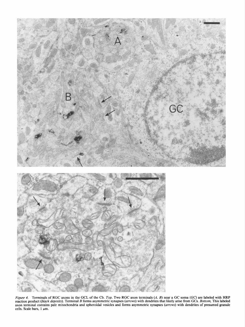
Figure 4. Terminals of RGC axons in the GCL of the Cb. Top, Two RGC axon terminals (A, B) near a GC soma (CC) are labeled with HRP reaction product (black deposits). Terminal B forms asymmetric synapses (arrows) with dendrites that likely arise from GCs. Bottom, This labeled axon terminal contains pale mitochondria and spheroidal vesicles and forms asymmetric synapses (arrows) with dendrites of presumed granule cells. Scale bars, 1 pm.

The Journal of Neuroscience, April 1992, 134) 1151
Table 4. Densities of PC somata
PN-grafted Cb
By distribution of RGC axons in the Cb All animals
GCL and Normal Cb GCL only ML only ML
Animals (n) 4 7 3 8 16‘J
PN grafts (n) - 8 3 10 21
PC somata/ 1000 pmb 9.9 k 0.3 8.0 f 0.5 8.4 + 0.7 6.6 + 0.8 7.4 + 0.4
* The total number of animals with RGC-Ch grafts in the three distribution groups is 18 instead of 16 because in two of the three animals in the “ML only” group, RGC axons grew into the GCL from a second graft. b Mean + SEM.
PC densities and the growth of RGC axons. The possibility that the predilection of RGC axons for the GCL was due to a selective loss of postsynaptic targets in the ML was investigated by comparing the numbers of PC somata per 1000 pm length of PC layer in 3 Mm sections. PCs were counted both in regions of the Cb where RGC axons were observed and in comparable areas of four normal animals (Table 4). There was a 25% re- duction in regional PC density in the RGC-innervated areas of the Cb (Table 4) but the distribution of RGC axons within the Cb did not correlate with PC density (Table 4). Furthermore, in animals in which RGC axons extended into the ML, there was little difference in PC density between those in which RGC axons formed synapses in the ML (three animals; mean, 8.5) and animals in which no synapses were formed (five animals; mean, 7.3). Therefore, a local decrease in the number of PC dendrites in the ML of experimental animals did not appear to be the basis for the predominant innervation of the GCL by RGC axons.
Reactive astrocytes and RGC axon distribution. In the three animals in which the Cb was processed for GFAP immuno- reactivity after the insertion of a PN segment into the vermis, the ends of the PN grafts were surrounded by a zone of GFAP staining with a radius of 100-200 pm. No difference in the intensity or extent of GFAP immunoreactivity was detected between the ML and GCL.
Ultrastructure of RGC-Cb terminals
Several ultrastructural characteristics of the RGC-Cb terminals were similar to those of normal and regenerated RGC terminals in the SC (Huerta and Hatting, 1984; Carter et al., 1989,199lb). In both the Cb (Fig. 4, bottom; 5, RGC-Cb) and SC, RGC terminals contained spherical vesicles and pale mitochondria and formed asymmetric, axodendritic synapses. However, the average size of RGC terminals in the cerebellar cortex (Table 3) was considerably greater than that of normal or regenerated RGC terminals in the SCs of hamsters studied with similar surgical, labeling, and analytical techniques (Carter et al., 1989; D. A. Carter, G. M. Bray, and A. J. Aguayo, unpublished ob- servations). RGC-Cb terminals had mean areas of 5.6 -t 0.5 pm2 in the GCL and 7.0 f 1.2 pm2 in the ML (mean f SEM; Table 3). In contrast, the areas were 0.97 + 0.02 and 1.77 f 0.07 Mm2 for similarly labeled control and regenerated RGC- SC terminals, respectively (Carter et al., 1989; Carter, Bray, and Aguayo, unpublished observations). The perimeter of RGC ter- minals in the Cb (11.1 f 0.8 pm in the GCL, 10.5 + 1.5 pm in the ML, Table 3) was also larger than that of either control (4.26 f 0.06 pm) or regenerated (5.7 + 0.14 pm) RGC-SC
terminals (Carter et al., 1989; Carter, Bray, and Aguayo, un- published observations).
The number of synapses/l0 bum perimeter was similar for RGC-Cb terminals (1.5 k 0.2; Table 3), control RGC-SC ter- minals (1.58 -t 0.06) and regenerated (1.81 + 0.09) RGC-SC terminals (Carter et al., 1989; Carter, Bray, and Aguayo, un- published observations). Due to their larger size, however, RGC- Cb terminals formed, on average, more synaptic contacts per single cross section (mean, 2.0kerminal; Tables 3, 5) than either control (0.7kerminal) or regenerated (1 .O/terminal) RGC-SC terminals (Carter et al., 1989).
There were no significant differences (p > 0.10) in terminal areas, perimeters, or synapses/ 10 pm of terminal perimeter be- tween RGC-Cb terminals in the GCL or the ML (Fig. 6; Table 3). Furthermore, the internal structure of RGC-Cb terminals was similar in both locations.
Efect of survival time. Whether using two-way ANOVA or one-way ANOVA with the data pooled for groups A and B, there were no statistically significant changes in the size of the RGC-Cb terminals or in the numbers of synapses with increas- ing survival times after inserting the graft into the Cb (Table 5, Fig. 6). Moreover, the nonsignificance of the two-way ANOVA test for interaction (p values > 0.10) suggests that the effect of age was similar in both groups.
Effects of denervation. While most RGC terminals and syn- apses were formed in the GCL in both the group A and B animals (see above and Table 2) the interruption of MF and climbing fiber afferents caused by cutting the middle and inferior cere- bellar peduncles (group A) influenced the innervation of the cerebellar cortex in various other ways. In the GCL, the group A animals had twice as many terminals and nearly three times as many synapses as the group B animals (Table 3). However, in the three animals in which a total of 12 terminals and 12 synapses were observed in the ML, 11 of the terminals and 10 of the synapses were observed in two hamsters with intact pe- duncles.
Denervation did not appear to affect the size of RGC-Cb terminals; there were no significant differences (all p values > 0.7) between the areas or perimeters of the terminals in groups A and B (Tables 3, 5; Fig. 6). The effect of denervation on the numbers of synapses/l0 pm terminal perimeter was less clear- cut. Although some of the p values approached 0.05 (Table 5) there was no consistent tendency for one group to have more synapses than the other; up to 5 months after graft insertion, there were more synapses in the group A animals but at later times this trend seemed to be reversed. Thus, it would be nec- essary to study more animals to be certain about the effect of

The Journal of Neuroscience, April 1992, 72(4) 1153
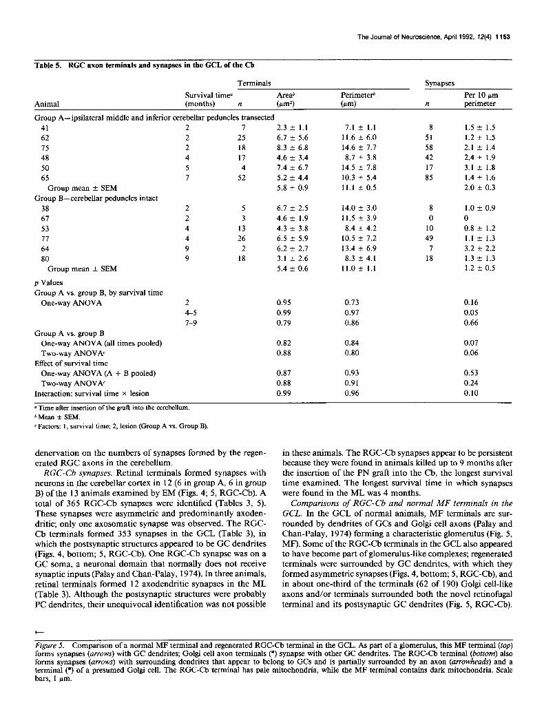
Table 5. RGC axon terminals and synapses in the GCL of the Cb
Animal
Terminals Synapses
Survival time” Areab Perimeteti Per 10 firn (months) n b-d (m-d n perimeter
Group A-ipsilateral middle and inferior cerebellar peduncles transected 41 62 15 48 50 65
Group mean + SEM Group B-cerebellar peduncles intact
38 67 53 77 64 80
Group mean + SEM
p Values Group A vs. group B, by survival time
One-way ANOVA
I
25 18 17 4
52
2.3 -t 1.1 7.1 + 1.1 6.1 + 5.6 11.6 + 6.0 8.3 + 6.8 14.6 + 7.7 4.6 k 3.4 8.7 + 3.8 1.4 k 6.7 14.5 AZ 7.8 5.2 k 4.4 10.3 + 5.4 5.8 + 0.9 11.1 + 0.5
8 51 58 42 17 85
1.5 * 1.5 1.2 + 1.5 2.1 f 1.4 2.4 + 1.9 3.1 + 1.8 1.4 + 1.6 2.0 k 0.3
5 3
13 26
2 18
6.7 k 2.5 14.0 k 3.0 4.6 k 1.9 11.5 k 3.9 4.3 k 3.8 8.4 + 4.2 6.5 z!z 5.9 10.5 + 7.2 6.2 e 2.1 13.4 + 6.9 3.1 k 2.6 8.3 f 4.1 5.4 k 0.6 11.0 + 1.1
8 0
10 49
7 18
1.0 * 0.9 0 0.8 k 1.2 1.1 t- 1.3 3.2 k 2.2 1.3 k 1.3 1.2 f 0.5
2 0.95 0.13 0.16 4-5 0.99 0.97 0.05 7-9 0.19 0.86 0.66
Group A vs. group B One-way ANOVA (all times pooled) Two-way ANOVh
Effect of survival time One-way ANOVA (A + B pooled) Two-way ANOVh
Interaction: survival time X lesion
0.82 0.84 0.07 0.88 0.80 0.06
0.87 0.93 0.53 0.88 0.91 0.24 0.99 0.96 0.10
0 Time after insertion of the graft into the cerebellum. b Mean + SEM. c Factors: 1, survival time; 2, lesion (Group A vs. Group B).
denervation on the numbers of synapses formed by the regen- erated RGC axons in the cerebellum.
RGC-Cb synapses. Retinal terminals formed synapses with neurons in the cerebellar cortex in 12 (6 in group A, 6 in group B) of the 13 animals examined by EM (Figs. 4; 5, RGC-Cb). A total of 365 RGC-Cb synapses were identified (Tables 3, 5). These synapses were asymmetric and predominantly axoden- dritic; only one axosomatic synapse was observed. The RGC- Cb terminals formed 353 synapses in the GCL (Table 3), in which the postsynaptic structures appeared to be GC dendrites (Figs. 4, bottom; 5, RGC-Cb). One RGC-Cb synapse was on a GC soma, a neuronal domain that normally does not receive synaptic inputs (Palay and Chan-Palay, 1974). In three animals, retinal terminals formed 12 axodendritic synapses in the ML (Table 3). Although the postsynaptic structures were probably PC dendrites, their unequivocal identification was not possible
in these animals. The RGC-Cb synapses appear to be persistent because they were found in animals killed up to 9 months after the insertion of the PN graft into the Cb, the longest survival time examined. The longest survival time in which synapses were found in the ML was 4 months.
Comparisons of RGC-Cb and normal MF terminals in the GCL. In the GCL of normal animals, MF terminals are sur- rounded by dendrites of GCs and Golgi cell axons (Palay and Chan-Palay, 1974) forming a characteristic glomerulus (Fig. 5, MF). Some of the RGC-Cb terminals in the GCL also appeared to have become part of glomerulus-like complexes; regenerated terminals were surrounded by GC dendrites, with which they formed asymmetric synapses (Figs. 4, bottom; 5, RGC-Cb), and in about one-third of the terminals (62 of 190) Golgi cell-like axons and/or terminals surrounded both the novel retinofugal terminal and its postsynaptic GC dendrites (Fig. 5, RGC-Cb).
t
Figure 5. Comparison of a normal MF terminal and regenerated RGC-Cb terminal in the GCL. As part of a glomerulus, this MF terminal (top) forms synapses (arrows) with GC dendrites; Golgi cell axon terminals (*) synapse with other GC dendrites. The RGC-Cb terminal (bottom) also forms synapses (arrows) with surrounding dendrites that appear to belong to GCs and is partially surrounded by an axon (arrowheads) and a terminal (*) of a presumed Golgi cell. The RGC-Cb terminal has pale mitochondria, while the MF terminal contains dark mitochondria. Scale bars, 1 pm.

1154 Zwimpfer et al. * Regenerative Synaptogenesis in Novel CNS Targets
All Terminals Cerebellar Months
’ f- GCL ML ; Peduncles ; Survival
Axon 6
Terminal 6
Area 4
(w’) 3
2
7
1
0 il
15 r
Axon 10 Terminal Perimeter
(rm) 5
3
1
I I
, cut intact 1 2 4-5 7-s
I I I I I I
' T I
Synopses/ ’ t -
n: 190 12 123 67 50 60 72
Figure 6. Histograms of the areas, perimeters, and numbers of syn- apses/10 pm terminal perimeter (mean + SEM) of regenerated RGC axon terminals in the GCL (solid bars) and ML (open bars) of the Cb. There were no significant differences (ANOVA, p > 0.05) in compar- isons of terminals in the GCL and ML (left panels). Within the GCL, neither the area, perimeter, nor synapses/ IO hrn terminal perimeter were significantly influenced (a > 0.3) bv transection of the cerebellar ne- dincles (m~ddlepanels) or by the’duration of survival (right panels): n, number of terminals.
It is not known if these glomerulus-like arrangements were a random occurrence or an attempted reconstitution of the glo- merulus that is normally present in the GCL.
Neither the mean area (p > 0.1) nor the mean perimeter (p > 0.3) of 190 RGC-Cb terminals in the GCL (groups A and B combined) was statistically different from those of 5 16 MF ter- minals in the GCL of normal hamsters (Table 3). Furthermore, there was no significant difference (p > 0.2) in the number of synapses/ 10 pm of perimeter between RGC-Cb and control MF terminals (Table 3). The mean area, perimeter, and synapses/ 10 pm of perimeter of the 12 RGC-Cb terminals in the ML were also not significantly different from those of MF terminals. Similar to MFs, the RGC-Cb terminals were filled with clear spherical vesicles and formed asymmetric synapses with GC dendrites (Fig. 5, Table 3). However, while mitochondria were pale in over 90% of RGC-Cb terminals in both the GCL and
ML, 97% of the normal MF terminals contained dark mito- chondria (Fig. 5, Table 3). Thus, the staining properties of RGC mitochondria were retained by the retinofugal axons that es- tablished persistent connections with cerebellar neurons in the GCL.
Discussion When nerve fibers were damaged, the recovery of useful con- nectivity requires the survival of the injured nerve cells, the regrowth of severed axons toward their normal fields of inner- vation, and the restoration of synaptic contacts with appropriate targets. While it is now known that several classes of CNS pro- jection neurons in adult mammals are capable of regenerating their axons over long distances, it is also apparent that condi- tions in their CNS environment do not permit the lengthy growth required for such axons to reach their distant targets. Therefore, it has been difficult to determine if the reexpression of the neu- ronal capacities to initiate and sustain axonal elongation can eventually lead to the selective retrieval of their postsynaptic partners and the avoidance of abnormal connections.
The regrowth of RGC axons
In adult rodents, the substitution of the ON by a long PN graft makes possible the regeneration of RGC axons into normal or unusual targets that are far removed from the eye. Regenerated RGC axons guided to one of their normal fields of innervation, the SC, were previously proven to form terminal arborizations and well-differentiated functional synapses within the superfi- cial, retinorecipient layers of the SC (Vidal-Sanz et al., 1987, 1991; Carter et al., 1989, 1991a; Keirstead et al., 1989; SauvC et al., 1991; Carter, Bray, and Aguayo, unpublished observa- tions). We have now shown that when RGC axons are routed along PN grafts to the Cb, a region of the brain that does not normally receive afferent fibers from the retina, they can also grow into the cerebellar cortex and form synapses. In these animals, the RGC axons that had elongated more than 2 cm along the PN grafts penetrated the Cb for up to 650 pm, an extension into the CNS that approximates that of the RGC axons that regenerate into the SC (Carter et al., 1989, 1991a; Carter, Bray, and Aguayo, unpublished observations). The re- generating RGC axons grew predominantly into the cerebellar cortex, while extension into the white matter was exceptional and only for up to 50 pm. This limited growth into the white matter is consistent with other studies that suggest that myelin components in the CNS of adult mammals inhibit cell adhesion and neurite outgrowth in vitro (Caroni and Schwab, 1988) and may block axonal extension in vivo (Schnell and Schwab, 1990).
The growth of the RGC axons into the Cb was more extensive in the animals in which the cerebellar peduncles had been tran- sected (group A) than in those with intact peduncles (group B). The 1.5-fold greater penetration of axons from the end of the grafts, the formation of nearly twice as many RGC-Cb terminals, and a three-fold increase in the number of synapses were signs of this effect of denervation. Such enhancement in axonal in- growth and synaptogenesis could reflect a greater availability of synaptic sites in the cerebellar cortex of the lesioned animals. Furthermore, an increased expression of molecules that facili- tate axonal growth has been reported after CNS denervation (Needels et al., 1986). While the expression of such factors has not been investigated in the lesioned Cb, it is known that in- terruption of the cerebellar peduncles (Pickel et al., 1973) or

The Journal of Neuroscience, April 1992, 12(4) 1155
partial destruction of nuclei that project fibers to the Cb (Rossi et al., 1989) can result in axonal sprouting and a more prominent arborization of intact cerebellar afferents.
Morphology of RGC-Cb terminals The novel RGC-Cb terminals were significantly larger and es- tablished more synapses per terminal than the regenerated RGC terminals in the SC (Carter et al., 1989; Carter, Bray, and Aguayo, unpublished observations). Others have also observed the for- mation of larger terminals by regenerating (Radel and Yoon, 1985; Carter et al., 1989; Itoh and Tessler, 1990b; Carter, Bray, and Aguayo, unpublished observations) or sprouting axons (Raisman, 1969; Chen and Hillman, 1982; Steward et al., 1988). In goldfish (Radel and Yoon, 1985) and hamsters (Carter et al., 1989; Carter, Bray, and Aguayo, unpublished observations) re- generated RGC-SC terminals are initially larger than controls but become smaller with time. In rats, regenerated RGC ter- minals in the SC, analyzed 18 months after joining the eye and the SC with a PN graft, showed no significant differences in size from age-matched controls (Vidal-Sanz et al., 199 1). It is there- fore puzzling that the size of the regenerated retinal terminals formed in the Cb did not decrease significantly over the 7 months spanned by the present study in hamsters.
Because the size of the RGC terminals in the GCL approxi- mated that of normal MF afferents to the GCL, it could be postulated that this layer of the Cb had a specific influence on the morphology of these novel terminals. Neuroanatomical in- vestigations of connections in the developing and mature CNS provide evidence that the target can indeed influence the shape and size of terminals (Ramon y Cajal, 19 1 1; Morest, 1968; Lund, 1969; Mugnaini, 1970; Mason and Gregory, 1984). Fur- thermore, the terminal morphology of retinal axons that form connections with nonretinal targets in immature rodents more closely resembles that of the normal afferents to such targets (Kalil and Schneider, 1975; Campbell and Frost, 1988). How- ever, large RGC terminals were also found in the ML of our experimental animals. In addition, in studies in adult hamsters where RGC axons were guided along PN grafts into other regions of the brain that normally do not receive direct retinal inputs (e.g., the central nucleus of the inferior colliculus or the visual and somatosensory cortices; Zwimpfer et al., 1990), synapses were also formed. In the inferior colliculus of these animals, the size of regenerated RGC terminals was also significantly larger than regenerated RGC terminals in the SC for up to 8 months (T. J. Zwimpfer, A. J. Aguayo, and G. M. Bray, unpublished observations). The persistence of large RGC axon terminals in these various parts of the CNS may be related to their aberrant location.
For the regenerated RGC-Cb axons, the number of synapses per 10 pm of terminal perimeter (1.5) was similar to that de- termined previously for regenerated (1.8) or normal (1.6) RGC terminals in the SC (Carter et al., 1989; Carter, Bray, and Aguayo, unpublished observations). One interpretation of these consis- tent relationships between the size of the terminals and the number of synapses formed is that the frequency of contacts influences the size of the terminals. Conversely, the formation of larger terminals, with their more abundant surface areas, might have made possible the establishment of a greater number of contacts. In either case, it can be assumed that interactions with postsynaptic elements capable of influencing the formation and turnover of the axonal membrane (Liuzzi and Lasek, 1987; Liuzzi, 1990) ultimately determines the size of these and other
terminals. The recent finding of a higher-than-normal density of terminals in the arbors made by some regenerated RGC axons that reinnervate the SC of adult hamsters might also denote a tendency of these growing fibers to form synapses soon after they penetrate their CNS targets (Carter, Bray, and Aguayo, unpublished observations). It is not known, however, if these changes are due to a greater propensity for synaptogenesis by regenerating as opposed to developing CNS axons or if adhesive interactions between growing axons and their substrates (Mo- nard, 1988) affect the extension of axons in these CNS regions.
The preference of RGC axons for the GCL Within the gray matter, RGC axons did not appear to extend or synapse randomly but displayed a predilection for the GCL, RGCs innervated the GCL in all but 1 of the 17 hamsters studied, and in 14 of these animals the growth was predomi- nantly or exclusively in the GCL. Moreover, 95% of RGC-Cb terminals and synapses were located in this layer. In goldfish, in which regenerating RGC axons grew into the Cb after uni- lateral (Lo and Levine, 1980) or bilateral (Sharma, 198 1) tectal ablations, the axons also extended selectively into the GCL (Lo and Levine, 1980).
The apparent predilection of retinal terminals for the GCL could have been due to a retraction of axons from the ML after an initial, less specific deployment. Indeed, withdrawal of retinal axons or axon collaterals is thought to contribute to the refine- ment of retinofugal connections during development (Kalil et al., 1986; O’Leary, 1987; Sretavan et al., 1988; Nakamura and O’Leary, 1989; Thompson and Holt, 1989; Bhide and Frost, 199 l), and in regeneration of retinotectal axons in the goldfish (Meyer et al., 1985; Rankin and Cook, 1986; Stuermer, 1988) and frog (Reh and Constantine-Paton, 1985). While the pref- erential innervation of the GCL in the adult hamsters was al- ready detectable at the shortest survival time studied (2 months), an earlier scrutiny would be needed to verify that the initial orientation of regenerated RGC axons entering the Cb was in- deed toward the GCL.
Avoidance of the ML might also have influenced RGC axons to grow preferentially within the GCL. Although in vitro studies of both chick (Walter et al., 1987a,b, 1990; Cox et al., 1990; Stahl et al., 1990) and goldfish (Vielmetter and Stuermer, 1989) retinotectal preparations have shown that the rostrocaudal ar- rangement of RGC axons in the tectum may be partly deter- mined by inhibitory influences, there is no indication that such inhibitory molecules also influence the laminar deployment of retinal axons in the SC. Furthermore, a blocking of axonal re- growth by components of CNS myelin (Caroni and Schwab, 1988; Schnell and Schwab, 1990) is unlikely to account for the distinct distribution of the regenerated RGC-Cb connections to the GCL because this layer contains more myelinated fibers than the ML (Palay and Chan-Palay, 1974). Moreover, the demon- stration that intact climbing fibers can sprout within the ML of the adult rat (Rossi et al., 1989) suggests that the ML is not a barrier to axonal extension within the cerebellar cortex.
During development, the migration of neurons and the dis- tribution of axonal projections in the CNS may be influenced by molecular boundaries (Keynes and Lumsden, 1990; Snow et al., 1990). If there were a comparable barrier in adult rodents between the GCL and the ML, the extension of RGC axons across the PC layer could be curtailed. Such a putative boundary might also clarify why PCs transplanted into the cerebellar cor- tex of PC degeneration mutant mice tend not to project beyond
1156 Zwimpfer et al. - Regenerative Synaptogenesis in Novel CNS Targets
the GCL-ML border (Sotelo et al., 1990; Sotelo and Alvarado- Mallart, 199 1). However, this mechanism would not explain why most axons arising from PN grafts positioned at the ML- GCL border tended to grow into the GCL. Although reactive astrocytes might act as a barrier (Liuzzi and Lasek, 1987; Faw- cett et al., 1989) that prevented the RGC axons from entering the ML or as a substrate that facilitated axon growth (David et al., 1990; Hall et al., 1991) into the GCL, the distribution of these cells, assessed by GFAP immunoreactivity, was similar in the ML and GCL.
Finally, the scarcity of RGC axonal extension into the ML did not seem to be due to a selective loss of postsynaptic targets in the ML caused by Cb damage during graft insertion. Com- pared to the normal Cb, the mean density of PC somata in the region of the cerebellar cortex surrounding the graft was de- creased by only 25% and PC density did not differ between animals with or without RGC innervation of the ML.
Role of denervation of the distribution of RGC-Cb connections Denervation influences the distribution of connections formed by axonal sprouts. In the adult mammalian CNS, the loss of afferent connections induces uninjured axons to sprout collat- erals that grow selectively into the denervated regions (Raisman, 1969, 1985; Cotman et al., 198 1; Rossi et al., 1989). However, within the narrow range covered by their growth, the sprouting axons appear to contact their normal targets preferentially even if other adjacent nerve cells are also denervated (Raisman, 1985). Furthermore, fetal neurons transplanted into the brains of neo- natal (Hankin and Lund, 1987) or adult (Gage et al., 1985) rats tended to extend axons selectively into nearby groups of host neurons that are their normal targets and avoid denervated regions that do not receive afferents from the type of cells con- tained in the graft.
In the group A hamsters of the present study, interruption of both MF and climbing fiber afferents caused by transecting the cerebellar peduncles would have affected neurons in both the GCL and ML. However, neurons in the GCL would be more extensively denervated than the PCs because parallel fiber inputs to the ML would be preserved (Palay and Chan-Palay, 1974). It is also important to recognize that in hamsters with intact or cut cerebellar peduncles, cells and axons are locally damaged by the insertion of the PN grafts. The distribution of such local damage would depend on the location of the PN grafts. With grafts in the GCL, some climbing and parallel fiber afferents to the ML and MF inputs to the GCL would be interrupted, de- nervating both the GCL and ML. However, the grafts located at or near the GCL-ML border would have denervated the ML more extensively by interrupting the climbing and parallel fiber inputs. Despite these three possible patterns ofdenervation within the Cb (GCL > ML, GCL = ML, and ML > GCL), RGC axons predominantly or exclusively innervated the GCL in 14 of the 17 experimental animals.
Synaptogenesis with abnormal targets Because neither the morphological features of the Cb nor the conditions related to the experimental procedure discussed above appear to explain the preference of RGC axons to extend and synapse in the GCL of the Cb, it is necessary to consider the possibility that the RGC axons may select the GCL because it expresses critical molecular constituents that are similar or iden- tical to those present in their normal targets. Certain molecules, which are highly expressed in both the SC and the GCL of the
Cb, are potential candidates for such a role; these may include brain-derived neurotrophic factor (BDNF) (Hofer et al., 1990) a protein known to influence RGC growth and survival (Johnson et al., 1986; Thanos et al., 1989) and the NMDA subtype of glutamate receptor (Cline and Constantine-Paton, 1989; Debski et al., 1990).
In vivo examples of the role of trophic factors in the guided growth and maintenance of connections include the anomalous invasion of peripheral sympathetic fibers into regions of the brainstem injected with NGF (Levi-Montalcini, 1976; Crutcher, 1987) and the extension of septohippocampal axons toward exogenous sources of NGF within the brain (Hagg et al., 1990) or into regions of increased NGF synthesis in the denervated hippocampus (Crutcher and Collins, 1986; Crutcher, 1987). Furthermore, in vitro, neurites from chick dorsal root ganglia are known to grow toward high concentrations of NGF (Gun- dersen and Barrett, 1979). However, such effects have not yet been investigated for BDNF or other NGF-related neurotro- phins.
Neurotransmitter-receptor interactions are thought to influ- ence the formation of normal (Cohen et al., 1987) and anom- alous (Landmesser, 1972; Schotzinger and Landis, 1990) con- nections in the PNS. In rat RGCs, N-acetylaspartylglutamate is a putative neurotransmitter (Anderson et al., 1987; Tsai et al., 1990) that can activate the NMDA subtype of glutamate recep- tor (Westerbrook et al., 1986; Sekiguchi et al., 1987). The NMDA receptor is present in the SC of the adult rat, especially in the superficial layers (Monaghan and Cotman, 1985) and, in the cat, has been shown to mediate postsynaptic responses in the retinogeniculate pathway (Kemp and Sillito, 1982). Mossy, climbing, and parallel fiber inputs to the Cb of adult rodents all appear to be glutaminergic (Foster and Roberts, 1983; Freeman et al., 1983) but the NMDA subtype is the predominant glu- tamate receptor in the GCL (Monaghan and Cotman, 1985; Olson et al., 1987). Furthermore, studies in the visual system of the frog suggest that the refinement of retinotectal connections may involve NMDA receptor activation (Cline et al., 1987; Cline and Constantine-Paton, 1989; Debski et al., 1990).
While the molecular determinants of intemeuronal recogni- tion and connectivity have not been elucidated, various exper- imental combinations of regenerating axons and target cells have demonstrated that unusual connections can be established in the PNS (Close, 1965; Landmesser, 1972; Bixby and Van Essen, 1979; McMahon and Wall, 1989; Schotzinger and Landis, 1990; for review, see Purves and Lichtman, 1985) and also in the CNS of the adult frog (Cantore and Scalia, 1987; Scalia, 1987) and goldfish (Yoon, 197 1; Schmidt, 1978; Meyer, 1979; Sharma, 198 l), as well as in the CNS of immature mammals (Kalil and Schneider, 1975; Frost, 198 1; Campbell and Frost, 1988; Sur et al., 1988). In addition, both normal and abnormal connec- tions have been demonstrated following transplantation of fetal neurons into the brains of adult mammals (McLoon and Lund, 1983; Nilsson et al., 1988; Clarke et al., 1990; Itoh and Tessler, 1990a).
In the damaged CNS, the formation of such aberrant con- nections might influence recovery in several ways. Premature synaptogenesis near the site of injury could curtail further axonal extension (Bernstein and Bernstein, 197 1) or create new circuits (Sur et al., 1988; MCtin and Frost, 1989; Roe et al., 1990) that can generate maladaptive behaviors (Schneider, 1973; Easter and Schmidt, 1977). On the other hand, novel connections might also help establish alternate pathways (M&in and Frost, 1989;

The Journal of Neuroscience, April 1992, 134) 1157
Roe et al., 1990) that compensate for lost or impaired functions. The existence of potential synaptic interactions in regions of the nervous system that normally have no anatomical or functional connections underscores the importance of the mechanisms that guide the long-range growth of axons to the proximity of their normal fields of innervation (Lumsden and Davies, 1986; Dodd and Jessell, 1988; Tessier-Lavigne et al., 1988; Harris, 1989; Godement et al., 1990). While advances have been made in promoting neuronal survival, axonal regrowth, and synapto- genesis, limitations in such long-range guidance in the mature CNS may continue to impose critical constraints on the recovery of useful connectivity in the injured mammalian nervous sys- tem. However, the apparent expression in the CNS of these animals of certain growth and synaptic preferences within the narrow field encompassed by terminal axonal arborizations also raises the possibility that appropriate connections may be re- stored spontaneously by guiding regenerating axons to the vi- cinity of their normal fields of innervation, or by enhancing the short-range extension of fibers interrupted near their targets.
References
Aguayo AJ, Carter DA, Zwimpfer TJ, Vidal-Sam M, Bray GM (1990) Axonal regeneration and synapse formation in the injured CNS of adult mammals. In: Brain repair (Bjiirklund A, Aguayo AJ, Ottoson D, eds), pp 25 l-272. London: Macmillan.
Anderson KJ, Borja MA, Cotman CW, Moffett JR, Namboodiri MAA, Neale JH (1987) N-acetylaspartylglutamate identified in the rat ret- inal ganglion cell and their projections in the brain. Brain Res 411: 172-177.
Bernstein JJ, Bernstein ME (197 1) Axonal regeneration and formation of synapses proximal to the site of lesion following hemisection of the rat sninal cord. EXD Neurol 30:336-35 1.
Bhide PG,-Frost DO (199 1) Stages of growth of hamster retinofugal axons: implications for developing axonal pathways with multiple targets. J Neurosci 11:485-504.
Bignami A, Dahl D (1976) The astroglial response to stabbing. Im- munofluorescence studies with antibodies to astrocyte-specific protein (GFA) in mammalian and submammalial vertebrates. Neuropathol Appl Neurobiol 2:99-l 10.
Bixby JL, Van Essen DC (1979) Competition between foreign and original nerves in adult mammalian skeletal muscle. Nature 282:726- 728.
Campbell G, Frost DO (1988) Synaptic organization of anomalous retinal projections to the somatosensory and auditory thalamus: target controlled morphogenesis of axon terminals and synaptic glomeruli. J Comp Neurol272:383408.
Cantore WA, Scalia F (1987) Ultrastructural evidence of the formation of synapses by retinal ganglion cell axons in two non-standard targets. J Comp Neurol261:137-147.
Caroni P, Schwab ME (1988) Two membrane protein fractions from rat central myelin with inhibitory properties for neurite growth and fibroblast spreading. J Cell Biol 106: 128 l-l 288.
Carter DA, Bray GM, Aguayo AJ (1989) Regenerated retinal ganglion cell axons can form well-differentiated retinal ganglion cell axons in the superior colliculus of adult hamsters. J Neurosci 9:40424050.
Carter DA, Bray GM, Aguayo AJ (199 la) Patterns of the arborizations made by retinal ganglion cell axons regenerating into the superior colliculus of adult hamsters. Sot Neurosci Abstr 17:568.
Carter DA, Aguayo AJ, Bray GM (199 lb) Retinal ganglion cell ter- minals in the hamster superior colliculus: an ultrastructural study. J Comp Neurol311:97-107.
Chen S, Hillman DE (1982) Plasticity of the parallel fiber-Purkinje cell synapse by spine takeover and new synapse formation in the adult rat. Brain Res 240:205-220.
Clarke DJ, Nilsson OG, Brundin P, Bjiirklund A (1990) Synaptic connections formed by grafts of different types of cholinergic neurons in the host hippocampus. Exp Neurol 107:l l-22.
Cline HT, Constantine-Paton M (1989) NMDA receptor antagonists disrupt the retinotectal topographic map. Neuron 3:4 13426.
Cline HT, Debski EA, Constantine-Paton M (1987) NMDA receptor
antagonist desegregates eye-specific stripes. Proc Nat1 Acad Sci USA 84~4342-4345.
Close R (1965) Effects of cross-union of motor nerves to fast and slow skeletal muscles. Nature 206:83 l-832.
Cohen MW, Rodriguez-Marin E, Wilson EM (1987) Distribution of synaptic specializations along isolated motor units formed in Xenopus nerve-muscle cultures. J Neurosci 7:2849-286 1.
Cotman CW, Nieto-Sampedro M, Harris EW (198 1) Synapse replace- ment in the nervous system of adult vertebrates. Physiol Rev 6 1:684- 784.
Cox EC, Miiller B, Bonhoeffer F (1990) Axonal guidance in the chick visual system: posterior tectal membranes induce collapse of growth cones from the temporal regina. Neuron 4:3 l-37.
Crutcher KA (1987) Sympathetic sprouting in the central nervous system: a model for studies of axonal growth in the mature mam- malian brain. Brain Res Rev 12:203-233.
Crutcher KA, Collins F (1986) Entorhinal lesions result in increased nerve growth factor-like growth-promoting activity in medium con- ditioned bv hiDDOCaIXIDa1 slices. Brain Res 399:383-389.
David S, Boucher-C, T&as 0, Giftochristos N (1990) Macrophages can modify the nonpermissive nature of the adult mammalian central nervous system. Neuron 5:463-469.
Debski EA, Cline HT, Constantine-Paton M (1990) Activity-depen- dent tuning and the NMDA receptor. J Neurobio12 1: 18-32.
Dodd J, Jesse11 TM (1988) Axon guidance and the patterning of neu- ronal projections in vertebrates. Science 2 1: 18-32.
Easter SS, Schmidt JT (1877) Reversed visuomotor behaviour me- diated by induced ipsilateral retinal projections in goldfish. J Neu- rophysiology 40: 1245-l 254.
Fawcett JW, Housden E, Smith-Thomas L, Meyer RL (1989) The growth of axons in three-dimensional astrocyte cultures. Dev Biol 135:449458.
Foster GA, Roberts PJ (1983) Neurochemical and pharmacological correlates of inferior olive destruction in the rat: attenuation of the events mediated by an endogenous glutamate-like substance. Neu- roscience 8:277-284.
Freeman ME, Lane JD, Smith JE (1983) Turnover rates of amino acid neurotransmitters in regions of rat cerebellum. J Neurochem 40: 144 l- 1447.
Frost DO (198 1) Orderly anomalous retinal projections to the medial geniculate, ventrobasal and lateral posterior nuclei of the hamster. J Comp Neurol203:227-256.
Frost DO (1984) Axonal growth and target selection during devel- opment: retinal projections to the ventrobasal complex and other “nonvisual” structures in neonatal Syrian hamsters. J Comp Neurol 230~576-592.
Gage FH, Bjijrklund A, Steveni U, Dunnett SB (1985) Grafting of embryonic CNS tissue to the damaged adult hippocampal formation. In: Neural grafting in the mammalian CNS (Bjorklund A, Stenevi U, eds), pp 559-573. New York: Elsevier.
Godement P, Salaun J, Mason CA (1990) Retinal axon pathfinding in the optic chiasm: divergence of crossed and uncrossed fibers. Neu- ron 5:173-186.
Gundersen RW, Barrett JN (1979) Neuronal chemotaxis: chick dorsal- root axons turn toward high concentrations of nerve growth factor. Science 206: 1079-1080.
Hagg T, Vahlsing HL, Manthorpe M, Varon S (1990) Nerve growth factor infusion into the denervated adult rat hippocampal formation promotes its cholinergic reinnervation. J Neurosci 10:3087-3092.
Hall S. Greason N, Rickard S (1991) Interaction of rearowina PNS axons with transplanted aggregates’of cultures CNS &a in ho. J Neurocytol 20:299-309.
Hankin MH, Lund RD (1987) Role of the target in directing the outgrowth ofretinal axons: transplants reveal surface-related and sur- face-independent cues. J Comp Neurol263:455-466.
Harris WA (1989) Local positional cues in the neuroepithelium guide retinal axons in embryonic Xenopus brain. Nature 339:218-221.
Hofer M, Pagliusi SR, Hohn A, Leibrock J, Barde Y-A (1990) Regional distribution of brain-derived neurotrophic factor mRNA in the adult mouse brain. EMBO J 9:2459-2464.
Huerta MF, Hatting JK (1984) The mammalian superior colliculus: studies of its morphology and connections. In: Comparative neurol- ogy of the optic tectum (Vanegas H, ed), pp 687-773. New York Plenum.
Itoh Y, Tessler A (199Oa) Ultrastructural organization of regenerated

1158 Zwimpfer et al. * Regenerative Synaptogenesis in Novel CNS Targets
adult dorsal root axons within transplants of fetal spinal cord. J Comp Neurol 292:396411.
Itoh I, Tessler A (1990b) Regeneration of adult dorsal root axons into transplants of fetal spinal cord and brain: a comparison of growth and synapse formation in appropriate and inappropriate targets. J Comp Neurol 302:272-293.
Johnson JE, Barde Y-A, Schwab M, Thoenen H (1986) Brain-derived neurotrophic factor supports the survival of cultured rat retinal gan- glion cells. J Neurosci 6:303 l-3038.
Kalil RE, Schneider GE (1975) Abnormal synaptic connections of the optic tract in the thalamus after midbrain lesions in newborn ham- sters. Brain Res 100:690-698.
Kalil RE, Dubin MW, Scott G, Stark LA (1986) Elimination of action potentials blocks the structural development of retinogeniculate syn- apses. Nature 323: 156-l 58.
Keirstead SA, Rasminsky M, Fukuda Y, Carter DA, Aguayo AJ, Vidal- Sanz M (1989) Electrophysiologic responses in hamster superior colliculus evoked by regenerating retinal axons. Science 246:255-258.
Kemp JA, Sillito AM (1982) The nature of the excitatory transmitter mediation X and Y cell inputs to the cat dorsal lateral geniculate nucleus. J Physiol (Lond) 323:377-39 1.
Kevnes R. Lumsden A (1990) Segmentation and the or&in of regional diversity in the vertebrate nerv&s system. Neuron 4: i-9. -
Landmesser L (1972) Pharmacological properties, cholinesterase ac- tivity and anatomy of nerve-muscle junction in vagus-innervated frog sartorius. J Physiol (Lond) 2201243-256.
Larsell 0 (1952) The morphogenesis and adult pattern of the lobules and fissures of the cerebellum of the white rat. J Comp Neurol 97: 28 l-356.
Lemann W, Saper CB, Rye DB, Wainer BH (1985) Stabilization of TMB reaction product for electron microscopic retrograde and an- teroarade fiber tracina. Brain Res Bull 14:277-28 1.
Levi-Montalcini (1976) The nerve growth factor: its role in growth, differentiation and function of the sympathetic adrenergic neuron. Prog Brain Res 45~235-258.
Liuzzi FJ (1990) Proteolysis is a critical step in the physiological stop pathway:.mechanisms involved in the blockade ofaxonal regeneration bv mammalian astrocvtes. Brain Res 5 12:277-283.
Liuzzi FJ, Lasek RJ (1987) Astrocytes block axonal regeneration in mammals by activating the physiological stop pathway. Science 237: 642-645.
Lo RYS, Levine R (1980) Time course and pattern of optic fiber regeneration following tectal lobe removal in the goldfish. J Comp Neurol 191:295-314.
Lumsden AGS, Davies AM (1986) Chemotropic effect of specific tar- get epithelium in the developing mammalian nervous system. Nature 323:538-539.
Lund RD (1969) Synaptic patterns of the superficial layers of the superior colliculus of the rat. J Comp Neurol 135: 197-208.
Mason CA, Gregory E (1984) Postnatal maturation ofcerebellar mossy and climbing fibers: transient expression of dual features on single axons. J Neurosci 4: 17 15-l 735.
McLoon SC, Lund RD (1983) Development of fetal retina, tectum and cortex transplanted to the superior colliculus ofadult rats. J Comp Neural 217:376-389.
McMahon SB, Wall P (1989) Changes in spinal cord reflexes after cross-anastomosis of cutaneous and muscle nerves in the adult rat. Nature 342~272-274.
M&in C, Frost DO (1989) Visual responses of neurons in somatosen- sory cortex of hamsters with experimentally induced retinal projec- tions to somatosensorv thalamus. Proc Nat1 Acad Sci USA 86:357- 361.
Meyer RL (1979) Retinotectal projection in goldfish to an inappro- priate region with a reversal in polarity. Science 205:8 19-821.
Mever RL. Sakurai K. Schauwecker E (1985) Topot?raphv of regen- crating optic fibers in goldfish traced with local wheat germ injectTons into retina: evidence for discontinuous microtopography in the reti- notectal projection. J Comp Neurol 239:27-43.
Monaghan DT, Cotman CW (1985) Distribution of N-methyl-o-as- partate-sensitive t$H]glutamate-binding sites in rat brain. J Neu- rosci 5:2909-2919.
Monard D (1988) Cell-derived proteases and protease inhibitors as regulators of neurite outgrowth. Trends Neurosci 11:541-544.
Morest DK (1968) The collateral system of the medial nucleus of the trapezoid body of the cat, its neuronal architecture and relation to the olivo-cochlear bundle. Brain Res 9:288-3 11.
Mugnaini E (1970) Neurones as synaptic targets. In: Excitatory syn- aptic mechanisms (Anderson P, Jansen JKS, eds), pp 149-169. Oslo: Universitetsforlaget.
Nakamura H, O’Leary DDM (1989) Inaccuracies in initial growth and arborization of chick retinotectal axons followed by course corrections and axon remodelling to develop topographic order. J Neurosci 9: 3776-3795.
Needels DL, Nieto-Sampedro M, Cotman CW (1986) Induction of a neurite-promoting factor in rat brain following injury or deafferen- tiation. Neuroscience 18:5 17-526.
Nilsson OG, Clarke DH, Brundin P, Bjijrklund A (1988) Comparison of growth and reinnervation properties of cholinergic neurons from different brain regions grafted to the hippocampus. J Comp Neurol 2681204-222.
O’Leary DDM ( 1987) Remodelling of early axonal projections through the selective eliminaton of neurons and long axon collaterals. In: Selective neuronal death (Bock G, O’Connor M, eds), pp 113-142. Chichester: Wiley.
Olson JMM, Greenamyre JT, Penney JB, Young AB (1987) Autora- diographic localization of cerebellar excitatory amino acid binding sites in the mouse. Neuroscience 22:9 13-923.
Palay SL, Chan-Palay V (1974) Cerebellar cortex, pp l-287. New York: Springer.
Pickel VM, Krebs H, Bloom FE (1973) Proliferation of norepineph- tine-containing axons in rat cerebellar cortex after peduncle lesions. Brain Res 59~169-179.
Purves D, Lichtman I (1985) Principles of neural development, pp 229-270. Sunderland, MA: Sinauer.
Radel JD, Yoon MG (1985) Time-course of ultrastructural changes in the regenerated optic fiber terminals of goldfish. Brain Res 342: 168-171.
Raisman G (1969) Neuronal plasticity in the septal nuclei of the adult rat. Brain Res 14:2548.
Raisman G (1985) Synapse formation in the septal nuclei of adult rats. In: Synaptic plasticity (Cotman CW, ed), pp 13-38. New York: Guilford.
Ramon y Cajal S (19 11) Histologie du systeme nerveux de l’homme et des vertebres, vol 1. Paris: A. Maloine [reprint (1955) pp 754- 838. Madrid: Consejo Superior de Investigaciones Cientificas, Institut Ramon y Cajal].
Rankin ECC, Cook JE (1986) Topographic refinement of the regen- erating retinotectal projection of the goldfish in standard laboratory conditions: a quantitative WGA-HRP studv. EXD Brain Res 63:409- _ - 420.
Reh TA, Constantine-Paton M (1985) Eye-specific segregation re- quires neural activity in three-eyed Rana pipiens. J Neurosci 5: 1132- 1143 __ ._.
Roe AW, Pallas SL, Hahm J, Sur M (1990) A map of visual space induced in primarv auditow cortex. Science 250:818-820.
Rossi F, Wiklund L, van der Want JJL, Strata P (1989) Climbing fibre plasticity in the cerebellum of the adult rat. Eur J Neurosci 1:543- 547.
Sauvt Y, Rasminsky M, Carter DA (199 1) Distribution and charac- terization of responses to light in reinnervated hamster superior col- liculus. Sot Neurosci Abstr 17:568.
Scalia F (1987) Synapse formation in the olfactory cortex by regen- erating optic axons: ultrastructural evidence for polyspecific che- moaffinity. J Comp Neurol 263:497-5 13.
Schmidt JT (1978) Retinal fibers alter tectal positional markers during the expansion of the half retinal projection in goldfish. J Comp Neurol 1771279-300.
Schneider GE (1973) Early lesions of superior colliculus: factors af- fecting the formation of abnormal retinal projections. Brain Behav Evol 8:73-109.
Schnell L, Schwab ME (1990) Axonal regeneration in the rat spinal cord produced by an antibody against myelin-associated neurite growth inhibitors. Nature 3431269-272.
Schotzinger RJ, Landis SC (1990) Acquisition of cholinergic and pep- tidergic properties of sympathetic innervation of rat sweat glands requires interaction with normal target. Neuron 5:91-100.
Sekiguchi M, Okamato K, Sakai Y (1987) Excitatory action of N-acetylaspartylglutamate on Purkinje cells in guinea pig cerebellar slices: an intrasomatic study. Brain Res 423:23-33.
Sharma SC (198 1) Retinal projection in a non-visual area after bilat- eral tectal ablation in goldfish. Nature 291:66-67.
Snow DM, Steindler DA, Silver J (1990) Molecular and cellular char-

The Journal of Neuroscience, April 1992, 72(4) 1159
acterization of the glial roof plate of the spinal cord and optic tectum: a possible role for a proteoglycan in the development of an axon barrier. Dev Biol 138:359-376.
Sotelo C, Alvarado-Mallart RM (1991) The reconstruction of cere- bellar circuits. Trends Neurosci 14:3X)-355.
Sotelo C, Alvarado-Mallart RM, Gardette R, Crepe1 F ( 1990) Fate of grafted embryonic Purkinje cells in the cerebellum of the adult “Pur- kinje cell degeneration” mutant mouse. I. Development of reciprocal graft-host interactions. J Comp Neurol 295: 165-187.
Sretavan DW, Shatz CJ, Styker MP (1988) Modification of retinal ganglion cell axon morphology by prenatal infusion of tetrodotoxin. Nature 336:468-471.
Stahl B, Muller B, von Boxberg Y, Cox EC, Bonhoeffer F (1990) Bio- chemical characterization of a putative axonal guidance molecule of the chick visual system. Neuron 5:735-743.
Steward 0, Vinsant SL, Davis L (1988) The process of reinnervation in the dentate gyrus of adult rats: an ultrastructural study of changes in presynaptic terminals as a result of sprouting. J Comp Neuro1267: 203-210.
Stuermer CA0 (1988) Trajectories of regenerating retinal axons in the tectum: I. A comparison of normal and regenerated axons at late regeneration stages. J Comp Neurol 267:55-68.
Sur M, Garraghty PE, Roe AW (1988) Experimentally induced visual projections into auditory thalamus and cortex. Science 242:1437- 1441.
Tessier-Lavigne M, Placzek M, Lumsden AGS, Dodd J, Jesse11 TM (1988) Chemotropic guidance of developing axons in the mammalian central nervous system. Nature 326:775-778.
Thanos S, Bahr M, Barde Y-A, Vanselow J (1989) Survival and axonal elongation of adult rat retinal ganglion cells. In vitro effects of lesioned sciatic nerve and brain derived neurotrophic factor. Eur J Neurosci 1:19-26.
Thompson I, Holt C (1989) Effects of intraocular tetrodotoxin on the development of the retinocollicular pathway in the Syrian hamster. J Comp Neurol 282:371-388.
Tsai G, Stauch BL, Vomov JJ, Deshpande JK, Coyle JT (1990) Se-
lective release of N-acetylaspartylglutamate from rat optic nerve ter- minals in vivo. Brain Res 5 18:3 13-3 16.
Vidal-Sanz M, Bray GM, Villegas-Perez MP, Thanos S, Aguayo AJ (1987) Axonal regeneration and synapse formation in the superior colliculus by retinal ganglion cells in the adult rat. J Neurosci 7:2894- 2909.
Vidal-Sanz M, Bray GM, Aguayo AJ (199 1) Regenerated synapses persist in the superior colliculus after the regrowth of retinal ganglion cell axons. J Neurocytol20:940-952.
Vielmetter J, Stuermer CA0 (1989) Goldfish retinal axons respond to position-specific properties of tectal cell membranes in vitro. Neu- ron 2:1331-1339.
Walter J, Kern-Veits B, Huf J, Stolze B, Bonhoeffer F (1987a) Rec- ognition of position-specific properties of tectal cell membranes by retinal axons in vitro. Development 101:685-696.
Walter J, Henke-Fahle S, Bonhoeffer F (1987b) Avoidance ofposterior tectal membranes by temporal retinal axons. Development 10 1:909- 913.
Walter J, Muller B, Bonhoeffer F (1990) Axonal guidance by an avoid- ance mechanism. J Physiol (Paris) 84: 104-l 10.
Westerbrook GL, Mayer ML, Namboordi MAA, Neale JH (1986) High concentrations of N-acetylaspartylglutamate (NAAG) selectively ac- tivate NMDA receptors in mouse spinal cord neurons in cell culture. J Neurosci 6:3385-3392.
Yoon MG (197 1) Reorganization of retinotectal projection following surgical operations on the optic tectum in the goldfish. Exp Neurol 33:395-411.
Zwimpfer TJ, Aguayo AJ, Bray GM (1989) Synapse formation by regenerating retinal ganglion cell axons directed into an inappropriate target (the cerebellar cortex) in adult hamsters. Sot Neurosci Abstr 15:458.
Zwimpfer TJ, Inoue HI, Aguayo AJ, Bray GM (1990) Regenerating retinal ganglion cell axons can form synapses with neurons in four different non-retinal targets in the adult hamster. Sot Neurosci Abstr 16:41.