sympathetic responses to central hypovolemia: new insights ... · sympathetic responses to central...
TRANSCRIPT

REVIEW ARTICLEpublished: 26 April 2012
doi: 10.3389/fphys.2012.00110
Sympathetic responses to central hypovolemia: newinsights from microneurographic recordingsKathy L. Ryan1*, Caroline A. Rickards2, Carmen Hinojosa-Laborde1,William H. Cooke2 and
Victor A. Convertino1
1 U.S. Army Institute of Surgical Research, Fort Sam Houston, TX, USA2 Department of Health and Kinesiology, The University of Texas at San Antonio, San Antonio, TX, USA
Edited by:
Elisabeth Lambert, Baker IDI Heartand Diabetes Institute, Australia
Reviewed by:
Roland Veelken, University ofErlangen Nuremberg, GermanyDeborah A. Scheuer, University ofFlorida, USA
*Correspondence:
Kathy L. Ryan, Research Division,U.S. Army Institute of SurgicalResearch, 3698 Chambers Pass, FortSam Houston, TX 78234, USA.e-mail: [email protected]
Hemorrhage remains a major cause of mortality following traumatic injury in both militaryand civilian settings. Lower body negative pressure (LBNP) has been used as an experi-mental model to study the compensatory phase of hemorrhage in conscious humans, asit elicits central hypovolemia like that induced by hemorrhage. One physiological compen-satory mechanism that changes during the course of central hypovolemia induced by bothLBNP and hemorrhage is a baroreflex-mediated increase in muscle sympathetic nerve activ-ity (MSNA), as assessed with microneurography.The purpose of this review is to describerecent results obtained using microneurography in our laboratory as well as those of othersthat have revealed new insights into mechanisms underlying compensatory increases inMSNA during progressive reductions in central blood volume and how MSNA is alteredat the point of hemodynamic decompensation. We will also review recent work that hascompared direct MSNA recordings with non-invasive surrogates of MSNA to determine theappropriateness of using such surrogates in assessing the clinical status of hemorrhagingpatients.
Keywords: hemorrhage, sympathetic activity, MSNA, baroreflex function, central hypovolemia, LBNP
Hemorrhage remains a major cause of mortality following trau-matic injury in both military and civilian settings. Although manycombat-related deaths are not survivable due to the severity ofinjury, approximately 51% of casualties who died of wounds (i.e.,expired after reaching a hospital) in Operations Iraqi Freedom(OIF) and Enduring Freedom (OEF) have been classified as poten-tially survivable; of these, 80% of the deaths were due to theinability to control bleeding and effectively resuscitate hemorrhag-ing patients (Eastridge et al., 2011). In the civilian setting, estimatesof mortality due to hemorrhage following trauma range from 21to 39% of all trauma deaths, with exsanguination being the mostcommon cause of death among those found dead upon arrival ofemergency medical services (EMS) personnel (Sauaia et al., 1995;Stewart et al., 2003). Consistently, hemorrhage is the second lead-ing cause of death after trauma, with only central nervous system(CNS) injury accounting for more mortality (Kauvar et al., 2006).Severe CNS injury, however, is often irreparable, while hemor-rhage is more amenable to development of advanced interventionsto reduce morbidity and mortality (Kauvar et al., 2006). Hence,research efforts have been concentrated on improving techniquesused to diagnose the severity of blood loss, control bleeding, andreturn volume to the hemorrhaging patient.
To our knowledge, the first direct measurement of sympatheticnerve activity during hemorrhage was performed in chloralose-anesthetized cats by Gernandt et al. (1946). From this directmeasurement as well as indirect measurements, primarily madein anesthetized animal models, it was clear by 1967 that the sym-pathetic nervous system was activated during the early phasesof hemorrhage as a compensatory response to maintain arterial
blood pressure (Chien, 1967). Subsequently, sympathoexcitationduring the early stages of hemorrhage was demonstrated viadirect measurement of activity in nerves supplying renal, hepatic,adrenal, splenic, and cardiac vascular beds in a variety of animalspecies (Ninomiya et al., 1971; Skoog et al., 1985; Koyama et al.,1988, 1992; Malpas et al., 1998). Schadt and Ludbrook (1991) laterproposed that the physiological response to hemorrhage occurs intwo phases. Phase I, evident in animals until 25–35% of blood vol-ume is lost, consists of sympathoexcitation, which contributes tothe maintenance of baseline levels of blood pressure. Phase II thenoccurs when compensatory mechanisms are exhausted, sympa-thoinhibition occurs, and blood pressure decreases precipitously(Schadt and Ludbrook, 1991).
Lower body negative pressure (LBNP) was introduced in the1960s as an experimental perturbation to study the physiologicalresponses produced by central hypovolemia in healthy humans;from the earliest studies, LBNP was seen as a model to study theacute responses to hemorrhage (Wolthuis et al., 1974). In 2001,the notion of using LBNP to develop clinical assessment toolsfor determination of the severity of hemorrhage and accuratedefinition of resuscitative strategies was advanced (Convertino,2001). In 2004, we extended this concept by summarizing litera-ture that suggested that LBNP could be used to investigate both thecompensatory phase of hemorrhage and subsequent decompen-sation (defined herein as the loss of compensatory physiologicalresponses that maintain blood pressure) in conscious humans(Cooke et al., 2004). Application of negative pressure to the lowerbody redistributes blood from the central thoracic and splanch-nic circulations into the legs, thereby reducing venous return
www.frontiersin.org April 2012 | Volume 3 | Article 110 | 1
Ryan et al. MSNA during central hypovolemia
and cardiac filling, producing central hypovolemia without actualblood loss. Indeed, the regional vascular changes elicited by LBNPhave been demonstrated to be similar to those induced by hemor-rhage (Taneja et al., 2007). Furthermore, LBNP produces periph-eral tissue dysoxia that is not sustained long enough to inducealterations in systemic lactate, pH, and base excess (Ward et al.,2010). While LBNP is a useful model to study the compensatoryresponses to loss of central blood volume and the subsequent lossof these responses, LBNP does not model prolonged hemorrhageeventuating in circulatory shock with accompanying metabolicacidosis. Additionally, physiological responses induced by LBNPare not accompanied by tissue trauma or pain as in hemorrhageinduced by traumatic injury. Table 1 shows ranges of effectivecentral blood loss (or fluid redistribution) produced by differentlevels of LBNP; we initially proposed these ranges of central hypov-olemia based on comparison of cardiovascular responses to LBNPand hemorrhage (Cooke et al., 2004), but these equivalencies havemore recently been confirmed via computer modeling (Summerset al., 2009).
Since 2004, we have used LBNP both to increase our under-standing of physiological responses to hemorrhage and to developnew means of assessing the physiological status of bleedingpatients (Convertino et al., 2008; Ward et al., 2010). Just as inanimal models, one physiological compensatory mechanism thatchanges early in humans during the course of both LBNP andhemorrhage is a baroreflex-mediated increase in muscle sympa-thetic nerve activity (MSNA), usually accessed via placement of anelectrode in the peroneal nerve (Rea et al., 1991). The purpose ofthis review is to describe recent results obtained using microneu-rography that have revealed new insights into how compensatoryincreases in MSNA occur during central hypovolemia, how MSNAis altered at the point of hemodynamic decompensation, andwhether non-invasive surrogates of MSNA are appropriate for usein assessing the physiological status of hemorrhaging patients.
MSNA DURING THE COMPENSATORY PHASE OF CENTRALHYPOVOLEMIAAt low levels of central hypovolemia elicited by mild LBNP inhumans, MSNA increases in the absence of alterations in heartrate (HR) or blood pressure (Sundlof and Wallin, 1978a; Victorand Leimbach, 1987). This initial increase in MSNA occurs in a
Table 1 | Ranges of fluid displacement induced by lower body negative
pressure and equivalent amounts of blood loss during hemorrhage.
LBNP Hemorrhage
10–20 mmHg, 400–550 ml fluid
displaced
400–550 ml, (∼10% of total blood
volume)
20–40 mmHg, 500–1000 ml fluid
displaced
550–1000 ml, (∼10–20% of total
blood volume)
≥40 mmHg, ≥1000 ml fluid
displaced
>1000 ml, >20% of total blood
volume
Hemorrhage data are from humans and represent approximations and ranges
derived from the literature. LBNP, lower body negative pressure. From Cooke
et al. (2004).
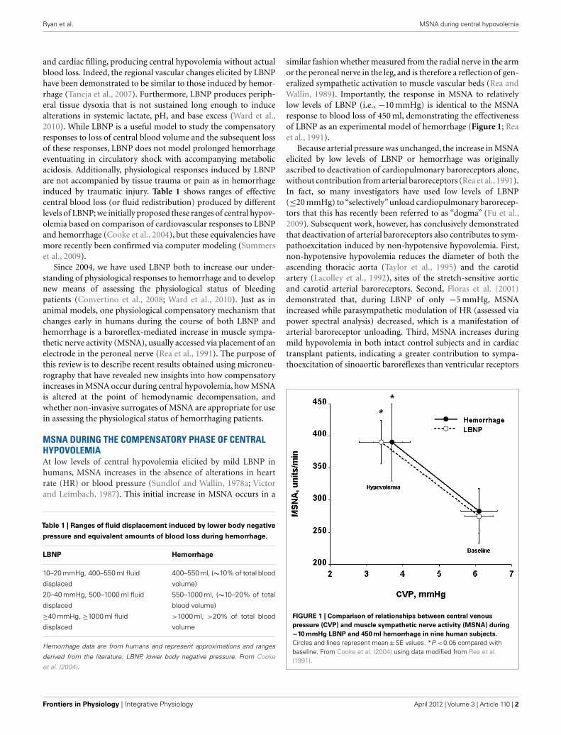
similar fashion whether measured from the radial nerve in the armor the peroneal nerve in the leg, and is therefore a reflection of gen-eralized sympathetic activation to muscle vascular beds (Rea andWallin, 1989). Importantly, the response in MSNA to relativelylow levels of LBNP (i.e., −10 mmHg) is identical to the MSNAresponse to blood loss of 450 ml, demonstrating the effectivenessof LBNP as an experimental model of hemorrhage (Figure 1; Reaet al., 1991).
Because arterial pressure was unchanged, the increase in MSNAelicited by low levels of LBNP or hemorrhage was originallyascribed to deactivation of cardiopulmonary baroreceptors alone,without contribution from arterial baroreceptors (Rea et al., 1991).In fact, so many investigators have used low levels of LBNP(≤20 mmHg) to “selectively” unload cardiopulmonary barorecep-tors that this has recently been referred to as “dogma” (Fu et al.,2009). Subsequent work, however, has conclusively demonstratedthat deactivation of arterial baroreceptors also contributes to sym-pathoexcitation induced by non-hypotensive hypovolemia. First,non-hypotensive hypovolemia reduces the diameter of both theascending thoracic aorta (Taylor et al., 1995) and the carotidartery (Lacolley et al., 1992), sites of the stretch-sensitive aorticand carotid arterial baroreceptors. Second, Floras et al. (2001)demonstrated that, during LBNP of only −5 mmHg, MSNAincreased while parasympathetic modulation of HR (assessed viapower spectral analysis) decreased, which is a manifestation ofarterial baroreceptor unloading. Third, MSNA increases duringmild hypovolemia in both intact control subjects and in cardiactransplant patients, indicating a greater contribution to sympa-thoexcitation of sinoaortic baroreflexes than ventricular receptors
FIGURE 1 | Comparison of relationships between central venous
pressure (CVP) and muscle sympathetic nerve activity (MSNA) during
−10 mmHg LBNP and 450 ml hemorrhage in nine human subjects.
Circles and lines represent mean ± SE values. *P < 0.05 compared withbaseline. From Cooke et al. (2004) using data modified from Rea et al.(1991).
Frontiers in Physiology | Integrative Physiology April 2012 | Volume 3 | Article 110 | 2
Ryan et al. MSNA during central hypovolemia
(Jacobsen et al., 1993). Finally, Fu et al. (2009) recently observedthat transient reductions in stroke volume and blood pressure[both systolic (SAP) and diastolic (DAP) pressures] occur at theonset of even mild levels of LBNP. Taken together, it has becomeclear that the sympathoexcitation produced by even low levels ofcentral hypovolemia is a result of reflex-mediated deactivation ofboth cardiopulmonary and arterial baroreceptors. Additionally,recent evidence suggests that reflexes from muscle afferents stim-ulated during venous distension may also directly evoke increasesin MSNA (Cui et al., 2009, 2011a). While venous distension shouldnot occur during hemorrhage, the possibility that reflexes arisingfrom distension of leg veins contribute to the sympathoexcitationobserved during LBNP cannot be discounted.
Once initiated, MSNA has been proposed to linearly increaseduring progressive central hypovolemia, although much of thedata supporting this suggestion until recently has been accruedusing lower levels of LBNP (≤30 mmHg; Convertino and Cooke,2002; Cooke et al., 2004). This is because of the inherent difficultyin maintaining nerve recordings during higher levels of negativepressure, as the microelectrode is often displaced during the LBNPprocedure. To mitigate this issue, Khan et al. (2002) applied nega-tive pressure to only one leg while performing microneurographyin the other. Using this protocol, these investigators demonstratedthat progressive reductions in LBNP (up to −50 mmHg) produceconcomitant graded increases in MSNA, but they did not measurethe degree of central hypovolemia induced by LBNP in order toquantify the relationship between volume loss and sympatheticactivation (Khan et al., 2002).
Over the past 6 years, we have created a large database of humanexperiments (>200 subjects) in which progressive central hypov-olemia has been used to produce hemodynamic decompensation(i.e., presyncope). In each subject, chamber decompression wasapplied for 5 min at −15, −30, −45, and −60 mmHg, and then
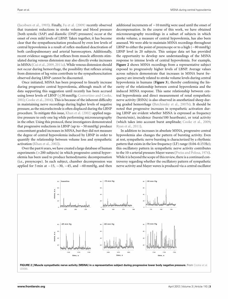
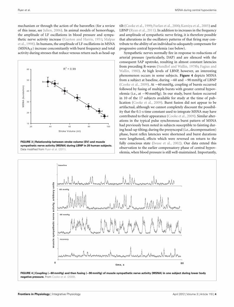
additional increments of −10 mmHg were used until the onset ofdecompensation. In the course of this work, we have obtainedmicroneurography recordings in a subset of subjects in whichstroke volume, a measure of central hypovolemia, has also beenassessed. We were able to maintain MSNA recordings throughoutLBNP to either the point of presyncope or to a high (−80 mmHg)LBNP level in 20 subjects. This unique data set has providedthe opportunity to develop new understandings of the MSNAresponse to intense levels of central hypovolemia. For example,Figure 2 shows MSNA recordings from a representative subjectexposed to progressively higher levels of LBNP. Average valuesacross subjects demonstrate that increases in MSNA burst fre-quency are inversely related to stroke volume levels during centralhypovolemia in humans (Figure 3), thereby confirming the lin-earity of the relationship between central hypovolemia and theinduced MSNA response. This same relationship between cen-tral hypovolemia and direct measurement of renal sympatheticnerve activity (RSNA) is also observed in anesthetized sheep dur-ing graded hemorrhage (Batchinsky et al., 2007b). It should benoted that progressive increases in sympathetic activation dur-ing LBNP are evident whether MSNA is expressed as frequency(bursts/min), incidence (bursts/100 heartbeats), or total activity(which takes into account burst amplitude; Cooke et al., 2009;Ryan et al., 2011).
In addition to increases in absolute MSNA, progressive centralhypovolemia also changes the pattern of bursting activity. Evenat rest, sympathetic nerve bursting is characterized by a rhythmicpattern that exists in the low frequency (LF) range (0.04–0.15 Hz);this oscillatory pattern in sympathetic nerve activity contributesto the 10-s arterial pressure Mayer waves (Preiss and Polosa, 1974).While it is beyond the scope of this review, there is a continued con-troversy regarding whether the oscillatory pattern of sympatheticnerve activity and Mayer waves is produced via a central oscillator
FIGURE 2 | Muscle sympathetic nerve activity (MSNA) in a representative subject during progressive lower body negative pressure. From Cooke et al.(2008).
www.frontiersin.org April 2012 | Volume 3 | Article 110 | 3
Ryan et al. MSNA during central hypovolemia
mechanism or through the action of the baroreflex (for a reviewof this issue, see Julien, 2006). In animal models of hemorrhage,the amplitude of LF oscillations in blood pressure and sympa-thetic nerve activity increase (Guyton and Harris, 1951; Malpaset al., 1998). In humans, the amplitude of LF oscillations in MSNA(MSNALF) increase concomitantly with burst frequency and totalactivity during stresses that reduce venous return such as head-up
FIGURE 3 | Relationship between stroke volume (SV) and muscle
sympathetic nerve activity (MSNA) during LBNP in 20 human subjects.
Data modified from Ryan et al. (2011).
tilt (Cooke et al., 1999; Furlan et al., 2000; Kamiya et al., 2005) andLBNP (Ryan et al., 2011). In addition to increases in the frequencyand amplitude of sympathetic nerve firing, it is therefore possiblethat alterations in the oscillatory patterns of that firing may con-tribute to the ability of an individual to adequately compensate forprogressive central hypovolemia (see below).
Sympathetic nerves normally fire in response to reductions ofarterial pressure (particularly, DAP) and are silenced with theconsequent SAP upstroke, resulting in almost constant latenciesfrom preceding R-waves (Sundlof and Wallin, 1978b; Fagius andWallin, 1980). At high levels of LBNP, however, an interestingphenomenon occurs in some subjects. Figure 4 depicts MSNAfrom a subject at baseline, during −60 and −90 mmHg of LBNP(Cooke et al., 2009). At −60 mmHg, coupling of bursts occurredfollowed by fusing of multiple bursts with greater central hypov-olemia (i.e., at −90 mmHg). In our study, burst fusion occurredin 10 of the 17 subjects available for study at the time of pub-lication (Cooke et al., 2009). Burst fusion did not appear to beartifactual, although we cannot completely discount the possibil-ity that the 0.1-s time constant used to integrate MSNA may havecontributed to their appearance (Cooke et al., 2009). Similar alter-ations in the typical pulse synchronous burst pattern of MSNAhad previously been noted in subjects susceptible to fainting dur-ing head-up tilting; during the presyncopal (i.e., decompensation)phase, burst reflex latencies were shortened and burst durationswere lengthened, effects which were reversed on return to thefully conscious state (Iwase et al., 2002). Our data extend thisobservation to the earlier compensatory phase of central hypov-olemia, when blood pressure is still well-maintained. Importantly,
FIGURE 4 | Coupling (−60 mmHg) and then fusing (−90 mmHg) of muscle sympathetic nerve activity (MSNA) in one subject during lower body
negative pressure. From Cooke et al. (2009).
Frontiers in Physiology | Integrative Physiology April 2012 | Volume 3 | Article 110 | 4
Ryan et al. MSNA during central hypovolemia
fusion of RSNA has also been observed in anesthetized sheep madehypotensive by severe hemorrhage (Batchinsky et al., 2007b). Wehave suggested that this phenomenon may represent sympatheticbaroreflex deafferentation (Cooke et al., 2009), as the fused burstsobserved during intense LBNP are similar in both their pulse asyn-chrony and their structure to those observed after bilateral blocksof glossopharyngeal and vagus nerves (Fagius et al., 1985). It isalso possible that loss of pulse-synchrony and continuous burstfiring may occur in some individuals as a strategy to continueto maintain blood pressure during severe reductions in venousreturn. In support of this concept, Salmanpour et al. (2011) haverecently shown that extreme baroreceptor unloading requiringhigh sympathetic outflow (induced by −80 mmHg LBNP) recruitsa subpopulation of large postganglionic axons in some but not allindividuals.
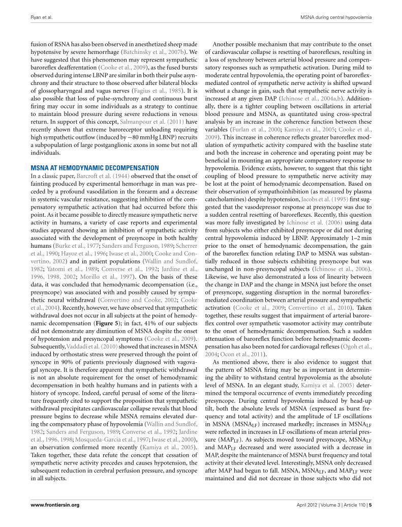
MSNA AT HEMODYNAMIC DECOMPENSATIONIn a classic paper, Barcroft et al. (1944) observed that the onset offainting produced by experimental hemorrhage in man was pre-ceded by a profound vasodilation in the forearm and a decreasein systemic vascular resistance, suggesting inhibition of the com-pensatory sympathetic activation that had occurred before thispoint. As it became possible to directly measure sympathetic nerveactivity in humans, a variety of case reports and experimentalstudies appeared showing an inhibition of sympathetic activityassociated with the development of presyncope in both healthyhumans (Burke et al., 1977; Sanders and Ferguson, 1989; Scherreret al., 1990; Hayoz et al., 1996; Iwase et al., 2000; Cooke and Con-vertino, 2002) and in patient populations (Wallin and Sundlof,1982; Yatomi et al., 1989; Converse et al., 1992; Jardine et al.,1996, 1998, 2002; Morillo et al., 1997). On the basis of thesedata, it was concluded that hemodynamic decompensation (i.e.,presyncope) was associated with and possibly caused by sympa-thetic neural withdrawal (Convertino and Cooke, 2002; Cookeet al., 2004). Recently, however, we have observed that sympatheticwithdrawal does not occur in all subjects at the point of hemody-namic decompensation (Figure 5); in fact, 41% of our subjectsdid not demonstrate any diminution of MSNA despite the onsetof hypotension and presyncopal symptoms (Cooke et al., 2009).Subsequently,Vaddadi et al. (2010) showed that increases in MSNAinduced by orthostatic stress were preserved through the point ofsyncope in 90% of patients previously diagnosed with vagova-gal syncope. It is therefore apparent that sympathetic withdrawalis not an absolute requirement for the onset of hemodynamicdecompensation in both healthy humans and in patients with ahistory of syncope. Indeed, careful perusal of some of the litera-ture frequently cited to support the proposition that sympatheticwithdrawal precipitates cardiovascular collapse reveals that bloodpressure begins to decrease while MSNA remains elevated dur-ing the compensatory phase of hypovolemia (Wallin and Sundlof,1982; Sanders and Ferguson, 1989; Converse et al., 1992; Jardineet al., 1996, 1998; Mosqueda-Garcia et al., 1997; Iwase et al., 2000),an observation confirmed more recently (Kamiya et al., 2005).Taken together, these data refute the concept that cessation ofsympathetic nerve activity precedes and causes hypotension, thesubsequent reduction in cerebral perfusion pressure, and syncopein all subjects.
Another possible mechanism that may contribute to the onsetof cardiovascular collapse is resetting of baroreflexes, resulting ina loss of synchrony between arterial blood pressure and compen-satory responses such as sympathetic activation. During mild tomoderate central hypovolemia, the operating point of baroreflex-mediated control of sympathetic nerve activity is shifted upwardwithout a change in gain, such that sympathetic nerve activity isincreased at any given DAP (Ichinose et al., 2004a,b). Addition-ally, there is a tighter coupling between oscillations in arterialblood pressure and MSNA, as quantitated using cross-spectralanalysis by an increase in the coherence function between thesevariables (Furlan et al., 2000; Kamiya et al., 2005; Cooke et al.,2009). This increase in coherence reflects greater baroreflex mod-ulation of sympathetic activity compared with the baseline stateand both the increase in coherence and operating point may bebeneficial in mounting an appropriate compensatory response tohypovolemia. Evidence exists, however, to suggest that this tightcoupling of blood pressure to sympathetic nerve activity maybe lost at the point of hemodynamic decompensation. Based ontheir observation of sympathoinhibition (as measured by plasmacatecholamines) despite hypotension, Jacobs et al. (1995) first sug-gested that the vasodepressor response at presyncope was due toa sudden central resetting of baroreflexes. Recently, this questionwas more fully investigated by Ichinose et al. (2006) using datafrom subjects who either exhibited presyncope or did not duringcentral hypovolemia induced by LBNP. Approximately 1–2 minprior to the onset of hemodynamic decompensation, the gainof the baroreflex function relating DAP to MSNA was substan-tially reduced in those subjects exhibiting presyncope but wasunchanged in non-presyncopal subjects (Ichinose et al., 2006).Likewise, we have also demonstrated a loss of linearity betweenthe change in DAP and the change in MSNA just before the onsetof presyncope, suggesting disruption in the normal baroreflex-mediated coordination between arterial pressure and sympatheticactivation (Cooke et al., 2009; Convertino et al., 2010). Takentogether, these results suggest that impairment of arterial barore-flex control over sympathetic vasomotor activity may contributeto the onset of hemodynamic decompensation. Such a suddenattenuation of baroreflex function before hemodynamic decom-pensation has also been noted for cardiovagal reflexes (Ogoh et al.,2004; Ocon et al., 2011).
As mentioned above, there is also evidence to suggest thatthe pattern of MSNA firing may be as important in determin-ing the ability to withstand central hypovolemia as the absolutelevel of MSNA. In an elegant study, Kamiya et al. (2005) deter-mined the temporal occurrence of events immediately precedingpresyncope. During central hypovolemia induced by head-uptilt, both the absolute levels of MSNA (expressed as burst fre-quency and total activity) and the amplitude of LF oscillationsin MSNA (MSNALF) increased markedly; increases in MSNALF
were reflected in increases in LF oscillations of mean arterial pres-sure (MAPLF). As subjects moved toward presyncope, MSNALF
and MAPLF decreased and were associated with a decrease inMAP, despite the maintenance of MSNA burst frequency and totalactivity at their elevated level. Interestingly, MSNA only decreasedafter MAP had begun to fall. MSNA, MSNALF, and MAPLF weremaintained and did not decrease in those subjects who did not
www.frontiersin.org April 2012 | Volume 3 | Article 110 | 5

Ryan et al. MSNA during central hypovolemia
FIGURE 5 | Arterial pressure (AP) and muscle sympathetic nerve
activity (MSNA) for three representative subjects 2 min before the
onset of presyncope. The lowest arterial pressure (BP) recorded for eachsubject is shown in the upper right corner of each AP panel. In subjectA185, MSNA decreased in the last 2 min of LBNP (−120 to −60 s,82 bursts/min; −60 s to presyncope, 64 bursts/min), but was stillmaintained at high levels relative to the pre-LBNP control (36 bursts/min).
Subject A075 displayed burst fusion, elevated MSNA over control(25 bursts/min), and no withdrawal of MSNA at presyncope (−120 to−60 s, 53 bursts/min; −60 s to presyncope, 54 bursts/min). Subject A199(a low tolerant subject) did not display large increases in MSNA from thepre-LBNP control value (17 bursts/min) and also did not demonstrateMSNA withdrawal at presyncope (−120 to −60 s, 22 bursts/min; −60 s topresyncope, 21 bursts/min). From Cooke et al. (2009).
exhibit hypotension and presyncope (Kamiya et al., 2005). Wehave observed a similar phenomenon in a subject whose toleranceto central hypovolemia was improved through the use of inspira-tory resistance breathing (Figure 6). In this experiment, subjectswere exposed to LBNP to the point of presyncope in separateexperiments performed at least 2 weeks apart. In one experiment,subjects breathed through a device that did not provide resistanceto inspiration (sham), while in the other experiment, subjectsbreathed through a device that provided resistance to inspiration
(active). Because resistance breathing improved LBNP tolerance,data were analyzed at the end of LBNP during the sham experimentand at this same absolute time point during the active experiment(i.e., well before the onset of presyncope during inspiratory resis-tance breathing). During breathing with the sham device, MSNAincreased from 12 bursts/min at baseline to 44 bursts/min at theend of LBNP, while MSNA increased from 10 to 54 bursts/minat this same absolute time point (i.e., prior to presyncope) duringresistance breathing. Importantly, MSNALF increased from 1.15 to
Frontiers in Physiology | Integrative Physiology April 2012 | Volume 3 | Article 110 | 6
Ryan et al. MSNA during central hypovolemia
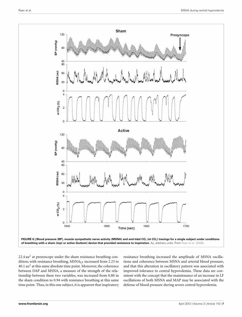
FIGURE 6 | Blood pressure (BP), muscle sympathetic nerve activity (MSNA) and end-tidal CO2 (et CO2) tracings for a single subject under conditions
of breathing with a sham (top) or active (bottom) device that provided resistance to inspiration. Au, arbitrary units. From Ryan et al. (2008).
22.4 au2 at presyncope under the sham resistance breathing con-dition; with resistance breathing, MSNALF increased from 2.23 to40.1 au2 at this same absolute time point. Moreover, the coherencebetween DAP and MSNA, a measure of the strength of the rela-tionship between these two variables, was increased from 0.80 inthe sham condition to 0.94 with resistance breathing at this sametime point. Thus, in this one subject, it is apparent that inspiratory
resistance breathing increased the amplitude of MSNA oscilla-tions and coherence between MSNA and arterial blood pressure,and that this alteration in oscillatory pattern was associated withimproved tolerance to central hypovolemia. These data are con-sistent with the concept that the maintenance of an increase in LFoscillations of both MSNA and MAP may be associated with thedefense of blood pressure during severe central hypovolemia.
www.frontiersin.org April 2012 | Volume 3 | Article 110 | 7
Ryan et al. MSNA during central hypovolemia
Thus, there are several possible mechanisms involving alter-ations in activation of sympathetic nerve activity to explain hemo-dynamic decompensation during severe hypovolemia. Since sym-pathetic withdrawal occurs in some individuals before presyncopebut not in others, it is no longer thought to be a prerequisite forthe ensuing hypotension (Cooke et al., 2009). It is also possiblethat there is a central resetting of arterial baroreflex function thatalters sympathetic outflow (Ichinose et al., 2006); this scenario maybe especially prominent in those subjects in whom sympatheticinhibition occurs despite progressive hypotension. Finally, thereis evidence to suggest that an increase in LF oscillations in bothMSNA and MAP may be protective during central hypovolemiaand that loss of these oscillations might precipitate hypotension(Kamiya et al., 2005). It is important to note that these mechanismsare not mutually exclusive and the contributions of each to theprocess have yet to be fully revealed. Furthermore, it is also possibleand even probable that different mechanisms may predominate indifferent individuals. Because of the scope of this review, we havechosen to focus on those mechanisms preceding presyncope thatinvolve loss of compensatory alterations in MSNA, but it is likelythat loss of other compensatory responses may also contributeto the inability to maintain blood pressure during severe hypo-volemia. One intriguing hypothesis put forward by Dietz et al.(1997) is that marked peripheral vasodilation is a major contribu-tor to the fall in arterial pressure preceding vasovagal syncope. Inthis regard, it is of interest that, during prolonged LBNP at a lowlevel (−15 mmHg), vasodilation of the forearm musculature wasobserved despite the continued presence of a sustained compen-satory increase in MSNA, suggesting that “sympathetic escape”occurs (Joyner et al., 1990). This observation is currently beinginvestigated.
Implicit in the preceding discussion is the notion that thereare individual differences in the ability of healthy humans totolerate central hypovolemia before reaching the point of car-diovascular collapse. Indeed, it has been known for many yearsthat there is a great deal of variability in the ability of patients(Davis, 1949) and animals (Chien, 1967; Kim and Shoemaker,1970) to survive traumatic hemorrhage; we have recently learnedthat there is a genetic basis underlying this variability (Klemckeet al., 2008, 2011). Likewise, individual differences in tolerance tocentral hypovolemia induced by LBNP have also been described(Sather et al., 1986) and attributed to differences in the releaseof vasoactive hormones (Convertino and Sather, 2000b; Green-leaf et al., 2000), compensatory tachycardia and vasoconstriction(Convertino and Sather, 2000a,b;Greenleaf et al., 2000), cardiacbaroreflex gain (Convertino and Sather, 2000a; Convertino et al.,in press), baroreflex gain of sympathetic nerve activation (Wijey-sundera et al., 2001), and central blood volume and cerebral bloodvelocity (Levine et al., 1994). In this regard, we have recently shownthat subjects demonstrating high tolerance (HT) to LBNP have agreater ability to increase MSNA than subjects with low toler-ance (LT; Convertino et al., in press). Additionally, the ability tosustain the compensatory mechanisms described above involv-ing both the baroreflex modulation of sympathetic activation andthe oscillatory component of that activation may also act to deter-mine tolerance to central hypovolemia. For example, Ichinose et al.(2006) described a sudden resetting of the DAP–MSNA baroreflex
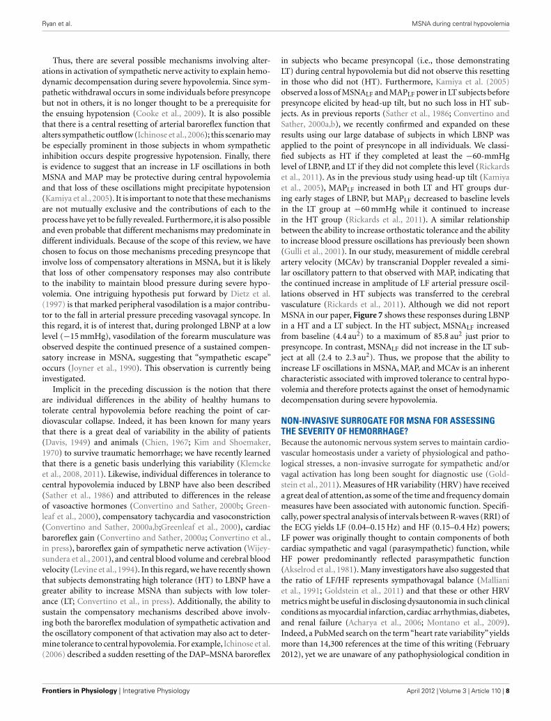
in subjects who became presyncopal (i.e., those demonstratingLT) during central hypovolemia but did not observe this resettingin those who did not (HT). Furthermore, Kamiya et al. (2005)observed a loss of MSNALF and MAPLF power in LT subjects beforepresyncope elicited by head-up tilt, but no such loss in HT sub-jects. As in previous reports (Sather et al., 1986; Convertino andSather, 2000a,b), we recently confirmed and expanded on theseresults using our large database of subjects in which LBNP wasapplied to the point of presyncope in all individuals. We classi-fied subjects as HT if they completed at least the −60-mmHglevel of LBNP, and LT if they did not complete this level (Rickardset al., 2011). As in the previous study using head-up tilt (Kamiyaet al., 2005), MAPLF increased in both LT and HT groups dur-ing early stages of LBNP, but MAPLF decreased to baseline levelsin the LT group at −60 mmHg while it continued to increasein the HT group (Rickards et al., 2011). A similar relationshipbetween the ability to increase orthostatic tolerance and the abilityto increase blood pressure oscillations has previously been shown(Gulli et al., 2001). In our study, measurement of middle cerebralartery velocity (MCAv) by transcranial Doppler revealed a simi-lar oscillatory pattern to that observed with MAP, indicating thatthe continued increase in amplitude of LF arterial pressure oscil-lations observed in HT subjects was transferred to the cerebralvasculature (Rickards et al., 2011). Although we did not reportMSNA in our paper, Figure 7 shows these responses during LBNPin a HT and a LT subject. In the HT subject, MSNALF increasedfrom baseline (4.4 au2) to a maximum of 85.8 au2 just prior topresyncope. In contrast, MSNALF did not increase in the LT sub-ject at all (2.4 to 2.3 au2). Thus, we propose that the ability toincrease LF oscillations in MSNA, MAP, and MCAv is an inherentcharacteristic associated with improved tolerance to central hypo-volemia and therefore protects against the onset of hemodynamicdecompensation during severe hypovolemia.
NON-INVASIVE SURROGATE FOR MSNA FOR ASSESSINGTHE SEVERITY OF HEMORRHAGE?Because the autonomic nervous system serves to maintain cardio-vascular homeostasis under a variety of physiological and patho-logical stresses, a non-invasive surrogate for sympathetic and/orvagal activation has long been sought for diagnostic use (Gold-stein et al., 2011). Measures of HR variability (HRV) have receiveda great deal of attention, as some of the time and frequency domainmeasures have been associated with autonomic function. Specifi-cally, power spectral analysis of intervals between R-waves (RRI) ofthe ECG yields LF (0.04–0.15 Hz) and HF (0.15–0.4 Hz) powers;LF power was originally thought to contain components of bothcardiac sympathetic and vagal (parasympathetic) function, whileHF power predominantly reflected parasympathetic function(Akselrod et al., 1981). Many investigators have also suggested thatthe ratio of LF/HF represents sympathovagal balance (Mallianiet al., 1991; Goldstein et al., 2011) and that these or other HRVmetrics might be useful in disclosing dysautonomia in such clinicalconditions as myocardial infarction, cardiac arrhythmias, diabetes,and renal failure (Acharya et al., 2006; Montano et al., 2009).Indeed, a PubMed search on the term “heart rate variability” yieldsmore than 14,300 references at the time of this writing (February2012), yet we are unaware of any pathophysiological condition in
Frontiers in Physiology | Integrative Physiology April 2012 | Volume 3 | Article 110 | 8
Ryan et al. MSNA during central hypovolemia
FIGURE 7 | Representative tracings from a high tolerant and low tolerant subject at baseline (BL), sub-maximal LBNP (i.e., the LBNP level before the
level at which presyncope was reached; SM) and immediately before presyncope (indicated by arrow; PS).
which HRV is currently used as a standard of care for diagnosisand/or treatment. The use of HRV is appealing because calculationrequires only non-invasive collection of a standard ECG.
Because increases in sympathetic activation occur in a linearfashion with decreases in stroke volume (Figure 3), we proposedthat HRV metrics might be useful non-invasive surrogates ofMSNA for assessing the degree and progression of hemorrhagein trauma victims (Cooke and Convertino, 2005). In 2008, wedemonstrated using our LBNP model of simulated hemorrhagethat, on average, RRIHF power and some time domain metricsassociated with parasympathetic function are inversely related todirect measurement of MSNA. RRILF power, on the other hand,was not associated with MSNA (Cooke et al., 2008); it has beenmade clear more recently that RRILF is not a measure of cardiacsympathetic activity (Billman, 2011; Goldstein et al., 2011). Fromthese data, we concluded that HRV might be of clinical utility inassessing autonomic function during hemorrhage, although weand others noted that correlations between MSNA and HRV inindividual subjects were not as strong as those derived from groupmeans (Floras et al., 2001; Cooke et al., 2008). Application of powerspectral analysis to ECG recordings collected from actual traumapatients during air transport to the hospital further suggested thatthe use of these metrics might be appropriate for clinical assess-ment of the severity of hemorrhage, as group means of some HRVmetrics differed between patients who lived and those who died upto 24 h later (Cooke et al., 2006a,b). Subsequent studies extendedthese observations to the use of non-linear HRV metrics to predictmortality (Batchinsky et al., 2007a) and to discriminate betweentrauma patients who required a life-saving intervention and thosewho did not (Cancio et al., 2008).
Importantly, all of the conclusions delineated above were devel-oped based on standard analyses of group mean data. As our
understanding evolved, however, we began to take the “next step”to assess whether HRV metrics could be useful in determining thephysiological status of individuals rather than a group as a whole,a requirement that must be met for successful application of anymetric for diagnosing individual patients. In doing so, we becameaware of a number of challenges with all of the HRV metrics thatwe examined (time domain, frequency domain, and non-linearmetrics). First, there are issues of large inter-individual variabilityand poor reproducibility within subjects during the same record-ing session (Rickards et al., 2010a) as well as across days (Tan et al.,2009). Second, accurate determination of HRV requires long (insome cases, up to 800 beats) segments of ECG recordings thatdo not contain electromagnetic noise or ectopic beats, but theseoccur more frequently in trauma patients than in healthy individu-als (Sethuraman et al., 2010). Third, increases in HR will always beassociated with decreases in HRV, purely as a mathematical func-tion of the curvilinear nature of the relationship between HR andRRI (Sacha and Pluta, 2008). Decreases in HRV are therefore notspecific to central hypovolemia but may be elicited by any physi-ological stressor that induces tachycardia such as physical move-ment (Rickards et al., 2008), pain, or anxiety, which are commonin conscious trauma patients. Fourth, HRV metrics are not able todiscriminate between HT and LT subjects early in the progressionof hypovolemia; an effective triage tool should be able to alert med-ical personnel to those patients that will progress to hemodynamicdecompensation more quickly (Hinojosa-Laborde et al., 2011).Finally, although group means of HRV metrics are highly corre-lated with decreases in stroke volume during central hypovolemia,analysis of the individual trajectories for these metrics demon-strated poor and inconsistent correlations with stroke volume atthe individual subject level, even under controlled laboratory con-ditions (Ryan et al., 2010). Armed with this new understanding,
www.frontiersin.org April 2012 | Volume 3 | Article 110 | 9
Ryan et al. MSNA during central hypovolemia
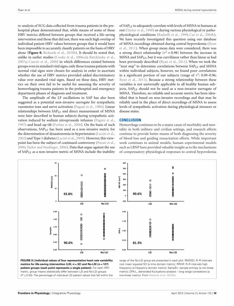
re-analysis of ECG data collected from trauma patients in the pre-hospital phase demonstrated that, while means of some of theseHRV metrics differed between groups that received a life-savingintervention and those that did not, there was such high overlap ofindividual patient HRV values between groups that it would havebeen impossible to accurately classify patients on the basis of HRValone (Figure 8; Rickards et al., 2010b). It should be noted that,unlike in earlier studies (Cooke et al., 2006a,b; Batchinsky et al.,2007a; Cancio et al., 2008) in which differences existed betweengroups even in standard vital signs,only those trauma patients withnormal vital signs were chosen for analysis in order to ascertainwhether the use of HRV metrics provided added discriminatoryvalue over standard vital signs. Based on these data, HRV met-rics on their own fail to be useful for assessing the severity ofhemorrhaging trauma patients in the prehospital and emergencydepartment phases of diagnosis and treatment.
The amplitude of the LF oscillations in SAP has also beensuggested as a potential non-invasive surrogate for sympatheticvasomotor tone and nerve activation (Pagani et al., 1986). Linearrelationships between SAPLF and direct measurement of MSNAwere later described in human subjects during sympathetic acti-vation induced by sodium nitroprusside infusion (Pagani et al.,1997) and head-up tilt (Furlan et al., 2000). On the basis of suchobservations, SAPLF has been used as a non-invasive metric forthe determination of dysautonomia in hypertension (Lucini et al.,2002) and Type 1 diabetes (Lucini et al., 2009). However, this view-point has been the subject of continued controversy (Parati et al.,2006; Taylor and Studinger, 2006). Data that argue against the useof SAPLF as a non-invasive metric of MSNA include the inability
of SAPLF to adequately correlate with levels of MSNA in humans atrest (Taylor et al., 1998) or during various physiological or patho-physiological conditions (Radaelli et al., 1999; Cui et al., 2004b).We have recently investigated this question using our databaseof MSNA recordings obtained during central hypovolemia (Ryanet al., 2011). When group mean data were considered, there wasa strong direct relationship (r2 = 0.98) between the increase inMSNA and SAPLF, but it was curvilinear rather than linear as hadbeen previously described (Ryan et al., 2011). When we took the“next step” to determine correlations between SAPLF and MSNAwithin individual subjects, however, we found poor correlationsin a significant portion of our subjects (range of r2: 0.09–0.96;Ryan et al., 2011). Because a strong relationship between thesevariables is not universally applicable to all healthy human sub-jects, SAPLF should not be used as a non-invasive surrogate ofMSNA. Therefore, no reliable and accurate metric has been iden-tified that is based on non-invasive recordings and that may bereliably used in the place of direct recordings of MSNA to assesslevels of sympathetic activation during physiological stressors ordisease states.
CONCLUSIONHemorrhage continues to be a major cause of morbidity and mor-tality in both military and civilian settings, and research effortscontinue to provide better means of both diagnosing the severityof blood loss and guiding resuscitation efforts. While importantwork continues in animal models, human experimental modelssuch as LBNP have provided valuable insight as to the mechanismsof compensatory physiological responses to central hypovolemia
FIGURE 8 | Individual values of four representative heart rate variability
metrics for life-saving intervention (LSI; n = 32) and No-LSI (n = 127)
patient groups (each point represents a single patient). For each HRVmetric, group means statistically differ between LSI and No-LSI groups(P ≤ 0.03). The percentage of individual LSI patient values that fall within the
range of the No-LSI group are presented in each plot. RMSSD, R–R intervalsroot mean squared SD (a time domain metric); RRIHF, R–R intervals highfrequency (a frequency domain metric); SampEn, sample entropy (a non-linearmetric); DFA-L, detrended fluctuations analysis – long range correlations (anon-linear metric). From Rickards et al. (2010b).
Frontiers in Physiology | Integrative Physiology April 2012 | Volume 3 | Article 110 | 10
Ryan et al. MSNA during central hypovolemia
without the confounding factors of anesthesia, species differences,and tissue injury. During even mild LBNP, both arterial and car-diopulmonary baroreceptors are unloaded, resulting in activationof the sympathetic nervous system to increase HR and vasomo-tor tone. Importantly, we now know that sympathetic activationentails not only an increase in sympathetic nerve firing but alsoan alteration in the pattern of firing, such that the amplitude ofLF oscillations in MSNA and, consequently, blood pressure is alsoincreased; both of these responses seem to be protective in that theyare associated with improved tolerance to central hypovolemia. Atsome point, which occurs at different levels of central hypovolemiafor individual subjects, this compensation fails and hypotensionensues. Although it was once thought that sympathetic withdrawalalways precipitated hemodynamic decompensation and hypoten-sion, it is now clear that a diminution of absolute levels of MSNAfiring is not required in all individuals. Instead, it is possiblethat loss of the compensatory increase in MSNALF may also beinvolved, particularly in those subjects demonstrating HT to cen-tral hypovolemia. Additionally, there may be an acute resettingof the baroreflex at the level of the CNS such that coherencebetween arterial blood pressure and sympathetic nerve activityis lost. Determination of the mechanisms underlying the develop-ment of presyncope in both healthy human subjects and patientswith diseases characterized by episodes of fainting continues to bean ongoing area of research.
The search for a non-invasive surrogate of MSNA for bothresearch and clinical purposes continues. Certainly, an easilyobtainable non-invasive metric of sympathetic activation could beof great importance for assessment of the severity of hemorrhage.However, metrics based on determination of variability in bothRRIs and arterial blood pressure do not fulfill the necessary cri-teria to perform effectively in this role. Before implementation ofany such metric, it is essential that investigators determine whetherthe metric will be able to reliably track MSNA within individualsubjects rather than simply rely on analyses based on group mean
data. Importantly, gender differences in the MSNA response tocentral hypovolemia must also be taken into account (Fu et al.,2005; Carter et al., 2009). For use in trauma victims suspected ofhemorrhage, it will also be necessary for laboratory determinationof the efficacy of the metric in the face of blood loss combinedwith other physiological stressors which can impact sympatheticactivation, such as heat stress (Cui et al., 2004a, 2011b), dehy-dration (Kimmerly and Shoemaker, 2002, 2003; Fu et al., 2005),mental or emotional stress (Carter et al., 2008), and ingestion ofalcohol (Carter et al., 2011), nicotine, or other drugs. All of thesestressors are commonly observed in conjunction with traumatichemorrhage in civilian and/or military settings. Because of thecomplexity of this problem, it is possible that clinical assessmentof the severity of hemorrhage during the acute phase may be bet-ter realized using artificial intelligence technologies that reflect theintegration of the sympathetic nervous response to hemorrhagewith changes in circulatory pressure and volume (Convertino et al.,2011).
DISCLAIMERThe opinions or assertions contained herein are the private viewsof the authors and are not to be construed as official or as reflect-ing the views of the Department of the Army or the Departmentof Defense.
ACKNOWLEDGMENTSThe authors thank our research subjects, who cheerfully cooper-ated with the experiments described herein, and Mr. Gary Munizfor his superb technical assistance. All studies performed at the U.S.Army Institute of Surgical Research were conducted under proto-cols reviewed and approved by the US Army Medical Researchand Materiel Command Institutional Review Board and in accor-dance with the approved protocols. This work was supported bythe Combat Casualty Care Research Area Directorate of the USArmy Medical Research and Materiel Command.
REFERENCESAcharya, U. R., Joseph, K. P., Kan-
nathal, N., Lim, C. M., and Suri, J.S. (2006). Heart rate variability: areview. Med. Biol. Eng. Comput. 44,1031–1051.
Akselrod, S., Gordon, D., Ubel, F. A.,Shannon, D. C., Berger, A. C., andCohen, R. J. (1981). Power spectrumanalysis of heart rate fluctuation: aquantitative probe of beat-to-beatcardiovascular control. Science 213,220–222.
Barcroft, H., Edholm, O. G., McMichael,J., and Sharpey-Schafer, E. P. (1944).Posthaemorrhagic fainting: study bycardiac output and forearm flow.Lancet 1, 489–490.
Batchinsky, A. I., Cancio, L. C., Salinas,J., Kuusela, T., Cooke, W. H., Wang,J. J., Boehme, M., Convertino, V. A.,and Holcomb, J. B. (2007a). Prehos-pital loss of R-to-R interval com-plexity is associated with mortality
in trauma patients. J. Trauma 63,512–518.
Batchinsky, A. I., Cooke, W. H., Kuusela,T. A., Jordan, B. S., Wang, J. J., andCancio, L. C. (2007b). Sympatheticnerve activity and heart rate vari-ability during severe hemorrhagicshock in sheep. Auton. Neurosci. 136,43–51.
Billman, G. E. (2011). Heart ratevariability – a historical per-spective. Front. Physiol. 2:86.doi:10.3389/fphys.2011.00086
Burke, D., Sundlof, G., and Wallin, G.(1977). Postural effects on musclenerve sympathetic activity in man.J. Physiol. (Lond.) 272, 399–414.
Cancio, L. C., Batchinsky, A. I., Sali-nas, J., Kuusela, T., Convertino, V.A., Wade, C. E., and Holcomb, J.B. (2008). Heart-rate complexity forprediction of prehospital lifesavinginterventions in trauma patients. J.Trauma 65, 813–819.
Carter, J. R., Durocher, J. J., and Kern, R.P. (2008). Neural and cardiovascu-lar responses to emotional stress inhumans. Am. J. Physiol. Regul. Integr.Comp. Physiol. 295, R1898–R1903.
Carter, J. R., Lawrence, J. E., and Klein,J. C. (2009). Menstrual cycle alterssympathetic neural responses toorthostatic stress in young, eumen-orrheic women. Am. J. Physiol.Endocrinol. Metab. 297, E85–E91.
Carter, J. R., Stream, S. F., Durocher,J. J., and Larson, R. A. (2011).Influence of acute alcohol ingestionon sympathetic neural responses toorthostatic stress in humans. Am.J. Physiol. Endocrinol. Metab. 300,E771–E778.
Chien, S. (1967). Role of the sym-pathetic nervous system in hemor-rhage. Physiol. Rev. 47, 214–288.
Converse, R. L. Jr., Jacobsen, T. N., Jost,C. M., Toto, R. D., Grayburn, P. A.,Obregon, T. M., Fouad-Tarazi, F.,
and Victor, R. G. (1992). Paradox-ical withdrawal of reflex vasocon-striction as a cause of hemodialysis-induced hypotension. J. Clin. Invest.90, 1657–1665.
Convertino, V. A. (2001). Lower bodynegative pressure as a tool forresearch in aerospace physiology andmilitary medicine. J. Gravit. Physiol.8, 1–14.
Convertino, V. A., and Cooke, W. H.(2002). Relationship between strokevolume and sympathetic nerve activ-ity: new insights about autonomicmechanisms of syncope. J. Gravit.Physiol. 9, P63–P66.
Convertino, V. A., Moulton, S. L., Gru-dic, G. Z., Rickards, C. A., Hinojosa-Laborde, C., Gerhardt, R. T., Black-bourne, L. H., and Ryan, K. L.(2011). Use of advanced machine-learning techniques for noninva-sive monitoring of hemorrhage. J.Trauma 71, S25–S32.
www.frontiersin.org April 2012 | Volume 3 | Article 110 | 11
Ryan et al. MSNA during central hypovolemia
Convertino, V. A., Ryan, K. L., Rickards,C. A., Salinas, J., McManus, J. G.,Cooke, W. H., and Holcomb, J.B. (2008). Physiological and med-ical monitoring for en route careof combat casualties. J. Trauma 64,S342–S353.
Convertino, V. A., and Sather, T. M.(2000a). Effects of cholinergic andbeta-adrenergic blockade on ortho-static tolerance in healthy subjects.Clin. Auton. Res. 10, 327–336.
Convertino, V. A., and Sather, T. M.(2000b). Vasoactive neuroendocrineresponses associated with toleranceto lower body negative pressure inhumans. Clin. Physiol. 20, 177–184.
Convertino, V. A., Rickards, C. A., andRyan, K. L. (2010). Responses ofsympathetic nerve activity to pre-syncope: new insights about mech-anisms of fainting. J. Gravit. Physiol.17, P27–P30.
Convertino, V. A., Rickards, C. A., andRyan, K. L. (in press). Toleranceto central hypovolemia: autonomicmechanisms associated with heartrate and vasoconstrictor reserves inhumans. Clin. Auton. Res.
Cooke, W. H., and Convertino, V. A.(2002). Association between vaso-vagal hypotension and low sympa-thetic neural activity during presyn-cope. Clin. Auton. Res. 12, 483–486.
Cooke, W. H., and Convertino, V.A. (2005). Heart rate variabil-ity and spontaneous baroreflexsequences: implications for auto-nomic monitoring during hemor-rhage. J. Trauma 58, 798–805.
Cooke, W. H., Hoag, J. B., Crossman,A. A., Kuusela, T. A., Tahvanainen,K. U., and Eckberg, D. L. (1999).Human responses to upright tilt:a window on central autonomicintegration. J. Physiol. (Lond.) 517,617–628.
Cooke, W. H., Rickards, C. A., Ryan,K. L., and Convertino, V. A. (2008).Autonomic compensation to simu-lated hemorrhage monitored withheart period variability. Crit. CareMed. 36, 1892–1899.
Cooke, W. H., Rickards, C. A., Ryan, K.L., Kuusela, T. A., and Convertino,V. A. (2009). Muscle sympatheticnerve activity during intense lowerbody negative pressure to presyn-cope in humans. J. Physiol. (Lond.)587, 4987–4999.
Cooke, W. H., Ryan, K. L., and Con-vertino, V. A. (2004). Lower bodynegative pressure as a model to studyprogression to acute hemorrhagicshock in humans. J. Appl. Physiol. 96,1249–1261.
Cooke, W. H., Salinas, J., Convertino, V.A., Ludwig, D. A., Hinds, D., Duke,
J. H., Moore, F. A., and Holcomb, J.B. (2006a). Heart rate variability andits association with mortality in pre-hospital trauma patients. J. Trauma60, 363–370; discussion 370.
Cooke, W. H., Salinas, J., McManus, J.G., Ryan, K. L., Rickards, C. A., Hol-comb, J. B., and Convertino, V. A.(2006b). Heart period variability intrauma patients may predict mortal-ity and allow remote triage. Aviat.Space Environ. Med. 77, 1107–1112.
Cui, J., Leuenberger, U. A., Gao, Z.,and Sinoway, L. I. (2011a). Sympa-thetic and cardiovascular responsesto venous distension in an occludedlimb. Am. J. Physiol. Regul. Integr.Comp. Physiol. 301, R1831–R1837.
Cui, J., Shibasaki, M., Low, D. A., Keller,D. M., Davis, S. L., and Crandall,C. G. (2011b). Muscle sympatheticresponses during orthostasis in heat-stressed individuals. Clin. Auton.Res. 21, 381–387.
Cui, J., McQuillan, P., Moradkhan,R., Pagana, C., and Sinoway, L. I.(2009). Sympathetic responses dur-ing saline infusion into the veins ofan occluded limb. J. Physiol. (Lond.)587, 3619–3628.
Cui, J., Wilson, T. E., and Crandall, C. G.(2004a). Muscle sympathetic nerveactivity during lower body nega-tive pressure is accentuated in heat-stressed humans. J. Appl. Physiol. 96,2103–2108.
Cui, J., Zhang, R., Wilson, T. E., andCrandall, C. G. (2004b). Spectralanalysis of muscle sympathetic nerveactivity in heat-stressed humans.Am. J. Physiol. Heart Circ. Physiol.286, H1101–H1106.
Davis, H. A. (1949). Shock and AlliedForms of Failure of the Circulation.New York: Grune & Stratton.
Dietz, N. M., Joyner, M. J., and Shep-herd, J. T. (1997). Vasovagal syn-cope and skeletal muscle vasodi-latation: the continuing conun-drum. Pacing Clin. Electrophysiol. 20,775–780.
Eastridge, B. J., Hardin, M., Cantrell, J.,Oetjen-Gerdes, L., Zubko, T., Mallak,C., Wade, C. E., Simmons, J., Mace,J., Mabry, R., Bolenbaucher, R., andBlackbourne, L. H. (2011). Died ofwounds on the battlefield: causa-tion and implications for improvingcombat casualty care. J. Trauma 71,S4–S8.
Fagius, J., and Wallin, B. G. (1980). Sym-pathetic reflex latencies and conduc-tion velocities in normal man. J.Neurol. Sci. 47, 433–448.
Fagius, J., Wallin, B. G., Sundlof,G., Nerhed, C., and Englesson,S. (1985). Sympathetic outflowin man after anaesthesia of the
glossopharyngeal and vagus nerves.Brain 108, 423–438.
Floras, J. S., Butler, G. C., Ando, S. I.,Brooks, S. C., Pollard, M. J., andPicton, P. (2001). Differential sym-pathetic nerve and heart rate spec-tral effects of nonhypotensive lowerbody negative pressure. Am. J. Phys-iol. Regul. Integr. Comp. Physiol. 281,R468–R475.
Fu, Q., Shibata, S., Hastings, J. L., Prasad,A., Palmer, M. D., and Levine, B. D.(2009). Evidence for unloading arte-rial baroreceptors during low levelsof lower body negative pressure inhumans. Am. J. Physiol. Heart Circ.Physiol. 296, H480–H488.
Fu, Q., Witkowski, S., Okazaki, K., andLevine, B. D. (2005). Effects of gen-der and hypovolemia on sympa-thetic neural responses to orthosta-tic stress. Am. J. Physiol. Regul. Integr.Comp. Physiol. 289, R109–R116.
Furlan, R., Porta, A., Costa, F., Tank,J., Baker, L., Schiavi, R., Robert-son, D., Malliani, A., and Mosqueda-Garcia, R. (2000). Oscillatory pat-terns in sympathetic neural dis-charge and cardiovascular variablesduring orthostatic stimulus. Circu-lation 101, 886–892.
Gernandt, B., Liljestrand, G., and Zot-terman, Y. (1946). Efferent impulsesin the splanchnic nerve. Acta Physiol.Scand. 11, 231–247.
Goldstein, D. S., Bentho, O., Park, M.Y., and Sharabi, Y. (2011). Low-frequency power of heart rate vari-ability is not a measure of cardiacsympathetic tone but may be a mea-sure of modulation of cardiac auto-nomic outflows by baroreflexes. Exp.Physiol. 96, 1255–1261.
Greenleaf, J. E., Petersen, T. W.,Gabrielsen, A., Pump, B., Bie, P.,Christensen, N. J., Warberg, J., Vide-baek, R., Simonson, S. R., and Norsk,P. (2000). Low LBNP tolerance inmen is associated with attenuatedactivation of the renin-angiotensinsystem. Am. J. Physiol. Regul. Integr.Comp. Physiol. 279, R822–R829.
Gulli, G., Wight, V. L., Hainsworth, R.,and Cevese, A. (2001). Spectral andcross-spectral autoregressive analy-sis of cardiovascular variables in sub-jects with different degrees of ortho-static tolerance. Clin. Auton. Res. 11,19–27.
Guyton, A. C., and Harris, J. W.(1951). Pressoreceptor-autonomicoscillation: a probable cause of vaso-motor waves. Am. J. Physiol. 165,158–166.
Hayoz, D., Noll, G., Passino, C., Weber,R., Wenzel, R., and Bernardi, L.(1996). Progressive withdrawalof muscle nerve sympathetic
activity preceding vaso-vagal syn-cope during lower-body negativepressure. Clin. Sci. 91(Suppl.),50–51.
Hinojosa-Laborde, C., Rickards, C. A.,Ryan, K. L., and Convertino, V. A.(2011). Heart rate variability duringsimulated hemorrhage with lowerbody negative pressure in high andlow tolerant subjects. Front. Physiol.2:85. doi:10.3389/fphys.2011.00085
Ichinose, M., Saito, M., Fujii, N., Kondo,N., and Nishiyasu, T. (2006). Modu-lation of the control of muscle sym-pathetic nerve activity during severeorthostatic stress. J. Physiol. (Lond.)576, 947–958.
Ichinose, M., Saito, M., Kitano, A.,Hayashi, K., Kondo, N., andNishiyasu, T. (2004a). Modulationof arterial baroreflex dynamicresponse during mild orthostaticstress in humans. J. Physiol. (Lond.)557, 321–330.
Ichinose, M., Saito, M., Ogawa, T.,Hayashi, K., Kondo, N., andNishiyasu, T. (2004b). Modulationof control of muscle sympatheticnerve activity during ortho-static stress in humans. Am. J.Physiol. Heart Circ. Physiol. 287,H2147–H2153.
Iwase, S., Mano, T., Kamiya, A., Niimi,Y., Fu, Q., and Suzumura, A. (2002).Syncopal attack alters the burstproperties of muscle sympatheticnerve activity in humans. Auton.Neurosci. 95, 141–145.
Iwase, S., Sugiyama, Y., Miwa, C.,Kamiya, A., Mano, T., Ohira, Y.,Shenkman, B., Egorov, A. I., andKozlovskaya, I. B. (2000). Effects ofthree days of dry immersion on mus-cle sympathetic nerve activity andarterial blood pressure in humans.J. Auton. Nerv. Syst. 79, 156–164.
Jacobs, M. C., Goldstein, D. S., Willem-sen, J. J., Smits, P., Thien, T., Dionne,R. A., and Lenders, J. W. (1995). Neu-rohumoral antecedents of vasode-pressor reactions. Eur. J. Clin. Invest.25, 754–761.
Jacobsen, T. N., Morgan, B. J., Scher-rer, U., Vissing, S. F., Lange, R. A.,Johnson, N., Ring, W. S., Rahko, P. S.,Hanson, P., and Victor, R. G. (1993).Relative contributions of cardiopul-monary and sinoaortic baroreflexesin causing sympathetic activation inthe human skeletal muscle circula-tion during orthostatic stress. Circ.Res. 73, 367–378.
Jardine, D. L., Ikram, H., and Crozier,I. G. (1996). Autonomic control ofasystolic vasovagal syncope. Heart75, 528–530.
Jardine, D. L., Ikram, H., Frampton,C. M., Frethey, R., Bennett, S. I.,
Frontiers in Physiology | Integrative Physiology April 2012 | Volume 3 | Article 110 | 12
Ryan et al. MSNA during central hypovolemia
and Crozier, I. G. (1998). Autonomiccontrol of vasovagal syncope. Am. J.Physiol. 274, H2110–H2115.
Jardine, D. L., Melton, I. C., Crozier, I.G., English, S., Bennett, S. I., Framp-ton, C. M., and Ikram, H. (2002).Decrease in cardiac output and mus-cle sympathetic activity during vaso-vagal syncope. Am. J. Physiol. HeartCirc. Physiol. 282, H1804–H1809.
Joyner, M. J., Shepherd, J. T., and Seals,D. R. (1990). Sustained increasesin sympathetic outflow during pro-longed lower body negative pres-sure in humans. J. Appl. Physiol. 68,1004–1009.
Julien, C. (2006). The enigma of Mayerwaves: facts and models. Cardiovasc.Res. 70, 12–21.
Kamiya, A., Hayano, J., Kawada, T.,Michikami, D., Yamamoto, K., Ari-umi, H., Shimizu, S., Uemura, K.,Miyamoto, T., Aiba, T., Sunagawa,K., and Sugimachi, M. (2005).Low-frequency oscillation of sympa-thetic nerve activity decreases dur-ing development of tilt-induced syn-cope preceding sympathetic with-drawal and bradycardia. Am. J. Phys-iol. Heart Circ. Physiol. 289, H1758–H1769.
Kauvar, D. S., Lefering, R., and Wade,C. E. (2006). Impact of hemorrhageon trauma outcome: an overview ofepidemiology, clinical presentations,and therapeutic considerations. J.Trauma 60, S3–S11.
Khan, M. H., Sinoway, L. I., andMacLean, D. A. (2002). Effects ofgraded LBNP on MSNA and inter-stitial norepinephrine. Am. J. Phys-iol. Heart Circ. Physiol. 283, H2038–H2044.
Kim, S. I., and Shoemaker, W. C.(1970). Comparison of cardiores-piratory changes in surviving andnonsurviving shock dogs. Arch. Surg.100, 275–279.
Kimmerly, D. S., and Shoemaker, J.K. (2002). Hypovolemia and neu-rovascular control during orthosta-tic stress. Am. J. Physiol. Heart Circ.Physiol. 282, H645–H655.
Kimmerly, D. S., and Shoemaker, J.K. (2003). Hypovolemia and MSNAdischarge patterns: assessing andinterpreting sympathetic responses.Am. J. Physiol. Heart Circ. Physiol.284, H1198–H1204.
Klemcke, H. G., Baer, D. G., Pankratz, V.S., Cox, A., Cortez, D. S., Garrett, M.R., Joe, B., and Ryan, K. L. (2008).Is survival time after hemorrhage aheritable, quantitative trait? An ini-tial assessment. Shock 29, 748–753.
Klemcke, H. G., Joe, B., Calderon, M. L.,Rose, R., Oh, T., Aden, J., and Ryan,K. L. (2011). Genetic influences on
survival time after severe hemor-rhage in inbred rat strains. Physiol.Genomics 43, 758–765.
Koyama, S., Aibiki, M., Kanai, K., Fujita,T., and Miyakawa, K. (1988). Role ofcentral nervous system in renal nerveactivity during prolonged hemor-rhagic shock in dogs. Am. J. Physiol.254, R761–R769.
Koyama, S., Sawano, F., Matsuda, Y.,Saeki, Y., Shibamoto, T., Hayashi, T.Jr., Matsubayashi,Y., and Kawamoto,M. (1992). Spatial and temporal dif-fering control of sympathetic activi-ties during hemorrhage. Am. J. Phys-iol. 262, R579–R585.
Lacolley, P. J., Pannier, B. M., Slama, M.A., Cuche, J. L., Hoeks, A. P., Lau-rent, S., London, G. M., and Safar, M.E. (1992). Carotid arterial haemody-namics after mild degrees of lower-body negative pressure in man. Clin.Sci. 83, 535–540.
Levine, B. D., Giller, C. A., Lane, L.D., Buckey, J. C., and Blomqvist,C. G. (1994). Cerebral versus sys-temic hemodynamics during gradedorthostatic stress in humans. Circu-lation 90, 298–306.
Lucini, D., Mela, G. S., Malliani, A.,and Pagani, M. (2002). Impair-ment in cardiac autonomic regu-lation preceding arterial hyperten-sion in humans: insights from spec-tral analysis of beat-by-beat cardio-vascular variability. Circulation 106,2673–2679.
Lucini, D., Zuccotti, G., Malacarne, M.,Scaramuzza, A., Riboni, S., Palombo,C., and Pagani, M. (2009). Early pro-gression of the autonomic dysfunc-tion observed in pediatric type 1diabetes mellitus. Hypertension 54,987–994.
Malliani, A., Pagani, M., Lombardi, F.,and Cerutti, S. (1991). Cardiovas-cular neural regulation explored inthe frequency domain. Circulation84, 482–492.
Malpas, S. C., Evans, R. G., Head, G.A., and Lukoshkova, E. V. (1998).Contribution of renal nerves to renalblood flow variability during hem-orrhage. Am. J. Physiol. 274, R1283–R1294.
Montano, N., Porta, A., Cogliati, C.,Costantino, G., Tobaldini, E., Casali,K. R., and Iellamo, F. (2009). Heartrate variability explored in the fre-quency domain: a tool to inves-tigate the link between heart andbehavior. Neurosci. Biobehav. Rev. 33,71–80.
Morillo, C. A., Eckberg, D. L., Ellenbo-gen, K. A., Beightol, L. A., Hoag, J.B., Tahvanainen, K. U., Kuusela, T.A., and Diedrich, A. M. (1997). Vagaland sympathetic mechanisms in
patients with orthostatic vasovagalsyncope. Circulation 96, 2509–2513.
Mosqueda-Garcia, R., Furlan, R.,Fernandez-Violante, R., Desai, T.,Snell, M., Jarai, Z., Ananthram, V.,Robertson, R. M., and Robertson,D. (1997). Sympathetic and barore-ceptor reflex function in neurallymediated syncope evoked by tilt. J.Clin. Invest. 99, 2736–2744.
Ninomiya, I., Nisimaru, N., and Irisawa,H. (1971). Sympathetic nerve activ-ity to the spleen, kidney, and heart inresponse to baroceptor input. Am. J.Physiol. 221, 1346–1351.
Ocon, A. J., Medow, M. S., Taneja,I., and Stewart, J. M. (2011). Res-piration drives phase synchroniza-tion between blood pressure andRR interval following loss of car-diovagal baroreflex during vasovagalsyncope. Am. J. Physiol. Heart Circ.Physiol. 300, H527–H540.
Ogoh, S., Volianitis, S., Raven, P. B., andSecher,N. H. (2004). Carotid barore-flex function ceases during vasovagalsyncope. Clin. Auton. Res. 14, 30–33.
Pagani, M., Lombardi, F., Guzzetti, S.,Rimoldi, O., Furlan, R., Pizzinelli, P.,Sandrone, G., Malfatto, G., Dell’orto,S., Piccaluga, E., Turiel, M., Baselli,G., Cerutti, S., and Malliani, A.(1986). Power spectral analysis ofheart rate and arterial pressure vari-abilities as a marker of sympatho-vagal interaction in man and con-scious dog. Circ. Res. 59, 178–193.
Pagani, M., Montano, N., Porta, A.,Malliani, A., Abboud, F. M., Birkett,C., and Somers, V. K. (1997). Rela-tionship between spectral compo-nents of cardiovascular variabilitiesand direct measures of muscle sym-pathetic nerve activity in humans.Circulation 95, 1441–1448.
Parati, G., Mancia, G., Di Rienzo, M.,and Castiglioni, P. (2006). Point: car-diovascular variability is/is not anindex of autonomic control of circu-lation. J. Appl. Physiol. 101, 676–678;discussion 678–682.
Preiss, G., and Polosa, C. (1974). Pat-terns of sympathetic neuron activityassociated with Mayer waves. Am. J.Physiol. 226, 724–730.
Radaelli, A., Perlangeli, S., Cerutti, M.C., Mircoli, L., Mori, I., Boselli, L.,Bonaita, M., Terzoli, L., Candotti,G., Signorini, G., and Ferrari, A.U. (1999). Altered blood pressurevariability in patients with conges-tive heart failure. J. Hypertens. 17,1905–1910.
Rea, R. F., Hamdan, M., Clary, M.P., Randels, M. J., Dayton, P. J.,and Strauss, R. G. (1991). Compari-son of muscle sympathetic responsesto hemorrhage and lower body
negative pressure in humans. J. Appl.Physiol. 70, 1401–1405.
Rea, R. F., and Wallin, B. G. (1989). Sym-pathetic nerve activity in arm and legmuscles during lower body negativepressure in humans. J. Appl. Physiol.66, 2778–2781.
Rickards, C. A., Ryan, K. L., and Con-vertino, V. A. (2010a). Characteriza-tion of common measures of heartperiod variability in healthy humansubjects: implications for patientmonitoring. J. Clin. Monit. Comput.24, 61–70.
Rickards, C. A., Ryan, K. L., Ludwig, D.A., and Convertino, V. A. (2010b).Is heart period variability associatedwith the administration of lifesavinginterventions in individual prehos-pital trauma patients with normalstandard vital signs? Crit. Care Med.38, 1666–1673.
Rickards, C. A., Ryan, K. L., Cooke,W. H., and Convertino, V. A.(2011). Tolerance to central hypo-volemia: the influence of oscilla-tions in arterial pressure and cerebralblood velocity. J. Appl. Physiol. 111,1048–1058.
Rickards, C. A., Ryan, K. L., Cooke, W.H., Romero, S. A., and Convertino,V.A. (2008). Combat stress or hemor-rhage? Evidence for a decision-assistalgorithm for remote triage. Aviat.Space Environ. Med. 79, 670–676.
Ryan, K. L., Cooke, W. H., Rickards, C.A., Lurie, K. G., and Convertino,V. A.(2008). Breathing through an inspi-ratory threshold device improvesstroke volume during central hypo-volemia in humans. J. Appl. Physiol.104, 1402–1409.
Ryan, K. L., Rickards, C. A., Hinojosa-Laborde, C., Cooke, W. H., andConvertino, V. A. (2011). Arterialpressure oscillations are not associ-ated with muscle sympathetic nerveactivity in individuals exposed tocentral hypovolaemia. J. Physiol.(Lond.) 589, 5311–5322.
Ryan, K. L., Rickards, C. A., Ludwig,D. A., and Convertino, V. A. (2010).Tracking central hypovolemia withECG in humans: cautions for the useof heart period variability in patientmonitoring. Shock 33, 583–589.
Sacha, J., and Pluta, W. (2008). Alter-ations of an average heart ratechange heart rate variability due tomathematical reasons. Int. J. Cardiol.128, 444–447.
Salmanpour, A., Brown, L. J., Steinback,C. D., Usselman, C. W., Goswami, R.,and Shoemaker, J. K. (2011). Rela-tionship between size and latency ofaction potentials in human musclesympathetic nerve activity. J. Neuro-physiol. 105, 2830–2842.
www.frontiersin.org April 2012 | Volume 3 | Article 110 | 13
Ryan et al. MSNA during central hypovolemia
Sanders, J. S., and Ferguson, D. W.(1989). Profound sympathoinhibi-tion complicating hypovolemia inhumans. Ann. Intern. Med. 111,439–441.
Sather, T. M., Goldwater, D. J., Mont-gomery, L. D., and Convertino, V.A. (1986). Cardiovascular dynamicsassociated with tolerance to lowerbody negative pressure. Aviat. SpaceEnviron. Med. 57, 413–419.
Sauaia, A., Moore, F. A., Moore, E. E.,Moser, K. S., Brennan, R., Read, R. A.,and Pons, P. T. (1995). Epidemiologyof trauma deaths: a reassessment. J.Trauma 38, 185–193.
Schadt, J. C., and Ludbrook, J. (1991).Hemodynamic and neurohumoralresponses to acute hypovolemia inconscious mammals. Am. J. Physiol.260, H305–H318.
Scherrer, U., Vissing, S., Morgan, B.J., Hanson, P., and Victor, R. G.(1990).Vasovagal syncope after infu-sion of a vasodilator in a heart-transplant recipient. N. Engl. J. Med.322, 602–604.
Sethuraman, G., Ryan, K. L., Rickards,C. A., and Convertino, V. A. (2010).Ectopy in trauma patients: cautionsfor use of heart period variabilityin medical monitoring. Aviat. SpaceEnviron. Med. 81, 125–129.
Skoog, P., Mansson, J., and Thoren, P.(1985). Changes in renal sympa-thetic outflow during hypotensivehaemorrhage in rats. Acta Physiol.Scand. 125, 655–660.
Stewart, R. M., Myers, J. G., Dent, D.L., Ermis, P., Gray, G. A., Villarreal,R., Blow, O., Woods, B., McFarland,M., Garavaglia, J., Root, H. D., and
Pruitt, B. A. Jr. (2003). Seven hun-dred fifty-three consecutive deathsin a level I trauma center: theargument for injury prevention. J.Trauma 54, 66–70; discussion 70–71.
Summers, R. L., Ward, K. R., Witten,T., Convertino, V. A., Ryan, K. L.,Coleman, T. G., and Hester, R. L.(2009). Validation of a computa-tional platform for the analysis of thephysiologic mechanisms of a humanexperimental model of hemorrhage.Resuscitation 80, 1405–1410.
Sundlof, G., and Wallin, B. G. (1978a).Effect of lower body negative pres-sure on human muscle nerve sym-pathetic activity. J. Physiol. (Lond.)278, 525–532.
Sundlof, G., and Wallin, B. G. (1978b).Human muscle nerve sympatheticactivity at rest. Relationship to bloodpressure and age. J. Physiol. (Lond.)274, 621–637.
Tan, C. O., Cohen, M. A., Eckberg, D. L.,and Taylor, J. A. (2009). Fractal prop-erties of human heart period vari-ability: physiological and method-ological implications. J. Physiol.(Lond.) 587, 3929–3941.
Taneja, I., Moran, C., Medow, M. S.,Glover, J. L., Montgomery, L. D., andStewart, J. M. (2007). Differentialeffects of lower body negative pres-sure and upright tilt on splanchnicblood volume. Am. J. Physiol. HeartCirc. Physiol. 292, H1420–H1426.
Taylor, J. A., Halliwill, J. R., Brown,T. E., Hayano, J., and Eckberg, D.L. (1995). ‘Non-hypotensive’ hypo-volaemia reduces ascending aorticdimensions in humans. J. Physiol.(Lond.) 483, 289–298.
Taylor, J. A., and Studinger, P. (2006).Counterpoint: cardiovascular vari-ability is not an index of autonomiccontrol of the circulation. J. Appl.Physiol. 101, 678–681; discussion681.
Taylor, J. A., Williams, T. D., Seals, D.R., and Davy, K. P. (1998). Low-frequency arterial pressure fluctua-tions do not reflect sympathetic out-flow: gender and age differences. Am.J. Physiol. 274, H1194–H1201.
Vaddadi, G., Esler, M. D., Dawood, T.,and Lambert, E. (2010). Persistenceof muscle sympathetic nerve activityduring vasovagal syncope. Eur. HeartJ. 31, 2027–2033.
Victor, R. G., and Leimbach, W. N. Jr.(1987). Effects of lower body neg-ative pressure on sympathetic dis-charge to leg muscles in humans. J.Appl. Physiol. 63, 2558–2562.
Wallin, B. G., and Sundlof, G. (1982).Sympathetic outflow to muscles dur-ing vasovagal syncope. J. Auton.Nerv. Syst. 6, 287–291.
Ward, K. R., Tiba, M. H., Ryan, K. L.,Filho, I. P., Rickards, C. A.,Witten, T.,Soller, B. R., Ludwig, D. A., and Con-vertino, V. A. (2010). Oxygen trans-port characterization of a humanmodel of progressive hemorrhage.Resuscitation 81, 987–993.
Wijeysundera, D. N., Butler, G. C.,Ando, S., Pollard, M., Picton, P.,and Floras, J. S. (2001). Attenu-ated cardiac baroreflex in men withpresyncope evoked by lower bodynegative pressure. Clin. Sci. 100,303–309.
Wolthuis, R. A., Bergman, S. A.,and Nicogossian, A. E. (1974).
Physiological effects of locallyapplied reduced pressure in man.Physiol. Rev. 54, 566–595.
Yatomi, A., Iguchi, A., Uemura, K.,Sakamoto, N., Iwase, S., and Mano,T. (1989). A rare case of recurrentvasodepressive attacks of 2-hoursduration: analysis of the mecha-nism by muscle sympathetic nerveactivity recording. Clin. Cardiol. 12,164–168.
Conflict of Interest Statement: Theauthors declare that the research wasconducted in the absence of any com-mercial or financial relationships thatcould be construed as a potential con-flict of interest.
Received: 16 February 2012; paper pend-ing published: 15 March 2012; accepted:03 April 2012; published online: 26 April2012.Citation: Ryan KL, Rickards CA,Hinojosa-Laborde C, Cooke WH andConvertino VA (2012) Sympatheticresponses to central hypovolemia:new insights from microneurographicrecordings. Front. Physio. 3:110. doi:10.3389/fphys.2012.00110This article was submitted to Frontiersin Integrative Physiology, a specialty ofFrontiers in Physiology.Copyright © 2012 Ryan, Rickards,Hinojosa-Laborde, Cooke and Con-vertino. This is an open-access articledistributed under the terms of the Cre-ative Commons Attribution Non Com-mercial License, which permits non-commercial use, distribution, and repro-duction in other forums, provided theoriginal authors and source are credited.
Frontiers in Physiology | Integrative Physiology April 2012 | Volume 3 | Article 110 | 14