surgical & anesthetic challenge giant occipital
TRANSCRIPT

Page 1/11
Giant Occipital Encephalocele: A Case Report,Surgical & Anesthetic ChallengeSoha Zahid ( [email protected] )
Jinnah Sindh Medical University https://orcid.org/0000-0002-8195-4265Ahtesham Khizar
Pakistan Institute of Medical Sciences, Islamabad, Pakistan
Case report
Keywords: Giant, Encephalocele, Meningoencephalocele, Neural Tube Defect
Posted Date: March 22nd, 2021
DOI: https://doi.org/10.21203/rs.3.rs-330010/v1
License: This work is licensed under a Creative Commons Attribution 4.0 International License. Read Full License

Page 2/11
AbstractBackground: An encephalocele is a congenital neural tube defect characterized by herniation of cranialcontents through a defect in the cranium and is caused by failure of the closure of the cranial part of thedeveloping neural tube. An encephalocele is termed as “giant encephalocele” when the size ofencephalocele is larger than the size of the head. They depend on size of the sac, percentage of neuraltissue content, hydrocephalus, infection, and other associated pathologies for a favorable neurologicaloutcome.
Case Presentation: We report a case of a four month old boy with a Giant Occipital Encephalocele about21 x 15 x 19 cm in size, who was a surgical and anesthetic challenge for us. Intubation was achieved inlateral position. Part of occipital and cerebellar parenchyma was present in the sac and bony defect wasapproximately 2.5 cm in occipital bone in midline. We did Surgical Excision and Repair with a goodoverall outcome.
Conclusion: Perioperative management of a Giant Occipital Encephalocele is a challenge for bothanesthesiologist and neurosurgeon. Managing such a case demands search for other congenitalabnormalities, expertise in handling airway and proper intraoperative care. Careful planning andperioperative management are essential for a successful outcome.
Background:Encephalocele is a rare neural tube defect, occurring in 1 per every 5,000 births worldwide from which70% are occipital.[1] It is characterized by the herniation of several cranial contents in the initial weeks offetal life, through a defect in the cranium caused by inappropriate closure of the developing cranial partof the neural tube.[2] The size of an encephalocele varies from a few centimeters to an enormous swellingand is termed as “giant encephalocele”, when the size of encephalocele is larger than the size of thehead.[2]
Such malformations are dependent on their size of the sac, percentage of neural tissue content,hydrocephalus, infection, and other associated pathologies for a favorable neurological outcome.Preoperative neurological status of the patients and the amount of cranial contents herniating into thesac are the vital factors in determining the long term prognosis. The anaesthetic management ofoccipital meningoencephalocele presents a challenge due to di�culty in securing the airway while beingin a prone position, blood loss and perioperative care.[3]
Case Presentation:A 4-month-old baby boy presented to us through the pediatric emergency department with the history of afall from his mother's lap. This baby boy had a huge swelling on the back of his head since birth and itincreased in size over the period of time. Mother had no antenatal check-ups. Baby was delivered by

Page 3/11
Cesarean section at term, in some local hospital and was taken to a local pediatrician for evaluation. Hewas not advised for neurosurgical opinion and the child was taken back to home. He grew up till the ageof 4 months, when it became di�cult for the mother to nurse the child and he fell from her lap and wastaken to another nearby hospital who referred the child to our neurosurgery department.
On examination, the child appeared active. He had a huge occipital swelling measuring about 21 x 15 x19 cm in size. (Fig. 1,A & B) The overlying skin was intact, though few areas of skin necrosis due topressure were seen. No obvious traumatic injury noted. Swelling was cystic and transilluminant. Anteriorfontanelle was lax and open, measuring about 3.5 x 2.5 cm. Child was moving all four limbs, power andtone were normal. With the aforementioned clinical �ndings, a diagnosis of Giant Occipital Encephalocelewas made.
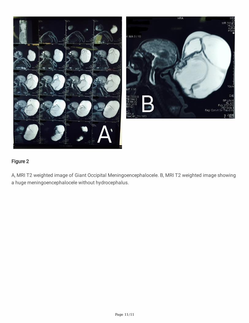
Routine investigations were normal. His Magnetic Resonance Imaging (MRI) Brain Plain was done whichreported a Cerebrospinal Fluid (CSF) containing sac with brain parenchymal tissue herniating through adefect in posterior occipital bones in midline. It measured approximately 21 x 15 x 19 cm. Bilateral lateraland third ventricle were normal. So, they concluded it to be a huge meningoencephalocele withouthydrocephalus. (Fig. 2, A & B)
Parents were counselled in detail about it and after taking informed written consent, we proceeded forsurgical excision and repair. It was a great challenge for our anesthetist to intubate this child. Intubationwas achieved in lateral position. After intubation, the child was kept in lateral position. Cleaned anddraped while holding the encephalocele in our hands. Incision was given along the neck of sac anddeepened through skin layers. Dissection done layer by layer and excised the sac along with dysplasticbrain tissue. Part of occipital and cerebellar parenchyma was found in the sac. Dural venous sinuseswere not involved. Dural layer was identi�ed, brain tissue reduced through a bony defect of 2.5 cm size inoccipital bone in midline and closure done with coated Vicryl (polyglactin 910) 4 − 0 suture. Valsalvamaneuver performed. No CSF leak noted. Subcuticular layer closed with Vicryl (polyglactin 910) 3 − 0suture. Skin closure done with Prolene (polypropylene) 3 − 0 suture. Child had smooth extubation andstarted to feed after 3 hours of surgery. There was minimal blood loss during the surgery, so bloodtransfusion was not done. The baby didn't develop hydrocephalus and was discharged after 3 days.
Discussion:The meningeal membrane covering the giant encephalocele is either covered by a normal, dysplastic or athin membrane. Clinically, the presentation of giant occipital encephaloceles is evident due to theircharacteristic swelling.[2] The contents of occipital encephalocele mainly include meninges and occipitallobes. It may also comprise of ventricles, cerebellum, and brain stem. The factors in�uencing theoutcome in the occipital encephalocele patients are site, size and herniation of the brain into the sac. Site,size, and the amount of brain herniated inside the sac determines the prognosis. The presence ofbrainstem or occipital lobe with or without the dural sinuses in the sac along with hydrocephalus alsoin�uence the outcome of the case.[4]

Page 4/11
The large-sized swellings may possibly have a remarkable brain herniation, an abnormality of theunderlying brain and microcephaly. The clinical examination comprises the examination of its size,extent, amount of protrusion and its location along with the size of bony defect.[2] The size of the headholds a signi�cance importance for clinical suspicion of microcephaly or hydrocephalus and extracranialanomalies.[2] MRI brain is the usual investigation of choice along with the three dimensional ComputedTomography (CT) that further helps in evaluating the deformity and hence the surgical procedure cantake place as soon as possible to avoid further neurological de�cits.[5]
Giant encephaloceles are rare; surgical procedures are a challenging task for the anesthesiologists, aswell as the neurosurgeons. The challenges present are mainly due to its complicated site, enormous size,associated bulging contents resulting in intracranial anomalies, intraoperative blood loss, and prolongedanesthesia.[6] The major aim of the anesthesiologist is to avoid the premature rupture of theencephalocele intraoperatively. The occipital site of the encephalocele causes a hindrance with theintubation as due to a di�cult airway caused by the restriction of the neck movement. This further leadsto an inability of having an optimal tracheal intubation position.[3]
The operative procedure also includes the management of possible loss of large quantities of CSFcausing superimposed electrolyte imbalance. Infants with encephalocele can develop suddenhypothermia due to dysfunction of autonomic control below the present defect.[3] Thus immediateconsideration and management has to be given to hypothermia, blood loss, and its associatedcomplications. The surgery is advised to be done as soon as possible to avoid life threateningcomplications such as Central Nervous System (CNS) infections, respiratory distress, aspirationpneumonia, irreversible impairment of vagus nerve and hypothermia.[5]
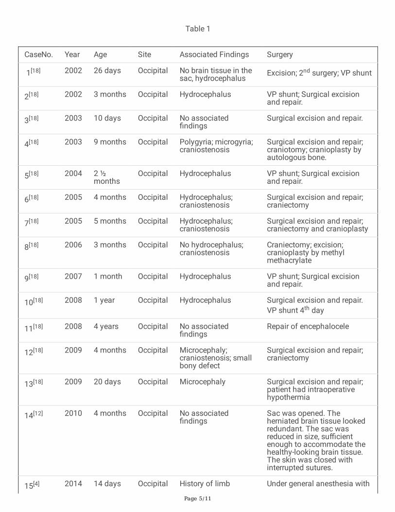
The table below, comprises all the reported cases to the author's knowledge. (Table 1)[3, 4, 9–21] The mainaim is to portray the different and most commonly used surgical procedures in the management of giantoccipital encephaloceles. The most frequently used method is simple resection and dural repair.Expansion Cranioplasty is one of the surgical procedures that consists of a mesh to provide room for theprotruded sac. Another technique used is done through ventricular volume reduction. It is a two-steptechnique; for start it increases the ventricular pressure inducing hydrocephalus followed by aventriculoperitoneal shunt. The ventricles then contract and the protruded tissue repositions itself insidethe cranium. For the herniated cerebellar and occipital parenchyma, an incision is made in the tentoriumto create an infratentorial area for the herniated tissue to retract.[7]
Page 5/11
Table 1
CaseNo. Year Age Site Associated Findings Surgery
1[18] 2002 26 days Occipital No brain tissue in thesac, hydrocephalus
Excision; 2nd surgery; VP shunt
2[18] 2002 3 months Occipital Hydrocephalus VP shunt; Surgical excisionand repair.
3[18] 2003 10 days Occipital No associated�ndings
Surgical excision and repair.
4[18] 2003 9 months Occipital Polygyria; microgyria;craniostenosis
Surgical excision and repair;craniotomy; cranioplasty byautologous bone.
5[18] 2004 2 ½months
Occipital Hydrocephalus VP shunt; Surgical excisionand repair.
6[18] 2005 4 months Occipital Hydrocephalus;craniostenosis
Surgical excision and repair;craniectomy
7[18] 2005 5 months Occipital Hydrocephalus;craniostenosis
Surgical excision and repair;craniectomy and cranioplasty
8[18] 2006 3 months Occipital No hydrocephalus;craniostenosis
Craniectomy; excision;cranioplasty by methylmethacrylate
9[18] 2007 1 month Occipital Hydrocephalus VP shunt; Surgical excisionand repair.
10[18] 2008 1 year Occipital Hydrocephalus Surgical excision and repair.VP shunt 4th day
11[18] 2008 4 years Occipital No associated�ndings
Repair of encephalocele
12[18] 2009 4 months Occipital Microcephaly;craniostenosis; smallbony defect
Surgical excision and repair;craniectomy
13[18] 2009 20 days Occipital Microcephaly Surgical excision and repair;patient had intraoperativehypothermia
14[12] 2010 4 months Occipital No associated�ndings
Sac was opened. Theherniated brain tissue lookedredundant. The sac wasreduced in size, su�cientenough to accommodate thehealthy-looking brain tissue.The skin was closed withinterrupted sutures.
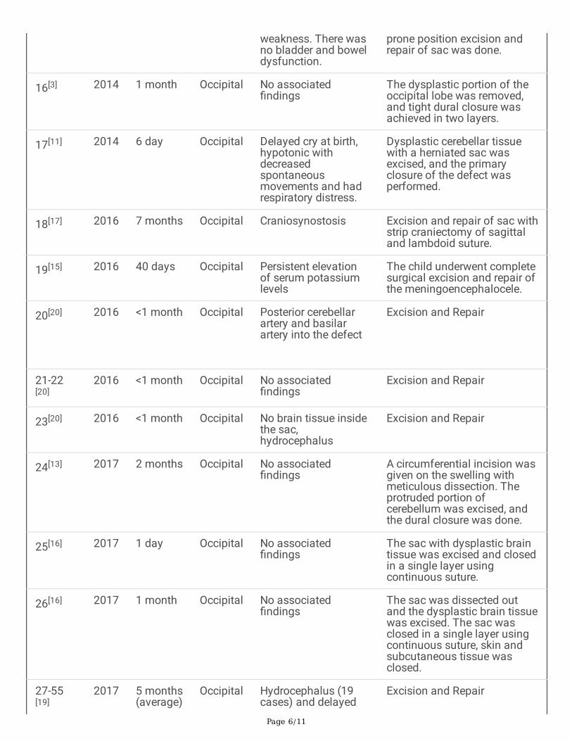
15[4] 2014 14 days Occipital History of limb Under general anesthesia with
Page 6/11
weakness. There wasno bladder and boweldysfunction.
prone position excision andrepair of sac was done.
16[3] 2014 1 month Occipital No associated�ndings
The dysplastic portion of theoccipital lobe was removed,and tight dural closure wasachieved in two layers.
17[11] 2014 6 day Occipital Delayed cry at birth,hypotonic withdecreasedspontaneousmovements and hadrespiratory distress.
Dysplastic cerebellar tissuewith a herniated sac wasexcised, and the primaryclosure of the defect wasperformed.
18[17] 2016 7 months Occipital Craniosynostosis Excision and repair of sac withstrip craniectomy of sagittaland lambdoid suture.
19[15] 2016 40 days Occipital Persistent elevationof serum potassiumlevels
The child underwent completesurgical excision and repair ofthe meningoencephalocele.
20[20] 2016 <1 month Occipital Posterior cerebellarartery and basilarartery into the defect
Excision and Repair
21-22[20]
2016 <1 month Occipital No associated�ndings
Excision and Repair
23[20] 2016 <1 month Occipital No brain tissue insidethe sac, hydrocephalus
Excision and Repair
24[13] 2017 2 months Occipital No associated�ndings
A circumferential incision wasgiven on the swelling withmeticulous dissection. Theprotruded portion ofcerebellum was excised, andthe dural closure was done.
25[16] 2017 1 day Occipital No associated�ndings
The sac with dysplastic braintissue was excised and closedin a single layer usingcontinuous suture.
26[16] 2017 1 month Occipital No associated�ndings
The sac was dissected outand the dysplastic brain tissuewas excised. The sac wasclosed in a single layer usingcontinuous suture, skin andsubcutaneous tissue wasclosed.
27-55[19]
2017 5 months(average)
Occipital Hydrocephalus (19cases) and delayed
Excision and Repair

Page 7/11
milestones (17 cases)
56[21] 2018 1 day Occipital No associated�ndings
No associated �ndings.
57[14] 2019 4 months Occipital No associated�ndings
The encephalocele wasexcised and the dura wasidenti�ed, and a watertightclosure was carried out.
58[9] 2019 6 months Occipital Delayed motormilestones in theform of inability tohold her head
Excision of the redundantneural tissue, and closure ofthe dura done and skin wasreconstructed.
59[10] 2019 10th day Occipital No associated�ndings
A transverse incision wasgiven
over the parietal massfollowed by surgical excisionand repair.
60[6] 2019 3 months Occipital The child had adelayed milestonewith poor feeding andabsent neck holding.ECHO showedpresence of a small 5mm ASD with L→Rshunt
Posterior paramedian incisionwas given, encephalocele sacalong with some brain tissuewas excised followed byprimary duraplasty.
Modern day neurosurgical techniques along with neuroimaging and neonatal intensive care withneurological facilities have greatly improved morbidity and mortality rate in the management ofencephaloceles.[8] Postoperatively, complications such as hypothermia, raised intracranial pressure (ICP),apnea, cardiac arrest, CSF leak, and infection can arise and hence need to be managed effectively.However, in our case no complications were present. Alongside all present di�culties, intubation andanaesthetic management in our patient were successfully achieved.
Conclusion:Perioperative management of a Giant Occipital Encephalocele is an extensive team challenge for bothanesthesiologist and neurosurgeon. These patients have a di�culty in attaining the supine position.Endotracheal intubation is achieved in the lateral position. Prognosis is mainly dependent on the extentof the invasion of cranial contents into the sac. Due to the herniation of parts of the brain, there is asigni�cant rise in the di�culty level of the surgical procedure. There is always a higher risk of infectioninvolved in giant encephaloceles usually due to CSF leakage. In order to avoid superimposedcomplications, carrying out the operative procedure at an early age is highly bene�cial.
Abbreviations

Page 8/11
CSF: Cerebrospinal Fluid
MRI: Magnetic Resonance Imaging
CT: Computed Tomography
CNS: Central Nervous System
ICP: Intracranial Pressure
Declarations
Ethics approval and consent to participate:Not applicable.
Consent for Publication:Written informed consent was taken from the mother of the child for publication of case report andaccompanying images.
Availability of data and materials:All data is within the article.
Competing interests:None of the authors has any con�ict of interest to disclose. We con�rm that we have read the Journal'sposition on issues involved in ethical publication and a�rm that this case report is consistent with thoseguidelines.
Funding:No funding was required for this work.
Authors' contributions:SZ and AK wrote the manuscript. AK is involved in the surgical management of the patient. Both authorsread and approved the �nal manuscript.
Page 9/11
Acknowledgements:Not applicable.
References1. Stephanie A, Black JA, Galvez MA, Rehman. Alan Jay Schwartz; Images in Anesthesiology: Airway
Management in an Infant with a Giant Occipital Encephalocele. Anesthesiology. 2014;120:1504.
2. Ghritlaharey RK. A Brief Review of Giant Occipital Encephalocele. Journal of neurosciences in ruralpractice. 2018;9(4):455–6.
3. Pahuja H, Deshmukh. Shubhda & palsodkar, supriya & lande, surabhi. (2015). Anaestheticmanagement of neonate with giant occipital Meningoencephalocele.. International Journal ofResearch in Medical Sciences. 31. 1. 10.5455/2320–6012.ijrms20150165.
4. Nath HD, Mahapatra AK, Borkar SA. A giant occipital encephalocele with spontaneous hemorrhageinto the sac: A rare case report. Asian journal of neurosurgery. 2014;9(3):158–60.
5. Velho V, Naik H, Survashe P, Guthe S, Bhide A, Bhople L, Guha A. Management strategies of cranialencephaloceles: A neurosurgical challenge. Asian J Neurosurg. 2019;14:718–24.
�. Ganeriwal V, et al. Giant meningoencephalocele with Arnold-Chiari type III malformation andanaesthetic challenges: A rare case report. Saudi journal of anaesthesia vol. 2019;13(2):136–9.doi:10.4103/sja.SJA_616_18.
7. Alwahab A, Kharsa A, Nugud A, Nugud S. Occipital Meningoencephalocele case report and review ofcurrent literature. Chinese Neurosurgical Journal. 2017;3(1).
�. Rehman L, Farooq G, Bukhari I. Neurosurgical interventions for occipital encephalocele. Asian JNeurosurg. 2018;13:233–7.
9. Naik V, Marulasiddappa V, Gowda Naveen MA, Pai SB, Bysani P, Amreesh SB. Giant Encephalocoele:A Rare Case Report and Review of Literature. Asian journal of neurosurgery. 2019;14(1):289–91.
10. Antunes M, Pizzol D, Calgaro S, Di Gennaro F, Colangelo A. Case report Giant Encephalocele:successful management in limited-resource settings. EuroMediterranean Biomedical Journal.2019;14:114–6. 10.3269/1970-5492.2019.14.26.
11. Bulut M, Yavuz A, Bora A, Gülşen İ, Özkaçmaz S, Sösüncü E. Chiari III Malformation with a GiantEncephalocele Sac: Case Report and a review of the literature. Pediatr Neurosurg. 2013;49(5):316–9.
12. Agarwal A, Chandak AV, Kakani A, Reddy S. A giant occipital encephalocele. APSP Journal of CaseReports. 2010 Jul;1(2):16.
13. Kumar V, Kulwant SBhaikhel, Saurabh S, Chauhan R. Giant Occipital Meningoencephalocele in aNeonate: A Therapeutic Challenge. Journal of Pediatric Neurosciences. 2017;12:46. 10.4103/1817-1745.205655.
14. Murthy PS, Kalinayakanahalli Ramkrishnappa SK. Giant Occipital Encephalocele in an Infant: ASurgical Challenge. Journal of Pediatric Neurosciences. 2019 Oct-Dec;14(4):218–21.

Page 10/11
15. Agrawal AC, Umamaheshwara RV, Hegde KV, Suneetha P, Kolikipudi DS. Giant high occipitalencephalocele. Romanian Neurosurgery. 2016;30:122–6.
1�. Kaif D, Ahmad D, Kumar Singh D, Pandey D. Giant Occipital Encephalocele – A Report of Two RareCases. Journal of Medical Science clinical Research. 2017;05(02):18162–5.
17. Dasgupta S, Saha S, Baranwal S, Banga M, Sandeep B. A rare case of giant occipital encephalocelewith craniosynostosis. Asian Journal of Medical Sciences. 2016;7(4):98–100.
1�. Mahapatra A. K: Giant Encephalocele: A Study of 14 Patients. Pediatr Neurosurg. 2011;47:406–11.
19. Mahajan CMD, DM*; Rath GP, MBBS, MD, DM*; Bithal PK, MD*; Mahapatra AK. MS, MCh†Perioperative Management of Children With Giant Encephalocele: A Clinical Report of 29 Cases.Journal of Neurosurgical Anesthesiology: July. 2017;Vol. 29(- Issue 3):322–9.
20. Ozdemir N, Ozdemir SA, Ozer EA. Management of the giant occipital encephaloceles in the neonates.Early Hum Dev. 2016 Dec;(103):229–234.
21. Kanesen D, et al. Giant Occipital Encephalocele with Chiari Malformation Type 3. Journal ofneurosciences in rural practice vol. 2018;9(4):619–21. doi:10.4103/jnrp.jnrp_103_18.
Figures
Figure 1
A, Giant Occipital Encephalocele B, Giant Occipital Encephalocele (21 x 15 x 19 cm in size)
Page 11/11
Figure 2
A, MRI T2 weighted image of Giant Occipital Meningoencephalocele. B, MRI T2 weighted image showinga huge meningoencephalocele without hydrocephalus.