occipital meningoencephalocele – anaesthetic …
TRANSCRIPT
www.wjpr.net Vol 3, Issue 3, 2014.
4761
Ahlawat et al. World Journal of Pharmaceutical Research
OCCIPITAL MENINGOENCEPHALOCELE – ANAESTHETIC
CHALLENGES: A CASE REPORT
Dr. Geeta Ahlawat, Associate Professor; Dr. Swati Chhabra, Assistant Professor;
Dr. Mangal Singh Ahlawat
Senior Resident,Pt. B. D. Sharma PGIMS, Rohtak.
ABSTRACT
Introduction: The anesthetic management of meningoencephalocele is
a challange because of the positioning, handling of the airway and the
difficulty in perioperative care.
Case Report: A 3 month old neonate presented for surgical excision
of giant occipital meningoencephalocele. Despite the difficulties,
intubation and peroperative anesthetic management of the patient was
successfully achieved. Conclusion: Previous similar case reports were
reviewed and potential perioperative complications are highlighted.
Key Words: paediatric difficult airway, Encephalocele, Occipital.
INTRODUCTION
The term cephalocele refers to a defect in the skull and dura with extracranial extension of
intracranial structures. Meningoencephalocele consists of a herniation of cerebrospinal fluid,
brain tissue and meninges through the skull defect. The cause of cephalocele has not been
fully determined. Many differences in type and frequency of cephalocele among various
ethnic groups have been observed. In Southeast Asia, the incidence is slightly higher, with
approximately 1 in 5,000 live births. In this case report, we have included a detailed
discussion describing the special anesthetic considerations of these patients.
Case report
A neglected 3 month old female neonate presented with a giant cystic swelling in the
occipital region and was scheduled for surgical excision. The baby was delivered by cesarean
section in a private nursing home. The neonate with small swelling in occipital region was
transferred to our hospital and admitted to the pediatric clinic for preoperative preparation but
initially parents did not gave consent for surgery. The swelling kept enlarging, infant had
World Journal of Pharmaceutical ReseaRch
Volume 3, Issue 3, 4761-4765. Case Study ISSN 2277 – 7105
Article Received on 02 March 2014, Revised on 25 March 2014, Accepted on 23 April 2014
*Correspondence for
Author
Dr. Mangal Singh Ahlawat
Senior Resident,Pt. B. D.
Sharma PGIMS, Rohtak
www.wjpr.net Vol 3, Issue 3, 2014.
4762
Ahlawat et al. World Journal of Pharmaceutical Research
difficulty in breast feeding, difficulty in supine postioning during sleep, then parents again
visited our hospitals out- patient department.
On preanesthetic evaluation, cardiovascular, respiratory and neurological system examination
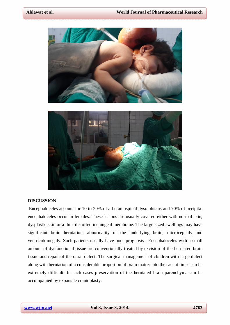
was normal. There was a swelling that measured 16x7 cm arising from posterior part of the
head. No other congenital anomaly was detected. Magnetic resonance imaging (MRI) showed
giant occipital meningoencephalocele with a minimal herniation of occipital lobe into the
swelling.
Laboratory evaluations were within normal limits. After attaching precordial stethoscope and
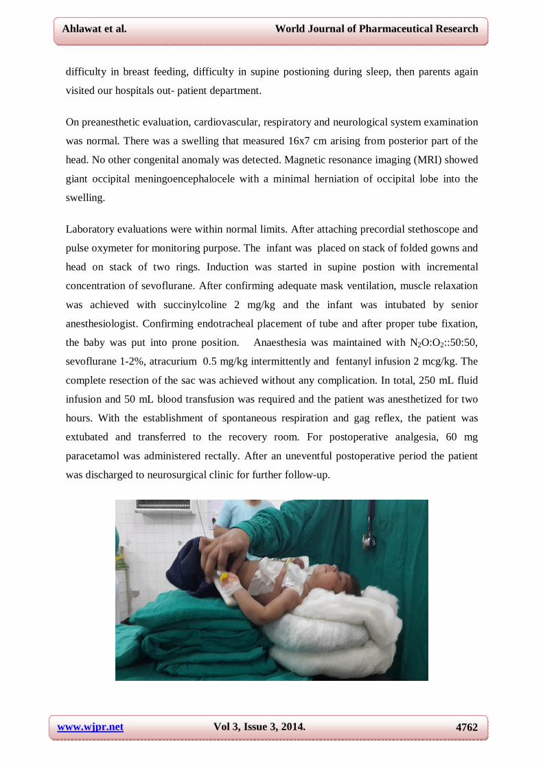
pulse oxymeter for monitoring purpose. The infant was placed on stack of folded gowns and
head on stack of two rings. Induction was started in supine postion with incremental
concentration of sevoflurane. After confirming adequate mask ventilation, muscle relaxation
was achieved with succinylcoline 2 mg/kg and the infant was intubated by senior
anesthesiologist. Confirming endotracheal placement of tube and after proper tube fixation,
the baby was put into prone position. Anaesthesia was maintained with N2O:O2::50:50,
sevoflurane 1-2%, atracurium 0.5 mg/kg intermittently and fentanyl infusion 2 mcg/kg. The
complete resection of the sac was achieved without any complication. In total, 250 mL fluid
infusion and 50 mL blood transfusion was required and the patient was anesthetized for two
hours. With the establishment of spontaneous respiration and gag reflex, the patient was
extubated and transferred to the recovery room. For postoperative analgesia, 60 mg
paracetamol was administered rectally. After an uneventful postoperative period the patient
was discharged to neurosurgical clinic for further follow-up.
www.wjpr.net Vol 3, Issue 3, 2014.
4763
Ahlawat et al. World Journal of Pharmaceutical Research
DISCUSSION
Encephaloceles account for 10 to 20% of all craniospinal dysraphisms and 70% of occipital
encephaloceles occur in females. These lesions are usually covered either with normal skin,
dysplastic skin or a thin, distorted meningeal membrane. The large sized swellings may have
significant brain herniation, abnormality of the underlying brain, microcephaly and
ventriculomegaly. Such patients usually have poor prognosis . Encephaloceles with a small
amount of dysfunctional tissue are conventionally treated by excision of the herniated brain
tissue and repair of the dural defect. The surgical management of children with large defect
along with herniation of a considerable proportion of brain matter into the sac, at times can be
extremely difficult. In such cases preservation of the herniated brain parenchyma can be
accompanied by expansile cranioplasty.
www.wjpr.net Vol 3, Issue 3, 2014.
4764
Ahlawat et al. World Journal of Pharmaceutical Research
Patients with giant encephalocele and large amount of brain tissue in the sac usually die
either shortly after birth or as a result of operation. A microcephalic child with neurological
deficit and a sac containing cerebrum, cerebellum and brain stem structures, carry a poor
prognosis. In such patients, it is generally impossible to foretell whether the infant will die
quickly or will continue to live for many months or years, as size of the encephalocele itself
is not a guide to prognosis. Ultimate result depends on the amount of normal brain tissue left
inside the skull after the operation. Surgery thus just facilitates nursing of the baby. This
infant was neurologically well developed. Furthermore less functional tissue in the sac made
the surgical excision of the sac easy and safe.
Once the decision to operate has been made, a perioperative plan must be formulated by an
anesthesiologist based on airway management, fluid balance and prevention of hypothermia.
In our case, the occipital meningoencephalocele made supine position of head impossible
due to the limited-neck extension and the likelihood of rupturing the membranes covering the
spinal cord or brain. So we decided to place patient on raised platform of rolled-up blankets
and intubate patient in supine position other alternative approaches of airway management
includes place the child in lateral position, while an assistant temporarily supports the head
or placing the child' s head beyond the edge of the table with an assistant supporting
it. Before administering neuromuscular blocking agents and intubation, adequate mask
ventilation must be verified. Latex allergy precautions should be used with these children
for their first anesthetic procedure.
CONCLUSION
Perioperative management of patients with giant meningoencephalocele may be challenging
for both anesthesiologist and neurosurgeon. These patients must be managed closely with an
interdisciplinary approach.
REFERENCES
1. Barkovich AJ. Pediatric Neuroimaging. Philadelphia: Lippincott Williams & Wilkins
2000:267–1.
2. Nyberg DA, Mc Gahan JP, Pretorius DH, Pilu G. Diagnostic Imaging of Fetal Anomalies.
Philadelphia: Lippincott Williams & Wilkins 2003:298.
3. McLone D. Pediatric Neurosurgery: Surgery of the developing system. Philadelphia:
Saunders Company 2001:204.
www.wjpr.net Vol 3, Issue 3, 2014.
4765
Ahlawat et al. World Journal of Pharmaceutical Research
4. Singh K, Garasia M, Ambardehar M, Thota R, Dewoolkar L, Mehta K. Giant occipital
meningoencephalocele: Anaesthetic implications. The Internet Jounal of Anesthesiology
2007;13(2).
Dey N, Gombar KK, Khanna AK, Khandelwal P. Airway management in neonates with
occipital encephalocele: adjustment and modifications. Paediatr Anaesth
2007;17(11):1119–20.