supporting information - pnas · n-terminal part of the α-helix directly interacts with actin...
TRANSCRIPT

Supporting InformationDucka et al. 10.1073/pnas.1005347107SI TextAbbreviations. SpirD, the D WH2 domain of Spire (residues461–480); SpirCD, the Spire containing CD WH2 domains (re-sidues 432–480); SpirBCD, the Spire containing triple WH2 do-mains (BCD, residues 396–480), SpirABCD, the Spire containingquadruple WH2 domains (ABCD, residues 371–480); SpirDDD,the artificial Spire WH2 construct comprising three identical Drepeats.
Materials andMethods. Preparation of proteins.The vectors (pGEX-6P-1) containing Drosophila Spire constructs were transformedinto BL21(DE3) BL21-CodonPlus(DE3)-RIL expression cells(Stratagene) and grown for approximately 4 h at 37 °C until theOD at 600 nm reached 0.9. Protein expression was carried outat 18 °C for 14–16 h after induction with 0.5 mM isopropyl-β-thio-galactopyranoside (Fermentas). The cells were harvested by cen-trifugation at 5,000 rpm for 15 min, and pellets were resuspendedin PBS containing 1 mM EDTA, 1 mM DTT, and 0.5 mM PMSF.The lysis was performed by sonication followed by centrifugationat 20,000 rpm for 30 min. The supernatant containing GST-taggedSpire constructs was applied on Glutathione Sepharose 4 FastFlow beads (GE Healthcare). Bound protein was washed with50 mM Tris (pH 7.0), 150 mM NaCl, 1 mM EDTA, and 1 mMDTTand cleaved by PreScisson protease (GE Healthcare) to re-lease the Spire protein. The cleaved Spire proteins were collected,concentrated, and further purified by Superdex 75 (SpirCD,SpirBCD, SpirDDD, and SpirABCD) or Superdex 30 (SpirCand SpirD) in PBS buffer. The His-tagged SpirNT encoded in apET 16b vector was expressed in BL21(DE3) Codonþ RIL andpurified by Ni-NTA agarose beads (Qiagen) followed by gel filtra-tion on Superdex 200 (GE Healthcare). The purity and homoge-neity of proteins were confirmed by mass spectrometry andN-terminal Edman sequencing (data not shown). The foldingproperties of all constructs were further analyzed by 1D NMRspectra recorded by a 650-MHz magnet. AP-actin (Drosiphila5C A204E/P243K cytoplasmatic actin) was prepared as describedby Rould et al. (1). The rabbit skeletal muscle actin was extractedaccording to the method of Spudich andWatt (2) and further pur-ified on a Sephacryl S-200 gel filtration column (GE Healthcare).Latrunculin B (Sigma) was used to prevent spontaneous poly-merization of rabbit actin.
Crystallization. Purified SpirABCD, SpirBCD, aswell as SpirDDD,and SpirCD were mixed with AP-actin at a molar ratio of 1∶4.1,1∶3.1, and 1∶2∶1, respectively, and separated by gel filtration onSuperdex 200 (GE Healthcare) in 5 mM Tris (pH 7.0), 50 mMNaCl, 0.2 mMATP, and 5mM β-mercaptoethanol. Fractions fromthe peaks shown in Fig. S3Awere analyzed by SDS-PAGE, pooled,and concentrated to 10–12 mg∕mL. The complexes were crystal-lized both at 4 °C and 20 °C in several crystallization conditions,using the sitting and hanging drop vapor diffusion method. The2- to 3-μL drops consisted of a 1∶1 or 2∶1 ðvol∕volÞ mixture ofprotein solution and a well solution. Crystals appeared after 2 daysand grew to the final size after 2–4 weeks of incubation. The bestdiffracting crystals of the SpirABCD/AP-actin, SpirBCD/AP-ac-tin, and SpirDDD/AP-actin complexes grew in 0.2-M ammoniumformate pH 6.6, 20% PEG 3350, and the crystals of SpirCD/AP-actin appeared in 0.2-M ammonium sulphate, 0.1-M Mes pH 6.5,20% PEG 8000. All crystals were soaked in cryosolutions contai-ning mother liquor supplemented with 20% 2-methyl-2,4-penta-nediol or glycerol and were flash frozen in liquid nitrogen. Crystalsof the SpirD/actin-latrunculin B complex appeared after oneweekat 4 °C in the solution containing 0.2 M magnesium formate, pH5.9 and 20% PEG 3350. Crystals were treated as described above.To exclude the possibility of proteolytic cleavage of Spire duringcrystallization, we dissolved several crystals of the complexes andanalyzed themby SDS-PAGEandmass spectrometry, proving thatboth SpirCD and SpirBCD remain intact during crystallization.
Data collection and structure determination. X-ray datasets of theSpirD/actin-latrunculin B (1.6 Å), SpirBCD/AP-actin (2.0 Å),SpirABCD/AP-actin (2.2 Å), SpirDDD/AP-actin (2.1 Å), andSpirCD/AP-actin (2.6 Å) were collected on the Swiss LightSource beamline PXII at the Paul Scherrer Institut. The datasetswere integrated, scaled, and merged by XDS and XSCALE pro-grams (3) in space group P212121 for SpirD-actin-latrunculin B,P65 for SpirCD/AP-actin, SpirABCD/AP-actin, and SpirDDD/AP-actin, and P21 for SpirBCD/AP-actin (Table S1). The struc-turesweredeterminedbymolecular replacement using theMolrepprogram from the CCP4 suite (4) and the structures of AP-actinand rabbit skeletal muscle actin complexed with latrunculin Aas a search model. Model building and refinement were carriedout with the program XtalViev/Xfit (5) and REFMAC5 (4). Datacollection refinement and statistics are shown in the Table S1.
Ducka et al. www.pnas.org/cgi/doi/10.1073/pnas.1005347107 1 of 6

1. Rould MA, Wan Q, Joel PB, Lowey S, Trybus KM (2006) Crystal structures of expressednon-polymerizable monomeric actin in the ADP and ATP states. J Biol Chem281:31909–31919.
2. Spudich JA, Watt S (1971) The regulation of rabbit skeletal muscle contraction. I.Biochemical studies of the interaction of the tropomyosin-troponin complex withactin and the proteolytic fragments of myosin. J Biol Chem 246:4866–4871.
3. Kabsch W (1993) Automatic processing of rotation diffraction data from crystals ofinitially unknown symmetry and cell constants. J Appl Crystallogr. 26:795–800.
4. CCP4 (Collaborative Computational Project, Number 4) (1994) The CCP4 suite:Programs for protein crystallography. Acta Crystallogr D Biol Crystallogr 50:760–763.
5. McRee DE (1999) XtalView/Xfit. A versatile program for manipulating atomiccoordinates and electron density. J Struc Biol 125:156–165.
Fig. S1. Amino acid sequence alignment of the WH2-containing actin nucleators. Dm_Spire (Drosophila melanogaster; Q9U1K1); Hs_Spire1 (Homo sapiens;Q08AE8); Hs_Spire2 (Homo sapiens; Q8WWL2); Hs_Cobl (Homo sapiens; O75128); Hs_NWASP (Homo sapiens; O00401). Conserved residues are colored asfollows: green, hydrophobic; cyan, basic; residues present in all WH2 domains are marked with asterisks. The residues directly interacting with actin areunderlined. Sequence of the Spire protein studied in this work is shown in red (WH2 domain) and magenta (linkers). The secondary structure is depictedabove the sequence.
Fig. S2. Titration of the 15N-labeled SpirCD with unlabeled (14N) AP-actin. The 15N-HSQC spectra of SpirCD before (red), and after addition of actin (blue). Only1H-15N NMR signals of SpirCD are visible. NMR indicates that free SpirCD is not structured in solution giving a typical NMR spectrum of the polypeptide chain ina random conformation (1, 2) On forming the complex with actin, most of the NMR signals (blue) of SpirCD disappear. This is because upon binding the SpirCDresidues participate in a well-defined structure of a large SpirCD/actin complex (1, 2). The observed 1∕T2 transverse relaxation rate of the bound Spire in thecomplexes increases thus significantly and broadening of NMR resonances results in the disappearance of its signals in the spectra. No strong, sharp NMR signalsare visible in the blue spectrum of the complex, indicating that there are no highly flexible residues present in the actin-bound SpirCD.
1. D’Silva L, et al. (2005). Monitoring the effects of antagonists on protein-protein interactions with NMR spectroscopy. J Am Chem Soc 127:13220–13226.2. Wüthrich K (1986) NMR of Proteins and Nucleic Acids (Wiley, New York).
Ducka et al. www.pnas.org/cgi/doi/10.1073/pnas.1005347107 2 of 6
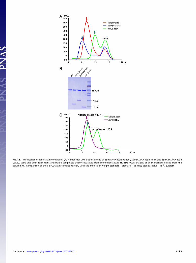
Fig. S3. Purification of Spire-actin complexes. (A) A Superdex 200 elution profile of SpirCD/AP-actin (green), SpirBCD/AP-actin (red), and SpirABCD/AP-actin(blue). Spire and actin form tight and stable complexes clearly separated from monomeric actin. (B) SDS-PAGE analysis of peak fractions eluted from thecolumn. (C) Comparison of the SpirCD-actin complex (green) with the molecular weight standard—aldolase (158 kDa; Stokes radius—48 Å) (violet).
Ducka et al. www.pnas.org/cgi/doi/10.1073/pnas.1005347107 3 of 6
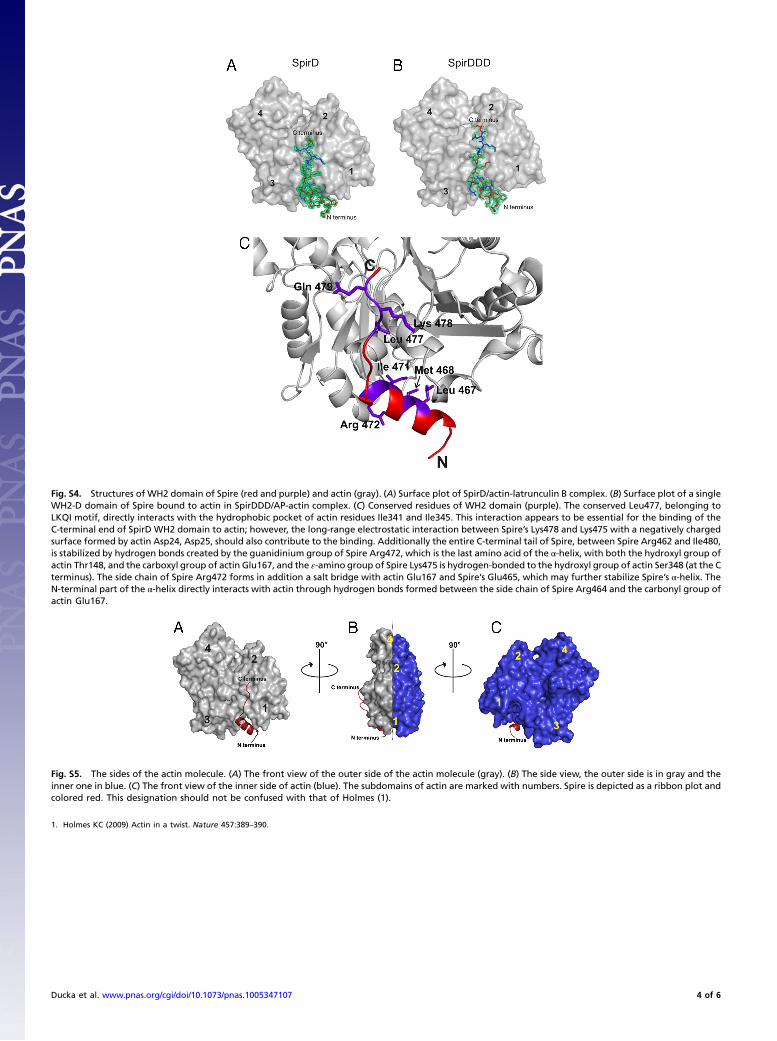
Fig. S4. Structures of WH2 domain of Spire (red and purple) and actin (gray). (A) Surface plot of SpirD/actin-latrunculin B complex. (B) Surface plot of a singleWH2-D domain of Spire bound to actin in SpirDDD/AP-actin complex. (C) Conserved residues of WH2 domain (purple). The conserved Leu477, belonging toLKQI motif, directly interacts with the hydrophobic pocket of actin residues Ile341 and Ile345. This interaction appears to be essential for the binding of theC-terminal end of SpirD WH2 domain to actin; however, the long-range electrostatic interaction between Spire’s Lys478 and Lys475 with a negatively chargedsurface formed by actin Asp24, Asp25, should also contribute to the binding. Additionally the entire C-terminal tail of Spire, between Spire Arg462 and Ile480,is stabilized by hydrogen bonds created by the guanidinium group of Spire Arg472, which is the last amino acid of the α-helix, with both the hydroxyl group ofactin Thr148, and the carboxyl group of actin Glu167, and the ε-amino group of Spire Lys475 is hydrogen-bonded to the hydroxyl group of actin Ser348 (at the Cterminus). The side chain of Spire Arg472 forms in addition a salt bridge with actin Glu167 and Spire’s Glu465, which may further stabilize Spire’s α-helix. TheN-terminal part of the α-helix directly interacts with actin through hydrogen bonds formed between the side chain of Spire Arg464 and the carbonyl group ofactin Glu167.
Fig. S5. The sides of the actin molecule. (A) The front view of the outer side of the actin molecule (gray). (B) The side view, the outer side is in gray and theinner one in blue. (C) The front view of the inner side of actin (blue). The subdomains of actin are marked with numbers. Spire is depicted as a ribbon plot andcolored red. This designation should not be confused with that of Holmes (1).
1. Holmes KC (2009) Actin in a twist. Nature 457:389–390.
Ducka et al. www.pnas.org/cgi/doi/10.1073/pnas.1005347107 4 of 6

Fig. S6. Stereo representation of the SpirABCD/AP-actin complex in the “compacted side-to-side” configuration. Actin molecules colored green, blue, andcyan belong to one asymmetric unit (in the P21 space group). The fourth actin of the complex (colored in brown) belongs to the neighboring asymmetric unit.The WH2 domains are shown in red; “missing” linkers are depicted as ribbons and colored in yellow.
Fig. S7. Model of actin nucleation by Spire. (A) The crystal structure of a SpirBCD-actin primary nucleus in the “elongated side-to-side” arrangement (side viewperpendicular to the long axis). (B and C) Model of G-actin recruitment and nucleus rearrangement. (D) Filament formation and elongation by addition of actinmonomers to the barbed end of growing filament. (E) The filament has been formed.
Ducka et al. www.pnas.org/cgi/doi/10.1073/pnas.1005347107 5 of 6

Movie S1. A proposed model of actin nucleation by Spire. Spire (red ribbons) aligns actin molecules (colored green, blue, cyan, and pink) into the elongatedside-to-side arrangement and forms the primary nucleus. Binding of free actins (colored in gray) induces transition to the F-actin conformation, followed byelongation upon addition of actin monomers to the barbed end of the growing filament.
Movie S1 (WMV)
Table S1. Data collection and refinement statistics
SpirD SpirCD SpirBCD SpirABCD SpirDDD
Data collectionSpace group P212121 P65* P21* P65 P65Cell constant:a 52.96 125.51 125.30 126.08 125.82b 71.81 125.51 55.95 126.08 125.82c 100.72 56.1 125.37∕β ¼ 120.036 56.37 56.02Resolution range (Å) 50 − 1.5 50 − 2.6 50 − 2.0 50 − 2.2 50 − 2.1Observed reflections 380,081 84,313 370,098 228,321 266,481Unique reflections 49,209 11,584 90,708 22,506 26,371Whole resolution range:Completeness (%) 79.0 73.6 80.8 76.2 86.4Rmerge 4.9 8.6 5.6 6.7 3.0I/σ (I) 21.4 12.6 19.0 15.54 32.6Last resolution shell:Resolution range (Å) 1.5 − 1.6 2.6 − 2.8 2.0 − 2.1 2.2 − 2.3 2.1 − 2.2Completeness (%) 38.1 40.5 47.6 38.5 36.9Rmerge 20.3 29.7 19.5 26.9 23.6I/σ (I) 4.4 4.4 5.0 3.9 4.0RefinementNo. of reflections 46,688 9,765 78,737 20,832 23,718Resolution (Å) 39 − 1.5 20 − 2.8 50 − 2.0 50 − 2.0 50 − 2.0R factor (%) 17.2 24.1 19.0 18.4 25.6Rfree (%) 21.6 33.0 23.9 25.1 27.5Average B (Å2) 19.8 51.0 32.8 42.8 57.8rms bond length (Å) 0.027 0.012 0.011 0.018 0.006rms angles (°) 2.4 1.5 1.2 1.5 0.9Content of asymmetric unitNo. of complexes 1 1 3 1 1No. of protein residues/atoms 367/3,418 381/2,974 1,132/9,613 377/2,912 378/2,967No. of solvent atoms 489 30 781 142 63PDB Data Bank ID code 3MN5 3MMV 3MN6 3MN9 3MN7
*Space groups P65 and P21 are equivalent (See the section: Crystal structures of the SpirCD/AP-actin, SpirBCD/AP-actin, SpirDDD/AP-actin and SpirABCD/AP-actin complexes.)
Ducka et al. www.pnas.org/cgi/doi/10.1073/pnas.1005347107 6 of 6