supplemental information reciprocal regulation of hif-1 ... · 0 15 30 60 90 120 0 15 30 60 90120...
TRANSCRIPT
Molecular Cell, Volume 53
Supplemental Information
Reciprocal Regulation of HIF-1 and LincRNA-p21
Modulates the Warburg Effect Fan Yang, Huafeng Zhang, Yide Mei, and Mian Wu
A
lincRNA-p21 ROR-797- -788 +1 -1518- -1515 +1
479 476 397 394 1316 1313 1
Figure S1
lincRNA-A7
Malat1NEAT1
HOTAIR
-162- -159 -82- -79 +1
-479- -476 -397- -394
-1500- -1497 +1
-1686- -1683-258- -255
-1316- -1313 +1
-661- -664
-1118- -1115 +1
B C20 Glut1B C
25
50
75
rage
RN
A c
opy
num
bers
f l
incR
NA
-p21
per
cel
l
5
10
15
lincRNA-p21
Rel
ativ
e R
NA
leve
l
D E5
Normoxia 24hH i 24h
1 2 3 4 5 6
Hypoxia (h) 0 6 12 24 48 72HIF-1αβ-Actin
0A
ve ofHeLa MCF7 H1299 LO2 IMR90
Hypoxia(h)0
0 6 12 24 48 72
0.5
1
elat
ive
RN
A le
vel
of li
ncR
NA
-p21
2
3
4
5
otal
cel
l num
ber (
x105
)
Hypoxia 24h
sh-lincRNA-p210
Re
1 2 3
0
1T
shRNA Ctrl lincRNA-p21
p21-
1
p21-
2F G7.8
7.4 ***
***
***
shR
NA
-ctrl
shR
NA
-linc
RN
A-p
shR
NA
-linc
RN
A-p
Nor
mox
ia
7.0
6.6
6.2
pH v
alue
NH
ypox
ia 5.8
Med
ium
with
out c
ells
sh-c
trl
sh-li
ncR
NA
-p21
-1
sh-li
ncR
NA
-p21
-2
sh-li
ncR
NA
-p21
-3

A B
Figure S2
Normoxia + + - -
Digoxin
β ActinHIF-1α
+ + - -Hypoxia - - + + - - + +
- - - - ++++
β-Actin
HIF-1αN H N H N H N H
sh-ctrl + + + +- - - -sh-lincRNA-p21 - - - -+ + + +
sh-HIF-1α - - - - ++ + +
RN
A-p
21-1
RN
A-p
21-2
RN
A-p
21-3
C
1 2 3 4 5 6 7 8β-Actin
1 2 3 4 5 6 7 8
1 2 3 4 5
β-Actin
HIF-1αNormoxia + - - - -
Hypoxia - + + + +
sh-c
trl
sh-li
ncR
sh-li
ncR
sh-li
ncR

Figure S3
sh-ctrl + -
sh-VHLsh-lincRNA-p21 -
- - + - - -- + + - - + +
- + - + - + - +
BA
CHX 0 15 30 60 90 120 0 15 30 60 90120 (min)sh-ctrl sh-lincRNA-p21
IP:
Cβ-ActinVHL
N H N Hsh-ctrl + + - -
sh-VHL - - + +
1 2 3 4 5 6 7 8β-ActinHyp564-HIF-1α
N N N N H H H H
Hyp564-HIF-1α/Actin
β-ActinHyp564-HIF-1α
CHX 0 15 30 60 90 120 0 15 30 60 90120 (min)
10.
950.
570.
450.
180.
14
0.45
0.42
0.23
0.15
0.121
sh-ctrl + - + -sh-lincRNA-p21
N N H H
Input
N N H H+ - + -
- + - + - + - +
IB:Hyp564-HIF-1α
Anti-VHL
IB:VHLIB:β-Actin
1 2 3 4 5 6 7 8
β
1 2 3 4
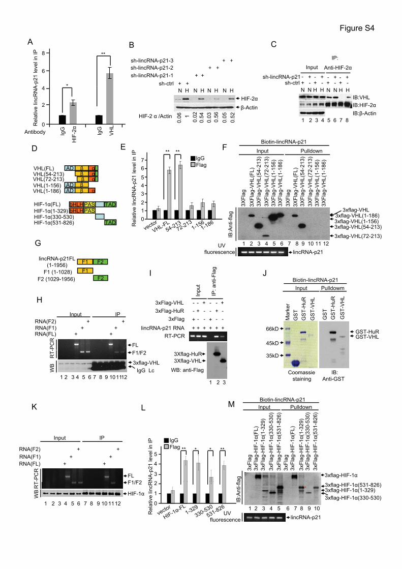
Figure S4
*
**8
6
4A-p
21 le
vel i
n IP
A C
sh-ctrl + - + -sh-lincRNA-p21
Input
+ - + -- + - + - + - +
IP:
Anti-HIF-2α
B
sh-ctrl + +sh-lincRNA-p21-1 + +sh-lincRNA-p21-2 + +sh-lincRNA-p21-3 + +
ED Input Pulldown
Biotin-lincRNA-p21P ** **
IgG
IgG
HIF
-2α
VH
L
4
2
0Rel
ativ
e lin
cRN
A
Antibody
1 2 3 4 5 6 7 8IB:β-Actin
IB:VHLIB:HIF-2α
N N H H N N H H
β-ActinHIF-2α
N H N H N H N H
0.06
0.02
0.54
0.03
0.56
0.05
0.521HIF-2 α /Actin
F Input Pulldown
3xflag-VHL3xflag-VHL(1-186)
3XFl
ag3X
Flag
-VH
L(FL
)3X
Flag
-VH
L(54
-213
)3X
Flag
-VH
L(72
-213
)3X
Flag
-VH
L(1-
156)
3XFl
ag-V
HL(
1-18
6)3X
Flag
3XFl
ag-V
HL(
FL)
3XFl
ag-V
HL(
54-2
13)
3XFl
ag-V
HL(
72-2
13)
3XFl
ag-V
HL(
1-15
6)3X
Flag
-VH
L(1-
186)
flag
ativ
e lin
cRN
A-p
21 le
vel i
n I P
76
543210
IgGFlag
bHLHPAS TADHIF-1α(FL)HIF-1α(1-329)HIF-1α(330-530)
bHLHPAS
AD β αVHL(FL)VHL(54-213)VHL(72-213)VHL(1-156)VHL(1-186)
β αβ α
AD βAD β α
G
g ( )3xflag-VHL(1-156)3xflag-VHL(54-213)
3xflag-VHL(72-213)1 2 3 4 5 6 7 8 9 10 11 12
IB:A
nti-f
UVfluorescence lincRNA-p21
Rel 0HIF 1α(330 530)
HIF-1α(531-826) TAD
F1 F2
F1 (1-1028)F2 (1029-1956)
lincRNA-p21FL (1-1956)
F1F2
nti-F
lag
Biotin-lincRNA-p21I J
Input IP
RNA(FL) + +RNA(F1) + +RNA(F2) + +
-PC
R
FL
H
Inpu
t
IP: a
n
lincRNA-p21 RNA + + + + + +
3xFlag - -- -3xFlag-HuR - + - - + -3xFlag-VHL - - + - - +
RT-PCR
+ +
66kD GST-HuRGST-VHL
45kD
Mar
ker
GS
T
GS
T-V
HL
GS
T-H
uR
GS
TG
ST-
HuR
GS
T-V
HL
Input Pulldowm
M
WB
RT- F1/F2
1 2 3 4 5 6 7 8 9 101112
3xflag-VHLIgG Lc
K Input Pulldown
0) 6) 0) 6)
Biotin-lincRNA-p21L
3Xflag-VHL3Xflag-HuR
1 2 3
WB: anti-Flag Coomassie staining
IB:Anti-GST
35kD
Input IP
RNA(FL) + +RNA(F1) + +RNA(F2) + +
T-P
CR
FLF1/F2
5
4
3
2RN
A-p
21 le
vel i
n IP IgG
Flag ** *** *
3xflag-HIF-1α
3xFl
ag3x
Flag
-HIF
-1α(
FL)
3xFl
ag-H
IF-1α(
1-32
9)3x
Flag
-HIF
-1α(
330-
530
3xFl
ag-H
IF-1α(
531-
826
3xFl
ag3x
Flag
-HIF
-1α(
FL)
3xFl
ag-H
IF-1α(
1-32
9)3x
Flag
-HIF
-1α(
330-
530
3xFl
ag-H
IF-1α(
531-
826
ag
WB
RT F1/F2
HIF-1α
1 2 3 4 5 6 7 8 9 10 1112
2
1
0
Rel
ativ
e lin
cR 3xflag-HIF-1α(531-826)3xflag-HIF-1α(1-329)3xflag-HIF-1α(330-530)
1 2 3 4 5 6 7 8 9 10
IB:A
nti-f
l
UVfluorescence lincRNA-p21
*
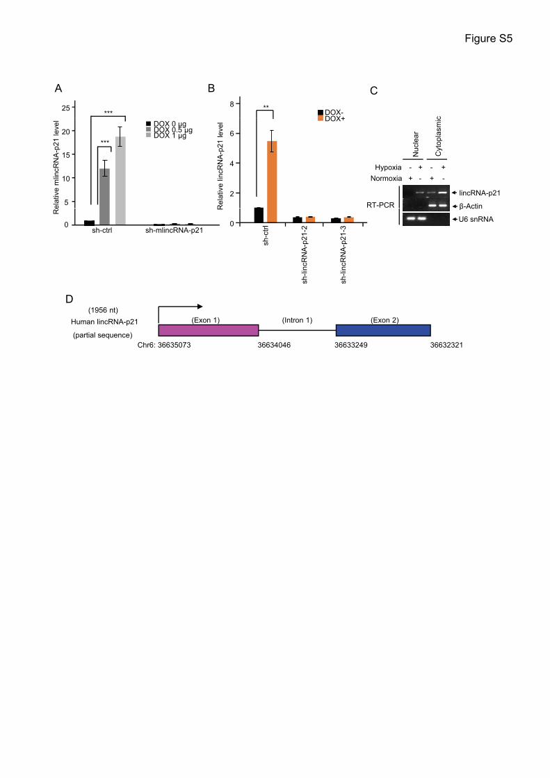
Figure S5
A B
***25 **8
DOX-DOX+
C
c
elat
ive
mlin
cRN
A-p
21 le
vel
***
5
10
15
20DOX 0 μgDOX 0.5 μgDOX 1 μg
Rel
ativ
e lin
cRN
A-p
21 le
vel
6
4
2
DOX+
β-Actin
lincRNA-p21
Normoxia + - + -Hypoxia - -+ +
Nuc
lear
Cyt
opla
smic
RT-PCR
Re
sh-ctrl sh-mlincRNA-p210
R0
sh-c
trl
sh-li
ncR
NA
-p21
-2
sh-li
ncR
NA
-p21
-3
U6 snRNA
β Actin
D
Human lincRNA-p21
(partial sequence)Chr6: 36635073 36634046 36633249 36632321
(1956 nt)(Exon 1) (Intron 1) (Exon 2)
D

Supplemental Figure Legends
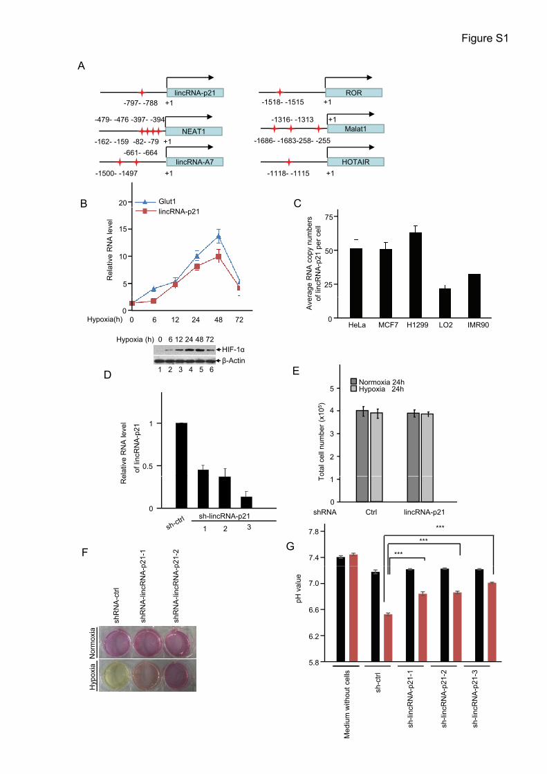
Figure S1, related to Figure 1
(A) Schematic representation of consensus hypoxia-responsive binding sites (CGTG)
(Red stars) in the indicated lncRNA promoter region. Arrowheads indicate the
orientation of transcription.
(B) HeLa cells were cultured under hypoxic (1% O2) conditions for the indicated
periods of time. Total RNA was subjected to real-time RT-PCR analysis. The data are
shown as mean ±SD of three independent experiments. HIF-1α expression upon
hypoxia treatment was also analyzed by Western blotting.
(C) The copy numbers of lincRNA-p21 transcript per cell in the indicated tumor cells
and non-transformed cells were quantified using a quantitative real-time RT-PCR
assay. Data shown are mean ± SD (n=3).
(D) HeLa cells were infected with lentiviruses expressing either control or three
different sets of lincRNA-p21 shRNAs. Twenty-four hours after infection, total RNA
was subjected to real-time RT-PCR analysis. Shown data are mean±SD (n=3).
(E) 2×10⁵ HeLa cells expressing either control shRNA or lincRNA-p21 shRNA were
cultured under normoxic or hypoxic conditions for 24 hrs. Cell numbers were then
counted. The data are shown as mean±SD of three independent experiments.
(F) HeLa cells expressing either control shRNA, lincRNA-p21-1 shRNA or
lincRNA-p21-2 shRNA were cultured under normoxic or hypoxic conditions for 24
hrs. Acidification of the culture medium was evaluated by visually inspecting the
color of the medium.
(G) HeLa cells expressing either control shRNA or the indicated lincRNA-p21
shRNAs were cultured under normoxic or hypoxic conditions for 24 hrs. pH value of
the culture medium was then measured. The data are shown as mean±SD of three
independent experiments.
Figure S2, related to Figure 2
(A) HeLa cells expressing either control shRNA or lincRNA-p21 shRNA were
co-transfected with the HIF-1α-responsive element (HRE) reporter construct and

Renilla luciferase plasmid. Twelve hours later, cells were cultured under normoxic or
hypoxic conditions in the presence or absence of Digoxin for additional 24 hrs. Hif-1α
expression was then examined by Western blot analysis.
(B) HeLa cells expressing control shRNA, lincRNA-p21 shRNA, HIF-1α shRNA or
both lincRNA-p21 and HIF-1α shRNAs were co-transfected with the
HIF-1α-responsive element (HRE) reporter construct and Renilla luciferase plasmid.
Twelve hours later, cells were cultured under normoxic or hypoxic conditions for
additional 24 hrs before Hif-1α expression was determined by Western blot analysis.
(C) HeLa cells expressing control shRNA, lincRNA-p21-1 shRNA, lincRNA-p21-2
shRNA or lincRNA-p21-3 shRNA were cultured under normoxic or hypoxic
conditions for 24 hrs. Cell lysates were then analyzed by Western blotting.
Figure S3, related to Figure 3
(A) HeLa cells expressing either control shRNA or lincRNA-p21 shRNA were
transfected with p3xflag-myc-cmv-24-HIF-1α plasmid. Twelve hours later, cells were
cultured under hypoxic conditions for additional 24 hrs. Cells were then treated with
CHX (25 μg/ml) for the indicated periods of time before cells were harvested for
Western blot analysis.
(B) HeLa cells expressing control shRNA, lincRNA-p21 shRNA, VHL shRNA or
both VHL and lincRNA-p21 shRNAs were cultured under normoxic or hypoxic
conditions for 24 hrs. Cell lysates were then analyzed by Western blotting. Successful
knockdown of VHL was also confirmed by Western blot analysis.
(C) HeLa cells expressing either control shRNA or lincRNA-p21 shRNA were
cultured under normoxic or hypoxic conditions in the presence of MG132 (5 μM) for
24 hrs. Cell lysates were immunoprecipitated with anti-VHL antibody. The
immunoprecipitates and input were then analyzed by Western blotting with the
indicated antibodies.
Figure S4, related to Figure 4

(A) HeLa cells were cultured under normoxic or hypoxic conditions for 24 hrs before
IP-real-time RT-PCR assays were carried out. Shown data are mean±SD (n=3).
(B) HeLa cells expressing either control shRNA or the indicated lincRNA-p21 shRNA
were cultured under normoxic or hypoxic conditions for 24 hrs. Cell lysates were then
analyzed by Western blotting with the indicated antibodies.
(C) HeLa cells expressing either control shRNA or lincRNA-p21 shRNA were
cultured under normoxic or hypoxic conditions in the presence of MG132 (5 μM) for
24 hrs. Cell lysates were immunoprecipitated with anti-HIF-2α antibody. The
immunoprecipitates and input were analyzed by Western blotting.
(D) Schematic illustration of full length VHL and HIF-1α and their deletion mutants
used in this study.
(E) HeLa cells were transfected with p3×flag-myc-cmv-24-VHL or its deletion
mutants as indicated. Twelve hours after transfection, cells were cultured under
hypoxic conditions for additional 24 h. Cell lysates were immunoprecipitated with
anti-Flag antibody or an isotype-matched IgG before lincRNA-p21 present in the
immunoprecipitates was examined by real-time RT-PCR. Data shown are mean ± SD
(n=3).
(F) In vitro transcribed biotin labeled lincRNA-p21 was incubated with lysates from
HEK293T cells expressing full length VHL or the indicated VHL deletion mutants for
3 hrs. A biotin pull-down assay was then carried out.
(G) Schematic illustration of full length lincRNA-p21 and its mutants used in this
study.
(H) HeLa cells were transfected with p3×flag-myc-cmv-24-VHL. Twenty-four hours
after transfection, cell lysates were incubated with in vitro transcribed RNAs
corresponding to different fragments of lincRNA-p21, followed by
immunoprecipitation with anti-Flag antibody. RNAs present in the input and
immunoprecipitates were analyzed by RT-PCR.
(I) In vitro transcribed lincRNA-p21 was incubated with purified flag-tagged HuR or
VHL bound with M2 beads. After incubation, beads were washed and bead-bound

RNA was eluted as templates for RT-PCR analysis. HuR was used as a positive
control in this assay because it has been shown to bind lincRNA-p21.
(J) In vitro transcribed biotin labeled lincRNA-p21 was incubated with purified
recombinant GST, GST-HuR or GST-VHL proteins bound with glutathione agarose
beads. After incubation, beads were washed and beads-bound proteins were eluted,
followed by Western blot analysis with anti-GST antibody. The input proteins were
also visualized by Coomassie blue staining. HuR was used as a positive control in this
assay because it has been shown to bind lincRNA-p21.
(K) HeLa cells were transfected with pcDNA-V5/His-HIF-1α. Twenty-four hours
after transfection, cell lysates were incubated with in vitro transcribed RNAs
corresponding to different fragments of lincRNA-p21, followed by
immunoprecipitation with anti-HIF-1α antibody. RNAs present in the input and
immunoprecipitates were analyzed by RT-PCR.
(L) HeLa cells were transfected with p3×flag-myc-cmv-24-HIF-1α or its various
deletion mutants as indicated. Twelve hours after transfection, cells were cultured
under hypoxic conditions for additional 24 h. Cell lysates were immunoprecipitated
with anti-Flag antibody or an isotype-matched IgG. LincRNA-p21 present in the
immunoprecipitates was examined by real-time RT-PCR. Data shown are mean ± SD
(n=3).
(M) HEK293T cells were transfected with the indicated plasmids expressing full
length HIF-1α or its various deletion mutants as indicated. Cell lysates were then
incubated with in vitro transcribed biotin labeled lincRNA-p21 for 3 h before a biotin
pull-down assay was carried out. * indicates non-specific band.
Figure S5, related to Figure 6
(A) MEF cells expressing control shRNA or mlincRNA-p21 shRNA were treated with
the indicated concentration of doxorubicin for 24 hrs. Cell lysates were then subjected
to RNA extraction, followed by real-time RT-PCR analysis. Data are represented as
mean±SD (n=3).

(B) HeLa cells expressing control shRNA, lincRNA-p21-2 shRNA or lincRNA-p21-3
shRNA were treated with 1 μg/ml doxorubicin for 24 hrs before real-time RT-PCR
analysis were performed. Data are represented as mean±SD ( n=3).
(C) HeLa cells were cultured under normoxic (20% O2) or hypoxic (1% O2)
conditions for 24 hrs. The cytosolic and nuclear fractions were then prepared. The
distribution of lincRNA-p21 was detected by RT-PCR. U6 snRNA and β-Actin
mRNA were useded as a nuclear marker and a cytosolic marker, respectively.
(D) Genomic localization of human lincRNA-p21 partial sequence.
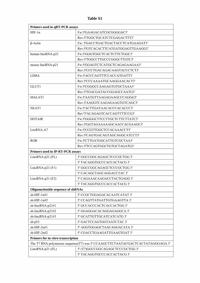
Primers used in qRT-PCR assays HIF-1α Fw:5'GAAGACATCGCGGGGAC3'
Rev:5'TGGCTGCATCTCGAGACTTT3' β-Actin Fw: 5'GACCTGACTGACTACCTCATGAAGAT3’
Rev:5'GTCACACTTCATGATGGAGTTGAAGG3’ human lincRNA-p21 Fw:5'GGGTGGCTCACTCTTCTGGC3'
Rev:5'TGGCCTTGCCCGGGCTTGTC3' mouse lincRNA-p21 Fw:5'GGAGTCTCATGCTCAGAGAAGAA3'
Rev:5'CCCTGACAGACAAGTACCCTCT3' LDHA Fw:5'ACCCAGTTTCCACCATGATT3'
Rev:5'CCCAAAATGCAAGGAACACT3' GLUT1 Fw:5'CGGGCCAAGAGTGTGCTAAA3’
Rev:5'TGACGATACCGGAGCCAATG3’ MALAT1 Fw:5'AATGTTAAGAGAAGCCCAGGG3'
Rev:5'AAGGTCAAGAGAAGTGTCAGC3' NEAT1 Fw:5'ACTTGATAACACCCACACCC3'
Rev"5'ACAGAGTCACCAGTTTTCCG3' HOTAIR Fw:5'GGGGCTTCCTTGCTCTTCTTATC3'
Rev:5'GGTAGAAAAAGCAACCACGAAGC3' LncRNA-A7 Fw:5'CCGTTGGCTCCACAAACCT3'
Rev:5'CAGTGACAGTAGCAGGCATCCT3' ROR Fw:5'CTTGATGGCATTGTCGCTAA3'
Rev:5'TCCAGTGGCTGTGCTAGATG3' Primers used in IP-RT-PCR assays LincRNA-p21 (FL) 5' GGCCGGCAGAGCTCCCGCTGG 3'
5' TACAGGTGCCCACCACTACG 3' LincRNA-p21 (F1) 5' GGCCGGCAGAGCTCCCGCTGG 3'
5' CACAGCTAGCAGGACCTAC 3' LincRNA-p21 (F2) 5' CAGAAACAAGACCTACTGAGG 3'
5' TACAGGTGCCCACCACTACG 3' Oligonucleotide sequence of shRNAs sh-HIF-1α#1 5' CCGCTGGAGACACAATCATAT 3' sh-HIF-1α#2 5' CCAGTTATGATTGTGAAGTTA 3' sh-lincRNA-p21#1 5' GCCACCCACTCACCACTGG 3' sh-lincRNA-p21#2 5' GGAGGACACAGGAGAGGCA 3' sh-lincRNA-p21#3 5' GCATTGTTGCATCATCATG 3' sh-p53 5' GACTCCAGTGGTAATCTAC 3' sh-HIF-2α#1 5' AGGTGGAGCTAACAGGACATA 3' sh-HIF-2α#2 5' CGACCTGAAGATTGAAGTGAT 3' Primers for in vitro transcription The T7 RNA polymerase sequence(T7) was 5' CCAAGCTTCTAATACGACTCACTATAGGGAGA 3'LincRNA-p21 (FL) 5' (T7)GGCCGGCAGAGCTCCCGCTGG 3'
5' TACAGGTGCCCACCACTACG 3'
Table S1

LincRNA-p21 (F1) 5' (T7)GGCCGGCAGAGCTCCCGCTGG 3' 5' CACAGCTAGCAGGACCTAC 3'
LincRNA-p21 (F2) 5' (T7)CAGAAACAAGACCTACTGAGG 3' 5' TACAGGTGCCCACCACTACG 3'
β-Tubulin CDS 5' (T7)ATGAGGGAAATCGTGCACATCC 3' 5' TTAGGCCTCTTCGGCCTCC 3'
Primers used in ChIP assays LDHA HRE Fw: 5' TTGGAGGGCAGCACCTTACTTAGA 3’
Rev: 5' GCCTTAAGTGGAACAGCTATGCTGAC 3’ P1 Fw: 5' GCTGCCCAGACTGGAGTGC 3'
Rev: 5'TGGGGCCACACACTGCCT 3' P2 Fw: 5' CTCAAGTGATCCGCCTGCCTC 3'
Rev: 5' CATCCTGGACCCCAGGCTG 3' 18S rRNA Fw: 5' CGGCGACGACCCATTCGAAC 3’
Rev: 5' GAATCGAACCCTGATTCCCCGTC 3’