sulfate reducing bacteria, nitrate reducing bacteria … · sulfate reducing bacteria, nitrate...
TRANSCRIPT
SULFATE REDUCING BACTERIA, NITRATE
REDUCING BACTERIA AND THEIR INTERACTIONS
IN A SOURING OFFSHORE OIL RESERVOIR SYSTEM
By
©Fuqiang Fan
A thesis submitted to the School of Graduate Studies
in partial fulfillment of the requirements for the
Degree of Doctor of Philosophy
Faculty of Engineering of Applied Science
Memorial University of Newfoundland
October, 2018
St. John’s Newfoundland and Labrador Canada
I
ABSTRACT
The troublesome souring issues, especially those occurred in offshore oilfields, have plagued
petroleum and environmental industries for decades. To control reservoir souring, the nitrate
addition have been noticed in recognition of their safety and operational effectiveness. The
interactions between sulfate reducing bacteria (SRB) and nitrate reducing bacteria (NRB) are key
mechanisms in the nitrate-mediated souring control. However, much is still unknown towards the
effective profiling of SRB and the detailed NRB-SRB interactions. Although NRB produced
biosurfactants might be promising bio-agents affecting NRB-SRB interactions, very limited
studies tackled the production of biosurfactants by natural NRB strains. Systematic investigation
of their unique roles in enhancing NRB competence over SRB was not documented. This thesis
targeted on filling the above stated gaps and examined SRB, NRB and their interactions in a
souring offshore oil reservoir system.
A method based on phospholipid fatty acid (PLFA) profiling of microbial communities in
offshore produced water was developed and optimized. The developed method was further
applied to profile microorganisms and trace SRB. Biosurfactant producing NRB was isolated and
the associated biosurfactant product was used for tracking NRB-SRB-biosurfactant interactions.
The outputs of this thesis include: (1) the established PLFA based protocol for profiling SRB in
offshore reservoirs; (2) the successful isolation and identification of biosurfactant producing
NRB coupled with subsequent biosurfactant generation and characterization; and (3) the findings
to confirm, for the first time, that NRB-produced biosurfactants could significantly strengthen
SRB inhibition by NRB. The thesis has resulted in promising products and scientific
observations for aiding souring control in the challenging offshore reservoir environments.
II
ACKNOWLEDGMENT
First and foremost, I would like to express my deep sense of gratitude to my supervisor, Dr.
Baiyu (Helen) Zhang. I am indebted for her excellent supervision, constructive criticism,
extensive encouragement and invaluable guidance throughout the course of my research degree.
I also highly appreciate Dr. Penny L. Morrill and Dr. Tahir Husain for serving in the supervisory
committee and spending time on reviewing my work. Dr. Penny L. Morrill has provided me with
constant support, encouragement, and guidance while Dr. Tahir Husain has inspired and supported
me with his practical advice and graceful patience. Special thanks to Dr. Bing Chen for his
generous guidance with his expertise, experience and encouragement.
I gratefully acknowledge the Faculty of Engineering and Applied Science, School of Graduate
Studies at the Memorial University of Newfoundland (MUN), and Suncor Energy Inc. for
financial support. My sincere thanks also go to Lidan Tao, Brettney Pilgrim, Jamie Warren, Linda
Winsor, Jeanette Wells, and Christopher Deacon for their assistance to the physicochemical and
genomic analysis. My further appreciation goes to my colleagues Dr. Jisi Zheng, Dr. Bo Liu,
Qinhong Cai, Jiabin Liu, Weiyun Lin, Zhiwen Zhu, Jingjing Ling, Xing Song, Xixi Li, Samira
Narimannezhad and Dr. Yinchen Ma for their friendship, valuable collaboration and assistance.
Special thanks are given to Adam Taylor and Shawn Beson for their kind assistance in the lab.
Finally, I wish to express my special appreciation to my parents and my siblings for their selfless
love and continued support which are the true source of inspiration in my pursuit of doctorate
degree. The deepest thanks are expressed to my girlfriend Tianjun Tan, for her remote but
endless love, enormous support and unlimited patience on the other side of the Earth.
III
TABLE OF CONTENTS
ABSTRACT……………………………………………………………………………………….I
ACKNOWLEDGMENT................................................................................................................. II
TABLE OF CONTENTS .............................................................................................................. III
LIST OF FIGURES ................................................................................................................... VIII
LIST OF TABLES ....................................................................................................................... XII
LIST OF SYMBOLS AND ABBREVIATIONS ...................................................................... XIII
CHAPTER 1 INTRODUCTION ................................................................................................... 1
1.1 Background ........................................................................................................................... 2
1.2 Statement of Problems .......................................................................................................... 8
1.3 Research Objectives ............................................................................................................ 11
1.4 Structure of the Thesis ........................................................................................................ 12
CHAPTER 2 LITERATURE REVIEW ...................................................................................... 14
2.1 Mechanisms of Offshore Reservoir Souring....................................................................... 15
2.1.1 Routes of H2S generation in offshore reservoirs.......................................................... 15
2.1.2 Roles of SRB in offshore reservoir souring ................................................................. 19
2.2 Microbial Monitoring of Offshore Reservoir Souring ........................................................ 20
2.2.1 Microbial characterization methodologies ................................................................... 21
IV
2.2.2 PLFA with FAME quantification ................................................................................ 26
2.3 Offshore Reservoir Souring Control ................................................................................... 31
2.3.1 Popular control techniques ........................................................................................... 31
2.3.2 Nitrate/Nitrite injection ................................................................................................ 35
2.4 Biosurfactants and Reservoir Souring Control ................................................................... 41
2.4.1 Introduction of biosurfactants ...................................................................................... 41
2.4.2 Biosurfactant producers in reservoirs .......................................................................... 55
2.4.3 Anti-souring effects of biosurfactants .......................................................................... 57
2.5 Summary ............................................................................................................................. 64
CHAPTER 3 PLFA ANALYSIS FOR PROFILING MICROBIAL COMMUNITIES IN
OFFSHORE PRODUCED WATER ............................................................................................ 71
3.1 Background ......................................................................................................................... 72
3.2 Materials and Methods ........................................................................................................ 76
3.2.1 Chemicals, reagents and glassware .............................................................................. 76
3.2.2 Sample collection ......................................................................................................... 77
3.2.3 Extraction of lipids from water samples ...................................................................... 77
3.2.4 SPE optimization ......................................................................................................... 78
3.2.5 Derivatization of FAME optimization ......................................................................... 80
3.2.6 Gas chromatography-mass spectrometry (GC-MS) determination of FAMEs ........... 80
3.2.7 Identification, quantification and validation ................................................................ 81
V
3.3 Results and Discussion ....................................................................................................... 84
3.3.1 Extraction efficiency in phase partition ....................................................................... 84
3.3.2 Determination of elution solvent volumes in SPE ....................................................... 85
3.3.3 Derivatization to FAMEs ............................................................................................. 91
3.3.4 Analytical performance ................................................................................................ 99
3.3.5 Analysis of offshore produced water samples ........................................................... 102
3.4 Summary ........................................................................................................................... 106
3.5 Appendix ........................................................................................................................... 107
CHAPTER 4 PROFILING OF SRB IN AN OFFSHORE OIL RESERVOIR USING PLFA
BIOMARKERS………………………………………………………………………………...109
4.1 Background ....................................................................................................................... 110
4.2 Materials and Methods ...................................................................................................... 113
4.2.1 Standards, reagents, apparatus and sample collection ............................................... 113
4.2.2 Extraction of lipids and preparation of FAMEs ......................................................... 114
4.2.3 GC-MS analyses ........................................................................................................ 115
4.2.4 Statistical analysis ...................................................................................................... 116
4.3 Results and Discussion ..................................................................................................... 117
4.3.1 In-situ measurements ................................................................................................. 117
4.3.2 PLFA analytical performance .................................................................................... 119
4.3.3 Fatty acids profiles ..................................................................................................... 120
VI
4.3.4 Identification of SRB ................................................................................................. 126
4.3.5 Patterns of microbial community structure and SRB migration behavior in reservoir
............................................................................................................................................. 130
4.4 Summary ........................................................................................................................... 140
CHAPTER 5 ISOLATION OF NRB FROM AN OFFSHORE RESERVOIR AND THE
ASSOCIATED BIOSURFACTANT PRODUCTION ............................................................... 141
5.1 Background ....................................................................................................................... 142
5.2 Materials and Methods ...................................................................................................... 145
5.2.1 Source and collection of inoculum ............................................................................ 145
5.2.2 Isolation and identification of NRB ........................................................................... 146
5.2.3 Screening of the NRB isolates for biosurfactant producers ....................................... 148
5.2.4 Performance demonstration of the selected strain ..................................................... 150
5.2.5 Biosurfactant production and characterization .......................................................... 151
5.3 Results and Discussion ..................................................................................................... 155
5.3.1 Phylogenetic analysis and morphological characteristics of isolates ........................ 155
5.3.2 Screening of biosurfactant-producing NRB ............................................................... 159
5.3.3 Performance demonstration of the selected isolate .................................................... 163
5.3.4 Characterization of NRB-generated biosurfactant product ........................................ 167
5.4 Summary ........................................................................................................................... 176

VII
CHAPTER 6 INTERACTIONS OF SRB, NRB SCREENED FROM OFFSHORE OIL
RESERVOIR AND NRB PRODUCED BIOSURFACTANTS IN MICROCOSMS ................ 178
6.1 Background ....................................................................................................................... 179
6.2 Materials and Methods ...................................................................................................... 181
6.2.1 Media and enrichment cultures .................................................................................. 181
6.2.2 Experimental setup..................................................................................................... 182
6.2.3 Chemical and physical analyses ................................................................................. 184
6.2.4 Microbial PLFA analysis ........................................................................................... 186
6.3 Results and Discussion ..................................................................................................... 186
6.3.1 NRB-SRB interactions under various nitrate conditions ........................................... 186
6.3.2 Analysis of microbial communities ........................................................................... 190
6.3.3 Souring control by nitrate and biosurfactant injection ............................................... 199
6.4 Summary ........................................................................................................................... 207
CHAPTER 7 CONCLUSIONS AND RECOMMENDATIONS ............................................... 209
7.1 Conclusions ....................................................................................................................... 210
7.2 Research Contributions ..................................................................................................... 214
7.3 Selective Publications ....................................................................................................... 215
7.4 Recommendations for Future Research ............................................................................ 217
REFERENCES…………………………………………………………………………………220
VIII
LIST OF FIGURES
Figure 1.1 Schematics for water re-injection process .................................................................... 3
Figure 2.1 Arrangement of phospholipids in the membrane of a living cell ............................... 27
Figure 2.2 Structures of glycolipid biosurfactants (A) Rhamnolipid from Pseudomonas
aeruinosa. (B) Trehalolipid from Rhodococcus erythropolis. (C) Sophorolipid from Torulopsis
bombicola. ..................................................................................................................................... 42
Figure 2.3 Diagrams of the generalized micelle with and without oil emulsification ................. 51
Figure 2.4 The changes of surface tension, interfacial tension and solubilization of the surfactant
over the increase of surfactant concentration................................................................................ 53
Figure 2.5 Mechanism of oil recovery enhanced by biosurfactants ............................................ 61
Figure 3.1 (a) Different volumes of DCM, acetone and methanol on the elution recoveries of
phospholipids. (b) Elution recoveries of phospholipids from SPE columns using 6 mL, 10 mL
and 14mL methanol. ..................................................................................................................... 88
Figure 3.2 Elution behaviour of phospholipids from SPE columns under two different elution
rate (0.6 mL/min and 1.2 mL/min). The phospholipids were eluted in 8 fractions continuously as
stated in the methods ..................................................................................................................... 90
Figure 3.3 The process of base-catalyzed transesterification of lipids ........................................ 92
Figure 3.4 Influence of methylation agent and volumes of acetic acids used for neutralization on
FAME derivatization .................................................................................................................... 95
Figure 3.5 Influence of reaction temperature and reaction time on FAME derivatization .......... 98
IX
Figure 3.6 Chromatogram of standard mixture containing 29 FAMEs by GC-MS analysis on
DB-5MS capillary column (number is accorded with Table 3.1) ............................................... 101
Figure 3.7 FAMEs detected from phospholipids in offshore produced water based on the optimal
PLFA method .............................................................................................................................. 105
Figure 3.8 Calibration curves of 29 FAMEs from 12 levels of sample concentration ranging
from 0.01 and 20 mg/L ............................................................................................................... 108
Figure 4.1 Capillary chromatograph of PLFAs (as methyl esters) from 29 external standards 121
Figure 4.2 Biplot of species based on PLFA profiles and reservoir water samples of the five
wells from principal component analysis ................................................................................... 125
Figure 4.3 FAMEs detected from phospholipids in offshore injection water and produced water
C4 and G2 ................................................................................................................................... 132
Figure 4.4 FAMEs detected from phospholipids in offshore injection water and produced water
C2 ................................................................................................................................................ 135
Figure 4.5 Changing patterns of possible SRB biomarkers and significant fatty acids associated
with SRB in the injection water and produced water samples.................................................... 136
Figure 4.6 Total biomass indicated by 9 PLFAs associated with SRB and their relationship with
redox potential ............................................................................................................................ 139
Figure 5.1 Phylogenetic tree of the isolated species from 16S rRNA gene sequences based on a
neighbor-joining analysis of 1000 resampled datasets ............................................................... 158
Figure 5.2 Secretion of amphipathic biosurfactants reduce the surface tension of the culture
supernatant as indicated by the degree of flatness of droplet on parafilm. ................................. 160
X
Figure 5.3 Evaluation parameters of the five Pseudomonas stutzeri strains in two growth media.
Surface tension and E24% in kerosene and hexadecane were indicated in 2a while Biomass,
OD600 and nitrate consumption were indicated in 2b.................................................................. 162
Figure 5.4 The changes of surface tension, cell growth and nitrate consumption of the
Pseudomonas stutzeri CX3 on glucose (3a) and glycerol (3b) medium versus the incubation time
..................................................................................................................................................... 166
Figure 5.5 FT-IR transmittance spectrum of the extracted biosurfactant product generated by
Pseudomonas stutzeri CX3 grown on glycerol medium............................................................. 172
Figure 5.6 Stability of the biosurfactant product produced by Pseudomonas stutzeri CX3 under
various environmental conditions. (a) Temperature; (b) Salinity; c) pH; (d) Critical micelle
concentration (CMC) of the biosurfactant product at 35 mg/L .................................................. 174
Figure 6.1 Redox potential, pH, sulfide and nitrate concentration changes in samples collected
on Day 5 and Day 11 during the competitive interactions of SRB and NRB strains ................. 188
Figure 6.2 PLFA profiles of P. stutzeri CX3 and D. escambiense ATCC 51164 strains grew on
NRB growth medium and SRB growth medium, respectively. .................................................. 191
Figure 6.3 Principal component analysis (PCA) showing the effect of different nitrate and
biosurfactant treatment on PLFA profiles of SRB and NRB strains on Day 11. PC1 and PC2
factors represented 61.8% and 35.2% of the total variance, respectively. .................................. 192
Figure 6.4 Representative PLFA patterns of microbial communities detected in microcosms with
various nitrate conditions on Day 11. ......................................................................................... 195
XI
Figure 6.5 Changes in the ratios of trans/cis of C16:1Δ9 and C18:1Δ11 and the ratios of
cyclopropyl PLFAs to their monoenoic precursors (cy/pre) at various growing conditions.
Numbers 1-8 in samples indicate runs in the first round of experiments. .................................. 198
Figure 6.6 Time course analyses of changes in physicochemical parameters after the inoculation
of P. stutzeri CX3 for souring mitigation. NRB-produced biosurfactants and varying nitrate
concentrations were added in the soured microcosms: (a) 1 g/L KNO3 (b) 3 g/L KNO3 (c) 3 g/L
KNO3 and 0.1 g/L biosurfactants (d) 5 g/L KNO3 ...................................................................... 202
Figure 6.7 Relationships between these time points and the connection of the parameters: (a)
PCA of the monitored time points and parameters during the 19 days of the biosurfactant-
involved NRB-SRB interaction. PC1 and PC2 factors represented 84.8% and 7.5% of the total
variance, respectively. (b) Eh, pH and the concentrations of sulfide and DIC on Day 19 in the
souring mitigation activities. ....................................................................................................... 206
XII
LIST OF TABLES
Table 2.1 Advantages and disadvantages of nitrate/nitrite injection ........................................... 38
Table 2.2 Various solvent systems and developer employed in TLC method ............................. 54
Table 2.3 Microorganisms and the effect of produced biosurfactants on interfacial tension and
residual oil recovery in model porous systems ............................................................................. 58
Table 2.4 Common methods of microbial characterization ......................................................... 65
Table 3.1 Retention time, identification and quantification ions (m/z), LODs, calibration range,
linearity, recovery rates, and repeatability of the twenty-nine FAMEs included in the analysis . 82
Table 4.1 Results for samples from the injection well and producing wells measured from the
surface facilities (C2, C4, F2 and G2 were the well numbers where produced water samples were
collected) ..................................................................................................................................... 118
Table 4.2 The absolute content and relative mole percentage of the fatty acids in water samples
from the injection well and producing wells............................................................................... 122
Table 5.1 Identification of the isolated possible biosurfactant producers ................................. 157
Table 5.2 The lipid components determined by TLC-FID and fatty acid profiles by GC-FID of
the biosurfactant product............................................................................................................. 169
Table 6.1 Experimental setting of the first round investigation of biosurfactant-involved NRB-
SRB interaction ........................................................................................................................... 183
Table 6.2 Experimental setting of the second round investigation of biosurfactant-involved
NRB-SRB interaction ................................................................................................................. 185
XIII
LIST OF SYMBOLS AND ABBREVIATIONS
ATR attenuated total reflectance
BATH bacterial adhesion to hydrocarbons
BCFA branched saturated fatty acids
BLAST basic local alignment search tool
CLPP community-level physiological profiling
CMC critical micelle concentration
CMD critical micelle dilution
CoA coenzyme A
CSB Coleville synthetic brine
DCM dichloromethane
DIC dissolved inorganic carbon
DO dissolved oxygen
DOC dissolved organic carbon
DSR dissimilatory sulfate reductase
Eh redox potential
EI electron ionization
EI24 emulsification index
ELDS enhanced laser diode spectroscopy
EOR enhanced oil recovery
FAME fatty acid methyl ester
XIV
FID flame ionization detector
FISH fluorescence in situ hybridization
FT-IR fourier transform infrared spectroscopy
GC gas chromatography
GC-MS gas chromatography coupled with mass spectroscopy
HE hydrogen embrittlement
HIC hydrogen induced cracking
hNRB heterotrophic nitrate reducing bacteria
HRI highly reactive intermediates
H2S hydrogen sulfide
LOD limits of detection
MEOR microbially enhanced oil recovery
MIC microbially induced corrosion
MPN most probable number
MUFA monounsaturated fatty acids
NMR nuclear magnetic resonance
NRB nitrate reducing bacteria
NR-SOB nitrate reducing, sulfide oxidizing bacteria
NL Newfoundland and Labrador
NRPOP Northern Region Persistent Pollution Control
OD optical density
PC phosphocholine
PC1 the first principal component
XV
PC2 the second principal component
PCA principal component analysis
PCR polymerase chain reaction
PE phosphoethanolamine
PLFA phospholipid fatty acid
PUFA polyunsaturated fatty acids
PWRI produced water re-injection
r2 coefficients of the calibration curves
RF response factors
RNA ribonucleic acid
RSD relative standard deviation
SDS sodium dodecylbenzene sulfonate
SIM selective ion monitoring
SPE solid phase extraction
SOB sulfur oxidizing bacteria
SOHIC stress orientated hydrogen induced cracking
SRB sulfate reducing bacteria
SSCC sulfide stress corrosion cracking
SSFAs straight-chain saturated fatty acids
TDC total dissolved carbon
TLC thin layer chromatography
UV ultra violet
VFAs volatile fatty acids

1
CHAPTER 1
INTRODUCTION
2
1.1 Background
Oil reservoirs are of great importance to the current global economic development and thus
considerable efforts have been placed to exploit and recover the petroleum resources. Generally,
oil recovery activities by petroleum industry can be divided into 3 classes: primary, secondary and
tertiary. These recovery methods follow a natural progression of oil production from the start to a
point where it is no longer economical to produce from a hydrocarbon reservoir (Muggeridge et al.
2014). When primary oil recovery becomes no longer feasible, secondary oil recovery commences.
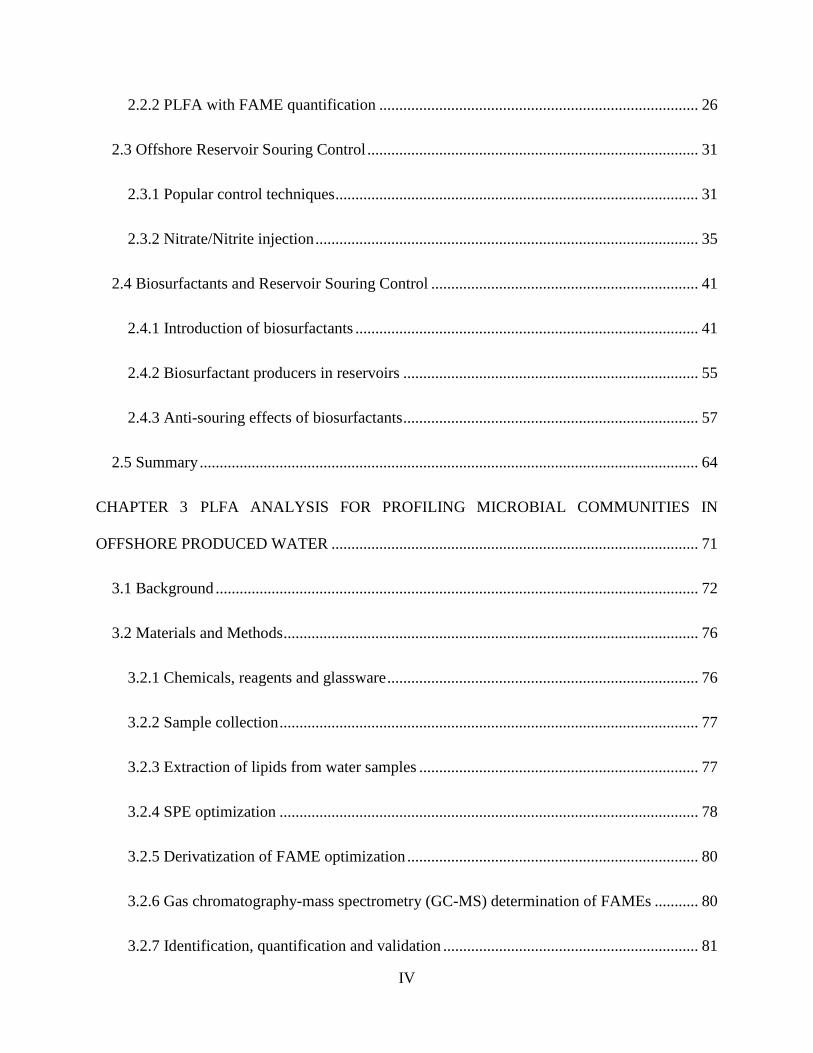
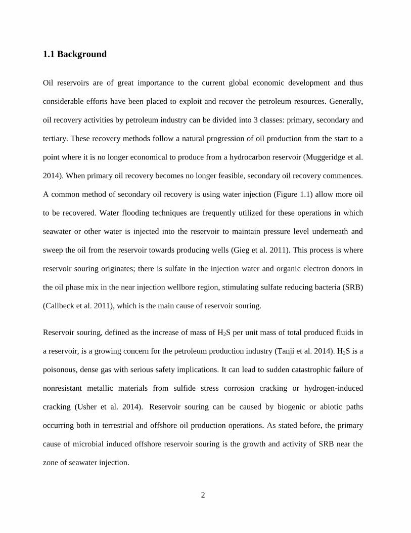
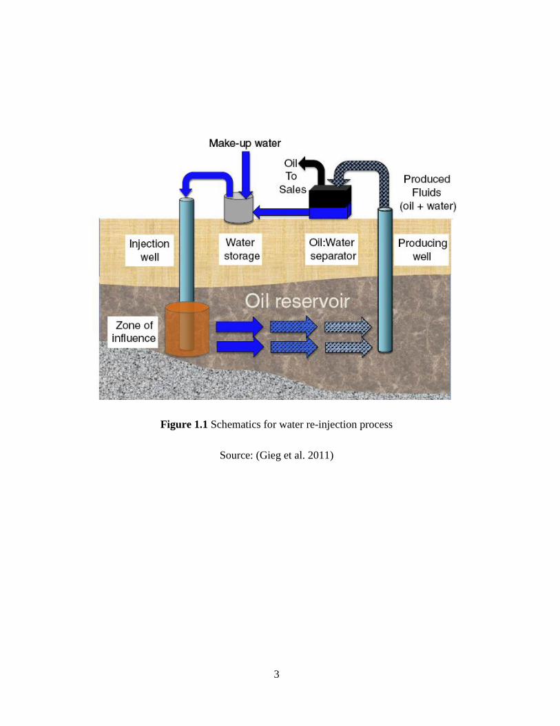
A common method of secondary oil recovery is using water injection (Figure 1.1) allow more oil
to be recovered. Water flooding techniques are frequently utilized for these operations in which
seawater or other water is injected into the reservoir to maintain pressure level underneath and
sweep the oil from the reservoir towards producing wells (Gieg et al. 2011). This process is where
reservoir souring originates; there is sulfate in the injection water and organic electron donors in
the oil phase mix in the near injection wellbore region, stimulating sulfate reducing bacteria (SRB)
(Callbeck et al. 2011), which is the main cause of reservoir souring.
Reservoir souring, defined as the increase of mass of H2S per unit mass of total produced fluids in
a reservoir, is a growing concern for the petroleum production industry (Tanji et al. 2014). H2S is a
poisonous, dense gas with serious safety implications. It can lead to sudden catastrophic failure of
nonresistant metallic materials from sulfide stress corrosion cracking or hydrogen-induced
cracking (Usher et al. 2014). Reservoir souring can be caused by biogenic or abiotic paths
occurring both in terrestrial and offshore oil production operations. As stated before, the primary
cause of microbial induced offshore reservoir souring is the growth and activity of SRB near the
zone of seawater injection.
3
Figure 1.1 Schematics for water re-injection process
Source: (Gieg et al. 2011)
4
The presence of sulfate is one of the main causes as it is sufficient in offshore injected seawater.
Carbon sources, as well as other nutrient sources in reservoirs, also influence the rate of sulfate
reduction by SRB. Several studies show that in water flooded reservoirs, volatile fatty acids (VFAs,
acetate, butyrate and propionate) and labile hydrocarbons such as alkanes and monoaromatics (e.g.,
toluene) are the biggest carbon sources in oilfield fluids for SRB growth (Grigoryan et al. 2009;
Grigoryan et al. 2008). These readily metabolized carbon sources frequently presented in the
injection water used for water flooding provide carbon sources for the SRB in the vicinity of the
injection well (Cavallaro et al. 2005; Grigoryan et al. 2008). Although concentrations of nitrogen
sources as nutrients are typically sufficient in oil reservoirs, phosphorous concentrations are
usually low, potentially limiting in situ microbial metabolism (Head et al. 2003).
There are different microbial communities in each oil reservoir depending on conditions such as
temperature, availability of substrates, salinity, and chemical compositions. Low-temperature
reservoirs facilitate the growth of mesophilic bacteria while high-temperature reservoirs are
typically dominated by thermophilic bacteria (Lin et al. 2014). If the temperature is higher than
100 ºC, the reservoir can naturally constrain the growth of any SRB (Gieg et al. 2011). Souring
can still occur in hot reservoirs of >100 ºC in the vicinity of the water injection well. This is
because the relatively cool water injection displaces the hot fluids from the zone of injection,
resulting in more favorable conditions for SRB growth (~50-70 ºC) (Gieg et al. 2011). In addition,
abiotic reactions are also of great importance in the production of H2S due to complex reactions
(Mueller and Nielsen 1996; Seto and Beliveau 2000). Specially, many iron-containing minerals are
capable of reacting with H2S forming iron sulfide, pyrite, or pyrrhotite for H2S scavenging. The
H2S adsorption capacity of the iron-bearing rock in the offshore reservoir is closely related to the
breakthrough of reservoir souring.
5
H2S lowers air quality and can be lethal to humans when high concentrations are inhaled; it can
easily escape the contaminated reservoir and may accumulate in a poorly vented area, such as
produced water tanks, flow lines, etc. A maximum of eight hours exposure to concentrations
greater than 100 ppm H2S will cause hemorrhage and death (Ballerino-Regan and Longmire 2010).
Concentrations above 600 ppm can be fatal in three to five minutes. In extreme cases of souring
the well may ultimately be shut down due to dangerous levels of the gas.
Sulfide is also closely correlated with corrosion issues. SRB are commonly considered the main
culprits of microbially induced corrosion (MIC), especially in anoxic, sulfate-rich environments
(Enning and Garrelfs 2014). The corrosion intensity level of co-produced water from an oil
reservoir can change over its lifetime. This is particularly marked in fields that are initially
considered clean, but produce more H2S in later life, in some cases at concentrations of up to many
thousands of parts per million by volume in the gas phase. Whilst carbon dioxide can cause very
severe corrosion (i.e., general and pitting) of steels, H2S corrosion is more localised, and can cause
sulfide stress corrosion cracking (SSCC), hydrogen embrittlement (HE), hydrogen induced
cracking (HIC) or stress orientated hydrogen induced cracking (SOHIC) (Ziaei et al. 2013). Hence,
increasing H2S will not necessarily cause a pro-rata increase in general corrosion rate, but rather
lay susceptible materials prone to catastrophic failure. Highly stressed, high strength steel can fail
in a matter of minutes in the presence of 50 ppm H2S. At high pressure, as little as 0.1 ppm H2S
can greatly reduce the time to failure of highly stressed, high strength steel (Amosa et al. 2013).
Besides, sulfide generation by SRB lowers oil quality and increase the cost of oil refinement.
Reservoir plugging due to the precipitation of ferrous sulfide slows the flow rate of water into the
reservoir reducing the efficacy of water injection as a secondary recovery method and hinders the

6
production speed of the well. The safety, corrosion, reduced oil quality and reservoir plugging
issues will all ultimately lead to higher operational costs of offshore oil production.
Due to the negative impacts of sulfide production in the three aspects stated above, mitigation
strategies are highly desired by the offshore oil and gas industry for the control of sulfide
generation and its release into the environment (Tang et al. 2009). Till now, various active and
passive measures have been proposed for reservoir souring mitigation, such as reverse osmosis or
membrane filtration to remove sulfide and carbon nutrients in injection water, sulfide scavengers
addition, the precipitation of metal sulfides and molybdate amendment (Gieg et al. 2011). The
methods listed may encounter effectiveness or cost issues in offshore reservoir souring control. For
this reason, biocides and nitrate/nitrite are commonly used in practical applications.
Compared with the application of nitrate/nitrite, chemical components in the reservoir may
scavenge biocides through reaction and may limit the application depth of biocides (Nemati et al.
2001). In addition, biocide treatments are difficult and expensive to deliver at sufficient
concentrations to the active souring zone as the enormous surface area of reservoir rock provides
ample sites for biocide sorption (Ezeuko et al. 2013). In contrast, nitrate and nitrite are very mobile
chemicals in subsurface environments and do not adsorb to most porous materials. Thus,
nitrate/nitrite transport should not be a limiting factor. Previous studies indicate that nitrate/nitrite
addition can be more effective in achieving longer periods of souring inhibition when compared to
biocides (Gieg et al. 2011; Reinsel et al. 1996). Therefore, nitrate/nitrite with convenient (high
solubility in water and compatibility with other chemicals), inexpensive and environmentally
friendly features has attracted increased attention from researchers in offshore reservoir souring
control.
7
The application of nitrate/nitrite could be very effective for reservoir souring control, by
promoting nitrate reducing bacteria (NRB), consuming nutrients that SRB require to grow, and
thus inhibiting SRB activity (Gieg et al. 2011). This treatment could be particularly effective and
may extend remaining production life under conditions of relatively low VFA content in produced
water and relatively short mean residence time of injection water (Stott 2012). Nitrite is more
preferable than nitrate for souring prevention in some high-temperature oil fields as it reacts
directly with sulfide and provides long‐lasting inhibition of sulfate reduction (Kaster et al. 2007).
Reinsel et al. (1996) found that glutaraldehyde (a type of biocide) did not have any long‐term
inhibitory effects, but instead lowered the SRB population. After glutaraldehyde removal, SRB
reportedly multiplied at their original growth rate and simultaneously produced H2S. On the other
hand, H2S concentration began to increase six days after nitrite was removed from the column.
This suggests that SRB were not killed by nitrite, but rather were still inhibited after nitrite was
removed; the original number of cells was still present and these were able to produce H2S once
the inhibitory effect was removed (Reinsel et al. 1996).
Effectively addressing souring‐related issues requires careful consideration of the operational
conditions encountered in specific offshore environments. Prior to the application of nitrate/nitrite
injection treatment, a better understanding is needed of the reservoir souring process, especially
the nature of NRB‐SRB interactions. It‘s thus highly desired to track the activities of SRB and
NRB, and to investigate their interactions in reservoirs, thus providing effective aid in offshore
reservoir souring control.
8
1.2 Statement of Problems
Unlike inland conditions, offshore reservoir souring control by nitrate amendment may encounter
many difficulties, which include the harsh marine environment, remote location, limited platform
space and the need for remote/unmanned control systems. As a result, the approaches and
technologies that are used to address souring in onshore operations may not be effective in dealing
with souring in offshore operations. The major problems have been identified and stated below:
1) Lack of routine determination tools for microbial analysis of offshore reservoir
samples
Produced water is a mixture of original water from different geological formations and the liquids
injected into the hydrocarbon zone, it could be used as a mirror to reflect the undergoing chemical
and biological activities beneath the seabed as a result of offshore oil and gas operations (Li et al.
2007a). SRB are responsible for the bacterial problems (Hubert and Voordouw 2007), thus the
profiling of these microbial groups from produced water is highly desired for reservoir souring
control. To measure microbial diversity and biomass, culture-independent methods provide
obvious advantages over culture techniques. The latter ones are generally time-consuming, labor-
intensive and most of the microorganisms are still recalcitrant to cultivation (Zengler 2009). As a
culture-independent technique, phospholipid fatty acid (PLFA) analysis has the potential to be an
inexpensive and quantitative method for microbial profiling of a large number of complex samples.
PLFAs have been extensively applied as biomarkers to characterize microorganisms in a variety of
solid and aqueous environmental samples (Dijkman et al. 2010; Drenovsky et al. 2010; Mills et al.
2006; Yu et al. 2009).
9
The performance of PLFA analysis is highly depended on the specific matrix extracted. To date,
the microbial profiling of offshore produced water with a complicated matrix and high salinity
using PLFA analysis is with extremely limited documentation. To examine the performance of
PLFA analysis for microbial profiling of produced water, operation conditions during extraction,
purification and derivatization of fatty acid methyl esters (FAMEs) in previous studies are not
directly applicable and need to be further evaluated. The extraction steps, parameters of
phospholipid purification as well as FAME derivatization are required to be further examined.
2) Unclear SRB transformation patterns in offshore reservoir wells
PLFA analysis offers a great potential of microbial community analysis in routine environmental
monitoring. Boschker et al. (2001) investigated the bacterial populations and pathways involved in
acetate and propionate consumption in anoxic brackish sediment. Labeled acetate and propionate
(13
C) were incorporated into PLFAs after incubation and the results showed that they were
predominantly consumed by different, specialized groups of SRB. Uranium-bearing sandstones
from the Dongsheng deposit were found with the abundant presence of C15-C18 fatty acids (Jiang
et al. 2012). Characteristic biomarkers of SRB Desulfovibrio and Desulfobacter sp. were found
and involved in bacterial sulfate reduction to sulfide. Even though PLFA profiling has been used
as SRB biomarkers in various solid and fluid samples, it is rarely applied in offshore reservoir
water analysis to specifically elucidate the mechanism of reservoir souring induced by SRB. There
is still a lack of basic understanding of the complex biomass and microbial community structure
information from the various reservoir conditions regarding souring.
3) Insufficient mechanism studies for NRB-SRB interactions
NRB are well known for their denitrifying capacity in which nitrates or nitrites are converted into
nitrogen-containing gases. This function enables NRB to play significant roles in the mitigation
10
and control of sulfide induced reservoir souring problems in offshore oil fields (Gieg et al. 2011).
Although nitrate-mediated souring control has been extensively studied in the laboratory (Callbeck
et al. 2011; Chen et al. 2017; Zhao et al. 2009) and in the field (Bodtker et al. 2008; Shartau et al.
2010; Voordouw et al. 2009), much is still unknown about the detailed microbial mechanisms
involved in NRB-SRB interactions during nitrate/nitrite injections for reservoir souring mitigation.
Hui et al. (2012) evaluated the microbial community structure and functionally distinct groups in
three kinds of produced water samples from Daqing oil reservoir. The isolates affiliated to
Pseudomonas stutzeri PTG4-15 (DP26, BP39, and PW5) were initially identified as NRB,
biosurfactant producing bacteria, and polymer-producing bacteria. Fallon et al. (2010) confirmed
that biosurfactants can be naturally derived from NRB. As microorganisms capable of utilizing
hydrocarbons as carbon and energy sources, NRB will produce surface-active agents as by-
products to facilitate hydrophobic degradation (Ron and Rosenberg 2002). The biosurfactants can
enhance the competence among species by increasing the bioavailability of entrapped organics in
the porous media (Pacwa-Płociniczak et al. 2011). The specific biosurfactants might also repress
the growth of certain targeted strain through their antimicrobial properties (Rodrigues et al. 2006).
Besides, as possible combination agents in biofilm matrix formation (Osterreicher-Ravid et al.
2000), biosurfactants could synergistically improve the bacterial adaptation capability to harsh
environments. The interactions of SRB and NRB are of great importance for reservoir souring
control by nitrate amendment. Meanwhile, biosurfactants produced by natural NRB are promising
bio-agents for enhancing NRB competence over SRB. However, biosurfactant producing NRB
isolated from oil reservoirs have been rarely reported, and associated biosurfactant production is
extremely limited in the literature (Zhao et al. 2016). Till now, there has been no published study
11
tackling the systematic investigation of NRB-SRB interactions with the involvement of
biosurfactants produced by natural NRB.
1.3 Research Objectives
The specific objectives of this research, therefore, are to develop a monitoring technique used for
routine analysis of samples with high oily and salinity properties in the marine environment, and fill
knowledge and technical gaps in biosurfactant-aided nitrate injection techniques for souring
remediation. The major research tasks include:
1) to develop a cost‐efficient SRB quantification methodology for their profiling in offshore
oil reservoirs;
2) to profile microorganisms and trace indigenous SRB populations in water samples from an
offshore oil reservoir;
3) to screen NRB species from the produced water samples after the reservoir was injected
with nitrate/nitrite to stimulate the growth of indigenous nitrate reducing microorganisms;
4) to isolate and evaluate the performance of biosurfactant producing NRB, and subsequently
produce and characterize the biosurfactant product through the metabolism of NRB; and
5) to investigate the NRB-SRB interactions under various nitrate and biosurfactant treatments,
track the associated microbial community structure changes and elucidate the mechanisms
involved in the processes.
12
1.4 Structure of the Thesis
Chapter 2 focused on the comprehensive review of reservoir souring mechanisms, the
monitoring techniques used for microbial control and detection, and the current technology
advances for controlling souring in hydrocarbon reservoirs. The application of biosurfactants in
reservoir system and remediation of reservoir souring was also discussed in detail.
Chapter 3 tackled the method development of PLFA analysis for profiling microbial
communities in offshore produced water. The elution parameters in solid phase extraction (SPE)
purification were adapted for treating the oily samples and their volumes were determined to
induce a high recovery for the fraction of phospholipids. The impact of parameters including
alkaline reagent, the volumes of acid used for neutralization, the time and temperature for
transesterification and the analytical performance of GC-MS were studied.
Chapter 4 provided PLFA profiles of microorganisms and SRB in produced water samples
from an offshore oil reservoir. The presence of SRB and SOB species and their relationship with
the redox environment of the reservoir wellbores was discussed. The species distribution patterns
were interpreted to elucidate the biological souring process.
Chapter 5 presented the isolation of NRB from an offshore reservoir and the associated
biosurfactant production. The possible biosurfactant producers were screened and the associated
biosurfactant production and characterization by the isolates were conducted.
Chapter 6 described the interactions of SRB, NRB screened from the offshore oil reservoir
and NRB produced biosurfactants in microcosms under non-sour and sour conditions. Various
nitrate and biosurfactant treatments were applied in the NRB-SRB interaction while using PLFA
13
biomarkers to trace the community responses. The potential of NRB produced biosurfactants in the
nitrate-dependent suppression of SRB activities and souring control was presented.
Chapter 7 concluded this study with summarized contribution and recommendations for
future research.
14
CHAPTER 2
LITERATURE REVIEW
15
2.1 Mechanisms of Offshore Reservoir Souring
2.1.1 Routes of H2S generation in offshore reservoirs
There are two types of H2S generation, abiotic meaning not living, and biogenic which comes
from SRB. Some offshore reservoirs are sour due to non-biogenic mechanisms that include the
thermophilic decomposition of sulfur containing hydrocarbons, the dissolution of pyrite or
thermochemical sulfate reduction. These mechanisms are influenced by the nature of reservoir
rock, oil composition and thermal maturity, high temperatures and aquathermolysis in thermal
recovery operations (Frazer and Bolling 1991; Khatib and Salanitro 1997; Seto and Beliveau
2000). Most biogenic souring in offshore oil reservoirs comes from the production of H2S through
the respiration of SRB.
In iron-deficient reservoirs, particularly in carbonate reservoirs associated with evaporates, an
abiotic mechanism that gives rise to H2S presence is the thermochemical reduction of sulfate,
which happens at temperatures above 100ºC. The flow of hydrogen sulfide from other geological
formations to initially sweet reservoirs, either because of geomechemical processes resulting from
reservoir depletion, or of failure in well cementations, are examples of abiotic H2S appearance
later in the field exploitation stage (Al-Eid et al. 2001). Reduction of the H2S solubility in the
water phase resulting from the reservoir pressure decrease can also be considered a late abiotic
reservoir souring mechanism (Seto and Beliveau 2000).
Heterotrophic SRB get energy for live and growth by oxidizing carbon sources (electron donors)
with the reduction of respired sulfate (electron acceptor) to sulfide. Microbial H2S generation
depends on various concurrent factors that allow the development of an active population of SRB.
Adequate ranges of temperature, pressure, water salinity, pH and redox potential; availability of
16
sulfate and of water-soluble organic carbon; provision of other nutrients, mainly nitrogen and
phosphorus, required for SRB biomass growth (Sunde and Torsvik 2005).
Thermochemical sulfate reduction
Thermochemical sulfate reduction is recognized as common and widespread geochemical
mechanism for souring by many case studies and theoretical reviews (Machel 2001).
Thermochemical sulfate reduction is a feasible mechanism evidenced by the Leblanc process for
soda manufacture, in which a mixture of sodium sulfate and coke is heated for a considerable
period at high temperature. However, the strongly endothermic process is normally operated at
1000 ºC and is not significant below 700 ºC. Sulfate is sufficiently present in injected seawater and
yields the large quantities of observed H2S in produced fluids. There are also substances present,
particularly in the oil which could be reducing agents for this process and it has been shown that
clay and other minerals are a rich source of catalysts for a wide variety of reactions. Perhaps under
milder conditions than usually employed in the Leblanc process it is possible with this
combination for such a reaction to proceed.
According to petroleum geologists, high H2S concentration accumulated in natural environments
(‗sour gas‘ fields) are formed when sulfate is reduced by petroleum (mostly methane and the n-
alkanes) at depths usually greater than 3000 m (Anderson and Thom 2008). Although the lower
thermal limit for thermochemical sulfate reduction is somewhat controversial, many studies
generally consider the minimum temperatures range from 100 to 140 ºC (Cai et al. 2003). In a
temperature of below 140 ºC, thermochemical sulfate reduction is very slow or inhibited, even
though the involved reactions are characterized by large negative free energy changes of reaction
(Machel 2001). Under proper reservoir conditions, both elemental sulfur and polysulfides are
17
capable of oxidizing some organic molecules under basic conditions (Goldstein and Aizenshtat
1994). Sulfate alone will not react only if lower oxidation state sulfur is present.
Thermal decomposition
Thermal decomposition of organic sulfur compounds which are present in some crude oils is
another mechanism which may lead to the production of relatively low concentration of H2S
(Worden and Smalley 1996). The disadvantage of such mechanisms is that they require high
temperatures and are not associated with sulfate reduction, nor are they related to seawater
injection. Generally, the H2S production by thermal decomposition of organics appears to be
proportional to the sulfur content of the oil (Ritchie et al. 1985). Organic sulfur ranging from the
very stable aromatic sulfides to very unstable thiocarbamates play different roles in the kinetics of
H2S production. Under proper thermal conditions, disulfides and thiols have been recognized as
the most reactive sulfur species, followed by aliphatic sulfides, thiophenic compounds (Cortese-
Krott et al. 2017; Giles et al. 2003; Kelemen et al. 1991). Meawhile, benzothiophenic compounds
appear to be the most stable organically bound sulfur species during thermal decomposition of
organic sulfur compounds.
An example of thiocarbamate hydrolysis is:
RCS2R + 3H2O RCO2H + 2H2S + ROH (2-1)
and thioether reduction:
RSR 2RH + H2S (2-2)
Dissolution of pyritic materials
18
Pyrite, FeS2, is widely distributed in formation rocks and therefore has to be considered as
(Gallego-Torres et al. 2015), although it is not obvious why the process should not have been
proceeding during the prehistory of the field. Pyrite contained in reservoir rock can be leached out
as particles of small dimension which may react with the environment according to the following
reactions:
FeS2 + 8H2O Fe2+
+ 2SO42-
+ 16H+ + 14e
- (2-3)
or
FeS2 + 4H+ + 2e
- Fe
2+ + 2H2S (2-4)
Pyrite oxidation (2-3) is known to be a slow process which requires the presence of an oxidant
such as oxygen or the involvement of oxidizing bacteria (Ma and Lin 2013). The reaction leads to
an acidic environment, which favors the formation of sulfide in petroleum reservoirs. Pyrite
reduction (2-4) is very possible at lower pH values. The theoretical calculation of the reaction
progress of (2-4) is very complex. Literature values for the pK of the solubility product for iron
sulfide vary from 16.9 to 18.8, which would reflect a 100 fold variation in calculated H2S
concentration (Eden et al. 1993). The pyrite reduction under acidic conditions could be a route,
and in the case of seawater injection (with the presence of sufficient sulfate) it is more likely route.
Redox reactions involving bisulfite oxygen scavengers
This mechanism has been suggested by a number of oil companies (Lasebikan et al. 2010).
Oxygen scavengers used in injection waters by the oil and gas industry in water flood and
production systems invariably comprise sulfite, and in many cases, ammonium bisulfite. These
compounds are redox poising agents as corrosion inhibitors and are known to remove oxygen and
19
stimulate the growth of SRB. The rise of H2S is not clearly associated with bisulfite injection by
purely chemical reactions. However, a relationship between the content of sulfide measured and
increase in ammonium bisulfite concentration in brine fluids at low pH was observed (Lasebikan
et al. 2010). As only a relatively low concentration of bisulfite is injected into the reservoir, they
may function as weak catalysts to give rise to high levels of H2S. These compounds typically react
completely with oxygen to yield sulfate, but the sulfate yields are negligible compared with the
sulfate concentration in seawater (2650 mg/L) (Eden et al. 1993):
2NH4HSO3 + O2 (NH4)2SO4 + H2SO4 (2-5)
In a system where bisulfite injection are well operated, the excess ammonium bisulfite injected
into reservoirs is generally less than l mg/L (Eden et al. 1993). Thus, it is not likely to be the
principal sulfur source for hydrogen sulfide, although it is considerably easier to reduce to sulfide
than the sulfate ion. It is more likely that the bisulfite could be involved either as a catalyst in the
conversion of some other sulfur-containing substance, or that it is modifying the surface of an inert
sulfur-containing solid in the reservoir so making it more reactive, thus generating H2S (Eden et al.
1993). Since the reactivity of metal sulfide products varies significantly depending on their
crystalline form and especially the nature of the surface, this possibility is promising.
2.1.2 Roles of SRB in offshore reservoir souring
Souring in offshore oilfield systems is heavily due to SRB, a diverse group of nonpathogenic,
anaerobic microorganisms that respire sulfate to produce hydrogen sulfide. However in order for
this to occur, there must be free electrons as an external energy present. Such biological souring is
a detrimental, widespread phenomenon in the petroleum industry, occurring in offshore and
onshore facilities in a wide range of growth conditions. In high temperature reservoirs most of the
20
souring will occur mainly in offshore facilities (topside processing of injection water) and in close
proximity to the injection site where the hot reservoir fluids mix with relatively cool injection
water resulting in more ideal conditions for SRB activity. Sulfate reducers exist either
indigenously in deep subsurface reservoirs or can be ―inoculated‖ into a reservoir system during
oilfield development or the oilfield production phase (Gieg et al. 2011). Souring happens when
microorganisms enzymatically reduce sulfate, thiosalts, or sulfur to gain energy for growth using
the dissimilatory sulfate reductase (DSR) enzyme (Chang et al. 2001). Sulfide is formed directly
by the activities of SRB. These consist of at least two genera (Desulfovibrio and
Desulfotomaculum) of obligate anaerobes which oxidize hydrogen and organic compounds using
sulfate by the DSR enzyme (Larsen et al. 2000). This DSR is apparent in mud at pond bottoms, in
bogs and on the sea-bed. Sulfate concentration is high in seawater and consequently its reduction
is an important factor in H2S production. The H2S formed in the biosphere is largely converted to
sulfur: only a small part of it subsequently becomes isolated in the form of insoluble sulfides of
heavy metals.
Desulfovibrio and Desulfotomaculum seem to be unrelated to each other and their relation to other
bacterial groups is obscure. The better known genus Desulfovibrio is usually mesophilic and
sometimes halophilic (preferring saline conditions) (Eden et al. 1993). Desulfotomaculum species
are somewhat more difficult to isolate and purify. They are characterized by spore formation and
are sometimes thermophilic.
2.2 Microbial Monitoring of Offshore Reservoir Souring
In order to effectively counter reservoir souring, its specific cause must be known. A vast majority
of reservoir souring derives from microbial activity in the offshore reservoir. There are many
21
techniques for SRB quantification and characterizing the microbial community in general. To find
the appropriate method of microbial characterization for in offshore reservoirs, details of
commonly used microbiological, molecular biological and biochemical techniques must be known
and compared with one another, weighing the pros and cons.
2.2.1 Microbial characterization methodologies
Microbiological methods
Metabolic assays
Community-level physiological profiling (CLPP) is a method that uses commercially available 96-
well microtiter plates, usually consisting of 95 different carbon sources, nutrients, and a
tetrazolium dye. The oxidation of the carbon substrate is concomitant with reduction of the dye.
The carbon utilization patterns are then analyzed using mutivariant statistical techniques to
evaluate the degree of similarity among environmental samples. Differences in sole carbon source
utilization have been used to distinguish among different bacterial types for over 50 years (Garland
1997). The automated microbial identification system, Biolog, based mainly on aerobic metabolic
activities, has contributed a great deal to our understanding of carbon source utilization (Muñiz et
al. 2014). This rapid, community-level approach for assessing the utilization patterns of sole
carbon sources is now being used to study microbial community dynamics. This approach has
been widely used for assessing the relative similarity between aerobic heterotrophic microbial
communities across spatial, temporal, and experimental gradients (Garland 1997).
Cell counting techniques
22
Cell counting techniques are methods of community characterization that cannot provide any
information other than the number of cells in a community sample. These methods provide no
information about community phylogeny, diversity or physiology when used alone. However, cell
counting techniques are often used in conjunction with other methods that address these facets of
community characterization to provide a more complete description of the community (Abbaci et
al. 2008; Reed et al. 2002). The cell count of a community is one of the most basic characteristics
of a community and is only a starting point for meaningful characterization.
Molecular biological methods
Polymerase chain reaction (PCR) -Based Gene Sequencing
PCR is a type of technique used in molecular biology to amplify segment of DNA from whole-cell
extracts or from total community DNA of an environmental sample across several orders of to
facilitate analysis (Spiegelman et al. 2005). DNA is exposed to a thermostable polymerase and is
subject to repetitive cycles of template strand denaturation, oligonucleotide primer annealing, and
polymerization of the template-primer duplex. This process results in the exponential amplification
of the template DNA. The key to PCR is the use of oligonucleotide primers designed to be
complementary to the desired gene or genetic region. During PCR, double-stranded DNA is
separated into single strands at high temperature, a process known as denaturation. Two
oligonucleotide primers then anneal to complementary regions of the denatured DNA, which flank
the desired sequence. Following this, a heat-stable DNA polymerase creates a new strand of DNA
by extending the primer, using the complementary strand as a template. A new cycle begins as this
new double-stranded molecule is denatured again. Multiple repetitions of this cycle lead to an
exponential amplification of the target gene(s) or genetic region (Kubista et al. 2006).
23
PCR is the simplest and currently the most widely used method to obtain 16S rRNA genes for
detailed characterization of microbial communities. It is the foundation upon which most genetic
polymorphism-based techniques are based. It is an effective way to inspect isolated genes of a
SRB; however it must be coupled with denaturant gradient gel electrophoresis (DGGE) or other
separation methods (temperature gradient gel electrophoresis, TGGE) for SRB quantification of
large communities. Although the analysis of a microbial community by PCR and cloning provides
a convenient and rapid alternative to some other culture-independent techniques, there are several
factors that could skew diversity estimates (Farrelly et al. 1995). PCR reactions are very sensitive
to reaction conditions and even duplicates might not give quantitatively identical results
(Schneegurt and Kulpa, 1998). Due to the sensitivity and specificity of the PCR reactions, minor
contamination can lead to false-positive signals and false-negative amplifications are often seen
(Bossler and Van Deerlin 2009). Available technology does not allow for the separation of
multiple bands amplified from a highly diverse bacterial community (Smith and Osborn 2009).
Another concern of PCR when combined with DGGE is the assignment of particular bands to
individual populations, particularly where multiple bands occur. Individual organisms could
potentially contribute to multiple bands on a DGGE gel since sequences between rRNA operons of
an individual organism can vary significantly (Boon et al. 2002).
Fluorescent In Situ Hybridization (FISH)
Fluorescence in situ hybridization (FISH) is a cytogenetic technique developed by biomedical
researchers in the early 1980s (Langer-Safer et al. 1982). It enables in situ phylogenetic
identification and enumeration of individual microbial cells by whole cell hybridization with
ribosomal RNA targeted oligonucleotide probes, which are covalently mono-labeled with
fluorescent dye molecules. This limits the sensitivity of the method and aggravates the use of FISH
24
for identification of prokaryotes with low ribosome content per cell. The intensity of fluorescent
signals is correlated to cellular rRNA contents and growth rates, which provide insight into the
metabolic state of the cells. FISH can be combined with flow cytometry for a high-resolution
automated analysis of mixed microbial populations. The FISH method was used to follow the
dynamics of bacterial populations in agricultural soils treated with s-triazine herbicides (Barra
Caracciolo et al. 2010). A variety of molecular probes were used to target specific phylogenetic
groups of bacteria such as α, β, γ, and δ subdivisions of Proteobacteria and Planctomycetes.
Biochemical methods
Bisbenzimidazole-CsCl-gradient fractionation
Bisbenzimidazole-CsCl-gradient fractionation is a method of DNA fractionation based on overall %
G-C content, which produces a characteristic community profile of relative abundance of DNA
vs. % G-C content. In this method, community DNA is exposed to bisbenzimidazole, a non-
intercalating dye that preferentially binds to A + T regions of the DNA. This binding alters the
buoyant density of the DNA in proportion to the amount of dye bound. This establishes a means of
physically separating the DNA on the basis of G-C vs. A-T content (Holben and Harris 1995);
when passed by centrifugation through a linear gradient of CsCl, the DNA–bisbenzimidazole
complex separates linearly by buoyancy, i.e., by relative amount of bound bisbenzimidazole, i.e.,
by A-T/G-C content. Bisbenzimidazole is fluorescent under long-wave UV illumination, to a
degree proportional to the amount of bisbenzimidazole present. This allows for the measurement
of the relative abundance of DNA at each point in the CsCl gradient (i.e., at varying %G-C), thus
establishing the parameters for the community profile.
Lipid analyses
25
Quinone profiling is a culture-independent lipid biomarker assay that uses the taxon-dependent
specificity of microbial quinones to create a community profile with moderate taxonomic
specificity. Quinones are essential lipid components of respiratory and photosynthetic electron
transport systems in microorganisms. Quinone profiling is a biochemical method of fingerprinting
entire microbial communities based on the distribution and relative abundance of various species
of quinones in the community, a method of community analysis that predates all genetic
fingerprinting techniques (Collins et al. 1979). Many molecular species of respiratory quinones
can be characteristic of bacteria at the level of genera or higher-level taxa, depending on the
species of molecule. Ubiquinones (a subgroup) are also used to identify genera of fungi, yeast, and
yeast-like fungi (Kuraishi et al. 2000; Okada et al. 1996).
PLFAs and FAMEs are culture-independent lipid biomarker assays in which the nature and
distribution of various membrane lipids are used to construct the phylogeny and metabolic activity
profiles for a microbial community. Membrane lipids can potentially provide a great deal of
information about the organisms from which they are derived. Microbes alter the lipid composition
of their membranes in response to differing environmental conditions, for example by enhancing
membrane fluidity by increasing the proportion of unsaturated fatty acids in response to cold
temperatures (Okada et al. 1996; Zheng et al. 2011). As such, membrane lipids can provide
information about the physiological states of a given microbe or community (Wixon and Balser
2013). Also, with the construction of reference libraries upon the variable microbial characteristics,
taxonomic information can also be derived from the nature and distribution of various lipid
subspecies isolated from a given microorganism.
26
2.2.2 PLFA with FAME quantification
2.2.2.1 Characteristics of PLFA
Phospholipids are the key component of cellular membrane in living cells. Viable microbes have
an intact membrane containing fatty acids as components of its phospholipids, which are not found
in storage products or in dead cells. Lipids usually make up less than 5% of the dry weight of
bacteria and are both structurally and functionally diverse. The results from sediments and soils
with substrate additions indicate that rapid changes in microbial community structure can be
detected by changes in PLFA patterns (Frostegård et al. 2011). This suggests that PLFA analysis is
suitable for detecting rapid changes in living populations. Taxonomically, fatty acids in the range
C2 to C24 have provided the greatest information and are present across a diverse range of
microorganisms (Banowetz et al. 2006).
Phospholipids consist of a single molecule of glycerol (3C alcohol), two OH groups of the glycerol
are bound to the two fatty acid chains (hydrophobic tail) and one OH group is bonded to a
phosphate group (hydrophilic head). Thus these lipids are asymmetric, having hydrophilic and
hydrophobic regions and in the membrane they form a bilayer with hydrophilic ends towards the
outer surface of the membrane and hydrophobic ends buried in the interior (Figure 2.1). PLFA can
be classified into ester-linked phospholipid fatty acids (EL-PLFAs, 60–90% of the total) and non-
ester linked phospholipid fatty acids (NEL-PLFAs, 10–40% of the total).

27
Figure 2.1 Arrangement of phospholipids in the membrane of a living cell
Source: (Kaur et al. 2005)
28
2.2.2.2 Evaluation of PLFA analysis
PLFA are useful biomarkers or signatures for fingerprinting the soil microbial community because
of the relative abundance of certain PLFAs, which differ considerably among the specific group of
microorganisms (Joergensen and Wichern 2008). PLFA with FAME quantification share the dual
advantages of being rapid and inexpensive to perform. GC/MS equipment is routinely used in
most analytical chemistry labs, and the cost of running individual samples is negligible. Also,
PLFA profile analysis holds competitive advantage over the rest of the conventional methods
(culturable technique) to study the soil microbial community structure, as it accounts for larger
proportion of the soil microbial community. Digressing from the rapid and inexpensive analyses
for the high number of samples needed for microbial ecology investigations, PLFA analysis has a
relatively high throughput, identifies only the viable bacteria population (Øvreås 2000). The ability
of PLFA and FAME to rapidly and inexpensively identify cultured isolates has been used to
extensively characterize community members originally identified by less specific mechanisms of
analysis (Wixon and Balser 2013).
As to the property of providing quantitative insight into the soil viable/active microbial biomass
from the concentration of total PLFA, this is because the phospholipids are rapidly degraded after
cell death and are not found in the storage products. A significant correlation has been observed
between total phospholipid content and other methods used for measuring microbial biomass, such
as acridine orange direct counts of microorganism and also with ATP content (Balkwill et al.
1988). Certain bacterial groups with specific biogeochemical activity such as SRB have already
been thoroughly characterized by PLFA profiling and were found to possess several characteristic
PLFA biomarkers (Córdova-Kreylos et al. 2006; Mohanty et al. 2008).
29
There are a few limitations associated with PLFA biomarker analysis, which may limit its use at
the regional and global scale. It does not reveal any information at the species-level, archae
bacteria cannot be determined using this method and databases for interpretation of biomarkers are
centered on fatty acids from microorganisms from pure cultures. Since growth conditions alter the
distribution of lipid species, the proper use of existing reference libraries of lipid composition
requires that the samples are cultivated under the exact same conditions as the strains used to make
the reference library. It is difficult to make major changes to the sample preparation method
without altering the fatty acid profile of the bacteria of interest, which would then require
preparation of a new reference library (Buyer 2002).
Furthermore, calibration of changes in stress biomarkers under diverse ecosystems, soil type and
climate, linking of PLFA profiles with functions of ecosystems, and automation of the technique
needs to be strengthened for the implementation of this bioindicator in the regional assessment of
environmental impact of agriculture and its incorporation in soil quality indices. Banks et al. (2014)
investigated the soil microbial community response to surfactants and herbicides in two soils and
proposed the necessity of long-term microbial community studies using PLFA analysis on a wide
array of soil types and management practices. The further enhancement of PLFA profiling in
investigating the changes in microbial community structure of agricultural soils was also suggested
(García-Orenes et al. 2013). Nevertheless, the full potential of PLFA as a bioindicator of
environmental monitoring and assessment at higher scales of resolution is certainly growing as
databases and novel methods focusing on functions are being developed.
30
2.2.2.3 PLFA with FAME for SRB quantification
Bacteria contain characteristic lipid fatty acids in the C12-C19 region which distinguish them from
eukaryotic organisms and in certain cases from each other (Parkes et al. 1993). Such properties
enable fatty acids to be used to study complex sedimentary communities in situ, thus avoiding the
limitations of isolation techniques. SRB are typical bacteria in that their membranes are composed
primarily of phospholipids with ester-bound fatty acids that can be analyzed as FAME. Taylor and
Parkes (1983) have shown that the lipid fatty acids of the SRB Desulfobacter, Desulfobulbus and
Desulfovibrio desulfuricans contain a number of characteristic acids which have the potential to
act as biomarkers for these bacterial types in complex sedimentary environments. Also, the lipid
composition of the Gram-negative SRB, especially polar lipid-derived fatty acids, have been
studied extensively and several uncommon PLFA, e.g. cy-C17:0, 10Me C16:0, i-C17:1 (cis-10),
C15:1 (cis-9) and C17:1 (cis-11), have been suggested as specific biomarkers for the different
groups of SRB (Córdova-Kreylos et al. 2006; Mohanty et al. 2008). Although these PLFAs may
also be found in non-SRB (e.g. 10Me C16:0 in actinomycetes), they are suitable to distinguish
among the different groups of SRB since they have a high biological specificity on them and are
produced only by a limited group of microorganisms. Their usage was limited under specific
environments, such as sulfide-rich conditions. They were applied as biomarkers of Desulfobacter,
Desulfotomaculum, and Desulfovibrio in uranium-contaminated subsurface sediment to evaluate
the geochemical and microbial community response to ethanol amendment (Mohanty et al. 2008).
PLFA biomarkers indicative of bacterial sulfate reducers have been identified in many studies. The
lipid marker Br-C17:1 (especially i-C17:1 (cis-10)) has been associated with Desulfovibrio
(Córdova-Kreylos et al. 2006); 10Me C16:0 and C17:1 (especially C17:1 (cis-11)) were
recognized as a major fatty acid component for Desulfobacter (Jiang et al. 2012; Kaksonen et al.
31
2004) and Desulfobulbus (Córdova-Kreylos et al. 2006), respectively. These biomarkers were
determined for a small subset of isolates and may not be present in, or exclusive to, all members of
the groups they are reported to represent. Caution has been urged when trying to interpret
responses in the Actionbacteria (actino) class due to overlapping PLFAs with SRB (e.g., 10Me
C16:0) (Jaatinen et al. 2006).
2.3 Offshore Reservoir Souring Control
2.3.1 Popular control techniques
2.3.1.1 Prophylaxis
Early prevention of souring (prophylaxis) can be achieved by using injection water that is naturally
low in sulfate concentration, VFAs, and biomass, or by removing sulfate (as well as thiosulfate
and sulfite) from injection water using technologies such as reverse osmosis or membrane
filtration (Robinson et al. 2010). Although this technology is typically applied at a significant
capital cost, efforts have been made in technology improvements to reduce its cost. The desulfated
water is reported to be achieved through a nanofiltration membrane process and is normally used
for prevention of sulfate scale in oilfields (Davis and Mcelhiney 2002). The reduction of sulfate by
lowering the concentration of sulfate in reservoirs to below that of VFAs will limit production of
H2S. Aerated water in water flooding could also be considered, thus inducing more oxidizing
environment to inhibit SRB. However, oxygenated seawater is corrosive to infrastructure, so this is
not applicable for offshore oil wells. Prophylactic additions of nitrate/nitrite to reservoir wellbores
was reported to be effective for souring control in high-temperature offshore operations (Larsen et
al. 2004).
32
2.3.1.2 Physical and chemical treatments
Physical or chemical treatments are used for controlling microbial souring after its breakthrough.
These options include using non-metallic pipes or polymer coatings inside pipelines, adding
sulfide scavengers such as amines or sodium hydroxide in product streams and installing vapor
recovery units on surface tank installations to remove H2S gas. Biocides can be injected into
above-ground facilities and added to the injection waters to help suppress microbial numbers and
activity (discussed below). For above ground and subsea piping, biocide treatment can be coined
with the use of mechanical cleaning ―pigs‖ (Larsen et al. 2004) that are run through pipelines to
disrupt and remove biologically active deposits from the pipe surface. This physical treatment
helps to reduce corrosive activities by sulfidogenic biofilms and removes potential inoculum from
the injection water stream. In extreme cases of souring, the best option may be to shut in the
affected wells or physically isolate problematic zones of the reservoir. Jesus et al. (2015)
investigated reservoir souring control using the chemical treatment of molybdate. Control of
sulfate reduction by adding molybdate in short-term tests was investigated to to determine the
minimum inhibitory concentration of molybdate in sulfate-rich medium. The results revealed that
0.08 mM (12.8 mg/L) molybdate, while the molar ratio molybdate/sulfate is 0.004, is sufficient to
inhibit the activity of SRB for 7 days.
If these approaches are not practical or economic, chemical treatments listed below can be used to
control the microbial process responsible for sulfide production. Chemicals that are widely
injected into the reservoir or applied in surface facilities for microbial control can be divided into
two classes: (1) non-specific biocides and (2) nitrate or nitrite, which more specifically inhibit
sulfidogenesis or oxidize existing sulfide.
33
2.3.1.3 Biocides
Commonly used biocides include glutaraldehyde, tetrakis (hydroxymethyl) phosphonium sulfate,
benzalkonium chloride, formaldehyde, sodium hypochlorite, and cocodiamines, among others
(Kaur et al. 2009; Videla et al. 2005). THPS is a recently developed broad spectrum biocide with
reduced environmental toxicity when compared to traditional biocides. It also has the ability to
dissolve ferrous sulfide precipitates that might otherwise protect planktonic cells from biocide
(Videla et al. 2005).
Biocides have the advantage of being easy to administer so they are routinely used in above-
ground facilities and pipelines. However, they are difficult to push deep into the reservoir, which
makes treatment of SRB communities far from the injection well challenging. Biocides have other
drawbacks, they are expensive and require repeated or constant application to be effective, some
are potentially hazardous to oilfield personnel and the environment, and their continued use can
lead to the creation of biocide-resistant microbial populations, either through biochemical
resistance or because of lack of penetration through biofilms in situ or in pipelines. In the latter
case, cycling of biocides or combinations of two biocides is used to temporarily overcome
resistance.
Recent studies have used molecular biology approaches to understand the response of SRB to
biocides, particularly regarding the development of resistance. Whole genome microarrays of
Desulfovibrio vulgaris Hildenborough (Lee et al. 2010) revealed that exposure to glutaraldehyde, a
nonspecific biocide, has caused upregulation of 179 genes and downregulation of 77 genes. This
diversified response was likely due to the broad range of cell targets affected by glutaraldehyde.
Mutating individual genes that responded to glutaraldehyde did not rescue the mutants from
34
subsequent exposure to the biocide, suggesting that development of resistance to glutaraldehyde is
less likely than for biocides like THPS and benzalkonium chloride. Fewer changes in gene
expression were observed after exposure to these two biocides, and the genes involved differed
from the glutaraldehyde response, being more specific to energy metabolism, motility, chemotaxis,
solute transport, and ribosome structure (Lee et al. 2010).
Combinations of biocides and co-inhibitors such as biodegradable chelators have also been
proposed. Chelators are assumed to contribute to treatment efficacy by increasing membrane
permeability to the biocide. Incubating planktonic cultures of D. vulgaris ATCC 7757 and
Desulfovibrio desulfuricans ATCC 14563 with the disodium salts of ethylenediamine disuccinate
(EDDS) or N-(2-hydroxyethyl) iminodiacetic acid had no apparent effect on the cells, but cell
motility was reduced when combined with either glutaraldehyde or THPS (Wen et al. 2010). A
parallel test of motility inhibition exposed D. desulfuricans subsp. aestuarii ATCC 14563 grown
on steel coupons to EDDS plus glutaraldehyde. This combination resulted in a qualitative decrease
in biofilm establishment, as observed by scanning electron microscopy, and a lower concentration
of glutaraldehyde required to treat established biofilms (Wen et al. 2009). Thus, the synergy
between chelators and biocides might reduce the dispersal of inoculum in a pipeline or reservoir by
decreasing planktonic cell motility although, notably, such a strategy does not control
sulfidogenesis.
2.3.1.4 Other treatment approaches
Unconventional souring treatments recently proposed include ―sterilization‖ of re-injection water
by pulsed electric fields, and/or ultrasound (Xin et al. 2008; Xin et al. 2009). Each of these
treatment methods is used for microbial inactivation and are common in the food industry for
35
killing bacteria and making the food safe to eat (Piyasena et al. 2003). It can be applied to souring
treatment since many SRB will be rendered inactive and unable to continue conversion into sulfide
thus slowing down or inhibiting reservoir souring. Inoculation of oilfield systems with a
competitive microbial strain as an antagonist has also been proposed for souring control (Zuo
2007). For example, Bacillus sp. Strain B21 was found to outperform the biocide THPS by
reducing SRB growth (Gana et al. 2011). However, the effect was apparent only during
exponential growth of the antagonist, and it was tested only against planktonic SRB cultures rather
than established biofilms. Whether competitive inoculation is practical and effective in real
offshore reservoir environments is unknown.
In addition, Duangmanee (2009) used an innovative, low-maintenance, low-cost biological sulfide
removal technology to remove sulfides simultaneously from both gas and liquid phase in a pilot-
scale facility. Redox potential was used as the controlling parameter to precisely regulate air
injection to the sulfide oxidizing unit attached to the digester. The micro-aeration technique
provided just enough oxygen to partially oxidize sulfides to elemental sulfur without inhibiting
methanogenesis. In this technology, abiotic sulfide oxidation was reported to account for 95% of
overall sulfide oxidation and no inoculation of special bacteria, addition of nutrients and, or pH
control chemicals are required.
2.3.2 Nitrate/Nitrite injection
Utilization of NRB for souring control was environmentally benign for oil reservoirs, because they
were completely indigenous in the oil fields and highly compatible with the underground
environment. Nitrate/nitrite injection has been an effective method for souring control as NRB and
nitrate reducing, sulfur oxidizing bacteria (NR-SOB) outcompete and reduce SRB activity, thus
36
reducing H2S production (Haghshenas et al. 2012). This technology can also be used to inhibit H2S
production within reservoirs over long terms rather instead of short terms.
2.3.2.1 Introduction
Nitrate/nitrite injection is a procedure used to inhibit the growth of SRB to help control reservoir
souring. Remediation by nitrate/nitrite injection is an economically feasible, successful and
relatively environmentally friendly approach to souring control (Gieg et al. 2011). NRB are
stimulated by nitrate addition and yield many species during anaerobic respiration including:
nitrite (NO2-), nitrogen gas (N2), and ammonia (NH4). The end products of NRB are less harmful
than end products of SRB (Eq. 2-8 and 2-9) (Eckford and Fedorak 2002). Nitrate reduction also
has a much more favorable Gibbs free energy than sulfate reduction (Reinsel et al. 1996). This is
one of the means by which NRB inhibit SRB through the easy acquisition of energy:
5CH3CO2- + 8NO3
- + 3H
+ 10HCO3
- + 4N2 + 4H2O (2-8)
ΔG = -495 KJ (mol NO3-)
CH3CO2- + SO4
2- 2HCO3
- + HS
- (2-9)
ΔG = -47 KJ (mol SO42-
)
NRB outcompete SRB for electron donors because of the significant thermodynamic advantage
(Eckford and Fedorak 2002). Nitrate reduction provides more energy for microbial growth;
therefore NRB grow faster and outcompete SRB (Haghshenas et al. 2012). Levels as low as 0.71
mM of nitrate have been shown to inhibit souring, even though high concentrations of sulfate (9.4
mM) and organic acids (2.27-4.35 mM) were present (Reinsel et al. 1996). However Kaster et al.
(2007) found that nitrate alone was ineffective at inhibiting thermophilic SRB, but nitrite proved
37
very effective. This suggests that nitrite may be a better option for high-temperature reservoirs.
Although, it is important to recognize that nitrite is a by-product of NRB respiration, and thus
nitrate could be effective as long as it can be oxidized to nitrite.
Another method in which nitrate injection remediates souring is by the growth of NR-SOB. The
NR-SOB gain energy by oxidizing reduced sulfur compounds to sulfate and elemental sulfur. NR-
SOB are also capable of nitrate reduction with the primary end product being N2. This removes
and suppresses sulfide production (Eckford and Fedorak 2002). If NR-SOB have the same level of
activity as SRB, then in theory the net rate of sulfide generation is zero (Haghshenas et al. 2012).
Nitrate/nitrite injection stimulates the growth of NRB and NR-SOB. Both oxidized forms of
nitrogen aid in the mitigation of H2S gas by biocompetitive exclusion of SRB and direct oxidation
of sulfide respectively. Unlike biocides, nitrate/nitrite injection does not involve harsh chemicals
and it is therefore an attractive method for H2S mitigation. It is the most viable and promising
option for reservoir souring control.
2.3.2.2 Advantages/disadvantages
Nitrate/nitrite injection has many advantages and disadvantages as seen in the following table
(Table 2.1). Overall, there are more advantages than disadvantages. The process is effective and
cost-efficient in comparison to other options of H2S gas inhibition.
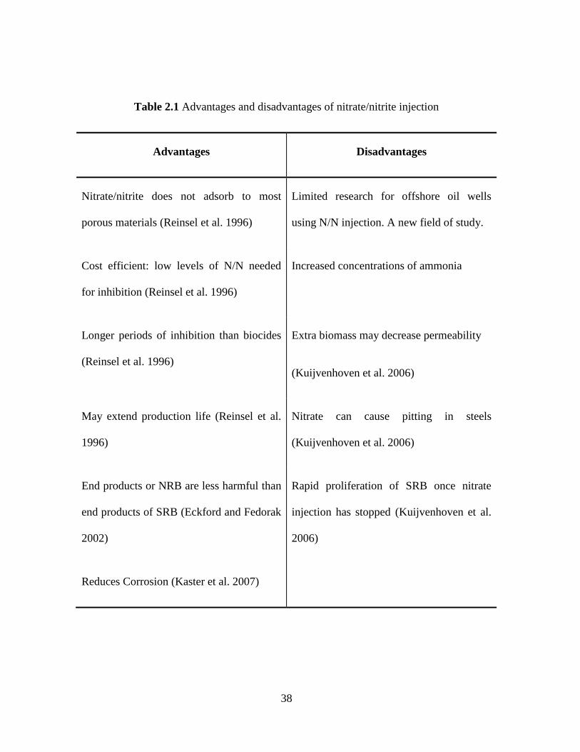
38
Table 2.1 Advantages and disadvantages of nitrate/nitrite injection
Advantages Disadvantages
Nitrate/nitrite does not adsorb to most
porous materials (Reinsel et al. 1996)
Limited research for offshore oil wells
using N/N injection. A new field of study.
Cost efficient: low levels of N/N needed
for inhibition (Reinsel et al. 1996)
Increased concentrations of ammonia
Longer periods of inhibition than biocides
(Reinsel et al. 1996)
Extra biomass may decrease permeability
(Kuijvenhoven et al. 2006)
May extend production life (Reinsel et al.
1996)
Nitrate can cause pitting in steels
(Kuijvenhoven et al. 2006)
End products or NRB are less harmful than
end products of SRB (Eckford and Fedorak
2002)
Rapid proliferation of SRB once nitrate
injection has stopped (Kuijvenhoven et al.
2006)
Reduces Corrosion (Kaster et al. 2007)
39
2.3.2.3 Field applications
Kaster et al. (2007) studied the effects of nitrate/nitrite injection on sulfide production in the
Ekofisk field of the Norwegian sector in the North Sea. The field is a deep reservoir where
thermophilic SRB contribute to most of the sulfide production at 60°C near injection wells. It was
hypothesized that nitrite may be preferable for some high-temperature oil fields as it reacts directly
with sulfide. Nitrite inhibits DSR, which is an enzyme that catalyzes the reduction of sulfite to
sulfide. DSR has a strong affinity for nitrite by reducing it to ammonia, thus inhibiting DSR usage
by SRB (Kaster et al. 2007).
Experiments were conducted in an upflow bioreactor filled with Ekofisk Chalk inoculated with 0.1
ml/min of produced waters that had 0.1 mM of phosphate added to it. Two strains of bacteria (NS-
tSRB1 and NS-tSRB2) were studied with differing additions of organic acids. Nitrate/nitrite
injection did not take place until the bioreactor was stabilized with a sulfide concentration of 4-6
mM. It was observed that 2 and 10 mM of nitrate addition had little to no effect on sulfide
production for both NS- tSRB1 and NS- tSRB2. It is also noted that no nitrite was detected after
nitrate addition. Conversely, nitrite addition strongly inhibited sulfate reduction. 0.25 mM of
nitrite inhibited sulfate reduction permanently for NS- tSRB1, and for 1100 hrs for NS- tSRB2.
Results suggested that the type of organic acid used as an electron donor has an influence on the
survival of SRB during nitrate/nitrite injection.
Nitrate proved effective for two high-temperature oil fields in the Norwegian and Danish sectors
of the North Sea, Veslefikk and Halfdan fields, respectively (Kaster et al. 2007). Thermophilic
nitrate reducing bacteria (tNRB) must have converted nitrate into nitrite, thus inhibiting tSRB of
organics derived from oil in these cases. This was not observed in Ekofisk, suggesting that tNRB
40
are not present, or they could have been lost during enrichment with sulfate. Overall, 0.25 mM of
added nitrite effectively controlled souring for the North Sea field.
Bodtker et al. (2008) studied the long-term effects of nitrate injection on SRB activity, corrosion
rates, and bacterial community composition of the Veslefrikk and Gullfacks fields in the North Sea.
Biocides were traditionally used to mitigate sulfide production, but nitrate injection was accepted
as a more effective and environmentally safe method. Nitrate injection enriches the NRB, which
outcompete SRB due to the more favorable energy potential of nitrate reduction compared to
sulfate reduction. Overall, SRB are inhibited and corrosion rates decrease.
The Veslefrikk field experienced a 50–fold reduction in H2S production (SRB activity) and a
reduction in the corrosion rate immediately following continuous nitrate injection (1999-2001).
NR-SOB formed major populations, and three of the four major populations that were observed
before biocide treatment were no longer observed after 1 year with nitrate treatment. Sulfate
reduction rate is given as the amount of H2S produced per biofilm area per day, and remained at
≤0.3 μg H2S/cm2/day during nitrate treatment. A decrease in corrosion rate was also observed. The
Gullfaks field experienced an initial decrease in number and activity of SRB, and an increase in
the numbers of NRB. The SRB activity has remained low during the 8 years of nitrate injection at
≤0.9 μg H2S/cm2/day. There was also a significant reduction in corrosion rate of up to 40%. The
long-term nitrate injection treatment has provided efficient inhibition of SRB activity and a
decrease in corrosion rate. This has enabled a stable NRB dominated biofilm to develop. Nitrate
injection has proven to be an effective and safe way to mitigate biogenic sulfide production in
offshore oil wells practicing secondary oil recovery.
41
2.4 Biosurfactants and Reservoir Souring Control
2.4.1 Introduction of biosurfactants
2.4.1.1 Definition and classification of biosurfactants
Biosurfactants are extracellular, surface-active substances synthesized by living cells. They have
the properties of reducing surface tension, stabilizing emulsions, promoting foaming and are
generally non-toxic and biodegradable (Saharan et al. 2011). Biosurfactants are amphipathic
molecules with hydrophilic as well as hydrophobic groups which impart their functional properties.
They are high value products and efficient replacers of chemically synthesized surface-active
agents due to their diversity, specific activity, ease of application, biodegradability and
performance in extreme environments (Marchant and Banat 2012).
Biosurfactants are of two types, low molecular weight biosurfactants which are generally
glycolipids and lipopeptides and high molecular weight biosurfactants, also called bioemulsifiers,
which are generally lipopolysaccharides, lipoproteins or a combination of these (Christofi and
Ivshina 2002). Generally, the former group effectively reduced the surface/interfacial tension,
while the latter group tended to stabilize oil-in-water emulsions but did not reduce much surface
tension (Rosenberg and Ron 1999). Most known biosurfactants are glycolipids. They are
carbohydrates in combination with long-chain aliphatic acids or hydroxyaliphatic acids. Among
the glycolipids, the best known are rhamnolipids, trehalolipids, and sophorolipids. Figure 2.2
shows the structure of the three classes of glycolipid biosurfactants.

42
Figure 2.2 Structures of glycolipid biosurfactants (A) Rhamnolipid from Pseudomonas aeruinosa.
(B) Trehalolipid from Rhodococcus erythropolis. (C) Sophorolipid from Torulopsis bombicola.
Source: (Desai and Banat 1997)

43
In addition, several bacteria and yeast produce large quantities of fatty acid and phospholipids
surfactants during growth on n-alkanes (Santos et al. 2016). Emulsan, liposan, mannoprotein and
polysaccharide-protein complexes are known to be the best-studied polymeric biosurfactants
(Desai and Banat 1997).
2.4.1.2 Functions of biosurfactants
The basic function of a biosurfactant is to reduce the surface or interfacial tension between two
mediums (e.g. water and air or oil and water). Biosurfactants are amphipathic meaning they have
hydrophobic and hydrophilic parts, as explained earlier in the report. Biosurfactants can also be
used to form stable emulsions, and for solubilization and desorption of hydrophobic compounds
from particle surfaces (Pacwa-Płociniczak et al. 2011). These functions can shape the ecological
niches for bacteria within microbial communities by altering the availability of substrates for
growth and by allowing bacteria to enter into liquid phases having different levels of
hydrophobicity.
The most significant role of microbial surfactants is documented for adhesion of the cells to the
interfaces. Adhesion is shown to be a prerequisite for the growth of Acinetobacter calcoaceticus
RAG-1 on liquid hydrocarbons under two conditions: low cell density and limited agitation
(Rosenberg and Ron 1999). Besides, biosurfactants can enhance growth on bound substrates by
desorbing them from surfaces or by increasing their apparent water solubility (Rosenberg and Ron
1999). Surfactants that lower interfacial tension are particularly effective in mobilizing bound
hydrophobic molecules and making them available for biodegradation. Another important
characteristic of the biosurfactants is that above the CMC, they form micelles (stable aggregates of
10 to 200 molecules), which brings about a sudden variation in the relation between the
44
concentration and the surface tension of the solution that can increase the solubility of
hydrophobic organics (Cho et al. 2002).
According to Puchkov et al. (2002), the biosurfactants could also be an evolutionary defense
strategy of microbe as evidenced by high mycocidal activity of the mycocidal complex secreted by
C. humicola. Similar analogy can be made for the lipopeptides biosurfactant producing strains of B.
subtilis. The lipopeptide (antibiotic) would have a strong influence on the survival of B. subtilis in
its natural habitat, the soil and the rhizosphere (Nielsen and Sorensen 2003).
2.4.1.3 Advantages of biosurfactants
Biosurfactants have gained considerable attention in recent years due to their unique properties
versus their synthetic counterparts.
Selectivity for specific interfaces
Biological molecules have been found to show more specificity as compared to the chemically
synthesized materials. Microbial surfactants show a specificity due to the presence of specific
functional groups, allowing specificity in the detoxification of specific pollutants, and activity
under conditions of extreme temperatures, pH and salinity (De Cassia et al. 2014). For example,
specificity of emulsan towards a mixture of aliphatic and aromatic hydrocarbons (Bach et al.
2003)and that of solubilizing factor of Pseudomonas PG1 towards pristine (Sekhon et al. 2011).
Resistance to environmental changes
Biosurfactants have chemical diversity which results in a wide variety of physico-chemical
properties suited for applications (De Cassia et al. 2014). For example, biosurfactants were noted
for their promising application enhanced oil recovery to remove and recover the residual oil.
45
Biosurfactant solutions at 0.01% and 0.05% produced by Pseudomonas sp. 2B removed 89% and
92% of the sand oil, respectively, while the synthetic surfactant, sodium dodecyl sulfate, removed
only 63% of the contaminated oil (Aparna et al. 2012). The extracted biosurfactant product was a
glycolipid type and was found to be stable over a pH range of 4 to 10, a temperature range of 4 to
121° C and salinity from 0 to 15% NaCl.
Diversity of biosurfactants
Biosurfactants have enormous diversity, and could be produced from various sources, such as
industrial wastes with simple and inexpensive procedures (Muthusamy et al. 2008). A single isolate
often generates chemical variations of the same surfactant, resulting in the production of a
surfactant mixture with an associated characteristic surface (Bodour et al. 2003). In fact, even
small variations in the structure of a surfactant can significantly affect its functions and potential
industrial applications (Symmank et al. 2002).
Low toxicity and easy biodegradation
Another main advantage of biosurfactants is their lower toxicity than traditional surfactants.
Sometimes the process used in the production of synthetic surfactants could release toxic
byproducts, as in the case of sodium dodecylbenzene sulfonate (SDS) production, where corrosive
and toxic chemicals are used and quite frequently discharged as pollutants. When focusing on
environmentally sound products, potentially toxic and sparing biodegradable synthetic surfactants
could be replaced by biosurfactants, which are naturally produced and nontoxic. Furthermore,
microbial surfactants like all natural products are susceptible to degradations by microorganisms in
water and soil (Khan and Butt 2016). Hence, concerns regarding the negative environmental risks
after the application are ignorable.
46
2.4.1.4 Screening of biosurfactant producing microorganisms
Oil reservoirs have a great potential in producing microorganisms that may produce biosurfactants
(Anandaraj and Thivakaran 2010). Currently, the understanding of biosurfactants as a class of
molecules remains limited. This is partially because the present body of knowledge has been
developed around a relatively small number of well-characterized biosurfactants. Contributing to
this is the lack of a concerted effort to perform a comprehensive screening for biosurfactants and
the microorganisms that produce them. There is a very limited amount of commercially available
biosurfactants (e.g. surfactin, sophorolipids and rhamnolipids) (Santos et al. 2016; Walter et al.
2010). The development of a variety of new biosurfactants by new strains provides a solution in
overcoming the economic obstacles of the production of biosurfactants. Therefore, efforts in the
discovery of new biosurfactant producing microbes must be made by applying a broad range of
different screening methods. The principal aim in screening for new biosurfactants is finding new
structures with strong interfacial activity, low critical micelle concentration (CMC), high emulsion
capacity, good solubility and activity in a broad pH range. Commercial viable biosurfactants have
to be economically competitive. Additionally, another reason in screening is discovering good
production strains with high yields (Walter et al. 2010).
There are eight different screening methods that have been reported as criteria to screen
biosurfactant producing microbes. Namely hemolytic assay (Yoshida et al. 2015), bacterial
adhesion to hydrocarbons (BATH) assay (Volchenko et al. 2007), emulsification assay (Afshar et
al. 2008), drop collapse assay, oil spreading assay (Nwaguma et al. 2016), du-nouy-ring method,
microplate assay (Vaux and Cottingham 2001) and CTAB agar plate. Each of these methods has
their own advantages and disadvantages. Surface tension measurement is the best way to discover
biosurfactant producing microorganisms in a sample (Bodour et al. 2003; Thavasi et al. 2011). The
47
Du-Nouy-Ring method which is a variation of surface tension measurement is the most common
method used for this type of measurement. One limitation is that it needs to use a tensiometer
which may not be readily available in a microbiology laboratory.
2.4.1.5 Biosurfactant production
There are numerous factors that affect biosurfactant production by microorganisms.
Microorganisms depend on vital nutrients and a suitable growth environment to sustain life and
thrive. Biosurfactant production is affected if the microorganisms are not getting proper nutrition
or inhabit an area with poor growth conditions. The nutrients will be discussed as culture medium
composition and growth conditions that will be discussed as environmental factors, both of which
have significant influence on the biosurfactant production.
The use of different carbon sources changes the structure of the biosurfactants produced and,
consequently, its properties. These changes may be welcomed when some properties are sought for
a particular application (Amaral et al. 2010). Also, the composition and characteristics of
biosurfactants are influenced by the nature of the nitrogen source as well as the presence of iron,
magnesium, manganese, phosphorus and sulfur.
Main carbon sources come from 3 categories: carbohydrates, hydrocarbons and vegetable oils.
Water-soluble carbon sources such as glycerol, glucose, mannitol and ethanol were all used for
rhamnolipid production by Pseudomonas spp. However, biosurfactant production was inferior to
that obtained with water-immiscible substrates such as n-alkane or olive oil (Thampayak et al.
2008). Similarly, different nitrogen compounds have been used for the production of
biosurfactants, such as urea, peptone, yeast extract, ammonium sulfate, ammonium nitrate, sodium
nitrate, meat extract and malt extract, etc. Different elements, such as iron and manganese, are also
48
reported to affect the yield of biosurfactants, for example, the addition of iron and manganese to
the culture medium increased the production of biosurfactant by Bacillus subtilis (Gudiña et al.
2015). The ratios of different elements such as C:N, C:P, C:Fe or C:Mg mare main factors
affecting biosurfactant production (Batista et al. 2010).
Environmental factors are extremely important in the yield and characteristics of the biosurfactants
produced. In order to obtain large quantities of biosurfactant it is necessary to optimize the process
conditions because the biosurfactant production may be induced by changes in pH, temperature,
aeration or agitation speed. For instance, the effect of pH in the biosurfactant production by
Virgibacillus salarius was investigated in the pH values varying from 5 to 12. The biosurfactant
generation increased with the increase of medium pH, but maximum biosurfactant production was
obtained at pH 9 for the strain V. salarius (KSA-T) (Elazzazy et al. 2015). The strain was found to
be moderately thermophilic and the maximum biosurfactant production was observed in the
temperature range of 45–50 °C. A lower culture temperature might make microoragnisms
hibernate partially, and its enzyme system for biosurfactant production couldn‘t be activated
completely. On the other hand, a higher culture temperature may have an adverse effect on the
nucleic acid and the enzyme system of the strain. Aeration and agitation rates are also important
factors that influence the production of biosurfactants, since they facilitate the oxygen transfer
from the gas phase to the aqueous phase and it may also be linked to the physiological function of
microbial emulsifiers.
2.4.1.6 Biosurfactant characterization
Physical-chemical parameters
49
There are several physical-chemical parameters that can be applied for the characterization of
biosurfactants. Surface tension, interfacial tension and CMC are quantitatively measured in
biosurfactant production. Whether these three factors are looked at individually or collectively,
they can indicate if a given surfactant will be successful in its intended application.
The surface tension of a liquid is the measurement of the interfacial free energy per unit area of the
boundary between the liquid and the air above it (Rosen 1989). In a liquid, intermolecular forces
(van der Waals) act upon each molecule in every direction by other surrounding molecules. At the
surface of the liquid (in contact with air), however, there exists a difference in these forces. At the
surface the molecules interact more strongly with the molecules in the interior of the liquid than
they do with the widely spaced gas molecules above it (Rosen 1989). Therefore, the surface
molecules are contracted or drawn into the liquid and a surface tension develops.
Surface tension is most commonly measured by a tensiometer. The surface tension is measured by
the force that is required to remove the ring from the liquid or to pull the plate a given distance up
while still remaining in the fluid. Surface tensions are equivalent to a force per unit length and are
commonly given in units of mN/m or dyn/cm. Biosurfactants are characterized by the measure of
their effectiveness and efficiency at reducing surface tensions. Effectiveness is the measure of the
minimum value to which surface tension can be lowered, while efficiency is measured by the
concentration of a surfactant required to produce some significant reduction in the surface tension
of water (Czajka et al. 2015). The most effective biosurfactants are those that have a short,
branched hydrophobic moiety. The more efficient biosurfactants have a linear, long hydrophobic
moiety.
50
Interfacial tension is very similar to surface tension except that a liquid is in contact with another
liquid phase rather than air. At the interface between the condensed phases, the dissimilar
molecules in the adjacent layers facing each other across the interface also have potential energies
different from those in their respective phases (Rosen 1989). Therefore, a tension develops at the
boundary between the two liquids. The less miscible the two liquids are, the greater the resultant
interfacial tensions will be. On the other hand, if the two liquids miscible well with each other,
there will be no interfacial tension between them.
A unique phenomenon of surfactants is the self-assembly of molecules into dynamic clusters
called micelles (Shchekin et al. 2016). Each singular biosurfactant molecule is termed a monomer.
The monomers arrange themselves into micelles once a CMC is reached in the solution (Figure
2.3). Micelles can be spherical, cylindrical, lamellar or vesicular depending upon several factors
such as temperature, hydrocarbon chain length, etc. In aqueous media, the surfactant molecules are
oriented, in all of these structures, with their polar heads toward the aqueous phase and their
hydrophobic tail groups away from it (Rosen 1989). In a non-polar media, the structure of the
micelle is reversed with the hydrophobic tails pointing out into the media.
CMCs are different for each type of surfactant. The CMC is determined by measuring the
reduction in surface tension produced by serially diluting the solution. The point at which the
surface tension first begins to rise is the critical micelle dilution (CMD). The CMC is a crucial
point because surface tension will decrease continually as surfactants are added until the CMC is
reached. After this point, no further significant surface tension reductions will take place
regardless of how much more surfactant is added. CMC is altered by metabolic acids or alcohols
that are produced by organisms and subsequently react with biosurfactants and by ionic species
such as salts (Palladino and Ragone 2011; Sidim and Acar 2013).
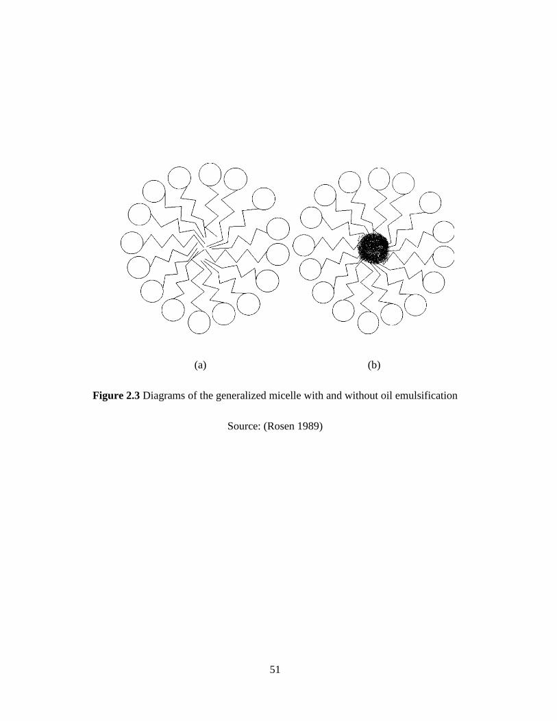
51
(a) (b)
Figure 2.3 Diagrams of the generalized micelle with and without oil emulsification
Source: (Rosen 1989)
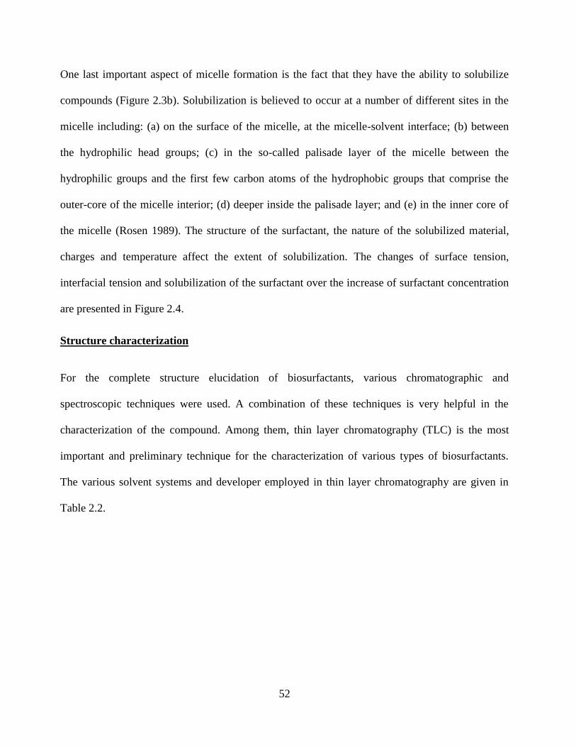
52
One last important aspect of micelle formation is the fact that they have the ability to solubilize
compounds (Figure 2.3b). Solubilization is believed to occur at a number of different sites in the
micelle including: (a) on the surface of the micelle, at the micelle-solvent interface; (b) between
the hydrophilic head groups; (c) in the so-called palisade layer of the micelle between the
hydrophilic groups and the first few carbon atoms of the hydrophobic groups that comprise the
outer-core of the micelle interior; (d) deeper inside the palisade layer; and (e) in the inner core of
the micelle (Rosen 1989). The structure of the surfactant, the nature of the solubilized material,
charges and temperature affect the extent of solubilization. The changes of surface tension,
interfacial tension and solubilization of the surfactant over the increase of surfactant concentration
are presented in Figure 2.4.
Structure characterization
For the complete structure elucidation of biosurfactants, various chromatographic and
spectroscopic techniques were used. A combination of these techniques is very helpful in the
characterization of the compound. Among them, thin layer chromatography (TLC) is the most
important and preliminary technique for the characterization of various types of biosurfactants.
The various solvent systems and developer employed in thin layer chromatography are given in
Table 2.2.
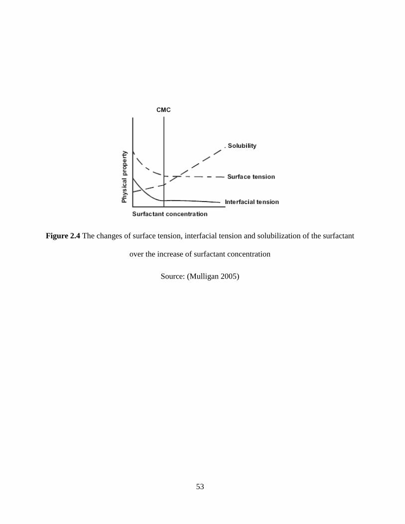
53
Figure 2.4 The changes of surface tension, interfacial tension and solubilization of the surfactant
over the increase of surfactant concentration
Source: (Mulligan 2005)
54
Table 2.2 Various solvent systems and developer employed in TLC method
Biosurfactant
type
Solvent System
Identification of
functional group
Developer References
Sophorolipid Chloroform:
Methanol: Water
(65:15:2)
Diacylated
lactone
Not mentioned Cavalero and
Cooper
(2003)
Protein-lipid-
polysaccharide
complex
(i) Hexane: Isopropyl
ether: Acetic acid
(15:10:1)
(ii) Chloroform:
Methanol: Water
(65:24:4)
Protein-40.2%,
Carbohydrate-
24.0% Lipid-
19.5%
Ninhydrin-free amino
groups Iodine vapors-
Lipids α-
napthol-H2SO4 - Sugar
Sarubbo et al.
(2007)
Glycolipid Chloroform:
Methanol: Water
(65:15:2)
Not mentioned α-napthol Folch et al.
(1957)
Sophorolipid Chloroform:
Methanol (8:2)
Not mentioned Anthrone/sulfuric acid Konishi et al.
(2008)
Sophorolipid Chloroform:
Methanol: Acetic
acid (65:25:4)
Not mentioned Iodine vapour and
Molisch reagent
Thaniyavarn
et al. (2008)
Source: Modified from Bhardwaj et al. (2013)
55
Additionally, GC-MS analysis for the confirmation of fatty acids, HPLC chromatogram for lipid
structure elucidation, and proton nuclear magnetic resonance (1H-NMR) spectrum as well as
carbon nuclear magnetic resonance (13
C-NMR) spectrum for functional groups of biosurfactants
were also used in the structure elucidation of biosurfactants (Kim et al. 1999; Konishi et al. 2008;
Thaniyavarn et al. 2008). For instance, in the structure elucidation of sophorolipids produced by
the Candida bombicola, hydroxyl-acid methyl esters were liberated by the methanolysis and were
confirmed by GC-MS. The 16-hydroxydecanoic acid was confirmed by comparing the
fragmentation pattern with the standard 16-hydroxyhexadecanoic acid purchased from Sigma
Aldrich. Also, an isomer 15-hydroxyhexadecanoic acid was confirmed because of the availability
of the same fragmentation pattern in the library (Cavalero and Cooper 2003).
2.4.2 Biosurfactant producers in reservoirs
Biosurfactant or surface-active compounds are a heterogeneous group of surface active molecules
with both a lipophilic and hydrophilic moieties produced by microorganisms, which either adhere
to cell surface or are excreted extracellulary in the growth medium (Santos et al. 2016). These
surface active molecules reduce surface tension at air-water interfaces and interfacial tension in
both aqueous solutions and hydrocarbon mixtures (Batista et al. 2006). Several types of
biosurfactants have been isolated and characterized, including glycolipids, phospholipids,
lipopeptides, natural lipids, fatty acids, lipopolysacharides and other fully characterized. The
majority of known biosurfactants are synthesized by microorganisms grown on water immiscible
hydrocarbons, but some have been produced on such water-soluble substrates as glucose, glycerol
and ethanol (Tabatabaee et al. 2005). Chemically-synthesized surfactants have been used in the oil
industry to aid clean up of oil spills, as well as to enhance oil recovery from oil reservoirs. These
compounds are not biodegradable and can be toxic to the environment. Biosurfactants have special
56
advantages over their commercially manufactured counterparts because of their lower toxicity,
biodegradable nature, and effectiveness at extreme temperature, pH, salinity and ease of synthesis.
Due to the presence of hydrophilic and lipophilic moieties in their structure, biosurfactants are able
to partition at the oil-air or the oil-water interfaces and to lower surface or interfacial tension,
respectively. They are potential candidates for much commercial application in the oil recovery
industries (Bordoloi and Konwar 2008; Shibulal et al. 2014).
Oil reservoirs contain diverse and metabolically active microbial communities. Knowledge of the
microbial ecology of oil reservoirs can be used to stimulate the beneficial activities of
microorganisms to enhance oil recovery. Microorganisms produce a variety of products that are
potentially useful for enhancing oil production. Several products have become commercially
viable technologies such as paraffin control. Several aerobic and anaerobic thermophiles tolerant
of pressure and moderate salinity have been isolated which are able to mobilize crude oil in the
laboratory. The most common biosurfactants used in microbially enhanced oil recovery (MEOR)
are lipopeptides produced by Bacillus and some Pseudomonas spp., glycolipids (rhamnolipids)
produced by Pseudomonas sp., and trehalose lipids produced by Rhodococcus sp. (Amani et al.
2010; Cai et al. 2014; Youssef et al. 2004). Lipopeptides and rhamnolipid biosurfactants lower
interfacial tension between the hydrocarbon (crude oil or pure hydrocarbons) and aqueous phases
to values of 0.1 mN/m or lower (Nguyen et al. 2008; Wang et al. 2007).
Biosurfactant producers isolated from a number of oil reservoirs are effective in mobilizing
residual oil from a variety of laboratory test systems. Table 2.3 summarized the biosurfactant
producers reported in the oil reservoir and related lab-scale experiments. In the oil and gas industry,
NRB induced biosurfactants have been initially investigated to aid enhanced oil recovery (EOR).
Fallon et al. (2010) have confirmed that biosurfactants can be naturally derived from NRB. Pure
57
NRB strains from ATCC (an independent, private, nonprofit biological resource center and
research organization) have been purchased and their effects on EOR haven been determined. To
date, there has been little relevant research related to biosurfactant-aided reservoir souring control,
though its application to reservoir souring control is theoretically possible.
2.4.3 Anti-souring effects of biosurfactants
2.4.3.1 Role of biosurfactants in oil reservoir
The majority of the world‘s energy is still from nonrenewable fossil fuel source. The average
recovery of the total oil from mature oilfields around the world is somewhere between 20% and 40%
by currently used methods (Muggeridge et al. 2014). Traditional primary and secondary
production methods typically recover one-third of oil in place, leaving two thirds behind.
Improving oil recovery to release more trapped oil in oil fields is essential options for increased
production rates. More advanced methods are desired and referred to as EOR. In Canada, for
example, 70% of oil discovered was found during the earliest 20% of drilling. About 130 billion
barrels have been produced to date and up to another 170 billion barrels are considered a long-term
target for advanced EOR technology (Bott 1999). Given the declining reserves and the low
probability of locating significant new fields, industries sought additional oil in old reservoirs,
making Canada a proving ground for EOR techniques.
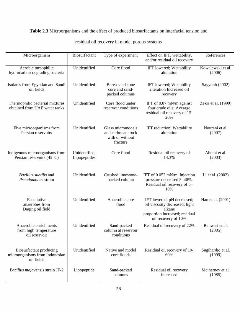
58
Table 2.3 Microorganisms and the effect of produced biosurfactants on interfacial tension and
residual oil recovery in model porous systems
Microorganism Biosurfactant Type of experiment Effect on IFT, wettability, and/or residual oil recovery
References
Aerobic mesophilic hydrocarbon-degrading bacteria
Unidentified Core flood IFT lowered; Wettability alteration
Kowalewski et al. (2006)
Isolates from Egyptian and Saudi oil fields
Unidentified Berea sandstone core and sand-
packed columns
IFT lowered; Wettability alteration Increased oil
recovery
Sayyouh (2002)
Thermophilic bacterial mixtures obtained from UAE water tanks
Unidentified Core flood under reservoir conditions
IFT of 0.07 mN/m against four crude oils; Average
residual oil recovery of 15-20%
Zekri et al. (1999)
Five microorganisms from Persian reservoirs
Unidentified Glass micromodels and carbonate rock
with or without fracture
IFT reduction; Wettability alteration
Nourani et al. (2007)
Indigenous microorganisms from Persian reservoirs (45 C)
Unidentified, Lipopeptides
Core flood Residual oil recovery of 14.3%
Abtahi et al. (2003)
Bacillus subtilis and Pseudomonas strain
Unidentified Crushed limestone-packed column
IFT of 0.052 mN/m, Injection pressure decreased 5–40%, Residual oil recovery of 5–
10%
Li et al. (2002)
Facultative anaerobes from Daqing oil field
Unidentified Anaerobic core flood
IFT lowered; pH decreased; oil viscosity decreased; light
alkane proportion increased; residual
oil recovery of 10%
Han et al. (2001)
Anaerobic enrichments from high temperature
oil reservoir
Unidentified Sand-packed column at reservoir
conditions
Residual oil recovery of 22% Banwari et al. (2005)
Biosurfactant producing microorganisms from Indonesian
oil fields
Unidentified Native and model core floods
Residual oil recovery of 10-60%
Sugihardjo et al. (1999)
Bacillus mojavensis strain JF-2 Lipopeptide Sand-packed columns
Residual oil recovery increased
Mcinerney et al. (1985)
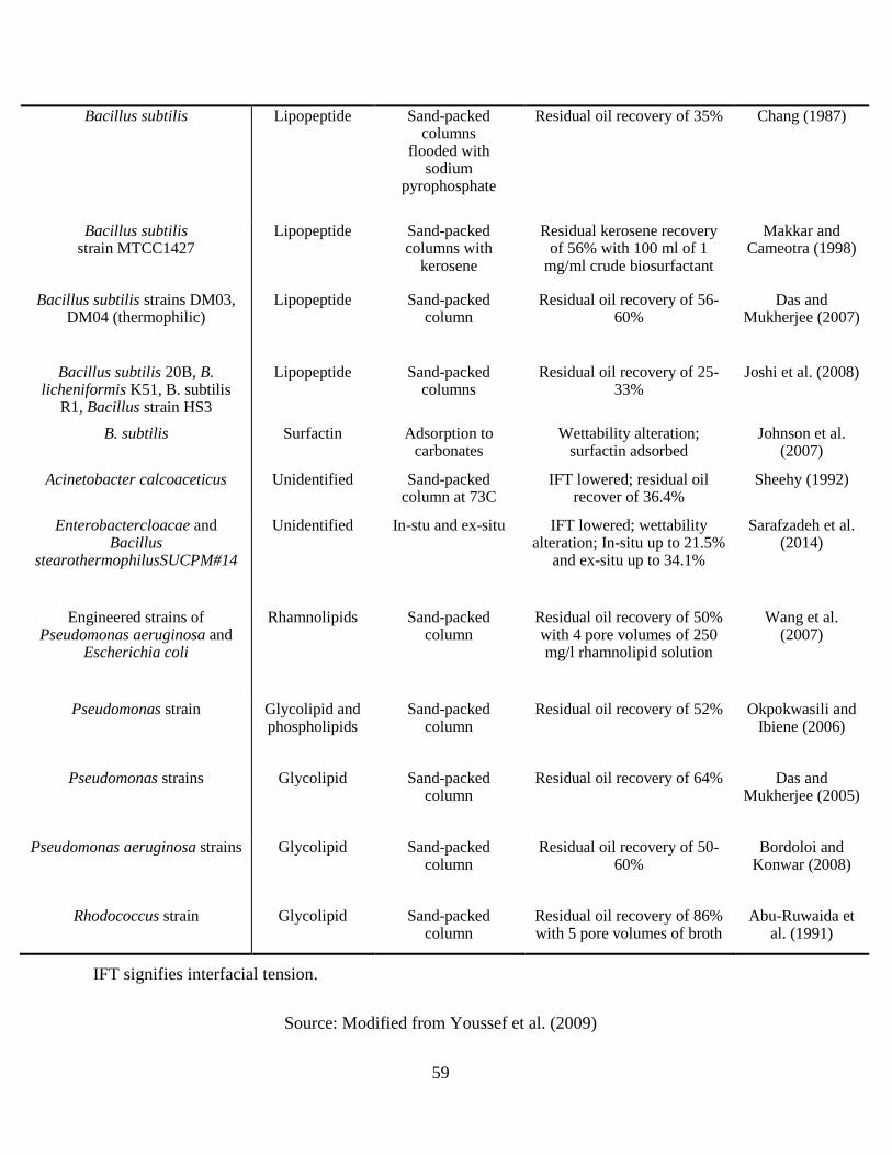
59
Bacillus subtilis Lipopeptide Sand-packed columns
flooded with sodium
pyrophosphate
Residual oil recovery of 35% Chang (1987)
Bacillus subtilis strain MTCC1427
Lipopeptide Sand-packed columns with
kerosene
Residual kerosene recovery of 56% with 100 ml of 1
mg/ml crude biosurfactant
Makkar and Cameotra (1998)
Bacillus subtilis strains DM03, DM04 (thermophilic)
Lipopeptide Sand-packed column
Residual oil recovery of 56-60%
Das and Mukherjee (2007)
Bacillus subtilis 20B, B. licheniformis K51, B. subtilis
R1, Bacillus strain HS3
Lipopeptide Sand-packed columns
Residual oil recovery of 25-33%
Joshi et al. (2008)
B. subtilis Surfactin Adsorption to carbonates
Wettability alteration; surfactin adsorbed
Johnson et al. (2007)
Acinetobacter calcoaceticus Unidentified Sand-packed column at 73C
IFT lowered; residual oil recover of 36.4%
Sheehy (1992)
Enterobactercloacae and Bacillus
stearothermophilusSUCPM#14
Unidentified In-stu and ex-situ IFT lowered; wettability alteration; In-situ up to 21.5%
and ex-situ up to 34.1%
Sarafzadeh et al. (2014)
Engineered strains of Pseudomonas aeruginosa and
Escherichia coli
Rhamnolipids Sand-packed column
Residual oil recovery of 50% with 4 pore volumes of 250 mg/l rhamnolipid solution
Wang et al. (2007)
Pseudomonas strain Glycolipid and phospholipids
Sand-packed column
Residual oil recovery of 52% Okpokwasili and Ibiene (2006)
Pseudomonas strains Glycolipid Sand-packed column
Residual oil recovery of 64% Das and Mukherjee (2005)
Pseudomonas aeruginosa strains Glycolipid Sand-packed column
Residual oil recovery of 50-60%
Bordoloi and Konwar (2008)
Rhodococcus strain Glycolipid Sand-packed column
Residual oil recovery of 86% with 5 pore volumes of broth
Abu-Ruwaida et al. (1991)
IFT signifies interfacial tension.
Source: Modified from Youssef et al. (2009)
60
Surfactant EOR represents one of the most promising advanced methods to recover a substantial
proportion of the residual oil. In this technology, an aqueous surfactant formulation is injected into
a mature oil reservoir. Where this solution contacts the small blobs of oil trapped in the pores of
the reservoir rock, it dramatically reduces the interfacial tension and mobilizes this trapped oil.
This reduces the capillary forces preventing oil from moving through rock pores (Figure 2.5). The
biosurfactants act as a separating agent making the oil move more freely away from rocks and
crevices so that it may travel easily out of the well. Biosurfactants can also bind tightly to the oil-
water interface and form emulsion. This stabilizes the desorbed oil in water and allows removal of
oil along with the injection water.
Indigenous or injected biosurfactant producing microorganisms are exploited in oil recovery in oil-
producing wells. MEOR is often implemented by direct injection of nutrients with microbes that
are able of producing desired products for mobilization of oil, by injection of a consortium or
specific microorganisms or by injection of the purified microbial products (e.g., biosurfactants).
These processes are followed by reservoir repressurization, interfacial reduction of tension/oil
viscosity and selective plugging of the most permeable zones to move the additional oil to the
producing wells. The main roles of biosurfactants in oil reservoirs thus lay in the enhanced oil
exploitation activities.
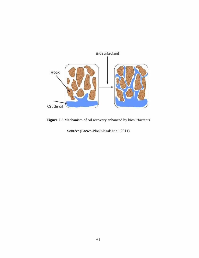
61
Figure 2.5 Mechanism of oil recovery enhanced by biosurfactants
Source: (Pacwa-Płociniczak et al. 2011)
62
2.4.3.2 Anti-souring effects of biosurfactants
Oil production by water injection often results in increased sulfide levels (souring), because SRB
couple the oxidation of degradable oil organics to the reduction of sulfate to sulfide. High levels of
sulfide represent increased toxicity and corrosion risk. Souring also negatively affects the quality
of gas stored in subsurface reservoirs, because the H2S must be removed prior to distribution to
consumers. Souring can be prevented or reversed by the periodic, batchwise application of
biocides or by the continuous, field-wide injection of nitrate. Application of nitrate to constrain
souring has been especially successful for seawater injection in offshore operations.
Continuous injection of nitrate or oxygen has been reported to result in MEOR by in situ
production of biosurfactants or of partially reduced, highly reactive intermediates, or by removal
of oil-dissolved sulfur by resident bacteria. This MEOR mechanism is critical, not only for
improving conventional oil production, but also for the reservoir control in a long run. Hui et al.
(2012) evaluated the microbial community structure and functionally distinct groups in three kinds
of produced water samples from the shallow, mesothermic and low-salinity Daqing oil reservoir
using both culture-dependent and culture-independent methods. They found some isolated strains
were simultaneously detected in different functional groups, and even more, from different
produced water samples. The isolates affiliated to Pseudomonas stutzeri PTG4-15 (DP26, BP39,
and PW5) were identified as NRB, biosurfactant producing bacteria, and polymer-producing
bacteria. Acinetobacter haemolyticus BA56 represented by DT20 and BP25 was functionally
related to both nitrate reducing and biosurfactant producing bacteria. In their study, the widespread
occurrence of close relatives to Pseudomonas putida BBAL5-01 within varying functional groups
(NRB, fermentative bacteria, and biosurfactants and biopolymer producing bacteria, and also,
polyacrylamide degrading bacteria) was identified. It indicates that it may be a common
63
indigenous bacterium in petroleum reservoirs and may have a significant impact on
biogeochemical cycles in oil reservoirs. Thus, anaerobic, indigenous NRB have the potential to
produce specific biosurfactants in the reservoir.
NRB and SRB are both oil-degrading bacteria and they existed as competitors in the reservoir
system. As oil-degrading bacteria, they can utilize only a limited group of hydrocarbons, so
bacteria attached and growing on an oil droplet become nutrient-starved once this group of
hydrocarbons is depleted. If the biosurfactant is cell-bound it can cause the microbial cell surface
to become more hydrophobic, depending on its orientation. For example, the cell surface
hydrophobicity of Pseudomonas aeruginosa was greatly increased by the presence of cell-bound
rhamnolipid (Sotirova et al. 2009). In addition, Acinetobacter strains produced cell-bound
emulsifier to reduce the cell-surface hydrophobicity was also reported (Patil and Chopade 2001;
Vasileva-Tonkova et al. 2011). These data suggest that microorganisms can use their
biosurfactants to regulate their cell-surface properties to attach or detach from surfaces of
substrates according to need. As indicated by A. calcoaceticus RAG-1 growing on crude oil,
RAG-1 utilized only relatively long-chain n-alkanes for growth after attached to the to the oil
droplet (Rosenberg 1993). Afer the depletion of these compounds, RAG-1 would detach the the
starved cells from the substrate although aromatics and cyclic paraffins are still enriched. In this
process, the ‗emulsifier‘ desorbed the cell for new fresh substrate. Meanwhile, a polymeric film
on the n-alkane depleted oil droplet was formed to mark the oil droplet as ―used‖, as the
hydrophilic outer surface is hard to be attached by the bacterium again (Garrett et al. 2008). The
detachment of bacteria from the depleted oil drop enables them to move to other drops where they
metabolize the specific group of utilizable hydrocarbons. This biosurfactant-induced effect will
lead to the NRB more competitive to SRB while nitrate/nitrite was injected for simulating NRB.
64
The developed biosurfactants will exist in pore throats to stimulate the growth of targeted
beneficial microorganisms (NRB) that live in all oil and gas reservoirs and improve their ability to
utilize hydrocarbons (Ron and Rosenberg 2002), thus out‐ competing harmful SRB for basic
carbon nutrients. The SRB will be inhibited from producing new hydrogen sulfide/iron sulfide,
and the existing sulfides will be removed by bacterial degradation, resulting in effective control of
reservoir souring.
2.5 Summary
There are copious amounts of methods for the characterization of the microbial community and
only popular methodologies are discussed here. The table below (Table 2.4) summarized the
characteristics of each widely applied method for microbial characterization.
Direct detection methods such as most probable number (MPN) and direct cell counting are
traditional, straight-forward and culture dependent ways of estimating microbial biomass of the
SRB community. But, they are labor intensive since the bacteria must be closely monitored when
culturing samples and they do not provide any details on community phylogeny, diversity or
physiology when used alone (Spiegelman et al. 2005). These techniques cannot be used in samples
containing low SRB numbers since only 0.001-15% of the total number of visible cells can be
retrieved by isolation (Xu et al. 2014) and interference of debris affects the determinations of SRB
numbers in turbid samples. Direct detection need also be used together with other culture-
independent methods to characterize microbial communities since they cannot provide quantitative
information.
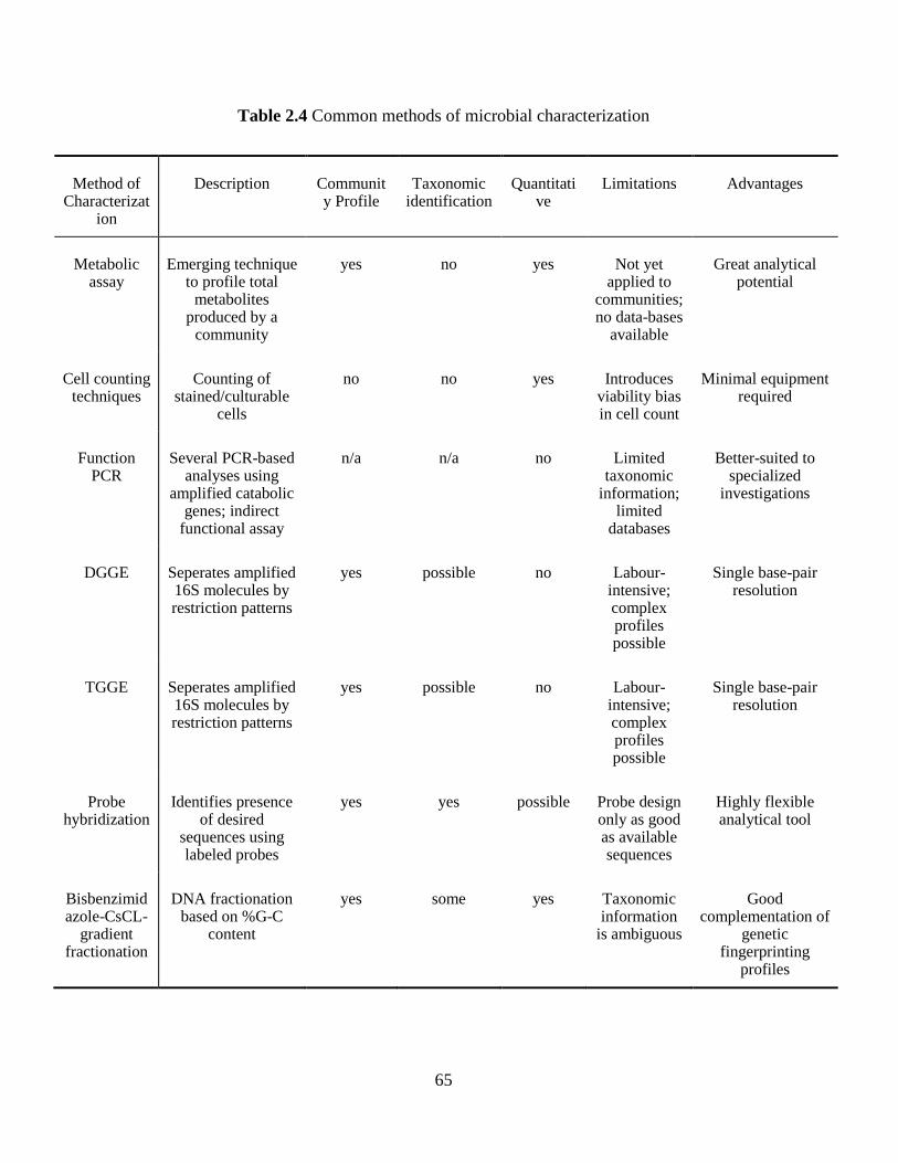
65
Table 2.4 Common methods of microbial characterization
Method of Characterizat
ion
Description Community Profile
Taxonomic identification
Quantitative
Limitations Advantages
Metabolic assay
Emerging technique to profile total
metabolites produced by a
community
yes no yes Not yet applied to
communities; no data-bases
available
Great analytical potential
Cell counting techniques
Counting of stained/culturable
cells
no no yes Introduces viability bias in cell count
Minimal equipment required
Function PCR
Several PCR-based analyses using
amplified catabolic genes; indirect
functional assay
n/a n/a no Limited taxonomic
information; limited
databases
Better-suited to specialized
investigations
DGGE Seperates amplified 16S molecules by restriction patterns
yes possible no Labour-intensive; complex profiles possible
Single base-pair resolution
TGGE Seperates amplified 16S molecules by restriction patterns
yes possible no Labour-intensive; complex profiles possible
Single base-pair resolution
Probe hybridization
Identifies presence of desired
sequences using labeled probes
yes yes possible Probe design only as good as available sequences
Highly flexible analytical tool
Bisbenzimidazole-CsCL-
gradient fractionation
DNA fractionation based on %G-C
content
yes some yes Taxonomic information
is ambiguous
Good complementation of
genetic fingerprinting
profiles

66
PLFA + FAME
Culture-independent
community profile based on the
distribution of various membrane
lipids
yes some yes Taxonomic identification
is delicate and limited
Fast and inexpensive; good
for comparing communities
Modified from Spiegelman et al. (2005)
67
Molecular methods offer the potential of determining the whole range of prokaryotic taxa without
running into the problem of selective laboratory enrichment and growth media. Also, nucleic-acid
analyses are reliable, reproducible and rapid, allowing a large number of samples to be analyzed
simultaneously. However, there are many drawbacks in the methodologies, including inconsistent
DNA recovery, inclusion of DNA from non-viable cells, kinetic biases, high cost of sampling in
complex environments and the need to develop specific primers and probes requires extensive
knowledge in sequencing. In practical operations, the storage of samples prior to processing can
bias results. Shifts in active functional groups of prokaryotes have been observed when samples
are stored aerobically or left at room temperature (Von Wintzingerode et al. 1997). Also, there are
no bias-free extraction methods currently available for DNA-based methods (Pan et al. 2010). So,
this method can be difficult to apply to extremely complex communities that produce hundreds of
bands on a DGGE profile, which become difficult to visualize individually.
As to the biochemical methods, from Bisbenzimidazole-CsCl-gradient fractionation, no
information can be obtained below the genus level from these profiles, since related, but possibly
quite different species can appear in a single % G-C peak (Holben and Harris 1995). That is, % G-
C profiles give information about the base composition of DNA, but cannot, on their own, be used
to determine the presence or abundance of a particular species. Quinone profiling generates fairly
non-specific profiles that and cannot themselves be subject to further specific analysis (such as
sequencing or probing). Thus, it was used in conjunction with other techniques as a backup or
confirmatory method to reinforce the assay results.
68
PLFA analysis is a fast, relatively simple and inexpensive method for SRB quantification.
Compared to the molecular methods used in microbial ecology, PLFAs offer a robust estimation of
three important attributes of microbial communities: viable biomass, microbial community
structure, and the physiological status (Piotrowska-Seget and Mrozik 2003). PLFA analysis has
many advantages associated with it. Most importantly, it is cheap and has the ability to
characterize large microbial communities rapidly which is optimal for our research, to develop a
cost-efficient SRB quantification methodology. Chemically, phospholipids consist of a glycerol
linked to one polar phosphatidyl head group and two non-polar fatty acyl side-chains. The fatty
acyl side-chains vary in composition (i.e., length, alkyl branches, substituent and number of double
bonds) between eukaryotes and prokaryotes, as well as among many prokaryotic groups
(Joergensen and Wichern 2008). This property makes PLFAs useful as biomarkers to determine
the presence and abundance of broad functional microbial groups such as fungi, Gram-positive and
negative bacteria etc.
Secondary oil recovery is a process used to maintain reservoir pressure in wells all over the world.
When seawater is injected into a reservoir, souring is almost inevitable. Biocides are a common
treatment used to control biomass within the well, however it is slowly becoming less popular due
to the high costs, high risk, and inadequate souring control. Nitrate/nitrite injection is a relatively
new way in which to mitigate H2S gas. By adding nitrate or nitrite to a reservoir, NRB and NR-
SOB within the injection water and reservoir are stimulated. The growth of NRB and NR-SOB is
detrimental to SRB as they are outcompeted and inhibited. NRB have a thermodynamic advantage
over SRB, and NR-SOB feed directly off of the sulfide or help to increase the redox potential,
which is unfavorable to SRB.
69
It has been well established that microorganisms can utilize hydrocarbons as a carbon and energy
source. Different types of bacteria, yeasts, and fungi produce metabolic products or membrane
components behaving similar to surfactants when grow on substrates insoluble in water, which are
generally named biosurfactants. Biosurfactants are a diverse group of surface-active chemical
compounds, which are amphiphilic molecules with both hydrophilic and hydrophobic domains.
Biosurfactants have an affinity for the interface between polar and nonpolar environments where
they can mediate the surface tension between two phases in a mixture such as oil and water or at
the air water interface of an aqueous solution of surface-active molecules. Besides reducing the
surface tension of a liquid, biosurfactants may also have emulsion-stabilizing capability. This
allows the "mixing" of hydrophobic substance such as hydrocarbons in aqueous solutions.
Compared with traditional chemical surfactants, biosurfactants have many advantages for further
applications, such as biodegradability, surface activity and environmental friendly property.
Many factors can influence the growth of bacteria and all biosynthetic mechanisms required for
biosurfactant production, e.g. carbon source, nitrogen source and amount, temperature, pH and
cation availability. As a consequence, these factors affect the "growth-stage" production of
biosurfactants, and may also work in concert to form a complex set of conditions favoring
biosurfactant production. As the demand to identify new biosurfactant producing microbes
increases, different screening methods have been developed to screen biosurfactant producing
microbes. In the literature review we listed hemolytic assay, BATH assay, emulsification assay,
drop collapse assay, oil spreading, du-nouy-ring method, microplate and CTAB agar plate, with
advantages and disadvantages of each discussed. Physical-chemical parameters including Surface
tension, interfacial tension and CMC which are quantitatively measured in biosurfactant
production were discussed for biosurfactants characterization. To elucidate the complete structure
70
of biosurfactants, various chromatographic and spectroscopic techniques like TLC, HPLC, LC-MS
and 13
C-NMR were listed and evaluated.
Biosurfactants can partition at the oil-water or water-air interfaces with differing polarities to
reduce the interfacial and/or surface tension. Such properties make them good candidates for EOR.
The appropriate biosurfactants are able to improve oil recovery processes, inhibit the production of
H2S, to be of no secondary pollution, and be operated easily and economically. In the nitrate/nitrite
injection operations for reservoir souring control, biosurfactants naturally derived from NRB have
potential use in characterizing NRB-SRB interaction and generating biosurfactant aided
technology for reservoir souring control. Biosurfactants can be produced as intermediate products
of microbial activities. The most critical issue associated with the biosurfactant development and
application is to determine whether an additional biosurfactant will promote (or limit) activities of
the inherent microorganisms. After playing the roles of improving media conditions and
promoting microbial activities, the biosurfactant (as an organic matter) could then be used as a
preferential substrate by inherent microorganisms. Ideally, a biosurfactant should be degradable by
the microorganisms at a slow rate to maintain its enhancement effectiveness. Indigenous NRB
would be stimulated through nitrate additions and the growth of SRB can be suppressed, thus the
production of H2S in the oil reservoirs can be well controlled. However, in this process although
the anaerobic, indigenous NRB have the potential to produce specific biosurfactants in the
reservoir, very little study was reported upon biosurfactants generation by NRB and their specific
roles in the interaction are seldom examined. In our project, biosurfactants naturally derived from
NRB will be monitored and related characterization will be conducted during the nitrate/nitrite
injection batch experiments in the lab-scale reactor.
71
CHAPTER 3
PLFA ANALYSIS FOR PROFILING MICROBIAL
COMMUNITIES IN OFFSHORE PRODUCED WATER1
1 This chapter is based on the following paper:
F. Fan, B. Zhang, P.L. Morrill, Phospholipid fatty acid (PLFA) analysis for profiling microbial
communities in offshore produced water, Marine pollution bulletin, 122 (2017) 194-206.
Role: Fuqiang Fan solely worked on this study and acted as the first author of this manuscript under
the guidance of Dr. Baiyu Zhang and Dr. Penny Morrill.
72
3.1 Background
Offshore produced water is commonly derived from the formation water in the deep reservoir
aquifer during secondary oil recovery operations. Water flooding techniques are frequently utilized
for these operations in which seawater or other water is injected into the reservoir to maintain
pressure level underneath and sweep the oil from the reservoir towards producing wells (Gieg et al.
2011). Since produced water is a mixture of original water from different geological formations
and the liquids injected into the hydrocarbon zone, it could be used as a mirror to reflect the
undergoing chemical and biological activities beneath the seabed as a result of offshore oil and gas
operations. Microbial communities in offshore produced water are of great scientific significance
and relevant to many industrial applications (Li et al. 2007a).
Characteristics of the deep subsurface petroleum reservoir are high temperature, high pressure,
high salinity and anoxic conditions, and with multiphase fluids of oil, gas and water.
Microorganisms inhabiting in such extreme harsh environments thus gained great attention in
recent years (Van Hamme et al. 2003). The mesophilic and thermophilic bacteria and archaea were
found in great distribution, and many of these organisms have potential to be involved in organic
and inorganic compound metabolisms (Magot et al. 2000). In addition, during offshore oil
productions, biological souring induced by the reduction of sulfate ions from injection water poses
severe processing and environmental concerns. SRBs are responsible for the bacterial problems
(Hubert and Voordouw 2007), thus the profiling of these microbial groups from produced water is
highly desired for reservoir souring control. Hasegawa et al. (2014) identified crude-oil
components and microorganisms in oil-field water responsible for crude-oil souring and the results
indicated that the degradation of these compounds was mediated by SRB (Desulfotignum spp.) via
73
the fumarate-addition pathway. Significant activity of SRB was observed in samples from the
offshore Bonga field (120 km southwest of Warri, Nigeria), although the field was amended with
calcium nitrate to limit reservoir souring (Okoro et al. 2014). Tanji et al. (2014) collected a
microbial consortium from an oil-water separator to investigate the mechanisms of souring, and
the relative abundance of SRB was observed to increase along with souring. Thus, the
microbiological analysis of offshore produced water places great importance on the investigation
of biological reservoir souring control.
To measure microbial diversity and biomass, culture-independent methods provide obvious
advantages over culture techniques. The latter ones are generally time-consuming, labor-intensive
and most of the microorganisms are still recalcitrant to cultivation (Zengler 2009). PLFA analysis
has been widely used as a culture-independent technique. Phospholipids are essential components
of microbial membranes (Powl et al. 2007) and they vary between eukaryotes and prokaryotes as
well as many different species among prokaryotes (Joergensen and Wichern 2008). Phospholipids
decompose rapidly after cell death in the environment (Lanekoff and Karlsson 2010). PLFA
analysis thus provides robust information on the microbial community structure, the abundance of
viable microbial groups, and their physiological status. Nielsen and Petersen (2000) estimated that
the fatty acids (FAs) yields from non-microbial sources accounted for no more than 5-10% in
PLFA analysis. PLFA analysis has the potential to be an inexpensive and quantitative method for
microbial profiling of a large number of complex samples. PLFAs have been extensively applied
as biomarkers to characterize microorganisms in soils, drinking waters, groundwaters, sediments
and biomats (Dijkman et al. 2010; Drenovsky et al. 2010; Franzmann et al. 1996; Mills et al. 2006;
Yu et al. 2009). These studies showed that the performance of PLFA analysis highly depended on
74
the specific matrix extracted. Till now, no studies have been published for microbial profiling of
offshore produced water with a complicated matrix and high salinity using PLFA analysis.
The procedures of PLFA analysis have been examined previously including extraction
(Papadopoulou et al. 2011; Wu et al. 2009), purification (Mills and Goldhaber 2010) and
derivatization of FAMEs for gas chromatography (GC) determination (Gómez-Brandón et al. 2008;
Rosenfeld 2002) in various types of samples. In terms of the extraction, phase partition was always
involved after one-phase extraction of total lipids from diverse samples (Fang et al. 2000; Fang
and Findlay 1996; Sturt et al. 2004). Nevertheless, lipid extraction efficiency during the phase
partition was not clearly elucidated previously and insufficient liquid-liquid extraction can lead to
considerable losses of these lipids. After the extraction process, the neutral lipid, glycolipid and
phospholipid fractions (F1, F2, F3) are sequentially eluted from a silica gel column
with dichloromethane (DCM), acetone, and methanol, receptively. Mills and Goldhaber (2010)
examined the recovery of all phospholipid classes using three commercial silica columns and
concluded a methanol to silica ratio of 20:1 was sufficient to quantitatively recover the
phospholipid standards. However, limited studies on phospholipid recovery were conducted on
non-commercial columns and the appropriate methanol amount should be examined to adapt to the
silica capacities. Furthermore, the proper volumes of DCM and acetone need to be determined for
removal of non-polar oil and lipid components extracted from oily produced water.
The phospholipids in the F3 fraction need to be derivatized to FAMEs for analysis by GC. In terms
of FAME preparation, the base-catalyzed method was recommended during transesterification to
profile comprehensive microbial communities since fatty acids containing methyl groups could not
be detected by the acid-catalyzed method (Chowdhury and Dick 2012). Zhang et al. (2007) used
mild-alkali methanolysis for the generation of FAMEs and investigated the patterns of microbial

75
communities in paddy soil with fertilizer treatments. Different fertilizer practices were proved to
have varying degrees of influence on the community structure of specific microbial groups. PLFAs
were also released as FAMEs by mild alkaline transesterification and the profiles were
successfully employed to illustrate the microbial community structure in drinking water biofilters
fed with varied concentrations of acetate or glucose (Yu et al. 2009). Previous studies indicated
that transesterification parameters such as types of methylation agent, volumes of acids for
neutralization and specific reaction conditions in base-catalyzed method could significantly impact
the PLFA analytical performance (Christie 1982; Ruiz-Rodriguez et al. 2010). Therefore, to
examine the performance of PLFA analysis for microbial profiling of produced water, operation
conditions during extraction, purification and derivatization of FAMEs in previous studies are not
directly applicable and need to be further evaluated.
The objective of this study was to optimize the fatty acid extraction from offshore produced water
matrix for the purpose of PLFA quantification and microbial community profiling of these fluids.
The extraction steps and parameters that were studied included phase partition efficiency during
extraction; volumes of DCM, acetone and methanol during purification; as well as types of
methylation agent, volumes of acids and associated specific reaction conditions during FAME
derivatization. This study provided a suitable and efficient GC-based analytical method for
intensive detection of PLFAs in oily saline offshore produced water, which would aid the
investigation of biological reservoir souring control among scientific and industrial activities.
76
3.2 Materials and Methods
3.2.1 Chemicals, reagents and glassware
The ultra-high purity water, silica gel (60-120 mesh), sodium hydroxide and solvents including
DCM, methanol, hexane, toluene and acetone of reagent grade or higher quality were purchased
from VWR® International (Mississauga, Ontario, Canada) and Fisher Scientific (Ottawa, Ontario,
Canada). The commercial 3 mL SPE tubes (miniature champagne column) were bought from
Supelco Inc. (Bellefonte, Pennsylvania, USA). GC supplies, including deactivated single tapered
glass inlet liners and J&W Scientific DB-5MS UI fused silica capillary columns, were obtained
from Agilent®
Technologies Inc. (Mississauga, Ontario, Canada). Potassium phosphate monobasic,
potassium phosphate dibasic and potassium hydroxide were obtained from Sigma-Aldrich
(Oakville, Ontario, Canada). Analytical standards of FAMEs: Bacterial Acid Methyl Esters CP
Mixture (26 methyl esters), C14:1 (cis-9) (methyl tetradecanoate, cis-9), 10Me C16:0 (methyl 10-
methylhexadecanoate), C16:1 (trans-9) (methyl hexadecenoate, trans-9), C18:1 (trans-11) (methyl
octadecenoate, trans-11), C19:0 (methyl nonadecanoate), and C21:0 (methyl heneicosanoate) were
purchased from Matreya LLC (Pleasant Gap, Pennsylvania, USA). C18:1 (cis-11) (methyl
octadecenoate, cis-11) was purchased from Sigma-Aldrich (Oakville, Ontario, Canada).
Phospholipid standards C16:1 (cis-9) PC (1,2-dipalmitoleoyl-sn-glycero-3-phosphocholine), C18:1
(cis-9) PC (1,2-dioleoyl-sn-glycero-3-phosphocholine), C19:0 PC (1, 2-dinonadecanoyl-sn-
glycero-3-phosphocholine) were purchased from Avanti Polar Lipids, Inc. (Alabaster, Alabama,
USA). Extraction tubes and all other glassware were primarily washed with detergent and then
deeply rinsed with chromic acid lotion to remove all types of oil, fingerprints, rust and other dirt.
77
3.2.2 Sample collection
Produced water samples were collected from a local offshore oil and gas platform in
Newfoundland and Labrador, Canada after oil-water separation. Right after the collection from the
offshore sampling port, 500 mL water sample was filtered through 0.2 μm nylon membrane filter
(47 mm in diameter) through a vacuum pump. The filter paper was then transferred into a 10 mL
amber vial with 9.5 mL extraction solvents (methanol: DCM: 125 mM phosphate buffer at pH 7.4
= 2:1:0.8) inside. Triplicated samples were collected followed by the filtration treatment and
sealed with screw caps containing Teflon lined septa. All samples were stored in a freezer in the
dark and then packaged with frozen ice packs while they were transported to the laboratory.
3.2.3 Extraction of lipids from water samples
3.2.3.1 Extraction solvent
PLFAs are commonly extracted from environmental samples by the modified Bligh and Dyer
method following by purification with silicic acid chromatography (Axelsson and Gentili 2014).
Phosphate buffer was then used to replace water in the one-phase mixture containing chloroform,
methanol, and water to improve the extraction efficiency (Bossio and Scow 1998; Nielsen and
Petersen 2000). Whereas further modifications involved the replacement of chloroform with less-
toxic DCM to reduce the risk of carcinogenicity (threshold limit 20–25 times higher than
chloroform) (Cequier-Sánchez et al. 2008). DCM has a higher density (1.3266 g/cm3) than water
and it allows the phase separation of the extraction mixture by adding more volumes of DCM and
phosphate buffer. DCM was thus selected as the extraction solvent in this study. After this liquid-
liquid extraction, the target lipids distribute mainly in the organic phase and this phase is collected
for further treatment. Since some polar lipids extracted from microbial communities may distribute
78
in the water phase (methanol included) as well, insufficient liquid-liquid extraction could lead to a
certain amount of loss of these lipids.
3.2.3.2 Phase separation optimization
To optimize for phase separation of lipids in the sample, total lipids were extracted using a
modified Bligh and Dyer extraction method (Fang and Findlay 1996). The nylon membrane filter
with suspensions from produced water samples was extracted in an amber vial with 5 mL
methanol, 2.5 mL DCM and 2.0 mL phosphate buffer at pH 7.4 (2: 1: 0.8). The mixture was
placed on a vortex mixer for 30 s at 3000 rpm to form fine droplets cloud and this vortex operation
was further processed twice. The amber vial was left overnight in the dark at -20 oC. Then 2.5 mL
DCM and 2.5 mL phosphate buffer were added for phase separation, such that the final ratio of
DCM to methanol to water ratio was 1: 1: 0.9. The lower organic phase was transferred and
collected in a glass tube. The membrane filter was subjected to secondary and tertiary total lipids
extraction with additional 4.5 mL and 4 mL mixture of methanol, DCM and phosphate buffer (2: 1:
0.8), respectively. After the same partition procedure, all the organic phase solvents containing the
extracted lipids from phase partition was collected and evaporated under a gentle stream of
nitrogen. The phospholipid recovery of the phase separation was quantified by spiking the
vortexed sample with phospholipid standards C16:1 (cis-9) PC, C18:1 (cis-9) PC, and C19:0 PC.
All samples were extracted in triplicate.
3.2.4 SPE optimization
The total lipids were redissolved into the tube within 2 mL hexane: DCM (70:30, v/v) and then
fractionated into neutral lipids, glycolipids and phospholipids with DCM, acetone and methanol
respectively. SPE technology including pre-packed non-commercial columns (0.1 g silica gel) and

79
vacuum pump was used to assist elution. Before applying the sample, SPE tubes were initially
conditioned by flushing 3 mL methanol and 3 mL DCM through them. The lipid extracts were
then loaded and the column was washed sequentially with 4 mL DCM, 4 mL acetone, and 10 mL
of methanol. The phospholipids were collected in the methanol fraction. The methanol fraction
was then evaporated to dryness under a stream of nitrogen and stored at -20 oC before
derivatization.
SPE efficiency was tested with varying volumes of DCM, acetone, methanol; as well as the
solvent elution speed. The elution solvents of DCM and acetone are assumed to remove neutral
lipids, glycolipids and the interference of oil components from the sample matrix. However, an
excess use of these solvents may influence the final recovery of phospholipids in the specific non-
commercial column. Thus three different volumes of methanol (2 mL, 4 mL and 6 mL) and two
different volumes of DCM and acetone (2 mL and 4 mL) were used. The elution behavior was
further studied through spiking the standards (C16:1 (cis-9) PC, C18:1 (cis-9) PC, and C19:0 PC)
into 2 mL hexane: DCM (70:30, v/v). The elution from the column was separated into 8 fractions:
(1) the 2 mL solution residue, (2) the 4 mL DCM, (3) the 4 mL acetone, (4) the first 4 mL
methanol, (5) the second 2 mL methanol (4-6 mL), (6) the third 2 mL methanol (6-8 L), (7) the
fourth 2 mL methanol (8-10 mL), and (8) the last 2 mL methanol (10-12mL). All fractions were
analyzed for the spiked phospholipids standards (C16:1 (cis-9) PC, C18:1 (cis-9) PC, and C19:0
PC) after derivatization. The process control was conducted through spiking the standards (C16:1
(cis-9) PC, C18:1 (cis-9) PC, and C19:0 PC) before SPE into 0.5 mL of methanol: toluene (1:1,
v/v) and further transferring them to FAMEs (the corresponding concentrations were 3.8, 3.1, and
5.1 mg/L). Four blanks were obtained through spiking C19:0 PC into 0.5 mL of methanol: toluene
80
(1:1, v/v) in each sample to eliminate the errors of FAMEs derived from solvents and other
sources. The SPE efficiency test was conducted in triplicate to determine its reproducibility.
3.2.5 Derivatization of FAME optimization
The solvent fractions to be analyzed for phospholipids were re-dissolved in 0.5 mL of methanol:
toluene (1:1, v/v) and subjected to a mild alkaline trans-methylation procedure to produce FAMEs.
Then, 0.5 mL of 0.2 N KOH in methanol was added to the fraction. The solution was mixed in a
vortex and heated in a water bath at 37oC for 12 min. The reaction mixture was then cooled to
room temperature and was neutralized with 0.5 mL of 0.2 N acetic acid. DCM and ultrapure water
both at 1 mL were added for phase separation. The bottom phase containing the FAMEs was
collected and the upper layer was re-extracted with 1 mL DCM. Both of the DCM phases obtained
were collected and concentrated to 400 μL under nitrogen flow. To optimize the derivatization
steps, the volumes of acetic acid used for neutralization (0 mL, 0.5 mL, 1.0 mL and 1.5 mL), the
type of methylation agent (KOH and NaOH), the reaction time (4 min, 12min and 20 min) and
temperature (22oC, 37
oC and 52
oC) were investigated in the treatment. Three phospholipid
standards mentioned above was spiked in the solvents before the methanolysis procedure. The
derivatization of PLFA optimization test was conducted in triplicate to determine its
reproducibility.
3.2.6 Gas chromatography-mass spectrometry (GC-MS) determination of FAMEs
The FAMEs were transferred into autosampler glass vials and analysed on a GC-MS system
(Agilent 7890A GC system coupled with a 5975C MSD) interfaced with an Agilent 7693 auto-
sampler. Data acquisition, processing and evaluation were carried out using Agilent ChemStation
software Version 2.01. Twenty-nine FAMEs were separated on a 30 m × 250 μm (internal

81
diameter, i.d.) × 0.25μm DB-5MS UI fused silica capillary column. An electronic pressure control
(7.65 psi) was utilized to maintain a constant carrier gas (Helium of ultrahigh purity) flow of 1.0
mL/min throughout the oven program. Sample injections (3μL) were conducted using a
split/splitless injector (single tapered inlet liner, pulsed splitless mode) at 200 °C under a pulse
pressure of 25 psi. Both full scan and selected ion monitoring (SIM) mode were used for FAME
determination. The initial oven temperature was 50 oC, then it was raised to 150
oC at 20 °C/min,
to 180 oC at 3°C/min, to 210
oC at 1°C/min, and finally to 280
oC at 35°C/min with a running time
of 47 min. The mass spectrometer was operated in the electron impact (EI) mode at 70 eV in the
scan range of 50–550 m/z.
3.2.7 Identification, quantification and validation
A mixture of 29 FAMEs was prepared and used as external standards. High resolution was
achieved by the series of FAMEs that have the same primary ion and close retention times (Table
3.1). SIM mode is used to identify the methyl ester peaks and the presence of methyl esters was
further confirmed by comparing their relative retention times with those of FAME standards. Fatty
acids are designated by the total number of carbon atoms to the number of double bonds (e.g. a 16-
carbon alkanoic acid is C16:0). The position of the double bond is indicated by a Δ number closest
to the carboxyl end of the fatty acid molecule with the geometry of either cis (c) or trans (t).
Prefixes i and a are given for iso- and anteiso-branched FAMEs, respectively. The suffix 10Me
indicates a methyl group at the 10th C atom, while OH stands for hydroxy and the cyclopropyl
group is indicated by ‗cy‘.
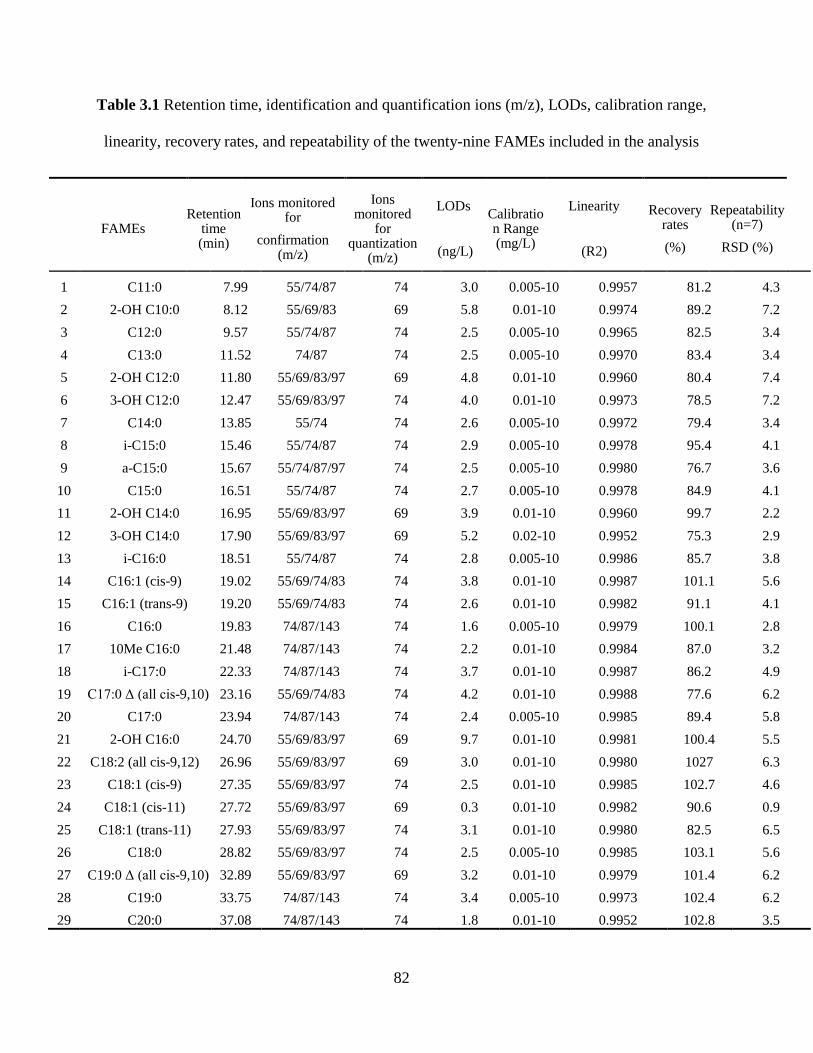
82
Table 3.1 Retention time, identification and quantification ions (m/z), LODs, calibration range,
linearity, recovery rates, and repeatability of the twenty-nine FAMEs included in the analysis
FAMEs Retention
time (min)
Ions monitored for
confirmation (m/z)
Ions monitored
for quantization
(m/z)
LODs
(ng/L)
Calibration Range (mg/L)
Linearity
(R2)
Recovery rates
(%)
Repeatability (n=7)
RSD (%)
1 C11:0 7.99 55/74/87 74 3.0 0.005-10 0.9957 81.2 4.3
2 2-OH C10:0 8.12 55/69/83 69 5.8 0.01-10 0.9974 89.2 7.2
3 C12:0 9.57 55/74/87 74 2.5 0.005-10 0.9965 82.5 3.4
4 C13:0 11.52 74/87 74 2.5 0.005-10 0.9970 83.4 3.4
5 2-OH C12:0 11.80 55/69/83/97 69 4.8 0.01-10 0.9960 80.4 7.4
6 3-OH C12:0 12.47 55/69/83/97 74 4.0 0.01-10 0.9973 78.5 7.2
7 C14:0 13.85 55/74 74 2.6 0.005-10 0.9972 79.4 3.4
8 i-C15:0 15.46 55/74/87 74 2.9 0.005-10 0.9978 95.4 4.1
9 a-C15:0 15.67 55/74/87/97 74 2.5 0.005-10 0.9980 76.7 3.6
10 C15:0 16.51 55/74/87 74 2.7 0.005-10 0.9978 84.9 4.1
11 2-OH C14:0 16.95 55/69/83/97 69 3.9 0.01-10 0.9960 99.7 2.2
12 3-OH C14:0 17.90 55/69/83/97 69 5.2 0.02-10 0.9952 75.3 2.9
13 i-C16:0 18.51 55/74/87 74 2.8 0.005-10 0.9986 85.7 3.8
14 C16:1 (cis-9) 19.02 55/69/74/83 74 3.8 0.01-10 0.9987 101.1 5.6
15 C16:1 (trans-9) 19.20 55/69/74/83 74 2.6 0.01-10 0.9982 91.1 4.1
16 C16:0 19.83 74/87/143 74 1.6 0.005-10 0.9979 100.1 2.8
17 10Me C16:0 21.48 74/87/143 74 2.2 0.01-10 0.9984 87.0 3.2
18 i-C17:0 22.33 74/87/143 74 3.7 0.01-10 0.9987 86.2 4.9
19 C17:0 Δ (all cis-9,10) 23.16 55/69/74/83 74 4.2 0.01-10 0.9988 77.6 6.2
20 C17:0 23.94 74/87/143 74 2.4 0.005-10 0.9985 89.4 5.8
21 2-OH C16:0 24.70 55/69/83/97 69 9.7 0.01-10 0.9981 100.4 5.5
22 C18:2 (all cis-9,12) 26.96 55/69/83/97 69 3.0 0.01-10 0.9980 1027 6.3
23 C18:1 (cis-9) 27.35 55/69/83/97 74 2.5 0.01-10 0.9985 102.7 4.6
24 C18:1 (cis-11) 27.72 55/69/83/97 69 0.3 0.01-10 0.9982 90.6 0.9
25 C18:1 (trans-11) 27.93 55/69/83/97 74 3.1 0.01-10 0.9980 82.5 6.5
26 C18:0 28.82 55/69/83/97 74 2.5 0.005-10 0.9985 103.1 5.6
27 C19:0 Δ (all cis-9,10) 32.89 55/69/83/97 69 3.2 0.01-10 0.9979 101.4 6.2
28 C19:0 33.75 74/87/143 74 3.4 0.005-10 0.9973 102.4 6.2
29 C20:0 37.08 74/87/143 74 1.8 0.01-10 0.9952 102.8 3.5
83
Quantitative analyses of individual fatty acids were performed based on the GC/MS response
(peak area) relative to that of two internal standards (C14:1 (cis-9) and C21:0) with known
concentrations. Method blanks were included in each set of samples and all results were blank
corrected accordingly. The area responses of the characteristic m/z against concentration for each
compound and internal standard were analyzed and response factors (RFs) for each compound
were calculated using Equation 1 (EPA, 1999).
(1)
where:
As = Area of the characteristic m/z for the parameter to be measured.
Ais = Area of the characteristic m/z for the internal standard.
Cis= Known concentration of the internal standard.
Cs = Known concentration of the parameter to be measured.
Then the RF value over the working range can be obtained and the concentration in the sample
was calculated using the determined RF and Equation 2.
(2)
where:
As = Area of the characteristic m/z for the parameter or surrogate standard to be measured.
Ais = Area of the characteristic m/z for the internal standard.
Cis = Concentration of the internal standard.
84
The selection of internal standards was based on a pre-test of offshore water samples to confirm
there were no recognizable chromatographic peaks of C14:1 (cis-9) and C21:0. Eleven calibration
standards spiked with multiple concentrations were prepared by diluting the stock in DCM. The
calibration range was between 0.005 and 10 mg/L. The analytical performance of the optimized
methodology coupled with GC-MS was evaluated by measuring the linearity, limits of detection
(LODs), recovery rates (RRs%) from the methyl esterification and relative standard deviation
(RSD%). The errors in all the analyses were calculated from triplicate or higher numbers of
sample analysis. Paired t tests were used for the statistical evaluation of differences in the analysis.
A p value less than 0.05 was used to indicate the significant difference between the tested groups.
3.3 Results and Discussion
3.3.1 Extraction efficiency in phase partition
The one-phase mixture of methanol, DCM and phosphate buffer at pH 7.4 (2:1:0.8, v/v) was
efficient in total lipids extractions, due to the presence of solvents with various polarity. Methanol
was water-miscible organic solvent and was scattered in both organic and aqueous phases after the
solvent partitioning when the ratio of DCM to methanol to water ratio was changed to 1: 1: 0.9. It
was assumed that partitioning of solutes (the total lipids extracted) between two liquids would stay
in a dynamic equilibrium (Berthod and Carda-Broch 2004) and compounds are separated based
on their solubility. Phospholipids (C16:1 (cis-9) PC, C18:1 (cis-9) PC, and C19:0 PC) spiked in
the vortexed samples were allowed to distribute in dynamic equilibrium in phase partition. The
organic phase was collected and the aqueous phase was again processed with 1 mL DCM. The two
organic phases were combined and subjected to FAME derivatization. Triplicate samples with the
same amount of phospholipids were also subjected to FAME derivatization directly. Results
85
indicated that 99.7% of C16:1 (cis-9), 99.2% of C18:1 (cis-9), and 99.4% of C19:0 were recovered
during the two-phase partition when the FAMEs from standards control were taken as a reference.
The RSDs of the analyses were all below 7.5%. The robust recovery indicated the lipid loss in the
phase separation was negligible under current experimental settings.
3.3.2 Determination of elution solvent volumes in SPE
SPE is based on the hydrophobic behavior of dissolved organic compounds on the packed column
(Roubeuf et al. 2000). DCM, acetone and methanol are solvents with increasing polarity to elute
increasingly polar lipids: neutral lipids, low-polarity lipids and polar lipids (Heinzelmann et al.
2014; Hutchins et al. 2008). The phospholipid isolation from the lipid extract was performed with
miniature champagne tubes paced with 0.1 g silica gel. An initial column conditioning step of
methanol was typically used to dehydrate the silica (Dobbs and Findlay 1993) to avoid any partial
hydration, which would reduce the polarity of silicic acid and may also potentiate the
chromatographic partitioning (Dickson et al. 2009). The phospholipids recoveries in methanol
fractions were also observed to be reduced for the phosphoethanolamine (PE) and phosphocholine
(PC) groups without methanol preconditioning (Mills and Goldhaber 2010), thus a methanol
preconditioning step was necessary. Based on the polarity principle, a DCM preconditioning step
was also conducted for the efficient removal of any oily organics in packed SPE tubes.
The influence of two different volumes of DCM and acetone and the elution behavior with three
different volumes of methanol were shown in Figure 3.1a. While the values of parameters were
selected according to previous studies (Bondia-Pons et al. 2006; Bossio and Scow 1998; Buyer
and Sasser 2012; Mills and Goldhaber 2010), significant differences were detected from serial
volumes of methanol for phospholipid elution. In contrast, the volumes of DCM and acetone have
86
little effect on the target lipid recoveries even though only 0.1 g silica gel was packed. Elution
volumes of methanol seemed not to be adequate for high throughput of phospholipid fraction in
the settings. Thus 4 mL, 6 mL, 8 mL, 10 mL and 12 mL volumes of methanol were used to
examine elution efficiency of phospholipids. Insufficient methanol eluant may result in incomplete
elution of phospholipids from silica columns and may explain the low reported recoveries (60-
75%) of PC standards by Billings and Ziegler (2008). Mills and Goldhaber (2010) investigated the
elution behavior of four phospholipid classes using three commercial SPE columns (packed with
0.5 g silica) and 10 mL methanol was found to be sufficient to quantitatively recover the
phospholipid standards. Wu et al. (2009) also used 10 mL methanol elution on home-made SPE
tubes to obtain phospholipids from two soils with different organic carbon concentrations. From
the results shown in Figure 3.1b, the recoveries of phospholipids C16:1 (cis-9) PC, C18:1 (cis-9)
PC, and C19:0 PC reached 92.9%, 96.3% and 92.8%, respectively, from 10 mL methanol elution
and the RSDs were all below 6.5%. Thus, 10 mL methanol in phospholipid elution could provide
an accurate characterization of microbial communities. Methanol elution was widely applied in
quantitative phospholipid studies on SPE columns from multiple sources (Bondia-Pons et al. 2006;
Bossio and Scow 1998; Chowdhury and Dick 2012; Fang and Findlay 1996), but the details of
their separation recoveries were not reported. Avalli and Contarini (2005) recovered 96% of a
phospholipid mixture from SPE cartridges packed with 1 g silica using 2 mL methanol followed
by 2 mL methanol: chloroform: water (5:3:2 v/v/v). Polar lipids are strongly retained by hydrogen
bonding and dipolar interactions within the silica column, thus more polar solvent mixture
demonstrated better elution behavior. However, further phase separation was involved in this step
and the complexity was increased, which prolonged the analysis procedures and may lead to
potential target loss.
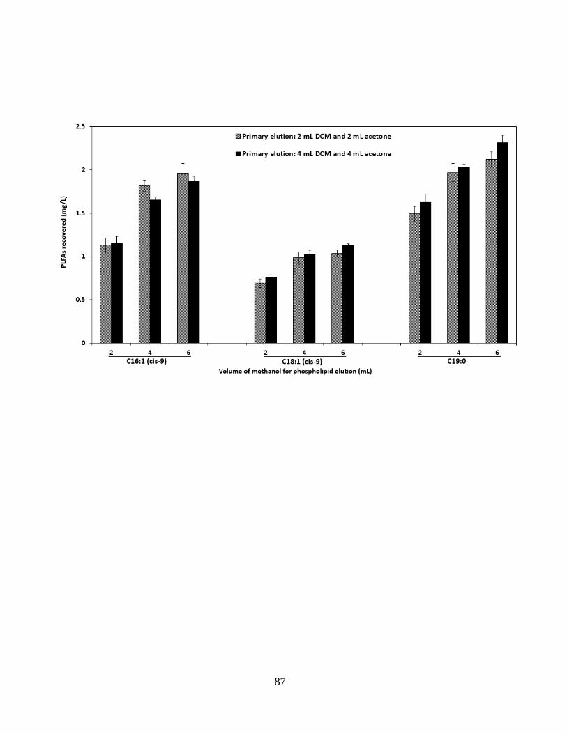
87
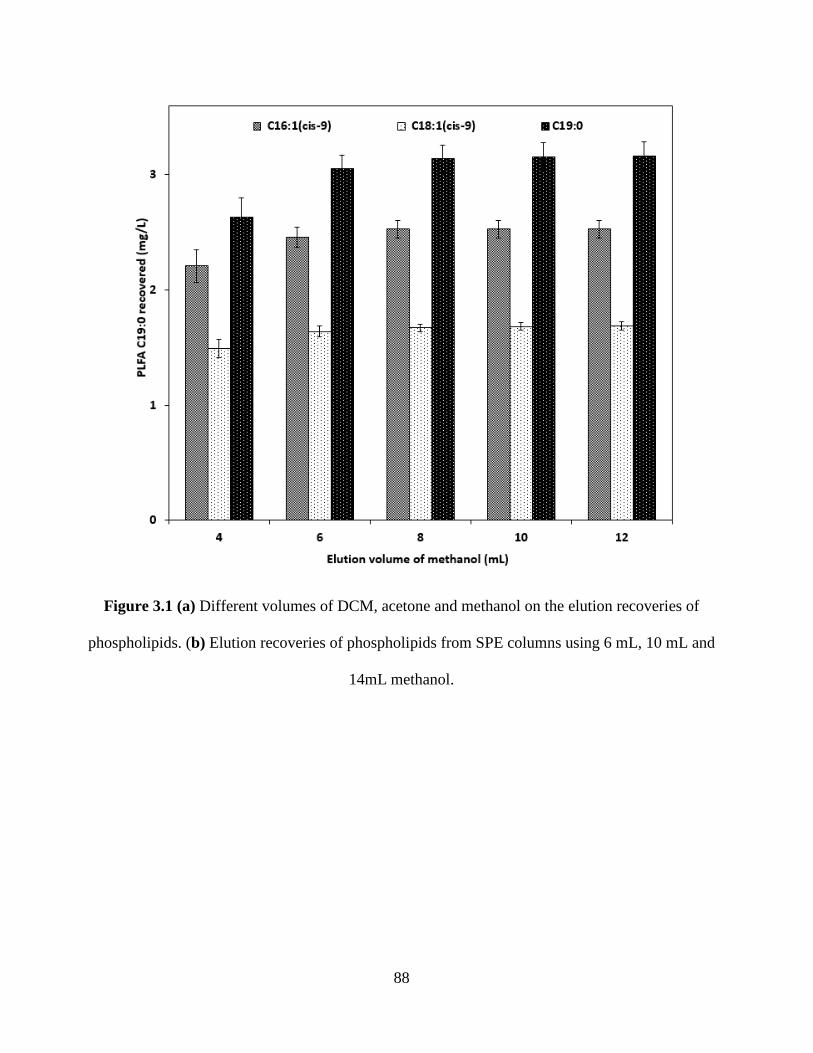
88
Figure 3.1 (a) Different volumes of DCM, acetone and methanol on the elution recoveries of
phospholipids. (b) Elution recoveries of phospholipids from SPE columns using 6 mL, 10 mL and
14mL methanol.
89
The DCM-acetone-methanol elution protocol was further examined using two different elution rate
and the results were shown in Figure 3.2. Little or no phospholipids were detected from the
solution residue, DCM or acetone fraction for all the three spiked standards. It was revealed that
the polar solute was transferred from the liquid phase into the silica absorbent. Elution behavior
was slightly different when a higher elution rate was applied. The volume of the first fraction
eluted with 4 mL methanol was reduced when elution flow rate became higher, and the volume of
the subsequent phospholipid fraction recovered by the second 2 mL methanol was thus increased.
This phenomenon from the first methanol fraction indicated that sufficient contact time would
release more polar lipids from their interaction with the silica absorbent while applying an
equivalent amount of solvent. Complete recovery of phospholipids may require more eluent if the
elution speed was increased. Mills and Goldhaber (2010) used different treatments on the solvent
elution rate (1 mL/min and 3 mL/min) to study the phospholipid recovery on SPE columns and
found the same elution tendency in the two methanol fractions. Even though, 10 mL of methanol
gave a convincing performance for the complete phospholipid recovery in this SPE procedure.
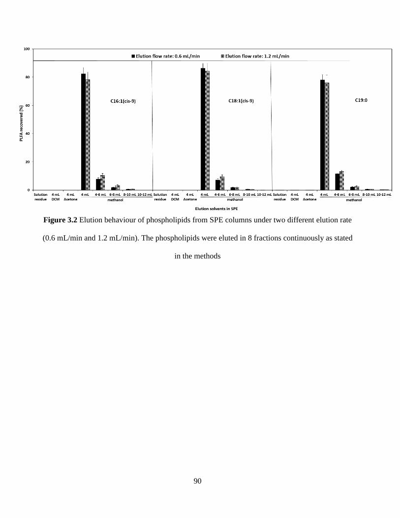
90
Figure 3.2 Elution behaviour of phospholipids from SPE columns under two different elution rate
(0.6 mL/min and 1.2 mL/min). The phospholipids were eluted in 8 fractions continuously as stated
in the methods
91
3.3.3 Derivatization to FAMEs
The derivatization method to FAMEs has significantly influenced the detectable concentration of
fatty acids. Acid-catalyzed methylation is an effective method to esterify free fatty acids and
transesterify fatty acids linked by ester bonds to glycerol or cholesterol. The acidic HCl together
with methanol solvent esterify all fatty acids at approximately the same rate (Weston et al. 2008).
However, its major drawback was that the PLFAs with methyl groups could not be detected,
possibly due to underlying mechanisms of addition-elimination reactions that occur during
esterification (Chowdhury and Dick 2012). To get a full picture of microbial profiling, a mild
alkaline trans-methylation procedure was selected in this study.
Base-catalyzed methylation reaction involves the cleaving of the ester bond by an alcohol. The
original ester firstly forms an anionic intermediate (b) (Figure 3.3) in the presence of a base such
as an alcoholate anion (a). The intermediate (b) can dissociate back to its former state or tend to
form a new ester (c) when the anion (a) was derived from a large excess of the alcohol. Due to the
presence of a large number of anion (a), the equilibrium will be displaced in favor of new ester (c)
production. This transesterification will virtually result in the sole product (c). Referred from the
optimal molarity is 0.5 to 2 N sodium or potassium methoxide in anhydrous methanol (Demirbas
2008), 0.2 N KOH/NaOH in methanol was adopted. Since the quality of the alkaline reagent and
the way it is prepared were potential factors that may affect methylation, the performance of KOH
and NaOH as the catalyst was compared under stated conditions.
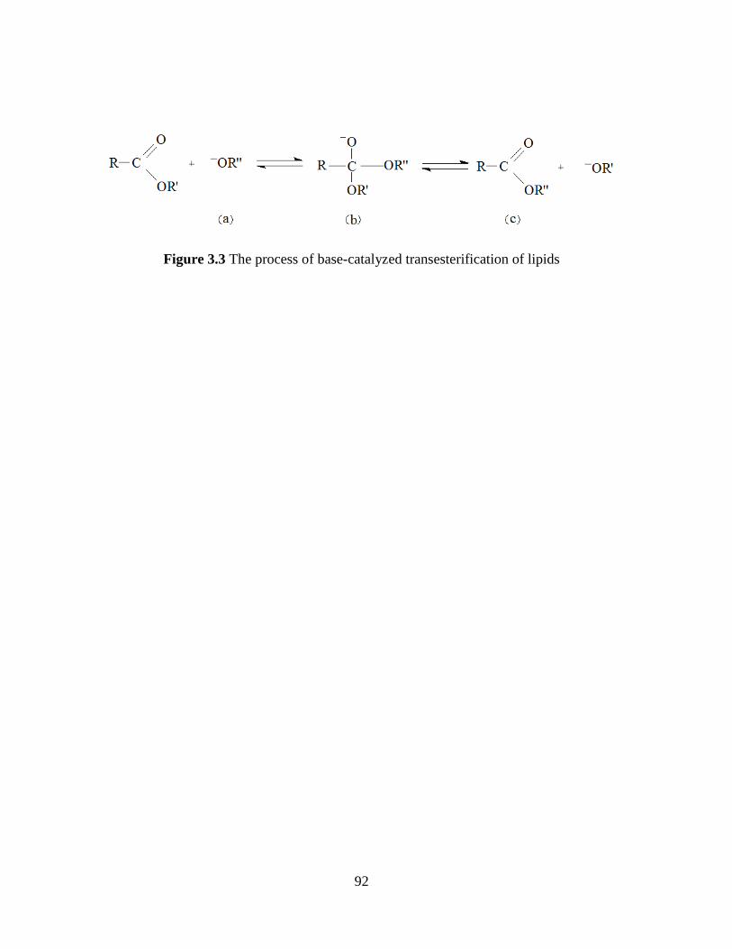
92
Figure 3.3 The process of base-catalyzed transesterification of lipids
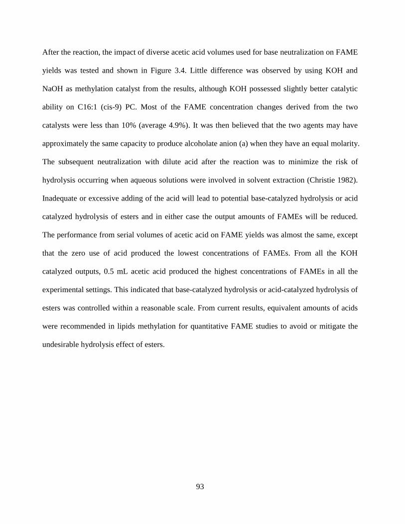
93
After the reaction, the impact of diverse acetic acid volumes used for base neutralization on FAME
yields was tested and shown in Figure 3.4. Little difference was observed by using KOH and
NaOH as methylation catalyst from the results, although KOH possessed slightly better catalytic
ability on C16:1 (cis-9) PC. Most of the FAME concentration changes derived from the two
catalysts were less than 10% (average 4.9%). It was then believed that the two agents may have
approximately the same capacity to produce alcoholate anion (a) when they have an equal molarity.
The subsequent neutralization with dilute acid after the reaction was to minimize the risk of
hydrolysis occurring when aqueous solutions were involved in solvent extraction (Christie 1982).
Inadequate or excessive adding of the acid will lead to potential base-catalyzed hydrolysis or acid
catalyzed hydrolysis of esters and in either case the output amounts of FAMEs will be reduced.
The performance from serial volumes of acetic acid on FAME yields was almost the same, except
that the zero use of acid produced the lowest concentrations of FAMEs. From all the KOH
catalyzed outputs, 0.5 mL acetic acid produced the highest concentrations of FAMEs in all the
experimental settings. This indicated that base-catalyzed hydrolysis or acid-catalyzed hydrolysis of
esters was controlled within a reasonable scale. From current results, equivalent amounts of acids
were recommended in lipids methylation for quantitative FAME studies to avoid or mitigate the
undesirable hydrolysis effect of esters.

94
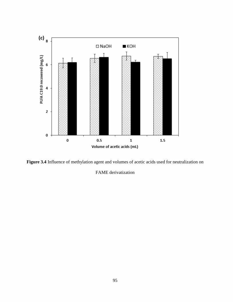
95
Figure 3.4 Influence of methylation agent and volumes of acetic acids used for neutralization on
FAME derivatization
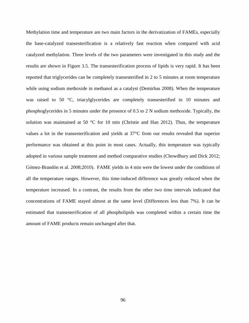
96
Methylation time and temperature are two main factors in the derivatization of FAMEs, especially
the base-catalyzed transesterification is a relatively fast reaction when compared with acid
catalyzed methylation. Three levels of the two parameters were investigated in this study and the
results are shown in Figure 3.5. The transesterification process of lipids is very rapid. It has been
reported that triglycerides can be completely transesterified in 2 to 5 minutes at room temperature
while using sodium methoxide in methanol as a catalyst (Demirbas 2008). When the temperature
was raised to 50 °C, triacylglycerides are completely transesterified in 10 minutes and
phosphoglycerides in 5 minutes under the presence of 0.5 to 2 N sodium methoxide. Typically, the
solution was maintained at 50 °C for 10 min (Christie and Han 2012). Thus, the temperature
values a lot in the transesterification and yields at 37°C from our results revealed that superior
performance was obtained at this point in most cases. Actually, this temperature was typically
adopted in various sample treatment and method comparative studies (Chowdhury and Dick 2012;
Gómez-Brandón et al. 2008;2010). FAME yields in 4 min were the lowest under the conditions of
all the temperature ranges. However, this time-induced difference was greatly reduced when the
temperature increased. In a contrast, the results from the other two time intervals indicated that
concentrations of FAME stayed almost at the same level (Differences less than 7%). It can be
estimated that transesterification of all phospholipids was completed within a certain time the
amount of FAME products remain unchanged after that.
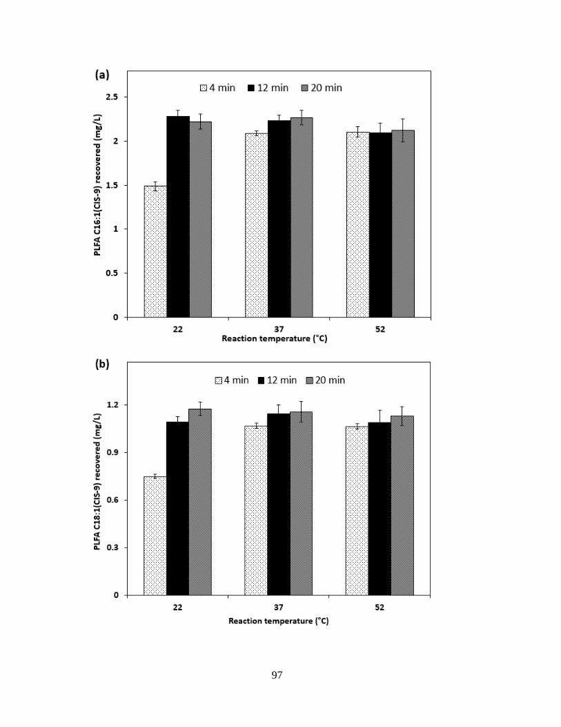
97
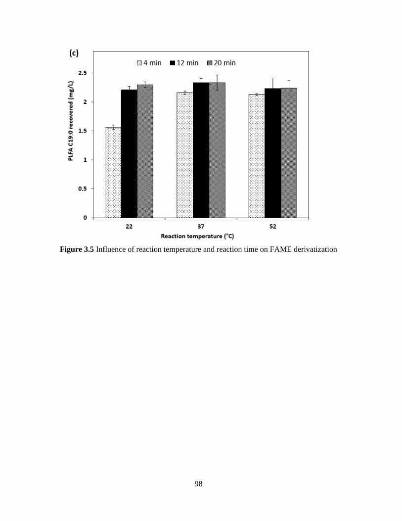
98
Figure 3.5 Influence of reaction temperature and reaction time on FAME derivatization
99
3.3.4 Analytical performance
3.3.4.1 Method linearity
The linearity of an analytical procedure is its ability (within a given range) to induce responses
which are directly proportional to the concentration (amount) of analyte in the sample. The
linearity of the method was verified through the measurement of serial dilutions of 29 FAME
standard mixtures.
Eleven standards spiked with multiple concentrations ranging from 0.01 and 10 mg/L were used to
create calibration curves and more details are shown in Table 3.1. The 11 standard concentrations
were 0.005 mg/L, 0.01 mg/L, 0.02 mg/L, 0.05 mg/L, 0.1 mg/L, 0.2 mg/L, 0.5 mg/L, 1 mg/L, 2
mg/L, 5 mg/L, and 10 mg/L. Calibration curves were built based on the analytical responses of
FAMEs among these concentrations. The coefficients of determination (r2) for the obtained
calibration curves were all higher than 0.995. The wide dynamic range and the high values of r-
squared are robust verification of GC-MS based quantification for FAMEs derived from
phospholipids. While two internal standards (C14:1 (cis-9) and C21:0) were involved in
determining the values of RFs for each methyl ester, an accurate FAME determination in a wide
range was obtained from RFs in the stated wide range.
3.3.4.2 Limit of detection
Detection limit is defined as the statistically calculated minimum concentration that can be
measured with 99% confidence that the reported value is greater than zero (Bernal 2014). LODs
were calculated by using following equation:
(3)
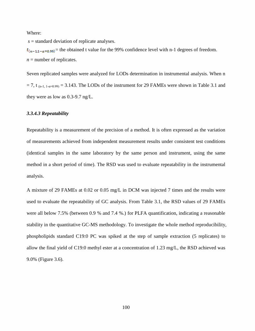
100
Where:
s = standard deviation of replicate analyses.
= the obtained t value for the 99% confidence level with n-1 degrees of freedom.
n = number of replicates.
Seven replicated samples were analyzed for LODs determination in instrumental analysis. When n
= 7, t (n-1, 1-α=0.99) = 3.143. The LODs of the instrument for 29 FAMEs were shown in Table 3.1 and
they were as low as 0.3-9.7 ng/L.
3.3.4.3 Repeatability
Repeatability is a measurement of the precision of a method. It is often expressed as the variation
of measurements achieved from independent measurement results under consistent test conditions
(identical samples in the same laboratory by the same person and instrument, using the same
method in a short period of time). The RSD was used to evaluate repeatability in the instrumental
analysis.
A mixture of 29 FAMEs at 0.02 or 0.05 mg/L in DCM was injected 7 times and the results were
used to evaluate the repeatability of GC analysis. From Table 3.1, the RSD values of 29 FAMEs
were all below 7.5% (between 0.9 % and 7.4 %.) for PLFA quantification, indicating a reasonable
stability in the quantitative GC-MS methodology. To investigate the whole method reproducibility,
phospholipids standard C19:0 PC was spiked at the step of sample extraction (5 replicates) to
allow the final yield of C19:0 methyl ester at a concentration of 1.23 mg/L, the RSD achieved was
9.0% (Figure 3.6).
101
Figure 3.6 Chromatogram of standard mixture containing 29 FAMEs by GC-MS analysis on DB-
5MS capillary column (number is accorded with Table 3.1)
1
2
3 4
5
6
7
8
9
10
11 12
13
14
15
16
17
18
19
20
21
22
23 24
25 26
27
28
29
0
50000
100000
150000
200000
250000
300000
350000
400000
5 10 15 20 25 30 35 40
Retention time (min)
Response of 29 FAMEs
Ab
un
dan
ce

102
3.3.4.4 Method recovery performance
Recovery is the detection of a known amount of an analytical parameter added to the sample
matrix and included throughout the method of analysis. Since PLFA quantitative study involved
the derivatization of PLFAs to FAMEs for GC analysis, the method recovery was investigated in
two portions. Firstly, triplicate samples were spiked with phospholipid C19:0 PC during the
treatment of sample extraction and FAME derivatization, respectively, and the concentrations of
fatty acid C19:0 obtained (A and B) were used to investigate the method recovery performance.
Results showed that 90.6% (A/B) of C19:0 PC was recovered before the treatment of FAME
derivatization in the methodology. Solvents (toluene: methanol-1:1) were spiked with 29 FAMEs
in the treatment of transesterification to test the recovery during the phase separation and
concentration steps. Results indicated that all 29 FAMEs exhibited recoveries greater than 75.3%,
while 24 of them showed recoveries ranging from 80.4% to 103.1% (all RSDs less than 8.8%).
3.3.5 Analysis of offshore produced water samples
Offshore produced water is characterized by its complex composition (e.g., treating chemicals,
formation solids, heavy metals, salts, dissolved and dispersed oils) with high salinity and
petroleum hydrocarbon content (Sirivedhin et al. 2004), and the general oil/water volume ratio is
1:3 (Fakhru‘l-Razi et al. 2009). Even though, over 90% recovery of phospholipid was achieved in
this study from triplicate produced water samples when C19:0 PC was spiked into the extraction
solvent at the beginning of sample processing. Before spiking C19:0 PC into the samples,
preliminary analysis of the reservoir samples indicated that no identifiable peaks of C19:0 were
observed. After the water sampling and analysis, 14 types of PLFAs were detected from waters in
a producing well of an offshore oil and gas platform and the results were illustrated in Figure 3.7.
103
The four most abundant fatty acids from the complex matrix were C14:0, C16:1(cis-9), C16:0 and
C18:0. Other typical fatty acids C12:0, C15:0 and C18:1(cis-9) were also found in considerable
amounts. The even-numbered fatty acids dominated in the PLFA profiles of offshore produced
water (88.4% of total amount) and this reflected the natural pathway for their biosynthesis from
the two-carbon building-block acetyl CoA (Estelmann et al. 2011). The only hydroxy fatty acid
(HFA) detected was 2-OH C12:0 and the polyunsaturated fatty acid (PUFA) was solely
represented by C18:2 (all cis-9, 12), which could be documented in marine hydrocarbon degrading
bacteria (Zhang et al. 2012). Fatty acids i-C15:0, a-C15:0 and a-C16:0 were main branched
saturated fatty acids (BCFA), but they are minor components as reflected from the whole PLFA
distributions. Straight-chain saturated fatty acids (SSFAs) were major compounds in PLFA profile
and were reported to be associated with swarming motility in Proteus mirabilis and Serratia
marcescens (Lai et al. 2005; Liaw et al. 2004). This interesting perspective was very likely
reasonable in explaining the resulting PLFA distributions in our samples, since the mobility of
bacterial cells was of crucial importance for their survival in commonly porous reservoir
environments.
From this point, monounsaturated fatty acids (MUFAs) and BCFAs were also associated with their
indicative uses. According to previous studies, they are signature biomarkers of different microbial
species to varying extents. For instance, the branched-chain fatty acids (iso, anteiso in C14-C18)
are widely accepted as general indicators for gram-positive bacteria (Ruess and Chamberlain
2010). However, significant amounts of i-C15:0 were previously found in Desulfovibrio
desulfurricans with various growth substrates, and the percentage fatty acid composition of i-
C15:0 in this strain increased from 9.9% to 12.6% when substrate changed from CO2 to lactate
(Taylor and Parkes 1983). The presence of C18:1(cis-9) is common to many psychrophilic
104
microorganisms such as saprophytic fungi (De Deyn et al. 2011), Pseudomonas sp. and some
Colwellia/Vibrio sp., but the occurrence of C18:1(cis-9) as a major fatty acid in sulfur oxidizing
bacteria (SOB) has never been reported (Guezennec et al. 1998). Specifically, a higher percentage
of C18:1(cis-9) was found in SRB species Desulfobacter curvatus and Desulfobacter latus in
marine mud (Kohring et al. 1994). The fatty acid C18:1(cis-9), potential marker to trace SRB
activities under certain circumstances, was also chosen to represent Desulfotomaculum in SRB-
related biogeochemical processes under the environments of subsurface sediment (Mohanty et al.
2008). The coexistence of C16:1 (cis-9) and C18:1 (cis-11) could indicate the presence of SOB.
The dominance of C16:1 (cis-9) (16.7 to 37.4%) and C18:1 (cis-11) (11.8 to 16.8%) has also been
observed in thiotrophic bacterial mats in the Barbados Trench (Guezennec and Fialamedioni 1996).
Zhang et al. (2005) studied the metabolic functions of microbial mat in sulfide-rich marine
sediments associated with gas hydrates in the Gulf of Mexico, and concluded that C16:1 (cis-9)
and C18:1 (cis-11) can be used as signature biomarkers for SOB in H2S-rich marine sediments.
Our results of PLFA species in reservoir samples indicated relatively higher level of sulfate
reducing activities but lower sulfur oxidizing rates. Therefore, PLFA profiles, as a significant
indicator of microbial activities, could provide very reasonable interpretation and guidance
towards biological reservoir souring and associated mitigation activities under continuous
monitoring conditions.
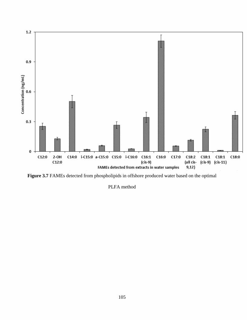
105
Figure 3.7 FAMEs detected from phospholipids in offshore produced water based on the optimal
PLFA method
106
3.4 Summary
A method for the determination of PLFAs in profiling microbial communities in offshore
produced water has been developed. A three-stage extraction process was confirmed and the
extraction efficiency in phase partition was evaluated. The elution parameters in SPE purification
were adapted for treating the oily samples and their volumes were determined to induce a high
recovery for the fraction of phospholipids. The impact of parameters including alkaline reagent,
the volumes of acid used for neutralization, the time and temperature for transesterification were
studied. The GC-MS quantitative analysis was validated by examining the method linearity, LODs
and repeatability. Method recovery performance from phospholipids preparation and FAME
derivatization were studied. Results indicated that the developed method exhibited high recoveries
and repeatability, remarkable selectivity and linearity, and acceptable quantification limits for
PLFA analysis. With reliable accuracy and precision, the method was applied to profile microbes
in offshore produced water samples. The developed method would be applicable for routine
analysis of samples with high oily and salinity properties in the marine environment.

107
3.5 Appendix
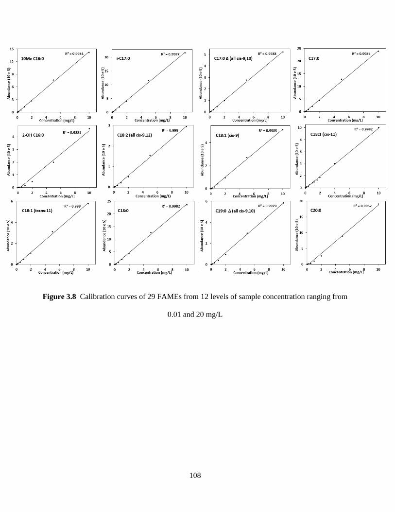
108
Figure 3.8 Calibration curves of 29 FAMEs from 12 levels of sample concentration ranging from
0.01 and 20 mg/L
109
CHAPTER 4
PROFILING OF SRB IN AN OFFSHORE OIL RESERVOIR
USING PLFA BIOMARKERS2
2 This chapter is based on the following paper:
F. Fan, B. Zhang, P.L. Morrill, T. Husain, Profiling of Sulfate-Reducing Bacteria in an Offshore
Oil Reservoir Using Phospholipid Fatty Acid (PLFA) Biomarkers, Water, Air, & Soil Pollution,
228 (2017) 410
Role: Fuqiang Fan solely worked on this study and acted as the first author of this manuscript. Dr.
Baiyu Zhang is my supervisor. Dr. Penny Morrill and Dr. Tahir Husain helped guide the research
and polish the manuscript.

110
4.1 Background
The Oil and Gas industry has been well aware of the harmful activities of SRB with regards to
reservoir souring and MIC for many years (Enning and Garrelfs 2014). As anaerobic
microorganisms, SRB are a major concern in the petroleum industry primarily because of their
ability to generate substantial amounts of hydrogen sulfide and insoluble ferrous sulfide in the
presence of iron.
SRB produce hydrogen sulfide through respiration and they gain energy for growth, in which the
sulfate functions as an electron acceptor and organic matter or molecular hydrogen are employed
as electron donors. In secondary oil recovery activities, seawater is usually injected into reservoirs
to maintain pressure level underneath and recover the remaining oil from the reservoir (Gieg et al.
2011). SRB are prevalent in many natural as well as engineered aquatic environments, and they
flourish in the presence of sulfate from seawater and oil components pre-existed in the reservoir
(Muyzer and Stams 2008). The subsequent undesirable production of hydrogen sulfide is closely
associated with negative effects in the form of reduced quality of hydrocarbon products, increased
iron sulfide scale precipitation and plugging, reduced injection efficiency in reservoir wells, and
increased pipeline corrosion (Hubert and Voordouw 2007). This production of hydrogen sulfide
also induces safety risks for operators and environmental concerns.
To effectively mitigate this situation, methods must be initially developed to characterize the
microbial community composition during the oil production activities, thus enabling the planning
for souring control solutions. Copious amounts of culture or molecular methods have been
developed for the detection and enumeration of viable SRB for routine monitoring and anomaly
investigation (Head et al. 1998; Kaster et al. 2009; Lenchi et al. 2013; Sun et al. 2014; Tanner
111
1989). Culture-dependent methods are typically laborious, time-consuming and cannot provide
unequivocal results. Moreover, more than 99% of naturally occurring microbes are considered
‗unculturable‘ using conventional cultivation techniques (Zhang et al. 2011a).
The two main culture-independent methods that are widely used for microbial profiling of
environmental samples include nucleic acid and PLFA analyses. Nucleic acid analysis methods
usually employ polymerase chain reaction (PCR) techniques and produce complex banding
patterns of DNA fragments on a gel to fingerprint microbial species. PCR-based methods have
been recognized as a highly sensitive and specific technique for species identification. However,
there are many drawbacks in the methodologies, including inconsistent DNA recovery, inherent
kinetic biases, the relatively high cost and the need to develop specific primers and probes
(Lueders and Friedrich 2003). PCR-based methods are also not suitable for characterizing samples
with complex communities (Pan et al. 2010). In contrast, PLFA analysis offers a great potential of
microbial community analysis in routine environmental monitoring.
Phospholipids are essential membrane constituents of bacterial cells and their types and amounts
vary from one to another (Guezennec and Fialamedioni 1996; Kaur et al. 2005). Since the content
of phospholipids stays relatively constant, microorganisms with the same taxonomic composition
can be identified by analyzing the diversity and specificity of their PLFAs (Kaur et al. 2005).
Phospholipids are rapidly decomposed after cell death in the environment, thus PLFA analysis is
widely accepted to indicate viable microbial biomass and provide a microbial community
‗fingerprint‘ (Zelles 1999). Due to their cellular abundance and chemically diverse nature, PLFAs
are commonly used as biomarkers to measure microbial biomass, nutritional/physiological status
and microbial community structure in soils, surface waters, groundwaters, sediments and biomats
112
(Acosta-Martínez et al. 2008; Dijkman et al. 2010; Drenovsky et al. 2010; Franzmann et al. 1996;
Moore-Kucera and Dick 2008; White and Ringelberg 1997; Yu et al. 2009).
Boschker et al. (2001) investigated the bacterial populations and pathways involved in acetate and
propionate consumption in anoxic brackish sediment based on PLFA analysis. Labeled acetate and
propionate (13
C) were incorporated into PLFAs after incubation and the results showed that they
were predominantly consumed by different, specialized groups of SRB. Guezennec and
Fialamedioni (1996) analyzed sediments from various locations in Barbados trench through PLFA
analysis and concluded anaerobic bacteria and presumably SRB were also present in these
sediments. Uranium-bearing sandstones from the Dongsheng deposit were also found with the
abundant presence of C15-C18 fatty acids (Jiang et al. 2012). Characteristic biomarkers of SRB
Desulfovibrio and Desulfobacter sp. were found and involved in bacterial sulfate reduction to
sulfide.
Even though PLFA profiling has been used as SRB biomarkers in various solid and fluid samples,
it is rarely applied in offshore reservoir water analysis to specifically elucidate the mechanism of
reservoir souring induced by SRB. The SRB transformation patterns between the injection wells
and producing wells under different redox conditions remained unclear. Furthermore, there is still
a lack of basic understanding of the complex biomass and microbial community structure
information from the various reservoir conditions regarding souring. In this study, we analyzed the
injection water and four produced water samples with different chemical and physical properties
using PLFA analysis in order to study the microbial community composition in various reducing
conditions and identify possible SRB species responsible for hydrogen sulfide production.
113
4.2 Materials and Methods
4.2.1 Standards, reagents, apparatus and sample collection
Analytical standards of FAMEs: Bacterial Acid Methyl Esters CP Mixture (26 methyl esters),
10Me C16:0 (Methyl 10-methylhexadecanoate), Trans-9 C16:1 (Methyl hexadecenoate, trans-9),
and C19:0 (Methyl nonadecanoate) were purchased from Matreya LLC (Pleasant Gap,
Pennsylvania, USA). Trans-11 C18:1 (trans-11-Octadecenoic methyl ester) was purchased from
Sigma-Aldrich (Oakville, Ontario, Canada). Phospholipid standards C16:1 (cis-9) PC (1,2-
dipalmitoleoyl-sn-glycero-3-phosphocholine), C18:1 (cis-9) PC (1,2-dioleoyl-sn-glycero-3-
phosphocholine), and C19:0 PC (1, 2-dinonadecanoyl-sn-glycero-3-phosphocholine) were
purchased from Avanti Polar Lipids, Inc. (Alabaster, Alabama, USA). The ultra-high purity water,
silica gel (60-120 mesh), solvents including chloroform, DCM, methanol, hexane, toluene and
acetone of reagent grade or higher quality were purchased from VWR® International (Mississauga,
Ontario, Canada) and Fisher Scientific (Ottawa, Ontario, Canada). The commercial 3 mL SPE
tubes (miniature champagne column) were bought from Supelco Inc. (Bellefonte, Pennsylvania,
USA). GC supplies, including deactivated single tapered glass inlet liners and J&W Scientific DB-
5MS UI fused silica capillary columns, were obtained from Agilent® Technologies Inc.
(Mississauga, Ontario, Canada). Extraction tubes and all other glassware were primarily washed
with detergent and then deeply rinsed with chromic acid cleaning solution to remove all types of
oil, fingerprints, rust and other dirt.
Triplicated offshore produced water and injection seawater samples were collected from four
producing wells and the injection well in an offshore oil and gas platform. Gaseous H2S
concentrations were detected through enhanced laser diode spectroscopy (ELDS) technology
114
(Moser et al. 2017). Before sampling in each location, in-situ measurements (pH, redox potential,
temperature, and dissolved oxygen) were conducted using field probes (pH, redox potential and
dissolved oxygen probes respectively) and the results were recorded. After the measurements of
the listed parameters, 500 mL water sample was filtered through 0.2 μm nylon membrane filter (47
mm in diameter) by a vacuum pump. The filter paper was then transferred into a 10 mL amber vial
with 9.5 mL extraction solvents (methanol: DCM: 125 mM phosphate buffer at pH 7.4 = 2:1:0.8)
inside. Total lipids were extracted from microbial biomass collected on the filters and the vials
were sealed with screw caps containing Teflon lined septa. All samples were stored in a freezer (-
20°C in the dark) before shipping and then transported to the laboratory in a package with frozen
ice packs for determination.
4.2.2 Extraction of lipids and preparation of FAMEs
Total lipids were extracted using a modified Bligh and Dyer extraction method (Fang and Findlay
1996). Approximately 0.1–0.7 g of freeze-dried sediment/mat sample was subjected to vortex-
assisted extraction in an amber vial with 9.5 mL extraction solvents (methanol: DCM: 125 mM
phosphate buffer at pH 7.4 = 2:1:0.8). The extraction mixture was allowed to stand overnight in
the dark at -20 oC. Additional 4.5 mL and 4 mL mixture of methanol, DCM and phosphate buffer
(2: 1: 0.8) were used for secondary and tertiary total lipids extraction from the membrane filter.
The lipids were then partitioned by adding DCM and water, such that the final ratio of DCM to
methanol to water was 1: 1: 0.9. The lower organic phase was transferred and collected altogether
from the extracts. Phospholipid standard C19:0 PC (1,2-dinonadecanoyl-sn-glycero-3-
phosphocholine) was spiked to examine the % recovery and four blanks spiked with C19:0 PC
were performed for process control.
115
The total lipid extract was dried under a gentle stream of nitrogen and was redissolved in hexane:
DCM (70:30, v/v). Neutral lipids, glycolipids and phospholipids were eluted with 4 mL of
chloroform, 4 mL of acetone and 10 mL of methanol on a home-made SPE tube (miniature
champagne column) packed with 0.1 g silica gel, respectively. The fraction of phospholipids was
evaporated to dryness, redissolved in 1 mL of methanol: toluene (1:1, v/v) and subjected to a mild
alkaline trans-methylation procedure to produce FAMEs prior to further quantitative analysis.
Potassium hydroxide with a concentration of 0.2 mol/L was used as methylation agent and the
FAMEs were finally concentrated to 400 μL in chloroform.
4.2.3 GC-MS analyses
FAMEs were analyzed by GC-MS system (Agilent 7890A GC system coupled with a 5975C MSD)
with an Agilent 7693 auto-sampler. Chromatographic separation was achieved by using a 30 m ×
250 μm (internal diameter, i.d.) × 0.25 μm DB-5MS UI fused silica capillary column. Sample
injections (3μL) were conducted using a split/splitless injector (single tapered inlet liner) in pulsed
splitless mode at 200 °C under a pulse pressure of 25 psi. The oven temperature program started at
70 oC, then it was raised to 150
oC at 20 °C/min, to 180
oC at 3°C/min, to 210
oC at 1°C/min, and
finally to 280 oC at 35°C/min with a running time of 47 min. Helium of ultrahigh purity was used
as the carrier gas at a flow rate of 1.0 mL/min with an electronic pressure control (7.65 psi). The
ion source was operated in the electron ionization (EI) mode at 70 eV and mass spectral data were
acquired through the scan range of 50–550 m/z. Full scanning and the SIM mode were used
simultaneously for FAME determination.
Individual compounds were identified from their mass spectra and were further confirmed by
comparing their relative retention times with those of well-known FAME standards.
116
Quantifications of FAMEs were based on response factors (RF) derived from a mixture of 29
external standards (FAMEs from C10 to C20) with C19:0 (methyl nonadecanoate) as an internal
standard. The area responses of the characteristic m/z against concentrations (twelve different
levels) for each compound were analyzed to derive RF for each compound. Replicate analyses (7×)
of samples were performed to ensure reproducibility (variation of ≤6.0%) in GC-MS quantification.
Fatty acids are designated by the total number of carbon atoms to the number of double bonds (e.g.
a 16-carbon alkanoic acid is C16:0). The position of the double bond is indicated by a Δ number
closest to the carboxyl end of the fatty acid molecule with the suffixes of either cis (c) or trans (t)
as the geometric isomers. Prefixes i and a are given for iso- and anteiso-branched FAMEs,
respectively. The suffix 10 ME indicates a methyl group at the 10th C atom, while OH stands for
hydroxy and the cyclopropyl ring is indicated as ‗cy‘.
4.2.4 Statistical analysis
Triplicate samples were prepared and analyzed to ensure the reproducibility of results, and the
error bars in the plotted data were derived from the standard deviations of the mean values of
triplicate samples. To assess the patterns of intercorrelations among PLFA species measured,
principal component analysis (PCA) was conducted using the software SPSS 18.0. The data were
orthogonally transformed into a new coordinate system and orthogonal variables called principal
components with the greatest variances were used as coordinates. The first principal component
(PC1, 59.1%) accounts for most of the original variability and the second principal component
(PC2, 27.5%) contains the second largest variance.
117
4.3 Results and Discussion
4.3.1 In-situ measurements
Cool seawater from the ocean was injected into the reservoir in order to maintain well pressure and
sweep the oil towards production wells. Since the reservoir was characterized by high temperature
and high pressure, seawater temperature was elevated when it was mixed with the formation fluids.
The hot mixed fluids cooled down during the pipeline of producing wells and above-ground
facilities. The temperature of produced water observed from the oil-water separator was
approximately 63°C initially, however, the temperature decreased with time. In general, the
intermediate zone where cool injection water was warmed to the reservoir temperature would
provide favorable thermal conditions for SRB activity (Gieg et al. 2011). As seen from Table 4.1,
the pH values were approximately 6.71 and showed little difference among the water samples of
the injection well and producing wells. The redox potentials, however, varied from one sampling
point to another were all below 100 mV and the results were accorded with low DO measurement
outputs.
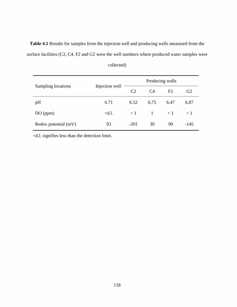
118
Table 4.1 Results for samples from the injection well and producing wells measured from the
surface facilities (C2, C4, F2 and G2 were the well numbers where produced water samples were
collected)
Sampling locations Injection well Producing wells
C2 C4 F2 G2
pH 6.71 6.52 6.75 6.47 6.87
DO (ppm) <d.l. < 1 1 < 1 < 1
Redox potential (mV) 93 -201 30 99 -145
<d.l. signifies less than the detection limit.
119
Water based oxygen scavengers are widely used in seawater injection systems as a tool to prevent
oxygen-induced corrosion, such that DO could not be detected in injection seawater. As a result,
the reducing conditions make the matrix susceptible to the growth of SRB in the reservoir (Enning
and Garrelfs 2014). From redox potential results, produced water samples from wellbore C2 and
G2 exhibited a more reducing environment than those from wellbore C4 and F2. The presence of
small amount of oxygen (1 ppm) in water samples from wellbore C4 may illustrate the cause of its
more oxidizing environment than others since DO values from other locations were all below 1
ppm. The wellbore F2, with the lowest pH value and the highest redox, has the potential to possess
differential characteristic bacterial species when compared to other producing wells.
4.3.2 PLFA analytical performance
A mixture of 29 FAMEs at 0.1 or 0.01 mg/L was measured 7 times to evaluate the reproducibility
of GC analysis. The results indicated that the RSD values of all the FAMEs were between 1.3 %
and 5.6 % while using RF as quantification tool and C19:0 (methyl nonadecanoate) as an internal
standard. Detection limit is defined as the statistically calculated minimum concentration that can
be measured with 99% confidence that the reported value is greater than zero (Bernal 2014).
Accordingly, the GC-MS detection limits of 29 FAMEs ranged from 0.4 to 12 ng/L. The analysis
incorporated triplicate blanks by spiking phospholipid standards C16:1 (cis-9) PC and C18:1 (cis-9)
PC into extraction solvents and allowed them running through the whole analytical procedure to
investigate the method recovery performance. The same amounts of phospholipid standards were
simultaneously spiked into FAME derivatization solvents and the results were taking as a
reference. More than 90 % of the phospholipids were recovered before the treatment of FAME
derivatization in the methodology and the RSDs of the blanks were all below 8.0%. The analysis
incorporated triplicate controls by spiking 29 FAMEs into 1 mL of methanol: toluene (1:1, v/v)
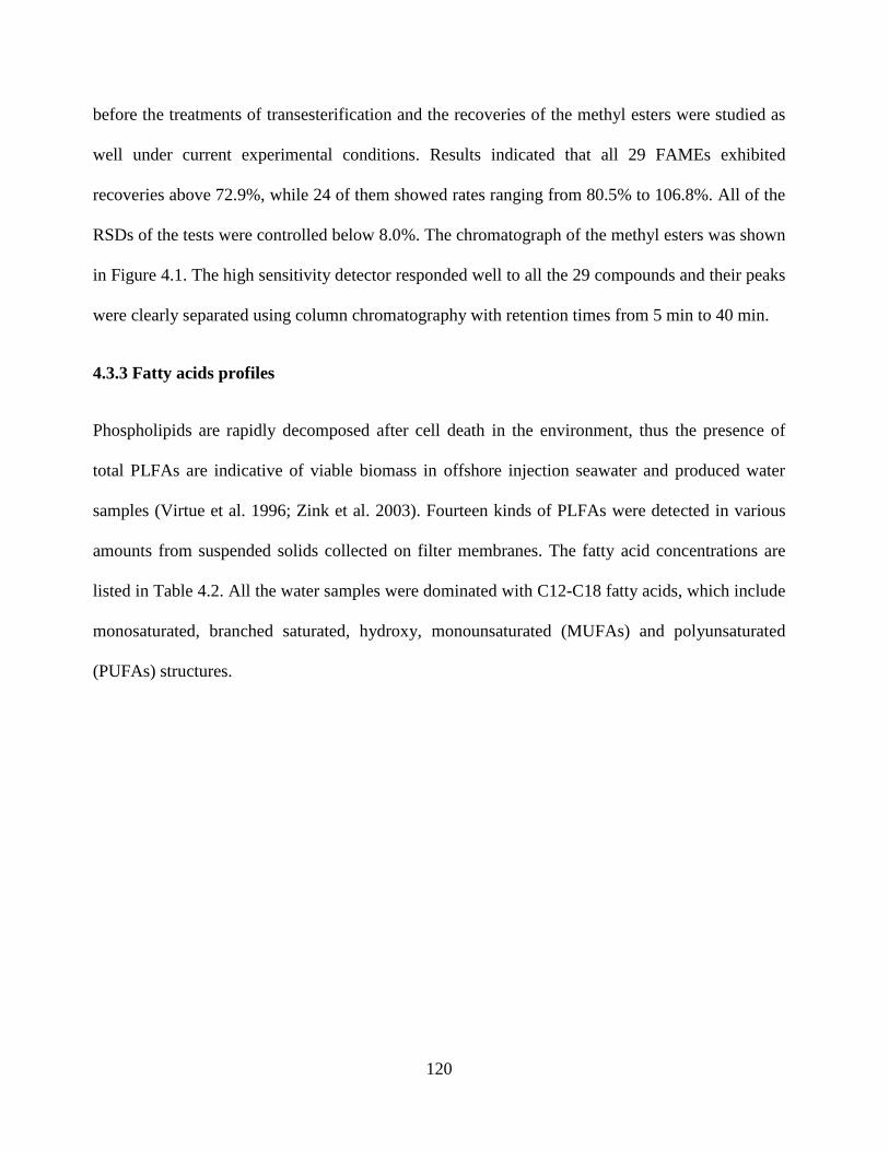
120
before the treatments of transesterification and the recoveries of the methyl esters were studied as
well under current experimental conditions. Results indicated that all 29 FAMEs exhibited
recoveries above 72.9%, while 24 of them showed rates ranging from 80.5% to 106.8%. All of the
RSDs of the tests were controlled below 8.0%. The chromatograph of the methyl esters was shown
in Figure 4.1. The high sensitivity detector responded well to all the 29 compounds and their peaks
were clearly separated using column chromatography with retention times from 5 min to 40 min.
4.3.3 Fatty acids profiles
Phospholipids are rapidly decomposed after cell death in the environment, thus the presence of
total PLFAs are indicative of viable biomass in offshore injection seawater and produced water
samples (Virtue et al. 1996; Zink et al. 2003). Fourteen kinds of PLFAs were detected in various
amounts from suspended solids collected on filter membranes. The fatty acid concentrations are
listed in Table 4.2. All the water samples were dominated with C12-C18 fatty acids, which include
monosaturated, branched saturated, hydroxy, monounsaturated (MUFAs) and polyunsaturated
(PUFAs) structures.
121
Figure 4.1 Capillary chromatograph of PLFAs (as methyl esters) from 29 external standards
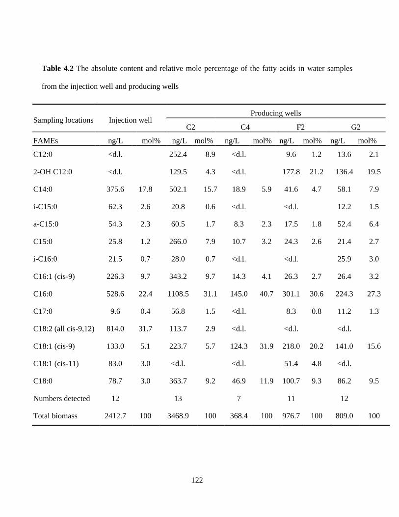
122
Table 4.2 The absolute content and relative mole percentage of the fatty acids in water samples
from the injection well and producing wells
Sampling locations Injection well Producing wells
C2 C4 F2 G2
FAMEs ng/L mol% ng/L mol% ng/L mol% ng/L mol% ng/L mol%
C12:0 <d.l.
252.4 8.9 <d.l.
9.6 1.2 13.6 2.1
2-OH C12:0 <d.l.
129.5 4.3 <d.l.
177.8 21.2 136.4 19.5
C14:0 375.6 17.8 502.1 15.7 18.9 5.9 41.6 4.7 58.1 7.9
i-C15:0 62.3 2.6 20.8 0.6 <d.l.
<d.l.
12.2 1.5
a-C15:0 54.3 2.3 60.5 1.7 8.3 2.3 17.5 1.8 52.4 6.4
C15:0 25.8 1.2 266.0 7.9 10.7 3.2 24.3 2.6 21.4 2.7
i-C16:0 21.5 0.7 28.0 0.7 <d.l.
<d.l.
25.9 3.0
C16:1 (cis-9) 226.3 9.7 343.2 9.7 14.3 4.1 26.3 2.7 26.4 3.2
C16:0 528.6 22.4 1108.5 31.1 145.0 40.7 301.1 30.6 224.3 27.3
C17:0 9.6 0.4 56.8 1.5 <d.l.
8.3 0.8 11.2 1.3
C18:2 (all cis-9,12) 814.0 31.7 113.7 2.9 <d.l.
<d.l.
<d.l.
C18:1 (cis-9) 133.0 5.1 223.7 5.7 124.3 31.9 218.0 20.2 141.0 15.6
C18:1 (cis-11) 83.0 3.0 <d.l.
<d.l.
51.4 4.8 <d.l.
C18:0 78.7 3.0 363.7 9.2 46.9 11.9 100.7 9.3 86.2 9.5
Numbers detected 12
13
7
11
12
Total biomass 2412.7 100 3468.9 100 368.4 100 976.7 100 809.0 100
123
In general, the fatty acids with carbon numbers of 15, 16 and 18 were major components in the
consortium. The PLFA profiles in all the wells were characterized by even-numbered fatty acids
(88%-94.9% of the total lipids) and their individual content was also higher than that of odd-
numbered fatty acids. In nearly all samples, C16:0 dominated the total fatty acids, and other fatty
acids with higher concentrations were C14:0, C16:1 (cis-9), C18:1 (cis-9), C18: 0, and 2-OH
C12:0. Higher concentrations of C18:2 (all cis-9,12) was only observed from injection water
samples. Fatty acids i-C15:0, a-C15:0 and a-C16:0 were main branched saturated fatty acids, but
their concentrations were all below 62.3 ng/L. MUFAs were represented by C16:1 (cis-9) and
C18:1 (cis-9), and their concentrations varied from 14.3 to 343.2 ng/L depending on the sources.
The sole PUFA was C18:2 (all cis-9,12) and it was only detected in the injection well and
producing wellbore C2. This PUFA was reported in great abundance in fungal fatty acids (Ruess
and Chamberlain 2010), but was also found in marine hydrocarbon degrading bacteria (Zhang et al.
2012). In particular, fatty acid 2-OH C12:0, which was only detected in water samples from
producing wells, was the single type of acid with hydroxy structure and interpreted as a portion of
Gram-negative bacteria in the microbial community (Ruess and Chamberlain 2010; Zelles 1999).
Ratios of specific PLFAs have also been used to track partly physiological change or stress
responses of the microorganisms (White and Ringelberg 1997). An increase in the ratios of
trans/cis PLFAs and cyclopropyl PLFAs to their monoenoic precursors (cy/pre) would typically
increase with stresses caused by insufficient nutrients, low pH, and other harsh environmental
conditions (Wixon and Balser 2013; Zelles 1999). The undetectable cyclopropyl fatty acids and
trans MUFAs in this study revealed that the communities existed under relatively suitable
conditions after long-term adaptation to the environments
124
The relationships between the PLFA components and the five sample units are shown in Figure
4.2. PLFA compositions in the reservoir samples of G2 and F2 exhibited similar trend while
producing wells C2 and C4 were obviously different in most of the PLFA patterns. The results
showed that the concentration differences in C18:2 (all cis-9,12), 2-OH C12:0, C18:1 (cis-11), and
i-C15:0 were the most significant when comparing samples from the injection well with samples
from the producing wells. Producing wells G2 and F2 exhibited similar PLFA compositions and
their total PLFA concentration (809.0 ± 71.7 ng/L and 976.7 ± 114.3 ng/L for G2 and F2,
respectively) were relatively close as well despite of their differences in redox potential. Microbial
community structures changed with varying temperature, mineralization, permeability and water
displacement factors in the oil reservoirs (Lin et al. 2014). The concentrations of total PLFAs
observed from the wells ranged from 368.4 to 3468.9 ng/L and were believed to be greatly
affected by the reservoir characteristics, e.g. geological properties and redox conditions.
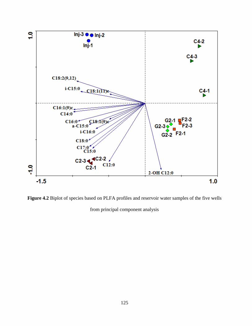
125
Figure 4.2 Biplot of species based on PLFA profiles and reservoir water samples of the five wells
from principal component analysis

126
4.3.4 Identification of SRB
Based on our fatty acid results, possible SRB biomarkers and significant PLFA profiles associated
with SRB were C18:1 (cis-9), C16:1 (cis-9), C18:1 (cis-11), i-C15:0, a-C15:0, C15:0, C17:0,
C14:0, and C18:0. Fatty acid C18:1 (cis-9) was found in unique presence among PLFA profiles of
Desulfotomaculum acetoxidans when Londry et al. (2004) cultivated the strain either
autotrophically or heterotrophically with acetate to investigate substrate usage by SRB species.
Mohanty et al. (2008) then used C18:1 (cis-9) as provisional indicative PLFA of SRB from the
genera of Desulfotomaculum and investigated the microbial community response and associated
sulfate reduction activities in the ethanol-amended slurries. Meanwhile, one species of the genus
Desulfobacter, D. latus, also contained C18:1 (cis-9) as the dominant PLFA (31%) when cultured
anaerobically (Kohring et al. 1994). This fatty acid also accounted for 15% and 14.2% in the major
PLFAs of SRB species, Desulfobacter curvatus and Desulfobotulus sapovorans (Kohring et al.
1994).
SOB have been examined by several researchers with their membrane lipids and fatty acids C16:1
(cis-9) or C18:1 (cis-11) was usually predominated in these studies (Guezennec et al. 1998; Jacq et
al. 1989; Katayama-Fujimura et al. 1982; Larkin 1980; Zhang et al. 2005). For instance, a
chemoautotrophic SOB, Thiomicrospira crunega, was isolated from a deep-sea hydrothermal vent
with large amounts of C16:1 (cis-9) or C18:1 (cis-11) dominant (Jannasch 1985). Two Thioploca
species from the Peru upwelling region were also characterized using fatty acid analysis and the
ratio of C16:1 (cis-9) ranged from 40.3 to 42.5% while the ratio of C18:1 (cis-11) was from 36 to
37.8% in these species (Mccaffrey et al. 1989). The dominance of C16:1 (cis-9) (16.7 to 37.4%)
and C18:1 (cis-11) (11.8 to 16.8%) has also been observed in thiotrophic bacterial mats in the
Barbados Trench (Guezennec and Fialamedioni 1996). It was then concluded that C16:1 (cis-9)
127
and C18:1 (cis-11) can be used as signature biomarkers for sulfur-oxidizing bacteria in H2S-rich
marine sediments (Zhang et al. 2005). On the other hand, Dowling et al. (1986) observed the
dominance of C16:1 (cis-9) (37.3%) from one of SRB species Desulfuromonas acetoxidans, while
the percentage of C18:1 (cis-11) was 0.8%. Interestingly, high levels of C16:1 (cis-9) (24.4%) and
C18:1 (cis-11) (24.1%) were also characteristic of Desulfotomaculum acetoxidans (Dowling et al.
1986) and it was proposed a Desulfotomaculum acetoxidans-like organism or group of organisms
dominated acetate-coupled sulfate reduction in estuarine and brackish sediments (Boschker et al.
1998). In our studies, the coexistence of C16:1 (cis-9) and C18:1 (cis-11) were only observed in
the injection well and wellbore F2, both of which were under relative oxidizing conditions. For the
other 3 production wells, only fatty acid C16:1 (cis-9) accounting for a lower ratio (3.3% -10.1%)
was found and the content of C18:1 (cis-11) was beyond our detection limit. Thus, fatty acid
C16:1 (cis-9) as the biomarker of SOB seemed not applicable in the reservoir wells with reducing
conditions, but the coexistence of C16:1 (cis-9) and C18:1 (cis-11) could indicate the presence of
SOB in the injection well and production well F2.
SRB were commonly found to contain odd-chain fatty acids, such as iso and anteiso C15:0 and
C17:0 (Edlund et al. 1985; Elvert et al. 2003; Goorissen et al. 2003; Zhang et al. 2005; Zhang et al.
2002), while the relative proportions of these fatty acids might be one criterion to evaluate the
possible presence of SRB. Significant amounts of C15:0 were observed when Desulfobulbus was
cultivated with all the growth substrates (propionate, lactate and H2/CO2) and this fatty acid alone
accounted for 23.0% of the total cellular fatty acids when on growth with propionate (Taylor and
Parkes 1983). The cellular fatty acids of Desulfovibrio desulfuricans grown on H2/CO2 was
dominated by branched iso fatty acids 75%, with i-C15:0 dominating (Taylor and Parkes 1983).
Fatty acid i-C15:0 was also reported to account for 8.9% and 23.0% when Desulfovibrio
128
desulfuricans grown on acetate and lactate (Londry et al. 2004). Beyond Desulfovibrio
desulfuricans, significant amounts of i-C15:0 was found in D. vulgaris, D. baculatus, D. simplex,
D. termitidis, Desulfomonas pigra (Vainshtein et al. 1992). Moreover, a-C15:0 with various
amounts (5-10%) predominanted in Desulfovibrio species, such as D. alcoholovorans, D.
carbinolicus, D. fructosovorans, D. giganteus, D. gigas, D. sulfodismutans (Vainshtein et al. 1992).
In addition, i-C15:0 was also reported as major compounds (48.6~68.3 in mol%) among
thermophilic Desulfotomaculum species when the strains grew with alcohols (Goorissen et al.
2003). Specially, the branched-chain fatty acids (iso, anteiso in C14-C18) have been widely
accepted as general indicators for gram-positive bacteria (Ruess and Chamberlain 2010), they (i.e.
i-C15:0 and a-C15:0) thus could not specifically represent the designated genera and species of
SRB. In addition, the odd chain PLFAs (e.g. C15:0 and C17:0) in bacteria are associated with the
composition of hydrocarbons in the substrate but not widely used as representative biomarkers for
specific microbes (Ruess and Chamberlain 2010). Therefore, the presence of these fatty acids (i.e.,
i-C15:0, a-C15:0, C15:0, and C17:0) could only be taken as enhanced references to describe SRB
occurrence when SRB were confirmed by other biomarkers based on published PLFA profiles of
SRB.
The even-number fatty acids C14:0 and C18:0 were both potential components in the PLFA
profile of SRB species and could be viewed as special references in determining the biomass of
microorganisms. Taylor and Parkes (1983) studied cellular fatty acid compositions of three sulfate
reducing species and found the major cellular fatty acids of Desulfobacter sp. grown on acetate
were dominated by C14:0 (23%) and C16:0 (44%). Additionally, a mole percent at 31.4% and
26.9% of fatty acid C14:0 was found in Desulfobacter AcBa and Desulfoarculus baarsii,
respectively, even though its content is below 6% for most of the SRB species (Kohring et al. 1994;
129
Londry et al. 2004). The PLFAs were reported to be dominated by C14:0 (19.1%), C16:0 (18.1%),
and C18:0 (25.9%) for Desulfomonile tiedjei when it was cultivated with formate/acetate and 3-
chlorobenzoate (Kohring et al. 1994). Furthermore, fatty acid C18:0 was present in many SRB
species with contents ranging from 0.1 to 10.6% (Kohring et al. 1994; Rütters et al. 2001; Taylor
and Parkes 1983).
According to the literature and PLFA profiles from the reservoir samples, the possible presence of
SRB and SOB were suggested. SRB species belonging to the genera Desulfotomaculum
(thermophilic) and Desulfobacter were present in a relatively high possibility (mainly indicated by
the biomarker C18:1 (cis-9)) and Desulfovibrio species may exist in the reservoir in a low
probability. The results of reservoir samples were consistent with the simultaneous microbial
community composition analysis based on DNA extraction (Okpala et al. 2017). As indicated by
the coexistence of C16:1 (cis-9) and C18:1 (cis-11), SOB species distributed in the injection well
and wellbore F2. Sulfidogenic communities Desulfotomaculum and Desulfovibrio were found in
the production water samples and were involved in the souring mediated corrosion of the oil-water
separation tanks in the north-eastern India oil fields (Agrawal et al. 2010). During another packed-
bed bioreactor study, SRB genus Desulfobacter were detected from a low-temperature oilfield in
Argentina through culturing and culture-independent techniques (Grigoryan et al. 2008).
Difference species of Desulfobacter, Desulfotomaculum and Desulfovibrio were also 3-
chlorobenzoate reported from other oilfield fluids (Grabowski et al. 2005; Lien and Beeder 1997;
Miranda-Tello et al. 2003; Tardy-Jacquenod et al. 1998). The three possible SRB genera were
consistent with the results of previous studies and were very likely the causes for souring in
oil reservoirs.
130
4.3.5 Patterns of microbial community structure and SRB migration behavior in reservoir
Changes in reservoir PLFA profiles can provide valuable guidance to explore the migration
behavior of target SRB communities in the course of above-ground water injection, water flooding
in the reservoir and above-ground water collection (Dijkman et al. 2010; Gieg et al. 2011). SRB,
notable for their utilization of sulfate as a terminal electron acceptor to produce reducing agent
hydrogen sulfide (Enning and Garrelfs 2014; Hubert and Voordouw 2007), are closely associated
with the redox potential of the reservoir samples. A redox potential of less than -100 mV at certain
pH conditions could provide a favorable environment for the reducing sulfidogenic process
(Church et al. 2007). Therefore, the redox potential of the reservoir samples can be used as a
supplementary index for identifying SRB thus aiding the PLFA profile analysis.
It was noted from the results that the redox potentials of the injection well, producing wells C4 and
G2 gradually decreased (Table 4.1). The varying redox potentials of the wells indicated different
levels of SRB/SOB activities in the reservoir microcosms. A comparison of the associated
microbial PLFA profiles thus can provide valuable information in the transformation patterns of
SRB/SOB communities in these wells. From Figure 4.3, samples from the relatively oxidizing
production wellbore C4 were detected with the lowest fatty acids content (368.4 ng/L) and the
simplest composition. The fatty acids detected from wellbore C4, such as C14:0, C16:0, C18:1
(cis-9), C18:0, were all high-abundance components in injection seawaters. It was speculated that
the main microbial components in wellbore C4 were very likely derived from microorganisms in
injection seawater and the main microbial species survived in reservoir conditions due to their high
biomass and adaptation to the reducing reservoir environment. As indicated by the presence of
C18:1 (cis-9), a group of SRB species very likely survived.
131
The wellbore G2, with higher fatty acid yields (809 ng/L) than wellbore C4, revealed more
obvious transformation of microbial community structure when seawater mixed with original
formation water beneath the seabed. All the seven fatty acids detected from wellbore C4 (likely
derived from microorganisms in seawater) were present in wellbore G2 and their content increased
to various extents. The dominating fatty acids C14:0, C16:0, C18:1 (cis-9), C18:0 in wellbore C4
and injection seawater were still in great abundance in wellbore G2. However, the dominating
fatty acids C16:1 (cis-9), C18:2 (all cis-9,12) and C18:1 (cis-11) in injection seawater were either
not discovered or had very limited distribution in wellbore G2 and C4. Interestingly, fatty acid 2-
OH C12:0, not observed in injection seawater, was found in great proportion from wellbore G2.
The change in microbial community composition might be due to two mechanisms: 1)
microorganisms inherited from seawater thrived in the reservoir environment, and 2) indigenous
microbes previously existed in the reservoir formation water before water flooding was applied.
The absence of fatty acids C18:2 (all cis-9,12) and C18:1 (cis-11) in producing wellbores G2 and
C4 would be caused by the environmental change from relatively oxidizing ocean to the anaerobic
reservoir. Therefore, the bacterial community structure changed to various degrees depending on
the reducing conditions in the reservoir while inheriting the main microbial components from
seawater.
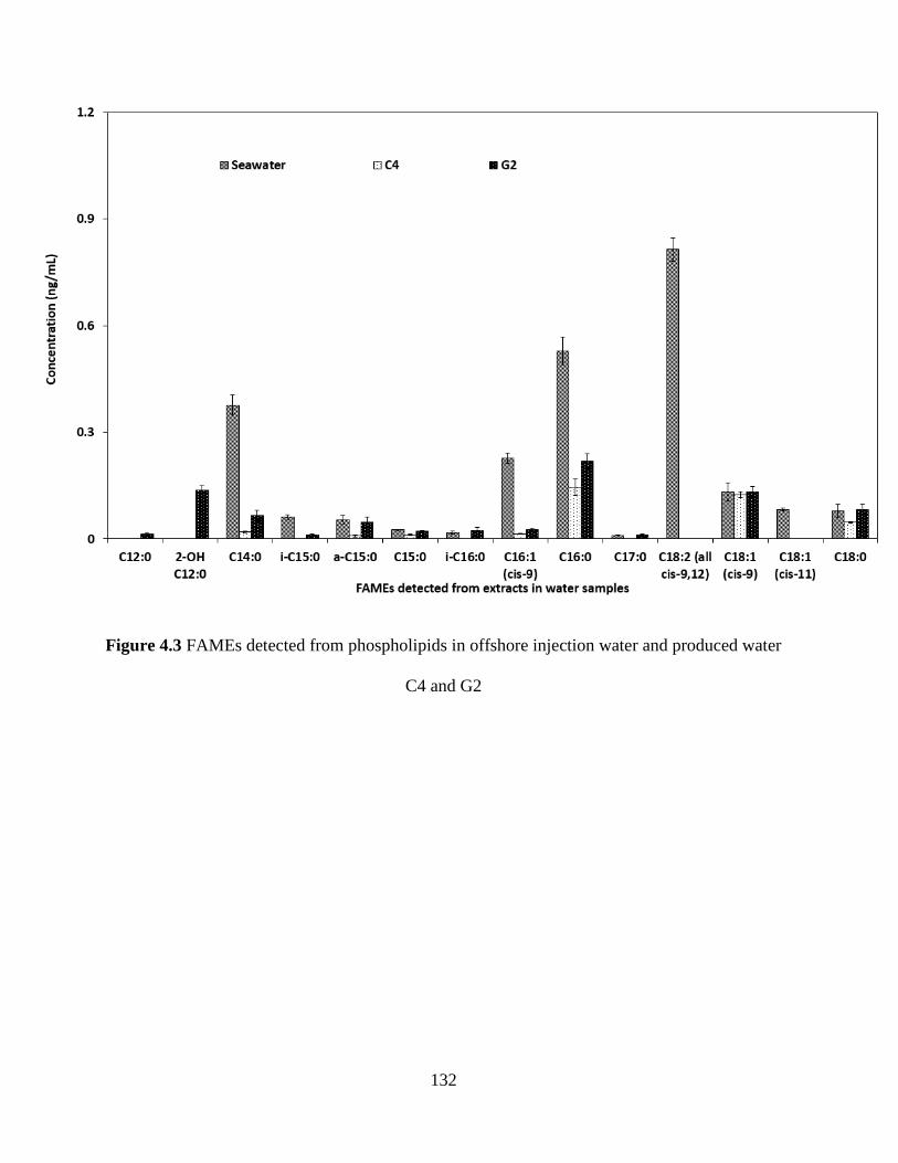
132
Figure 4.3 FAMEs detected from phospholipids in offshore injection water and produced water
C4 and G2
133
PLFA profile in the producing wellbore C2 was further compared with the microbial patterns in
the original injection well, as C2 exhibited the most reducing conditions in which the greatest
number of fatty acids and the highest biomass were found. Fatty acid C18:2 (all cis-9,12), not
detected in other producing wells, was found in a minor presence (2.9% of the total PLFAs) in
wellbore C2 even though the proportion was as high as 31.7% in seawater PLFA profiles. As
shown in Figure 4.4, PLFA profiles of wellbore C2 and injection seawater also shared most of the
same types of fatty acids. The geological environment near wellbore C2 was considered to be
more suitable than wellbore G2 for reducing microorganisms to survive and thrive in the water
flooding process. When cold seawater was mixed with hot fluids near the injection well, amenable
environmental conditions with suitable temperature was created for bacterial growth and bacteria
would progress into high-permeability zones and thrive there (Shibulal et al. 2014).
Two fatty acids with low-carbon numbers, C12:0 and 2-OH C12:0 were not detected from
injection seawater samples, but they were detected with concentrations of 252.4 ng/L and 129.5
ng/L in wellbore C2, respectively. The amenable conditions also stimulated the activities of SRB
and the H2S would ultimately be produced by SRB through the use of sulfate in seawater and oil
components existed in the reservoir (Usher et al. 2014). As revealed by the SRB biomarker C18:1
(cis-9), the most reducing condition in wellbore C2 was an indicator of the presence of H2S (120
ppm from the gas phase at the separator), which reflected the vibrant anaerobic microbial activities
(total PLFAs of 3468.9 ng/L) as well. In comparison to the pattern of PLFA concentrations in the
injection seawater, fatty acids C15:0, C17:0, and C18:0 increased significantly when water was
collected from wellbore C2, while compound C18:1 (cis-11) was below detection limits in this
structure transformation of microbial community. This phenomenon was attributed to the
disappearance of SOB species, as they are indicated by the coexistence of C16:1 (cis-9) and C18:1
134
(cis-11). The limited availability of oxygen and associated redox potential change are believed to
be among the main factors influencing the transformation microbial community structure.
Proportions of 9 PLFA subgroups (as mol%) stated in Section 3.4 provided specific information
on SRB and SOB species for the reservoir samples. As seen from Figure 4.5, while SOB species
(very likely Beggiatoa and Thioploca) were indicated by the coexistence of C16:1 (cis-9) and
C18:1 (cis-11) in the relatively oxidizing injection well and wellbore F2, they were below
detection limits in the reducing production wells (C2, G2 and C4) according to the disappearance
of fatty acid C18:1 (cis-11). The presence of SOB species could be regarded as one of the
indicators reflecting the relatively oxidizing environment in the producing well F2 (99 mV).
In general, the absolute content of compound C18:1 (cis-9) from producing wells was higher than
that from the injection well and this was very likely due to the increment of SRB species. Figure
4.5 indicated that the SRB biomarker C18:1 (cis-9) occupied a low proportion of total fatty acids
in the injection seawater. However, its proportion raised in nearly all the produced water samples
(C4, G2 and F2), a significant sign that the sulfidogenic species thrived in the warm reducing
reservoir environment.
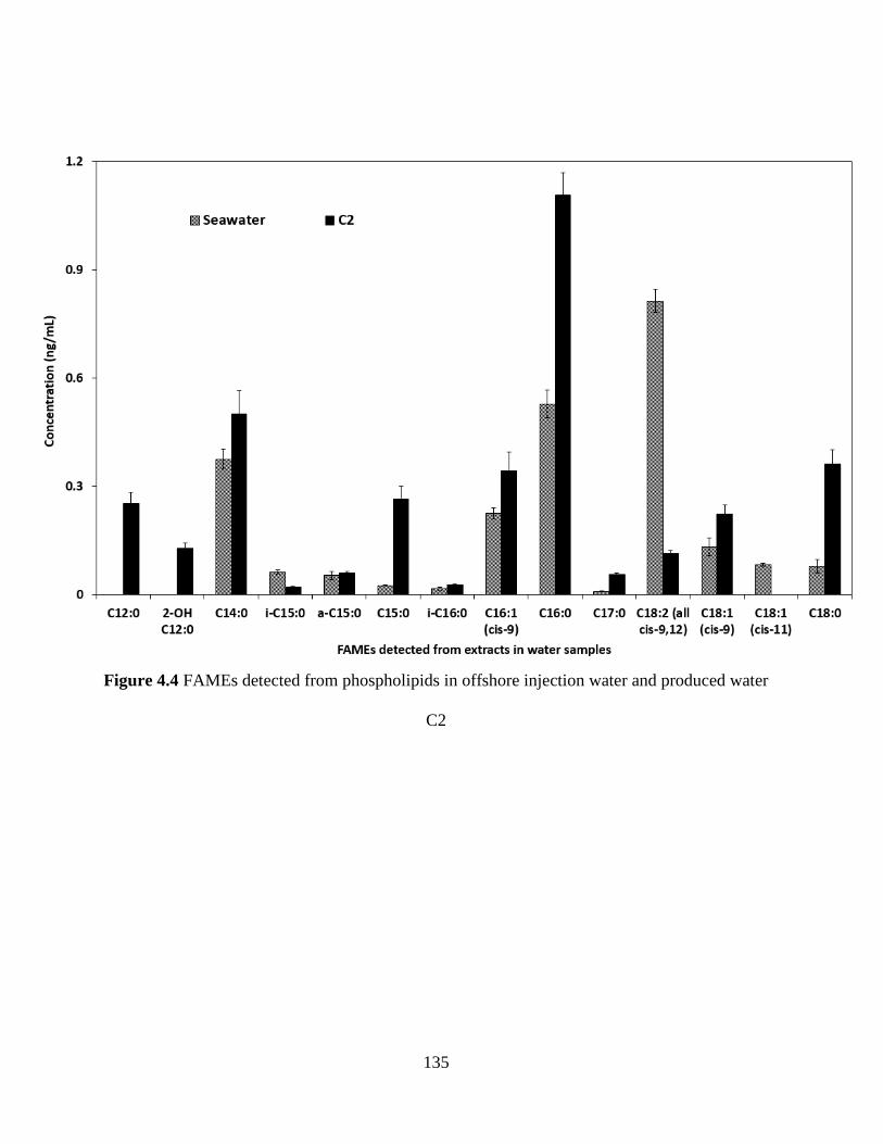
135
Figure 4.4 FAMEs detected from phospholipids in offshore injection water and produced water
C2
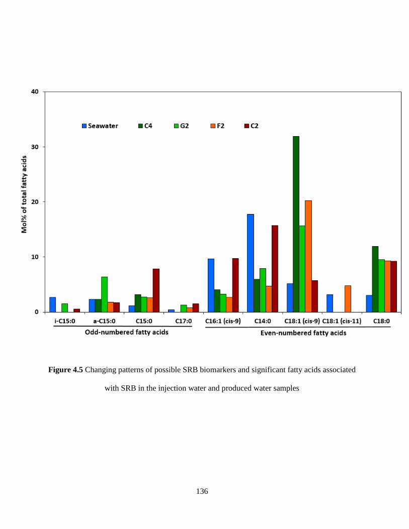
136
Figure 4.5 Changing patterns of possible SRB biomarkers and significant fatty acids associated
with SRB in the injection water and produced water samples
137
The SRB flourished in the anaerobic oil reservoir due to the conducive environment provided by
the removal of oxygen but the availability of sulfate from injected water, and the existence of
VFAs produced by microbial degradation of crude-oil components (Hasegawa et al. 2014). The
content of C18:1 (cis-9) in seawater (5.2%) and producing wellbore C2 (5.7%) were relatively low
when compared to other producing wells (ranging from 15.6% to 31.9%). This phenomenon might
be attributable to the relatively fierce competition for available carbon sources and other nutrients
that occurs among the microorganisms, since the two samples were both detected with high
biomass and the microbial consortia were diverse in the environment. Specially, C18:1 (cis-9) was
possible dominating SRB biomarker in producing wellbore C4, G2 and F2, while C16:1 (cis-9)
and C18:1 (cis-9) were both potential dominating SRB biomarker in producing wellbore C2. From
our results, SOB species were deemed to be absent in producing wellbore C2 according to the
appreciable presence of C16:1 (cis-9) but C18:1 (cis-11), and this differentiated C2 from the
injection well.
In addition, all the concentrations of detectable fatty acids from the 9 compounds were summed up
in Figure 4.6 to evaluate the sulfate-related microbial biomass in water samples. From the redox
potential results, producing well F2 showed almost the same redox condition as that in the
seawater. However, the abundance of PLFA in F2 decreased dramatically compared with the
injection water likely due to the disturbance of water flooding in reservoir F2. SOB and SRB
species were both detected in injection water and F2 as indicated by compounds C16:1 (cis-9),
C18:1 (cis-11), and C18:1 (cis-9). The limited microbial abundance in F2 could be attributed to
the restraints from harsh environments for indigenous microbes and the fact that only a small
group of exogenous bacteria adapted to the reservoir environment and survived (Ren et al. 2015).
Interestingly, the more reduced conditions were associated with higher bacterial abundance in
138
three producing wells (C4, G2, and C2) where very limited SOB biomarkers were found. Thus, the
presence of sulfate reducers and SOB, coupled with their biomass were closely associated with the
redox environment in the reservoir. In fact, the fatty acids analyzed in this study indicated that
most of the SRB were derived from the seawater and their microbial community structure varied
while the microorganisms adapted to different reservoir conditions. From Figure 4.6, a dynamic
change of microbial communities was obtained based on the distribution patterns of PLFA
abundance among the sample microcosms generated from the producing wells after water flooding
and from the injection well before water flooding. A five-step transformation pattern indicated as
(a) high SRB&SOB in seawater, (b) low SRB&SOB in F2, (c) low SRB in C4, (d) relatively high
SRB in G2, and (e) high SRB in C2 was used to elucidate the souring process induced by SRB. In
this regard, the PLFA profiles provided convincing reference and credible validation of SRB and
SOB activities in the designated reservoir.
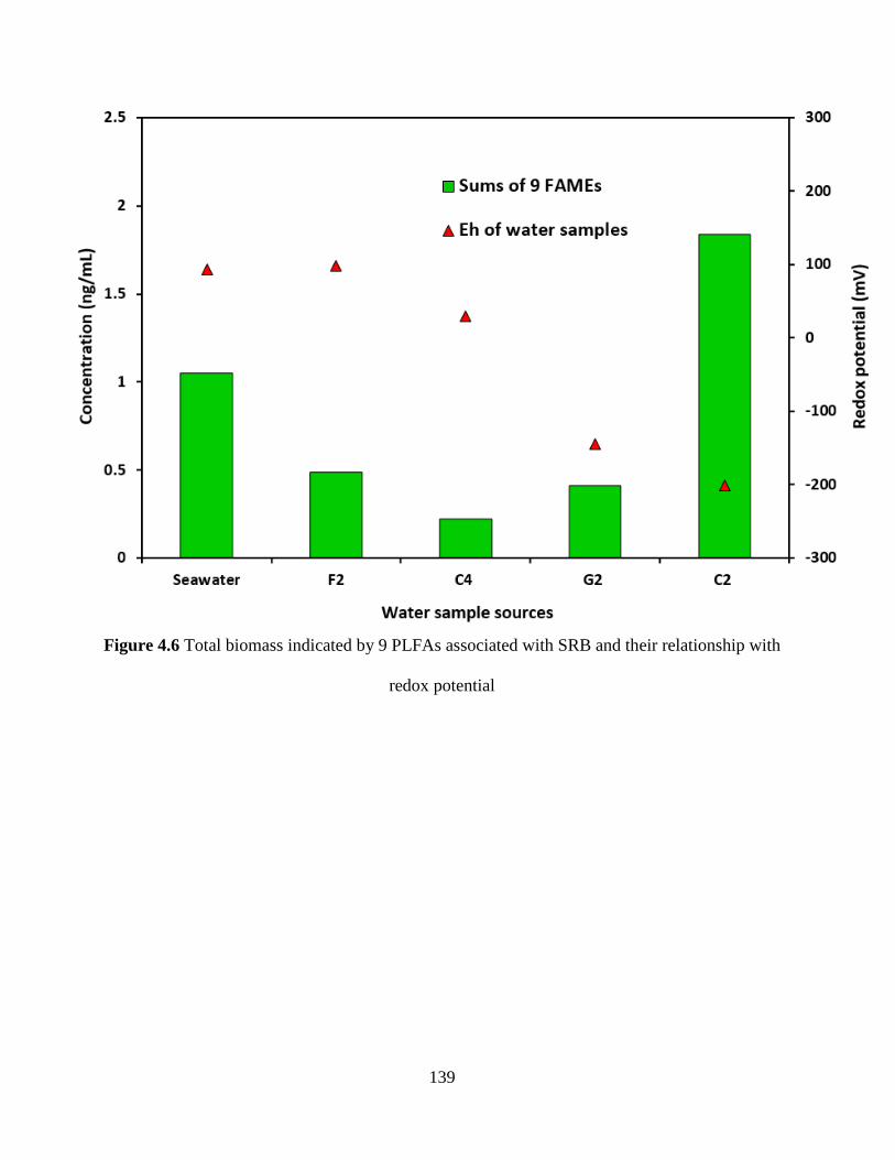
139
Figure 4.6 Total biomass indicated by 9 PLFAs associated with SRB and their relationship with
redox potential
140
4.4 Summary
In this study, PLFA analysis was conducted to characterize SRB in offshore reservoir injection
water and produced water samples. The in-situ measurements were performed to obtain parameters
including temperature, pH, DO, and redox potential. Analytical performance in the PLFA
determination was evaluated through the repeatability of GC, recoveries in the methodology and
capillary chromatograph. The fatty acid profiles were elucidated between samples of the injection
well and four producing wells, and the small differences with respect to the structure of the sulfate
reducing community were determined. The PLFA profiles were closely related to the redox
potential results and possible SRB biomarkers coupled with significant fatty acids related to SRB
were selected to analyze the possible SRB species. SRB Desulfotomaculum, Desulfobacter,
Desulfovibrio and SOB species were proposed to exist in various possibilities and their
transformation patterns in the reservoir were concluded. In this case, the fatty acid analysis
provided reasonable results to trace microorganisms in the offshore reservoir water samples and
can be potentially used as a routine monitoring tool in the implementation of reservoir souring
mitigation strategies.
141
CHAPTER 5
ISOLATION OF NRB FROM AN OFFSHORE RESERVOIR AND
THE ASSOCIATED BIOSURFACTANT PRODUCTION3
3 The contents in the chapter will result in the potential publication:
F. Fan, B. Zhang, P.L. Morrill, T. Husain, Isolation of nitrate-reducing bacteria from an offshore
reservoir and the associated biosurfactant production, RSC Advances, 8 (2018) 26596-26609.
Role: Fuqiang Fan solely worked on this study and acted as the first author of this manuscript. Dr.
Baiyu Zhang is my supervisor. Dr. Penny Morrill and Dr. Tahir Husain helped guide the research
and polish the manuscript.
142
5.1 Background
The activity of SRB has long been a major concern in oilfield water systems and offshore
petroleum reservoirs because these microorganisms are one of the main causative agents of
reservoir souring as well as MIC (Xu et al. 2012). Many oilfield products (produced water, gas, etc)
contain sulfides (H2S and HS-) as a result of the activity of SRB or other sulfidogenic bacteria.
SRB reduce sulfate in the injection water to sulfide, while oxidizing degradable organic electron
donors present in the offshore oil reservoir. The undesirable production of sulfides in offshore oil
reservoirs results in reduced quality of produced hydrocarbons, health and safety risks for
operators, and increased corrosivity of produced fluids (Hubert and Voordouw 2007; Okoro et al.
2014).
Although sulfides can be removed chemically after their production, in situ elimination through
continuous nitrate/nitrite injection has also proven to be effective, as demonstrated both in model
column (An et al. 2010; Grigoryan et al. 2008; Hubert et al. 2005), inland (Grigoryan et al. 2009;
Shartau et al. 2010) and offshore field studies (Dunsmore et al. 2006; Larsen et al. 2004). Nitrate
injection changes the microbial community in the subsurface from mainly SRB to one enriched in
NRB, which include the nitrate reducing, sulfide oxidizing bacteria (NR-SOB) that oxidize H2S
directly and the heterotrophic NRB (hNRB) that compete with SRB for degradable organic
electron donors. Additionally, both types of NRB also promote SRB inhibition via production of
nitrite formed in nitrate reduction pathways (Greene et al. 2003). NRB are well known for their
denitrifying capacity in which nitrates or nitrites are converted into nitrogen-containing gases. This
function enables NRB to play significant roles in the global nitrogen cycle (Rothenberger et al.
2009) and mitigation and control of sulfide induced reservoir souring problems in offshore oil
143
fields (Gieg et al. 2011). However, understanding of the detailed microbial mechanisms involved
in NRB-SRB interactions during nitrate/nitrite injections for offshore reservoirs souring mitigation
is currently limited.
Anaerobic, indigenous NRB have the potential to produce specific biosurfactants in deep
geological porous offshore reservoirs with diverse physiochemical in situ conditions.
Biosurfactants are surface-active compounds with both lipophilic and hydrophilic structural
moieties produced by microorganisms, which either adhere to the cell surface or are secreted
extracellularly in the growth medium (Rodrigues et al. 2006). These surface active molecules
reduce surface tension at air-water interfaces and interfacial tension in both aqueous solutions and
hydrocarbon mixtures (Ghribi et al. 2012). Notably, biosurfactants have various advantages over
their commercially manufactured counterparts because of their lower toxicity, biodegradable
nature, ease of biosynthesis and the effectiveness under extreme conditions such as temperature,
pH, and salinity (Mulligan 2005; Pacwa-Płociniczak et al. 2011). Fallon et al. (2010) have
confirmed that biosurfactants can be naturally derived from NRB. As microorganisms capable of
utilizing hydrocarbons as carbon and energy sources, NRB will produce surface-active agents as
by-products to facilitate hydrophobic degradation (Ron and Rosenberg 2002). The selective
surface-active agents or biosurfactants produced by NRB increase the surface area of hydrophobic
water-insoluble substrates (low molecular weight biosurfactants) and increase the solubility (high
molecular weight biosurfactants), thus improving the bioavailability of hydrocarbons for NRB
(Pacwa-Płociniczak et al. 2011). When emulsion occurs closely to the cell surface of NRB, each
cluster of cells creates its own microenvironment and stimulate the growth of NRB in oil and gas
reservoirs (Ron and Rosenberg 2002). This mechanism enables NRB to out‐compete harmful SRB
for basic carbon nutrients. The SRB will be inhibited from producing new hydrogen sulfide/iron
144
sulfide, and the existing sulfides will be removed by bacterial degradation, resulting in effective
control of offshore reservoir souring. Thus, at this point biosurfactants are interesting by-products
involved in the SRB/NRB competition.
To investigate how biosurfactants affect NRB-SRB competition in a reservoir, successful
screening of biosurfactant producing NRB and generation of associated biosurfactants are needed.
Oil reservoirs could provide a unique hydrocarbon-rich environment for biosurfactant screening
and the enrichment of diverse biosurfactant producers (Christofi and Ivshina 2002). The extreme
conditions (e.g., high temperature, high pressure, and low oxygen concentration) in oil reservoirs
would formulate a microbial community that may be distinguished from others (Grabowski et al.
2005). So far, biosurfactant producers identified from oil reservoirs are mainly from inland
reservoirs and limited to the genera of Bacillus (Al-Bahry et al. 2013; She et al. 2011; Simpson et
al. 2011; Wang et al. 2011; Youssef et al. 2007) and Pseudomonas (Gudiña et al. 2012; Lotfabad
et al. 2009; Pruthi and Cameotra 2003). Currently, very limited marine biosurfactant producers
from offshore oil and gas fields have been reported and it was unclear whether they were NRB
species or not. Cai et al. (2015) isolated marine biosurfactant producers from crude oil, formation
water, drilling mud, and treated produced water samples in offshore oil and gas fields. The
genotype and phylogenetic relation of these isolates were investigated and biosurfactant producers
were primarily found in the genera of Rhodococcus and Halomonas. However, most of these
species reported are general biosurfactant producers and they were screened under aerobic
conditions, which were not prevalent in the reducing reservoir environments. Hui et al. (2012)
evaluated the microbial community structure and functionally distinct groups in three kinds of
produced water samples from the shallow, mesothermic and low-salinity Daqing oil reservoir
using both culture-dependent and culture-independent methods. The isolates affiliated to
145
Pseudomonas stutzeri PTG4-15 (DP26, BP39, and PW5) were initially identified as nitrate
reducing bacteria, biosurfactant producing bacteria, and polymer-producing bacteria. This
indicates that anaerobic indigenous NRB have the potential to produce specific biosurfactants in
the reservoir.
Until now, biosurfactant producing NRB isolated from oil reservoirs have been rarely reported,
and associated biosurfactant production is extremely limited in the literature. No previous study
tackled the isolation of biosurfactant producing NRB from offshore reservoirs and subsequent
anaerobic biosurfactant production. Therefore, the aim of this study was to screen NRB strains
from an offshore reservoir, and conduct biosurfactant production and characterization. The
research outputs will not only help identify NRB and generate biosurfactants under anaerobic
conditions, but also provide technical and methodological support for further identifying NRB-
SRB interactions and generating methodologies for effective offshore reservoir souring control in
the future.
5.2 Materials and Methods
5.2.1 Source and collection of inoculum
Produced water samples in an offshore water flooding reservoir were collected for screening novel
biosurfactant producers. Injection wells on the platforms were injected with nitrate/nitrite to
stimulate the growth of indigenous nitrate reducing microorganisms in the reservoir or exogenous
NRB strains from seawater injection process. Produced water was collected in 1-liter sterile glass
bottles and then sealed immediately to maintain anoxic conditions. The samples were stored in a
refrigerator in darkness before shipping. Subsequently, the samples were packaged with frozen ice
packs and transported to the laboratory for enrichments and bacterial isolations. The major
146
constituents of the produced water include (in wt/vol) chloride, 4.0%; sodium, 2.5%; sulfate,
0.13%; calcium, 0.12%; and bicarbonate, 0.076%. Water samples were stored at 4 °C and were
taken for enrichment culture within one week of collection.
5.2.2 Isolation and identification of NRB
5.2.2.1 Growth media
Coleville synthetic brine (CSB) medium was selected for the NRB culturing based on the
properties of produced water and NRB growth requirement. The recipe was composed of (in 1 liter)
NaCl, 7.0 g; MgSO4 ∙7H2O, 0.68 g; CaCl2 ∙2H2O, 0.24 g; NH4Cl, 0.02 g; KH2PO4, 0.027 g;
NaC2H3O2 ∙3H2O, 0.68 g; KNO3, 1.0 g; NaHCO3, 1.9 g; resazurin, 0.0001 g; and ND trace metals
(0.5 mL H2SO4, 2.28 g MnSO4 ∙H2O, 0.5 g ZnSO4 ∙7H2O, 0.5 g H3BO3, 0.025 g CuSO4 ∙5H2O,
0.025 g Na2MoO4 ∙2H2O, and 0.045 g CoCl2 ∙ 6H2O per liter), 50 ml/liter (Gevertz et al. 2000). The
medium pH was then adjusted to between 7.0 and 7.5. After autoclaving, cooling, and
equilibration of the medium with chamber gas overnight, sterilized 2.5% (w/v) Na2S∙9H2O (final
concentration of 0.02% (w/v)) was added to remove residual oxygen. Solid growth media were
prepared by adding a certain amount of agar (2% w/v).
5.2.2.2 Enrichment and isolation
Enrichments were prepared by adding 5 ml of produced water to 125 mL medium in sterile conical
flasks under anaerobic conditions. To ensure the growth of microorganisms, three enrichment
recipes with various carbon sources were prepared in conical flasks, respectively. The first one
was adopted from Hui et al. (2012) and contained (in 1 liter) peptone, 20 g; beef extract, 10 g,
KNO3, 1.0 g; NaCl, 0.7 g; KCl, 0.7 g; MgCl2 ∙6H2O, 10 g; MgSO4 ∙7H2O, 5.4 g; CaCl2 ∙2H2O, 1.0
147
g. The second recipe was a raw screening medium which contained (in 1 liter) peptone, 5.0 g; beef
extract, 3.0 g; KNO3, 1.0 g and trace metals (same as in CSB medium). The third recipe used
citrate as the carbon source and contained L-asparagine 0.5 g; KNO3, 1.0 g; trisodium citrate, 0.85
g; MgSO4 ∙7H2O, 0.5 g; FeCl3 ∙6H2O, 0.05 g; CaCl2 ∙2H2O, 0.24 g; and KH2PO4, 0.05 g. Trace
metals were added following the trace element solution in CSB medium. All the media pHs were
buffered between 7.2 - 7.5 and further treated with sterilized Na2S∙9H2O.
Enrichment was initially conducted in a chamber filled with nitrogen until observable turbidity
occurred. The bacterial consortia were then inoculated into new media for further acclimatization.
The average bacterial acclimatization period was 15 days. After seven periods, 5 ml of each
medium broth were transferred into fresh liquid CSB medium and the citrate medium. The second
round of bacterial acclimatization in liquid CSB and citrate medium were conducted through three
periods. After that, the consortia were serially diluted to 106, 10
5, and 10
4 and then spread on solid
CSB and citrate medium agar plate, respectively. The resulting plates were incubated at room
temperature in a nitrogen-filled environment.
Routine growth and maintenance of broth isolates were in CSB medium. Bacterial growth status
was detected by observing an increase in optical density at 600 nm (OD600) or the cell dry weight
filtered from medium broth.
5.2.2.3 Identification and phylogenetic characterization of isolates
The purified isolates were then subjected to 16S ribosomal RNA sequencing and amplified with
universal bacterial primers 27F (5‘-AGA GTT TGA TYM TGG CTC AG-3‘) and 16SR10 (5‘-
ACG GCT ACC TTG TTA CGA CT-3‘). An aliquot of the each culture was used for a DNA
template in a polymerase chain reaction (PCR) using the primer pair. After gel electrophoresis
148
confirmation of successful PCR reaction, PCR products were subjected to a clean-up process and
measured by a NanoDrop spectrophotometer to determine the concentrations. Lastly, sequencing
reactions with the last PCR products were conducted and measured with Applied Biosystems 3730
DNA Analyzer in Creait Network of Memorial University of Newfoundland. The obtained DNA
sequences were aligned with previously published sequences from the GenBank database of
National Center for Biotechnology Information (NCBI) using the Basic Local Alignment Search
Tool (BLAST) program.
5.2.3 Screening of the NRB isolates for biosurfactant producers
5.2.3.1 Biosurfactant producing media
According to the morphological properties of the strains on the agar plate and associated 16S
rRNA results, 5 strains were selected for subsequent biosurfactant producer screening. Two media
with glycerol and glucose as carbon sources were selected for anaerobic biosurfactant production,
respectively. The first one modified from Zhao et al. (2014) contained (in 1 liter) glycerol, 46.6 g;
NaNO3, 3.0 g; K2HPO4, 4.0 g; KH2PO4, 5.7 g; MgSO4∙7H2O, 0.4 g; CaCl2 ∙2H2O, 0.17 g; NaCl, 2 g;
and yeast extract, 2.7 g. The second one was adjusted from a previous medium in NRB screening
medium and contained (in 1 liter) glucose, 10 g; NaNO3, 3.0 g; K2HPO4, 3.36 g; KH2PO4, 3.4 g;
MgSO4∙7H2O, 0.68 g; CaCl2 ∙2H2O, 0.24 g; NaCl, 2g; yeast extract, 3.0 g; and FeCl3, 0.05 g. Trace
metals were also added into the two media as described previously. After autoclaving, cooling, and
equilibration of the medium with chamber gas overnight, sterilized Na2S solution was also added
as mentioned above.
149
5.2.3.2 Biomass determination
Each isolate was incubated using the same conditions as in biosurfactant producing procedure for
228 hours. Subsequently, 10 mL culture sample was filtered through a pre-weighted 0.22 µm filter
membrane and washed 2 times with 10 mL distilled water. The filter membrane was then dried in
oven for 24 h and cooled down in desiccators before measuring the final weight. The biomass was
determined as cell dry weight (g/L).
5.2.3.3 Parafilm test
A 25 μL aliquot of bacterial broth was added to the hydrophobic surface of a parafilm. The shape
and the diameter of the droplet on the surface were inspected after 3 min. The negative control was
prepared with 0.5 M phosphate buffer (Morita et al. 2007).
5.2.3.4 Drop collapsing test
Drops of the cell-free supernatant were placed on an oil-coated, solid surface. The polar water
molecules are repelled from the hydrophobic surface with the absence of surfactants in the liquid
and the drops remain stable whereas the droplet spread out slightly or even collapse with the
presence of surfactants (Youssef et al. 2004).
5.2.3.5 Emulsification activity assay
The emulsification activity (E24, Eq.1) of the culture broth was determined by addition of 5 mL
culture broth to 5 mL hexadecane or kerosene and vortexed for 2 min to create an optimum
emulsion. Tests were performed in duplicate for quality assurance purpose and the results were
expressed using the average of two measurements.
150
E24 = HEL / HS ×100% (Eq.1)
where HEL is the height of the emulsion layer and HS is the height of the total solution.
5.2.3.6 Surface tension measurement
Culture samples were centrifuged at 10,000 rpm for 20 min to remove microbial cells and the
supernatant was subject to surface activity measurements. Surface tension was determined with a
surface tensiometer (DuNouy Tensiometer, Interfacial, CSC Scientific) at room temperature
according to the ring method. The values reported were the mean of triplicate measurements.
5.2.4 Performance demonstration of the selected strain
According to the results, one isolate was selected to further demonstrate the performance of
biosurfactant production. The two surfactant production media with glycerol and glucose as carbon
sources were used to investigate the performance of biosurfactant production. The incubation was
conducted at 30 °C while shaking at 200 rpm.
The isolate was firstly inoculated into the flask containing 20 mL fresh glucose medium and
incubated using the above conditions for 48 h. Then a 200 µL aliquot of the culture broth was
inoculated into each flask with 20 mL glycerol or glucose producing medium. The following two
tests at time intervals of 0, 12, 24, 36, 48, 60, 84, 128, 156, 180, 204 and 228 h were sampled with
the whole flask, respectively.
The OD600 of samples was employed as the index of bacterial growth (Safari et al. 2012).
Absorbance was measured at λ = 600 nm using a UV–Visible spectrophotometer. All culture
media were shaken for 5 s to homogenize the media before OD600 determination. The absorbance

151
of freshly autoclaved medium was adjusted to 0 as the blank control. The measurement of OD600
was performed in triplicate by sampling three times.
To observe the nitrate consumption by NRB, nitrate concentration in the culture was determined
using a two-wavelength approach (Gopalan et al. 2005). A rapid measurement of sample
absorption at 220 nm (A220) was conducted in a quartz cuvette and organic matter interference
was eliminated through the second measurement at 275 nm (A275). Nitrate concentration was
determined from standard curves prepared with NaNO3 (0.2–8 mg/L). Additionally, surface
tension changes over the period were monitored using the methodology mentioned above. CMD
analysis was conducted to evaluate the concentration of biosurfactants produced during the
investigation of cultivation kinetics.
5.2.5 Biosurfactant production and characterization
5.2.5.1 Extraction of the biosurfactant product
The procedures of biosurfactant extraction from the culture broth mainly followed the protocol
according Silva et al. (2010) after 228 hours of incubation of CX3 in glycerol medium. The culture
broth was initially centrifuged at 11000 rpm for 20 min to remove bacterial cells. The supernatant
was then acidified to pH 2.0 with concentrated HCl (1 mol/L) and kept at 4°C overnight to reduce
the rhamnolipid solubility. The biosurfactant product was further recovered through the addition of
two volumes of chloroform: methanol (v/v, 2: 1) mixture. After shaking the mixture for 2 h, the
lower organic phase was collected and evaporated to dryness by a rotary evaporator. The final
precipitate was collected.
152
5.2.5.2 Determination of critical micellar concentrations (CMC)
CMC is defined as the surfactant concentration necessary to initiate micelle formation. The CMC
of generated biosurfactant product was determined by plotting the surface tensions as a function of
biosurfactant concentration and it was defined from the intercept of two straight lines extrapolated
from the concentration-dependent and concentration-independent sections (De Oliveira et al. 2013).
5.2.5.3 TLC analysis
TLC analysis was conducted to preliminarily characterize the purified biosurfactant product. Ten
microliter biosurfactant solution at a concentration of 200 mg/L in methanol was applied to a
20×20 silica gel TLC plate (Sigma Aldrich). The biosurfactant product was separated using
CHCl3:CH3OH:H2O (70:10:0.5, v/v/v) as developing solvent system with different color
developing reagents. For detection of lipopeptide biosurfactants, ninhydrin reagent (0.5 g
ninhydrin in 100 mL anhydrous acetone) was sprayed on the dry plates and red spots were
visualized after keeping the plate at 105 ºC for 5 min. Anthrone reagent (1 g anthrone in 5 mL
sulfuric acid mixed with 95 mL ethanol) was used to reveal the presence of glycolipid
biosurfactants in yellow spots. Also, lipid content was further visualized by iodine chamber.
5.2.5.4 Lipid class determination
Lipid class composition was determined using an Iatroscan Mark VI TLC with flame ionization
detector (FID), silica coated Chromarods and a three-step development method (Parrish 1999).
The lipid extracts were applied to the Chromarods and focused to a narrow band using 100%
acetone. The first development system was hexane:diethyl ether:formic acid (99.95:1:00.05). The
rods were developed for 25 minutes, removed from the system for 5 minutes and replaced for 20
153
minutes. The second development was for 40 minutes in hexane:diethyl ether:formic acid
(79:20:1). The final development system had two steps, the first was 100% acetone for two 15
minute time periods, followed by two 10 minute periods in chloroform:methanol:chloroform-
extracted water (5:4:1). Before each solvent system the rods were dried in a constant humidity
chamber. After each development system the rods were scanned in the Iatroscan and the data were
collected using Peak Simple software (ver 3.67, SRI Inc). The Chromarods were calibrated using
standards from Sigma Chemicals (Sigma Chemicals, St. Louis, Mo., USA).
5.2.5.5 Fatty acid analysis
For all samples, lipid extracts were transesterified using sulfuric acid and methanol for 1 hour at
100oC. The FAMEs developed from the extracts were analyzed on a HP 6890 GC system with
FID and a 7683 autosampler. The GC column was a ZB wax+ (Phenomenex, U.S.A.). The column
length was 30m with an internal diameter of 0.32mm. The column temperature began at 65 ºC and
held this temperature for 0.5 minutes. The temperature ramped to 195 o
C at a rate of 40 ºC/min,
held for 15 minutes then ramped to a final temperature of 220 o
C at a rate of 2 ºC/min. This final
temperature was held for 0.75 minutes. The carrier gas was hydrogen and flowed at a rate of 2
ml/minute. The injector temperature started at 150 oC and ramped to a final temperature of 250
oC
at a rate of 120 o
C/minute. The detector temperature stayed constant at 260 o
C. Peaks were
identified using retention times from standards purchased from Supelco, 37 component FAME
mix (Product number 47885-U), Bacterial acid methyl ester mix (product number 47080-U),
PUFA 1 (product number 47033) and PUFA 3 (product number 47085-U). Chromatograms were
integrated using the Varian Galaxie Chromatography Data System, version 1.9.3.2. A quantitative
standard purchased from Nu-Chek Prep, Inc (product number GLC490) was used to check the GC

154
column about every 300 samples (or once a month) to ensure that the areas returned were as
expected.
5.2.5.6 Fourier transform infrared spectroscopy (FT-IR) analysis
IR spectroscopy was used for structure analysis of the extracted biosurfactant product based on the
oscillation patterns of chemical bonds at characteristic frequencies. The IR absorption spectrum of
the dried biosurfactant product was measured on a Bruker Tensor 27 FT-IR using 16 scans over
the range of 500-4000 cm-1
(KBr beamsplitter). The signals were collected in transmittance mode
with a Zn-Se attenuated total reflectance (ATR) spectroscopy which are commonly used for
rhamnolipid analysis (Heyd et al. 2008).
5.2.5.7 Stability characterization
The effect of temperature, pH, and salinity on the surface activity of generated biosurfactants was
investigated by changing surrounding conditions (Abouseoud et al. 2008b). Generally, 1 CMC of
biosurfactant solution was prepared and maintained at a constant temperature of 0, 20, 40, 60, 80,
100 °C for 120 min and cooled at room temperature to determine the thermal stability of the
biosurfactant. The pH influence on the biosurfactant activity was determined by adjusting the
biosurfactant solutions in the range 2.0-12.0 using HCl (2 N) and NaOH (2 N) solutions, and the
effect of salinity on the surface activity of the biosurfactant product was assessed by using various
concentrations of sodium chloride (0.5-20% in w/v). In each case, the stability of the biosurfactant
solution was evaluated by the change of surface tension values (1 CMC in all tests) and
determined in triplicate.
155
5.3 Results and Discussion
5.3.1 Phylogenetic analysis and morphological characteristics of isolates
These isolates were incubated under anaerobic conditions with nitrate as the electron acceptor and
acetate as the carbon source and electron donor. Most of the isolated NRB species were
anaerobically slow-growing and mature colonies were formed around ten days on the CSB
medium. As shown in Table 5.1, with sequences obtained by doing a Blast search, a similarity of
99% or 100% to Pseudomonas stutzeri was found by the alignment of the 16S rRNA gene
sequences of all the selected five NRB strains isolated from offshore produced water.
The morphology characteristics of isolates were summarized in Table 5.1. In general, all the
species could be summed into two categories: the brown one with entire margins and the light
color or transparent ones with raised or curled margins. Notably, some isolates, HF5 and FX6
formed flocculants in liquid biosurfactant producing medium. The components of the flocculants
were studied extensively before and were a mixture of polysaccharides, proteins, lipids,
glycolipids and glycoproteins (Zheng et al. 2008). Bioflocculants could be produced by various
functional microorganisms and are biodegradable, environmentally friendly and harmlessness to
humans. Microorganisms with high bioflocculant-producing ability thus can be utilized to produce
bioflocculants and can be used in industrial fields such as drinking and wastewater treatment,
downstream processing, and fermentation processes (Salehizadeh and Shojaosadati 2001). The
flocculated cells were previously reported to be immobilized inside the reactor for a continuous
fermentation system without cell separation and recycling units, and the yields from the reactor
were clean broths for further ethanol production (Andrietta et al. 2008). Viewed from this
156
perspective, the two strains have promising applications in batch, fed-
batch or continuous fermentation reactors for industrial nitrate removal activities.
The strain CX3 was noticeable due to its different margin and color in the agar plate during
morphology examination when compared with others (Table 5.1). During the growth on CSB
medium, the isolate formed visible regular and glistening colonies with the colony diameters
ranging from 1 to 3 mm. The precise taxonomic positions of the microbes were subsequently
determined through the genotypic analysis on the basis of partial 16S rRNA sequencing. Unlike
the other four strains, Pseudomonas stutzeri CX3 occupied a unique branch in the phylogenetic
tree generated from the five Pseudomonas stutzeri strains and other Pseudomonas species (Figure
5.1). The selected Pseudomonas stutzeri strains formed a stable phyletic group within a
heterogeneous cluster of Pseudomonas xanthomarina, and other Pseudomonas stutzeri strains,
which distributed in ubiquitous environments and were identified among denitrifiers in natural
materials (Lalucat et al. 2006). P. stutzeri is a highly diverse species of great physiological and
ecological versatility and is of interest due to their specific activities in nitrification and
denitrification processes (Sikorski et al. 2002). The strains of Pseudomonas stutzeri were also
anaerobically isolated from various environmental samples and used in the degradation of aliphatic
and aromatic hydrocarbons (Grimberg et al. 1996; Kaczorek et al. 2012).
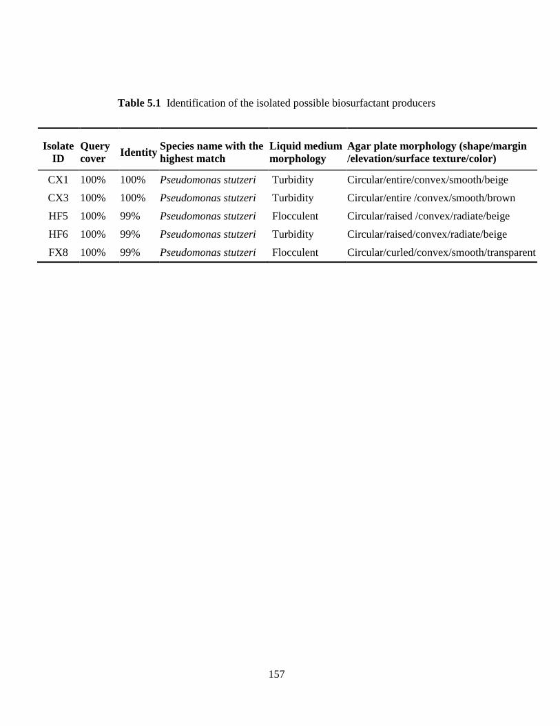
157
Table 5.1 Identification of the isolated possible biosurfactant producers
Isolate
ID
Query
cover Identity
Species name with the
highest match
Liquid medium
morphology
Agar plate morphology (shape/margin
/elevation/surface texture/color)
CX1 100% 100% Pseudomonas stutzeri Turbidity Circular/entire/convex/smooth/beige
CX3 100% 100% Pseudomonas stutzeri Turbidity Circular/entire /convex/smooth/brown
HF5 100% 99% Pseudomonas stutzeri Flocculent Circular/raised /convex/radiate/beige
HF6 100% 99% Pseudomonas stutzeri Turbidity Circular/raised/convex/radiate/beige
FX8 100% 99% Pseudomonas stutzeri Flocculent Circular/curled/convex/smooth/transparent
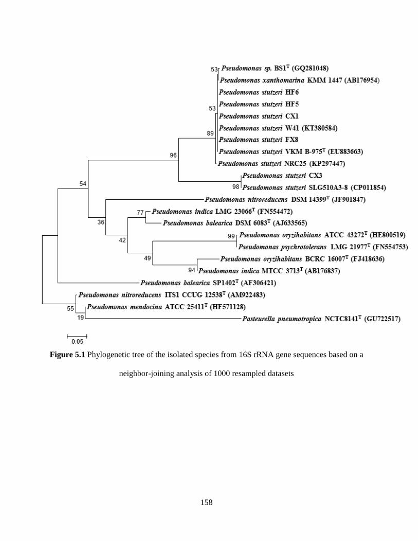
158
Figure 5.1 Phylogenetic tree of the isolated species from 16S rRNA gene sequences based on a
neighbor-joining analysis of 1000 resampled datasets
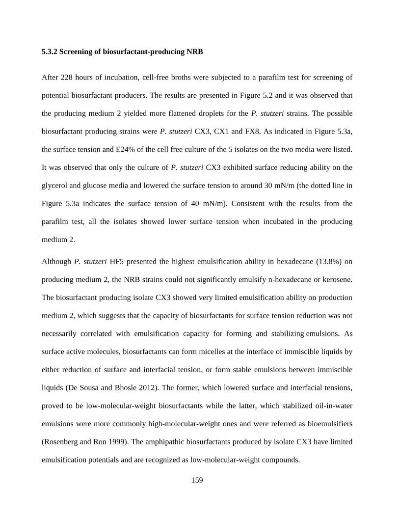
159
5.3.2 Screening of biosurfactant-producing NRB
After 228 hours of incubation, cell-free broths were subjected to a parafilm test for screening of
potential biosurfactant producers. The results are presented in Figure 5.2 and it was observed that
the producing medium 2 yielded more flattened droplets for the P. stutzeri strains. The possible
biosurfactant producing strains were P. stutzeri CX3, CX1 and FX8. As indicated in Figure 5.3a,
the surface tension and E24% of the cell free culture of the 5 isolates on the two media were listed.
It was observed that only the culture of P. stutzeri CX3 exhibited surface reducing ability on the
glycerol and glucose media and lowered the surface tension to around 30 mN/m (the dotted line in
Figure 5.3a indicates the surface tension of 40 mN/m). Consistent with the results from the
parafilm test, all the isolates showed lower surface tension when incubated in the producing
medium 2.
Although P. stutzeri HF5 presented the highest emulsification ability in hexadecane (13.8%) on
producing medium 2, the NRB strains could not significantly emulsify n-hexadecane or kerosene.
The biosurfactant producing isolate CX3 showed very limited emulsification ability on production
medium 2, which suggests that the capacity of biosurfactants for surface tension reduction was not
necessarily correlated with emulsification capacity for forming and stabilizing emulsions. As
surface active molecules, biosurfactants can form micelles at the interface of immiscible liquids by
either reduction of surface and interfacial tension, or form stable emulsions between immiscible
liquids (De Sousa and Bhosle 2012). The former, which lowered surface and interfacial tensions,
proved to be low-molecular-weight biosurfactants while the latter, which stabilized oil-in-water
emulsions were more commonly high-molecular-weight ones and were referred as bioemulsifiers
(Rosenberg and Ron 1999). The amphipathic biosurfactants produced by isolate CX3 have limited
emulsification potentials and are recognized as low-molecular-weight compounds.
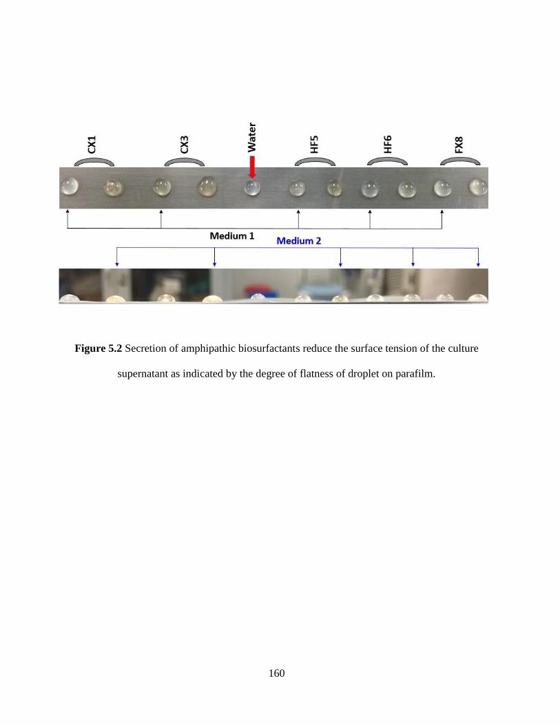
160
Figure 5.2 Secretion of amphipathic biosurfactants reduce the surface tension of the culture
supernatant as indicated by the degree of flatness of droplet on parafilm.
161
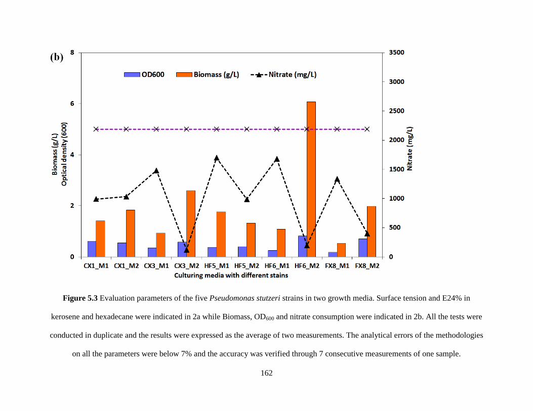
162
Figure 5.3 Evaluation parameters of the five Pseudomonas stutzeri strains in two growth media. Surface tension and E24% in
kerosene and hexadecane were indicated in 2a while Biomass, OD600 and nitrate consumption were indicated in 2b. All the tests were
conducted in duplicate and the results were expressed as the average of two measurements. The analytical errors of the methodologies
on all the parameters were below 7% and the accuracy was verified through 7 consecutive measurements of one sample.
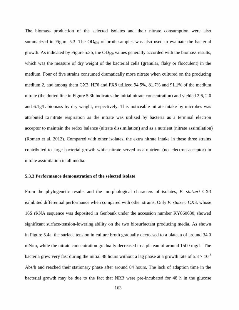
163
The biomass production of the selected isolates and their nitrate consumption were also
summarized in Figure 5.3. The OD600 of broth samples was also used to evaluate the bacterial
growth. As indicated by Figure 5.3b, the OD600 values generally accorded with the biomass results,
which was the measure of dry weight of the bacterial cells (granular, flaky or flocculent) in the
medium. Four of five strains consumed dramatically more nitrate when cultured on the producing
medium 2, and among them CX3, HF6 and FX8 utilized 94.5%, 81.7% and 91.1% of the medium
nitrate (the dotted line in Figure 5.3b indicates the initial nitrate concentration) and yielded 2.6, 2.0
and 6.1g/L biomass by dry weight, respectively. This noticeable nitrate intake by microbes was
attributed to nitrate respiration as the nitrate was utilized by bacteria as a terminal electron
acceptor to maintain the redox balance (nitrate dissimilation) and as a nutrient (nitrate assimilation)
(Romeo et al. 2012). Compared with other isolates, the extra nitrate intake in these three strains
contributed to large bacterial growth while nitrate served as a nutrient (not electron acceptor) in
nitrate assimilation in all media.
5.3.3 Performance demonstration of the selected isolate
From the phylogenetic results and the morphological characters of isolates, P. stutzeri CX3
exhibited differential performance when compared with other strains. Only P. stutzeri CX3, whose
16S rRNA sequence was deposited in Genbank under the accession number KY860630, showed
significant surface-tension-lowering ability on the two biosurfactant producing media. As shown
in Figure 5.4a, the surface tension in culture broth gradually decreased to a plateau of around 34.0
mN/m, while the nitrate concentration gradually decreased to a plateau of around 1500 mg/L. The
bacteria grew very fast during the initial 48 hours without a lag phase at a growth rate of 5.8 × 10⁻³
Abs/h and reached their stationary phase after around 84 hours. The lack of adaption time in the
bacterial growth may be due to the fact that NRB were pre-incubated for 48 h in the glucose
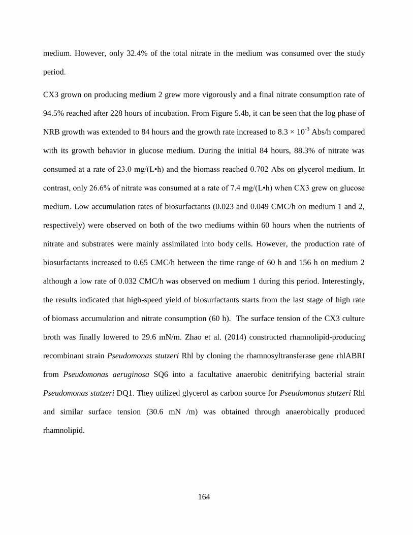
164
medium. However, only 32.4% of the total nitrate in the medium was consumed over the study
period.
CX3 grown on producing medium 2 grew more vigorously and a final nitrate consumption rate of
94.5% reached after 228 hours of incubation. From Figure 5.4b, it can be seen that the log phase of
NRB growth was extended to 84 hours and the growth rate increased to 8.3 × 10⁻³
Abs/h compared
with its growth behavior in glucose medium. During the initial 84 hours, 88.3% of nitrate was
consumed at a rate of 23.0 mg/(L•h) and the biomass reached 0.702 Abs on glycerol medium. In
contrast, only 26.6% of nitrate was consumed at a rate of 7.4 mg/(L•h) when CX3 grew on glucose
medium. Low accumulation rates of biosurfactants (0.023 and 0.049 CMC/h on medium 1 and 2,
respectively) were observed on both of the two mediums within 60 hours when the nutrients of
nitrate and substrates were mainly assimilated into body cells. However, the production rate of
biosurfactants increased to 0.65 CMC/h between the time range of 60 h and 156 h on medium 2
although a low rate of 0.032 CMC/h was observed on medium 1 during this period. Interestingly,
the results indicated that high-speed yield of biosurfactants starts from the last stage of high rate
of biomass accumulation and nitrate consumption (60 h). The surface tension of the CX3 culture
broth was finally lowered to 29.6 mN/m. Zhao et al. (2014) constructed rhamnolipid-producing
recombinant strain Pseudomonas stutzeri Rhl by cloning the rhamnosyltransferase gene rhlABRI
from Pseudomonas aeruginosa SQ6 into a facultative anaerobic denitrifying bacterial strain
Pseudomonas stutzeri DQ1. They utilized glycerol as carbon source for Pseudomonas stutzeri Rhl
and similar surface tension (30.6 mN /m) was obtained through anaerobically produced
rhamnolipid.
165
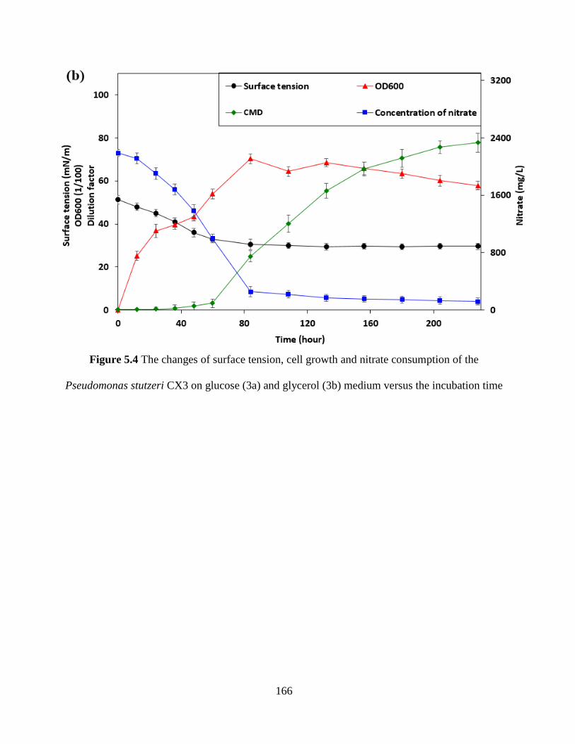
166
Figure 5.4 The changes of surface tension, cell growth and nitrate consumption of the
Pseudomonas stutzeri CX3 on glucose (3a) and glycerol (3b) medium versus the incubation time
167
5.3.4 Characterization of NRB-generated biosurfactant product
After the extraction, the final concentrated biosurfactant product by P. stutzeri CX3 was with
a yellow-brown colour. It could lower the surface tension of distilled water from 72.2 mN/m to
30.4 mN/m. The biosurfactant product was further characterized and the stability under various
environmental conditions was also examined to facilitate its potential use in the fields.
5.3.4.1 CMC of biosurfactants
CMC is an important indicator which determines the capability of biosurfactants to mobilize crude
oil from contaminated soils or the sand-oil mixture into the aqueous biosurfactant solution. Low
CMC is correlated with high efficiency of the biosurfactant product (Bharali et al. 2014). The
CMC value of the isolated product was determined by measuring the surface tension of different
concentrated solutions of the product with a tensiometer, and a sudden change in the surface
tension was observed. A lower CMC value of 35 mg/L was derived for the extracted biosurfactant
product. The biosurfactant product proved to be highly efficient when compared with the typical
CMC values (10-230 mg/L) previously reported for biosurfactants produced from different
microbial sources (Gogoi et al. 2016; Nitschke et al. 2005).
5.3.4.2 Preliminary TLC analysis
The lipid content in the biosurfactant product was preliminarily determined through the general
staining of iodine. TLC analysis also showed that light red spot on silica gel plates was generated
when using ninhydrin as color developing reagent, suggesting that only a small part of lipopeptide
existed in this biosurfactant. In contrast, the evident yellow spot proved the abundant presence of
168
glycolipid. Zhao et al. (2014) constructed an engineered strain Pseudomonas stutzeri Rhl and used
it for heterologous production of Rhamnolipid under anaerobic conditions. Accordingly, the main
components of glycolipid biosurfactants were most probable to be rhamnolipids. All the results
indicated that the product was very likely to be a mixture of a small part of lipopeptides and a large
part of glycolipid biosurfactants.
5.3.4.3 Lipid and fatty acid analysis
TLC-FID analysis revealed that this biosurfactant product was a mixture of seven lipid
components, which was dominated by the acetone mobile polar lipids and phospholipids. As
shown in Table 5.2, 46.7% of the components were acetone mobile polar lipids (mainly
glycolipids), accounting for nearly half the total lipids. Phospholipids, as the most abundant lipids
in cell membranes and dominating constituent in our lipids matrix (31.9 %), may be partially
originated from cell debris co-precipitated with the biosurfactant product during the extraction
process. The fact could also be inferred from the presence of small amounts of sterols in lipids (1%)
as cholesterol is an important structural component of a phospholipid bilayer (Van Der Paal et al.
2017). Additionally, hydrocarbons, triacylglycerols and free fatty acids were also found in various
amounts in the lipid mixture.
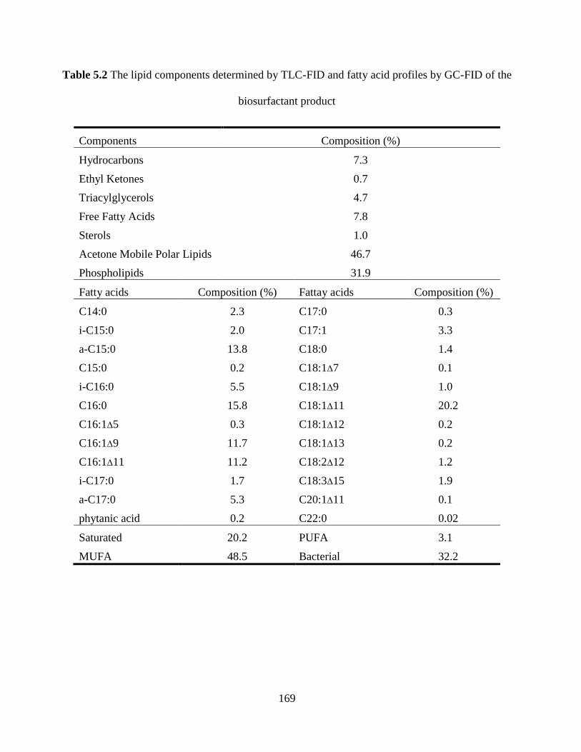
169
Table 5.2 The lipid components determined by TLC-FID and fatty acid profiles by GC-FID of the
biosurfactant product
Components Composition (%)
Hydrocarbons 7.3
Ethyl Ketones 0.7
Triacylglycerols 4.7
Free Fatty Acids 7.8
Sterols 1.0
Acetone Mobile Polar Lipids 46.7
Phospholipids 31.9
Fatty acids Composition (%) Fattay acids Composition (%)
C14:0 2.3 C17:0 0.3
i-C15:0 2.0 C17:1 3.3
a-C15:0 13.8 C18:0 1.4
C15:0 0.2 C18:1Δ7 0.1
i-C16:0 5.5 C18:1Δ9 1.0
C16:0 15.8 C18:1Δ11 20.2
C16:1Δ5 0.3 C18:1Δ12 0.2
C16:1Δ9 11.7 C18:1Δ13 0.2
C16:1Δ11 11.2 C18:2Δ12 1.2
i-C17:0 1.7 C18:3Δ15 1.9
a-C17:0 5.3 C20:1Δ11 0.1
phytanic acid 0.2 C22:0 0.02
Saturated 20.2 PUFA 3.1
MUFA 48.5 Bacterial 32.2
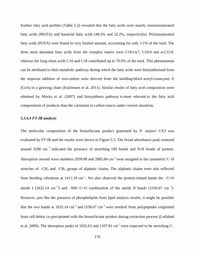
170
Further fatty acid profiles (Table 5.2) revealed that the fatty acids were mainly monounsaturated
fatty acids (MUFA) and bacterial fatty acids (48.5% and 32.2%, respectively). Polyunsaturated
fatty acids (PUFA) were found in very limited amount, accounting for only 3.1% of the total. The
three most abundant fatty acids from the complex matrix were C18:1w7, C16:0 and a-C15:0,
whereas the long-chain acids C16 and C18 contributed up to 70.9% of the total. This phenomenon
can be attributed to their metabolic pathway during which the fatty acids were biosynthesized from
the stepwise addition of two-carbon units derived from the building-block acetyl-coenzyme A
(CoA) to a growing chain (Estelmann et al. 2011). Similar results of fatty acid composition were
obtained by Morita et al. (2007) and biosynthesis pathway is more relevant to the fatty acid
compositions of products than the variations in carbon source under current situations.
5.3.4.4 FT-IR analysis
The molecular composition of the biosurfactant product generated by P. stutzeri CX3 was
evaluated by FT-IR and the results were shown in Figure 5.5. The broad absorbance peak centered
around 3298 cm−1 indicated the presence of stretching OH bonds and N-H bonds of protein.
Absorption around wave numbers 2939.08 and 2885.84 cm−1 were assigned to the symmetric C–H
stretches of –CH2 and –CH3 groups of aliphatic chains. The aliphatic chains were also reflected
from bending vibrations at 1411.19 cm−1. We also observed the protein-related bands the –C=O
amide I (1632.14 cm −1) and –NH/–C=O combination of the amide II bands (1536.67 cm −1
).
However, just like the presence of phospholipids from lipid analysis results, it might be possible
that the two bands at 1632.14 cm−1 and
1536.67 cm−1
were resulted from polypeptides originated
from cell debris co-precipitated with the biosurfactant product during extraction process (Lotfabad
et al. 2009). The absorption peaks of 1035.63 and 1107.81 cm−1 were expected to be stretching C–
171
O–C bonds, which indicated the presence of polysaccharide or polysaccharide-like substances in
the biosurfactant product. The FTIR spectra provided strong evidence for the presence
of glycolipids and lipopeptides in the biosurfactant product (Aparna et al. 2012; Joshi et al. 2008).
5.3.4.5 Stability studies
The applicability of biosurfactants in several fields is highly associated with their stability under
various environmental conditions. Therefore, after the generation of biosurfactant product by P.
stutzeri CX3, its stability was tested under a wide range of temperature, pH value and salinity
while maintaining the biosurfactant concentrations at CMC value (35 mg/L). From the results
shown in Figure 5.6, the biosurfactant activity was retained and insignificantly affected by most of
the experimental settings, especially only small variations in surface tension values were
determined under the wide range of temperatures between 0 and 121 °C and salinities between 0%
and 20%. Biosurfactants are widely applied in petroleum, pharmaceutical, health care and food
processing industries (Abouseoud et al. 2008a; Khopade et al. 2012) due to its robust stability and
the findings indicated the potential use of our product in these areas. As indicated in Figure 5.6,
biosurfactant solution achieved the lowest surface tension at a temperature around 40 °C, whereas
heating up to 100 °C or even autoclaving (121 °C) caused no significant effect on its thermal
stability. The results are accorded with the properties of rhamnolipid indicated by Abdel-Mawgoud
et al. (2009), which are main components of the biosurfactant product and maintain stable
structure over the temperature settings. The extreme thermal stability was reported by Seghal
Kiran et al. (2010) from Brevibacterium aureum MSA13 and Aparna et al. (2012) from
Pseudomonas sp. 2B, which enables the biosurfactant product to be applied in a high-temperature
reservoir.
172
Figure 5.5 FT-IR transmittance spectrum of the extracted biosurfactant product generated by
Pseudomonas stutzeri CX3 grown on glycerol medium
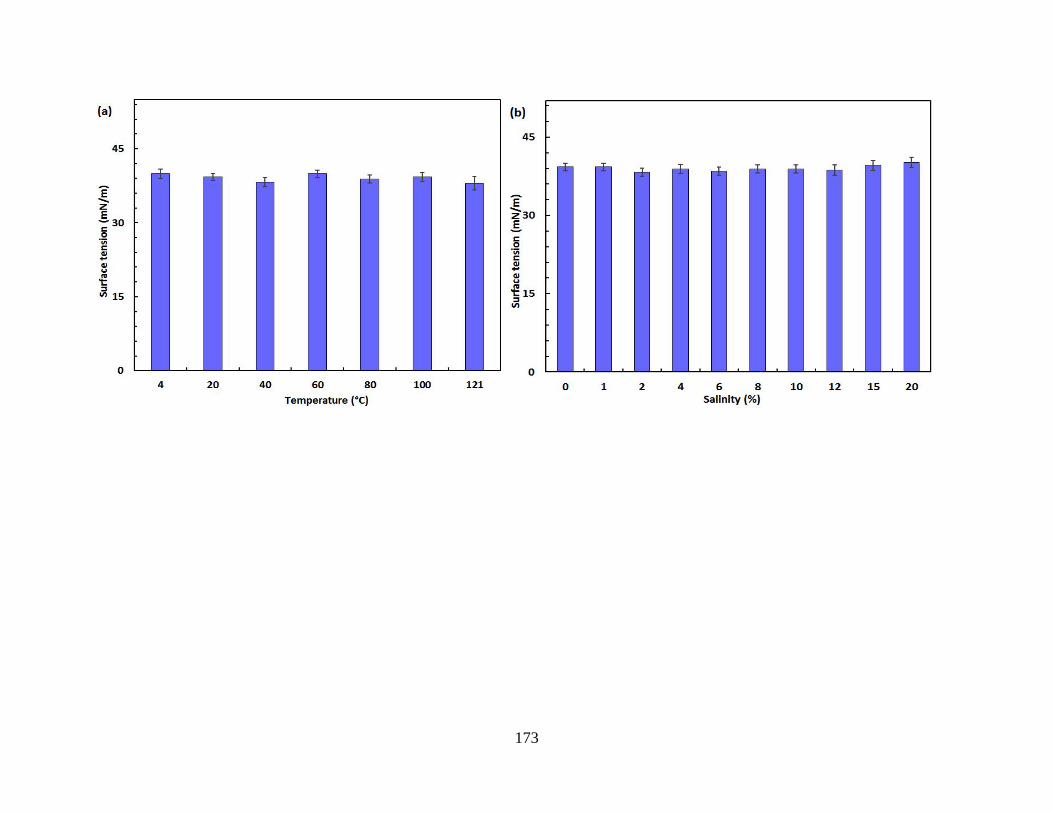
173
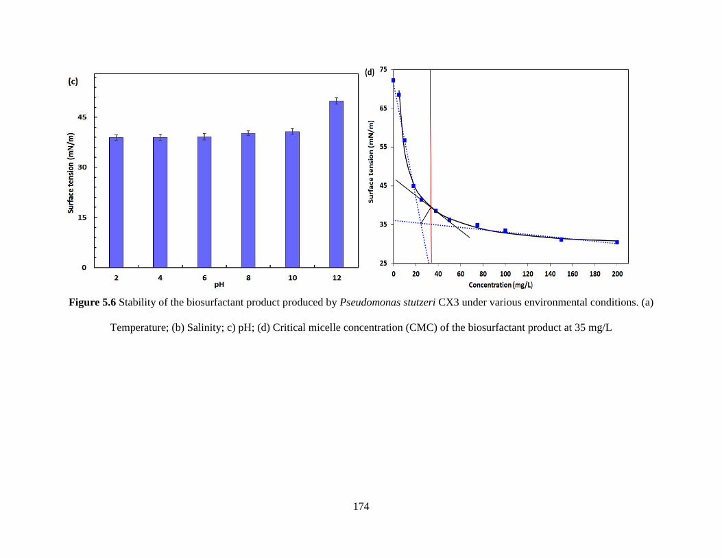
174
Figure 5.6 Stability of the biosurfactant product produced by Pseudomonas stutzeri CX3 under various environmental conditions. (a)
Temperature; (b) Salinity; c) pH; (d) Critical micelle concentration (CMC) of the biosurfactant product at 35 mg/L
175
The effect of salinity on the biosurfactant activity was examined and negligible changes were
observed in the increased concentration of NaCl up to 20% (w/v) (Figure 5.6b). Electrolytes in the
bulk solutions will shield the carboxylate groups of the rhamnolipid molecules, causing them to
behave more like nonionic than anionic surfactants (Helvaci et al. 2004). Although a relatively
lower surface tension was obtained at 2% NaCl concentration, the differences over the wide brine
concentration levels of 0 to 20% were not significant (<5%). The steady performance of surface
tension in salty waters allows the product to be potentially used in reservoir MEOR or
environmental bioremediations under saline environments. Singh and Tripathi (2013) isolated a
strain of Pseudomonas stutzeri from the formation water of an Indian coalbed and the isolate
produced copious amount of biosurfactant with the supplementation of coal. The biosurfactant
with rhamnolipid nature showed considerable emulsifying ability and great potential for in situ
biotransformation of coal into methane and bioremediation of PAHs from the oil-contaminated
sites. Correspondingly, our product with considerable surface-tension-lowering ability will reduce
the capillary force that holds oil and porous solid media together to mobilize the crude oil or oil in
MEOR or environmental bioremediation activities.
The surface activity of biosurfactant solution remained relatively stable between pH 2 and 10. A
negative biosurfactant performance was observed at pH 12 in which the surface tension raised to
49.9 mN/m. This detrimental effect is possibly caused by structure alteration of the biosurfactant
product under extreme pH conditions. The polar head of anionic rhamnolipids was more
negatively charged under more alkaline conditions (Silva et al. 2010). This is reflected by the fact
of increased solubility of the product.
As amphiphilic molecules released extracellularly by microorganisms, biosurfactants are known to
be beneficial for their producers in various ways. By promoting wetting, solubilization and

176
emulsification of various types of organics, biosurfactants could increase the surface area between
the oil and water phases, thereby increasing the bioavailability of entrapped oil in the porous
media (Pacwa-Płociniczak et al. 2011). Thus, biosurfactants could improve the nutrient conditions
of NRB producers and enhance the competence of NRB over SRB. Additionally, several
biosurfactants proved to have antibiotic effects (Rodrigues et al. 2006), which may have the
potential to inhibit the growth of SRB. Moreover, biosurfactants were found to be important agents
in the connection between microbial communities and biofilm formations (Osterreicher-Ravid et al.
2000), which may synergistically improve the resistance of the NRB producers to harsh
environment. Therefore, the generated biosurfactant product, once injected into the soured
reservoir externally, has great potential to assist NRB outcompeted SRB through these
mechanisms. The effects of NRB produced biosurfactants in offshore reservoir NRB/SRB
interactions, coupled with other environmental implications of this product, need to be further
investigated.
5.4 Summary
In this study, the offshore petroleum-reservoir brines following nitrate/nitrite injection were used
for the anaerobic screening of biosurfactant producing NRB. After periodic enrichment and
sophisticated screening of the microorganisms, five typical denitrifying strains were isolated and
found to be the species of Pseudomonas stutzeri according to their 16S rRNA sequencing results.
The isolates were further screened for possible biosurfactant producers on glycerol and glucose
media. The strain P. stutzeri CX3 was confirmed with biosurfactant production capacity through a
series of biosurfactant characterization tests (e.g. drop collapsing test, parafilm test and surfaced
tension determination). Better surface tension lowering ability was observed from the glycerol
177
medium and the consumption of nitrate by NRB was found in positive correlation with bacterial
growth and surface tension reduction. CX3 was selected to further demonstrate the performance of
biosurfactant production through two production media over a 228-hour monitoring. The nitrate
concentrations and surface tensions on the two media were both reduced to a relatively stable level
within 84 hours during which OD600 reached relatively high levels as well over the period. The
biosurfactant product generated by P. stutzeri CX3 was defined with a CMC as low as 35 mg/L,
and further characterized by TLC, GC-FID and FT-IR analysis. The main components of the
biosurfactant were recognized as glycolipids. The biosurfactant product demonstrated stable
performance during different environmental conditions with a wide range of temperature, pH
values, and salinity, which indicated its potential applications in environmental bioremediation,
petroleum and other various industrial fields. The successful isolation and identification of
biosurfactant producing NRB from laborious screenings on offshore reservoir samples, coupled
with subsequent biosurfactant characterization would provide new insight into NRB-SRB
interactions for offshore reservoir souring control investigations.
178
CHAPTER 6
INTERACTIONS OF SRB, NRB SCREENED FROM OFFSHORE
OIL RESERVOIR AND NRB PRODUCED BIOSURFACTANTS IN
MICROCOSMS4
4 The contents in the chapter will result in the potential publication:
F. Fan, B. Zhang, J. Liu, Q. Cai, W. Lin, B. Chen. Interactions of sulfate reducing bacteria (SRB),
biosurfactant producing nitrate reducing bacteria (NRB) screened from an offshore reservoir and
NRB produced biosurfactant in microcosms (submitted to the journal of Environmental Pollution)
Role: Fuqiang Fan solely worked on this study and acted as the first author of this manuscript. Dr.
Baiyu Zhang is my supervisor. Dr. Bing Chen helped guide the research and polish the manuscript. Jibin Liu, Qinhong Cai and Weiyun Lin participated in conducting experiments.
179
6.1 Background
Oilfield reservoir souring caused by SRB is among the major issues faced by the petroleum
industry, which occurred both in terrestrial and offshore oil production operations (Gieg et al. 2011;
Voordouw et al. 2009). The injection of water in secondary oil recovery to maintain pressure and
press the oil towards the production wells (Hook et al. 2014) often leads to the production of
sulfide (souring) by resident SRB or other sulfidogenic bacteria. The sulfide, as a byproduct of
SRB in respiration, is closely correlated with the deterioration of crude oil quality, health
and safety issues, and increased corrosion risk and operating cost (Hubert and Voordouw 2007;
Zhao et al. 2009). Hydrogen sulfide was also reported to inhibit the growth and metabolism of
biosurfactant producing bacteria, thus exerting negative effects on MEOR (Zhao et al. 2015a).
Nitrate injection is a promising strategy for souring control due to its relatively non-toxic and cost-
effective attributes and its convenience in practical distribution (Gieg et al. 2011; Xue and
Voordouw 2015). The mechanism of nitrate injection includes the boost of hNRB to outcompete
SRB for available nutrients (biocompetitive exclusion), the promotion of NR-SOB to directly
oxidize sulfide and the SRB repression through resultant nitrite (Fida et al. 2016; Gieg et al. 2011;
Hubert and Voordouw 2007). Nitrate-mediated souring control has been extensively studied in the
laboratory (Callbeck et al. 2011; Chen et al. 2017; Zhao et al. 2009) and in the field (Bodtker et al.
2008; Shartau et al. 2010; Voordouw et al. 2009). The studies mainly focused on improving
nitrate/nitrite inhibitory effect on SRB-induced sulfide production through varying environmental
factors (An et al. 2017; Chen et al. 2017; Gadekar et al. 2006; Hubert et al. 2003; Okpala et al.
2017) or combination with other protocols (Greene et al. 2006; Nemati et al. 2001; Xue and
Voordouw 2015) in the complicated matrix. Research interests of biological investigations were
also mainly placed on tracing and characterizing microbial communities under complicated
180
biological conditions (Callbeck et al. 2011; Gieg et al. 2011; Kamarisima et al. 2018; Ontiveros-
Valencia et al. 2012; Zhao et al. 2009). These studies have significantly enlightened our
understanding towards effective control of reservoir souring, different NRB-SRB interaction
patterns and the associated microbial community changes under various environments. However,
mechanism-based studies towards specific interactions of individual NRB and SRB strains were
rarely reported. Currently, much still is unknown about the detailed microbial mechanisms
involved in NRB-SRB interactions during nitrate/nitrite injections for reservoir souring mitigation.
Our previous studies have proved anaerobic indigenous NRB screened from an offshore reservoir
are biosurfactant producers (Fan et al. 2018). As amphiphilic molecules produced extracellularly
by certain strains, biosurfactants can enhance the competence among species by increasing the
bioavailability of entrapped organics in the porous media (Pacwa-Płociniczak et al. 2011). The
specific biosurfactants might also repress the growth of certain targeted strain through their
antimicrobial properties (Rodrigues et al. 2006). Besides, as possible combination agents in
biofilm matrix formation (Osterreicher-Ravid et al. 2000), biosurfactants could synergistically
improve the bacterial adaptation capability to harsh environments. Therefore, the NRB produced
biosurfactants might be unique and promising agents involved in NRB-SRB interaction, which
have great potential in assisting NRB to outcompete harmful SRB in reservoir environments.
However, marine biosurfactant producers are relatively rarely explored (Antoniou et al. 2015; Cai
et al. 2014), during which anaerobic biosurfactant producers screened from marine reservoir
conditions were very limited (Domingues et al. 2017; Zhao et al. 2015b). Reports regarding
biosurfactant producing NRB in marine environments are extremely limited (Zhao et al. 2016) and,
to our knowledge, there has been no published study tackling the systematic investigation of NRB-
SRB interactions with the involvement of biosurfactants produced by natural NRB.
181
In this work, NRB strain screened from a soured offshore reservoir was used to produce
biosurfactants and the biosurfactants involved NRB-SRB competition mechanism was examined
through multiple nitrate addition experiments in microcosm configurations. PLFA biomarkers
were used to trace and signify SRB and NRB community responses.
6.2 Materials and Methods
6.2.1 Media and enrichment cultures
The NRB strain Pseudomonas stutzeri CX3 (Genbank accession number KY860630) (Fan et al.
2018), a natural biosurfactant producer screened from produced water of an offshore reservoir at a
depth of 3200~3700 m below the sea floor (Okpala et al. 2017), was utilized for inhibition of SRB
and removal of sulfide simultaneously. The SRB strain Desulfomicrobium escambiense ATCC
51164, which commonly exist in oil reservoirs (Gieg et al. 2011; Zapata-Peñasco et al. 2016;
Zapata-Peñasco et al. 2013), was purchased from Leibniz Institute DSMZ (German Collection of
Microorganisms and Cell Cultures GmbH). Routine growth and maintenance of P. stutzeri CX3
was conducted on a NRB medium containing NaCl, 7 g; KNO3, 3 g; MgSO4 ∙7H2O, 0.68 g; CaCl2
∙2H2O, 0.24 g; NH4Cl, 0.02 g; KH2PO4, 0.027 g; NaHCO3, 1.9 g; sodium acetate, 1.36 g; and
trisodium citrate, 1.6 g. The biosurfactant (a mixture of glycolipid and lipopeptide) production of
P. stutzeri CX3 followed a protocol described by Zhao et al. (2014). The D. escambiense was
cultivated and maintained on a SRB growth medium containing Na2SO4, 4.5 g; NaCl, 5 g; KCl, 1.5
g; MgCl∙6H2O, 1.2 g; MgSO4 ∙7H2O, 0.3 g; CaCl2 ∙2H2O, 0.042 g; KH2PO4, 0.03 g; NH4Cl, 0.6 g;
sodium citrate, 3 g; sodium acetate, 2.04 g; sodium lactate 0.8 g; sodium propionate 2 g; yeast
extract 1 g; and selenite-tungstate solution (Tabuchi et al. 2010), 1 mL. The pH level of all media
182
was between 7.2 and 7.5. The media were further treated with sterilized Na2S∙9H2O. Trace metals
were added to all media following the trace element receipt in CSB medium (Tang et al. 2010).
6.2.2 Experimental setup
Biosurfactant-involved NRB-SRB interaction was investigated through two rounds of experiments,
which corresponded with non-sour (Round 1) and sour (Round 2) conditions.
To examine the NRB-SRB interaction patterns under various nitrate and/or biosurfactant
conditions, the first round of experiments was conducted on the SRB growth medium. NRB and
SRB strains were both inoculated at Time 0 in 150 mL Erlenmeyer flasks. Runs 1-4, 6 and 8
without biosurfactant addition were prepared in which KNO3 was supplemented at concentrations
of 0, 0.2, 0.5, 1, 3, and 5 g/L, respectively. In Runs 5 and 7, the KNO3 concentrations were 1 and 3
g/L, respectively, while biosurfactants produced by NRB were added at 0.1 g/L to both flasks. The
detailed setting of each run in Round 1 was listed in Table 6.1. The strains grew in sealed flasks at
a shaking rate of 150 rpm. Media samples were collected on Day 5 and Day 11, respectively.
Parameters including redox potential, pH, concentrations of sulfide, nitrate and microbial PLFA
were determined to signify the effect of SRB growth inhibition.
To further examine the anti-souring effect of nitrate and biosurfactant treatment, the second round
of experiments was carried out in 4 sealed 1000 mL Erlenmeyer flasks at 150 rpm.
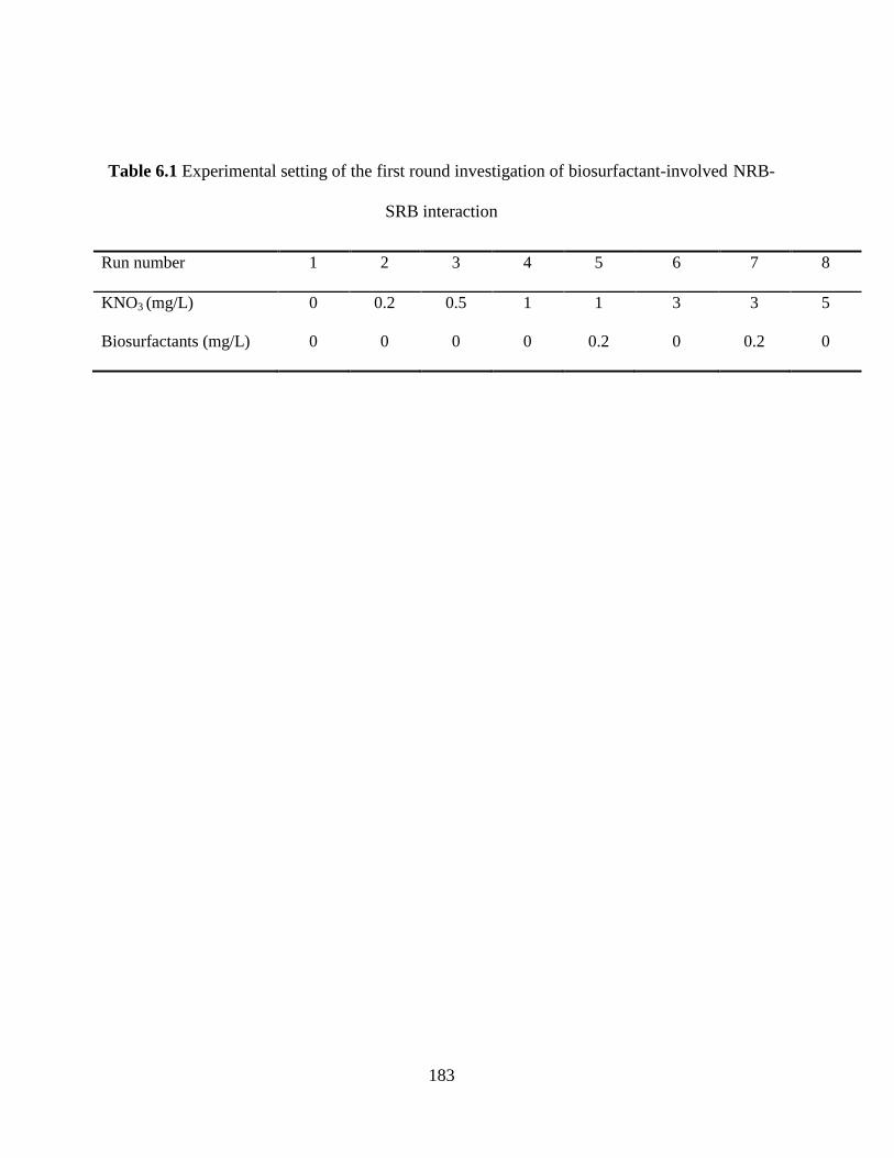
183
Table 6.1 Experimental setting of the first round investigation of biosurfactant-involved NRB-
SRB interaction
Run number 1 2 3 4 5 6 7 8
KNO3 (mg/L) 0 0.2 0.5 1 1 3 3 5
Biosurfactants (mg/L) 0 0 0 0 0.2 0 0.2 0
184
SRB strain was inoculated and cultivated for 15 days beforehand to yield a mature SRB culture. At
Time 0, 1 g Na2SO4, 1.5 g sodium citrate and KNO3 of 1, 3, and 5 g, respectively, were added into
the mature SRB culture (Runs A, B and D). Biosurfactants of 0.1 g were added in an extra run
(Run C, 3g KNO3 added) and the detailed setting of each run in Round 2 was listed in Table 6.2.
The seed culture of the NRB was inoculated in all runs at Time 0. The sampling was conducted on
odd days as well as on Days 6, 8 and 10. Parameters including redox potential, pH, concentrations
of sulfide, sulfate, nitrate, nitrite, dissolved organic carbon (DOC), dissolved inorganic carbon
(DIC) and total dissolved carbon (TDC) in NRB-SRB interactions were monitored. All the
microcosm experiments were conducted under anaerobic conditions.
6.2.3 Chemical and physical analyses
Concentrations of sulfide and nitrite in all media samples were determined colorimetrically with
N,Ndimethyl-p-phenylenediamine and sulfanilamide/n-(naphthyl)-ethylenediamine reagent,
respectively (Bridgewater 2012). Concentration of sulfate was assayed using a Dionex ion
chromatography (ICS-2100) system (Dionex Corporation, Sunnyvale, CA, USA). DOC, DIC and
TDC were analyzed using a high-temperature combustion total organic carbon analyzer (Shimadzu
Total Organic Carbon Analyzer (TOC-L)) (Anumol et al. 2015). The determination of nitrate
followed a two-wavelength approach described by Zhang et al. (2011b) Redox potential (Eh) was
measured using water proof ORPTestr 10 with a platinum band electrode. The pH measurements
of the water samples were conducted using a benchtop pH meter (Mettler Toledo). All the
measurements of sulfide, nitrite, nitrate, Eh and pH were carried out in triplicate and values
were presented as mean ± standard deviation. The analyses of other parameters were performed in
duplicate and the associated statistical analyses agreed to within 95% confidence.
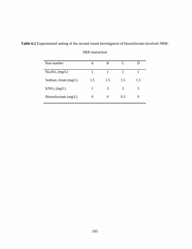
185
Table 6.2 Experimental setting of the second round investigation of biosurfactant-involved NRB-
SRB interaction
Run number A B C D
Na2SO4 (mg/L) 1 1 1 1
Sodium citrate (mg/L) 1.5 1.5 1.5 1.5
KNO3 (mg/L) 1 3 3 5
Biosurfactants (mg/L) 0 0 0.2 0
186
6.2.4 Microbial PLFA analysis
PLFA analysis was used to profile SRB strain cultivated on SRB medium, NRB strain cultivated
on NRB medium, and the microbial community structure during the first round of experiments.
PLFA profiling was conducted following a modified Bligh and Dyer extraction method (Fan et al.
2017a). Phospholipid standard C19:0 PC (1,2-dinonadecanoyl-sn-glycero-3-phosphocholine;
Avanti Polar Lipids) was used for determining phospholipid recovery. Internal standards C14:1
(cis-9) and C21:0 were spiked before GC-MS analysis (Ziegler et al. 2013). FAMEs were
determined from five standards: Bacterial Acid Methyl Esters CP Mixture, FIM-FAME-7
Mixture, 10Me C16:0, and C16:1 (trans-9) from Matreya LLC (Pleasant Gap, Pennsylvania, USA);
as well as C18:1 (trans-11) from Sigma-Aldrich (Oakville, Ontario, Canada). PCA was conducted
to compare PLFA composition of different settings and illustrate their correlations. The trans/cis
ratios of C16:1Δ9 and C18:1Δ11, cyclo/precursor ratios including cy-C17:0/C16:1 (cis-9) and (cy-
C17:0 + cy-C19:0)/(C16:1 (cis-9) + C18:1 (cis-11)) were used as indicators of physiological or
nutritional stress in bacterial communities (Moore-Kucera and Dick 2008; Wixon and Balser
2013). Triplicate measurements coupled with blank control were performed to demonstrate
analytical accuracy and values were presented as mean ± standard deviation.
6.3 Results and Discussion
6.3.1 NRB-SRB interactions under various nitrate conditions
The first round of experiments were performed to determine the effects of NRB and/or
biosurfactant addition on SRB growth inhibitory under various nitrate/sulfate loadings, and the
associated bacterial responses. In this round, the NRB and SRB strains were inoculated in the
system simultaneously, and then incubated for 11 days. Figure 6.1 presents the values of redox

187
potential, pH, sulfide and nitrate concentrations from samples collected on Day 5 and Day 11 in all
8 runs during the competitive NRB-SRB interactions. As shown in Figure 6.1, redox potentials in
Runs 2-4 all reached below -300 mV after five days. Redox potentials in biosurfactant-added
protocols (Runs 5 and 7) and Run 6 were reduced from initial -190 mV to below -400 mV.
Meanwhile, the sulfide concentration was significantly increased in Runs 5-7 (26.0-113.9 mg/L).
On Day 11, the redox potentials in nearly all the settings (except Run 5) rose back to above -204
mV. The lowest Eh value of -395 mV and the highest sulfide concentration (15.3 mg/L) were
achieved in Run 5 on Day 11 comparing to all other runs, which indicated the environmental
conditions could favorably support the growth of the SRB strain. Coincidentally, the lowest pH of
7.6 on Day 11 was also observed in Run 5, which implied the SRB growth might be inhibited by
alkaline conditions in other runs. Similar to Run 5, Run 1 was also noted because of its relative
lower redox potential (-204 mV) and pH value (7.8) on Day 11. Interestingly, although KNO3 was
added with various amounts (0.2- 5 g/L), the final nitrate concentrations in all Runs 2-7 reached a
stable amount (~130 mg/L), resulting in diverse percentage of nitrate consumption (36.1-95.1%).
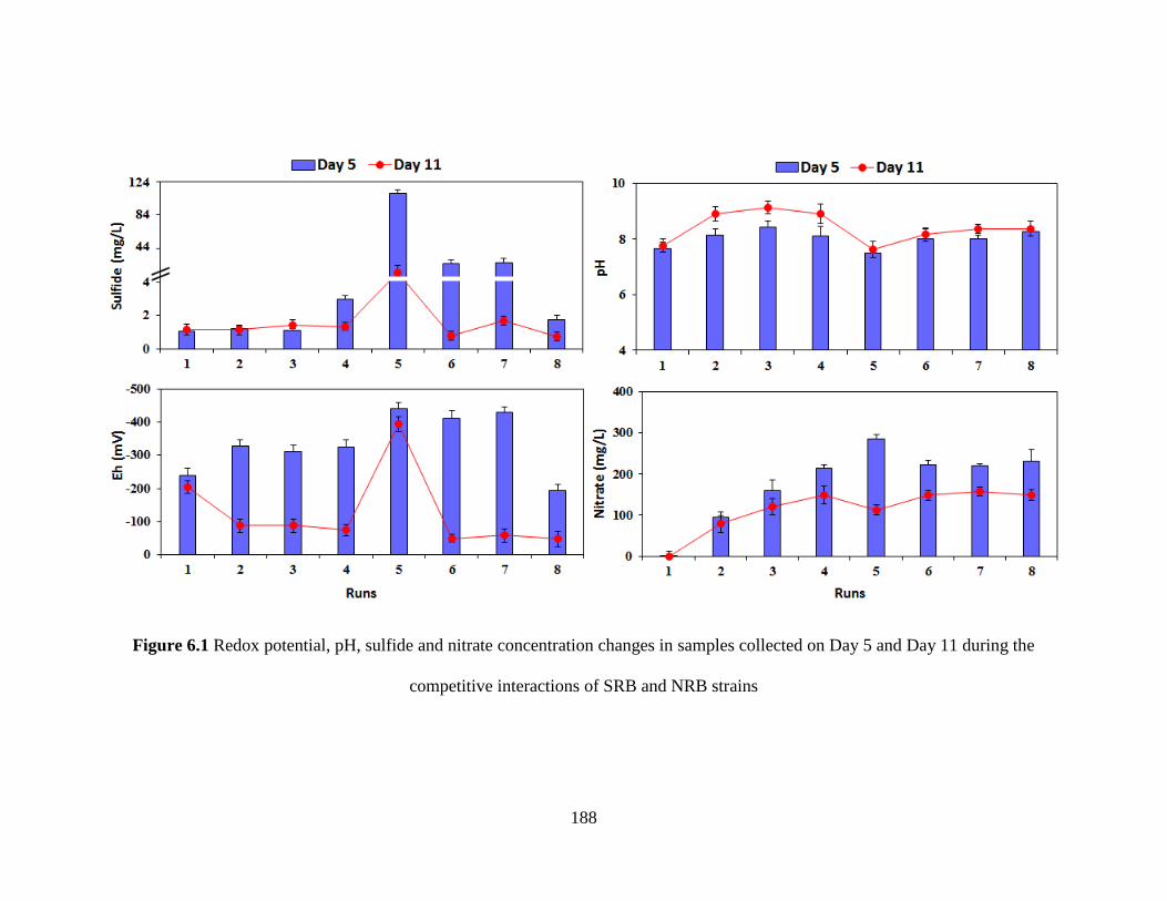
188
Figure 6.1 Redox potential, pH, sulfide and nitrate concentration changes in samples collected on Day 5 and Day 11 during the
competitive interactions of SRB and NRB strains
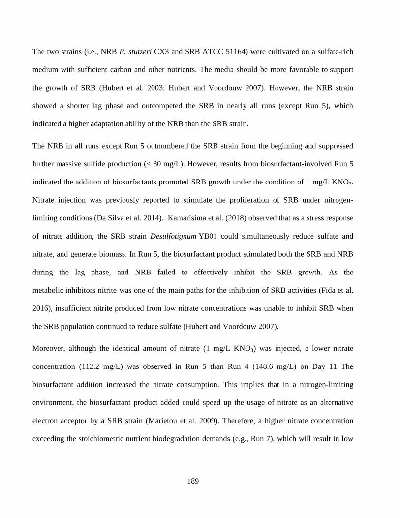
189
The two strains (i.e., NRB P. stutzeri CX3 and SRB ATCC 51164) were cultivated on a sulfate-rich
medium with sufficient carbon and other nutrients. The media should be more favorable to support
the growth of SRB (Hubert et al. 2003; Hubert and Voordouw 2007). However, the NRB strain
showed a shorter lag phase and outcompeted the SRB in nearly all runs (except Run 5), which
indicated a higher adaptation ability of the NRB than the SRB strain.
The NRB in all runs except Run 5 outnumbered the SRB strain from the beginning and suppressed
further massive sulfide production (< 30 mg/L). However, results from biosurfactant-involved Run 5
indicated the addition of biosurfactants promoted SRB growth under the condition of 1 mg/L KNO3.
Nitrate injection was previously reported to stimulate the proliferation of SRB under nitrogen-
limiting conditions (Da Silva et al. 2014). Kamarisima et al. (2018) observed that as a stress response
of nitrate addition, the SRB strain Desulfotignum YB01 could simultaneously reduce sulfate and
nitrate, and generate biomass. In Run 5, the biosurfactant product stimulated both the SRB and NRB
during the lag phase, and NRB failed to effectively inhibit the SRB growth. As the
metabolic inhibitors nitrite was one of the main paths for the inhibition of SRB activities (Fida et al.
2016), insufficient nitrite produced from low nitrate concentrations was unable to inhibit SRB when
the SRB population continued to reduce sulfate (Hubert and Voordouw 2007).
Moreover, although the identical amount of nitrate (1 mg/L KNO3) was injected, a lower nitrate
concentration (112.2 mg/L) was observed in Run 5 than Run 4 (148.6 mg/L) on Day 11 The
biosurfactant addition increased the nitrate consumption. This implies that in a nitrogen-limiting
environment, the biosurfactant product added could speed up the usage of nitrate as an alternative
electron acceptor by a SRB strain (Marietou et al. 2009). Therefore, a higher nitrate concentration
exceeding the stoichiometric nutrient biodegradation demands (e.g., Run 7), which will result in low
190
carbon/sulfate/sulfide to nitrate ratios in NRB-SRB interaction (An et al. 2010; Hubert et al. 2003), is
recommended for effective souring control.
6.3.2 Analysis of microbial communities
6.3.2.1 Principal component analysis (PCA)
PLFA were used as biomarkers to identify individual microbial species, trace the community
structure responses and describe changes in microbial metabolic fingerprints. The PLFA profiles of
NRB grown on NRB medium (pure NRB), as well as SRB cultivated on SRB medium (pure SRB)
were determined and shown in Figure 6.2. A total of 21 different PLFA biomarkers were identified
from the cell membrane of the SRB strain, of which a-C15:0 (49.2%) and C16:0 (16.2%) dominated.
In contrast, 16 fatty acids were observed from PLFA profiles of the NRB strain, of which C18:1 (cis-
11) (50.5%) and C16:0 (19.3%) dominated. Major fatty acids identified in the NRB strain include
C16:1 (cis-9) (10.4%) and cy-C19:0 (10.5%) as well. PCA of all the bacterial samples in Figure 6.3
indicated the correlation between the fatty acids and different samples. From Figure 6.3, the
dominating a-C15:0 was one of characteristic PLFAs of pure SRB, and major fatty acids C16:1 (cis-9)
and C18:1 (cis-11) were the representative components of pure NRB. Interestingly, fatty acids C16:1
(cis-9) and C18:1 (cis-11) are both closely correlated with the sulfide-oxidizing NRB strain, while
their coexistence has been previously recognized as signature biomarkers for other sulfur-oxidizing
bacteria in sulfide-rich marine sediment (Li et al. 2007b) and offshore reservoir injection water (Fan
et al. 2017b). Typical SRB biomarkers 10Me C16:0, i-C17:0, C17:1 (cis-10) and i-C17:1 (cis-10)
(Córdova-Kreylos et al. 2006; Mohanty et al. 2008), although detected with small amounts, all
showed close relationships with pure SRB in PCA. It was suggested that the linking between a-C15:0
and these SRB biomarkers could be used as the evidence of the SRB presence in a system.
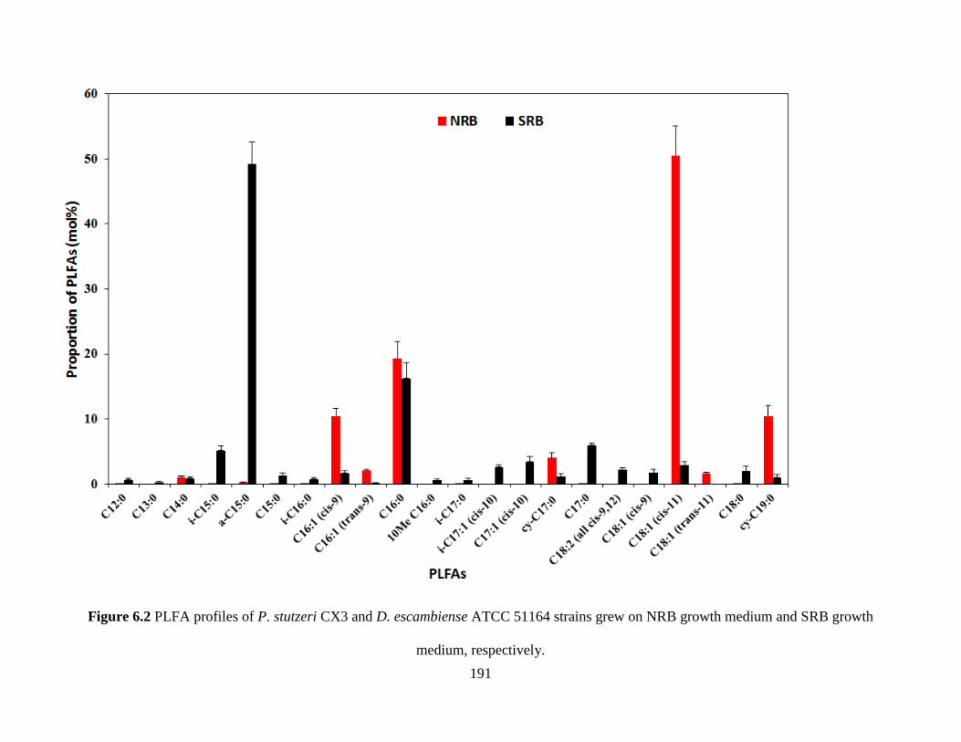
191
Figure 6.2 PLFA profiles of P. stutzeri CX3 and D. escambiense ATCC 51164 strains grew on NRB growth medium and SRB growth
medium, respectively.
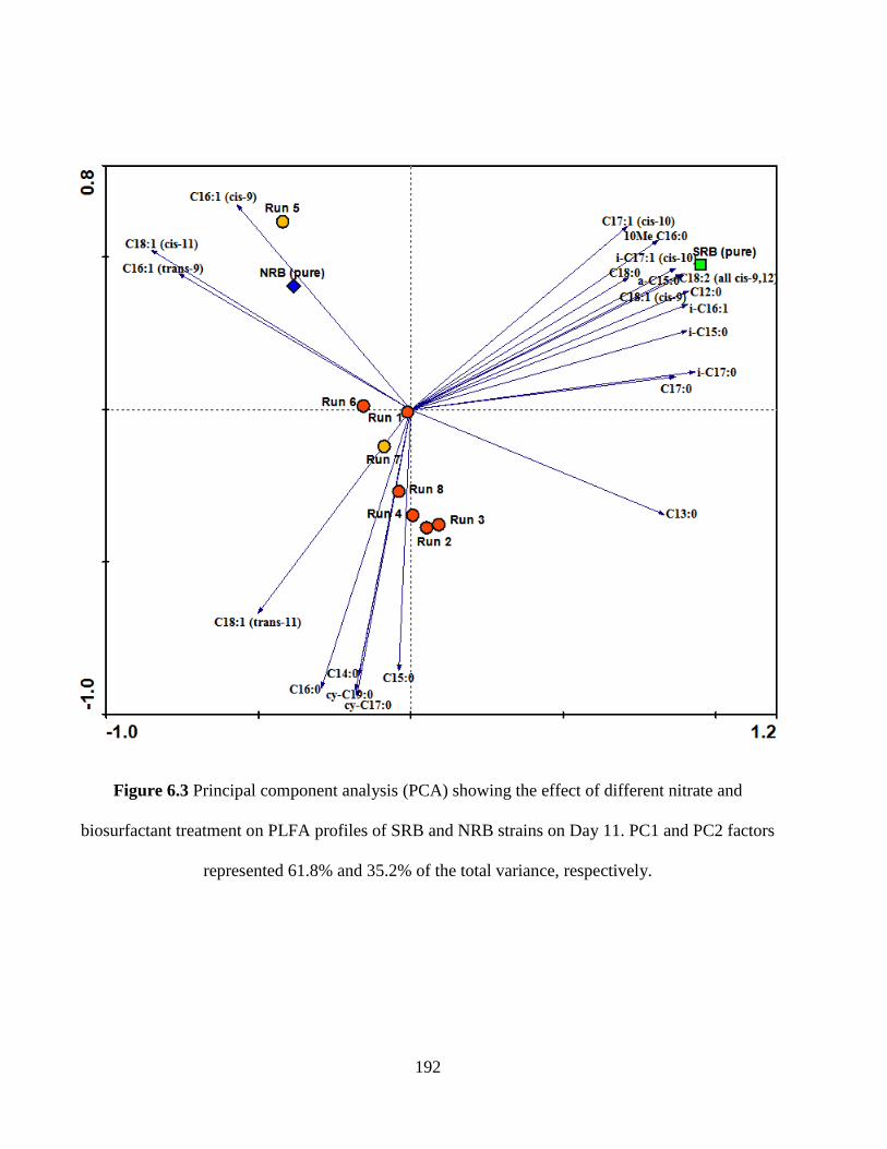
192
Figure 6.3 Principal component analysis (PCA) showing the effect of different nitrate and
biosurfactant treatment on PLFA profiles of SRB and NRB strains on Day 11. PC1 and PC2 factors
represented 61.8% and 35.2% of the total variance, respectively.
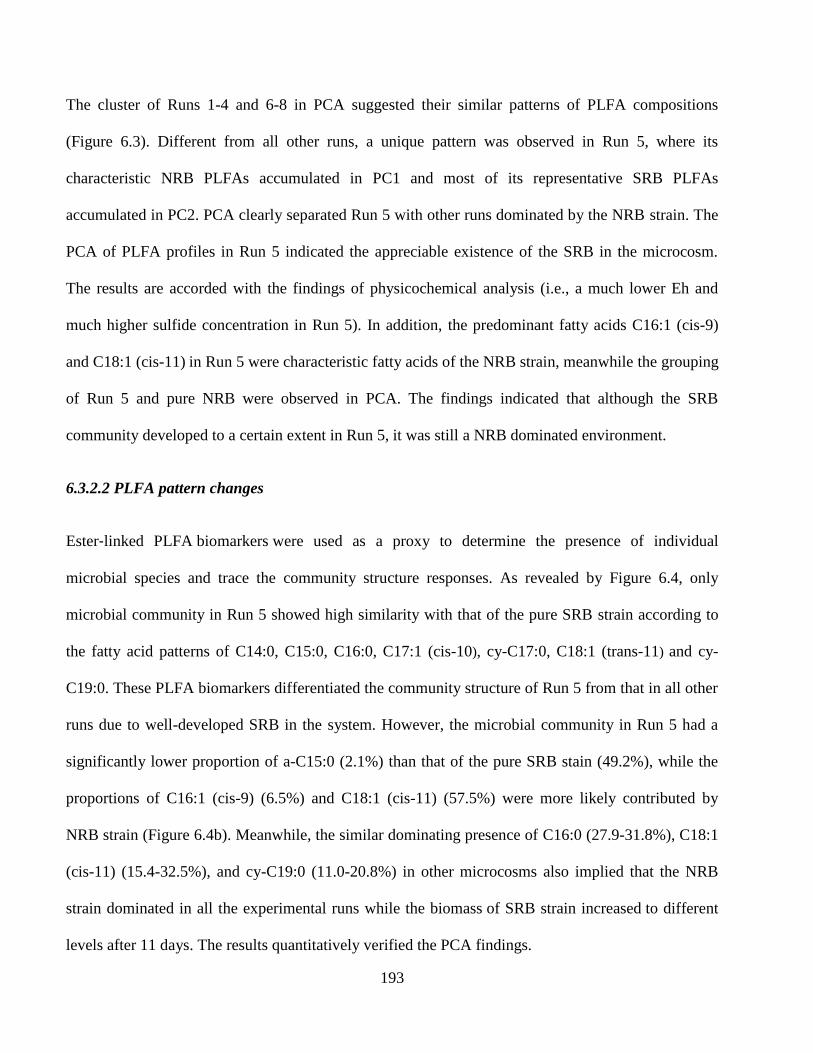
193
The cluster of Runs 1-4 and 6-8 in PCA suggested their similar patterns of PLFA compositions
(Figure 6.3). Different from all other runs, a unique pattern was observed in Run 5, where its
characteristic NRB PLFAs accumulated in PC1 and most of its representative SRB PLFAs
accumulated in PC2. PCA clearly separated Run 5 with other runs dominated by the NRB strain. The
PCA of PLFA profiles in Run 5 indicated the appreciable existence of the SRB in the microcosm.
The results are accorded with the findings of physicochemical analysis (i.e., a much lower Eh and
much higher sulfide concentration in Run 5). In addition, the predominant fatty acids C16:1 (cis-9)
and C18:1 (cis-11) in Run 5 were characteristic fatty acids of the NRB strain, meanwhile the grouping
of Run 5 and pure NRB were observed in PCA. The findings indicated that although the SRB
community developed to a certain extent in Run 5, it was still a NRB dominated environment.
6.3.2.2 PLFA pattern changes
Ester‐linked PLFA biomarkers were used as a proxy to determine the presence of individual
microbial species and trace the community structure responses. As revealed by Figure 6.4, only
microbial community in Run 5 showed high similarity with that of the pure SRB strain according to
the fatty acid patterns of C14:0, C15:0, C16:0, C17:1 (cis-10), cy-C17:0, C18:1 (trans-11) and cy-
C19:0. These PLFA biomarkers differentiated the community structure of Run 5 from that in all other
runs due to well-developed SRB in the system. However, the microbial community in Run 5 had a
significantly lower proportion of a-C15:0 (2.1%) than that of the pure SRB stain (49.2%), while the
proportions of C16:1 (cis-9) (6.5%) and C18:1 (cis-11) (57.5%) were more likely contributed by
NRB strain (Figure 6.4b). Meanwhile, the similar dominating presence of C16:0 (27.9-31.8%), C18:1
(cis-11) (15.4-32.5%), and cy-C19:0 (11.0-20.8%) in other microcosms also implied that the NRB
strain dominated in all the experimental runs while the biomass of SRB strain increased to different
levels after 11 days. The results quantitatively verified the PCA findings.
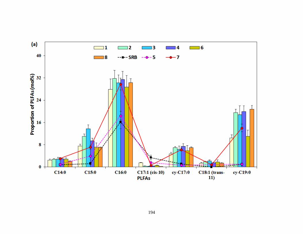
194
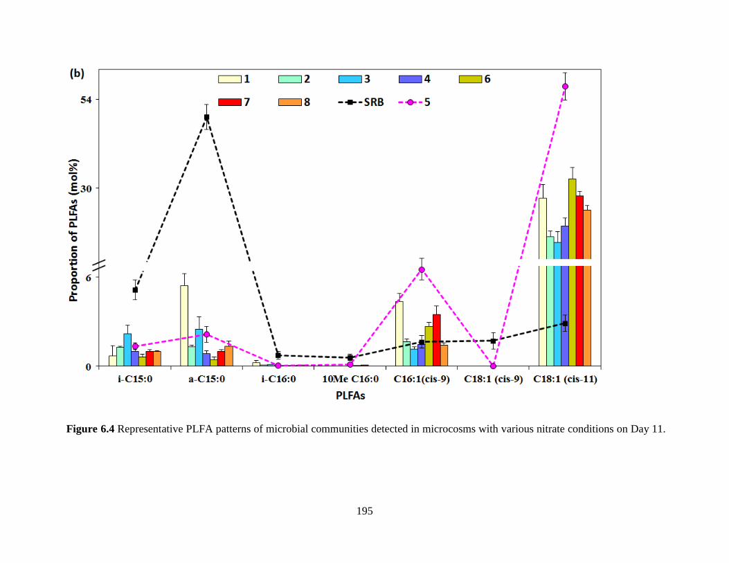
195
Figure 6.4 Representative PLFA patterns of microbial communities detected in microcosms with various nitrate conditions on Day 11.
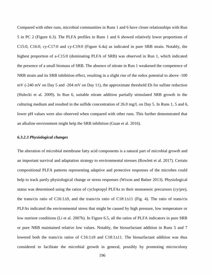
196
Compared with other runs, microbial communities in Runs 1 and 6 have closer relationships with Run
5 in PC 2 (Figure 6.3). The PLFA profiles in Runs 1 and 6 showed relatively lower proportions of
C15:0, C16:0, cy-C17:0 and cy-C19:0 (Figure 6.4a) as indicated in pure SRB strain. Notably, the
highest proportion of a-C15:0 (dominating PLFA of SRB) was observed in Run 1, which indicated
the presence of a small biomass of SRB. The absence of nitrate in Run 1 weakened the competence of
NRB strain and its SRB inhibition effect, resulting in a slight rise of the redox potential to above -100
mV (-240 mV on Day 5 and -204 mV on Day 11), the approximate threshold Eh for sulfate reduction
(Hulecki et al. 2009). In Run 6, suitable nitrate addition partially stimulated SRB growth in the
culturing medium and resulted in the sulfide concentration of 26.0 mg/L on Day 5. In Runs 1, 5 and 6,
lower pH values were also observed when compared with other runs. This further demonstrated that
an alkaline environment might help the SRB inhibition (Guan et al. 2016).
6.3.2.3 Physiological changes
The alteration of microbial membrane fatty acid components is a natural part of microbial growth and
an important survival and adaptation strategy to environmental stresses (Rowlett et al. 2017). Certain
compositional PLFA patterns representing adaptive and protective responses of the microbes could
help to track partly physiological change or stress responses (Wixon and Balser 2013). Physiological
status was determined using the ratios of cyclopropyl PLFAs to their monoenoic precursors (cy/pre),
the trans/cis ratio of C16:1Δ9, and the trans/cis ratio of C18:1Δ11 (Fig. 4). The ratio of trans/cis
PLFAs indicated the environmental stress that might be caused by high pressure, low temperature or
low nutrient conditions (Li et al. 2007b). In Figure 6.5, all the ratios of PLFA indicators in pure SRB
or pure NRB maintained relative low values. Notably, the biosurfactant addition in Runs 5 and 7
lowered both the trans/cis ratios of C16:1Δ9 and C18:1Δ11. The biosurfactant addition was thus
considered to facilitate the microbial growth in general, possibly by promoting microcolony
197
formation in the initial phase (Pamp and Tolker-Nielsen 2007). Very small amounts of cyclopropyl
PLFAs of C17:0 and C19:0 in Run 5 was observed and the ratios of cy/pre reached nearly zero,
suggesting the co-existence of the SRB and NRB in a mild environment. Overall, Runs 1-4, 6 and 8
without biosurfactant addition suffered different levels of nutritional fluctuation and environmental
stress, which maybe induced by limited nitrate availability, starvation, and pH changes (Kaur et al.
2005; Willers et al. 2015). On the contrary, the indicative stress analysis revealed the biosurfactant
addition significantly enhanced the action of bacterial mitigation towards the environmental stress.
The beneficial effect demonstrated by biosurfactant addition acted on both of the NRB and SRB, and
explained the obvious sulfide production in Run 5 but not in Run 4.
198
Figure 6.5 Changes in the ratios of trans/cis of C16:1Δ9 and C18:1Δ11 and the ratios of cyclopropyl
PLFAs to their monoenoic precursors (cy/pre) at various growing conditions. Numbers 1-8 in
samples indicate runs in the first round of experiments.
199
6.3.3 Souring control by nitrate and biosurfactant injection
6.3.3.1 Anti-souring effect of nitrate and biosurfactant addition
To investigate the anti-souring effect of nitrate and biosurfactant treatment, the second round of
experiments was conducted. The SRB strain was inoculated in the media for 15 days in each run to
create an extremely souring situation in which Eh below -400 mV was achieved and the sulfide
concentration reached ~200 mg/L. In all four experimental runs (Runs A to D), the NRB strain was
then introduced to the system together with nitrate (and biosurfactants in Run C) addition for souring
mitigation. Figure 6.6 showed the results of changes on Eh, pH and the concentrations of sulfide,
nitrate and DOC during 19 days of souring mitigation activities in all the four runs. From Figure 6.6,
sulfide concentration in all runs was significantly reduced over time with values changes from ~200
mg/L to below 2 mg/L within 10 days. The pattern of sulfide concentration changes then varied in the
4 experimental runs after 10 days, whereas the final values were all below 4 mg/L. On Day 19, Run A
achieved the highest sulfide concentration (3.8 mg/L) and the most reducing condition (-356 mV).
The redox potentials of the Runs B to D were between -265 to -96 mV on Day 19. The pH values in
Runs B to D were all elevated to above 8.5 while pH 7.6 was observed in Run A on Day 19. In spite
of the varying concentrations of nitrate injection (1-5 g/L KNO3) the final nitrate concentrations
reached a relatively constant and low level (96.9-146.7 mg/L). In general, the souring condition in the
microcosm was mitigated to different degrees through the various nitrate (and biosurfactant) addition
in all four runs. Specially, high levels of nitrate injection (3 or 5 g/L KNO3) resulted in effective
control of souring caused by SRB activities and improved the reducing conditions in the system.
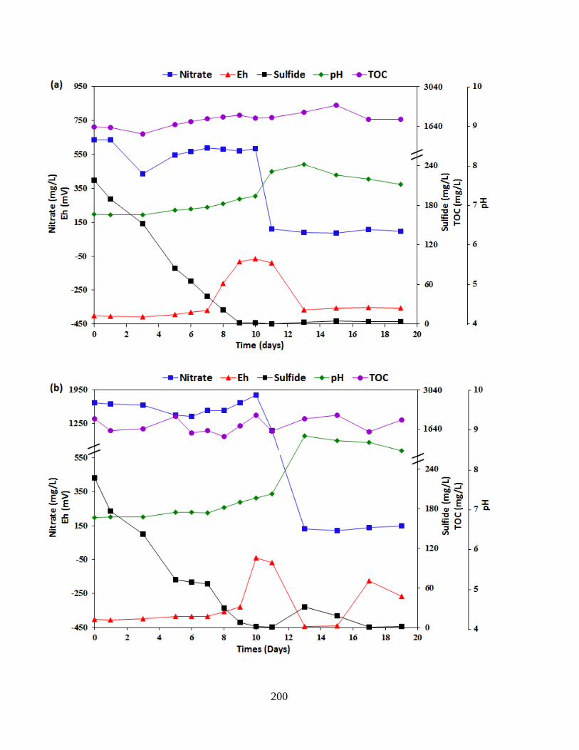
200
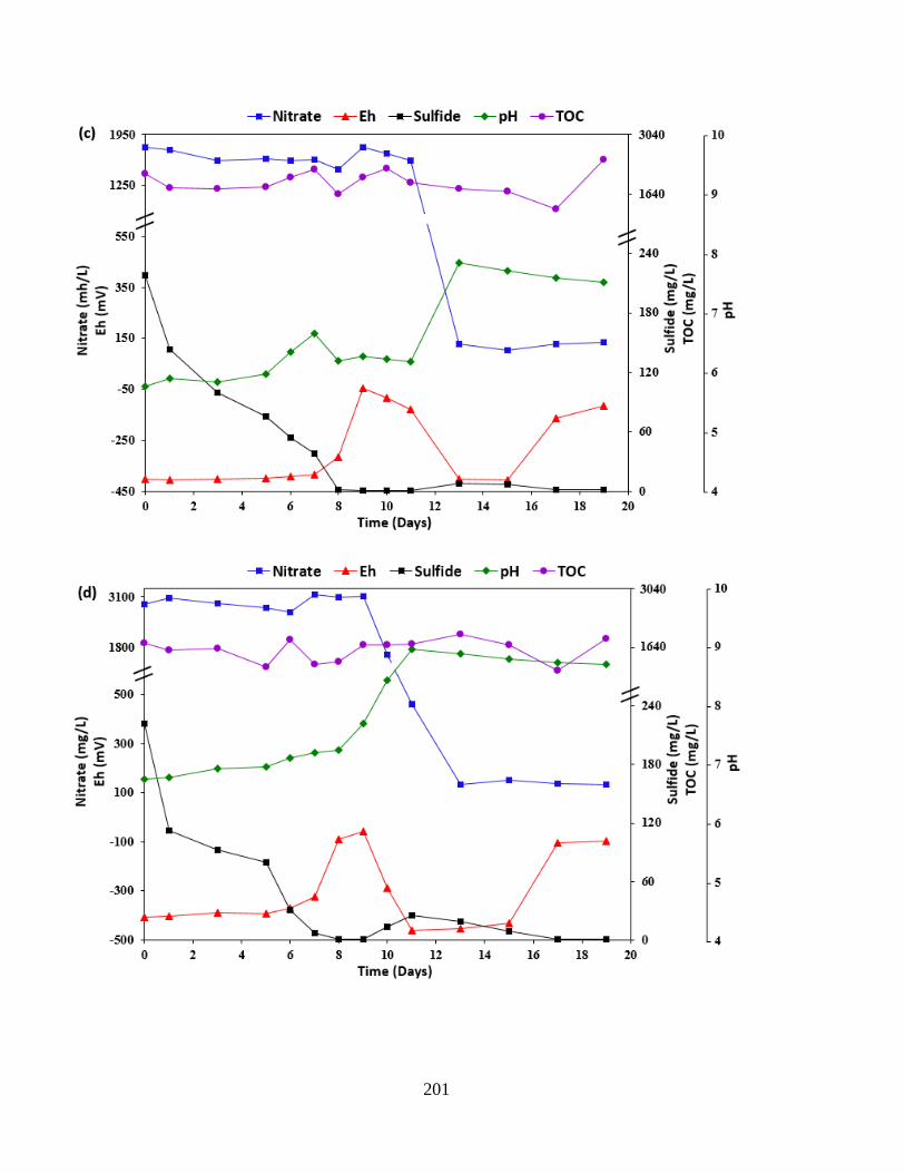
201
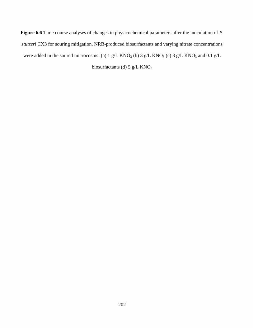
202
Figure 6.6 Time course analyses of changes in physicochemical parameters after the inoculation of P.
stutzeri CX3 for souring mitigation. NRB-produced biosurfactants and varying nitrate concentrations
were added in the soured microcosms: (a) 1 g/L KNO3 (b) 3 g/L KNO3 (c) 3 g/L KNO3 and 0.1 g/L
biosurfactants (d) 5 g/L KNO3
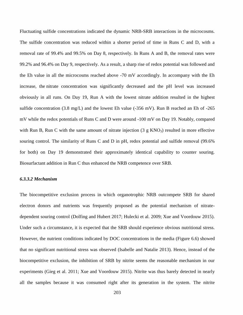
203
Fluctuating sulfide concentrations indicated the dynamic NRB-SRB interactions in the microcosms.
The sulfide concentration was reduced within a shorter period of time in Runs C and D, with a
removal rate of 99.4% and 99.5% on Day 8, respectively. In Runs A and B, the removal rates were
99.2% and 96.4% on Day 9, respectively. As a result, a sharp rise of redox potential was followed and
the Eh value in all the microcosms reached above -70 mV accordingly. In accompany with the Eh
increase, the nitrate concentration was significantly decreased and the pH level was increased
obviously in all runs. On Day 19, Run A with the lowest nitrate addition resulted in the highest
sulfide concentration (3.8 mg/L) and the lowest Eh value (-356 mV). Run B reached an Eh of -265
mV while the redox potentials of Runs C and D were around -100 mV on Day 19. Notably, compared
with Run B, Run C with the same amount of nitrate injection (3 g KNO3) resulted in more effective
souring control. The similarity of Runs C and D in pH, redox potential and sulfide removal (99.6%
for both) on Day 19 demonstrated their approximately identical capability to counter souring.
Biosurfactant addition in Run C thus enhanced the NRB competence over SRB.
6.3.3.2 Mechanism
The biocompetitive exclusion process in which organotrophic NRB outcompete SRB for shared
electron donors and nutrients was frequently proposed as the potential mechanism of nitrate-
dependent souring control (Dolfing and Hubert 2017; Hulecki et al. 2009; Xue and Voordouw 2015).
Under such a circumstance, it is expected that the SRB should experience obvious nutritional stress.
However, the nutrient conditions indicated by DOC concentrations in the media (Figure 6.6) showed
that no significant nutritional stress was observed (Isabelle and Natalie 2013). Hence, instead of the
biocompetitive exclusion, the inhibition of SRB by nitrite seems the reasonable mechanism in our
experiments (Gieg et al. 2011; Xue and Voordouw 2015). Nitrite was thus barely detected in nearly
all the samples because it was consumed right after its generation in the system. The nitrite
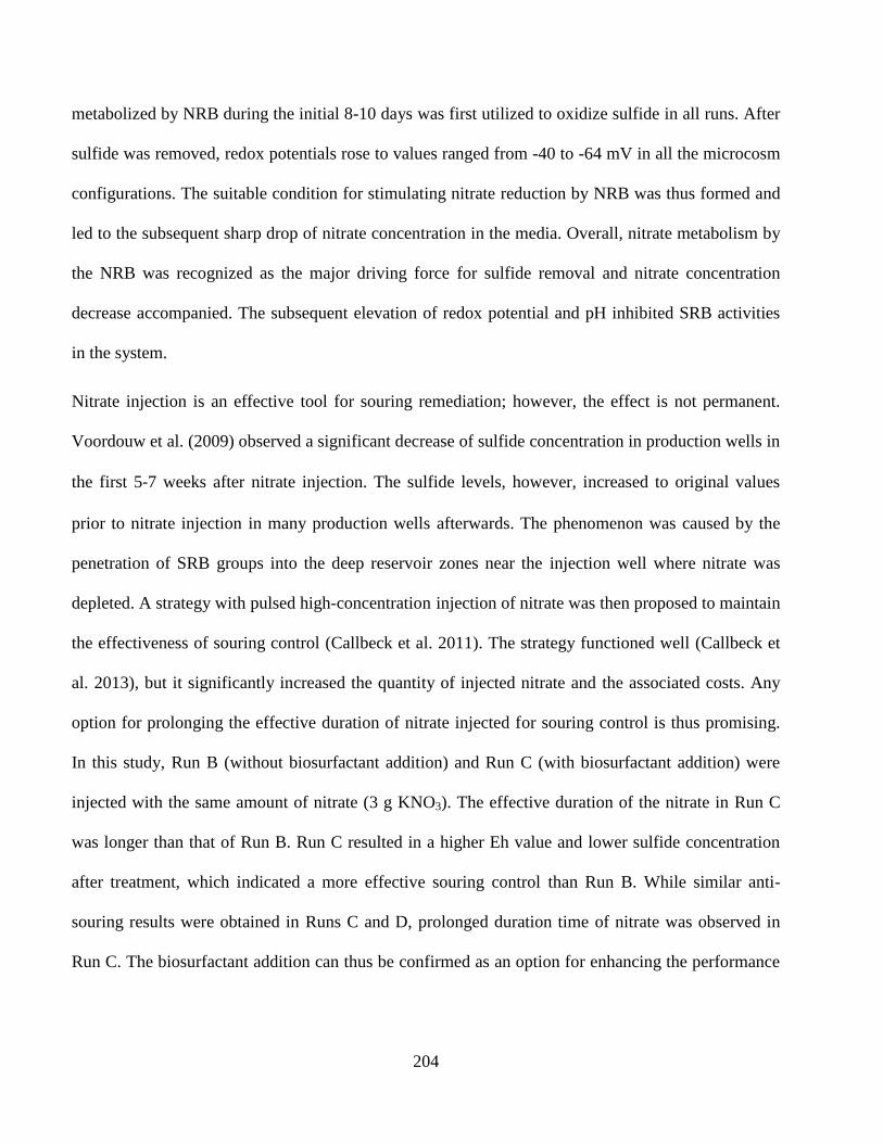
204
metabolized by NRB during the initial 8-10 days was first utilized to oxidize sulfide in all runs. After
sulfide was removed, redox potentials rose to values ranged from -40 to -64 mV in all the microcosm
configurations. The suitable condition for stimulating nitrate reduction by NRB was thus formed and
led to the subsequent sharp drop of nitrate concentration in the media. Overall, nitrate metabolism by
the NRB was recognized as the major driving force for sulfide removal and nitrate concentration
decrease accompanied. The subsequent elevation of redox potential and pH inhibited SRB activities
in the system.
Nitrate injection is an effective tool for souring remediation; however, the effect is not permanent.
Voordouw et al. (2009) observed a significant decrease of sulfide concentration in production wells in
the first 5-7 weeks after nitrate injection. The sulfide levels, however, increased to original values
prior to nitrate injection in many production wells afterwards. The phenomenon was caused by the
penetration of SRB groups into the deep reservoir zones near the injection well where nitrate was
depleted. A strategy with pulsed high-concentration injection of nitrate was then proposed to maintain
the effectiveness of souring control (Callbeck et al. 2011). The strategy functioned well (Callbeck et
al. 2013), but it significantly increased the quantity of injected nitrate and the associated costs. Any
option for prolonging the effective duration of nitrate injected for souring control is thus promising.
In this study, Run B (without biosurfactant addition) and Run C (with biosurfactant addition) were
injected with the same amount of nitrate (3 g KNO3). The effective duration of the nitrate in Run C
was longer than that of Run B. Run C resulted in a higher Eh value and lower sulfide concentration
after treatment, which indicated a more effective souring control than Run B. While similar anti-
souring results were obtained in Runs C and D, prolonged duration time of nitrate was observed in
Run C. The biosurfactant addition can thus be confirmed as an option for enhancing the performance
205
of nitrate injection in souring control. This is the first time that biosurfactant involved NRB-SRB
interaction was investigated in a souring system.
6.3.3.3 Parameter correlation
PCA was used to visualize the relationships between the time points and the connection of the
parameters monitored during the biosurfactant-involved NRB-SRB interaction in Run C (Figure 6a).
The variances between nitrate and pH, while significant, were not as strong as the variances between
sulfide, sulfate and Eh. This is because the major changes of nitrate and pH occurred only between
Day 9 and Day 13 while dynamic changes of other 3 parameters were observed in the whole process.
The inverse relationship between sulfide and sulfate was observed due to the shifts between the two
chemicals (Chen et al. 2017; Voordouw et al. 2009).
A positive correlation of sulfate and DOC in PCA was also observed. This is because sulfate, as an
essential nutrient for both NRB (Gadekar et al. 2006; Tang et al. 2010) and SRB, was consumed
along with the metabolizing of DOC (electron donor) by the two strains. Considering the DIC
(mainly CO32-
, HCO3-) formation increased the medium pH (Zhu and Dittrich 2016), which inhibited
SRB activities and elevated the medium Eh in the microcosm, these three factors (e.g. DIC, pH and
Eh) were closely related in Figure 6.7a.
The relationship of parameters including DIC, sulfide, pH and Eh on Day 19 in all runs was shown in
Figure 6.7b. Sulfide concentration, as the significant factor influencing the redox potential, was
negatively correlated with the redox potential. Accorded with the PCA results in Figure 6.7a, DIC
and pH are both positively correlated with redox potential on Day 19 in the systems.
206
Figure 6.7 Relationships between these time points and the connection of the parameters: (a) PCA of the monitored time points and
parameters during the 19 days of the biosurfactant-involved NRB-SRB interaction. PC1 and PC2 factors represented 84.8% and 7.5%
of the total variance, respectively. (b) Eh, pH and the concentrations of sulfide and DIC on Day 19 in the souring mitigation activities.
207
6.4 Summary
SRB are the main culprits of MIC (Enning and Garrelfs 2014) and the sulfide formed will lower the
value of petroleum products, increase health hazards and environmental threats (Chen et al. 2017;
Hubert and Voordouw 2007). The troublesome souring issue has plagued petroleum and
environmental industries for decades (Cheng et al. 2016). The individual impacts (Callbeck et al.
2013; Callbeck et al. 2011; Gieg et al. 2011; Kamarisima et al. 2018) and combining effects with
other chemical agents (Greene et al. 2006; Hulecki et al. 2009; Xue and Voordouw 2015) of nitrate
injection have been extensively examined owing to its convenient, inexpensive, and environmentally
friendly features. However, the nitrate-mediated control of microbial sulfide production may
encounter many issues, such as high nitrate demand (Da Silva et al. 2014), SRB resistance towards
nitrate as a stress response (Kamarisima et al. 2018; Korte et al. 2014), the alteration of SRB zone
into deeper reservoir (Callbeck et al. 2011; Voordouw et al. 2009) and incompetence of NRB due to
their limited thermotolerant capabilities (Fida et al. 2016; Okpala et al. 2017).
We, for the first time, gained insight into the significant positive effect of biosurfactants produced by
natural NRB P. stutzeri CX3 on the inhibition of SRB D. escambiense ATCC. Our results revealed
biosurfactant addition promoted the growth of both SRB and NRB under non-sour conditions.
Insufficient nitrate injection led to limited SRB inhibition due to the concurrent reduction of nitrate
and sulfate by the SRB strain. Under sour conditions, the biosurfactant addition within specific nitrate
levels increased the sulfide removal efficiency and significantly enhanced the nitrate inhibition of
SRB by NRB. Nitrite oxidation of sulfide was the major reason for sulfide removal. Notably, The
biosurfactant injection could reduce nitrate usage in souring mitigation. The prolonged effective
208
duration of nitrate will help nitrate/nitrite reach deeper zone in the system, resulting in long-term
suppression of SRB activities and better souring control.
The natural NRB-produced biosurfactants enhanced the competence of NRB in the microcosm at the
presence of soluble carbon nutrients. This enhancement may be strengthened when biosurfactants are
introduced into environments where hydrophobic oil organics are main carbon sources. The
amphiphilic properties of biosurfactants have the potential of helping NRB outcompete sulfidogenic
microbes through the mechanism of both biocometative exclusion and nitrite suppression. This
knowledge would extend the traditional perspective of biosurfactants in MEOR applications and lead
to management strategies for targeted control of souring in oil fields, and thus needs to be further
investigated.

209
CHAPTER 7
CONCLUSIONS AND RECOMMENDATIONS
210
7.1 Conclusions
Microbially induced reservoir souring, the undesirable production of hydrogen sulfide (H2S) in oil
reservoirs by SRB, has become a major concern during secondary oil recovery when water is injected
into the reservoir to recover the remaining oil. SRB are responsible for the majority of the bacterial
problems in oil production, and H2S is produced directly by SRB as a by-product of respiration. This
hazardous gas, a respiratory inhibitor, is both volatile and toxic. The unexpected production of H2S
has implications for reduced quality of produced hydrocarbons, for health and safety risks for
operators, and for increased corrosivity of produced fluids. Currently, reservoir souring has occurred
in approximately 70% of fields under water flooding (Elshahawi and Hashem 2005). The engineering,
environmental and safety costs associated with reservoir souring have attracted great attention across
the globe in recent decades. Hence, effective removal of sulfide or the inhibition of sulfide production
is highly desired by the oil and gas industry, especially in offshore operations.
The injection of nitrate/nitrite could be very effective for reservoir souring control by promoting NRB
to inhibit SRB activities. The possible mechanisms include the boost of hNRB to outcompete SRB
for available nutrients (biocompetitive exclusion) because of its thermodynamic advantage over SRB,
the promotion of nitrate reducing sulfide oxidizing bacteria (NR-SOB) to directly oxidize sulfide, and
the repression of SRB through resultant nitrite. Due to its convenient, inexpensive,
and environmentally friendly features, the injection of nitrate/nitrite has been widely investigated and
applied under both simulated and real reservoir conditions. However, to effectively address souring-
related issues, careful consideration of the operational conditions encountered in specific
environments is required. The approaches and technologies that are used to address souring in
onshore operations may not be effective in dealing with souring in offshore operations.
211
Nitrate injection has not been universally effective in reservoir souring control, the application of this
technique may encounter many issues, such as high nitrate demand, SRB resistance towards nitrate as
a stress response, the alteration of SRB zone into deeper reservoir and incompetence of NRB due to
their limited thermotolerant capabilities. Much still is unknown about the detailed microbial
mechanisms involved in NRB-SRB interactions during nitrate/nitrite injections for reservoir souring
mitigation. Meanwhile, biosurfactants produced by natural NRB are promising bio-agents for
enhancing NRB competence over SRB, which may assist in the enhanced suppression of SRB
activities and better souring control. However, there is still a lack of routine SRB monitoring tools in
offshore reservoir samples and systematic investigation of NRB-SRB-biosurfactant interactions. This
thesis, therefore, has presented SRB profiling of an offshore oil reservoir, isolated NRB for possible
biosurfactant producers, characterized the biosurfactant product and investigated the effect of
biosurfactants in NRB-SRB interaction. A summary of the method development of biosurfactant-based
nitrate injection for reservoir souring is described below:
1) A method based on PLFA analysis for profiling microbial communities in offshore produced
water was optimized. A three-stage extraction process was confirmed and the extraction efficiency in
phase partition was evaluated. The elution parameters in SPE purification were adapted for treating
the oily samples and their volumes were determined to induce a high recovery for the fraction of
phospholipids. Under the selected conditions, 92.9%, 96.3% and 92.8% of the spiked phospholipid
standards C16:1 (cis-9) PC, C18:1 (cis-9) PC, and C19:0 PC were recovered, respectively, using 10
mL methanol as elution solvent on a non-commercial SPE column. Over 90% of spiked C19:0 PC
was recovered before sample transesterification. Four parameters including alkaline reagent, volume
of acid for neutralization, time and temperature for FAME derivatization were examined. GC-MS
was used to analyze FAMEs and the method linearities, recoveries of 29 FAMEs during

212
transesterification, detection limits, relative standard deviations were presented. Results indicated that
the developed method exhibited high recoveries and repeatability, remarkable selectivity and linearity,
and acceptable quantitation limits for PLFA analysis. With reliable accuracy and precision, the
method was applied to profile microbes in offshore produced water samples.
2) PLFA analysis was conducted to profile microorganisms and trace SRB in water samples
from an offshore oil reservoir. From the results of spiked phospholipid standards, more than 90 % of
the phospholipids were recovered before the treatment of FAME derivatization while the RSDs were
below 8.0 %. The water samples from the injection well and four producing wells exhibited various
reducing conditions and were further subjected to PLFA analysis. Fourteen kinds of PLFAs were
detected in the five wellbores and the concentrations of total fatty acids ranged from 368.4 to 3468.9
ng/L. Possible SRB biomarkers and significant PLFAs associated with SRB including C14:0, i-C15:0,
a-C15:0, C15:0, C16:1 (cis-9), C17:0, C18:1 (cis-9) and C18:1 (cis-11) were selected for determining
the presence of SRB species and evaluating the sulfate-related microbial biomass. The possible
existence of SRB genera Desulfobacter, Desulfotomaculum, Desulfovibrio and SOB in the reservoir
were proposed based on PLFA profiles. The highest biomass was found in the most reducing well
where very limited SOB biomarkers were found. Results indicated that the presence of SRB and SOB
species was closely associated with the redox environment of the reservoir wellbores. The species
distribution patterns were interpreted to elucidate the biological souring process.
3) The offshore petroleum-reservoir brines following nitrate/nitrite injection were used for the
anaerobic screening of biosurfactant producing NRB. After periodic enrichment and sophisticated
screening of the microorganisms, five NRB strains were screened from offshore produced water
samples and all identified as Pseudomonas stutzeri according to their 16S rRNA sequencing results.
The strain P. stutzeri CX3 was confirmed with biosurfactant production capacity through a series of

213
biosurfactant characterization tests (e.g., drop collapsing test, parafilm test and surfaced tension
determination). Their biosurfactant producing abilities fed on either glucose or glycerol media were
investigated. P. stutzeri CX3 reduced the medium surface tension to 33.5 and 29.6 mN/m,
respectively, while growing on glucose or glycerol media. The CX3 strain was further inoculated to
examine its growth performance, resulting in 32.4% and 94.5% of nitrate consumption over 228
hours of monitoring in two media, respectively. The nitrate concentrations and surface tensions on the
two media were both reduced to a relatively stable level within 84 hours during which OD600 reached
relatively high levels as well over the period. The composition analysis of the biosurfactant product
generated by P. stutzeri CX3 was conducted through thin-layer chromatography, GC with FID and
fourier transform infrared spectroscopy (FT-IR). The biosurfactant product was identified as a
mixture of a small part of lipopeptide and a large part of glycolipid while its critical micellar
concentration (CMC) was as low as 35 mg/L. The biosurfactant product demonstrated high stability
over a wide range of temperature (4–121°C), pH (2-10), and salinity (0%–20% w/v) concentration.
4) The effectiveness of nitrate-mediated souring control highly depends on the interactions of
SRB and NRB while biosurfactants produced by natural NRB are promising bio-agents for enhancing
NRB competence over SRB. Biosurfactant-aided inhibitory control of SRB strain Desulfomicrobium
escambiense ATCC 51164 by NRB strain Pseudomonas stutzeri CX3was examined in two scenarios.
Under non-sour conditions, insufficient nitrate injection resulted in limited SRB inhibition due to the
concurrent reduction of nitrate and sulfate by SRB. PLFA biomarkers traced the community
responses and verified the existence of the two strains. Compositional PLFA patterns revealed
biosurfactant addition benefitted both SRB and NRB towards stressful conditions. The NRB strain
dominated in all the experimental runs while the biomass of SRB strain increased to different levels.
During the investigations, an alkaline environment helped the SRB inhibition. Under sour conditions,
214
nitrite oxidation of sulfide, instead of the biocompetitive exclusion, proved to be the primary
mechanism for sulfide removal. The subsequent elevation of redox potential and pH inhibited SRB
activities. The connection of the parameters monitored during the biosurfactant-involved NRB-SRB
interaction was elucidated. NRB-produced biosurfactants added at proper nitrate level significantly
enhanced the nitrate inhibition of SRB activities by P. stutzeri CX3 and resulted in more efficient
sulfide removal and effective duration of nitrate in the microcosms.
7.2 Research Contributions
According to the research findings, this study can be summarized and highlighted by the following
contributions:
1) A suitable and efficient GC-based analytical method for intensive detection of PLFAs in oily
saline offshore produced water was generated. Operation parameters affecting SPE purification and
FAME derivatization were optimized. Characteristic biomarkers of microorganisms in offshore
produced water were identified from PLFA profiles. The microbiological analysis of offshore
produced water places great importance on the investigation of biological reservoir souring control
among scientific and industrial activities.
2) Effective PLFA profiling was achieved in offshore reservoir water analysis to specifically
elucidate the mechanism of reservoir souring induced by SRB. The complex biomass and microbial
community structure information from the various reservoir conditions were revealed. The SRB and
SOB species, coupled with their transformation patterns between the injection wells and producing
wells under different redox conditions were proposed. The reasonable results in tracing
microorganisms indicated PLFA analysis can be potentially used as routine monitoring tool in
implementation of reservoir souring mitigation strategies.
215
3) The isolation of biosurfactant producing NRB from offshore reservoirs was achieved and
subsequent anaerobic biosurfactant production was investigated for the first time. Pseudomonas
stutzeri CX3 was identified as biosurfactant producer and the kinetic behavior of biosurfactant
production was investigated. Glycolipids are major components of the biosurfactant product while
high biosurfactant stability was demonstrated under various environmental conditions. The successful
isolation and identification of biosurfactant producing NRB from laborious screenings on offshore
reservoir samples, coupled with subsequent biosurfactant characterization provided valuable technical
and methodological support for effective offshore reservoir souring control and associated EOR
activities.
4) NRB-produced biosurfactants were for the first time reported to significantly strengthen SRB
inhibition by NRB. Research results indicated insufficient nitrate injection resulted in ineffective SRB
inhibition and biosurfactant addition benefitted both SRB and NRB towards stressful conditions.
Nitrate metabolism was the major driving force for sulfide removal while the elevated redox potential
and pH inhibited SRB activities. The biosurfactant injection could reduce nitrate usage in souring
mitigation. The prolonged effective duration of nitrate will help nitrate/nitrite reach deeper zone in
the system, resulting in long-term suppression of SRB activities and better souring control.
7.3 Selective Publications
1. F. Fan, B. Zhang, J. Liu, Q. Cai, et al. Interactions of sulfate reducing bacteria (SRB),
biosurfactant producing nitrate reducing bacteria (NRB) screened from an offshore reservoir and
NRB produced biosurfactants in microcosms, Environmental Pollution, 2018, under review
2. F. Fan, B. Zhang, P.L. Morrill, T. Husain, Isolation of nitrate-reducing bacteria from an offshore
reservoir and the associated biosurfactant production, RSC Advances, 8 (2018) 26596-26609.
216
3. X. Li, F. Fan, K. Zhang, B. Zhang, B. Chen, PLFA based tracking of mechanisms and profiling of
microbial community during enhanced soil bioremediation of hydrocarbons, International
Biodeterioration and Biodegradation, 132 (2018) 216-225 (equally contributing 1st author).
4. X. Zhao, F. Fan, H. Zhou, P. Zhang, G. Zhao, Microbial diversity and activity of an aged soil
contaminated by polycyclic aromatic hydrocarbons, Bioprocess and Biosystems Engineering, 41
(2018) 871-883.
5. F. Fan, X. Li, B. Zhang, Enhanced coastal soil bioremediation of petroleum hydrocarbons (PHCs)
and associated microbial community transformation. Costal Zone Canada (CZC 2018), July 15-19,
2018, St. John‘s, Canada
6. F. Fan, B. Zhang, P.L. Morrill, Phospholipid fatty acid (PLFA) analysis for profiling microbial
communities in offshore produced water, Marine Pollution Bulletin, 122 (2017) 194-206.
7. F. Fan, B. Zhang, P.L. Morrill, T. Husain, Profiling of Sulfate-Reducing Bacteria in an Offshore
Oil Reservoir Using Phospholipid Fatty Acid (PLFA) Biomarkers, Water, Air, & Soil Pollution, 228
(2017) 410.
8. W. Qin, F. Fan, Y. Zhu, Y. Wang, X. Liu, A. Ding, J. Dou, Comparative proteomic analysis and
characterization of benzo(a)pyrene removal by Microbacterium sp. strain M.CSW3 under denitrifying
conditions, Bioprocess and Biosystems Engineering, 40 (2017) 1825-1838.
9. Y. Pi, B. Chen, M. Bao, F. Fan, Q. Cai, L. Ze, B. Zhang, Microbial degradation of four crude oil
by biosurfactant producing strain Rhodococcus sp, Bioresource Technology, 232 (2017) 263-269.
10. F. Fan, B. Zhang, P.L. Morrill, Investigation of seasonal pattern of microbial community
structure and identification of sulfate-reducing bacteria (SRB) in seawater samples. Symposium on

217
Persistent and Emerging Organic Pollution in cold and coastal Environments (PEOPLE 2017),
October 16-17, 2017, St. John‘s, Canada
11. F. Fan, B. Zhang, P.L. Morrill, Phospholipid fatty acid analysis for identifying sulfate-reducing
bacteria in seawater samples using base and acid catalyzed methylation methods. EWRI World
Environmental & Water Resources Congress (EWRI 2016), May 22-26, 2016, West Palm Beach,
Florida, USA
7.4 Recommendations for Future Research
The current research efforts focus on method development of PLFA profiling, the identification of
SRB and associated souring mechanisms in an offshore oil reservoir. Biosurfactant producing NRB
was screened and the associated biosurfactant-involved NRB-SRB interactions in microcosms were
investigated. Future investigations can be carried out in the following aspects:
1) Based on the current results obtained in Chapter 3 and 4, phospholipid and FAME
degradation rates during the sampling, storage and sample analysis could be further investigated.
There is lack of data on other species that may produce similar PLFA profiles, thus more quantity and
types of samples are recommended to be analyzed for more precise PLFA profile elucidation. The
differences and similarities in geological and operation conditions among the sampling points needs
to be further investigated for better elucidation of the results.
2) Carbon sources are of great importance on the production of biosurfactants and the alternation
of culture conditions for the bacterial producers. The natural NRB-produced biosurfactants enhanced
the competence of NRB over SRB through the mechanism of nitrite suppression at the presence of
soluble carbon nutrients in the microcosm. However, as amphiphilic compounds with both
hydrophilic and hydrophobic moieties, biosurfactants will increase the bioavailability of entrapped oil
218
in the reservoir for the bacterial producers by promoting wetting, solubilization and emulsification of
various types of organics. Thus, the enhancement may be strengthened when biosurfactants are
introduced into environments where hydrophobic oil organics are main carbon sources. The
biosurfactants have the potential of helping NRB outcompete sulfidogenic microbes through the
mechanism of both biocometative exclusion and nitrite suppression. This knowledge would extend
the traditional perspective of biosurfactants in MEOR applications and lead to management strategies
for targeted control of souring in oil fields, and thus needs to be further investigated.
3) NRB strain Pseudomonas stutzeri CX3, with the treatment of nitrate and biosurfactants,
exhibited promising anti-souring potential under sour conditions. Further investigations of reservoir
souring control by nitrate and biosurfactant amendment can be conducted on continuous bioreactors
powered by a variable speed peristaltic pump. Under proper operation of the pumps and maintenance
of the anaerobic conditions by pressurized sterilized nitrogen gas, SRB enrichment culture or field
produced water can be introduced to simulate the reservoir fluids in water flooding process. Various
nitrate and biosurfactant amendments can be applied with the inoculation of Pseudomonas stutzeri
CX3. Sampling ports located near the influent and effluent regions can be used for the monitoring of
parameters including nitrate, nitrite, sulfate, sulfide, carbon source and redox potential changes in the
continuous reactors when nitrate/nitrite injection is conducted. The structural changes of the
microbial communities under the complicated biological conditions, especially the associated SRB
and NRB species, can be traced and characterized by PLFA analysis, quantitative fluorescence PCR
techniques and other diagnostic tools.
4) As facultative microorganisms, NRB strains of Pseudomonas stutzeri were promising
petroleum hydrocarbon degraders under aerobic or anaerobic conditions. The strains thus have great
potential in applications of oil spill cleanup as well as hydrocarbon bioremediation & soil reclamation.
219
The biosurfactant producing ability of Pseudomonas stutzeri CX3 will help enhance the
remediation efficiency by promoting the mobility and bioavailability of petroleum hydrocarbons and
subsequent biodegradation. PLFA analysis can be used to evaluate the physiological status of the
NRB strains and elucidate the associated biodegradation mechanisms linked to different soil
treatments.
220
REFERENCES
Abbaci, M., barberi-heyob, M., Blondel, W., Guillemin, F., & Didelon, J. (2008). Advantages and
limitations of commonly used methods to assay the molecular permeability of gap junctional
intercellular communication (Vol. 45).
Abdel-Mawgoud, A. M., Aboulwafa, M. M., & Hassouna, N. A. (2009). Characterization of
rhamnolipid produced by Pseudomonas aeruginosa isolate Bs20. Appl Biochem Biotechnol,
157(2), 329-345.
Abouseoud, M., Maachi, R., Amrane, A., Boudergua, S., & Nabi, A. (2008a). Evaluation of different
carbon and nitrogen sources in production of biosurfactant by Pseudomonas fluorescens.
Desalination, 223(1), 143-151.
Abouseoud, M., Yataghene, A., Amrane, A., & Maachi, R. (2008b). Biosurfactant production by free
and alginate entrapped cells of Pseudomonas fluorescens. Journal of Industrial Microbiology
& Biotechnology, 35(11), 1303-1308.
Abtahi, N., Roostaazad, R., & Ghadiri, F. (2003). Biosurfactant Production In MEOR For
Improvement Of Iran's Oil Reservoirs' Production Experimental Approach. Paper presented at
the SPE International Improved Oil Recovery Conference in Asia Pacific, Kuala Lumpur,
Malaysia.
Abu-Ruwaida, A. S., Banat, I. M., Haditirto, S., Salem, A., & Kadri, M. (1991). Isolation of
biosurfactant-producing bacteria, product characterization, and evaluation. Acta
Biotechnologica, 11(4), 315-324.
Acosta-Martínez, V., Rowland, D., Sorensen, R. B., & Yeater, K. M. (2008). Microbial community
structure and functionality under peanut-based cropping systems in a sandy soil. Biology and
Fertility of Soils, 44(5), 681-692.

221
Afshar, S., Lotfabad, T. B., Roostaazad, R., Najafabadi, A. R., & Noghabi, K. A. (2008).
Comparative approach for detection of biosurfactant-producing bacteria isolated from Ahvaz
petroleum excavation areas in south of Iran. Annals of Microbiology, 58(3), 555.
Agrawal, A., Vanbroekhoven, K., & Lal, B. (2010). Diversity of culturable sulfidogenic bacteria in
two oil–water separation tanks in the north-eastern oil fields of India. Anaerobe, 16(1), 12-18.
Al-Bahry, S. N., Elshafie, A. E., Al-Wahaibi, Y. M., Al-Bemani, A. S., Joshi, S. J., & Al-Lawati, A.
(2013). Isolation and Characterization of Biosurfactant/Biopolymer Producing Spore Forming
Bacteria from Oil Contaminated Sites and Oil Field of Oman. APCBEE Procedia, 5, 242-246.
Al-Eid, M. I., Kokal, S. L., Carrigan, W. J., Al-Dubaisi, J. M., Halpern, H. I., & Al-Juraid, J. I. (2001).
Investigation of H2S Migration in the Marjan Complex.
Amani, H., Sarrafzadeh, M. H., Haghighi, M., & Mehrnia, M. R. (2010). Comparative study of
biosurfactant producing bacteria in MEOR applications. Journal of Petroleum Science and
Engineering, 75(1), 209-214.
Amaral, P. F. F., Coelho, M. A. Z., Marrucho, I. M. J., & Coutinho, J. A. P. (2010). Biosurfactants
from Yeasts: Characteristics, Production and Application. In R. Sen (Ed.), Biosurfactants (pp.
236-249). New York, NY: Springer New York.
Amosa, M. K., Mohammed-Dabo, I. A., & Yaro, S. A. (2013). Environmental and Corrosion Control
Effects of Hydrogen Sulfide Scavengers in Oil and Gas Exploration. In R. Javaherdashti;C.
Nwaoha; & H. Tan (Eds.), Corrosion and Materials in the Oil and Gas Industries (pp. 454-
485). London: CRC Press.
An, B. A., Shen, Y., & Voordouw, G. (2017). Control of Sulfide Production in High Salinity Bakken
Shale Oil Reservoirs by Halophilic Bacteria Reducing Nitrate to Nitrite. Frontiers in
Microbiology, 8, 1164.
222
An, S., Tang, K., & Nemati, M. (2010). Simultaneous biodesulphurization and denitrification using
an oil reservoir microbial culture: Effects of sulphide loading rate and sulphide to nitrate
loading ratio. Water Res, 44(5), 1531-1541.
Anandaraj, B., & Thivakaran, P. (2010). Isolation and production of biosurfactant producing
organism from oil spilled soil (Vol. 1).
Anderson, G. M., & Thom, J. (2008). The role of thermochemical sulfate reduction in the origin of
Mississippi Valley-type deposits. II. Carbonate-sulfide relationships. Geofluids, 8(1), 27-34.
Andrietta, S. R., Steckelberg, C., & Andrietta, M. d. G. S. (2008). Study of flocculent yeast
performance in tower reactors for bioethanol production in a continuous fermentation process
with no cell recycling. Bioresource Technology, 99(8), 3002-3008.
Antoniou, E., Fodelianakis, S., Korkakaki, E., & Kalogerakis, N. (2015). Biosurfactant production
from marine hydrocarbon-degrading consortia and pure bacterial strains using crude oil as
carbon source. Frontiers in Microbiology, 6(274)
Anumol, T., Sgroi, M., Park, M., Roccaro, P., & Snyder, S. A. (2015). Predicting trace organic
compound breakthrough in granular activated carbon using fluorescence and UV absorbance
as surrogates. Water Res, 76, 76-87.
Aparna, A., Srinikethan, G., & Smitha, H. (2012). Production and characterization of biosurfactant
produced by a novel Pseudomonas sp. 2B. Colloids Surf B Biointerfaces, 95, 23-29.
Avalli, A., & Contarini, G. (2005). Determination of phospholipids in dairy products by
SPE/HPLC/ELSD. Journal of Chromatography A, 1071(1-2), 185-190.
Axelsson, M., & Gentili, F. (2014). A Single-Step Method for Rapid Extraction of Total Lipids from
Green Microalgae. PLoS One, 9(2), e89643.
223
Bach, H., Berdichevsky, Y., & Gutnick, D. (2003). An Exocellular Protein from the Oil-Degrading
Microbe Acinetobacter venetianus RAG-1 Enhances the Emulsifying Activity of the
Polymeric Bioemulsifier Emulsan. Applied and Environmental Microbiology, 69(5), 2608-
2615.
Balkwill, D. L., Leach, F. R., Wilson, J. T., McNabb, J. F., & White, D. C. (1988). Equivalence of
microbial biomass measures based on membrane lipid and cell wall components, adenosine
triphosphate, and direct counts in subsurface aquifer sediments. Microb Ecol, 16(1), 73-84.
Ballerino-Regan, D., & Longmire, A. W. (2010). Hydrogen sulfide exposure as a cause of sudden
occupational death. Arch Pathol Lab Med, 134(8), 1105.
Banks, M. L., Kennedy, A. C., Kremer, R. J., & Eivazi, F. (2014). Soil microbial community
response to surfactants and herbicides in two soils. Applied Soil Ecology, 74, 12-20.
Banowetz, G. M., Whittaker, G. W., Dierksen, K. P., Azevedo, M. D., Kennedy, A. C., Griffith, S. M.,
& Steiner, J. J. (2006). Fatty acid methyl ester analysis to identify sources of soil in surface
water. J Environ Qual, 35(1), 133-140.
Banwari, L., REDDY, M., Ramajaneya, Varaprasada, AGNIHOTRI, A., KUMAR, A., SARBHAI,
M., Prasad, SINGH, N., KHURANA, R., Karan, KHAZANCHI, S., Kishen, & MISRA, T.,
Ram. (2005). A process for enhanced recovery of crude oil from oil wells using novel
microbial consortium.
Barra Caracciolo, A., Bottoni, P., & Grenni, P. (2010). Fluorescence in situ hybridization in soil and
water ecosystems: A useful method for studying the effect of xenobiotics on bacterial
community structure (Vol. 92).
224
Batista, R. M., Rufino, R. D., Luna, J. M., de Souza, J. E., & Sarubbo, L. A. (2010). Effect of
medium components on the production of a biosurfactant from Candida tropicalis applied to
the removal of hydrophobic contaminants in soil. Water Environ Res, 82(5), 418-425.
Batista, S. B., Mounteer, A. H., Amorim, F. R., & Tótola, M. R. (2006). Isolation and
characterization of biosurfactant/bioemulsifier-producing bacteria from petroleum
contaminated sites. Bioresource Technology, 97(6), 868-875.
Bernal, E. (2014). Limit of Detection and Limit of Quantification Determination in Gas
Chromatography. In X. Guo (Ed.), Advances in Gas Chromatography (pp. 57-81). Rijeka:
InTech.
Berthod, A., & Carda-Broch, S. (2004). Determination of liquid–liquid partition coefficients by
separation methods. Journal of Chromatography A, 1037(1–2), 3-14.
Bharali, P., Singh, S. P., Dutta, N., Gogoi, S., Bora, L. C., Debnath, P., & Konwar, B. K. (2014).
Biodiesel derived waste glycerol as an economic substrate for biosurfactant production using
indigenous Pseudomonas aeruginosa. RSC Advances, 4(73), 38698-38706.
Bhardwaj, G., Cameotra, S., & Chopra, H. (2013). Biosurfactants from Fungi: A Review. Journal of
Petroleum & Environmental Biotechnology, 4(6), 160.
Billings, S. A., & Ziegler, S. E. (2008). Altered patterns of soil carbon substrate usage and
heterotrophic respiration in a pine forest with elevated CO2 and N fertilization. Global
Change Biology, 14(5), 1025-1036.
Bodour, A. A., Drees, K. P., & Maier, R. M. (2003). Distribution of biosurfactant-producing bacteria
in undisturbed and contaminated arid Southwestern soils. Appl Environ Microbiol, 69(6),
3280-3287.
225
Bodtker, G., Thorstenson, T., Lillebo, B. L., Thorbjornsen, B. E., Ulvoen, R. H., Sunde, E., &
Torsvik, T. (2008). The effect of long-term nitrate treatment on SRB activity, corrosion rate
and bacterial community composition in offshore water injection systems. J Ind Microbiol
Biotechnol, 35(12), 1625-1636.
Bondia-Pons, I., Morera-Pons, S., Castellote, A. I., & López-Sabater, M. C. (2006). Determination of
phospholipid fatty acids in biological samples by solid-phase extraction and fast gas
chromatography. Journal of Chromatography A, 1116(1–2), 204-208.
Boon, N., De Windt, W., Verstraete, W., & Top, E. M. (2002). Evaluation of nested PCR–DGGE
(denaturing gradient gel electrophoresis) with group-specific 16S rRNA primers for the
analysis of bacterial communities from different wastewater treatment plants. FEMS
Microbiology Ecology, 39(2), 101-112.
Bordoloi, N. K., & Konwar, B. K. (2008). Microbial surfactant-enhanced mineral oil recovery under
laboratory conditions. Colloids Surf B Biointerfaces, 63(1), 73-82.
Boschker, H. T., de Graaf, W., Koster, M., Meyer-Reil, L., & Cappenberg, T. E. (2001). Bacterial
populations and processes involved in acetate and propionate consumption in anoxic brackish
sediment. FEMS Microbiol Ecol, 35(1), 97-103.
Boschker, H. T. S., Nold, S. C., Wellsbury, P., Bos, D., de Graaf, W., Pel, R., Parkes, R. J., &
Cappenberg, T. E. (1998). Direct linking of microbial populations to specific biogeochemical
processes by 13
C-labelling of biomarkers. Nature, 392(6678), 801-805.
Bossio, D. A., & Scow, K. M. (1998). Impacts of Carbon and Flooding on Soil Microbial
Communities: Phospholipid Fatty Acid Profiles and Substrate Utilization Patterns. Microbial
Ecology, 35(3), 265-278.

226
Bossler, A., & Van Deerlin, V. (2009). Chapter 4 - Conventional and Real-Time Polymerase Chain
Reaction A2 - Tubbs, Raymond R. In M. H. Stoler (Ed.), Cell and Tissue Based Molecular
Pathology (pp. 33-49). Philadelphia: Churchill Livingstone.
Bott, R. (1999). Our Petroleum Challenge: Exploring Canada's Oil and Gas Industry. Calgary:
Petroleum communication.
Bridgewater, L. (2012). Standard methods for the examination of water and wastewater (E. W. RiceR.
B. BairdA. D. Eaton & L. S. Clesceri Eds. 22th Edition ed.). Washington, DC.: American
Water Works Association.
Buyer, J. S. (2002). Rapid sample processing and fast gas chromatography for identification of
bacteria by fatty acid analysis. Journal of Microbiological Methods, 51(2), 209-215.
Buyer, J. S., & Sasser, M. (2012). High throughput phospholipid fatty acid analysis of soils. Applied
Soil Ecology, 61, 127-130.
Cai, C., Worden, R. H., Bottrell, S. H., Wang, L., & Yang, C. (2003). Thermochemical sulphate
reduction and the generation of hydrogen sulphide and thiols (mercaptans) in Triassic
carbonate reservoirs from the Sichuan Basin, China. Chemical Geology, 202(1), 39-57.
Cai, Q., Zhang, B., Chen, B., Song, X., Zhu, Z., & Cao, T. (2015). Screening of biosurfactant-
producing bacteria from offshore oil and gas platforms in North Atlantic Canada. Environ
Monit Assess, 187(5), 284.
Cai, Q., Zhang, B., Chen, B., Zhu, Z., Lin, W., & Cao, T. (2014). Screening of biosurfactant
producers from petroleum hydrocarbon contaminated sources in cold marine environments.
Mar Pollut Bull, 86(1), 402-410.
227
Callbeck, C. M., Agrawal, A., & Voordouw, G. (2013). Acetate Production from Oil under Sulfate-
Reducing Conditions in Bioreactors Injected with Sulfate and Nitrate. Appl Environ Microbiol,
79(16), 5059-5068.
Callbeck, C. M., Dong, X., Chatterjee, I., Agrawal, A., Caffrey, S. M., Sensen, C. W., & Voordouw,
G. (2011). Microbial community succession in a bioreactor modeling a souring low-
temperature oil reservoir subjected to nitrate injection. Appl Microbiol Biotechnol, 91(3), 799-
810.
Cavalero, D. A., & Cooper, D. G. (2003). The effect of medium composition on the structure and
physical state of sophorolipids produced by Candida bombicola ATCC 22214. J Biotechnol,
103(1), 31-41.
Cavallaro, A. N., Martinez, M. E. G., Ostera, H., Panarello, H., & Cordero, R. R. (2005). Oilfield
Reservoir Souring During Waterflooding: A Case Study with Low Sulphate Concentration in
Formation and Injection Waters. Paper presented at the SPE International Symposium on
Oilfield Chemistry, The Woodlands, Texas.
Cequier-Sánchez, E., Rodríguez, C., Ravelo, Á. G., & Zárate, R. (2008). Dichloromethane as a
Solvent for Lipid Extraction and Assessment of Lipid Classes and Fatty Acids from Samples
of Different Natures. Journal of Agricultural and Food Chemistry, 56(12), 4297-4303.
Chang, Y.-J., Peacock, A. D., Long, P. E., Stephen, J. R., McKinley, J. P., Macnaughton, S. J.,
Hussain, A. K. M. A., Saxton, A. M., & White, D. C. (2001). Diversity and Characterization
of Sulfate-Reducing Bacteria in Groundwater at a Uranium Mill Tailings Site. Applied and
Environmental Microbiology, 67(7), 3149-3160.
Chang, Y. (1987). Preliminary studies assessing sodium pyrophosphate effects on microbially
mediated oil recovery. Annals of the New York Academy of Sciences, 506(1), 296-307.
228
Chen, C., Shen, Y., An, D., & Voordouw, G. (2017). Use of Acetate, Propionate, and Butyrate for
Reduction of Nitrate and Sulfate and Methanogenesis in Microcosms and Bioreactors
Simulating an Oil Reservoir. Appl Environ Microbiol, 83(7)
Cheng, Y., Hubbard, C. G., Li, L., Bouskill, N., Molins, S., Zheng, L., Sonnenthal, E., Conrad, M. E.,
Engelbrektson, A., Coates, J. D., & Ajo-Franklin, J. B. (2016). Reactive Transport Model of
Sulfur Cycling as Impacted by Perchlorate and Nitrate Treatments. Environ Sci Technol,
50(13), 7010-7018.
Cho, H. H., Park, J. W., & Liu, C. C. (2002). Effect of molecular structures on the solubility
enhancement of hydrophobic organic compounds by environmental amphiphiles. Environ
Toxicol Chem, 21(5), 999-1003.
Chowdhury, T. R., & Dick, R. P. (2012). Standardizing methylation method during phospholipid fatty
acid analysis to profile soil microbial communities. Journal of Microbiological Methods,
88(2), 285-291.
Christie, W. W. (1982). A simple procedure for rapid transmethylation of glycerolipids and
cholesteryl esters. J Lipid Res, 23(7), 1072-1075.
Christie, W. W., & Han, X. (2012). Chapter 3 - Lipid extraction, storage and sample handling Lipid
Analysis (Fourth edition) (pp. 55-66): Woodhead Publishing.
Christofi, N., & Ivshina, I. B. (2002). Microbial surfactants and their use in field studies of soil
remediation. J Appl Microbiol, 93(6), 915-929.
Church, C. D., Wilkin, R. T., Alpers, C. N., Rye, R. O., & McCleskey, R. B. (2007). Microbial
sulfate reduction and metal attenuation in pH 4 acid mine water. Geochemical Transactions, 8,
10-10.
229
Collins, M. D., Goodfellow, M., & Minnikin, D. E. (1979). Isoprenoid quinones in the classification
of coryneform and related bacteria. Journal of General Microbiology, 110(1), 127-136.
Córdova-Kreylos, A. L., Cao, Y., Green, P. G., Hwang, H.-M., Kuivila, K. M., LaMontagne, M. G.,
Van De Werfhorst, L. C., Holden, P. A., & Scow, K. M. (2006). Diversity, Composition, and
Geographical Distribution of Microbial Communities in California Salt Marsh Sediments.
Applied and Environmental Microbiology, 72(5), 3357-3366.
Cortese-Krott, M. M., Koning, A., Kuhnle, G. G. C., Nagy, P., Bianco, C. L., Pasch, A., Wink, D. A.,
Fukuto, J. M., Jackson, A. A., van Goor, H., Olson, K. R., & Feelisch, M. (2017). The
Reactive Species Interactome: Evolutionary Emergence, Biological Significance, and
Opportunities for Redox Metabolomics and Personalized Medicine. Antioxid Redox Signal,
27(10), 684-712.
Czajka, A., Hazell, G., & Eastoe, J. (2015). Surfactants at the Design Limit. Langmuir, 31(30), 8205-
8217.
da Silva, M. L., Soares, H. M., Furigo, A., Jr., Schmidell, W., & Corseuil, H. X. (2014). Effects of
nitrate injection on microbial enhanced oil recovery and oilfield reservoir souring. Appl
Biochem Biotechnol, 174(5), 1810-1821.
Das, K., & Mukherjee, A. K. (2005). Characterization of biochemical properties and biological
activities of biosurfactants produced by Pseudomonas aeruginosa mucoid and non-mucoid
strains isolated from hydrocarbon-contaminated soil samples. Appl Microbiol Biotechnol,
69(2), 192-199.
Das, K., & Mukherjee, A. K. (2007). Comparison of lipopeptide biosurfactants production by
Bacillus subtilis strains in submerged and solid state fermentation systems using a cheap
230
carbon source: Some industrial applications of biosurfactants. Process Biochemistry, 42(8),
1191-1199.
Davis, R. A., & McElhiney, J. E. (2002). The Advancement of Sulfate Removal from Seawater in
Offshore Waterflood Operations. Paper presented at the CORROSION 2002, Denver,
Colorado.
de Cassia, F. S. S. R., Almeida, D. G., Rufino, R. D., Luna, J. M., Santos, V. A., & Sarubbo, L. A.
(2014). Applications of biosurfactants in the petroleum industry and the remediation of oil
spills. Int J Mol Sci, 15(7), 12523-12542.
De Deyn, G. B., Quirk, H., Oakley, S., Ostle, N., & Bardgett, R. D. (2011). Rapid transfer of
photosynthetic carbon through the plant-soil system in differently managed species-rich
grasslands. Biogeosciences, 8(5), 1131-1139.
de Oliveira, D. W., Franca, I. W., Felix, A. K., Martins, J. J., Giro, M. E., Melo, V. M., & Goncalves,
L. R. (2013). Kinetic study of biosurfactant production by Bacillus subtilis LAMI005 grown
in clarified cashew apple juice. Colloids Surf B Biointerfaces, 101, 34-43.
de Sousa, T., & Bhosle, S. (2012). Isolation and characterization of a lipopeptide bioemulsifier
produced by Pseudomonas nitroreducens TSB.MJ10 isolated from a mangrove ecosystem.
Bioresource Technology, 123, 256-262.
Demirbas, A. (2008). Biodiesel from Triglycerides via Transesterification Biodiesel: A Realistic Fuel
Alternative for Diesel Engines (pp. 121-140). London: Springer London.
Desai, J. D., & Banat, I. (1997). Microbial Production of Surfactants and Their Commercial Potential
(Vol. 61).
231
Dickson, L., Bull, I. D., Gates, P. J., & Evershed, R. P. (2009). A simple modification of a silicic acid
lipid fractionation protocol to eliminate free fatty acids from glycolipid and phospholipid
fractions. J Microbiol Methods, 78(3), 249-254.
Dijkman, N. A., Boschker, H. T. S., Stal, L. J., & Kromkamp, J. C. (2010). Composition and
heterogeneity of the microbial community in a coastal microbial mat as revealed by the
analysis of pigments and phospholipid-derived fatty acids. Journal of Sea Research, 63(1),
62-70.
Dobbs, F. C., & Findlay, R. H. (1993). Analysis of microbial lipids to determine biomass and detect
the response of sedimentary microorganisms to disturbance. In S. B. Kemp PF, Sherr E, Cole
JJ (Ed.), Current Methods in Aquatic Microbial Ecology (pp. 347-358). Boca Raton, Florida:
Lewis Publishers.
Dolfing, J., & Hubert, C. R. J. (2017). Using Thermodynamics to Predict the Outcomes of Nitrate-
Based Oil Reservoir Souring Control Interventions. Frontiers in Microbiology, 8, 2575.
Domingues, P. M., Almeida, A., Serafim Leal, L., Gomes, N. C. M., & Cunha, Â. (2017). Bacterial
production of biosurfactants under microaerobic and anaerobic conditions. Reviews in
Environmental Science and Bio/Technology, 16(2), 239-272.
Dowling, N. J. E., Widdel, F., & White, D. C. (1986). Phospholipid Ester-linked Fatty Acid
Biomarkers of Acetate-oxidizing Sulphate-reducers and Other Sulphide-forming Bacteria.
Microbiology, 132(7), 1815-1825.
Drenovsky, R. E., Steenwerth, K. L., Jackson, L. E., & Scow, K. M. (2010). Land use and climatic
factors structure regional patterns in soil microbial communities. Glob Ecol Biogeogr, 19(1),
27-39.
232
Duangmanee, T. (2009). Micro-aeration for hydrogen sulfide removal from biogas. (Doctor of
philosophy), Iowa State University. (AAI3389738)
Dunsmore, B., Youldon, J., Thrasher, D. R., & Vance, I. (2006). Effects of nitrate treatment on a
mixed species, oil field microbial biofilm. J Ind Microbiol Biotechnol, 33(6), 454-462.
Eckford, R. E., & Fedorak, P. M. (2002). Chemical and microbiological changes in laboratory
incubations of nitrate amendment "sour" produced waters from three western Canadian oil
fields. J Ind Microbiol Biotechnol, 29(5), 243-254.
Eden, B., Laycock, P. J., Fielder, M., Great, B., Health, & Safety, E. (1993). Oilfield reservoir
souring. [Sheffield]: HSE.
Edlund, A., Nichols, P. D., Roffey, R., & White, D. C. (1985). Extractable and lipopolysaccharide
fatty acid and hydroxy acid profiles from Desulfovibrio species. J Lipid Res, 26(8), 982-988.
Elazzazy, A. M., Abdelmoneim, T. S., & Almaghrabi, O. A. (2015). Isolation and characterization of
biosurfactant production under extreme environmental conditions by alkali-halo-thermophilic
bacteria from Saudi Arabia. Saudi Journal of Biological Sciences, 22(4), 466-475.
Elshahawi, H., & Hashem, M. N. (2005). Accurate Measurement of the Hydrogen Sulfide Content in
Formation Fluid Samples-Case Studies. Paper presented at the SPE Annual Technical
Conference and Exhibition, Dallas, Texas.
Elvert, M., Boetius, A., Knittel, K., & Jorgensen, B. B. (2003). Characterization of specific
membrane fatty acids as chemotaxonomic markers for sulfate-reducing bacteria involved in
anaerobic oxidation of methane. Geomicrobiology Journal, 20(4), 403-419.
Enning, D., & Garrelfs, J. (2014). Corrosion of Iron by Sulfate-Reducing Bacteria: New Views of an
Old Problem. Applied and Environmental Microbiology, 80(4), 1226-1236.
233
Estelmann, S., Hugler, M., Eisenreich, W., Werner, K., Berg, I. A., Ramos-Vera, W. H., Say, R. F.,
Kockelkorn, D., Gad'on, N., & Fuchs, G. (2011). Labeling and enzyme studies of the central
carbon metabolism in Metallosphaera sedula. J Bacteriol, 193(5), 1191-1200.
Ezeuko, C. C., Sen, A., & Gates, I. D. (2013). Modelling biofilm-induced formation damage and
biocide treatment in subsurface geosystems. Microb Biotechnol, 6(1), 53-66.
Fakhru‘l-Razi, A., Pendashteh, A., Abdullah, L. C., Biak, D. R. A., Madaeni, S. S., & Abidin, Z. Z.
(2009). Review of technologies for oil and gas produced water treatment. Journal of
Hazardous Materials, 170(2–3), 530-551.
Fallon, R. D., Hnatow, L. L., Jackson, S. C., & Keeler, S. J. (2010). Method for identification of
novel anaerobic denitrifying bacteria utilizing petroleum components as sole carbon source,
The United States of America Patent No. US20100044031 A1.
Fan, F., Zhang, B., & Morrill, P. L. (2017a). Phospholipid fatty acid (PLFA) analysis for profiling
microbial communities in offshore produced water. Mar Pollut Bull, 122(1-2), 194-206.
Fan, F., Zhang, B., Morrill, P. L., & Husain, T. (2017b). Profiling of Sulfate-Reducing Bacteria in an
Offshore Oil Reservoir Using Phospholipid Fatty Acid (PLFA) Biomarkers. Water, Air, &
Soil Pollution, 228(11), 410.
Fan, F., Zhang, B., Morrill, P. L., & Husain, T. (2018). Isolation of nitrate-reducing bacteria from an
offshore reservoir and the associated biosurfactant production. RSC advances (Submitted)
Fang, J., Barcelona, M. J., & Semrau, J. D. (2000). Characterization of methanotrophic bacteria on
the basis of intact phospholipid profiles. Fems Microbiology Letters, 189(1), 67-72.
Fang, J., & Findlay, R. H. (1996). The use of a classic lipid extraction method for simultaneous
recovery of organic pollutants and microbial lipids from sediments. Journal of
Microbiological Methods, 27(1), 63-71.
234
Farrelly, V., Rainey, F. A., & Stackebrandt, E. (1995). Effect of genome size and rrn gene copy
number on PCR amplification of 16S rRNA genes from a mixture of bacterial species.
Applied & Environmental Microbiology, 61(7), 2798.
Fida, T. T., Chen, C., Okpala, G., & Voordouw, G. (2016). Implications of Limited Thermophilicity
of Nitrite Reduction for Control of Sulfide Production in Oil Reservoirs. Appl Environ
Microbiol, 82(14), 4190-4199.
Folch, J., Lees, M., & Sloane Stanley, G. H. (1957). A simple method for the isolation and
purification of total lipides from animal tissues. J Biol Chem, 226(1), 497-509.
Franzmann, P. D., Patterson, B. M., Power, T. R., Nichols, P. D., & Davis, G. B. (1996). Microbial
biomass in a shallow, urban aquifer contaminated with aromatic hydrocarbons: analysis by
phospholipid fatty acid content and composition. J Appl Bacteriol, 80(6), 617-625.
Frazer, L. C., & Bolling, J. D. (1991). Hydrogen Sulfide Forecasting Techniques for the Kuparuk
River Field. Paper presented at the International Arctic Technology Conference, Anchorage,
Alaska.
Frostegård, Å., Tunlid, A., & Bååth, E. (2011). Use and misuse of PLFA measurements in soils. Soil
Biology and Biochemistry, 43(8), 1621-1625.
Gadekar, S., Nemati, M., & Hill, G. A. (2006). Batch and continuous biooxidation of sulphide by
Thiomicrospira sp. CVO: reaction kinetics and stoichiometry. Water Res, 40(12), 2436-2446.
Gallego-Torres, D., Reolid, M., Nieto-Moreno, V., & Martínez-Casado, F. J. (2015). Pyrite framboid
size distribution as a record for relative variations in sedimentation rate: An example on the
Toarcian Oceanic Anoxic Event in Southiberian Palaeomargin. Sedimentary Geology, 330,
59-73.
235
Gana, M. L., Kebbouche-Gana, S., Touzi, A., Zorgani, M. A., Pauss, A., Lounici, H., & Mameri, N.
(2011). Antagonistic activity of Bacillus sp. obtained from an Algerian oilfield and chemical
biocide THPS against sulfate-reducing bacteria consortium inducing corrosion in the oil
industry. J Ind Microbiol Biotechnol, 38(3), 391-404.
García-Orenes, F., Morugán-Coronado, A., Zornoza, R., & Scow, K. (2013). Changes in Soil
Microbial Community Structure Influenced by Agricultural Management Practices in a
Mediterranean Agro-Ecosystem. PLoS One, 8(11), e80522.
Garland, J. L. (1997). Analysis and interpretation of community-level physiological profiles in
microbial ecology. FEMS Microbiology Ecology, 24(4), 289-300.
Garrett, T. R., Bhakoo, M., & Zhang, Z. (2008). Bacterial adhesion and biofilms on surfaces.
Progress in Natural Science, 18(9), 1049-1056.
Gevertz, D., Telang, A. J., Voordouw, G., & Jenneman, G. E. (2000). Isolation and characterization
of strains CVO and FWKO B, two novel nitrate-reducing, sulfide-oxidizing bacteria isolated
from oil field brine. Appl Environ Microbiol, 66(6), 2491-2501.
Ghribi, D., Abdelkefi-Mesrati, L., Mnif, I., Kammoun, R., Ayadi, I., Saadaoui, I., Maktouf, S., &
Chaabouni-Ellouze, S. (2012). Investigation of antimicrobial activity and statistical
optimization of Bacillus subtilis SPB1 biosurfactant production in solid-state fermentation. J
Biomed Biotechnol, 2012, 373682.
Gieg, L. M., Jack, T. R., & Foght, J. M. (2011). Biological souring and mitigation in oil reservoirs.
Appl Microbiol Biotechnol, 92(2), 263-282.
Giles, N. M., Watts, A. B., Giles, G. I., Fry, F. H., Littlechild, J. A., & Jacob, C. (2003). Metal and
redox modulation of cysteine protein function. Chem Biol, 10(8), 677-693.
236
Gogoi, D., Bhagowati, P., Gogoi, P., Bordoloi, N. K., Rafay, A., Dolui, S. K., & Mukherjee, A. K.
(2016). Structural and physico-chemical characterization of a dirhamnolipid biosurfactant
purified from Pseudomonas aeruginosa: application of crude biosurfactant in enhanced oil
recovery. RSC Advances, 6(74), 70669-70681.
Goldstein, T. P., & Aizenshtat, Z. (1994). Thermochemical sulfate reduction a review. Journal of
Thermal Analysis, 42(1), 241-290.
Gómez-Brandón, M., Lores, M., & Domínguez, J. (2008). Comparison of extraction and
derivatization methods for fatty acid analysis in solid environmental matrixes. Anal Bioanal
Chem, 392(3), 505-514.
Gómez-Brandón, M., Lores, M., & Domínguez, J. (2010). A new combination of extraction and
derivatization methods that reduces the complexity and preparation time in determining
phospholipid fatty acids in solid environmental samples. Bioresour Technol, 101(4), 1348-
1354.
Goorissen, H. P., Boschker, H. T., Stams, A. J., & Hansen, T. A. (2003). Isolation of thermophilic
Desulfotomaculum strains with methanol and sulfite from solfataric mud pools, and
characterization of Desulfotomaculum solfataricum sp. nov. Int J Syst Evol Microbiol, 53(Pt
5), 1223-1229.
Gopalan, R., Anad, A., & Sugumar, R. W. (2005). Nitrate Nitrogen Brucine Method A Laboratory
Manual for Environmental Chemistry (pp. 44-45). New Delhi, Bangalore: I.K. International
Publishing House Pvt. Ltd.
Grabowski, A., Nercessian, O., Fayolle, F., Blanchet, D., & Jeanthon, C. (2005). Microbial diversity
in production waters of a low-temperature biodegraded oil reservoir. FEMS Microbiol Ecol,
54(3), 427-443.
237
Greene, E. A., Brunelle, V., Jenneman, G. E., & Voordouw, G. (2006). Synergistic inhibition of
microbial sulfide production by combinations of the metabolic inhibitor nitrite and biocides.
Appl Environ Microbiol, 72(12), 7897-7901.
Greene, E. A., Hubert, C., Nemati, M., Jenneman, G. E., & Voordouw, G. (2003). Nitrite reductase
activity of sulphate-reducing bacteria prevents their inhibition by nitrate-reducing, sulphide-
oxidizing bacteria. Environ Microbiol, 5(7), 607-617.
Grigoryan, A., Lambo, A., Lin, S., Cornish, S. L., Jack, T. R., & Voordouw, G. (2009). Souring
Remediation by Field-wide Nitrate Injection in an Alberta Oil Field. Journal of Canadian
Petroleum Technology, 48(05), 58-61.
Grigoryan, A. A., Cornish, S. L., Buziak, B., Lin, S., Cavallaro, A., Arensdorf, J. J., & Voordouw, G.
(2008). Competitive Oxidation of Volatile Fatty Acids by Sulfate- and Nitrate-Reducing
Bacteria from an Oil Field in Argentina. Appl Environ Microbiol, 74(14), 4324-4335.
Grimberg, S. J., Stringfellow, W. T., & Aitken, M. D. (1996). Quantifying the biodegradation of
phenanthrene by Pseudomonas stutzeri P16 in the presence of a nonionic surfactant. Appl
Environ Microbiol, 62(7), 2387-2392.
Guan, F., Zhai, X., Duan, J., Zhang, M., & Hou, B. (2016). Influence of Sulfate-Reducing Bacteria on
the Corrosion Behavior of High Strength Steel EQ70 under Cathodic Polarization. PLoS One,
11(9), e0162315.
Gudiña, E. J., Fernandes, E. C., Rodrigues, A. I., Teixeira, J. A., & Rodrigues, L. R. (2015).
Biosurfactant production by Bacillus subtilis using corn steep liquor as culture medium.
Frontiers in Microbiology, 6, 59.
238
Gudiña, E. J., Pereira, J. F. B., Rodrigues, L. R., Coutinho, J. A. P., & Teixeira, J. A. (2012). Isolation
and study of microorganisms from oil samples for application in Microbial Enhanced Oil
Recovery. International Biodeterioration & Biodegradation, 68, 56-64.
Guezennec, J., & FialaMedioni, A. (1996). Bacterial abundance and diversity in the Barbados Trench
determined by phospholipid analysis. FEMS Microbiology Ecology, 19(2), 83-93.
Guezennec, J., Ortega-Morales, O., Raguenes, G., & Geesey, G. (1998). Bacterial colonization of
artificial substrate in the vicinity of deep-sea hydrothermal vents. FEMS Microbiology
Ecology, 26(2), 89-99.
Haghshenas, M., Sepehrnoori, K., Bryant, S. L., & Farhadinia, M. (2012). Modeling and Simulation
of Nitrate Injection for Reservoir-Souring Remediation.
Han, P., Sun, F., & Shi, M. (2001). Microbial EOR Laboratory Studies on the Microorganisms Using
Petroleum Hydrocarbon as a Sole Carbon Source. Paper presented at the SPE Asia Pacific
Improved Oil Recovery Conference, Kuala Lumpur, Malaysia.
Hasegawa, R., Toyama, K., Miyanaga, K., & Tanji, Y. (2014). Identification of crude-oil components
and microorganisms that cause souring under anaerobic conditions. Applied Microbiology and
Biotechnology, 98(4), 1853-1861.
Head, I. M., Jones, D. M., & Larter, S. R. (2003). Biological activity in the deep subsurface and the
origin of heavy oil. Nature, 426, 344.
Head, I. M., Saunders, J. R., & Pickup, R. W. (1998). Microbial Evolution, Diversity, and Ecology: A
Decade of Ribosomal RNA Analysis of Uncultivated Microorganisms. Microb Ecol, 35(1), 1-
21.
239
Heinzelmann, S. M., Bale, N. J., Hopmans, E. C., Sinninghe Damsté, J. S., Schouten, S., & van der
Meer, M. T. J. (2014). Critical Assessment of Glyco- and Phospholipid Separation by Using
Silica Chromatography. Applied and Environmental Microbiology, 80(1), 360-365.
Helvaci, S. S., Peker, S., & Ozdemir, G. (2004). Effect of electrolytes on the surface behavior of
rhamnolipids R1 and R2. Colloids Surf B Biointerfaces, 35(3-4), 225-233.
Heyd, M., Kohnert, A., Tan, T. H., Nusser, M., Kirschhofer, F., Brenner-Weiss, G., Franzreb, M., &
Berensmeier, S. (2008). Development and trends of biosurfactant analysis and purification
using rhamnolipids as an example. Anal Bioanal Chem, 391(5), 1579-1590.
Holben, W. E., & Harris, D. (1995). DNA-based monitoring of total bacterial community structure in
environmental samples. Molecular Ecology, 4(5), 627-632.
Hook, M., Davidsson, S., Johansson, S., & Tang, X. (2014). Decline and depletion rates of oil
production: a comprehensive investigation. Philos Trans A Math Phys Eng Sci, 372(2006),
20120448.
Hubert, C., Nemati, M., Jenneman, G., & Voordouw, G. (2003). Containment of biogenic sulfide
production in continuous up-flow packed-bed bioreactors with nitrate or nitrite. Biotechnol
Prog, 19(2), 338-345.
Hubert, C., Nemati, M., Jenneman, G., & Voordouw, G. (2005). Corrosion risk associated with
microbial souring control using nitrate or nitrite. Appl Microbiol Biotechnol, 68(2), 272-282.
Hubert, C., & Voordouw, G. (2007). Oil field souring control by nitrate-reducing Sulfurospirillum
spp. that outcompete sulfate-reducing bacteria for organic electron donors. Appl Environ
Microbiol, 73(8), 2644-2652.
240
Hui, L., Ai, M., Han, S., Guo, Y., Li, N., Shi, R., Zeng, X., Leng, S., Zhao, J., Liu, X., & Zhang, Y.
(2012). Microbial diversity and functionally distinct groups in produced water from the
Daqing Oilfield, China. Petroleum Science, 9(4), 469-484.
Hulecki, J. C., Foght, J. M., Gray, M. R., & Fedorak, P. M. (2009). Sulfide persistence in oil field
waters amended with nitrate and acetate. J Ind Microbiol Biotechnol, 36(12), 1499-1511.
Hutchins, P. M., Barkley, R. M., & Murphy, R. C. (2008). Separation of cellular nonpolar neutral
lipids by normal-phase chromatography and analysis by electrospray ionization mass
spectrometry. Journal of lipid research, 49(4), 804-813.
Isabelle, L., & Natalie, M. (2013). Dissolved organic matter photolysis in Canadian arctic thaw ponds.
Environmental Research Letters, 8(3), 035026.
Jaatinen, K., Fritze, H., Laine, J., & Laiho, R. (2006). Effects of short‐ and long‐term water‐level
drawdown on the populations and activity of aerobic decomposers in a boreal peatland.
Global Change Biology, 13(2), 491-510.
Jacq, E., Prieur, D., Nichols, P., White, D. C., Porter, T., & Geesey, G. G. (1989). Microscopic
Examination and Fatty-Acid Characterization of Filamentous Bacteria Colonizing Substrata
around Subtidal Hydrothermal Vents. Archives of Microbiology, 152(1), 64-71.
Jannasch, H. W. (1985). The Chemosynthetic Support of Life and the Microbial Diversity at Deep-
Sea Hydrothermal Vents. Proceedings of the Royal Society Series B-Biological Sciences,
225(1240), 277-297.
Jesus, E., R P Lima, L., Bernardez, L. A., & Almeida, P. (2015). Inhibition of microbial sulfate
reduction by molybdate. Brazilian Journal of Petroleum and Gas, 9, 95-106.
241
Jiang, L., Cai, C. F., Zhang, Y. D., Mao, S. Y., Sun, Y. G., Li, K. K., Xiang, L., & Zhang, C. M.
(2012). Lipids of sulfate-reducing bacteria and sulfur-oxidizing bacteria found in the
Dongsheng uranium deposit. Chinese Science Bulletin, 57(11), 1311-1319.
Joergensen, R. G., & Wichern, F. (2008). Quantitative assessment of the fungal contribution to
microbial tissue in soil. Soil Biology and Biochemistry, 40(12), 2977-2991.
Johnson, S. J., Salehi, M., Eisert, K. F., Liang, J.-T., Bala, G. A., & Fox, S. L. (2007). Using
Biosurfactants Produced From Agriculture Process Waste Streams To Improve Oil Recovery
in Fractured Carbonate Reservoirs. Paper presented at the International Symposium on
Oilfield Chemistry, Houston, Texas, U.S.A.
Joshi, S., Bharucha, C., & Desai, A. J. (2008). Production of biosurfactant and antifungal compound
by fermented food isolate Bacillus subtilis 20B. Bioresource Technology, 99(11), 4603-4608.
Kaczorek, E., Jesionowski, T., Giec, A., & Olszanowski, A. (2012). Cell surface properties of
Pseudomonas stutzeri in the process of diesel oil biodegradation. Biotechnology Letters, 34(5),
857-862.
Kaksonen, A. H., Plumb, J. J., Robertson, W. J., Franzmann, P. D., Gibson, J. A. E., & Puhakka, J. A.
(2004). Culturable Diversity and Community Fatty Acid Profiling of Sulfate-Reducing
Fluidized-Bed Reactors Treating Acidic, Metal-Containing Wastewater. Geomicrobiology
Journal, 21(7), 469-480.
Kamarisima, Hidaka, K., Miyanaga, K., & Tanji, Y. (2018). The presence of nitrate- and sulfate-
reducing bacteria contributes to ineffectiveness souring control by nitrate injection.
International Biodeterioration & Biodegradation
Kaster, K. M., Bonaunet, K., Berland, H., Kjeilen-Eilertsen, G., & Brakstad, O. G. (2009).
Characterisation of culture-independent and -dependent microbial communities in a high-
242
temperature offshore chalk petroleum reservoir. Antonie Van Leeuwenhoek International
Journal of General and Molecular Microbiology, 96(4), 423-439.
Kaster, K. M., Grigoriyan, A., Jenneman, G., & Voordouw, G. (2007). Effect of nitrate and nitrite on
sulfide production by two thermophilic, sulfate-reducing enrichments from an oil field in the
North Sea. Appl Microbiol Biotechnol, 75(1), 195-203.
Katayama-Fujimura, Y., Tsuzaki, N., & Kuraishi, H. (1982). Ubiquinone, Fatty acid and DNA Base
Composition Determination as a Guide to the Taxonomy of the Genus Thiobacillus.
Microbiology, 128(7), 1599-1611.
Kaur, A., Chaudhary, A., Kaur, A., Choudhary, R., & Kaushik, R. (2005). Phospholipid fatty acid - A
bioindicator of environment monitoring and assessment in soil ecosystem. Current Science,
89(7), 1103-1112.
Kaur, G., Mandal, A. K., Nihlani, M. C., & Lal, B. (2009). Control of sulfidogenic bacteria in
produced water from the Kathloni oilfield in northeast India. International Biodeterioration &
Biodegradation, 63(2), 151-155.
Kelemen, S. R., Gorbaty, M. L., George, G. N., Kwiatek, P. J., & Sansone, M. (1991). Thermal
reactivity of sulphur forms in coal. Fuel, 70(3), 396-402.
Khan, A., & Butt, A. (2016). Biosurfactants and Their Potential Applications for Microbes and
Mankind : An Overview (Vol. 11).
Khatib, Z. I., & Salanitro, J. R. (1997). Reservoir Souring: Analysis of Surveys and Experience in
Sour Waterfloods. Paper presented at the SPE Annual Technical Conference and Exhibition,
San Antonio, Texas.
243
Khopade, A., Biao, R., Liu, X., Mahadik, K., Zhang, L., & Kokare, C. (2012). Production and
stability studies of the biosurfactant isolated from marine Nocardiopsis sp. B4. Desalination,
285, 198-204.
Kim, H.-S., Yoon, B. D., Choung, D. H., Oh, H.-M., Katsuragi, T., & Tani, Y. (1999).
Characterization of a biosurfactant, mannosylerythritol lipid produced from Candida sp.
SY16 (Vol. 52).
Kohring, L. L., Ringelberg, D. B., Devereux, R., Stahl, D. A., Mittelman, M. W., & White, D. C.
(1994). Comparison of phylogenetic relationships based on phospholipid fatty acid profiles
and ribosomal RNA sequence similarities among dissimilatory sulfate-reducing bacteria.
Fems Microbiology Letters, 119(3), 303-308.
Konishi, M., Fukuoka, T., Morita, T., Imura, T., & Kitamoto, D. (2008). Production of New Types of
Sophorolipids by Candida batistae (Vol. 57).
Korte, H. L., Fels, S. R., Christensen, G. A., Price, M. N., Kuehl, J. V., Zane, G. M., Deutschbauer, A.
M., Arkin, A. P., & Wall, J. D. (2014). Genetic basis for nitrate resistance in Desulfovibrio
strains. Frontiers in Microbiology, 5, 153.
Kowalewski, E., Rueslåtten, I., Steen, K. H., Bødtker, G., & Torsæter, O. (2006). Microbial improved
oil recovery—bacterial induced wettability and interfacial tension effects on oil production.
Journal of Petroleum Science and Engineering, 52(1), 275-286.
Kubista, M., Andrade, J. M., Bengtsson, M., Forootan, A., Jonák, J., Lind, K., Sindelka, R., Sjöback,
R., Sjögreen, B., Strömbom, L., Ståhlberg, A., & Zoric, N. (2006). The real-time polymerase
chain reaction. Molecular Aspects of Medicine, 27(2), 95-125.
244
Kuijvenhoven, C., Noirot, J.-C., Bostock, A. M., Chappell, D., & Khan, A. (2006). Use of Nitrate to
Mitigate Reservoir Souring in Bonga Deepwater Development Offshore Nigeria. SPE
production & operations, 21(4), 467-474.
Kuraishi, H., Itoh, M., Katayama, Y., Ito, T., Hasegawa, A., & Sugiyama, J. (2000). Ubiquinone
systems in fungi. V. Distribution and taxonomic implications of ubiquinones in Eurotiales,
Onygenales and the related plectomycete genera, except for Aspergillus, Paecilomyces,
Penicillium, and their related teleomorphs. Antonie van Leeuwenhoek, 77(2), 179-186.
Lai, H. C., Soo, P. C., Wei, J. R., Yi, W. C., Liaw, S. J., Horng, Y. T., Lin, S. M., Ho, S. W., Swift, S.,
& Williams, P. (2005). The RssAB two-component signal transduction system in Serratia
marcescens regulates swarming motility and cell envelope architecture in response to
exogenous saturated fatty acids. J Bacteriol, 187(10), 3407-3414.
Lalucat, J., Bennasar, A., Bosch, R., Garcia-Valdes, E., & Palleroni, N. J. (2006). Biology of
Pseudomonas stutzeri. Microbiol Mol Biol Rev, 70(2), 510-547.
Lanekoff, I., & Karlsson, R. (2010). Analysis of intact ladderane phospholipids, originating from
viable anammox bacteria, using RP-LC-ESI-MS. Anal Bioanal Chem, 397(8), 3543-3551.
Langer-Safer, P. R., Levine, M., & Ward, D. C. (1982). Immunological method for mapping genes on
Drosophila polytene chromosomes. Proceedings of the National Academy of Sciences of the
United States of America, 79(14), 4381-4385.
Larkin, J. M. (1980). Isolation of Thiothrix in Pure Culture and Observation of a Filamentous
Epiphyte on Thiothrix. Current Microbiology, 4(3), 155-158.
Larsen, Lien, T., & Birkeland, N. K. (2000). Characterization of the desulforubidin operons from
Desulfobacter vibrioformis and Desulfobulbus rhabdoformis. Fems Microbiology Letters,
186(1), 41-46.
245
Larsen, J., Rod, M. H., & Zwolle, S. (2004). Prevention of Reservoir Souring in the Halfdan Field by
Nitrate Injection. Paper presented at the NACE International, Houston,TX.
Lasebikan, B. A., Akisanya, A. R., Deans, W. F., Macphee, D. E., & Boyle, L. (2010). The Effect Of
Ammonium Bisulfite On Sulfide In Brine/H2S Solution. Paper presented at the CORROSION
2010, San Antonio, Texas.
Lee, M. H., Caffrey, S. M., Voordouw, J. K., & Voordouw, G. (2010). Effects of biocides on gene
expression in the sulfate-reducing bacterium Desulfovibrio vulgaris Hildenborough. Appl
Microbiol Biotechnol, 87(3), 1109-1118.
Lenchi, N., Inceoglu, O., Kebbouche-Gana, S., Gana, M. L., Lliros, M., Servais, P., & Garcia-
Armisen, T. (2013). Diversity of Microbial Communities in Production and Injection Waters
of Algerian Oilfields Revealed by 16S rRNA Gene Amplicon 454 Pyrosequencing. PLoS One,
8(6)
Li, H., Yang, S. Z., Mu, B. Z., Rong, Z. F., & Zhang, J. (2007a). Molecular phylogenetic diversity of
the microbial community associated with a high-temperature petroleum reservoir at an
offshore oilfield. FEMS Microbiol Ecol, 60(1), 74-84.
Li, Q., Kang, C., Wang, H., Liu, C., & Zhang, C. (2002). Application of microbial enhanced oil
recovery technique to Daqing Oilfield. Biochemical Engineering Journal, 11(2), 197-199.
Li, Y., Peacock, A. D., White, D. C., Geyer, R., & Zhang, C. L. (2007b). Spatial patterns of bacterial
signature biomarkers in marine sediments of the Gulf of Mexico. Chemical Geology, 238(3),
168-179.
Liaw, S. J., Lai, H. C., & Wang, W. B. (2004). Modulation of swarming and virulence by fatty acids
through the RsbA protein in Proteus mirabilis. Infect Immun, 72(12), 6836-6845.
246
Lien, T., & Beeder, J. (1997). Desulfobacter vibrioformis sp. nov., a sulfate reducer from a water-oil
separation system. International Journal of Systematic Bacteriology, 47(4), 1124-1128.
Lin, J. Z., Hao, B., Cao, G. Z., Wang, J., Feng, Y., Tan, X. M., & Wang, W. D. (2014). A study on
the microbial community structure in oil reservoirs developed by water flooding. Journal of
Petroleum Science and Engineering, 122, 354-359.
Londry, K. L., Jahnke, L. L., & Marais, D. J. D. (2004). Stable carbon isotope ratios of lipid
biomarkers of sulfate-reducing bacteria. Applied and Environmental Microbiology, 70(2),
745-751.
Lotfabad, T. B., Shourian, M., Roostaazad, R., Najafabadi, A. R., Adelzadeh, M. R., & Noghabi, K.
A. (2009). An efficient biosurfactant-producing bacterium Pseudomonas aeruginosa MR01,
isolated from oil excavation areas in south of Iran. Colloids Surf B Biointerfaces, 69(2), 183-
193.
Lueders, T., & Friedrich, M. W. (2003). Evaluation of PCR amplification bias by terminal restriction
fragment length polymorphism analysis of small-subunit rRNA and mcrA genes by using
defined template mixtures of methanogenic pure cultures and soil DNA extracts. Applied and
Environmental Microbiology, 69(1), 320-326.
Ma, Y., & Lin, C. (2013). Microbial Oxidation of Fe2+ and Pyrite Exposed to Flux of Micromolar
H2O2 in Acidic Media. Scientific Reports, 3, 1979.
Machel, H. G. (2001). Bacterial and thermochemical sulfate reduction in diagenetic settings — old
and new insights. Sedimentary Geology, 140(1), 143-175.
Magot, M., Ollivier, B., & Patel, B. K. C. (2000). Microbiology of petroleum reservoirs. Antonie van
Leeuwenhoek, 77(2), 103-116.
247
Makkar, R. S., & Cameotra, S. S. (1998). Production of biosurfactant at mesophilic and thermophilic
conditions by a strain of Bacillus subtilis. Journal of Industrial Microbiology and
Biotechnology, 20(1), 48-52.
Marchant, R., & Banat, I. M. (2012). Microbial biosurfactants: challenges and opportunities for future
exploitation. Trends Biotechnol, 30(11), 558-565.
Marietou, A., Griffiths, L., & Cole, J. (2009). Preferential Reduction of the Thermodynamically Less
Favorable Electron Acceptor, Sulfate, by a Nitrate-Reducing Strain of the Sulfate-Reducing
Bacterium Desulfovibrio desulfuricans 27774. J Bacteriol, 191(3), 882-889.
Mccaffrey, M. A., Farrington, J. W., & Repeta, D. J. (1989). Geochemical Implications of the Lipid-
Composition of Thioploca Spp from the Peru Upwelling Region 15-Degrees-S. Organic
Geochemistry, 14(1), 61-68.
McInerney, M. J., Jenneman, G. E., Knapp, R. M., & Menzie, D. E. (1985). Biosurfactant and
enhanced oil recovery, 4,522,261. U. p. Office.
Mills, C. T., Dias, R. F., Graham, D., & Mandernack, K. W. (2006). Determination of phospholipid
fatty acid structures and stable carbon isotope compositions of deep-sea sediments of the
Northwest Pacific, ODP site 1179. Marine Chemistry, 98(2–4), 197-209.
Mills, C. T., & Goldhaber, M. B. (2010). On silica-based solid phase extraction techniques for
isolating microbial membrane phospholipids: Ensuring quantitative recovery of
phosphatidylcholine-derived fatty acids. Soil Biology and Biochemistry, 42(7), 1179-1182.
Miranda-Tello, E., Fardeau, M. L., Fernandez, L., Ramirez, F., Cayol, J. L., Thomas, P., Garcia, J. L.,
& Ollivier, B. (2003). Desulfovibrio capillatus sp nov., a novel sulfate-reducing bacterium
isolated from an oil field separator located in the Gulf of Mexico. Anaerobe, 9(2), 97-103.
248
Mohanty, S. R., Kollah, B., Hedrick, D. B., Peacock, A. D., Kukkadapu, R. K., & Roden, E. E.
(2008). Biogeochemical processes in ethanol stimulated uranium-contaminated suhsurface
sediments. Environmental Science & Technology, 42(12), 4384-4390.
Moore-Kucera, J., & Dick, R. P. (2008). PLFA profiling of microbial community structure and
seasonal shifts in soils of a Douglas-fir chronosequence. Microbial Ecology, 55(3), 500-511.
Morita, T., Konishi, M., Fukuoka, T., Imura, T., & Kitamoto, D. (2007). Physiological differences in
the formation of the glycolipid biosurfactants, mannosylerythritol lipids, between Pseudozyma
antarctica and Pseudozyma aphidis. Appl Microbiol Biotechnol, 74(2), 307-315.
Moser, H., Pölz, W., Waclawek, J. P., Ofner, J., & Lendl, B. (2017). Implementation of a quantum
cascade laser-based gas sensor prototype for sub-ppmv H(2)S measurements in a
petrochemical process gas stream. Anal Bioanal Chem, 409(3), 729-739.
Mueller, R. F., & Nielsen, P. H. (1996). Characterization of thermophilic consortia from two souring
oil reservoirs. Appl Environ Microbiol, 62(9), 3083-3087.
Muggeridge, A., Cockin, A., Webb, K., Frampton, H., Collins, I., Moulds, T., & Salino, P. (2014).
Recovery rates, enhanced oil recovery and technological limits. Philos Trans A Math Phys
Eng Sci, 372(2006), 20120320.
Mulligan, C. N. (2005). Environmental applications for biosurfactants. Environmental Pollution,
133(2), 183-198.
Muñiz, S., Lacarta, J., Pata, M. P., Jiménez, J. J., & Navarro, E. (2014). Analysis of the Diversity of
Substrate Utilisation of Soil Bacteria Exposed to Cd and Earthworm Activity Using
Generalised Additive Models. PLoS One, 9(1), e85057.
Muthusamy, K., Gopalakrishnan, S., Kochupappy Ravi, T., & Sivachidambaram, P. (2008).
Biosurfactants: Properties, commercial production and application (Vol. 94).
249
Muyzer, G., & Stams, A. J. M. (2008). The ecology and biotechnology of sulphate-reducing bacteria.
Nature Reviews Microbiology, 6(6), 441-454.
Nemati, M., Mazutinec, T. J., Jenneman, G. E., & Voordouw, G. (2001). Control of biogenic H(2)S
production with nitrite and molybdate. J Ind Microbiol Biotechnol, 26(6), 350-355.
Nguyen, T. T., Youssef, N. H., McInerney, M. J., & Sabatini, D. A. (2008). Rhamnolipid
biosurfactant mixtures for environmental remediation. Water Res, 42(6-7), 1735-1743.
Nielsen, P., & Petersen, S. O. (2000). Ester-linked polar lipid fatty acid profiles of soil microbial
communities: a comparison of extraction methods and evaluation of interference from humic
acids. Soil Biology and Biochemistry, 32(8–9), 1241-1249.
Nielsen, T. H., & Sorensen, J. (2003). Production of cyclic lipopeptides by Pseudomonas fluorescens
strains in bulk soil and in the sugar beet rhizosphere. Appl Environ Microbiol, 69(2), 861-868.
Nitschke, M., Costa, S. G., & Contiero, J. (2005). Rhamnolipid surfactants: an update on the general
aspects of these remarkable biomolecules. Biotechnol Prog, 21(6), 1593-1600.
Nourani, M., Panahi, H., Mohebbi, A., Haghighi, M., Roostaazad, R., & Biria, D. (2007). Laboratory
Studies of MEOR in the Micromodel as a Fractured System. Paper presented at the Eastern
Regional Meeting, Lexington, Kentucky USA.
Nwaguma, I. V., Chikere, C. B., & Okpokwasili, G. C. (2016). Isolation, characterization, and
application of biosurfactant by Klebsiella pneumoniae strain IVN51 isolated from
hydrocarbon-polluted soil in Ogoniland, Nigeria. Bioresources and Bioprocessing, 3(1), 40.
Okada, K., Takizawa, K., Maebayashi, Y., Xi, L., de Campos-Takaki, G. M., Nishimura, K., Miyaji,
M., & Fukushima, K. (1996). Ubiquinone systems of the genus Cladosporium and
morphologically similar taxa. FEMS Immunol Med Microbiol, 16(1), 39-43.
250
Okoro, C., Smith, S., Chiejina, L., Lumactud, R., An, D., Park, H. S., Voordouw, J., Lomans, B. P., &
Voordouw, G. (2014). Comparison of microbial communities involved in souring and
corrosion in offshore and onshore oil production facilities in Nigeria. J Ind Microbiol
Biotechnol, 41(4), 665-678.
Okpala, G. N., Chen, C., Fida, T., & Voordouw, G. (2017). Effect of Thermophilic Nitrate Reduction
on Sulfide Production in High Temperature Oil Reservoir Samples. Frontiers in Microbiology,
8(1573)
Okpokwasili, G., & Ibiene, A. (2006). Enhancement of recovery of residual oil using a biosurfactant
slug. African Journal of Biotechnology, 5, 453-456.
Ontiveros-Valencia, A., Ziv-El, M., Zhao, H. P., Feng, L., Rittmann, B. E., & Krajmalnik-Brown, R.
(2012). Interactions between nitrate-reducing and sulfate-reducing bacteria coexisting in a
hydrogen-fed biofilm. Environ Sci Technol, 46(20), 11289-11298.
Osterreicher-Ravid, D., Ron, E. Z., & Rosenberg, E. (2000). Horizontal transfer of an exopolymer
complex from one bacterial species to another. Environ Microbiol, 2(4), 366-372.
Øvreås, L. (2000). Population and community level approaches for analysing microbial diversity in
natural environments. Ecology Letters, 3(3), 236-251.
Pacwa-Płociniczak, M., Płaza, G. A., Piotrowska-Seget, Z., & Cameotra, S. S. (2011). Environmental
Applications of Biosurfactants: Recent Advances. International Journal of Molecular
Sciences, 12(1), 633-654.
Palladino, P., & Ragone, R. (2011). Ionic Strength Effects on the Critical Micellar Concentration of
Ionic and Nonionic Surfactants: The Binding Model. Langmuir, 27(23), 14065-14070.
Pamp, S. J., & Tolker-Nielsen, T. (2007). Multiple Roles of Biosurfactants in Structural Biofilm
Development by Pseudomonas aeruginosa. J Bacteriol, 189(6), 2531-2539.
251
Pan, Y., Bodrossy, L., Frenzel, P., Hestnes, A. G., Krause, S., Luke, C., Meima-Franke, M., Siljanen,
H., Svenning, M. M., & Bodelier, P. L. (2010). Impacts of inter- and intralaboratory variations
on the reproducibility of microbial community analyses. Appl Environ Microbiol, 76(22),
7451-7458.
Papadopoulou, E. S., Karpouzas, D. G., & Menkissoglu-Spiroudi, U. (2011). Extraction parameters
significantly influence the quantity and the profile of PLFAs extracted from soils. Microb
Ecol, 62(3), 704-714.
Parkes, R. J., Dowling, N. J. E., White, D. C., Herbert, R. A., & Gibson, G. R. (1993).
Characterization of sulphate-reducing bacterial populations within marine and estuarine
sediments with different rates of sulphate reduction. FEMS Microbiology Ecology, 11(3-4),
235-250.
Parrish, C. C. (1999). Determination of Total Lipid, Lipid Classes, and Fatty Acids in Aquatic
Samples. In M. T. Arts & B. C. Wainman (Eds.), Lipids in Freshwater Ecosystems (pp. 4-20).
New York, NY: Springer New York.
Patil, J. R., & Chopade, B. A. (2001). Studies on bioemulsifier production by Acinetobacter strains
isolated from healthy human skin. J Appl Microbiol, 91(2), 290-298.
Piotrowska-Seget, Z., & Mrozik, A. (2003). Signature Lipid Biomarker (SLB) Analysis in
Determining Changes in Community Structure of Soil Microorganisms (Vol. 12).
Piyasena, P., Mohareb, E., & McKellar, R. C. (2003). Inactivation of microbes using ultrasound: a
review. International Journal of Food Microbiology, 87(3), 207-216.
Powl, A. M., East, J. M., & Lee, A. G. (2007). Different effects of lipid chain length on the two sides
of a membrane and the lipid annulus of MscL. Biophys J, 93(1), 113-122.

252
Pruthi, V., & Cameotra, S. S. (2003). Effect of nutrients on optimal production of biosurfactants by
Pseudomonas putida—A gujarat oil field isolate. Journal of Surfactants and Detergents, 6(1),
65-68.
Puchkov, E. O., Zähringer, U., Lindner, B., Kulakovskaya, T. V., Seydel, U., & Wiese, A. (2002).
The mycocidal, membrane-active complex of Cryptococcus humicola is a new type of
cellobiose lipid with detergent features. Biochimica et Biophysica Acta (BBA) -
Biomembranes, 1558(2), 161-170.
Reed, D. W., Fujita, Y., Delwiche, M. E., Blackwelder, D. B., Sheridan, P. P., Uchida, T., & Colwell,
F. S. (2002). Microbial Communities from Methane Hydrate-Bearing Deep Marine Sediments
in a Forearc Basin. Applied and Environmental Microbiology, 68(8), 3759-3770.
Reinsel, M. A., Sears, J. T., Stewart, P. S., & McInerney, M. J. (1996). Control of microbial souring
by nitrate, nitrite or glutaraldehyde injection in a sandstone column. Journal of Industrial
Microbiology, 17(2), 128-136.
Ren, H. Y., Xiong, S. Z., Gao, G. J., Song, Y. T., Cao, G. Z., Zhao, L. P., & Zhang, X. J. (2015).
Bacteria in the injection water differently impacts the bacterial communities of production
wells in high-temperature petroleum reservoirs. Frontiers in Microbiology, 6
Ritchie, R. G. S., Roche, R. S., & Steedman, W. (1985). Non-isothermal programmed pyrolysis
studies of oil sand bitumens and bitumen fractions: 1. Athabasca asphaltene. Fuel, 64(3), 391-
399.
Robinson, K., Ginty, W. R., Samuelsen, E. H., Lundgaard, T., & Skovhus, T. L. (2010). Reservoir
Souring in a Field with Sulphate Removal: A Case Study. Paper presented at the SPE Annual
Technical Conference and Exhibition, Florence, Italy.
253
Rodrigues, L., Banat, I. M., Teixeira, J., & Oliveira, R. (2006). Biosurfactants: potential applications
in medicine. J Antimicrob Chemother, 57(4), 609-618.
Romeo, A., Sonnleitner, E., Sorger-Domenigg, T., Nakano, M., Eisenhaber, B., & Blasi, U. (2012).
Transcriptional regulation of nitrate assimilation in Pseudomonas aeruginosa occurs via
transcriptional antitermination within the nirBD-PA1779-cobA operon. Microbiology, 158(Pt
6), 1543-1552.
Ron, E. Z., & Rosenberg, E. (2002). Biosurfactants and oil bioremediation. Current Opinion in
Biotechnology, 13(3), 249-252.
Rosen, M. J. (1989). Surfactants in Emerging Technologies (Vol. 26). New York: Marcel Dekker, Inc.
Rosenberg, E. (1993). Exploiting microbial growth on hydrocarbons—new markets. Trends
Biotechnol, 11(10), 419-424.
Rosenberg, E., & Ron, E. Z. (1999). High- and low-molecular-mass microbial surfactants. Appl
Microbiol Biotechnol, 52(2), 154-162.
Rosenfeld, J. M. (2002). Application of analytical derivatizations to the quantitative and qualitative
determination of fatty acids. Analytica Chimica Acta, 465(1–2), 93-100.
Rothenberger, M. B., Burkholder, J. M., & Brownie, C. (2009). Long-term effects of changing land
use practices on surface water quality in a coastal river and lagoonal estuary. Environ Manage,
44(3), 505-523.
Roubeuf, V., Mounier, S., & Benaim, J. Y. (2000). Solid phase extraction applied to natural waters:
efficiency and selectivity. Organic Geochemistry, 31(1), 127-131.
Rowlett, V. W., Mallampalli, V. K. P. S., Karlstaedt, A., Dowhan, W., Taegtmeyer, H., Margolin, W.,
& Vitrac, H. (2017). Impact of Membrane Phospholipid Alterations in Escherichia coli on
Cellular Function and Bacterial Stress Adaptation. J Bacteriol, 199(13)

254
Ruess, L., & Chamberlain, P. M. (2010). The fat that matters: Soil food web analysis using fatty acids
and their carbon stable isotope signature. Soil Biology & Biochemistry, 42(11), 1898-1910.
Ruiz-Rodriguez, A., Reglero, G., & Ibañez, E. (2010). Recent trends in the advanced analysis of
bioactive fatty acids. Journal of Pharmaceutical and Biomedical Analysis, 51(2), 305-326.
Rütters, H., Sass, H., Cypionka, H., & Rullkotter, J. (2001). Monoalkylether phospholipids in the
sulfate-reducing bacteria Desulfosarcina variabilis and Desulforhabdus amnigenus. Archives
of Microbiology, 176(6), 435-442.
Safari, R., Motamedzadegan, A., Ovissipour, M., Regenstein, J. M., Gildberg, A., & Rasco, B. (2012).
Use of Hydrolysates from Yellowfin Tuna (Thunnus albacares) Heads as a Complex Nitrogen
Source for Lactic Acid Bacteria. Food and Bioprocess Technology, 5(1), 73-79.
Saharan, B., Sahu, R., & Sharma, D. (2011). A review on biosurfactants: Fermentation, current
developments and perspectives (Vol. 29).
Salehizadeh, H., & Shojaosadati, S. A. (2001). Extracellular biopolymeric flocculants: Recent trends
and biotechnological importance. Biotechnology Advances, 19(5), 371-385.
Santos, D. K. F., Rufino, R. D., Luna, J. M., Santos, V. A., & Sarubbo, L. A. (2016). Biosurfactants:
Multifunctional Biomolecules of the 21st Century. International Journal of Molecular
Sciences, 17(3), 401.
Sarafzadeh, P., Niazi, A., Oboodi, V., Ravanbakhsh, M., Hezave, A. Z., Ayatollahi, S. S., & Raeissi,
S. (2014). Investigating the efficiency of MEOR processes using Enterobacter cloacae and
Bacillus stearothermophilus SUCPM#14 (biosurfactant-producing strains) in carbonated
reservoirs. Journal of Petroleum Science and Engineering, 113, 46-53.
255
Sarubbo, L. A., Farias, C. B., & Campos-Takaki, G. M. (2007). Co-utilization of canola oil and
glucose on the production of a surfactant by Candida lipolytica. Current Microbiology, 54(1),
68-73.
Sayyouh, M. H. (2002). Microbial Enhanced Oil Recovery: Research Studies in the Arabic Area
During the Last Ten Years. Paper presented at the SPE/DOE Improved Oil Recovery
Symposium, Tulsa, Oklahoma.
Seghal Kiran, G., Anto Thomas, T., Selvin, J., Sabarathnam, B., & Lipton, A. P. (2010). Optimization
and characterization of a new lipopeptide biosurfactant produced by marine Brevibacterium
aureum MSA13 in solid state culture. Bioresour Technol, 101(7), 2389-2396.
Sekhon, K. K., Khanna, S., & Cameotra, S. S. (2011). Enhanced biosurfactant production through
cloning of three genes and role of esterase in biosurfactant release. Microbial Cell Factories,
10, 49-49.
Seto, C. J., & Beliveau, D. A. (2000). Reservoir Souring in the Caroline Field. Paper presented at the
SPE/CERI Gas Technology Symposium, Calgary, Alberta, Canada.
Shartau, S. L., Yurkiw, M., Lin, S., Grigoryan, A. A., Lambo, A., Park, H. S., Lomans, B. P., van der
Biezen, E., Jetten, M. S., & Voordouw, G. (2010). Ammonium concentrations in produced
waters from a mesothermic oil field subjected to nitrate injection decrease through formation
of denitrifying biomass and anammox activity. Appl Environ Microbiol, 76(15), 4977-4987.
Shchekin, A. K., Babintsev, I. A., & Adzhemyan, L. T. (2016). Full-time kinetics of self-assembly
and disassembly in micellar solution via the generalized Smoluchowski equation with fusion
and fission of surfactant aggregates. J Chem Phys, 145(17), 174105.
She, Y. H., Zhang, F., Xia, J. J., Kong, S. Q., Wang, Z. L., Shu, F. C., & Hu, J. M. (2011).
Investigation of Biosurfactant-Producing Indigenous Microorganisms that Enhance Residue
256
Oil Recovery in an Oil Reservoir After Polymer Flooding. Appl Biochem Biotechnol, 163(2),
223-234.
Sheehy, A. (1992). Recovery of oil from oil reservoirs, 5,083,610. U. p. office.
Shibulal, B., Al-Bahry, S. N., Al-Wahaibi, Y. M., Elshafie, A. E., Al-Bemani, A. S., & Joshi, S. J.
(2014). Microbial Enhanced Heavy Oil Recovery by the Aid of Inhabitant Spore-Forming
Bacteria: An Insight Review. Scientific World Journal
Sidim, T., & Acar, G. (2013). Alcohols Effect on Critic Micelle Concentration of Polysorbate 20 and
Cetyl Trimethyl Ammonium Bromine Mixed Solutions. Journal of Surfactants and
Detergents, 16(4), 601-607.
Sikorski, J., Teschner, N., & Wackernagel, W. (2002). Highly Different Levels of Natural
Transformation Are Associated with Genomic Subgroups within a Local Population of
Pseudomonas stutzeri from Soil. Applied and Environmental Microbiology, 68(2), 865-873.
Silva, S. N., Farias, C. B., Rufino, R. D., Luna, J. M., & Sarubbo, L. A. (2010). Glycerol as substrate
for the production of biosurfactant by Pseudomonas aeruginosa UCP0992. Colloids Surf B
Biointerfaces, 79(1), 174-183.
Simpson, D. R., Natraj, N. R., McInerney, M. J., & Duncan, K. E. (2011). Biosurfactant-producing
Bacillus are present in produced brines from Oklahoma oil reservoirs with a wide range of
salinities. Appl Microbiol Biotechnol, 91(4), 1083-1093.
Singh, D. N., & Tripathi, A. K. (2013). Coal induced production of a rhamnolipid biosurfactant by
Pseudomonas stutzeri, isolated from the formation water of Jharia coalbed. Bioresour Technol,
128, 215-221.
Sirivedhin, T., McCue, J., & Dallbauman, L. (2004). Reclaiming produced water for beneficial use:
salt removal by electrodialysis. Journal of Membrane Science, 243(1–2), 335-343.
257
Smith, C. J., & Osborn, A. M. (2009). Advantages and limitations of quantitative PCR (Q-PCR)-
based approaches in microbial ecology. FEMS Microbiol Ecol, 67(1), 6-20.
Sotirova, A., Spasova, D., Vasileva-Tonkova, E., & Galabova, D. (2009). Effects of rhamnolipid-
biosurfactant on cell surface of Pseudomonas aeruginosa. Microbiological Research, 164(3),
297-303.
Spiegelman, D., Whissell, G., & Greer, C. W. (2005). A survey of the methods for the
characterization of microbial consortia and communities. Can J Microbiol, 51(5), 355-386.
Stott, J. F. D. (2012). Implementation of Nitrate Treatment for Reservoir Souring Control:
Complexities and Pitfalls. Paper presented at the SPE International Conference & Workshop
on Oilfield Corrosion, Aberdeen, UK.
Sturt, H. F., Summons, R. E., Smith, K., Elvert, M., & Hinrichs, K. U. (2004). Intact polar membrane
lipids in prokaryotes and sediments deciphered by high-performance liquid
chromatography/electrospray ionization multistage mass spectrometry--new biomarkers for
biogeochemistry and microbial ecology. Rapid Commun Mass Spectrom, 18(6), 617-628.
Sugihardjo, Legowo, E. H., Partomo, & Pratomo, S. W. (1999). Microbial Core Flooding
Experiments Using Indigenous Microbes. Paper presented at the SPE Asia Pacific Improved
Oil Recovery Conference, Kuala Lumpur, Malaysia.
Sun, J., Hu, S. H., Sharma, K. R., Ni, B. J., & Yuan, Z. G. (2014). Stratified Microbial Structure and
Activity in Sulfide- and Methane-Producing Anaerobic Sewer Biofilms. Applied and
Environmental Microbiology, 80(22), 7042-7052.
Sunde, E., & Torsvik, T. (2005). Microbial Control of Hydrogen Sulfide Production in Oil Reservoirs
Petroleum Microbiology: American Society of Microbiology.
258
Symmank, H., Franke, P., Saenger, W., & Bernhard, F. (2002). Modification of biologically active
peptides: production of a novel lipohexapeptide after engineering of Bacillus subtilis surfactin
synthetase. Protein Eng, 15(11), 913-921.
Tabatabaee, A., Mazaheri Assadi, M., Noohi, A. A., & Sajadian, V. A. (2005). Isolation of
Biosurfactant Producing Bacteria from Oil Reservoirs (Vol. 2).
Tabuchi, K., Kojima, H., & Fukui, M. (2010). Seasonal changes in organic matter mineralization in a
sublittoral sediment and temperature-driven decoupling of key processes. Microb Ecol, 60(3),
551-560.
Tang, K., An, S., & Nemati, M. (2010). Evaluation of autotrophic and heterotrophic processes in
biofilm reactors used for removal of sulphide, nitrate and COD. Bioresour Technol, 101(21),
8109-8118.
Tang, K., Baskaran, V., & Nemati, M. (2009). Bacteria of the sulphur cycle: An overview of
microbiology, biokinetics and their role in petroleum and mining industries. Biochemical
Engineering Journal, 44(1), 73-94.
Tanji, Y., Toyama, K., Hasegawa, R., & Miyanaga, K. (2014). Biological souring of crude oil under
anaerobic conditions. Biochemical Engineering Journal, 90, 114-120.
Tanner, R. S. (1989). Monitoring Sulfate-Reducing Bacteria - Comparison of Enumeration Media.
Journal of Microbiological Methods, 10(2), 83-90.
Tardy-Jacquenod, C., Magot, M., Patel, B. K. C., Matheron, R., & Caumette, P. (1998).
Desulfotomaculum halophilum sp. nov., a halophilic sulfate-reducing bacterium isolated from
oil production facilities. International Journal of Systematic Bacteriology, 48, 333-338.
259
Taylor, J., & Parkes, R. J. (1983). The Cellular Fatty Acids of the Sulphate-reducing Bacteria,
Desulfobacter sp., Desulfobulbus sp. and Desulfovibrio desulfuricans. Microbiology, 129(11),
3303-3309.
Thampayak, I., Cheeptham, N., Pathom-aree, W., Leelapornpisid, P., & Lumyong, S. (2008).
Isolation and Identification of Biosurfactant Producing Actinomycetes From Soil. Research
Journal of Microbiology, 3, 499-507.
Thaniyavarn, J., Chianguthai, T., Sangvanich, P., Roongsawang, N., Washio, K., Morikawa, M., &
Thaniyavarn, S. (2008). Production of Sophorolipid Biosurfactant by Pichia anomala (Vol.
72).
Thavasi, R., Jayalakshmi, S., & Banat, I. M. (2011). Application of biosurfactant produced from
peanut oil cake by Lactobacillus delbrueckii in biodegradation of crude oil. Bioresour Technol,
102(3), 3366-3372.
Usher, K. M., Kaksonen, A. H., Cole, I., & Marney, D. (2014). Critical review: Microbially
influenced corrosion of buried carbon steel pipes. International Biodeterioration &
Biodegradation, 93, 84-106.
Vainshtein, M., Hippe, H., & Kroppenstedt, R. M. (1992). Cellular Fatty-Acid Composition of
Desulfovibrio Species and Its Use in Classification of Sulfate-Reducing Bacteria. Systematic
and Applied Microbiology, 15(4), 554-566.
Van der Paal, J., Verheyen, C., Neyts, E. C., & Bogaerts, A. (2017). Hampering Effect of Cholesterol
on the Permeation of Reactive Oxygen Species through Phospholipids Bilayer: Possible
Explanation for Plasma Cancer Selectivity. Scientific Reports, 7, 39526.
Van Hamme, J. D., Singh, A., & Ward, O. P. (2003). Recent advances in petroleum microbiology.
Microbiology and Molecular Biology Reviews, 67(4), 503-549.
260
Vasileva-Tonkova, E., Sotirova, A., & Galabova, D. (2011). The Effect of Rhamnolipid Biosurfactant
Produced by Pseudomonas fluorescens on Model Bacterial Strains and Isolates from Industrial
Wastewater. Current Microbiology, 62(2), 427-433.
Vaux, D., & Cottingham, M. (2001). Method and apparatus for measuring surface configuration, WO
2007/039729 A1.
Videla, H. A., Herrera, L. K., & Edyvean, G. (2005). An Updated Overview of SRB Induced
Corrosion and Protection of Carbon Steel. Paper presented at the CORROSION 2005,
Houston, Texas.
Virtue, P., Nichols, P. D., & Boon, P. I. (1996). Simultaneous estimation of microbial phospholipid
fatty acids and diether lipids by capillary gas chromatography. Journal of Microbiological
Methods, 25(2), 177-185.
Volchenko, N. N., Karasev, S. G., Nimchenko, D. V., & Karaseva, E. V. (2007). Cell hydrophobicity
as a criterion of selection of bacterial producers of biosurfactants. Microbiology, 76(1), 112-
114.
von Wintzingerode, F., Gobel, U. B., & Stackebrandt, E. (1997). Determination of microbial diversity
in environmental samples: pitfalls of PCR-based rRNA analysis. FEMS Microbiol Rev, 21(3),
213-229.
Voordouw, G., Grigoryan, A. A., Lambo, A., Lin, S., Park, H. S., Jack, T. R., Coombe, D., Clay, B.,
Zhang, F., Ertmoed, R., Miner, K., & Arensdorf, J. J. (2009). Sulfide remediation by pulsed
injection of nitrate into a low temperature Canadian heavy oil reservoir. Environ Sci Technol,
43(24), 9512-9518.
261
Walter, V., Syldatk, C., & Hausmann, R. (2010). Screening Concepts for the Isolation of
Biosurfactant Producing Microorganisms. In R. Sen (Ed.), Biosurfactants (pp. 1-13). New
York, NY: Springer New York.
Wang, J., Ji, G., Tian, J., Zhang, H., Dong, H., & Yu, L. (2011). Functional characterization of a
biosurfactant-producing thermo-tolerant bacteria isolated from an oil reservoir. Petroleum
Science, 8(3), 353-356.
Wang, Q., Fang, X., Bai, B., Liang, X., Shuler, P. J., Goddard, W. A., 3rd, & Tang, Y. (2007).
Engineering bacteria for production of rhamnolipid as an agent for enhanced oil recovery.
Biotechnol Bioeng, 98(4), 842-853.
Wen, J., Zhao, K., Gu, T., & Raad, I. (2010). Chelators enhanced biocide inhibition of planktonic
sulfate-reducing bacterial growth. World Journal of Microbiology and Biotechnology, 26(6),
1053-1057.
Wen, J., Zhao, K., Gu, T., & Raad, I. I. (2009). A green biocide enhancer for the treatment of sulfate-
reducing bacteria (SRB) biofilms on carbon steel surfaces using glutaraldehyde. International
Biodeterioration & Biodegradation, 63(8), 1102-1106.
Weston, T. R., Derner, J. D., Murrieta, C. M., Rule, D. C., & Hess, B. W. (2008). Comparison of
Catalysts for Direct Transesterification of Fatty Acids in Freeze-Dried Forage Samples. Crop
Science, 48(4), 1636-1641.
White, D. C., & Ringelberg, D. B. (1997). Utility of the signature lipid biomarker analysis in
determining in situ viable biomass, community structure and nutritional/physiological status
of deep subsurface microbiota. In P. S. Amy & D. L. Haldeman (Eds.), The microbiology of
the terrestrial deep subsurface (pp. 119-136): LCRC Press.
262
Willers, C., Jansen van Rensburg, P. J., & Claassens, S. (2015). Phospholipid fatty acid profiling of
microbial communities–a review of interpretations and recent applications. J Appl Microbiol,
119(5), 1207-1218.
Wixon, D. L., & Balser, T. C. (2013). Toward conceptual clarity: PLFA in warmed soils. Soil Biology
and Biochemistry, 57, 769-774.
Worden, R. H., & Smalley, P. C. (1996). H2S-producing reactions in deep carbonate gas reservoirs:
Khuff Formation, Abu Dhabi. Chemical Geology, 133(1), 157-171.
Wu, Y., Ding, N., Wang, G., Xu, J., Wu, J., & Brookes, P. C. (2009). Effects of different soil weights,
storage times and extraction methods on soil phospholipid fatty acid analyses. Geoderma,
150(1–2), 171-178.
Xin, Q., Zhang, X., & Lei, L. (2008). Inactivation of Bacteria in Oil Field Injection Water by Non-
thermal Plasma Treatment. Plasma Chemistry and Plasma Processing, 28(6), 689.
Xin, Q., Zhang, X., Li, Z., & Lei, L. (2009). Sterilization of oil-field re-injection water using
combination treatment of pulsed electric field and ultrasound. Ultrason Sonochem, 16(1), 1-3.
Xu, D., Li, Y., & Gu, T. (2014). D-Methionine as a biofilm dispersal signaling molecule enhanced
tetrakis hydroxymethyl phosphonium sulfate mitigation of Desulfovibrio vulgaris biofilm and
biocorrosion pitting. Materials and Corrosion, 65(8), 837-845.
Xu, D., Wen, J., Gu, T., & Raad, I. (2012). Biocide Cocktail Consisting of Glutaraldehyde, Ethylene
Diamine Disuccinate (EDDS), and Methanol for the Mitigation of Souring and Biocorrosion.
Corrosion, 68(11)
Xue, Y., & Voordouw, G. (2015). Control of Microbial Sulfide Production with Biocides and Nitrate
in Oil Reservoir Simulating Bioreactors. Frontiers in Microbiology, 6(1387)
263
Yoshida, Y., Miyata, T., Matsumoto, M., Shirotani-Ikejima, H., Uchida, Y., Ohyama, Y., Kokubo, T.,
& Fujimura, Y. (2015). A Novel Quantitative Hemolytic Assay Coupled with Restriction
Fragment Length Polymorphisms Analysis Enabled Early Diagnosis of Atypical Hemolytic
Uremic Syndrome and Identified Unique Predisposing Mutations in Japan. PLoS One, 10(5),
e0124655.
Youssef, N., Elshahed, M. S., & McInerney, M. J. (2009). Microbial processes in oil fields: culprits,
problems, and opportunities. Adv Appl Microbiol, 66, 141-251.
Youssef, N., Simpson, D. R., Duncan, K. E., McInerney, M. J., Folmsbee, M., Fincher, T., & Knapp,
R. M. (2007). In situ biosurfactant production by Bacillus strains injected into a limestone
petroleum reservoir. Appl Environ Microbiol, 73(4), 1239-1247.
Youssef, N. H., Duncan, K. E., Nagle, D. P., Savage, K. N., Knapp, R. M., & McInerney, M. J.
(2004). Comparison of methods to detect biosurfactant production by diverse microorganisms.
J Microbiol Methods, 56(3), 339-347.
Yu, X., Shi, X., Wei, B., Ye, L., & Zhang, S. (2009). PLFA profiles of drinking water biofilters with
different acetate and glucose loadings. Ecotoxicology, 18(6), 700-706.
Zapata-Peñasco, I., Salazar-Coria, L., Saucedo-García, M., Villa-Tanaca, L., & Hernández-Rodríguez,
C. (2016). Bisulfite reductase gene expression of thermophilic sulphate-reducing bacteria
from saline connate water of oil reservoirs with high temperature. International
Biodeterioration & Biodegradation, 108, 198-206.
Zapata-Peñasco, I., Salazar-Coria, L., Saucedo-García, M., Villa-Tanaka, L., & Hernández-Rodríguez,
C. (2013). Bisulfite reductase and nitrogenase genes retrieved from biocorrosive bacteria
in saline produced waters of offshore oil recovery facilities. International Biodeterioration &
Biodegradation, 81, 17-27.
264
Zekri, A. Y., Almehaideb, R. A., & Chaalal, O. (1999). Project of Increasing Oil Recovery from UAE
Reservoirs Using Bacteria Flooding. Paper presented at the SPE Annual Technical
Conference and Exhibition, Houston, Texas.
Zelles, L. (1999). Fatty acid patterns of phospholipids and lipopolysaccharides in the characterisation
of microbial communities in soil: a review. Biology and Fertility of Soils, 29(2), 111-129.
Zengler, K. (2009). Central role of the cell in microbial ecology. Microbiol Mol Biol Rev, 73(4), 712-
729.
Zhang, C. L., Huang, Z. Y., Cantu, J., Pancost, R. D., Brigmon, R. L., Lyons, T. W., & Sassen, R.
(2005). Lipid biomarkers and carbon isotope signatures of a microbial (Beggiatoa) mat
associated with gas hydrates in the Gulf of Mexico. Applied and Environmental Microbiology,
71(4), 2106-2112.
Zhang, C. L. L., Li, Y. L., Wall, J. D., Larsen, L., Sassen, R., Huang, Y. S., Wang, Y., Peacock, A.,
White, D. C., Horita, J., & Cole, D. R. (2002). Lipid and carbon isotopic evidence of methane-
oxidizing and sulfate-reducing bacteria in association with gas hydrates from the Gulf of
Mexico. Geology, 30(3), 239-242.
Zhang, J., Wang, R., Du, X., Li, F., & Dai, J. (2012). Characterization of contamination, source and
degradation of petroleum between upland and paddy fields based on geochemical
characteristics and phospholipid fatty acids. J Environ Sci (China), 24(11), 1995-2003.
Zhang, P., Gu, J., He, J., Gao, W., & al., e. (2011a). Next-Generation and Future DNA Sequencing
Technologies and Metagenomic. In R. W. Li (Ed.), Metagenomics and its Applications in
Agriculture, Biomedicine and Environmental Studies (pp. 79-106): New York: Nova Sciences
Publishers.
265
Zhang, Q. C., Wang, G. H., & Yao, H. Y. (2007). Phospholipid fatty acid patterns of microbial
communities in paddy soil under different fertilizer treatments. J Environ Sci (China), 19(1),
55-59.
Zhang, Y., Li, Y., Li, J., Hu, L., & Zheng, X. (2011b). Enhanced removal of nitrate by a novel
composite: Nanoscale zero valent iron supported on pillared clay. Chemical Engineering
Journal, 171(2), 526-531.
Zhao, F., Ma, F., Shi, R., Zhang, J., Han, S., & Zhang, Y. (2015a). Production of rhamnolipids by
Pseudomonas aeruginosa is inhibited by H2S but resumes in a co-culture with P. stutzeri:
applications for microbial enhanced oil recovery. Biotechnol Lett, 37(9), 1803-1808.
Zhao, F., Mandlaa, M., Hao, J., Liang, X., Shi, R., Han, S., & Zhang, Y. (2014). Optimization of
culture medium for anaerobic production of rhamnolipid by recombinant Pseudomonas
stutzeri Rhl for microbial enhanced oil recovery. Lett Appl Microbiol, 59(2), 231-237.
Zhao, F., Shi, R., Zhao, J., Li, G., Bai, X., Han, S., & Zhang, Y. (2015b). Heterologous production of
Pseudomonas aeruginosa rhamnolipid under anaerobic conditions for microbial enhanced oil
recovery. J Appl Microbiol, 118(2), 379-389.
Zhao, F., Zhou, J. D., Ma, F., Shi, R. J., Han, S. Q., Zhang, J., & Zhang, Y. (2016). Simultaneous
inhibition of sulfate-reducing bacteria, removal of H2S and production of rhamnolipid by
recombinant Pseudomonas stutzeri Rhl: Applications for microbial enhanced oil recovery.
Bioresour Technol, 207, 24-30.
Zhao, Y. G., Wang, A. J., & Ren, N. Q. (2009). Effect of sulfate absence and nitrate addition on
bacterial community in a sulfidogenic bioreactor. Journal of Hazardous Materials, 172(2),
1491-1497.
266
Zheng, G., Tian, B., Zhang, F., Tao, F., & Li, W. (2011). Plant adaptation to frequent alterations
between high and low temperatures: remodeling of membrane lipids and maintenance of
unsaturation levels. Plant, cell & environment, 34(9), 1431-1442.
Zheng, Y., Ye, Z.-L., Fang, X.-L., Li, Y.-H., & Cai, W.-M. (2008). Production and characteristics of
a bioflocculant produced by Bacillus sp. F19. Bioresource Technology, 99(16), 7686-7691.
Zhu, T., & Dittrich, M. (2016). Carbonate Precipitation through Microbial Activities in Natural
Environment, and Their Potential in Biotechnology: A Review. Frontiers in Bioengineering
and Biotechnology, 4, 4.
Ziaei, S. M. R., Kokabi, A. H., & Nasr-Esfehani, M. (2013). Sulfide stress corrosion cracking and
hydrogen induced cracking of A216-WCC wellhead flow control valve body. Case Studies in
Engineering Failure Analysis, 1(3), 223-234.
Ziegler, S. E., Billings, S. A., Lane, C. S., Li, J., & Fogel, M. L. (2013). Warming alters routing of
labile and slower-turnover carbon through distinct microbial groups in boreal forest organic
soils. Soil Biology and Biochemistry, 60, 23-32.
Zink, K. G., Wilkes, H., Disko, U., Elvert, M., & Horsfield, B. (2003). Intact phospholipids -
microbial "life markers" in marine deep subsurface sediments. Organic Geochemistry, 34(6),
755-769.
Zuo, R. (2007). Biofilms: strategies for metal corrosion inhibition employing microorganisms. Appl
Microbiol Biotechnol, 76(6), 1245-1253.