structure of the f-spondin domain of mindin, an integrin...
TRANSCRIPT

Structure of the F-spondin domain of mindin,an integrin ligand and pattern recognition molecule
Yili Li1, Chunzhang Cao2, Wei Jia3,Lily Yu1, Min Mo1, Qian Wang1,
Yuping Huang1, Jae-Min Lim4, MayumiIshihara4, Lance Wells4, Parastoo Azadi4,Howard Robinson5, You-Wen He3,Li Zhang2 and Roy A Mariuzza1,*1WM Keck Laboratory for Structural Biology, Center for AdvancedResearch in Biotechnology, University of Maryland BiotechnologyInstitute, Rockville, MD, USA, 2Department of Physiology, Center forVascular and Inflammatory Diseases, University of Maryland School ofMedicine, Baltimore, MD, USA, 3Department of Immunology, DukeUniversity Medical Center, Durham, NC, USA, 4Complex CarbohydrateResearch Center, University of Georgia, Athens, GA, USA and 5BiologyDepartment, Brookhaven National Laboratory, Upton, NY, USA
Mindin (spondin-2) is an extracellular matrix protein of
unknown structure that is required for efficient T-cell
priming by dendritic cells. Additionally, mindin functions
as a pattern recognition molecule for initiating innate
immune responses. These dual functions are mediated
by interactions with integrins and microbial pathogens,
respectively. Mindin comprises an N-terminal F-spondin
(FS) domain and C-terminal thrombospondin type 1 repeat
(TSR). We determined the structure of the FS domain at
1.8-A resolution. The structure revealed an eight-stranded
antiparallel b-sandwich motif resembling that of mem-
brane-targeting C2 domains, including a bound calcium
ion. We demonstrated that the FS domain mediates integ-
rin binding and identified the binding site by mutagenesis.
The mindin FS domain therefore represents a new integrin
ligand. We further showed that mindin recognizes lipopo-
lysaccharide (LPS) through its TSR domain, and obtained
evidence that C-mannosylation of the TSR influences LPS
binding. Through these dual interactions, the FS and TSR
domains of mindin promote activation of both adaptive
and innate immune responses.
The EMBO Journal (2009) 28, 286–297. doi:10.1038/
emboj.2008.288; Published online 15 January 2009
Subject Categories: cell & tissue architecture; structural
biology
Keywords: extracellular matrix; innate immunity; integrin;
mindin (spondin-2); structure
Introduction
The extracellular matrix (ECM) consists of a complex assort-
ment of glycoproteins and proteoglycans that are produced
locally by cells in the matrix and are assembled into an
organized meshwork (Kreis and Vale, 1999). The ECM serves
not only as a scaffold to provide structural support for tissues
but also regulates the behaviour of cells that contact it.
Interactions between cells and ECM components influence
cell migration, adhesion, differentiation and proliferation
(Kreis and Vale, 1999). Additionally, the ECM may provide
docking sites for invading microorganisms. For example,
microbial pathogens use adhesins to attach to ECM proteins,
such as fibronectin, collagen and thrombospondin (Patti et al,
1994). Conversely, the ECM may participate in immune
defence against infections by recruiting leukocytes to sites
of inflammation. Inflammatory cell recruitment is mediated
by interactions between integrins and several ECM proteins,
including fibronectin, vitronectin and laminin, which func-
tion as integrin ligands (Hynes, 2002; Shimaoka and Springer,
2003; Arnaout et al, 2007). ECM components may also
contribute to host defence by interacting with pattern recog-
nition molecules of the innate immune system, such as
Toll-like receptor 4 (TLR4) (Okamura et al, 2001; Lasarte
et al, 2007).
Mindin (also called spondin-2) is a highly conserved ECM
protein that is abundantly expressed in the spleen and lymph
nodes (He et al, 2004). It is a member of the mindin–
F-spondin (FS) family of secreted ECM proteins that also
includes mammalian FS, zebrafish mindin 1 and mindin 2,
and Drosophila M-spondin (Klar et al, 1992; Higashijima et al,
1997; Umemiya et al, 1997; Feinstein et al, 1999; Manda et al,
1999; Zisman et al, 2007). Mindin and FS promote the out-
growth and adhesion of embryonic hippocampal neurons in
rodents (Feinstein et al, 1999). Remarkably, mindin also
functions as a pattern recognition molecule for microbial
pathogens (He et al, 2004), and as an integrin ligand for
inflammatory cell recruitment and T-cell priming (Jia et al,
2005; Li et al, 2006).Mice lacking mindin are resistant to lipopolysaccharide
(LPS)-induced septic shock and exhibit an impaired ability
to clear bacterial infections (He et al, 2004). Macrophages
and mast cells from mindin-deficient mice show defective
responses to a broad spectrum of microbial stimuli.
Moreover, mindin agglutinates bacteria and binds directly
to LPS and lipoteichoic acid through their carbohydrate
moieties. Mindin also functions as an opsonin for macro-
phage phagocytosis of bacteria (He et al, 2004). Mice lacking
mindin exhibit defective clearance of influenza virus,
whereas mindin-deficient macrophages show impaired acti-
vation following influenza infection (Jia et al, 2008). Thus,
mindin is a pattern recognition molecule that is critical for
initiating innate immune responses to both bacterial and viral
pathogens (McDonald and Nunez, 2004; Jia et al, 2008).Mindin-deficient mice display severely impaired recruit-
ment of neutrophils and macrophages to inflammation sites
(Jia et al, 2005). Furthermore, neutrophils directly adhere to
immobilized mindin, and mindin promotes neutrophil
migration in vitro. These effects are mediated through theReceived: 4 August 2008; accepted: 12 December 2008; publishedonline: 15 January 2009
*Corresponding author. WM Keck Laboratory for Structural Biology,Center for Advanced Research in Biotechnology, University of MarylandBiotechnology Institute, 9600 Gudelsky Drive, Rockville, MD 20850,USA. Tel.: þ 1 240 314 6243; Fax: þ 1 240 314 6225;E-mail: [email protected]
The EMBO Journal (2009) 28, 286–297 | & 2009 European Molecular Biology Organization | All Rights Reserved 0261-4189/09
www.embojournal.org
The EMBO Journal VOL 28 | NO 3 | 2009 &2009 European Molecular Biology Organization
EMBO
THE
EMBOJOURNAL
THE
EMBOJOURNAL
286

interaction of mindin with aMb2 and a4b1 integrins on
neutrophils. Mindin–integrin interactions also have a key
function in T-cell priming by dendritic cells (DCs). Mice
lacking mindin have defective humoral immune responses
to T-dependent, but not T-independent, antigens (Li et al,
2006). DCs from mindin-deficient mice show an impaired
capacity to prime CD4þ T cells due to inefficient engagement
of T lymphocytes. Additionally, these DCs have reduced levels
of the Rho guanosine triphosphatases (GTPases) Rac1 and
Rac2, which control DC priming capability (Benvenuti et al,
2004). Mindin regulates Rac1 and Rac2 expression by signal-
ling through a4b1 and a5b1 integrins on DCs (Li et al, 2006).
Despite the importance of mindin in both innate and
adaptive immunity, no information is available on its three-
dimensional structure or ligand-binding sites. Indeed, such
information is lacking for any member of the mindin–FS
family of ECM proteins, each of which comprises a unique
N-terminal region (termed the FS domain) with no detectable
sequence homology to any known protein, and one (in the
case of mindin) or more (six in the case of FS) C-terminal
thrombospondin type 1 repeats (TSRs) (Higashijima et al,
1997). We determined the crystal structure of the FS domain
of human mindin and demonstrated that this domain med-
iates integrin binding. We further localized the binding site by
mutagenesis and characterized the integrin specificity of the
FS domain. In addition, we showed that mindin recognizes
LPS through its TSR domain.
Results
Structure determination
Initially, we attempted crystallizing full-length mindin (mind-
in-FL; residues 1–309), which we expressed as bacterial
inclusion bodies and folded in vitro. However, the protein
underwent slow degradation, and no crystals were obtained.
SDS–PAGE revealed a stable product of B25 kDa, which
amino-acid sequencing identified as an N-terminal fragment
of mindin. Further characterization by mass spectrometry
showed that the fragment comprised residues 1–223, which
contained the FS region. Notably, mindin-FL includes a 26-
residue segment between residues 223 and 250, the predicted
start of the TSR domain, which contains several potential
protease sites. A new expression construct was designed
accordingly, and the corresponding protein (mindin-FS) crys-
tallized readily. The structure was determined to 1.8 A reso-
lution by multiple wavelength anomalous dispersion (MAD)
using a selenomethionine-substituted crystal (Table I). The
final model contains two monomers in the asymmetric unit.
However, the interface between the molecules is relatively
small, in agreement with the behaviour of both mindin-FS
and mindin-FL as monomers in gel filtration (not shown).
The root mean squared (r.m.s.) difference in a-carbon posi-
tions for the two mindin-FS molecules in the asymmetric unit
is 0.43 A, indicating close similarity.
Overall structure
On the basis of sequence alignments, it was predicted that all
members of the mindin–FS family share three structural
features: two N-terminal FS domains (FS1 and FS2) and one
(in the case of mindin) or more TSRs (Higashijima et al,
1997). However, the structure of mindin-FS revealed that FS1
and FS2 actually form a single domain (Figure 1A and B), and
that full-length mindin therefore consists of one N-terminal FS
domain linked to one C-terminal TSR domain.
The overall fold of mindin-FS is that of an eight-stranded
antiparallel b-sandwich composed of two four-stranded
b-sheets, with two contiguous a-helices packed on one side
of the b-sandwich sheet. Unexpectedly, a Dali structure
homologue search (http://ekhidna.biocenter.helsinki.fi/dali/)
identified significant similarities between the eight-stranded
antiparallel b-sandwich motif of mindin and C2 domains
(Figure 1C), despite the absence of sequence homology. The
top hit (Z-score¼ 5.9) was the C2 domain of phosphoinositide
3-kinase (PI3K), a lipid kinase (Walker et al, 1999).
Superposition of the mindin-FS and PI3K C2 structures gave
an r.m.s. deviation of 3.3 A for 111 a-carbon atoms composing
the b-sandwich motif. C2 domains (B130 residues) are found
in diverse cytosolic proteins involved in membrane trafficking
and signal transduction, where they function as membrane-
targeting modules through Ca2þ -dependent or -independent
binding to phospholipids (Rizo and Sudhof, 1998; Cho and
Stahelin, 2005). On the basis of differences in the connectivity
of their b-strands, C2 domains have been subdivided into two
types of topology variants, I and II (Nalefski and Falke, 1996).
Mindin, similar to PI3K, possesses a type II topology.
The two b-sheets of mindin are composed of b-strands b1,
b4, b7 and b8, and strands b2, b3, b5 and b6, respectively,
using the labeling convention of type II C2 domains (Nalefski
and Falke, 1996) (Figure 1). In most C2 domains, the three
loops connecting b-strands b1 and b2, b3 and b4, and b5 and
b6 (designated calcium-binding region 1 (CBR1), CBR2 and
CBR3) form a site for binding one or more Ca2þ ions
(Figure 1C), although some C2 domains, for example PI3K
C2, do not bind Ca2þ . Mindin contains one bound Ca2þ ion
(Figure 1A and B). However, it is not located within the loops
corresponding to the CBRs of C2 domains (L1, L2 and L3),
but instead at a site formed mainly by the loop connecting
strands b7 and b8 (L4). This loop, which contains two short
b-strands (b7b and b8a), is exceptionally long (52 residues)
compared with the corresponding loop of C2 domains
(B8 residues). Additionally, mindin includes two a-helices
(a1 and a2), inserted between strands b2 and b3, that are not
found in C2 domains (Figure 1B and C). The mindin-FS
structure contains a single disulphide bond, linking Cys9
and Cys145. Because both residues are strictly conserved in
FS domains (Figure 3A), a corresponding disulphide is likely
present in all members of the mindin–FS family.
Calcium-binding site
The final refined structure contains one Ca2þ in each of the
two mindin molecules in the asymmetric unit. Both Ca2þ
ions were refined at full occupancy, with an average B value
(13 A2) less than the average main chain B value for the
protein (17 A2) (Table I), indicating high-affinity binding to
mindin. In each case, the bound Ca2þ sits on a small b-sheet
platform formed by strands b6, b7b and b8a (Figure 2A). It is
enveloped by residues 162–166 from loop L4, resulting in
nearly complete burial. Hence, the most likely role of Ca2þ in
mindin is to stabilize the protein structure, particularly the
conformation of the long L4 loop, rather than to mediate
ligand binding, at least directly. In Ca2þ -dependent C2
domains, by contrast, bound Ca2þ is available to directly
coordinate the phosphoryl group of phospholipids (Cho and
Stahelin, 2005).
Structure of mindin (spondin-2)Y Li et al
&2009 European Molecular Biology Organization The EMBO Journal VOL 28 | NO 3 | 2009 287
The Ca2þ ion is ligated by three aspartic acid residues
(Asp134, Asp162 and Asp166) and three water molecules
(Figure 2B). Asp166 binds Ca2þ through its main
chain carbonyl oxygen atom, whereas Asp134 and Asp162
function as bidentate ligands, each binding through its
two carboxyl group oxygen atoms. Hence, the Ca2þ ion in
mindin coordinates eight oxygen atoms, instead of the seven
that typically define the pentagonal bipyramidal coordination
geometry of Ca2þ in most Ca2þ -binding proteins (Strynadka
and James, 1989). Nevertheless, the coordination geometry
in mindin may still be regarded as pentagonal bipyramidal,
with one bidentate aspartate ligand at one of the vertices.
All metal–ligand distances (2.3–2.6 A) are within the
range reported for Ca2þ ions in proteins (Strynadka and
James, 1989). Moreover, sequence alignment of mindin,
M-spondin and FS showed absolute conservation of all
three Ca2þ -coordinating aspartate residues in mindin, as
well as residues 162–166 of L4 (Figure 3A), strongly
suggesting that all members of the mindin–FS family
bind Ca2þ .
Identification of the integrin-binding site
We used mutational analysis and cell adhesion assays to
identify the integrin-binding site of mindin. To permit direc-
tional coupling of the protein to streptavidin-coated plastic
surfaces, as well as to minimize denaturation due to immobi-
lization, peptide tags containing biotinylation sequences
were added to the C termini of mindin-FL and mindin-FS.
Both immobilized proteins mediated equally strong adhesion
of HEK 293 cells expressing aMb2 integrin (aMb2/293)
(Figure 4A), which demonstrates that the integrin-binding site
of mindin resides in its FS, rather than TSR, domain. Mock-
transfected HEK 293 cells did not adhere to mindin (Figure 4A).
Furthermore, adhesion was abolished by neutrophil inhibitory
factor (NIF), an aMb2 integrin antagonist (Muchowski et al,
1994) (Figure 4A), by the anti-aMb2 antibody M1/70, and by the
addition of EDTA (not shown), thereby confirming the specifi-
city and cation dependency of mindin binding to aMb2 integrin.
Moreover, primary murine peritoneal neutrophils adhered to
human mindin in a dose-dependent manner, which was further
enhanced by cellular activation with LPS (Figure 4C).
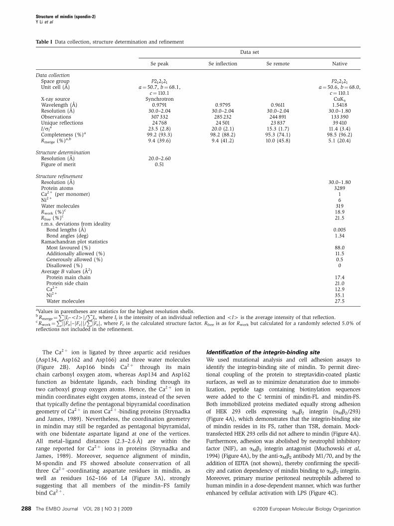
Table I Data collection, structure determination and refinement
Data set
Se peak Se inflection Se remote Native
Data collectionSpace group P212121 P212121
Unit cell (A) a¼ 50.7, b¼ 68.1,c¼ 110.1
a¼ 50.6, b¼ 68.0,c¼ 110.1
X-ray source Synchrotron CuKa
Wavelength (A) 0.9791 0.9795 0.9611 1.5418Resolution (A) 30.0–2.04 30.0–2.04 30.0–2.04 30.0–1.80Observations 307 332 285 232 244 891 133 390Unique reflections 24 768 24 501 23 837 39 410I/sI
a 23.5 (2.8) 20.0 (2.1) 15.3 (1.7) 11.4 (3.4)Completeness (%)a 99.2 (93.3) 98.2 (88.2) 95.3 (74.1) 98.5 (96.2)Rmerge (%)a,b 9.4 (39.6) 9.4 (41.2) 10.0 (45.8) 5.1 (20.4)
Structure determinationResolution (A) 20.0–2.60Figure of merit 0.51
Structure refinementResolution (A) 30.0–1.80Protein atoms 3289Ca2+ (per monomer) 1Ni2+ 6Water molecules 319Rwork (%)c 18.9Rfree (%)c 21.5r.m.s. deviations from ideality
Bond lengths (A) 0.005Bond angles (deg) 1.34
Ramachandran plot statisticsMost favoured (%) 88.0Additionally allowed (%) 11.5Generously allowed (%) 0.5Disallowed (%) 0
Average B values (A2)Protein main chain 17.4Protein side chain 21.0Ca2+ 12.9Ni2+ 35.1Water molecules 27.5
aValues in parentheses are statistics for the highest resolution shells.b Rmerge¼
P|Ij–oI4|/
PIj, where Ij is the intensity of an individual reflection and oI4 is the average intensity of that reflection.
c Rwork¼P
||Fo|–|Fc||/P
|Fo|, where Fc is the calculated structure factor. Rfree is as for Rwork but calculated for a randomly selected 5.0% ofreflections not included in the refinement.
Structure of mindin (spondin-2)Y Li et al
The EMBO Journal VOL 28 | NO 3 | 2009 &2009 European Molecular Biology Organization288

The integrin-binding site of mindin was localized further
by mutagenesis. Ligand binding to integrins is universally
dependent on bivalent cations, such that Mg2þ or Mn2þ at
the metal ion-dependent adhesion site (MIDAS) of the integ-
rin directly coordinates to the carboxyl group of a glutamate
or aspartate residue of the ligand (Shimaoka and Springer,
2003; Arnaout et al, 2007). However, the FS domain of
mindin contains 22 glutamate or aspartate residues on its
surface that could potentially ligate metals at the MIDAS. In
the crystal structure, we observed that Lys16, Glu96 and
Glu115 coordinated a cluster of five Ni2þ ions, which pre-
sumably originated from the crystallization buffer. As these
Ni2þ -binding residues should also have affinities for other
bivalent cations, we constructed the triple mutant K16A/
E96A/E115A. These mutations reduced adhesion of cells
expressing aMb2 integrin by B75% compared with wild-
type mindin (Figure 4A), implicating Glu96 or Glu115 as
the key metal-coordinating residue.
Glu96 is unlikely to be responsible for MIDAS binding,
based on the following observations: first, whereas human
mindin (the protein studied here) contains glutamate at
position 96, the corresponding residue in mouse (and rat)
mindin is alanine (Figure 3A). Second, mouse mindin inter-
acts with aMb2 integrin (Jia et al, 2005; Li et al, 2006). To
directly test whether Glu115 is involved in aMb2 binding,
we constructed the double mutant K16A/E115A. As these
Figure 1 Structure of the F-spondin domain of mindin and comparison with a Ca2þ -dependent C2 domain. (A) Side view of mindin-FS (ribbondiagram). The two four-stranded b-sheets, b6b5b2b3 (front) and b4b1b8b7 (back), are green and blue, respectively. Helices a1 and a2 are red.The bound Ca2þ ion is yellow. Loops L1–L4 at the top of the molecule are cyan. Residues mutated to alanine to localize the integrin-bindingsite are drawn in ball-and-stick representation, with carbon atoms in yellow, oxygen atoms in red and nitrogen atoms in blue. (B) Mindin-FS isrotated B1801 about the vertical axis with respect to the view in (A). (C) Side view of the C2 domain of cytosolic phospholipase A2 (PDBaccession code 1RLW). The orientation is similar to that of mindin-FS in (B). The two four-stranded b-sheets, b4b1b8b7 (front) and b6b5b2b3(back), are blue and green, respectively. The bound Ca2þ ion is yellow. Calcium-binding loops CBR1–CBR3 are cyan.
Figure 2 Calcium-binding site of mindin-FS. (A) Stereo view of the top of mindin-FS. Loops L1, L3 and L4 are red. Conserved residues withinthese loops that participate in key interloop interactions, or in coordinating the Ca2þ ion (yellow), are green. (B) Coordination geometry of theCa2þ ion. The calcium is ligated by Asp134, Asp162, Asp166, and three water molecules (Wat1–Wat3). Oxygen and carbon atoms are red andgreen, respectively.
Structure of mindin (spondin-2)Y Li et al
&2009 European Molecular Biology Organization The EMBO Journal VOL 28 | NO 3 | 2009 289

mutations reduced adhesion of aMb2-expressing cells to im-
mobilized mindin to nearly the same extent as the K16A/
E96A/E115 mutations (Figure 4A), we concluded that Glu115
most likely coordinates to the MIDAS of aMb2 integrin. As
controls, we introduced double (D182A/E186A) or triple
(D54A/R59A/K60A) mutations on other faces of the mindin
molecule (Figure 1A and B). However, these mutations did
not appreciably affect adhesion (Figure 4A), confirming the
specificity of the binding results.
Further examination of the human mindin sequence
revealed that Glu115 is embedded within the tripeptide
LEV (Figure 3A), which conforms to the consensus sequence
for LDV-binding integrins (L/I-D/E-V/S/T) (Hynes, 2002;
Humphries et al, 2006). Moreover, none of the other 21
solvent-accessible glutamate or aspartate residues of mindin
forms part of a known integrin recognition motif, in agree-
ment with localization of the integrin-binding site at Glu115.
The LEV motif is conserved in human, mouse and rat mindin,
but not in zebrafish mindin 1 (VLL), Drosophila M-spondin
(VFV) or in rat or zebrafish F-spondin (FSV) (Figure 3A).
Thus, within the mindin–FS family of ECM proteins, only
mammalian mindins appear to be integrin ligands, at least
with respect to the Glu115 site. However, zebrafish mindin 2
contains the sequence FEV at this position, which could
conceivably represent an integrin-binding site in other
vertebrates.
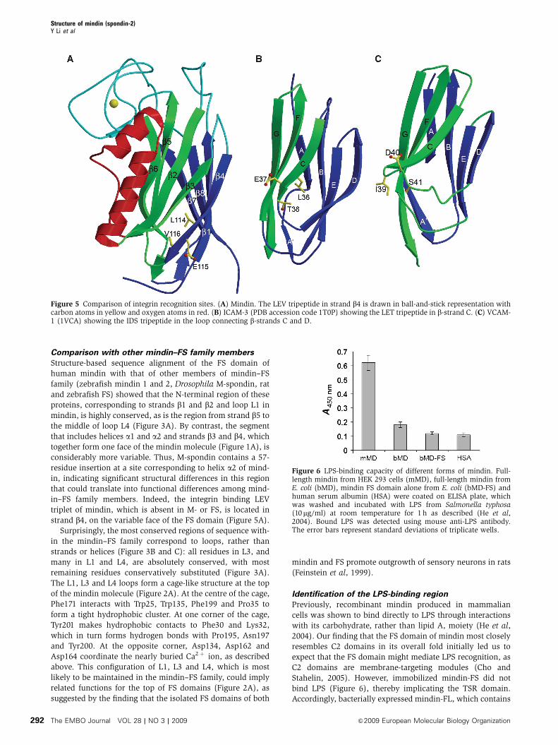
The LEV tripeptide of mindin is located at the end of strand
b4 near the base of the FS domain (Figure 5A). Interestingly,
Figure 3 Mindin–F-spondin family. (A) Structure-based sequence alignment of the F-spondin domains of mindin, M-spondin and F-spondin.Secondary structure elements are denoted by red cylinders (a-helices) and green arrow (b-strands); these are numbered sequentially. Loopslocated at the top of the mindin domain are labelled L1–L4. Residues that are strictly conserved are shaded red; residues that are well conservedare shaded pink. The integrin-binding triplet of mindin (LEV) is shaded yellow. Residues that coordinate the calcium ion are shaded blue.H, humans; M, mouse; R, rat; Z, zebrafish; D, Drosophila. Sequences were retrieved from SwissProt using the following accession numbers:human mindin (Q9BUD6); mouse mindin (Q8BMS2); rat mindin (Q9WV75); zebrafish mindin 1 (O42111); zebrafish mindin 2 (O42112);Drosophila M-spondin (O02092); rat F-spondin (P35446); zebrafish F-spondin 1 (O42113); zebrafish F-spondin-2 (O42114). Sequencealignments were performed with the program ClustalW (http://www.expasy.ch). (B) Distribution of conserved residues in the mindin-FSstructure, based on the sequence alignment in (A). Regions containing strictly conserved residues are highlighted in red; regions with wellconserved residues are highlighted in magenta. All other regions are grey. The calcium ion is yellow. Secondary structure elements are labelledaccording to (A). (C) Mindin-FS is rotated B1801 about the vertical axis with respect to the view in (B).
Structure of mindin (spondin-2)Y Li et al
The EMBO Journal VOL 28 | NO 3 | 2009 &2009 European Molecular Biology Organization290

this location is somewhat analogous to that of the integrin
binding LET triplet of ICAM-3, which is situated at the end of
b-strand C on the lower side of this immunoglobulin super-
family domain (Song et al, 2005) (Figure 5B). By contrast, the
metal-coordinating IDV triplet of VCAM-1 is found in a loop
connecting b-strands C and D (Shimaoka and Springer, 2003)
(Figure 5C).
Integrin specificity
Integrins are heterodimers composed of non-covalently
associated a- and b-subunits (Hynes, 2002; Shimaoka
and Springer, 2003; Arnaout et al, 2007). In mammals, 18
a-subunits and 8 b-subunits form 24 heterodimers that
recognize distinct but overlapping ligands. Half of integrin
a-subunits, including aM, contain inserted (I) domains (also
known as A domains), which are the principal ligand-binding
domains when present (Shimaoka and Springer, 2003).
Although HEK 293 cells expressing aMb2 bound strongly to
mindin, cells expressing aLb2 integrin showed no adhesion
(Figure 4B). To identify the source of this differential recogni-
tion, we constructed a chimaeric integrin by replacing the
I domain of aL with that of aM. As cells expressing the
aL(IaM)b2 chimaera adhered to mindin as well as cells
expressing aMb2 (Figure 4B), specificity is clearly mediated
through the I domains. However, other parts of the integrin
ab heterodimer also appear to contribute to mindin binding,
as suggested by a chimaera in which the a2 I domain of
integrin a2b1, which did not bind mindin (not shown), was
replaced by that of aMb2. In this context, the aM I domain only
partially restored adhesion to mindin (Figure 4B).
Figure 4 Cell adhesion to mindin. (A) aMb2-mediated cell adhesion to mindin. A total of 2�106 aMb2-expressing or mock-transfected HEK 293cells were allowed to adhere to 24-well non-tissue culture polystyrene plates precoated with recombinant mindin-FL, mindin-FS or its differentmutants. After incubation at 371C for 30 min, unbound cells were removed by three washes with DPBS and adherent cells were quantified bystaining with crystal violet, measuring absorption at 570 nm. Specificity of cell adhesion was verified using the aMb2-specific antagonist NIF,mock-transfected cells and biotinylated IgG or ovalbumin as the substrate (not shown). Data shown are the means±s.d. of triplicate wells andare representative of three independent experiments. (B) The aMb2-mediated cell adhesion to mindin is mediated by its aM I domain. Celladhesion to mindin-FL by different integrin receptors, including aMb2, aLb2, aL(IaM)b2 and a2(IaM)b1, where the aL or a2 I domain was replacedwith that of aM, were determined as above. Specificity was verified by the addition of the aMb2-specific antagonist NIF and by using mock-transfected HEK 293 cells. Data shown are the means ±s.d. of triplicate wells and are representative of two independent experiments.(C) Neutrophil adhesion to mindin is enhanced by LPS. Murine neutrophils were added to mindin-coated 24-well plates (0.5�106 cells perwell) in the presence or absence of 1 mg/ml LPS. After 10 min at 371C, unbound cells were removed by washing with PBS and bound cells werequantified by crystal violet staining, measuring absorption at 570 nm.
Structure of mindin (spondin-2)Y Li et al
&2009 European Molecular Biology Organization The EMBO Journal VOL 28 | NO 3 | 2009 291
Comparison with other mindin–FS family members
Structure-based sequence alignment of the FS domain of
human mindin with that of other members of mindin–FS
family (zebrafish mindin 1 and 2, Drosophila M-spondin, rat
and zebrafish FS) showed that the N-terminal region of these
proteins, corresponding to strands b1 and b2 and loop L1 in
mindin, is highly conserved, as is the region from strand b5 to
the middle of loop L4 (Figure 3A). By contrast, the segment
that includes helices a1 and a2 and strands b3 and b4, which
together form one face of the mindin molecule (Figure 1A), is
considerably more variable. Thus, M-spondin contains a 57-
residue insertion at a site corresponding to helix a2 of mind-
in, indicating significant structural differences in this region
that could translate into functional differences among mind-
in–FS family members. Indeed, the integrin binding LEV
triplet of mindin, which is absent in M- or FS, is located in
strand b4, on the variable face of the FS domain (Figure 5A).
Surprisingly, the most conserved regions of sequence with-
in the mindin–FS family correspond to loops, rather than
strands or helices (Figure 3B and C): all residues in L3, and
many in L1 and L4, are absolutely conserved, with most
remaining residues conservatively substituted (Figure 3A).
The L1, L3 and L4 loops form a cage-like structure at the top
of the mindin molecule (Figure 2A). At the centre of the cage,
Phe171 interacts with Trp25, Trp135, Phe199 and Pro35 to
form a tight hydrophobic cluster. At one corner of the cage,
Tyr201 makes hydrophobic contacts to Phe30 and Lys32,
which in turn forms hydrogen bonds with Pro195, Asn197
and Tyr200. At the opposite corner, Asp134, Asp162 and
Asp164 coordinate the nearly buried Ca2þ ion, as described
above. This configuration of L1, L3 and L4, which is most
likely to be maintained in the mindin–FS family, could imply
related functions for the top of FS domains (Figure 2A), as
suggested by the finding that the isolated FS domains of both
mindin and FS promote outgrowth of sensory neurons in rats
(Feinstein et al, 1999).
Identification of the LPS-binding region
Previously, recombinant mindin produced in mammalian
cells was shown to bind directly to LPS through interactions
with its carbohydrate, rather than lipid A, moiety (He et al,
2004). Our finding that the FS domain of mindin most closely
resembles C2 domains in its overall fold initially led us to
expect that the FS domain might mediate LPS recognition, as
C2 domains are membrane-targeting modules (Cho and
Stahelin, 2005). However, immobilized mindin-FS did not
bind LPS (Figure 6), thereby implicating the TSR domain.
Accordingly, bacterially expressed mindin-FL, which contains
Figure 5 Comparison of integrin recognition sites. (A) Mindin. The LEV tripeptide in strand b4 is drawn in ball-and-stick representation withcarbon atoms in yellow and oxygen atoms in red. (B) ICAM-3 (PDB accession code 1T0P) showing the LET tripeptide in b-strand C. (C) VCAM-1 (1VCA) showing the IDS tripeptide in the loop connecting b-strands C and D.
Figure 6 LPS-binding capacity of different forms of mindin. Full-length mindin from HEK 293 cells (mMD), full-length mindin fromE. coli (bMD), mindin FS domain alone from E. coli (bMD-FS) andhuman serum albumin (HSA) were coated on ELISA plate, whichwas washed and incubated with LPS from Salmonella typhosa(10mg/ml) at room temperature for 1 h as described (He et al,2004). Bound LPS was detected using mouse anti-LPS antibody.The error bars represent standard deviations of triplicate wells.
Structure of mindin (spondin-2)Y Li et al
The EMBO Journal VOL 28 | NO 3 | 2009 &2009 European Molecular Biology Organization292

both FS and TSR domains, was tested for LPS binding. In
contrast to full-length mindin produced in HEK 293 cells,
the bacterial protein showed no significant binding to LPS
(Figure 6).
To explain this functional difference between bacterial and
mammalian mindin, we analysed mindin from HEK 293 cells
for possible post-translational modifications. Previous studies
of the TSRs of FS, thrombospondin-1 and properdin identified
two types of protein glycosylation: O-fucosylation and
C-mannosylation (Hofsteenge et al, 2001; Gonzalez de Peredo
et al, 2002). Although the O-fucosylation site is absent in the
TSR of mindin, which has proline in place of serine/threonine
at position 266, this TSR does contain a potential site for
C-mannosylation at Trp257. This unusual modification in-
volves attachment of a single a-mannopyranosyl residue to
the C-2 atom of the first tryptophan in the recognition motif
W-X-X-W (W257SSW in mindin) (Doucey et al, 1998).
To determine whether mindin is C-mannosylated, the
protein was digested with trypsin and endoproteinase
Glu-C, and the resulting peptides were analysed by reversed
phase liquid chromatography coupled with tandem mass
spectrometry (RP-LC-MS/MS). The parent masses calculated
from the peptide sequence containing the C-mannosylation
recognition motif, VSLW@SSWGLC#GGPC#GK, allowed for
alkylated cysteine (#¼ 57.0215 Da) with and without manno-
sylated tryptophan (@¼ 162.0528 Da). A C-mannosylated
Figure 7 Identification of a C-mannosylation site in mindin. (A) LC-MS/MS result of C-mannosylated glycopeptides(VSLW@SSWGLC#GGPC#GK, þ 2, m/z 956.9320, @¼ 162.0528 Da and #¼ 57.0215 Da) produced by digestion of mindin with endoproteinaseGlu-C and trypsin. A full FTMS spectrum at 96.54 min of liquid chromatogram has the doubly charged C-mannosylated glycopeptides with 2.1p.p.m. mass accuracy. (B) An MS/MS spectrum shows the fragmentation of the glycopeptides for peptide identification and a cross-ringcleavage on zero and two carbons of mannose by neutral loss.
Structure of mindin (spondin-2)Y Li et al
&2009 European Molecular Biology Organization The EMBO Journal VOL 28 | NO 3 | 2009 293

glycopeptide (VSLW@SSWGLC#GGPC#GK, þ 2, at m/z
956.9301 as mono isotopic mass) was observed at
96.54 min (Figure 7A). The mass of the peptide indicated
two alkylated cysteines and a mannosylated tryptophan.
The MS/MS spectrum of þ 2, at m/z 956.93, confirmed the
peptide sequence (Figure 7B). In the y-ion series from the
fragmentation, C-mannosylated tryptophan was assigned with
348.13 Da differences (186.08 Da (W)þ 162.05 Da (@)) be-
tween the y-12 ion and y-13 ion from the fragmentations. The
120 Da loss, which is the typical ring cleavage of aromatic
C-glycosides (Figure 7B, inset), has been observed with loss of
m/z 120 and m/z 60 for singly and doubly charged ions,
respectively. The cross-ring cleavage of mannose occurred on
zero and two carbon bonds (0.2A/0.2X) by neutral loss, which
gave the most intense ion at m/z 897.06 and resulted in a
60 Da loss from the parent ion. Therefore, mindin produced in
HEK 293 cells is C-mannosylated at Trp257.
Analysis of the entire mindin sequence using the program
ExPASy (http://ca.expasy.org/tools/) identified Trp257 as the
only potential glycosylation site of any type. Consistent with
this result, LC-MS/MS analysis of peptides generated by
digesting mindin with trypsin and chymotrypsin revealed
no post-translational modifications elsewhere in the protein
(74% sequence coverage; not shown). As discussed below,
the C-mannosylated Trp257 residue of mindin TSR is located
on the proposed ligand-binding face of TSR domains
(Tan et al, 2002).
Discussion
The ECM protein mindin has multiple functions in nervous
system development and innate and adaptive immunity.
These include patterning axonal trajectory in the embryonic
spinal cord (Feinstein et al, 1999), acting as a pattern
recognition molecule for microbial pathogens (He et al,
2004; Jia et al, 2008), recruiting leukocytes to inflammation
sites (Jia et al, 2005), and priming CD4þ T cells (Li et al,
2006). Of these diverse functions, inflammatory cell recruit-
ment and T-cell priming are mediated by interactions of
mindin with integrins on neutrophils and DCs, respectively
(Jia et al, 2005; Li et al, 2006). We have demonstrated that
mindin binds integrins through its FS domain, which there-
fore constitutes a new integrin ligand. Interestingly, the
isolated FS domain of mindin has also been shown to
facilitate the outgrowth of dorsal root ganglia neurons,
although the relevant receptors have not been identified in
that case (Feinstein et al, 1999). In this respect, mindin
resembles FS, the founding member of the mindin–FS family,
which too is involved in axon guidance (Burstyn-Cohen et al,
1999; Tzarfaty-Majar et al, 2001; Zisman et al, 2007).
The structure of the FS domain of human mindin revealed
a broad surface which, based on sequence comparisons,
exhibits substantial topological variability within the mind-
in–FS family. Importantly, this surface contains the integrin
recognition motif that is unique to mammalian mindin.
Mindin-FS, ICAM-1 and ICAM-3 all utilize the L/I-D/E-V/S/
T triplet to bind integrins (LEV, IET and LET, respectively). In
the crystal structures of ICAM-1 and ICAM-3 bound to the I
domain of aL, the side chain of the central glutamate residue
projects from the ligand to coordinate to the MIDAS of the I
domain, whereas the side chains of the two flanking residues
point towards the ligand and do not contact the integrin
(Shimaoka et al, 2003; Song et al, 2005). The LEV triplet of
mindin-FS is similarly disposed, with the side chain of Glu115
fully accessible to solvent, but those of Leu114 and Val116
nearly completely buried in the FS domain. Apart from the
L/I-D/E-V/S/T motif, however, no residues of ICAM-1 or
ICAM-3 that contact the aL I domain in the complex struc-
tures are conserved in mindin-FS, implying a different set of
interactions outside the MIDAS.
Our data support a mosaic model of the mindin-binding
site within aMb2, where the aM I domain has a major function
and other domains, such as the aM b-propeller or the b2 I
domain, have supporting functions. Thus, mindin binding to
aMb2 is completely blocked by NIF, an aMb2-specific antago-
nist, the binding site of which is located exclusively on the aM
I domain (Zhang and Plow, 1997). Furthermore, substitution
of the aL I domain with that of aM conferred aLb2, an aMb2
homologue that does not bind mindin, with full mindin-
binding activity, suggesting that the I domain forms a major
binding interface with mindin. However, when the aM I
domain was inserted into a2, which complexes with a differ-
ent b-subunit (b1) and is less homologous to aM, only partial
binding was recovered. This suggests that other domains
within aMb2 may also contribute to binding, either by con-
tacting mindin directly or by altering the conformation of the
aM I domain. Our previous study showed that mindin also
interacts with a4b1 integrin on neutrophils (Jia et al, 2005). As
the a4 subunit lacks an I domain (Hynes, 2002), we anticipate
that mindin binding to a4b1 may occur through a different
mechanism than the one we describe here for aMb2.
TSR domains (B60 residues) have been identified in over
40 human proteins belonging to multiple families (Venter
et al, 2001). Structural studies of isolated TSRs have shown
that they are elongated molecules characterized by an unusual
antiparallel, three-stranded fold consisting of alternating
stacked layers of tryptophan and arginine residues (Tan
et al, 2002; Paakkonen et al, 2006). TSRs are found in secreted
proteins or in the extracellular portion of transmembrane
proteins that are involved in cell adhesion, neuronal develop-
ment and immunity (Tucker, 2004). Besides LPS, molecules
that bind to particular TSR modules include glycosaminogly-
cans, fibronectin and laminin. Mindin, which does not
bind fibronectin or laminin, recognizes LPS through its
carbohydrate, rather than lipid A, moiety (He et al, 2004).
Although no structure is available for any TSR bound to a
specific ligand, the second TSR module of thrombospondin-1
contains a long, positively-charged groove that is the likely
recognition surface for glycosaminoglycans (Tan et al, 2002)
(Figure 8A). A homology model of the mindin TSR domain
displayed a similar, though less electropositive, groove
(Figure 8B). Notably, the C-mannosylation site of thrombos-
pondin-1 TSR is located along one edge of its putative
glycosaminoglycan-binding groove, such that the mannose
attached to Trp423, which corresponds to Trp257 of mindin
TSR, could form part of the binding site (Tan et al, 2002)
(Figure 8A and B). Our finding that C-mannosylated
mindin from HEK 293 cells binds LPS, but that bacterially
expressed mindin does not, is consistent with a role for
glycosylation in ligand recognition. A role for glycosylation
in TSR function is also suggested by the finding that chemi-
cally synthesized C-mannosylated peptides derived from
TSRs enhanced LPS-induced signalling in macrophages
(Muroi et al, 2007).
Structure of mindin (spondin-2)Y Li et al
The EMBO Journal VOL 28 | NO 3 | 2009 &2009 European Molecular Biology Organization294

Although most of the sensors that detect and report
invading microbes are associated with immune cells (e.g.
TLRs, Nod-like receptors and RIG-I-like receptors), the ECM
is also essential for host defence against pathogens
(McDonald and Nunez, 2004). We have shown that the
ECM protein mindin interacts with integrins and LPS through
its FS and TSR domains, respectively. In this way, mindin
promotes activation of both adaptive and innate immune
responses to microbial invaders. The structural and binding
studies described here provide a molecular framework for
further investigating the mechanism of mindin’s multiple
actions in the immune system.
Materials and methods
Protein expression and purificationGene fragments encoding full-length human mindin (mindin-FL;residues Gln1-Val309), or the FS domain alone (mindin-FS; residuesGln1-Arg223) were inserted into pET-26b (Novagen). Both proteinswere expressed as inclusion bodies in Escherichia coli BL21(DE3)cells (Novagen). Inclusion bodies were dissolved in 8 M urea,50 mM Tris–HCl (pH 8.0) and 10 mM DTT. For in vitro folding,mindin-FL or mindin-FS inclusion bodies were diluted into foldingbuffer containing 0.8 M L-arginine-HCl, 50 mM Tris–HCl (pH 8.0),1 mM EDTA, 3 mM reduced glutathione and 0.9 mM oxidizedglutathione to a final protein concentration of 70 mg/l. After 72 hat 41C, the mixture was dialysed against 50 mM MES (pH 6.0).Mindin-FL and mindin-FS were purified using Superdex S-75 and
Mono Q columns (GE Healthcare). Mutants were produced andpurified similarly to the wild-type protein. For the production ofbiotinylated mindin, a tag (GSLNDIFEAQKIEWHE) was added tothe C termini of both mindin-FL and mindin-FS.
The FS domain of mindin contains only two methionineresidues, at positions 57 and 81. To assure sufficient anomaloussignal for MAD phasing, Leu215 was mutated to methionine. Forselenomethionine (SeMet) labeling, mindin-FS was expressed inmethionine-auxotroph E. coli B834(DE3) cells (Novagen) grown inSelenoMet medium (Athena Enzyme Systems).
Full-length mouse mindin containing a C-terminal Myc tag wasproduced in transfected HEK 293 cells as described (He et al, 2004).The protein was purified from culture supernatants using amonoclonal antibody to Myc (9E10) conjugated to Sepharose.
Crystallization and data collectionCrystals of mindin-FS (native or SeMet derivative) grew in hangingdrops at room temperature in 0–6% (w/v) polyethylene glycolmonomethyl ether 2000, 100 mM Tris–HCl (8.8–9.3) and 10 mMNiCl2. For data collection, crystals were transferred to a cryopro-tectant solution (mother liquor containing 20% (v/v) glycerol),prior to flash cooling. A native X-ray diffraction data set wascollected at 100 K with a Siemens HI-STAR area detector. MAD datafrom a SeMet crystal were collected to 2.04-A resolution at beamlineX29 of the Brookhaven National Synchrotron Light Source. All datawere indexed, integrated and scaled with the HKL2000 program(Otwinowski and Minor, 1997). Data collection statistics arepresented in Table I.
Structure determination and refinementThe mindin-FS structure was determined by MAD phasing with datacollected from the SeMet derivative at three different wavelengths(0.9791, 0.9795 and 0.9611 A). Six Se positions were determinedusing HKL2MAP (Pape and Schneider, 2004) or SOLVE (Terwilliger,2003), corresponding to three Se atoms per molecule in theasymmetric unit, and giving initial phases to 2.5-A resolution withan overall figure of merit of 0.51. Density modification andautomatic model building were performed with RESOLVE (Terwilli-ger, 2003), resulting in models for the two molecules in theasymmetric unit that were approximately 60 and 40% complete. Bycombining the partial structures, 80% of the model could be built,with initial Rwork of 40.4% and Rfree of 42.6% at 2.1-A resolution.
Model building and refinement were completed using the nativemindin-FS data set. Refinement was carried out using CNS1.1(Brunger et al, 1998), including iterative cycles of simulatedannealing, positional refinement and B-factor refinement, inter-spersed with model rebuilding into sA-weighted Fo–Fc and 2Fo–Fc
electron density maps using XtalView (McRee, 1999). The finalmodel of mindin-FS contains residues 8–218 of one monomer and8–222 of the other, 2 calcium ions, 6 nickel ions and 319 watermolecules. Refinement statistics are summarized in Table I. Stereo-chemical parameters were evaluated with PROCHECK (Laskowskiet al, 1993). Atomic coordinates and structure factors have beendeposited in the Protein Data Bank under accession code 3D34.
Cell adhesion assayCell adhesion to mindin was carried out as described (Li and Zhang,2003) with minor modifications. Twenty four-well non-tissueculture polystyrene plates were coated with 100 ml of 0.2mMstreptavidin at room temperature overnight. The coated plateswere blocked sequentially with 200ml of 1% bovine serum albumin(BSA) and 200ml of 0.5% polyvinylpyrrolidone in Dulbecco’sphosphate-buffered saline (DPBS) at room temperature for 1 heach. Biotinylated mindin and its different mutants (0.8mM) orcontrol IgG were added and the plates were incubated at roomtemperature for 30 min. After washing the plates with DPBS, 2�106
HEK 293 cells expressing different integrin receptors or their mock-transfected controls in DPBS plus 1 mM Ca2þ and 1 mM Mg2þ wereincubated with 100 nM NIF, 50mg/ml M1/70 or IgG for 15 min at 41Cand then added to each well. After 30 min at 371C, unbound cellswere removed by three washes with DPBS and adherent cells werefixed with 200 ml of 4% PFA in DPBS. The number of adherent cellswas quantified by staining the plates with 0.5% crystal violet andmeasuring absorption at 570 nm.
Figure 8 Electrostatic potential surface of TSRs. (A) The proposedligand recognition face of the second TSR module of thrombospon-din-1 with a mannose (Man) attached to Trp423 (Tan et al, 2002).Solvent-accessible surfaces are coloured according to electrostaticpotential, with positively charged regions in blue and negativelycharged regions in red. Electrostatic surface potentials were calcu-lated with GRASP (Nicholls et al, 1991). (B) The corresponding faceof a homology model of mindin TSR. A mannose (Man) is attachedto Trp257, which corresponds to Trp423 of the thrombospondin-1TSR. Homology modelling of the mindin TSR domain was per-formed with the program SWISS-MODEL (http://swissmodel.expasy.org) using the NMR solution structure of the first TSR domain of F-spondin as a template (PDB accession code 1VEX) (Paakkonen et al,2006).
Structure of mindin (spondin-2)Y Li et al
&2009 European Molecular Biology Organization The EMBO Journal VOL 28 | NO 3 | 2009 295

LPS-binding assayLPS binding was measured as described (He et al, 2004) with minormodifications. Purified full-length mindin from HEK 293 cells, full-length mindin from E. coli, mindin FS domain alone from E. coli orcontrol human serum albumin were coated on ELISA plates at 2mg/mlin 100ml in sodium bicarbonate buffer (pH 9.6) at 41C overnight.After washing with 0.2% Tween in PBS, the wells were blocked with3% BSA in PBS for 1 h at room temperature. Salmonella typhosa LPS(Sigma) at 10mg/ml was added and incubated at room temperaturefor 1 h. Bound LPS was detected with mouse anti-LPS polyclonalantibody followed by horseradish peroxidase-conjugated goat anti-mouse IgG antibody (Jackson Laboratory). After washing, 3,3,5,50-tetramethyl-benzidine was added; absorbance was measured at450 nm.
Mass spectrometryAffinity-purified mouse mindin from HEK 293 cells (20 mg) wasreduced with 25 mM DTT for 1 h at 551C and carboxyamidomethy-lated with 90 mM iodoacetamide in the dark for 45 min. Themodified protein was dialysed with a 7.5-kDa cutoff membrane(Millipore) against nanopure water at 41C overnight and dried in aSpeedVac concentrator. The dried mindin was resuspended in50 mM ammonium bicarbonate and digested with endoproteinaseGlu-C (Roche) at 251C for 20 h. The protein was further digestedwith trypsin (Promega) at 371C for 20 h. The Glu-C/tryptic peptideswere resuspended with 19.5ml of mobile phase A (0.1% formic acidin water) and 0.5 ml of mobile phase B (80% acetonitrile and 0.1%formic acid in water). The sample was loaded off-line onto ananospray tapered capillary column/emitter (360 mm� 75mm� 15mm) and separated through a 160 min linear gradient ofincreasing mobile phase B at a flow rate of B200 nl/min directlyinto the mass spectrometer.
LC-MS/MS analysis was performed on a LTQ Orbitrap XL massspectrometer (ThermoFisher) equipped with a nanospray ion
source. A full FTMS spectrum at 60 000 resolution was collectedat 400–2000 m/z, followed by three data-dependent MS/MS spectraof ITMS in the most intense ion peaks from parent mass listfollowing CID. The parent masses calculated from the peptidesequence VSLW@SSWGLC#GGPC#GK allowed for alkylated cysteine(#¼ 57.0215 Da) with and without mannosylated tryptophan(@¼ 162.0528 Da).
The resulting data were searched against the mouse mindinsequence (SwissProt accession number Q8BMS2), including con-taminant database using the TurboSequest algorithm (BioWorks3.3.1 SP1; ThermoFisher). DTA files were generated for spectra witha threshold of 15 ions, a TIC of 1e3 and a range of MHþ 400–6000m/z. The SEQUEST parameters were set to allow 30.0 p.p.m. ofprecursor ion mass tolerance and 0.5 Da of fragment ion tolerancewith monoisotopic mass. Tryptic and Glu-C peptides were allowedwith up to three missed internal cleavage sites and differentialmodifications were allowed for alkylated cysteine and mannosy-lated tryptophan.
Additional sequence mapping of mindin from HEK 293 cells wascarried by subjecting an SDS–PAGE gel slice containing mindin toin-gel digestion with trypsin, chymotrypsin or both enzymescombined. The resulting peptides were analysed by LC-MS/MS(Midwest Bio Services).
Acknowledgements
This study was supported by National Institutes of Health GrantsAI065612 (RAM), P01 HL54710 (LZ), and AI054658 and AI061364(Y-WH). Support for the data collected at beamline X29 of theNational Synchrotron Light Source comes from the Offices ofBiological and Environmental Research and of Basic EnergySciences of the US Department of Energy, and from the NationalCenter for Research Resources of the National Institutes of Health.
References
Arnaout MA, Goodman SL, Xiong JP (2007) Structure and me-chanics of integrin-based cell adhesion. Curr Opin Cell Biol 19:495–507
Benvenuti F, Hughes S, Walmsley M, Ruf S, Fetler L, Popoff M,Tybulewicz VL, Amigorena S (2004) Requirement of Rac1 andRac2 expression by mature dendritic cells for T cell priming.Science 305: 1150–1153
Burstyn-Cohen T, Tzarfaty V, Frumkin A, Feinstein Y, Stoeckli E,Klar A (1999) F-spondin is required for accurate pathfinding ofcommissural axons at the floor plate. Neuron 23: 233–246
Brunger AT, Adams PD, Clore GM, DeLano WL, Gros P,Grosse-Kunstleve RW, Jiang JS, Kuszewski J, Nilges M, PannuNS, Read RJ, Rice LM, Simonson T, Warren GL (1998)Crystallography and NMR systems: a new software suite formacromolecular structure determination. Acta Crystallogr Sec DBiol Crystallogr 54: 905–921
Cho W, Stahelin RV (2005) Membrane–protein interactions in cellsignaling and membrane trafficking. Annu Rev Biophys BiomolStruct 34: 119–151
Doucey M-A, Hess D, Cacan R, Hofsteenge J (1998) ProteinC-mannosylation is enzyme-catalyzed and uses dolichyl-phosphate-mannose as a precursor. Mol Biol Cell 9: 291–300
Feinstein Y, Borrell V, Garcia C, Burstyn-Cohen T, Tzarfaty V,Frumkin A, Nose A, Okamoto H, Higashijima S, Soriano E, KlarA (1999) F-spondin and mindin: two structurally and functionallyrelated genes expressed in the hippocampus that promote out-growth of embryonic hippocampal neurons. Development 126:3637–3648
Gonzalez de Peredo A, Klein D, Macek B, Hess D, Peter-Katalinic J,Hofsteenge J (2002) C-mannosylation and O-fucosylation ofthrombospondin type 1 repeats. Mol Cell Proteomics 1: 11–18
He Y-W, Li H, Zhang J, Hsu C-L, Lin E, Zhang N, Guo J, Forbush KA,Bevan MJ (2004) The extracellular matrix protein mindin is apattern-recognition molecule for microbial pathogens. NatImmunol 5: 88–97
Higashijima S, Nose A, Eguchi G, Hotta Y, Okamoto H (1997)Mindin/F-spondin family: novel ECM proteins expressed in thezebrafish embryonic axis. Dev Biol 192: 211–227
Hofsteenge J, Huwiler KG, Macek B, Hess D, Lawler J, Mosher DF,Peter-Katalinic J (2001) C-mannosylation and O-fucosylation ofthe thrombospondin type 1 module. J Biol Chem 276: 6485–6498
Humphries JD, Byron A, Humphries MJ (2006) Integrin ligands at aglance. J Cell Sci 119: 3901–3903
Hynes RO (2002) Integrins: bidirectional, allosteric signalingmachines. Cell 110: 673–687
Jia W, Li H, He Y-W (2005) The extracellular matrix protein mindinserves as an integrin ligand and is critical for inflammatory cellrecruitment. Blood 106: 3854–3859
Jia W, Li H, He Y-W (2008) Pattern recognition molecule mindinpromotes intranasal clearance of influenza viruses. J Immunol180: 6255–6261
Klar A, Baldassare M, Jessell TM (1992) F-spondin: a gene ex-pressed at high levels in the floor plate encodes a secreted proteinthat promotes neural cell adhesion and neurite extension. Cell 69:95–110
Kreis T, Vale R (eds) (1999) Guidebook to the Extracellular Matrix,Anchor, and Adhesion Proteins, 2nd edn Oxford University Press:Oxford
Lasarte JJ, Casares N, Gorraiz M, Hervas-Stubbs S, Arribillaga L,Mansilla C, Durantez M, Llopiz D, Sarobe P, Borras-Cuesta F,Prieto J, Leclerc C (2007) The extra domain A from fibronectintargets antigens to TLR4-expressing cells and induces cytotoxic Tcell responses in vivo. J Immunol 178: 748–756
Laskowski RA, MacArthur MW, Moss DS, Thornton JM (1993)PROCHECK: a program to check the stereo chemical quality ofprotein structures. J Appl Crystallogr 26: 283–291
Li H, Oliver T, Jia W, He Y-W (2006) Efficient dendritic cell primingof T lymphocytes depends on the extracellular protein mindin.EMBO J 25: 4097–4107
Li Y, Zhang L (2003) The fourth blade within the b-propeller isinvolved specifically in C3bi recognition by integrin aMb2. J BiolChem 278: 34395–34402
Manda R, Kohno T, Matsuno Y, Takenoshita S, Kuwano H, Yokota J(1999) Identification of genes (SPON2 and C20orf2) differentiallyexpressed between cancerous and noncancerous lung cells bymRNA differential display. Genomics 61: 5–14
Structure of mindin (spondin-2)Y Li et al
The EMBO Journal VOL 28 | NO 3 | 2009 &2009 European Molecular Biology Organization296

McDonald C, Nunez G (2004) Mindin the fort. Nat Immunol 5:16–18
McRee DE (1999) XtalView/Xfit–a versatile program for manipulat-ing atomic coordinates and electron density. J Struct Biol 125:156–165
Muchowski PJ, Zhang L, Chang ER, Soule HR, Plow EF, Moyle M(1994) Functional interaction between the integrin antagonistneutrophil inhibitory factor and the I domain of CD11b/CD18.J Biol Chem 269: 26419–26423
Muroi E, Manabe S, Ikezaki M, Urata Y, Sato S, Kondo T, Ito Y,Ihara Y (2007) C-mannosylated peptides derived from the throm-bospondin type 1 repeat enhance lipopolysaccharide-inducedsignaling in macrophage-like RAW264.7 cells. Glycobiology 17:1015–1028
Nalefski EA, Falke JJ (1996) The C2 domain calcium-binding motif:structural and functional diversity. Protein Sci 5: 2375–2390
Nicholls A, Sharp KA, Honig B (1991) Protein folding and associa-tion: insights from the interfacial and thermodynamic propertiesof hydrocarbons. Proteins 11: 281–296
Okamura Y, Watari M, Jerud ES, Young DW, Ishizaka ST, Rose J,Chow JC, Strauss III JF (2001) The extra domain A of fibronectinactivates Toll-like receptor 4. J Biol Chem 276: 10229–10233
Otwinowski Z, Minor W (1997) Processing of X-ray diffraction datacollected in oscillation mode. Methods Enzymol 276: 307–326
Paakkonen K, Tossavainen H, Permi P, Rakkolainen H, Rauvala H,Raulo E, Kilpelainen I., Guntert P (2006) Solution structures of thefirst and fourth TSR domains of F-spondin. Proteins 64: 665–672
Pape T, Schneider TR (2004) HKL2MAP: a graphical user interfacefor macromolecular phasing with SHELX programs. J Appl Cryst37: 843–844
Patti JM, Allen B, McGavin MJ, Hook M (1994) MSCRAMM-mediated adherence of microorganisms to host tissues. AnnuRev Microbiol 48: 585–617
Rizo J, Sudhof TC (1998) C2-domains, structure and function of auniversal Ca2+-binding domain. J Biol Chem 273: 15879–15882
Shimaoka M, Springer TA (2003) Therapeutic antagonists andconformational regulation of integrin function. Nat Rev DrugDisc 2: 703–715
Shimaoka M, Xiao T, Liu JH, Yang Y, Dong Y, Jun CD, McCormackA, Zhang R, Joachimiak A, Takagi J, Wang JH, Springer TA (2003)
Structures of the aL I domain and its complex with ICAM-1reveal a shape-shifting pathway for integrin regulation. Cell 112:99–111
Song G, Yang Y, Liu J, Casasnovas JM, Shimaoka M, Springer TA,Wang J-H (2005) An atomic resolution view of ICAM recognitionin a complex between the binding domains of ICAM-3 andintegrin aLb2. Proc Natl Acad Sci USA 102: 3366–3371
Strynadka NCJ, James MNG (1989) Crystal structures of thehelix-loop-helix calcium-binding proteins. Annu Rev Biochem58: 951–998
Tan K, Duquette M, Liu J, Dong Y, Zhang R, Joachimiak A, Lawler J,Wang J-H (2002) Crystal structure of the TSP-1 type 1 repeats: anovel layered fold and its biological implication. J Cell Biol 159:373–382
Terwilliger TC (2003) SOLVE and RESOLVE: automated structuresolution and density modification. Methods Enzymol 374: 22–37
Tucker RP (2004) The thrombospondin type 1 repeat family. IntJ Biochem Cell Biol 36: 969–974
Tzarfaty-Majar V, Burstyn-Cohen T, Klar A (2001) F-spondin is acontact-repellent molecule for embryonic motor neurons. ProcNatl Acad Sci USA 98: 4722–4727
Umemiya T, Takeichi M, Nose A (1997) M-spondin, a novel ECMprotein highly homologous to vertebrate F-spondin, is located atthe muscle attachment sites in the Drosophila embryo. Dev Biol186: 165–176
Venter JC, Adams MD, Myers EW, Li PW, Mural RJ, Sutton GG,Smith HO, Yandell M, Evans CA, Holt RA, Gocayne JD,Amanatides P, Ballew RM, Huson DH, Wortman JR, Zhang Q,Kodira CD, Zheng XH, Chen L, Skupski M et al (2001) Thesequence of the human genome. Science 291: 1304–1351
Walker EH, Perisic O, Ried C, Stephens L, Williams RL (1999)Structural insights into phosphoinositide 3-kinase catalysis andsignaling. Nature 402: 313–320
Zhang L, Plow EF (1997) Identification and reconstruction of thebinding site within aMb2 for a specific and high affinity ligand,NIF. J Biol Chem 272: 17558–17564
Zisman S, Marom K, Avraham O, Rinsky-Halivni L, Gai U, Kligun G,Tzarfaty-Majar V, Suzuki T, Klar A (2007) Proteolysis and mem-brane capture of F-spondin generates combinatorial guidanceclues from a single molecule. J Cell Biol 178: 1237–1249
Structure of mindin (spondin-2)Y Li et al
&2009 European Molecular Biology Organization The EMBO Journal VOL 28 | NO 3 | 2009 297