structure of native and expanded sobemoviruses by electron
TRANSCRIPT

doi:10.1006/jmbi.2000.4043 available online at http://www.idealibrary.com on J. Mol. Biol. (2000) 303, 197±211
Structure of Native and Expanded Sobemoviruses byElectron Cryo-Microscopy and Image Reconstruction
Natacha Opalka1,2, Mariana Tihova2, Christophe Brugidou3
Abhinav Kumar4, Roger N. Beachy1, Claude M. Fauquet3
and Mark Yeager2,4,5*
1Division of Plant Biology2Department of Cell Biology3International Laboratory forTropical AgriculturalBiotechnology (ILTAB/ORSTOM-TSRI), Division ofPlant Biology4Department of MolecularBiology, The Scripps ResearchInstitute, 10550 North TorreyPines Road, La JollaCA 92037, USA5Scripps Clinic, Division ofCardiovascular Diseases, 10666North Torrey Pines Road, LaJolla, CA 92037, USA
Present addresses: N. Opalka, LaMolecular Biophysics, The RockefelYork Avenue, New York, NY 10021Institut de recherche Pour le deÂveloAvenue Agropolis, BP5045, 34032 MFrance; A. Kumar, University of WSciences Center, K-428, Box 357742,USA; R. N. Beachy, Donald DanforCenter, 7425 Forsyth Boulevard, SuLouis, MO 63105, USA; C. M. FauqDanforth Plant Science Center, 8001Road, St. Louis, MO 63121, USA.
Abbreviations used: BPMV, beanCCMV, cowpea chlorotic mottle comlucerne transient streak virus; RYMvirus; SsbMV, sesbania mosaic virubean mosaic virus - bean; SCPMV,mosaic virus - cowpea; TBSV, tomaTYMV, turnip yellow mosaic tymovelectron microscopy.
E-mail address of the [email protected]
0022-2836/00/020197±15 $35.00/0
Rice yellow mottle virus (RYMV) and southern bean mosaic virus,cowpea strain (SCPMV) are members of the Sobemovirus genus of RNA-containing viruses. We used electron cryo-microscopy (cryo-EM) andicosahedral image analysis to examine the native structures of these twoviruses at 25 AÊ resolution. Both viruses have a single tightly packedcapsid layer with 180 subunits assembled on a T � 3 icosahedral lattice.Distinctive crown-like pentamers emanate from the 12 5-fold axes ofsymmetry. The exterior face of SCPMV displays deep valleys along the2-fold axes and protrusions at the quasi-3-fold axes. While having asimilar topography, the surface of RYMV is comparatively smooth. Twoconcentric shells of density reside beneath the capsid layer of RYMV andSCPMV, which we interpret as ordered regions of genomic RNA. In thepresence of divalent cations, SCPMV particles swell and fracture, whereasthe expanded form of RYMV is stable. We previously proposed that thecell-to-cell movement of RYMV in xylem involves chelation of Ca2� frompit membranes of infected cells, thereby stabilizing the capsid shells andallowing a pathway for spread of RYMV through destabilizedmembranes. In the context of this model, we propose that the expandedform of RYMV is an intermediate in the in vivo assembly of virions.
# 2000 Academic Press
Keywords: virus structure; electron cryo-microscopy; image processing;sobemovirus
*Corresponding authorboratory ofler University, 1230, USA; C. Brugidou,ppement, 911ontpellier cedex 1,
ashington, HealthSeattle, WA 98195,
th Plant Scienceite 385, Box 1098, St.uet, ILTAB/DonaldNatural Bridge
pod mottle virus;ovirus; LTSV,
V, rice yellow mottles; SBMV, southernsouthern beamto bushy stunt virus;irus; cryo-EM,
ing author:
Introduction
Rice yellow mottle virus (RYMV) and southernbean mosaic virus (SBMV) are members of thegenus Sobemovirus. First reported in Kenya(Bakker, 1974), the disease caused by RYMV isnow widespread in many western and easternAfrican countries. SBMV exclusively infects Legu-minosae and has been reported from the UnitedStates, Central and South America, Africa, Franceand India. The cowpea strain (SCPMV) used inthis study infects most cowpea cultivars (Shepherd& Fulton, 1962) and induces mosaic and leaf distor-tions. The genomes of RYMV and SCPMV consistof a single molecule of positive sense RNA with a50-linked protein (VPg). The complete genomesequences of RYMV (Ngon A Yassi et al., 1994) andSCPMV (Wu et al., 1987) reveal four open readingframes designated ORFs 1 to 4, predicted to encodeat least four proteins. ORF1 of RYMV encodes aprotein of 17.8 kDa that is involved in virus move-ment (Bonneau et al., 1998). The functions of the
# 2000 Academic Press

198 Electron Cryo-microscopy of Sobemoviruses
ORF1 and ORF3 proteins remain unknown. Bycomparison with other RNA viruses, ORF2encodes a polyprotein that apparently includes theVPg, a protease and a polymerase. ORF4 encodesthe coat protein of 26 kDa for RYMV and 28 kDafor SCPMV.
The pairwise similarity between the sequencesfor the capsid proteins of SCPMV and RYMV isonly 20.1 % (Table 1). In contrast, the sequences ofSBMV, SCPMV and sesbania mosaic virus(SsbMV) display much greater similarity (Table 1).Despite the presence of a number of substitutionsin the N-terminal region of RYMV compared toSCPMV, the residues in this region are highlybasic, a property thought to be important for pro-tein-RNA interactions and for the stability of thevirion. Ngon a Yassi et al. (1994) also proposedthat the N-terminal region of the coat protein ofRYMV and SCPMV has a bipartite nuclear target-ting motif, which could account for the presence ofvirions in the nuclei of infected cells (Francki et al.,1985). The stability of the capsid shell of sobemo-viruses is known to be dependent on the chelationof Mg2� and Ca2� (Hsu et al., 1976). AlthoughRYMV displays little sequence similarity withSCPMV, all amino acids involved in Ca2� bindingin SCPMV (D157, D160, E213, N278) are conservedin RYMV, except for E213 (Figure 1).
The X-ray structure of the cowpea strain ofcouther bean mosaic virus (SCPMV) at 2.8 AÊ resol-ution displays a solid capsid shell composed of 180identical protein subunits, which assemble withT � 3 icosahedral symmetry (Abad-Zapatero et al.,1980). The protein subunit consists of two domains.The R (random) domain (residues 1 to 64, 7 kDa)is partially ordered, and the S (shell) domain (resi-dues 65 to 260, 21 kDa) displays a canonical b-bar-rel motif. The highly basic N-terminal region of thecoat protein is thought to interact with RNA tostabilize the virion. For example, removal ofamino-terminal residues (1 to 30) results in theassembly of particles with a T � 1 capsid shell(Savithri & Erickson, 1983).
Interactions that are important for the in vitrostability of the sobemoviruses include a divalentcation protein-protein bond, a pH-dependent pro-tein-protein link and protein-RNA interactions(Hull, 1977). Upon alkali treatment in the presence
Table 1. Coat protein sequence comparisons in the Sobemovir
CfMV LTSV RYM
CfMV - 18.9 30.5LTSV 75.8 - 22.2RYMV 66.9 75.2 -SBMV 75.2 67.8 73.5SCPMV 75.7 66.0 75.8SsbMV 75.7 66.0 77.5
The upper right shows the percentage similarity between individuCfMV, cocksfoot mottle virus (MaÈkinen et al., 1995); LTSV, lucern
mottle virus (Ngon A Yassi et al., 1994); SBMV, southern bean momosaic virus (Wu et al., 1987); SsbMV, sesbania mosaic virus (Bhuva
of divalent cation chelators, the capsid shell swellsand becomes sensitive to enzymes and denatur-ants. Such swelling has been demonstrated forother plant viruses and may represent an inter-mediate state before disassembly and release of theRNA for binding by ribosomes and subsequenttranslation (Wilson, 1985). Interestingly, poliovirusundergoes capsid swelling during receptor attach-ment (Fricks & Hogle, 1990). However, the expan-sion of cowpea chlorotic mottle comovirus(CCMV) was not required for disassembly andtranslation of viral RNA (Albert et al., 1997).
Here, we report a comparison of the three-dimensional structures of RYMV and SCPMV at25 AÊ resolution derived from electron micrographsof unstained, frozen-hydrated specimens. Althoughthe capsid proteins exhibit only 20.1 % similarity,the subunits assemble on a T � 3 icosahedrallattice, and the capsid surfaces display similar pro-trusions clustered around the 5-fold axes of sym-metry. Although the swollen form of SCPMV wasunstable, the swollen form of RYMV was morestable and amenable to image analysis. We pro-pose that this expanded form of RYMV is an inter-mediate in the in vivo assembly of virions.
Results
Electron cryo-microscopy andimage processing
Native frozen-hydrated particles of RYMV andSCPMV exhibited distinctive circular and hexago-nal pro®les when examined by electron cryo-microscopy (Figure 2(a) and (b)). The absence ofdistortion and the constant size of the particlescon®rmed good preservation of the vitri®edsample. Although the particle edges were smooth,structural features were clearly visible within theparticle boundaries. In the presence of EDTA,many of the SCPMV particles swelled and frac-tured (Figure 2(d), background), whereas theswollen RYMV particles were stable and had a sizeclose to that of native SCPMV (Figure 2(c)).
Differences in the appearence of the individualparticle images of RYMV and SCPMV were con-sistent with random orientations of the virions inthe vitri®ed buffer, as was con®rmed by a plot of
us genus
V SBMV SCPMV SsbMV
18.5 19.3 19.323.8 23.0 26.420.5 20.1 18.4
- 75.9 69.023.8 - 62.830.4 36.8 -
al pairs, and the lower left shows the percentage divergence.e transient streak virus (Jeffries et al., 1995); RYMV, rice yellowsaic virus (Othman and Hull, 1995); SCPMV, southern cowpeaneshwari et al., 1995).

Figure 1. Alignment of the coat protein sequences of CfMV, LTSV, RYMV, SBMV, SCPMV and SsbMV. Bold lettersidentify identical residues. The locations for b-sheets, a-helices and Ca2�-binding sites are based on their positions inthe X-ray crystal structure of SCPMV.
Electron Cryo-microscopy of Sobemoviruses 199
the particle orientations over the icosahedral asym-metric unit (Figure 3(a)). Eigenvalue spectra pro-vide a test for errors resulting from a non-randomdistribution of particle orientations (Crowther et al.,1970). For both the compact forms of RYMV andSCPMV, and the expanded form of RYMV, theinverse eigenvalues were less than 0.1, which con-®rmed that the three-dimensional reconstructionswere reliably determined. Based on a cross-corre-lation cut-off value of 0.5, the resolution was�25 AÊ for the maps of native RYMV and SCPMVand �35 AÊ for the map of swollen RYMV(Figure 3(b)). The inability to re®ne the map ofswollen RYMV to higher resolution may re¯ectheterogeneity due to a decrease of stability uponexpansion.
Three-dimensional reconstructions of nativeRYMV and SCPMV
As was evident in the images, the three-dimen-sional reconstructions of native RYMV and
SCPMV revealed a single, solid capsid shell thatwas hexagonal when viewed down the 2-fold and3-fold axes of symmetry and circular when vieweddown the 5-fold vertex positions (Figure 4). Thecapsid shells were composed of 12 pentamers and20 hexamers arranged on a T � 3 icosahedral sym-metry lattice. Both viruses displayed pentamericprotrusions around the 5-fold axes of symmetry,which extended from radii of �153 to �160 AÊ forRYMV and from �155 to �164 AÊ for SCPMV(Figure 4). The protrusions at the 3-fold axes ofsymmetry were less prominent. Around the quasi-3-fold axes, SCPMV displayed a slightly highermass of density than for RYMV. The distancebetween the trimers was also shorter in SCPMVthan for RYMV (�53 AÊ for SCPMV and �59 AÊ forRYMV).
Protein distribution in native RYMV and SCPMV
Plots of the spherically averaged density of thenative, compact forms of RYMV and SCPMV

Figure 2. Electron micrographs of unstained, frozen-hydrated (a) native RYMV, (b) native SCPMV, (c) swollenRYMV and (d) swollen SCPMV. The native particles (a) and (b) are of uniform size and exhibit circular and hexago-nal pro®les. Samples of (c) swollen RYMV showed more uniformly sized particles compared with (d) swollenSCPMV in which the particles were more irregular and the background showed fragments of fractured particles. Thescale bar represents 1000 AÊ
200 Electron Cryo-microscopy of Sobemoviruses
reconstructions revealed capsid shells centered atradii of 130 and 138 AÊ , respectively (Figure 5). Theisosurface values for the protein shells displayed inFigures 4, 6, and 8 to 11 were adjusted to yieldvolumes for RYMV and SCPMV of 5.75 � 106 AÊ 3
and 6.19 � 106 AÊ 3, respectively, based on T � 3capsid shells formed by 180 subunits of molecularmass 26 kDa (for RYMV) and 28 kDa (forSCPMV), assuming a protein partial speci®cvolume of 0.74 cm3/g. Sectioned views of theRYMV and SCPMV three-dimensional reconstruc-tions along the 2-fold symmetry axis revealed solidcapsid shells with hexagonal pro®les and protru-sions at the 5-fold axes (Figure 6(a) and (b)). How-ever, the curvature and thickness of the capsidshell varied. The RYMV capsid exhibited a fairlysmooth surface relief. The thickness was nearlyconstant except at the inner surface of the 5-foldaxes where cavities were observed. In contrast, theinner surface of the SCPMV capsid showed a lar-ger mass of density at the 5-fold axes. In addition,
there were distinct protrusions along the quasi-3-fold axes that extended to a radius of 151 AÊ , anddepressions were seen at the 2-fold axes in theexterior face.
Ordered shells of RNA in native RYMVand SCPMV
As expected, the radial density plot of the X-raymap of SCPMV revealed no density withinthe capsid shell (Figure 5). The slight rippling withsmall peaks at �61 and �89 AÊ are ascribed toFourier series termination error because the �28 AÊ
distance between these peaks is close to the cut-offresolution of 25 AÊ . Note that the inner peakslabelled I and II in the RYMV and SCPMV pro®leswere substantially larger, which we interpret asorganized regions of genomic RNA that adopt apartial icosahedral arrangement. Peaks I and IIwere centered at �96 and �56 AÊ for RYMV and�101 and �65 AÊ for SCPMV. The isosurface values

Figure 3. (a) Plot of the re®ned y, f orientation anglesfor native RYMV (*), SCPMV (&) and swollen RYMV(~) particles. The plot demonstrates that the dataencompass a fairly uniform distribution of orientations.(b) Cross-correlation analysis. The particle image datasets were divided in half, and two independant recon-structions were computed. The maps for native RYMV(ÐÐ), SCPMV (- - - - - -) and swollen RYMV (± � ± � ± � ±)were compared as a function of spatial frequency. Thecorrelation coef®cients fall to a value of 0.5 at a resol-ution of �25 AÊ for the native RYMV and SCPMV recon-structions and �35 AÊ for swollen RYMV.
Electron Cryo-microscopy of Sobemoviruses 201
for these shells displayed in Figures 6, 7 and 11were adjusted to yield volumes for the RYMV andSBCMV RNA of 1.4 � 106 AÊ 3, based on the size ofthe genomic RNA (4.4 kB), assuming an anhydrousRNA partial speci®c volume of 0.55 cm3/g.Surface-shaded maps (Figure 7) showed that thetwo layers have a hexagonal shape when vieweddown a 2-fold axis of symmetry and were formedby an open fenestrated network of density. ForRYMV, layer I had projections at the 5-fold axesthat extended to a radius of �120 AÊ (Figure 7).Connections were also observed between layer Iand the capsid shell at the 2-fold axes. Layer II hada distinctive dodecahedral shape in which densitiesat the 5-fold vertices were connecting by rod-like
densities suggestive of duplex RNA. The twointernal layers of density were less well de®ned inSCPMV. The outermost layer was more rounded,and there was no connection between the globulardensities. Similarly, layer II displayed globulardensities clustered at the 5-fold vertex positions.
Comparison of cryo-EM and X-ray mapsof SCPMV
A density map derived from the 2.8 AÊ resolutionX-ray structure of SCPMV was computed at 25 AÊ
resolution and then rescaled by a factor of 3 % tomatch the cryo-EM map. There was a remarkablyclose correspondence in the surface and internalcontours of the X-ray and cryo-EM maps (Figure 8),which provided con®dence that the SCPMV cryo-EM structure was reliably determined. This closesimilarity was con®rmed by a correlation coef®-cient of 0.84 between these two reconstructions.
Comparison of SCPMV X-ray model and cryo-EM maps of native RYMV and SCPMV
A comparison between the X-ray model and thecryo-EM map of SCPMV was ®rst performed with-out any rigid body re®nement and yielded an R-factor of 0.56 (Figure 9(a)). After rigid body re®ne-ment, a best ®t was obtained with a substantiallyimproved R-factor of 0.36 (Figures 9(b) and 10(a)).A similar ®t of the SCPMV X-ray model into thecryo-EM map of RYMV yielded an R-factor of 0.28(Figures 9(c) and 10(b)).
Comparison of native and swollen RYMV
Upon chelation of divalent cations with EDTA,RYMV and SCPMV expand from peak radii of�130 to �139 AÊ and �138 to �148 AÊ , respectively(Figure 5). Specimens of swollen SCPMV containednumerous broken particles (Figure 2(d)), and forthe most spherical particles, unique orientationscould not be derived, which precluded determi-nation of a three-dimensional reconstruction.Although specimens of RYMV contained far fewerbroken particles, the three-dimensional reconstruc-tion could be derived only to 35 AÊ resolution, com-pared with a resolution of 25 AÊ for the nativeparticle. Therefore, the swollen particles are lessstable and more heterogeneous in size.
The three-dimensional reconstruction of swollenRYMV exhibited similar features with the compactform (Figure 11(a)). Although the expandedparticle was rounder, the hexagonal shape wasmaintained, and protrusions were seen at the5-fold axis. The expansion was most notable at theicosahedral 2-fold and 5-fold axes where thediameter increased �8 %. As noted, the twointernal layers of density within RYMV are inter-preted as ordered regions of genomic RNA(Figure 11(b) and (c) left), which were also seen inthe expanded particle (Figure 11(b) and (c) right).Particle expansion generated a smoother inner

Figure 4. Three-dimensional, surface-shaded density maps of RYMV (left) and SCPMV (right), viewed down (a) 2-fold, (b) 3-fold and (c) 5-fold axes of symmetry. Both viruses display a solid capsid surface with protrusions at the 5-fold vertex positions. The 5-fold and 3-fold symmetry axes are indicated in (a). The ends of the bars in (a) identifythe quasi-3-fold axes. The lengths of the bars indicate that the protrusions at the quasi-3-fold axes are separated by59 AÊ in RYMV and 53 AÊ in SCPMV. The scale bar represents 40 AÊ .
202 Electron Cryo-microscopy of Sobemoviruses
surface (Figure 11(b) right) with loss of the cavitybeneath the 5-fold symmetry axis seen in the nativeparticle (Figure 11(b) left).
Discussion
Icosahedral capsid structure of RYMV andSCPMV and comparison with other T�3plant viruses
Even though the sequence similarity betweenRYMV and SCPMV is only 20 % (Figure 1) and thediameter of RYMV is �7 % smaller (Figure 5),there is conservation of T � 3 icosahedral lattice
symmetry and considerable similarity in thetopology of the capsid shells (Figure 4). We areimpressed that three out of the four presumedCa2�-binding residues are conserved in RYMV(Figure 1). Lastly, the ®t of the SCPMV atomicmodel to the RYMV density is surprisingly good(Figures 9(c) and 10(b)). Therefore, we expect thatthe canonical b-fold and subunit packing withinSCPMV will be conserved in RYMV. Nevertheless,there are likely to be important differences in struc-ture, since the swollen form of SCPMV is muchmore unstable than RYMV.
Both RYMV and SCPMV display prominent pro-trusions at the 5-fold vertex positions. Turnip yel-

Figure 5. Radial density pro®les of RYMV (ÐÐ), SCPMV ( � � � � � � ), swollen RYMV (± ± ± ±), swollen SCPMV(Ð - Ð - Ð) and SCPMV X-ray (- - - - -) derived by spherically averaging the three-dimensional maps. The capsidshells are centered at peak radii of �130 and �138 AÊ for native RYMV and SCPMV. The shells expand to peak radiiof �139 and �148 AÊ for swollen RYMV and SCPMV, respectively. The pro®les derived from the cryo-EM mapsshow two inner peaks (labeled I and II) at radii of �96 and �65 AÊ for native RYMV, �101 and �56 AÊ for nativeSCPMV, and �100 and �66 AÊ for swollen RYMV. In comparison, the radial density pro®le derived from the X-raymap of SCPMV shows only slight rippling (small peaks at �89 and �61 AÊ ), which we ascribe to Fourier seriestermination errors because the distance between these peaks (�28 AÊ ) is close to the cut-off resolution of 25 AÊ . Thesubstantial peaks I and II in the cryo-EM pro®les are therefore ascribed to encapsidated RNA.
Electron Cryo-microscopy of Sobemoviruses 203
low mosaic virus (TYMV, genus Tymovirus)(BoÈ ttcher & Crowther, 1996; Canady et al., 1996)and CCMV (Speir et al., 1995) exhibit T � 3 icosa-hedral symmetry with pentameric protrusions atthe 5-fold axes. In addition, there are hexamericprotrusions at the quasi-6-fold positions, whichmake these viruses appear less angular thanRYMV and SCPMV. Association of the N terminiof six subunits at the quasi-6-fold symmetry axis isa unique feature of TYMV and CCMV. Previousstudies on T � 3 plant viruses showed that theN termini of only three subunits interact at thequasi-6-fold symmetry axes (Olson et al., 1983;Hogle et al., 1986; Silva & Rossmann, 1987). Intomato bushy stunt virus, the protruding Pdomains reside at the quasi-3-fold axes (Olson et al.,1983). The maps of SCPMV derived by X-ray crys-tallography and cryo-EM also reveal small protru-sions at the quasi-3-fold axes, which arise from the
insertion of a three-turn helix (aD helix) and a one-turn helix (aE helix) (Abad-Zapareto et al., 1980).These protrusions are less prominent in the cryo-EM structure of RYMV (Figures 4 and 10(b)). Inter-estingly, the RYMV sequence displays a deletion inthe region corresponding to the aD helix as well asproline residues that would tend to break a-helices.(In Figure 1, compare the sequence ATDYA-TAVGVNAN in SCPMV with SI - - - PSSVD-PN forRYMV). These differences may account for thereduced height of the protrusions at the quasi-3-fold axes in the RYMV cryo-EM map.
Despite the similarity between pentamers andhexamers on the exterior surface of RYMV andSCPMV, the interior surfaces differ. For RYMV,there are cavities at the pentameric axes, which arealso present in the capsid shell of TYMV (Canadyet al., 1996). In the case of CCMV, a channel is pre-sent in the pentamers which may be a consequence
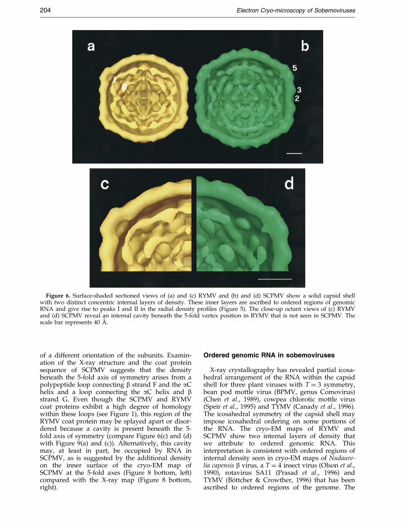
Figure 6. Surface-shaded sectioned views of (a) and (c) RYMV and (b) and (d) SCPMV show a solid capsid shellwith two distinct concentric internal layers of density. These inner layers are ascribed to ordered regions of genomicRNA and give rise to peaks I and II in the radial density pro®les (Figure 5). The close-up octant views of (c) RYMVand (d) SCPMV reveal an internal cavity beneath the 5-fold vertex position in RYMV that is not seen in SCPMV. Thescale bar represents 40 AÊ .
204 Electron Cryo-microscopy of Sobemoviruses
of a different orientation of the subunits. Examin-ation of the X-ray structure and the coat proteinsequence of SCPMV suggests that the densitybeneath the 5-fold axis of symmetry arises from apolypeptide loop connecting b strand F and the aChelix and a loop connecting the aC helix and bstrand G. Even though the SCPMV and RYMVcoat proteins exhibit a high degree of homologywithin these loops (see Figure 1), this region of theRYMV coat protein may be splayed apart or disor-dered because a cavity is present beneath the 5-fold axis of symmetry (compare Figure 6(c) and (d)with Figure 9(a) and (c)). Alternatively, this cavitymay, at least in part, be occupied by RNA inSCPMV, as is suggested by the additional densityon the inner surface of the cryo-EM map ofSCPMV at the 5-fold axes (Figure 8 bottom, left)compared with the X-ray map (Figure 8 bottom,right).
Ordered genomic RNA in sobemoviruses
X-ray crystallography has revealed partial icosa-hedral arrangement of the RNA within the capsidshell for three plant viruses with T � 3 symmetry,bean pod mottle virus (BPMV, genus Comovirus)(Chen et al., 1989), cowpea chlorotic mottle virus(Speir et al., 1995) and TYMV (Canady et al., 1996).The icosahedral symmetry of the capsid shell mayimpose icosahedral ordering on some portions ofthe RNA. The cryo-EM maps of RYMV andSCPMV show two internal layers of density thatwe attribute to ordered genomic RNA. Thisinterpretation is consistent with ordered regions ofinternal density seen in cryo-EM maps of Nudaure-lia capensis b virus, a T � 4 insect virus (Olson et al.,1990), rotavirus SA11 (Prasad et al., 1996) andTYMV (BoÈ ttcher & Crowther, 1996) that has beenascribed to ordered regions of the genome. The

Figure 7. The inner shells of RYMV (left) and SCPMV (right) (labeled I and II, here, and in Figure 5) are ascribedto ordered regions of genomic RNA. The dodecahedral appearance of shell II for RYMV is consistent with orderedduplex RNA. The scale bar represents 40 AÊ .
Electron Cryo-microscopy of Sobemoviruses 205
RNA within the capsid shell of RYMV appearsmore ordered than that of SCPMV. For example,the dodecahedral appearance of layer II in RYMV(Figure 7) presumably corresponds to duplexRNA, which is not seen in SCPMV. Thesedifferences may be related to weaker protein-RNAinteractions resulting from difference in the aminoacid sequences or biochemical differences such aspH and/or ionic strength. (Note that the RYMVparticles were maintained at pH 7, whereas theSCPMV particles were maintained at pH 5.5.)
Small-angle neutron scattering of SCPMV pro-vided information on the distribution of the RNAand protein within the capsid shell (KruÈ se et al.,1982). The scattering curves were consistent with amodel having four concentric shells centered atradii of 40, 67, 97 and 126 AÊ . The outermost layerwas assigned to the capsid shell, whereas the con-centric shells at lower radii contained signi®cantamounts of both RNA and some protein. The
results were consistent with localization of RNAand 15 % of the protein within the interior of thecapsid. The viral RNA was con®ned to radii lessthan 110 AÊ , in agreement with our results. Thescattering pro®les were interpreted by a modelwith a bilobal distribution of RNA and protein,similar to TBSV (Chauvin et al., 1978).
Compact and expanded forms of plant viruses
Chelation of divalent cations by EDTA results inexpansion of RYMV and SCPMV. Previous studieson the swollen forms of TBSV and CCMV showedthat the expansion is triggered by electrostaticrepulsion at the Ca2�-binding sites located near thequasi-3-fold axes (Robinson & Harrison, 1982;Speir et al., 1995). This repulsion caused a largeconformational change with formation of holes atthe quasi-3-fold axes. We presume that a similarmechanism may cause expansion in RYMV and

Figure 8. Three-dimensional surface-shaded density maps of SCPMV derived by cryo-EM (left) and X-ray crystallo-graphy (right) at 25 AÊ resolution. Note the close correspondence in the structure of the outer and inner surfaces ofthe capsid shell. To facilitate comparison with the X-ray structure, the two internal shells of density in the cryo-EMmap (see Figure 7) were removed. The additional density on the inner surface of the cryo-EM map of SCPMV at the5-fold axes (Figure 8 bottom, left) compared with the X-ray map (Figure 8 bottom, right) may be due to RNA incontact with the capsid at this site. The scale bar represents 40 AÊ .
206 Electron Cryo-microscopy of Sobemoviruses
SCPMV, since the X-ray structure of SCPMV doesshow bound Ca2�, and three out of four of theseresidues are conserved in the RYMV amino acidsequence. The lack of holes at the quasi-3-fold axesin the expanded form of RYMV is presumably dueto the low resolution of the reconstruction. Com-pared with RYMV, SCPMV particles tend to breakduring chelation of divalent cations, suggestingthat SCPMV is more unstable upon swelling. Thisloss of stability could be due to weakening of pro-tein-protein and/or protein-RNA associations. TheN-terminal polypeptide of both capsid proteinsmay interact with the genome in a different man-ner after swelling. For instance, the expanded formof TBSV undergoes a rearrangement of the N ter-
mini of the A and B subunits, which becomeordered and fold as b-sheets across the quasi-2-foldaxes (Robinson & Harrison, 1982).
Functional implications
We previously examined the translocation path-way of RYMV in systemically infected leaves andthe ultrastructural changes associated with RYMVinfection using Western immunoblotting, Northernblotting and thin-section electron microscopy(Opalka et al., 1998). In inoculated leaves, RYMVRNA and coat protein were ®rst detected at threeand ®ve days post-inoculation, respectively. By sixdays post-inoculation, RYMV had spread systemi-

Figure 9. A ribbon diagramdepicting the atomic structure ofSCPMV has been docked withinthe molecular envelope providedby cryo-EM and image analysis,(a) before and (b) after rigid-bodyre®nement. (b) After rigid-bodyre®nement, there is close corre-spondence between the X-ray andcryo-EM maps. (c) The good corre-spondence between the atomicstructure of SCPMV and the RYMVcryo-EM map suggests conserva-tion in the protein fold, in spite oflittle sequence similarity betweenthe SCPMV and RYMV coatproteins.
Electron Cryo-microscopy of Sobemoviruses 207
cally to leaves, and virus particles were observedin most cell types, including epidermal, mesophyll,bundle sheath, and vascular parenchymal cells.Most of the virions accumulated in large crystallinepatches in xylem parenchyma cells and sieveelements. Colocalization of a cell wall marker forcellulosic b-(1-4)-D-glucans and RYMV antibodiesover pit membranes suggested a pathway for virusmigration between vessels. We proposed that thepartial digestion of pit membranes resulting fromprogrammed cell death may permit virusmigration through them, concomitant with autoly-sis. In addition, displacement of Ca2� from pitmembranes to virus particles may contribute to thedisruption of the pit membranes and facilitate sys-temic virus transport. In the context of this model,we propose that the expanded form of RYMV is anintermediate in the in vivo assembly of virions.Since the native particle map could be re®ned to
higher resolution compared with the swollen, wepresume that the compact form is an inherentlymore stable particle with closer packing of subunitsto protect the encapsidated genome.
Materials and Methods
Virus preparation
RYMV-CI from the Ivory Coast (referred to here asRYMV) was puri®ed from infected rice plants (Oryzasativa L. variety IR8) as described (Fauquet & Thouvenel,1977). RYMV was retrieved from a 10 % to 40 % (w/v)continuous sucrose gradient. Samples (1 to 3 ml) werethen dialysed for 12 hours at 4 �C against 500 ml of20 mM potassium phosphate buffer (pH 7.0). SCPMV(kindly provided by Dr David Hacker) was isolatedfrom cowpea plants according to the method of Johnsonet al. (1974), in which the virus is puri®ed by sequentialprecipitation in 8 % (w/v) polyethylene glycol. The ®nal

Figure 10. Ribbon diagramsdepicting the atomic structuresof the A (yellow), B (red) andC (green) subunits of SCPMVdocked within the (a) SCPMV and(b) RYMV cryo-EM map. Pro-trusions at the quasi-3-fold axis aremore prominent in the cryo-EMmap of SCPMV than in the RYMVmap. The polypeptide containingthe aD and aE helices in the X-raystructure of SCPMV closelymatches the SCPMV cryo-EM mapat the quasi-3-fold axis. Notably,the corresponding region in theRYMV sequence contains a deletionas well as a proline residue. Thesesequence differences may accountfor the reduced height of the pro-trusions at the quasi-3-fold axes inthe RYMV cryo-EM map (locatedat the tips of the bars in Figure 4(a).
208 Electron Cryo-microscopy of Sobemoviruses
pellet after ultracentrifugation was resuspended at 3 to5 mg/ml in 10 mM sodium acetate (pH 5.2).
Preparation of expanded particles
Swollen particles were prepared by a modi®cation ofthe procedure of Rayment et al. (1979). Puri®ed samplesof native virions were dialysed for 24 hours at 4 �Cagainst 50 mM potassium phosphate buffer (pH 7.5) con-taining EDTA. Different concentrations of EDTA(10 mM, 20 mM and 30 mM) were used in order tode®ne the appropriate conditions for cryo-EM. Afterultracentrifugation for two hours, the pellet was resus-pended in 20 mM potassium phosphate buffer (pH 7.5)containing 10 mM EDTA.
Electron cryo-microscopy
Aliquots of the virus suspension were applied to elec-tron microscope grids coated with holey carbon ®lm pre-viously made hydrophilic by glow-discharge in anatmosphere of amylamine (Yeager et al., 1994). Virus par-ticles tended to preferentially adhere to the carbon ratherthan partition into the holes ®lled with vitri®ed buffer.This problem was solved by sequential application oftwo 5 ml droplets of sample (�5 mg/ml) to the grids.The ®rst application of the sample to the grid presum-ably saturated non-speci®c binding sites of the virus tothe carbon. The grid was blotted with ®lter paper, and asecond 5 ml aliquot was applied. After 60 seconds, the
droplet was blotted for eight seconds with preheated®lter-paper and then immediately plunged into a slushof liquid ethane (Yeager et al., 1990).
Grids were transferred to a Gatan 626 cryo-specimenholder (Gatan, Inc., Warrendale, PA) under liquidnitrogen and rapidly inserted into a Philips CM120transmission electron microscope operated at 100 kV.Grids were examined at low magni®cation (3600�) toidentify holes with minimal contamination and uniformice thickness. The area just adjacent to the region tobe photographed was examined at high magni®cation(88,000�) to locate Gaussian focus, correct for astigma-tism and ensure that drift was minimal. Low dose (5 to10 electrons/AÊ 2) micrographs were recorded at 0.8 to1.2 mm underfocus and a magni®cation of 45,000�.
Image processing
Optical diffraction was used to select images that dis-played minimal astigmatism and drift based on the Thonrings in the contrast transfer function. Selected imageswere digitized at a 25 mm interval using a Perkin Elmermicrodensitometer, corresponding to 5.5 AÊ on the speci-men. A DEC Alpha equipped with a Denali graphicsaccelerator was used for image processing. The programX3D, kindly provided by Drs James Conway and Alas-dair Steven, was used to extract particle images with acircular mask and subtract a background density. Forprocessing the images of native RYMV, an initial set oforigin and orientation values of 126 particles from a

Figure 11. (a) Three-dimensionalsurface-shaded density maps ofnative (left, yellow) and expandedRYMV (right, blue) viewed downa 2-fold symmetry axis. Theexpanded particle has similar sur-face topography but is rounderthan the native particle. (b) Sec-tioned views of both the native andexpanded particles show internaldensity that we attribute to orderedgenomic RNA, which has been dis-played separately in (c). Note thatparticle expansion is associatedwith loss of the cavity beneath the5-fold symmetry axes. The scalebar represents 40 AÊ .
Electron Cryo-microscopy of Sobemoviruses 209
single micrograph were determined using cross-corre-lation and common line procedures (Crowther, 1971;Fuller, 1987; Baker et al., 1988; Olson & Baker, 1989). Tooptimize the search procedure, calculations were per-formed using only a portion of the Fourier transform ofthe masked image (between 1/70 and 1/35 AÊ ÿ1) toremove both low and high frequency noise. Three par-ticles with well-de®ned orientations (�20 � phaseresidual) were used to compute an initial three-dimen-sional reconstruction using Fourier-Bessel inversion(Crowther, 1971). This initial map at 35 AÊ resolution wasused as the starting model for the Polar Fourier Trans-form (PFT) method (Baker & Cheng, 1996). Brie¯y, theFourier transform of the density distribution of theparticles was computed at each radius. A restrictedrange of particle radii (111 to 160 AÊ ) corresponding toradii encompassing the capsid shell was selected tooptimize the search procedure. In addition, images werelow-pass and high-pass ®ltered to optimize the determi-nation of the origin and orientation parameters (between1/70 and 1/35 AÊ ÿ1). The success of each cycle of re®ne-ment was assessed using the program CMP05 (kindlyprovided by Drs Tim Baker and Holland Cheng), whichcomputes correlation coef®cients comparing the densitydistribution of each particle with the correspondingprojected view of the three-dimensional model. For thePFT method, 12 cycles (six in global mode with 1o incre-ments and six in re®nement mode with 0.5 and then0.25 � increments) were performed by progressivelyincluding more particles in the map and by extendingthe resolution in the re®nement mode from 35 to 30 AÊ . A
®nal map at 30 AÊ resolution based on 12 particles wasthen used as a starting model to analyze a second micro-graph of RYMV particle images. A similar PFT strategyyielded a three-dimensional map at 25 AÊ resolutionbased on 50 particles.
For processing images of SCPMV particles, initial ori-gin and orientation values were determined for 200 par-ticles by cross-correlation and common lines procedures.A reconstruction at 35 AÊ resolution based on ®ve par-ticles was used as a starting model for the PFT method.The calculations were performed by including radii ofparticle images between 120 and 170 AÊ and by using therange of resolution as described above. Multiple cyclesof re®nement yielded a ®nal three-dimensional map at25 AÊ resolution derived from 41 particles.
For processing the images of the expanded RYMVparticles, the three-dimensional map of RYMV in thecompact form was radially scaled to ®t the radius of theexpanded particles. This expanded map was used as thestarting model for the PFT method. Multiple cycles ofre®nement, as described above, yielded a three-dimen-sional map at 35 AÊ resolution derived from 23 particles.A similar strategy was attempted for processing theimages of the expanded SCPMV particles. However,instability in the origin and orientation of particlesduring the search mode precluded determination of areliable three-dimensional reconstruction.
To assess the resolution of the maps, each data setwas divided in half to compute two independent recon-structions. The program EMMAP3DT was used to gener-

210 Electron Cryo-microscopy of Sobemoviruses
ate a list of structure factors, and the program EMCTF03used these data to compute the correlation coef®cient asa function of spatial frequency. All surface-shaded rep-resentations were visualized using AVS software(Sheehan et al., 1996).
Fit between X-ray SCPMV model and cryo-EM maps
The program XPLOR (BruÈ nger, 1996) was used to gen-erate structure factors of the SCPMV X-ray model, and amap at 25 AÊ resolution was generated by inverse Fouriertransformation using a temperature factor of 1000 AÊ 2.The program CMP05 was then used to determine aradial scale factor to ®t the cryo-EM and X-ray map. TheSCPMV cryo-EM map was contracted by 3 %, and theRYMV cryo-EM map was expanded by 4.3 % to bestmatch the SCPMV X-ray map. Prior to performing therigid-body re®nement in XPLOR, the cryo-EM map wasmodi®ed so that the density in the interior and exteriorof the shell (presumably noise) was ¯attened to zero.The structure factors calculated from this modi®ed den-sity were used in XPLOR during re®nement. Compari-son of the structure factors for the unre®ned X-raymodel and the cryo-EM map of SCPMV yielded an R-factor of 0.56. After 40 cycles of rigid-body re®nementwherein the three subunits, A, B, C of SCPMV, wereallowed to move independently, the R-factor decreasedto 0.36 for SCPMV and 0.28 for RYMV. The X-ray andcryo-EM maps were visualized using the program ``O''(Jones et al., 1991) to determine the view and select thenon-crystallographic symmetry matrices (from the set of60 icosehedral matrices) to generate selected Figures. Theprogram Bobscript (Esnouf, 1997) was used for ®nal ren-dering of the X-ray and cryo-EM maps in Figures 9and 10.
Note added in proof
Since submission of this paper, the X-ray crystal struc-ture of RYMV has been determined (C. Qu et al., unpub-lished results). In spite of low sequence similarity withSCPMV, the coat protein of RYMV does fold as a canoni-cal jellyroll b sandwich. However, a unique feature ofRYMV explains the increased stability compared withSCPMV. The bA arms (residues 31-53) between 2-foldrelated C subunits are swapped with each other, therebyproducing long-range interactions through the icosahe-dral surface lattice.
Acknowledgments
We thank S. Leitner for maintaining the plants. Wethank Timothy S. Baker, Holland Cheng, James Conway,Tony Crowther and Alasdair Steven for computer pro-grams. This work was supported by ORSTOM (FrenchScienti®c Research Institute for Development throughCooperation), by the Rockefeller Foundation, theNational Institute of Health (AI27161 to R.N.B. andAI31535 to M.Y.), the Scripps Family Chair (to R.N.B.),the Gustavus and Louise Pfeiffer Research Foundation(to M.Y.) and the Donald E. and Delia B. Baxter Foun-dation (to M.Y.). During this work M.Y. was an Estab-lished Investigator of the American Heart Associationand Bristol-Myers Squibb and is now the recipient of aClinical Scholar Award in Translational Research fromthe Burroughs Wellcome Fund.
References
Abad-Zapatero, C., Abdel-Meguid, S. S., Johnson, J. E.,Leslie, A. G. W., Rayment, I., Rossmann, M. G.,Suck, D. & Tsukihara, T. (1980). Structure ofsouthern bean mosaic virus at 2.8 AÊ resolution.Nature, 286, 33-39.
Albert, F. G., Fox, J. M. & Young, M. J. (1997). Virionswelling is not required for cotranslational disas-sembly of cowpea chlorotic mottle virus in vitro.J. Virol. 71, 4296-4299.
Baker, T. S. & Cheng, R. H. (1996). A model-basedapproach for determining orientations of biologicalmacromolecules imaged by cryoelectronmicroscopy. J. Struct. Biol. 116, 120-130.
Baker, T. S., Drak, J. & Bina, M. (1988). Reconstructionof the three-dimensional structure of simian virus40 and visualization of the chromatin core. Proc.Natl Acad. Sci. USA, 85, 422-426.
Bakker, W. (1974). Characterization and ecologicalaspects of rice yellow mottle virus in Kenya. InAgric. Res. Rep. 829, p. 152, Cent. Agric. Publ. Doc,Wageningen.
Bhuvaneshwari, M., Subramanya, H. S., Gopinath, K.,Savithri, H. S., Nayudu, M. V. & Murthy, M. R. N.(1995). Structure of Sesbania mosaic virus at 3 AÊ res-olution. Structure, 3, 1021-1030.
Bonneau, C., Brugidou, C., Chen, L., Beachy, R. N. &Fauquet, C. (1998). Expression of the rice yellowmottle virus P1 protein in vitro and in vivo and itsinvolvement in virus spread. Virology, 244, 79-86.
BoÈ ttcher, B. & Crowther, R. A. (1996). Difference ima-ging reveals ordered regions of RNA in turnip yel-low mosaic virus. Structure, 4, 387-394.
BruÈ nger, A. T. (1996). Recent developments for crystallo-graphic re®nement of macromolecules. Methods Mol.Biol. 56, 245-66.
Canady, M. A., Larson, S. B., Day, J. & McPherson, A.(1996). Crystal structure of turnip yellow mosaicvirus. Nature Struct. Biol. 3, 771-781.
Chauvin, C., Witz, J. & Jacrot, B. (1978). The structure oftomato bushy stunt virus: a model for protein-RNAinteraction. J. Mol. Biol. 124, 641-651.
Chen, Z., Stauffacher, C., Li, Y., Schmidt, T., Bomu, W.,Kamer, G., Shanks, M., Lomonossof, G. & Johnson,J. E. (1989). Protein-RNA interactions in an icosahe-dral virus at 3.0 AÊ resolution. Science, 245, 154-159.
Crowther, R. A. (1971). Procedures for three-dimensionalreconstruction of spherical viruses by Fourier syn-thesis from electron micrographs. Phil. Trans. Roy.Soc. ser. B, 261, 221-230.
Crowther, R. A., DeRosier, D. J. & Klug, A. (1970). Thereconstruction of a three-dimensional structure fromprojections and its applications to electronmicroscopy. Proc. Roy. Soc. ser. A, 317, 319-340.
Esnouf, R. M. (1997). An extensively modi®ed version ofMolScript that includes greatly enhanced coloringcapabilities. J. Mol. Graph. Model., 15, 132-134, (112-113 color plates).
Fauquet, C. & Thouvenel, J. C. (1977). Identi®cation ofrice yellow mottle virus in Ivory Coast. Plant Dis.Rep. 61, 443-446.
Francki, R. I. B., Milne, R. G. & Hatta, T. (1985). Atlas ofPlant Viruses, vol. 1, CRC Press, Boca Raton, FL,USA.
Fricks, C. E. & Hogle, J. M. (1990). Cell-induced confor-mational change in poliovirus: externalisation of theamino terminus of VP1 is responsible for liposomebinding. J. Virol. 64, 1934-1945.

Electron Cryo-microscopy of Sobemoviruses 211
Fuller, S. D. (1987). The T � 4 envelope of Sindbis virusis organized by interactions with a complementaryT � 3 capsid. Cell, 48, 923-934.
Hogle, J. M., Maeda, A. & Harrison, S. C. (1986). Struc-ture and assembly of turnip crinkle virus. I. X-raycrystallographic structure analysis at 3.2 AÊ resol-ution. J. Mol. Biol. 191, 625-638.
Hsu, C. H., Sehgal, O. P. & Pickett, E. E. (1976). Stabiliz-ing effect of divalent metal ions on virions ofsouthern bean mosaic virus. Virology, 69, 587-595.
Hull, R. (1977). The stabilization of the particles ofturnip rosette virus and of other members of thesouthern bean mosaic virus group. Virology, 79,58-66.
Jeffries, A. C., Rathjen, J. P. & Symons, R. H. (1995).Lucerne transient streak virus: complete genome,NCBI # U31286.
Johnson, J. E., Rossmann, M. G., Smiley, I. E. & Wagner,M. A. (1974). Single crystal X-ray diffraction studiesof southern bean mosaic virus. J. Ultrastruct. Res.46, 441-451.
Jones, T. A., Zou, J.-Y., Cowan, S. W. & Kjeldgaard, M.(1991). Improved methods for building proteinmodels in electron density maps and the location oferrors in these models. Acta Crystallog. sect. A, 47,110-119.
KruÈ se, J., Timmins, P. A. & Witz, J. (1982). A neutronscattering study of the structure of compact andswollen forms of southern bean mosaic virus. Virol-ogy, 119, 42-50.
MaÈkinen, K., Tamm, T., Nñss, V., Truve, E., Puurand,UÈ ., Munthe, T. & Saarma, M. (1995). Characteriz-ation of cocksfoot mottle sobemovirus genomicRNA and sequence comparison with related virus.J. Gen. Virol. 76, 2817-2825.
Ngon a Yassi, M., Ritzenthaler, C., Brugidou, C.,Fauquet, C. & Beachy, R. N. (1994). Nucleotidesequence and genome characterization of rice yel-low mottle virus RNA. J. Gen. Virol. 75, 249-257.
Olson, A. J., Bricogne, G. & Harrison, S. C. (1983). Struc-ture of tomato bushy stunt virus IV. The virusparticle at 2.9 AÊ resolution. J. Mol. Biol. 171, 61-93.
Olson, N. H. & Baker, T. S. (1989). Magni®cation cali-bration and the determination of spherical virusdiameters using cryo-electron microscopy. Ultrami-croscopy, 30, 281-298.
Olson, N. H., Baker, T. S., Johnson, J. E. & Hendry, D. A.(1990). The three-dimensional structure of frozen-hydrated Nudaurelia capensis b virus, a T � 4 insectvirus. J. Struct. Biol. 105, 111-122.
Opalka, N., Brugidou, C., Bonneau, C., Nicole, M.,Beachy, R. N., Yeager, M. & Fauquet, C. (1998).
Movement of rice yellow mottle virus betweenxylem cells through pit membranes. Proc. Natl Acad.Sci. USA, 95, 3323-3328.
Othman, Y. & Hull, R. (1995). Nucleotide sequence ofthe bean strain of southern bean mosaic virus. Virol-ogy, 206, 287-297.
Prasad, B. V. V., Rothnagel, R., Zeng, C. Q.-Y., Jakana,J., Lawton, J. A., Chiu, W. & Estes, M. K. (1996).Visualization of ordered genomic RNA and localiz-ation of transcriptional complexes in rotavirus.Nature, 382, 471-473.
Rayment, I., Johnson, J. E. & Rossmann, M. G. (1979).Metal-free southern bean mosaic virus crystals.J. Biol. Chem. 254, 5243-5245.
Robinson, I. K. & Harrison, S. C. (1982). Structure of theexpanded state of tomato bushy stunt virus. Nature,297, 563-568.
Savithri, H. S. & Erickson, J. W. (1983). The self-assem-bly of the cowpea strain of southern bean mosaicvirus: formation of T � 1 and T � 3 nucleoproteinparticles. Virology, 126, 328-335.
Sheehan, B., Fuller, S. D., Pique, M. E. & Yeager, M.(1996). AVS software for visualization in molecularmicroscopy. J. Struct. Biol. 116, 99-106.
Shepherd, R. J. & Fulton, R. W. (1962). Identity of aseed-borne virus of cowpea. Phytopathology, 52, 489.
Silva, A. M. & Rossmann, M. G. (1987). Re®ned struc-ture of southern bean mosaic virus at 2.9 AÊ resol-ution. J. Mol. Biol. 197, 69-87.
Speir, J. A., Munshi, S., Wang, G., Baker, T. S. &Johnson, J. E. (1995). Structures of the native andswollen forms of cowpea chlorotic mottle virusdetermined by X-ray crystallography and cryo-elec-tron micro-scopy. Structure, 3, 63-78.
Wilson, T. M. A. (1985). Nucleocapsid disassembly andearly gene expression by positive-strand RNAviruses. J. Gen. Virol. 66, 1201-1207.
Wu, S., Rinehart, C. A. & Kaesberg, P. (1987). Sequenceand organization of southern bean mosaic virusgenomic RNA. Virology, 161, 73-80.
Yeager, M., Dryden, K. A., Olson, N. H., Greenberg,H. B. & Baker, T. S. (1990). Three-dimensional struc-ture of rhesus rotavirus by cryoelectron microscopyand image reconstruction. J. Cell Biol. 110, 2133-2144.
Yeager, M., Berriman, J. A., Baker, T. S. & Bellamy, A. R.(1994). Three-dimensional structure of the rotavirushaemagglutinin VP4 by cryo-electron microscopyand difference map analysis. EMBO J. 13, 1011-1018.
Edited by W. Baumeister
(Received 18 May 2000; accepted 6 June 2000)