structure & function of the knee - mcccbehrensb/documents/week10b-final.pdf · structure &...
TRANSCRIPT

Structure & Function of the Knee
One of the most complex “simple” structures in the human body.
The “middle child” of the lower extremity.

Objectives
Identify the bones and bony landmarks of
the knee joint
Describe the supporting structures of the
knee

The Knee Joint
Consists of
• Tibiofemoral joint
• Patellofemoral joint
Two planes of motion
• Flexion and Extension
• Internal and External Rotation
–Any rotation occurs simultaneously with
flex/ext

Screw Home Mechanism
External rotation in the
last 20 degrees of
extension
A locking mechanism
Open Chain
--Tibial Ext Rotation on
femur
Closed Chain
--Femoral Int Rotation on
tibia

Osteology of the Knee
Distal femur
Right Femur
(ADDuctor tubercle)
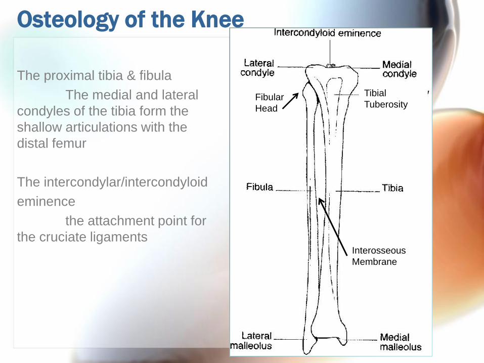
Osteology of the Knee
The proximal tibia & fibula
The medial and lateral
condyles of the tibia form the
shallow articulations with the
distal femur
The intercondylar/intercondyloid
eminence
the attachment point for
the cruciate ligaments
Tibial
TuberosityFibular
Head
Interosseous
Membrane

Osteology of the knee

Anatomy of the Knee: Anterior Aspect
• Femur
• Articular Cartilage
• Tibia
• Tibial Tuberosity
• Fibula (head)
• Medial Meniscus
• Lateral Meniscus
• Medial Collateral Ligament
• Lateral Collateral Ligament
• Anterior Cruciate Ligament

Anatomy of the Knee: Posterior Aspect•Femur
•Medial condyle
•Lateral condyle
•ADDuctor Tubercle
•Tibia
•Fibula•Fibular Head
•Medial Meniscus
•Lateral Meniscus
•Posterior Cruciate Ligament
•Lateral Collateral Ligament
•Medial Collateral Ligament

Anatomy of the Knee
Cruciate Ligaments
Anterior: (ACL)
-resists anterior motion
of the tibia on a fixed femur
-resists extremes of
knee extension
Posterior: (PCL)
-resists posterior motion
of the tibia on a fixed femur
-resists extremes of
knee flexion

Common Pathologies of the Knee
The menisci:
absorb shock and disperse large compressive
forces through the knee joint
They may not heal well:
inner 1/3: avascular (a)
middle 1/3: poor blood supply (b)
outer 1/3: good blood supply (c)

Genu Varum and Genu Valgum
Varus: inward
deviation of the
distal bone
Valgus
deformity:
outward
deviation of the
distal bone

Anatomy of the Knee: Genu “what?”
Genurecurvatum:
Hyperextension of the tibiofemoral joint placing
excessive stress on the structures in the popliteal space
Tibial nerve
Popliteal Vein
Popliteal Artery
Common Peroneal Nerve