structure & function of the knee - mcccbehrensb/documents/struckneebjb.pdf · structure &...
TRANSCRIPT
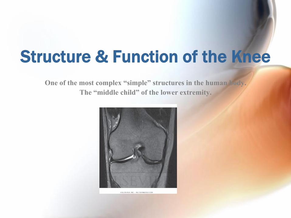
Structure & Function of the Knee
One of the most complex “simple” structures in the human body.
The “middle child” of the lower extremity.
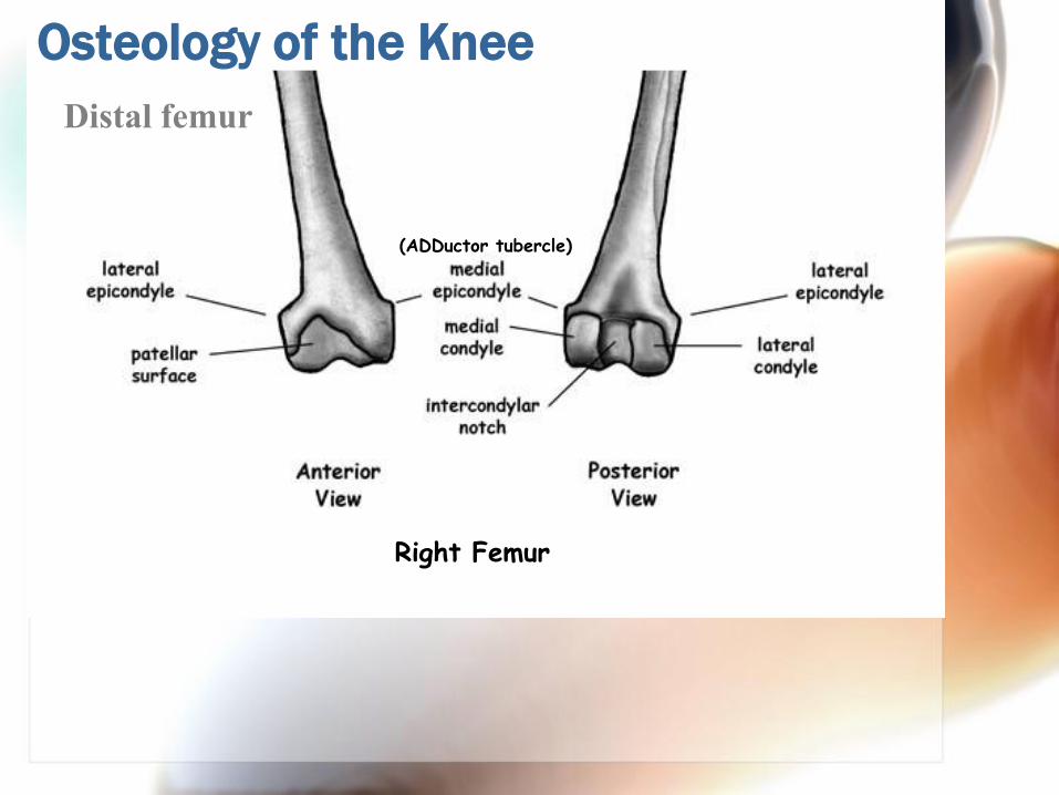
Osteology of the Knee
Distal femur
Right Femur
(ADDuctor tubercle)
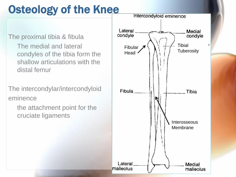
Osteology of the Knee
The proximal tibia & fibula
The medial and lateral
condyles of the tibia form the
shallow articulations with the
distal femur
The intercondylar/intercondyloid
eminence
the attachment point for the
cruciate ligaments
Tibial
TuberosityFibular
Head
Interosseous
Membrane
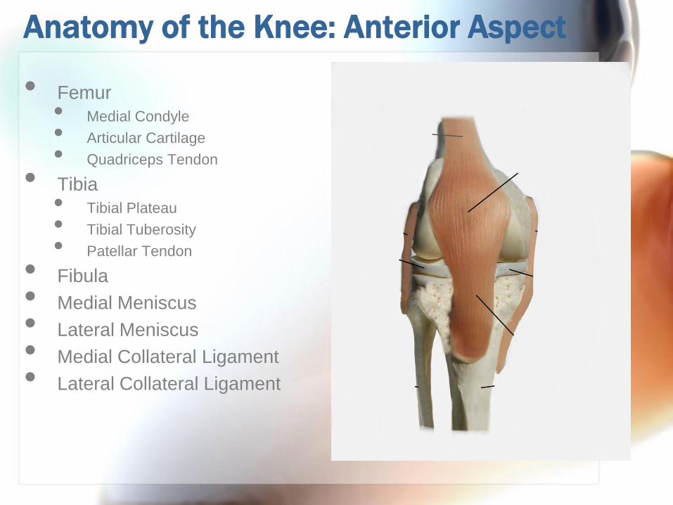
Anatomy of the Knee: Anterior Aspect
• Femur
• Medial Condyle
• Articular Cartilage
• Quadriceps Tendon
• Tibia
• Tibial Plateau
• Tibial Tuberosity
• Patellar Tendon
• Fibula
• Medial Meniscus
• Lateral Meniscus
• Medial Collateral Ligament
• Lateral Collateral Ligament
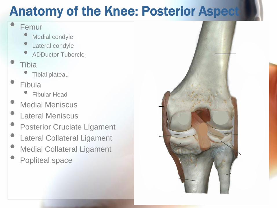
Anatomy of the Knee: Posterior Aspect• Femur
• Medial condyle
• Lateral condyle
• ADDuctor Tubercle
• Tibia
• Tibial plateau
• Fibula
• Fibular Head
• Medial Meniscus
• Lateral Meniscus
• Posterior Cruciate Ligament
• Lateral Collateral Ligament
• Medial Collateral Ligament
• Popliteal space
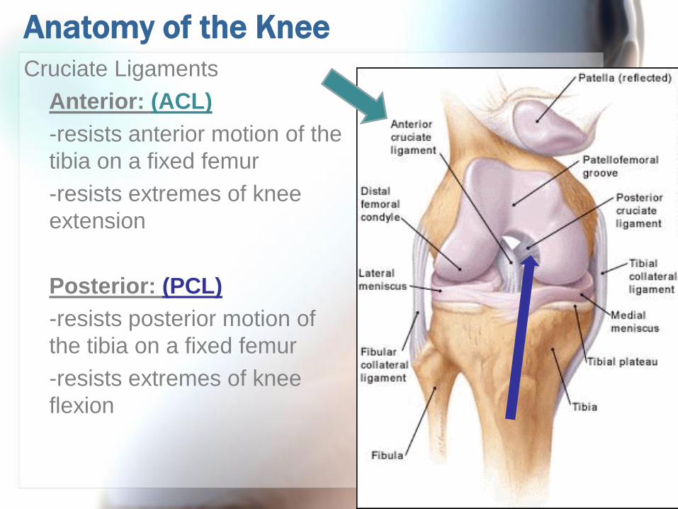
Anatomy of the Knee
Cruciate Ligaments
Anterior: (ACL)
-resists anterior motion of the
tibia on a fixed femur
-resists extremes of knee
extension
Posterior: (PCL)
-resists posterior motion of
the tibia on a fixed femur
-resists extremes of knee
flexion
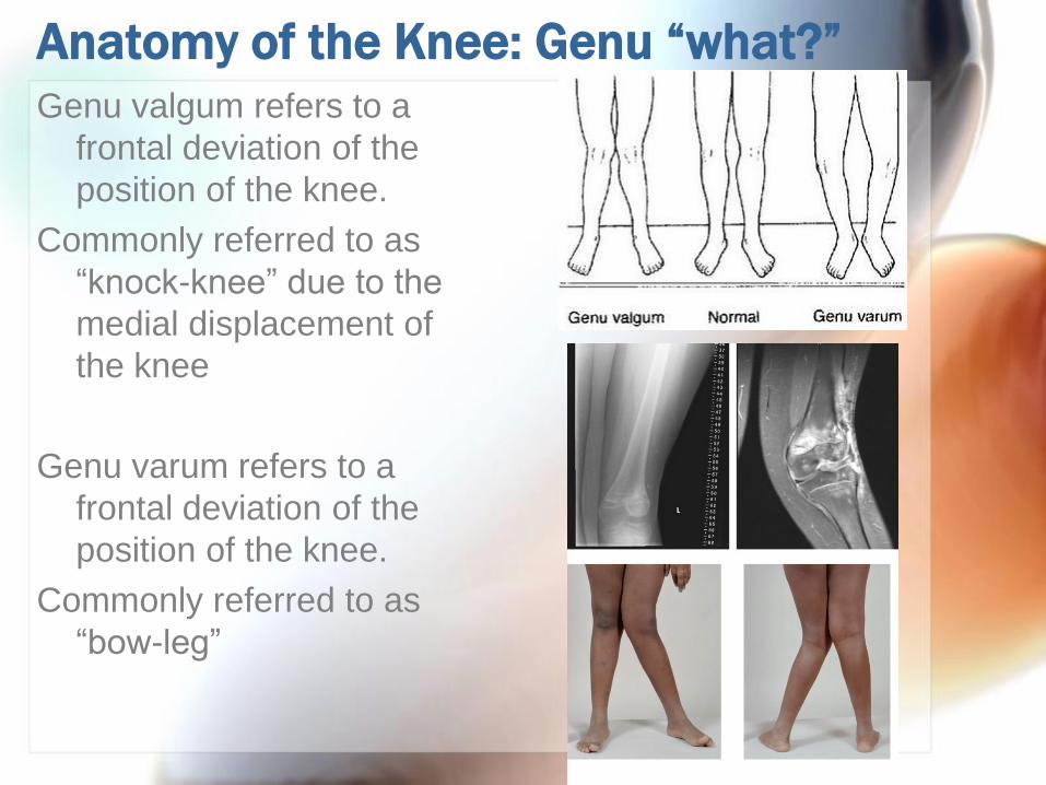
Anatomy of the Knee: Genu “what?”
Genu valgum refers to a
frontal deviation of the
position of the knee.
Commonly referred to as
“knock-knee” due to the
medial displacement of
the knee
Genu varum refers to a
frontal deviation of the
position of the knee.
Commonly referred to as
“bow-leg”
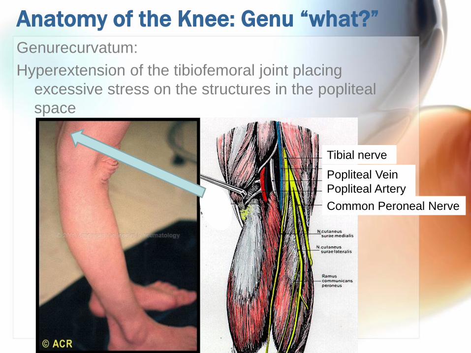
Anatomy of the Knee: Genu “what?”
Genurecurvatum:
Hyperextension of the tibiofemoral joint placing
excessive stress on the structures in the popliteal
space
Tibial nerve
Popliteal Vein
Popliteal Artery
Common Peroneal Nerve
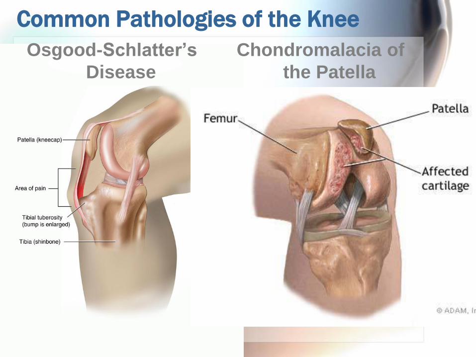
Common Pathologies of the Knee
Chondromalacia of
the Patella
Osgood-Schlatter’s
Disease

Common Pathologies of the Knee
The menisci:
absorb shock and disperse large compressive forces
through the knee joint
They may not heal well:
inner 1/3: avascular (a)
middle 1/3: poor blood supply (b)
outer 1/3: good blood supply (c)

Myology of the Knee
Your subtopic goes hereRectus Femoris
Origin Anterior-inferior iliac spine
Insertion Tibial tuberosity via the quadriceps
tendon
Innervation Femoral n.
Action Hip flexion, knee extension
“tidbit” One of the heads of the “quads”

Myology of the Knee
Vastus Medialis
Origin Medial lip of the linea aspera
and the intertrochanterid line
of the femur
Insertion Tibial tuberosity via the
patellar tendon
Innervation Femoral n.
Action Knee extension
“tidbit” •One of the heads of the
“quad”
•“VMO” one of the first
muscles of the knee to atrophy
post-operatively,
• responsible for last 10-15o of
knee extension
Vastus
Medialis
Obliquus

Myology of the Knee
Vastus Lateralis
Origin Lateral lip of the linea aspera,
intertrochanteric line, lateral
region of the gluteal tuberosity
Insertion Tibial tuberosity via the
patellar tendon
Innervation Femoral n.
Action Knee extension
“tidbit” Part of the “quads”
Myology of the Knee
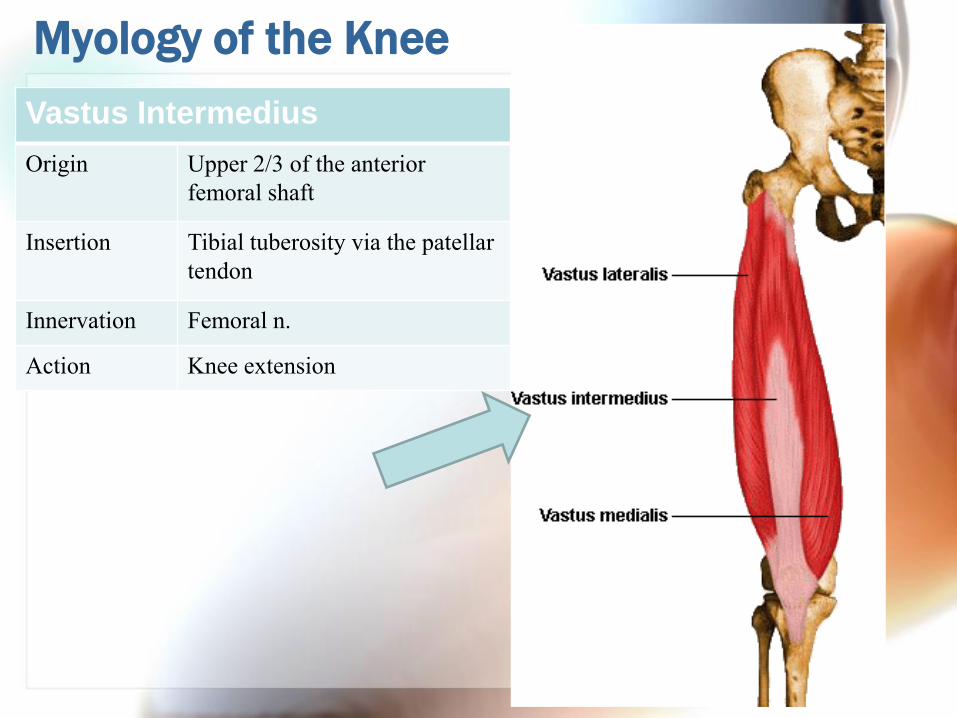
Vastus Intermedius
Origin Upper 2/3 of the anterior
femoral shaft
Insertion Tibial tuberosity via the patellar
tendon
Innervation Femoral n.
Action Knee extension

Q –Angle of the Knee
The line of force of the quadriceps can be described by
the Q-angle. It identifies patellofemoral tracking.
Females:
-greater angle
-greater incidence
of patellofemoral
joint pain
Q
Angle
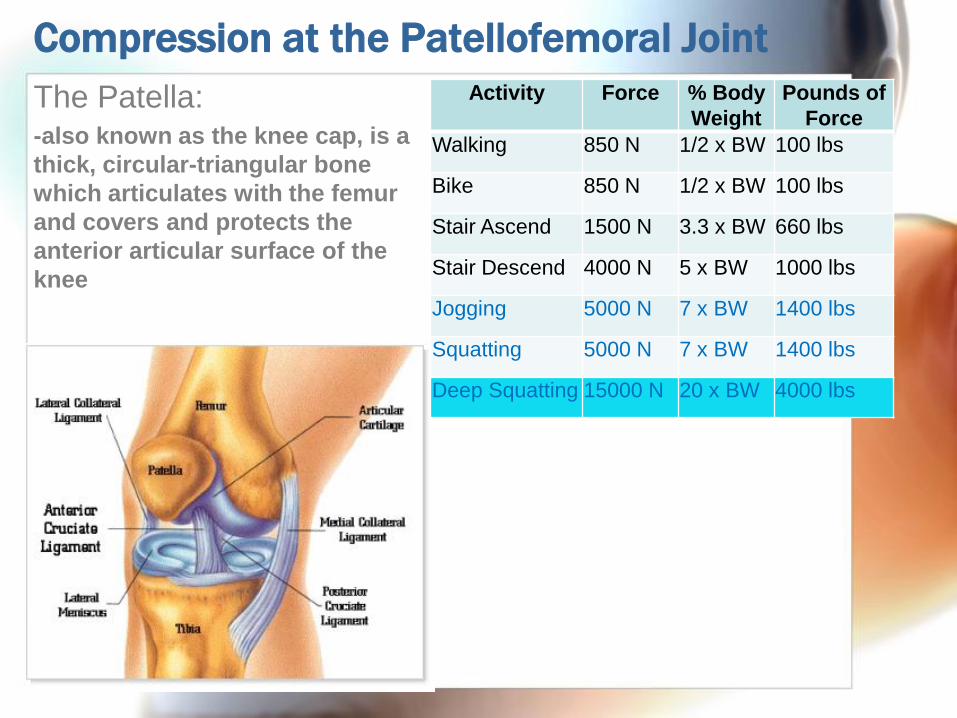
Compression at the Patellofemoral Joint
The Patella:-also known as the knee cap, is a
thick, circular-triangular bone
which articulates with the femur
and covers and protects the
anterior articular surface of the
knee
Activity Force % Body
Weight
Pounds of
Force
Walking 850 N 1/2 x BW 100 lbs
Bike 850 N 1/2 x BW 100 lbs
Stair Ascend 1500 N 3.3 x BW 660 lbs
Stair Descend 4000 N 5 x BW 1000 lbs
Jogging 5000 N 7 x BW 1400 lbs
Squatting 5000 N 7 x BW 1400 lbs
Deep Squatting 15000 N 20 x BW 4000 lbs
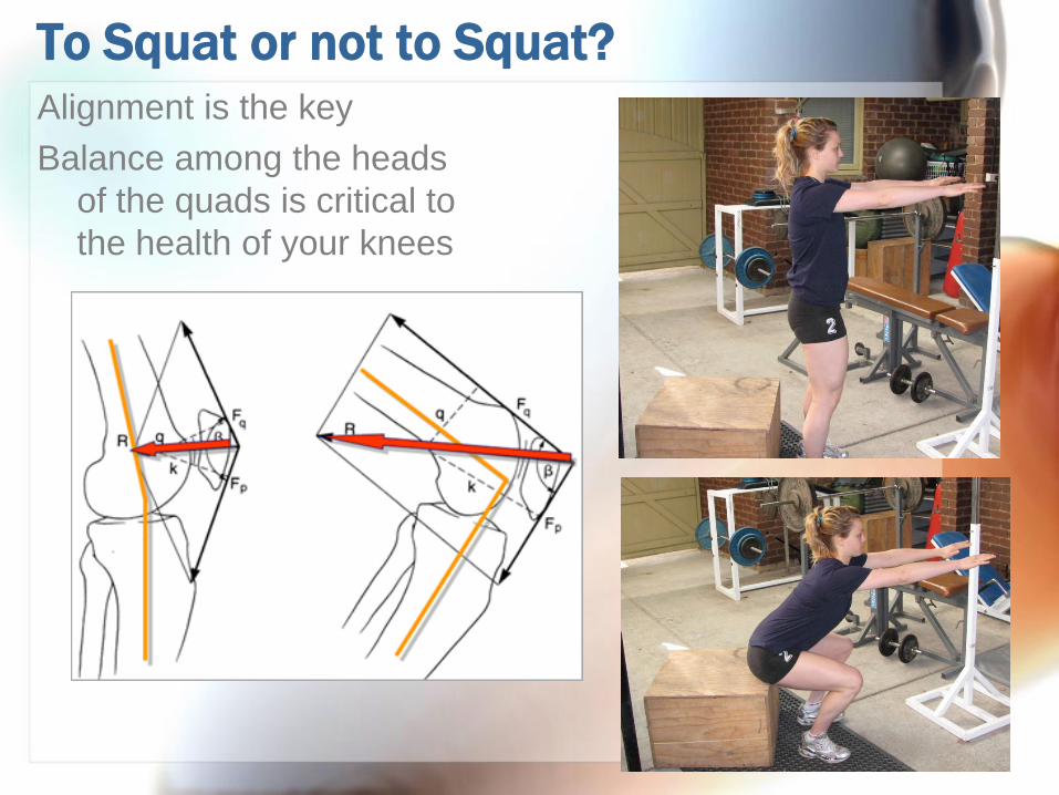
To Squat or not to Squat?
Alignment is the key
Balance among the heads
of the quads is critical to
the health of your knees
Myology of the Knee
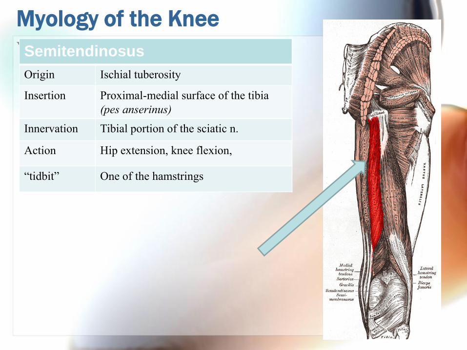
Your subtopic goes hereSemitendinosus
Origin Ischial tuberosity
Insertion Proximal-medial surface of the tibia
(pes anserinus)
Innervation Tibial portion of the sciatic n.
Action Hip extension, knee flexion,
“tidbit” One of the hamstrings

Myology of the Knee
Your subtopic goes hereBiceps Femoris
Origin Ischial tuberosity
Insertion Head of the fibula
Innervation Tibial portion of the sciatic n.
Action Hip extension, knee flexion
“tidbit” One of the hamstrings
A
A B C DBicep F Bicep F Semimem Semiten
Myology of the Knee
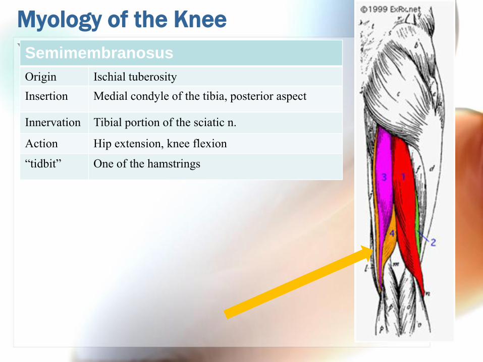
Your subtopic goes hereSemimembranosus
Origin Ischial tuberosity
Insertion Medial condyle of the tibia, posterior aspect
Innervation Tibial portion of the sciatic n.
Action Hip extension, knee flexion
“tidbit” One of the hamstrings
Myology of the Knee
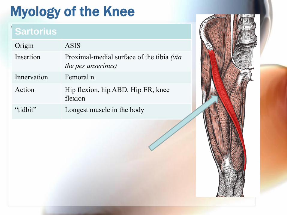
Your subtopic goes hereSartorius
Origin ASIS
Insertion Proximal-medial surface of the tibia (via
the pes anserinus)
Innervation Femoral n.
Action Hip flexion, hip ABD, Hip ER, knee
flexion
“tidbit” Longest muscle in the body
Myology of the Knee
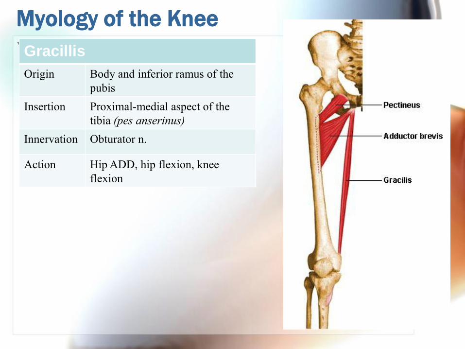
Your subtopic goes hereGracillis
Origin Body and inferior ramus of the
pubis
Insertion Proximal-medial aspect of the
tibia (pes anserinus)
Innervation Obturator n.
Action Hip ADD, hip flexion, knee
flexion
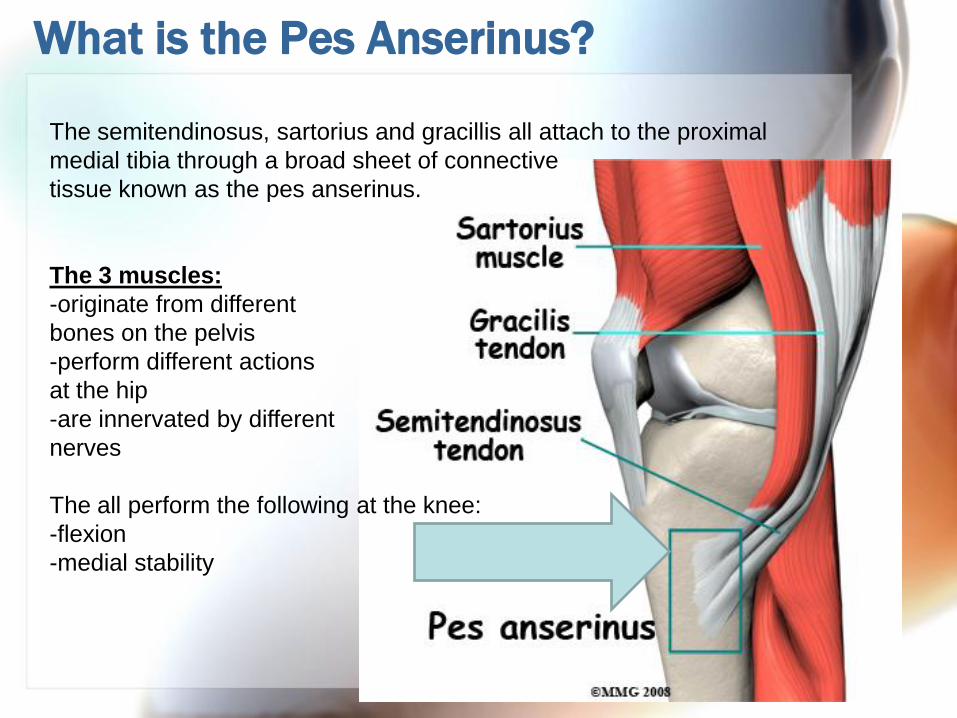
What is the Pes Anserinus?
The semitendinosus, sartorius and gracillis all attach to the proximal
medial tibia through a broad sheet of connective
tissue known as the pes anserinus.
The 3 muscles:
-originate from different
bones on the pelvis
-perform different actions
at the hip
-are innervated by different
nerves
The all perform the following at the knee:
-flexion
-medial stability

Myology of the Knee
Popliteus
Origin Posterior aspect of the
lateral femoral condyle
Insertion Posterior surface of the
proximal tibia
Innervation Tibial n.
Action Initiates knee flexion

Myology of the Knee
Gastrocnemius
Origin Medial head: posterior aspect of the
medial femoral condyle
Lateral head: posterior aspect of the
lateral femoral condyle
Insertion Calcaneal tuberosity via the Achilles
tendon
Innervation Tibial n.
Action Flexion of the knee, plantar flexion,

What can you identify? (in her knee)
Quadriceps
Vastus medialis
Vastus lateralis
Vastus intermedius?
Rectus femoris
Sartorius
Anything else?