structural properties of a highly ... - saiz lab
TRANSCRIPT

Structural Properties of a Highly Polyunsaturated Lipid Bilayer fromMolecular Dynamics Simulations
Leonor Saiz and Michael L. KleinCenter for Molecular Modeling, University of Pennsylvania, Philadelphia, Pennsylvania 19104-6323 USA
ABSTRACT The structure of a fully hydrated mixed (saturated/polyunsaturated) chain lipid bilayer in the biologically relevantliquid crystalline phase has been examined by performing a molecular dynamics study. The model membrane, a 1-stearoyl-2-docosahexaenoyl-sn-glycero-3-phosphocholine (SDPC, 18:0/22:6 PC) lipid bilayer, was investigated at constant (room)temperature and (ambient) pressure, and the results obtained in the nanosecond time scale reproduced quite well theavailable experimental data. Polyunsaturated fatty acids are found in high concentrations in neuronal and retinal tissues andare essential for the development of human brain function. The docosahexaenoic fatty acid, in particular, is fundamental forthe proper function of the visual receptor rhodopsin. The lipid bilayer order has been investigated through the orientationalorder parameters. The water-lipid interface has been explored thoroughly in terms of its dimensions and the organization ofthe different components. Several types of interactions occurring in the system have been analyzed, specifically, thewater-hydrocarbon chain, lipid-lipid and lipid-water interactions. The distribution of dihedral angles along the chains and themolecular conformations of the polyunsaturated chain of the lipids have also been studied. Special attention has beenfocused on the microscopic (molecular) origin of the effects of polyunsaturations on the different physical properties ofmembranes.
INTRODUCTION
Polyunsaturated fatty acid chains are an essential compo-nent of biomembranes. For instance, the retinal rod outersegment disk membrane is exceptionally rich in polyunsat-urated fatty acids, with;50% of docosahexaenoic fatty acid(DHA). Contents of DHA close to native levels are neededfor the proper function of the visual receptor rhodopsin(Brown, 1994; Litman and Mitchell, 1996), the first three-dimensional crystal structure of which has recently beenresolved at 2.8-Å resolution (Palczewski et al., 2000). Inaddition, polyunsaturated fatty acid chains are found in highconcentrations in cerebral gray matter and synaptic plasmamembranes and, from nutritional studies, it is well knownthat polyunsaturated lipids are important in the developmentof human brain function (Bloom, 1998).
Polyunsaturated phospholipid bilayers are characterizedby low temperatures for the main (gel-to-liquid-crystalline)phase transition,Tm (Lipowsky and Sackmann, 1995). Thisallows fluidity of model membranes under physiologicalconditions in contrast to lipids with saturated chains ofsimilar lengths (Small, 1986; Barry et al., 1991). In biolog-ical membranes, this fluidity is regulated by varying itscomposition (lipid and cholesterol content, for instance)since cells are usually constrained to an environment wheretemperature and pressure are fixed. For polyunsaturatedlipids or even phospholipids with mixed saturated/polyun-saturated chains, the fluid lamellar phase can be achieved at
room temperature for a unicomponent lipid bilayer. In thecase of the 1-stearoyl-2-docosahexaenoyl-sn-glycero-3-phosphocholine (SDPC, 18:0/22:6 PC) lipid, in particular,the main order-disorder (Lb93La) transition temperature inmultilamellar dispersions containing 50 wt % H2O wasmeasured by NMR spectroscopy and found to beTm 527.7°C 6 0.4 (25.3°C 6 0.7) on decreasing (increasing)the temperature (Barry et al., 1991).
Using computer modeling two linear conformations forDHA were predicted (Applegate and Glomset, 1986): heli-cal and an angle-iron-shaped form. Although the lowerenergy conformations correspond to a hairpin shaped mol-ecule, the former were thought to be more suitable forbiological interests, in which DHA can be paired withstearoyl in typical biological membranes. That study sug-gested that this kind of mixed-chain lipids with a saturatedchain in the sn1 position and the DHA in the sn2 positionmay form chain arrays with relatively tight packing incertain conditions. The importance of polyunsaturated lipidsin the local properties of membranes has recently beenstudied experimentally and specific lipid-protein and lipid-lipid interactions have been interpreted in terms of modelswith lateral segregation and the formation of domains (Lit-man and Mitchell, 1996). The aim of the present work isthus to study at a microscopic (atomistic) level these type ofcomplex systems under physiological conditions. In thisway, we are able to probe for the first time the molecularorigin of the peculiarities that the system confers to themembranes of which it is a component.
In this paper we report the results of a molecular dynam-ics (MD) study of an SDPC lipid bilayer in the liquidcrystalline phase at room temperature and ambient pressure.The MD simulations include structural properties of thelipid bilayer as well as conformational properties of the lipid
Received for publication 13 October 2000 and in final form 18 April 2001.
Address reprint requests to Dr. Leonor Saiz, University of Pennsylva-nia, Center for Molecular Modeling, 321 S. 34th St., Philadelphia, PA19104 -6323. Tel.: 215-573-4773; Fax: 215-573-6233; E-mail:[email protected].
© 2001 by the Biophysical Society
0006-3495/01/07/204/13 $2.00
204 Biophysical Journal Volume 81 July 2001 204–216

molecules. To our knowledge, earlier studies of water-lipidphosphatidylcholine (PC) systems in the fluid lamellarphase were restricted to disaturated lipids (Merz and Roux,1996) or lipids with a low degree of unsaturation. Thosesystems were constituted mainly by dimonounsaturated lip-ids, such as dioleoyl phosphatidylcholine (DOPC, 18:1/18:1) (Feller et al., 1997), or mixed saturated/unsaturatedchains with one double bond, such as palmitoyl oleoylphosphatidylcholine (POPC, 16:0/18:1) (Chiu et al., 1999),or two double bonds, such as palmitoyl linoleoyl phosphati-dylcholine (PLPC, 16:0/18:2) (Hyvo¨nen et al., 1997). In thecase of systems with unsaturations, the maximum numberof double bonds in a chain was two, and in the case of mixedchains, the simulations were not performed using a flexiblecell geometry, which has been proved to be essential, atleast during equilibration (Tobias et al., 1997; Venable etal., 2000). In all cases, except for simulations of dimyristoylphosphatidylcholine (DMPC) and DOPC, the simulatedtemperatures were.50°C.
Computer simulation details
The initial configuration for the computer simulation wasconstituted by a monolayer of 32 lipid molecules arrangedin the xy plane, which transformed into a bilayer after arotation of 90° about thez axis and of 90° about one of theaxis in thexy plane at the center of the bilayer. The bilayersystem was then solvated by a water slab and the watermolecules overlapped with the headgroups and those lo-cated deeper than the carbonyl region were removed. Theinitial cell dimensions were chosen to give an area per lipid,A, of A ' 69 Å2/lipid, and a lamellar spacing,d, of d ' 66Å. These values were chosen to agree with the NMR andx-ray experimental data at room temperature (Koenig et al.,1997). The initial lipid conformation was the following: 1)the headgroups were pointing away from the acyl chainregion toward the water-rich zone, 2) the saturated chainwas built as all-trans, the minimum energy conformation atzero temperature, 3) the unsaturated chain consisted of anangle-iron structure for the double-bond region with thedouble bonds adopting acis conformation, which is one ofthe conformations predicted by computer modeling forDHA in membrane environments (Applegate and Glomset,1986), and 4) the molecular axes with the smallest inertiamoments of the lipids were oriented randomly in thexyplane. The system with the previous characteristics wasconstituted by 64 lipid molecules and'27.5 water mole-cules per lipid (nw), which corresponds to a fully hydratedlipid bilayer.
To be able to perform a quantitative analysis of theinternal structure of the acyl chains, we have considered thefollowing definitions for the different conformations asso-ciated with the dihedral angles (gk and bk) related to thetorsional motions about the different bonds. For the satu-rated chain, thetrans conformation is defined by the range
120° # gk , 240°, whereas thegaucheconformations aredefined by the ranges 0°# gk , 120° (gauche1) and 240°# gk , 360° (gauche2). For the polyunsaturated chain, theskew1 (skew2) conformations of the dihedral angles aregiven by the ranges 60°# bk , 180° (180°# bk , 300°)and thecis conformation corresponds to dihedral angles inthe interval260° # bk , 60°. For the latter chain type, wehave considered the usual definitions for the dihedral anglesbetween consecutivecis double bonds, which correspond tothe structures with the following different shapes (Apple-gate and Glomset, 1986): helical (Askew6skew6Askew6skew6A; where “A” indicates the position of thedouble bonds), angle-iron (Askew6skew6Askew7skew7
A), and hairpin (Askew6skew7Askew7skew6A ).The molecular and potential model used for the lipid
molecules was the recent version of the all-atom CHARMMforce field (CHARMM27) for lipids (Schlenkrich et al.,1996; Feller and MacKerell, 2000), which has been shownto give excellent results for disaturated lipids and for lipidswith a low degree of unsaturation (Feller et al., 1997;Hyvonen et al., 1997). All the motions involving hydrogenatoms were frozen since those degrees of freedom are notexpected to be relevant for the properties analyzed and thisallowed us to use a longer time step in the integration of theclassical equations of motion. We used a rigid TIP3P modelfor water (Jorgensen et al., 1983), which is consistent withthe force field chosen for the lipid. The intermolecular partsof the force fields are pairwise additive functions, whichconsist of simple Lennard-Jones plus Coulomb terms.
The MD simulations were performed at constant tem-perature,T 5 303 K, and pressure,p 5 1 atm, andconsisted of an equilibration period of 1.8 ns and anequilibrium run of 1 ns. During the equilibration period,the system was initially simulated at constant volume for500 ps. Once the system was equilibrated at constantvolume, it needed;1300 ps to get a good convergence ofthe energy and the cell dimensions at constant pressureunder NPT conditions using the Nose´-Hoover thermostatchain extended system isothermal-isobaric dynamicsmethod with an orthorhombic simulation cell, as imple-mented in the PINY2D computational package (Tucker-man et al., 2000). The reversible multiple time stepalgorithm (Martyna et al., 1996) permitted the use of alonger time step (5 fs) while the smallest time step was 1fs. After equilibration, different properties were evalu-ated over a production run of 1 ns.
We used periodic boundary conditions and the constraintswere handled by means of the SHAKE/ROLL and RAT-TLE/ROLL methods (Martyna et al., 1996). The short-range forces were computed using a cutoff of;10 Å, andthe minimum image convention, and the long-range forceswere taken into account by means of the particle meshEwald summation technique (Frenkel and Smit, 1996).
Structure of a Polyunsaturated Membrane 205
Biophysical Journal 81(1) 204–216
Convergence and membrane dimensions
Due to the complexity (high degree of unsaturation) of thestudied lipid, a long equilibration period (1.8 ns) wasneeded to achieve a convergence of the dimensions of thesystem. After the equilibrium was reached, the dimensionsof the lipid bilayer evaluated over an additional 1 ns wereA 5 61.46 0.5 Å2 andd 5 70.06 0.5 Å, for the area perlipid and lamellar spacing, respectively. These data corre-spond to deviations of'10% (area per lipid) and'5%(lamellar spacing) from the initial set-up values.
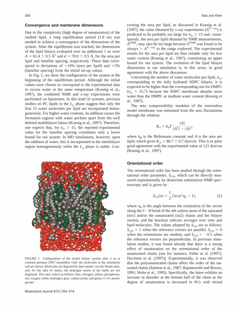
In Fig. 1, we show the configuration of the system at thebeginning of the equilibrium period. Although the initialvalues were chosen to correspond to the experimental datain excess water at the same temperature (Koenig et al.,1997), the combined NMR and x-ray experiments wereperformed on liposomes. In this kind of systems, previousstudies on PC lipids in theLa phase suggest that only thefirst 15 water molecules per lipid are incorporated homo-geneously. For higher water contents, its addition causes theformation regions with water pockets apart from the welldefined multibilayer lattice (Koenig et al., 1997). Therefore,one expects that, fornw . 15, the reported experimentalvalue for the lamellar spacing constitutes only a lowerbound for our system. In MD simulations, however, uponthe addition of water, this is incorporated in the interbilayerregion homogeneously while theLa phase is stable. Con-
cerning the area per lipid, as discussed in Koenig et al.(1997), the value obtained by x-ray experiments (AX2ray) ispredicted to be probably too large fornw . 15 and, conse-quently, the area per lipid obtained by NMR measurements,ANMR, may also be too large becauseANMR was found to bealways. AX2ray in the range explored. The experimentalresults for the area per lipid are thus reliable only for lowwater content (Koenig et al., 1997), constituting an upperbound for our system. The evolution of the lipid bilayerdimensions in our simulation is, in this sense, in goodagreement with the above discussion.
Concerning the number of water molecules per lipid,nw,corresponding to the fully hydrated SDPC bilayer, it isexpected to be higher than the corresponding one for DMPC(nw 5 25.7) because the SDPC membrane absorbs morewater than the DMPC at medium water content (Koenig etal., 1997).
The area compressibility modulus of the tensionlessmodel membrane was estimated from the area fluctuationsthrough the relation:
KA 5 kBT^A&
^A2& 2 ^A&2,
wherekB is the Boltzmann constant andA is the area perlipid, which givesKA 5 86.76 0.7 dyn/cm. This is in quitegood agreement with the experimental value of 121 dyn/cm(Koenig et al., 1997).
Orientational order
The orientational order has been studied through the orien-tational order parameter,SCD, which can be directly mea-sured experimentally by deuterium substitution NMR spec-troscopy and is given by
SCD~n! 51
2^3cos2hn 2 1&, (1)
wherehn is the angle between the orientation of the vectoralong theC2H bond of thenth carbon atom of the saturated(sn1) and/or the unsaturated (sn2) chains and the bilayernormal, and the brackets indicate averages over time andlipid molecules. The values adopted bySCD are as follows:SCD 5 1 when the reference vectors are parallel,SCD 5 0when the orientations are random, andSCD 5 20.5 whenthe reference vectors are perpendicular. In previous simu-lation studies, it was found already that there is a strongeffect of unsaturation on the orientational order of theunsaturated chains (see for instance, Feller et al. (1997);Hyvonen et al. (1997)). Experimentally, it was observedthat the polyunsaturated chains affect the order of the sat-urated chains (Salmon et al., 1987; Rajamoorthi and Brown,1991; Holte et al., 1995). Specifically, the latter exhibits anincrease in disorder at the bottom half of the chain as thedegree of unsaturation is increased in PCs with mixed
FIGURE 1 Configuration of the model bilayer system after 2 ns atconstant pressure (NPT ensemble). Only the molecules in the simulationcell are shown. Molecules are depicted by their atomic van der Waals radii,and, for the sake of clarity, the hydrogen atoms of the lipids are notdisplayed. The color code is as follows: blue, nitrogen; yellow, phosphorus;red, oxygen; white, hydrogen; gray, carbon atoms; and green, CAC atomicgroups.
206 Saiz and Klein
Biophysical Journal 81(1) 204–216

chains (Holte et al., 1995), whereas the effect of increasingtemperature on the chain order consisted of a decrease inorder in the first two-thirds of the saturated chain.
In Fig. 2, we plot the results obtained forSCD as afunction of the position,n, of the carbon atoms along thesaturated and the polyunsaturated chains. As a generaltrend, the different molecular structure and dynamics of thepolyunsaturated chain leads to values of the order parame-ters significantly lower than those of the saturated chain, ingood agreement with experiments (Safley et al., 2000). Theorientational order parameters for the DHA chain have beenfound to be strongly affected by unsaturation. This effect iscorrelated with the unsaturation position, especially at thefirst and second double bonds and in less degree at the thirdone. This correlation with unsaturation position suggests adependence of the order parameters on the specific confor-mation of the polyunsaturated chain (Saiz and Klein, 2001).A decrease in order occurs at the position of the first doublebond (C4AC5) andSCD takes very small values betweenthe first two bonds. The effect is just the opposite at thesecondcis double bond (C7AC8) where the order is in-creased, althoughSCD takes smaller values than at thebeginning of the chain. After the third double bond(C10AC11),SCD decays to'.05 for positions deeper intothe bilayer interior but the behavior is not visibly as stronglycorrelated with thecisdouble bond position as in the case ofthe first two double bonds. This can be related with the factthat from computer modeling it was shown how the molec-ular shape (and packing) of diacyl glycerols depends on theposition of the unsaturations (Applegate and Glomset,1991a,b). For instance, stearoyl acid when paired with DHAin the sn2 position, the situation considered in this study,was classified in the same group as arachidonyl and eico-satrienoyl acids, which have at least three double bondsstarting at the beginning of the chain. Diacylglycerols withtwo or three unsaturations starting at the middle of thechains pertained to the same type as distearoyl, and mole-
cules with only onecis double bond at the middle of thechain showed a different behavior. Our results suggest thatthe addition of morecis double bonds after the first threewhen the unsaturation begins close to the headgroup doesnot have a strong effect on the order of the polyunsaturatedchain.
A similar correlation of the chain order with the positionof a characteristic atomic group in the lipid chains waspreviously found (Husslein et al., 1998). There, the authorsreported an MD simulation of diphytanol phosphatidylcho-line (DPhPC) where the behavior ofSCD(n) suggested astepwise decrease in order, correlated with the positions ofthe methyl group substitutions.
Density profiles
The electron density profiles (EDPs) have been computedassuming a gaussian distribution located at the atomic po-sitions with variance,s, equal tos 5 23/2sLJ, wheresLJ isthe range of the Lennard-Jones potential. Hence,sLJ corre-sponds to the width of the gaussian distribution at half-height. The prefactors for each gaussian correspond to theatomic numbers,ni. The EDPs computed in this way areproportional to the density profiles measured along thebilayer normal obtained by x-ray scattering experiments atlow angles (White and Wiener, 1992, 1996) and are givenby
rX2ray~z!dz} Oi51
N ni
V
1
Î2ps2 e2 ~z2 zi!2/2s2dz, (2)
whereV is the volume of the slab betweenz andz 1 dz.The EDP has been calculated separately for the different
atomic groups of the lipid molecules. Maintaining the de-composition of White and Wiener (1992, 1996), the SDPCmolecule is constituted by the following groups: cholinegroup ([CH32]3N2CH22CH2, denoted by CHOL.), phos-phate group (PO4), glycerol (C3H5, denoted by GLYC.),carbonyl groups (COO), alkene (HCACH, denoted byCAC), methylene (CH2) and methyl (CH3) groups. It isworth noting that the CAC groups are located only in theunsaturated chain, whereas methyl and methylene groupsare located in both chains. The component due to thecarbonyl groups has been evaluated for the saturated(C(1)OO) and the unsaturated (C(2)OO) chains. In Fig. 3a,we show the results obtained for the (total) EDP, and thecontributions arising from the water and lipid moleculesseparately. The components due to the acyl chains aredepicted in Fig. 3a as well. The contributions of the lipidheadgroups, glycerol and the carbonyl groups to the EDPare decomposed in Fig. 3b and compared to that of water.
The total EDP has the expected characteristics: 1) ahigher density at the position of the lipid-water interface,corresponding to the headgroup, glycerol, and carbonylgroups of the lipids and water distributions; 2) a lower
FIGURE 2 Orientational order parameter as a function of the position ofthe carbon atom along the chains,SCD(n). The results of the saturated (sn1)and polyunsaturated (sn2) chains are plotted separately. Error bars aregiven as the standard deviations.
Structure of a Polyunsaturated Membrane 207
Biophysical Journal 81(1) 204–216

density in the bulk water and hydrocarbon region; and 3) a(slight) depletion at the center of the bilayer. The differ-ences found between the two monolayers give an idea of theerrors present in the simulations, whereas the asymmetry,splitting of the headgroup distributions shown in Fig. 3b, isa consequence of shape deformations, fluctuations in cur-vature, of the model membrane. The decomposition of thetotal EDP into its components allow us to ascribe thedistinct features of the curves to the different quasimolecu-lar groups. Therefore, the first shoulder in the total distri-bution close to the bilayer center is due to the carbonylgroups and their covalently bonded ester oxygens, and themaximum corresponds to the phosphate and choline groups.It is interesting to note that the water distribution is con-strained between the glycerol distributions of oppositemonolayers, which is in excellent accord with the diffrac-tion results for a DOPC lipid bilayer at low hydration(White and Wiener, 1992). Furthermore, we found a re-markable overlap between the water distribution and thedouble-bond region of the lipid chains, and between CACdistributions of lipids at the two opposite leaflets of thebilayer. These two features suggested that the double bonds
might ferry water across the bilayer explaining, in this way,the enhanced permeability of membranes upon increase ofunsaturation (White and Wiener, 1992). This mechanismwas already confronted with simulations of DOPC (Feller etal., 1997) and PLPC (Hyvo¨nen et al., 1997) bilayers. In thepresent study, however, due to the higher hydration andhigher degree of unsaturation of the system, the overlap ofwater and CAC curves is visibly enhanced and the CACdistributions continuously expand the membrane interior.The favorable interactions between water molecules and thehydrocarbon region of the lipid will be considered in detailbelow.
The localization of the different quasiatomic groups atboth sides of the membrane provides an approximate mea-sure of the bilayer thickness. Nevertheless, it is difficult togive a unique definition for this quantity due to the com-plexity of the lipid bilayer, which can be divided intolipid-water interface and hydrocarbon region. The head-group region lies at the interface and constitutes the mostpolar zone of the system with effective charges of11e and21e for the choline and phosphate groups, respectively. Inthis broad region, water is polarized and its effects coun-teract those of the lipid headgroup dipoles (Saiz and Klein,2000). So far, several definitions have been proposed for thebilayer thickness that are generally accepted, namely, theLuzzati thickness,dl, the Small thickness,dL, and the dis-tance between the maximums in the EDP,dpp. In the case ofthe SDPC lipid bilayer, we obtaineddl 5 dChol.2Chol. ' 45.6Å, dL 5 dGLYC.2GLYC.'37.6 Å, anddpp ' 42.7 Å, (basi-cally, dPO42PO4 ' 43.3 Å). The distance between the cho-line and the glycerol groups, actually (dL 2 dl)/2, gives anestimate of the width of the water-lipid interface, which inour case amounts fordinterface' 4 Å. A minimum of'10 Åof bulk water is located between the two interfaces of theSDPC lipid bilayer. Hence, our system is constituted by alow polar region in the core of the bilayer of'37.6 Å, twointerfaces of'4 Å, each, and a bulk water slab of'24.4 Å.
The fact that the bilayer thickness is (slightly) smallerthan the dimensions of the simulation cell in the bilayerplane indicates that the shape fluctuations of the membranewe observed represent bending deformations (fluctuationsin curvature) since shape fluctuations represent bendingmodes of the model membrane for characteristic lengthssomewhat larger than the bilayer system (Goetz et al.,1999). The bending rigidity of the system,k, can be esti-mated from the membrane thickness,lme, and the areacompressibility modulus,KA, through the relationship:k 5KAlme
2 /48, obtained by classical elasticity theory for thinsolidlike films (Goetz et al., 1999). Although this relation-ship give excellent results for disaturated lipids, in the caseof lipids with unsaturations it seems to predict a value forthek/KA ratio somewhat higher than the experiments (Raw-icz et al., 2000). Forlme 5 dL ' 37.6 Å, which correspondsto the deformable hydrocarbon region, we obtainedk 5(0.25 6 .01) 3 10219 J which represents;6 times the
FIGURE 3 Electron density profiles. (a) Total, components arising fromthe water and lipid molecules, and subcomponents coming from the hy-drocarbon chains, namely, methyl, methylene and alkene groups; (b) com-ponents due to the lipid headgroups (phosphate, PO4, and choline, CHOL.,groups), glycerol (GLYC.), and carbonyl groups (C(1)OO and C(2)OO forthe saturated and polyunsaturated chains, respectively) and water mole-cules. The bilayer center is located atz 5 0 Å.
208 Saiz and Klein
Biophysical Journal 81(1) 204–216

thermal energy,kBT ' 4.18 3 10221 J. This result isconsistent with experimental data for similar systems, eventhough, due to the dimensions of the system, we are just atthe onset of the bending modes (collective undulations). Ina recent simulation, however, an extensive study and aspectral decomposition of the mesoscopic undulations andthickness fluctuations modes was performed by simplifyingthe model and extending temporal and spatial scales (Lin-dahl and Edholm, 2000).
Hydrocarbon region and water-hydrocarbonchain interaction
To get more insight into the interactions of water with thehydrocarbon region of the lipid, we computed the numberdensity profile along the bilayer normal for the acyl-chaincarbon atoms as a function of their position in the chain. InFig. 4, we plot the results obtained for the carbon atoms ofthe saturated and polyunsaturated chains and for the watermolecules. There, the water number density was augmentedconveniently for the sake of clarity. Our results show thatcarbon atoms at the end of the chains and, especially, thoseparticipating in the double bonds (C4AC5, C7AC8,C10AC11, C13AC14, C16AC17, and C19AC20) canreach the lipid-water interface. Furthermore, it is remark-able the disorder (accessible space and mobility) of theunsaturated chain atoms, which present quite broad distri-butions, especially when saturated and polyunsaturated
chains are compared. It is worth realizing that the saturated-chain distributions (Fig. 4,top) are not specific of thestudied system but display the common features of saturatedchains in lipid bilayers. Note also that both chains showcarbon atoms located at distances deeper than the bilayercenter, manifesting an interpenetration of the hydrocarbonregion of the monolayers. The broad distributions of thepolyunsaturated chain carbon atoms, especially after thefirst cis double bond, and the accessibility of the interfaceby the CAC atomic groups is in good agreement withCantor’s theoretical (lattice statistical thermodynamics) cal-culations on the effect of unsaturations on the lateral pres-sure profiles in lipid bilayers (Cantor, 1999). Cantor foundthat the addition of unsaturated chains redistributes pres-sures from a broad region close to the center of the lipidbilayer to a region centered at;425 Å below the lipid-water interface. For polyunsaturated lipids, this effect wasfound to be more pronounced. Cantor reported also a cor-relation with the position of the firstcis double bond and,thus, the effect on the pressure profiles was more markedwhen the polyunsaturation began closer to the headgroupand continued deeper into the bilayer. The situation mani-fested in Fig. 4 (top) and (bottom) indicates as well themolecular origin of the low area compressibility modulus(high projected area fluctuations) of SDPC membranes(Koenig et al., 1997; Rawicz et al., 2000) when comparedwith disaturated lipid bilayers.
The water permeability of lipid membranes is known tobe affected by the introduction ofcis double bonds in thelipids. For instance, the apparent coefficient for water per-meability at 21°C measured recently by micropipette aspi-ration (Olbrich et al., 2000) varies from;30 to 403 1026
nm/ns for mono- and dimonounsaturated PCs, whereas,with two or morecisdouble bonds in the chain, the apparentpermeability rises to;50 3 1026 nm/ns for C18:0/2, to;903 1026 nm/ns for diC18:2, and to;1503 1026 nm/nsfor diC18:3. Those results are in qualitative agreement withprevious measurements (see for instance, Huster et al.(1997), in which the authors reported a value of 2396 6731026 nm/ns for the water permeability coefficient of SDPCat 25°C). The marked overlap between the water distribu-tion and the double bond region of the lipid chains observedin the EDPs suggested a mechanism for the enhanced per-meability of membranes upon increase of unsaturation(White and Wiener, 1992). However, it is difficult to predictfavorable interactions between water and the CAC groupsonly from this fact and also to quantitatively compare thebehavior observed on computer simulations of differentlipid bilayers (Feller et al., 1997), which usually were per-formed at different conditions (temperature, hydration,membrane dimensions, etc.).
The use of a mixed chain (saturated/polyunsaturated)lipid allows us to probe the interactions of the two differentchains with water under the same circumstances. Hence, wehave evaluated the three-dimensional radial distribution
FIGURE 4 Number density of the acyl-chain carbon atoms along thebilayer normal for the saturated (top) and polyunsaturated (bottom) chainsand of the water molecules (bottom). The distribution corresponding towater has been augmented for the sake of clarity.Bold curvesstand foratoms located at the beginning and at the end of the chains and the bilayercenter is located atz 5 0 Å.
Structure of a Polyunsaturated Membrane 209
Biophysical Journal 81(1) 204–216

functions (RDFs) of the oxygen atoms of the water mole-cules around the carbon atoms of the methyl (C(20.27e) 2H3
(10.09e); with the effective charges in units of e, the elec-tron charge, indicated in parenthesis), methylene (C(20.18e)
2 H2(10.09e)) and alkene (C(20.15e)5 H(10.15e)) groups of the
polyunsaturated chain and for the carbons of the methylenegroups of the saturated chain located at the same position asthose considered for the polyunsaturated chain. The resultsobtained are plotted in Fig. 5. Due to the different length ofthe chains, the interactions of some atoms at the end of thepolyunsaturated chain were not included in the calculations.The structure found in thegXOw(r) (X 5 CH3, CH2, CH)functions for the polyunsaturated chain is an indication ofpreferential interactions of carbon atoms and the oxygenatoms of water. The first maximum ('3.8 Å) corresponds tothe distance between the carbon atom interacting with thewater oxygen atom, whereas the second maximum ('4.9 Å)corresponds to that between the carbon atom covalentlybonded to a carbon atom interacting with the water Ow
atom. In general, the probability of finding a water moleculearound a carbon atom in the tails is clearly higher for thepolyunsaturated chain than for the saturated one. ForX 5CH and CH3, the curves corresponding to the two chains arequalitatively different. For the former, the peak in theg(r)for the polyunsaturated chain is indicative of favorableinteractions with water, whereas for the saturated chain isstructureless (when compared with that forX 5 CH2 in thesame chain). For the latter, although becomes nonzero at thesame distance, indicating that some end atoms are reachingthe water-lipid interface, there is not any affinity, whereasthere is definitely some for the polyunsaturated chain. Thesimilar qualitative results found forX 5 CH2 are probably
due to the major contribution of the atoms at the beginningof the chains.
Headgroup-headgroup and headgroup-waterinteractions
Lipid headgroups and water molecules are strongly orga-nized at the interface (Saiz and Klein, 2000), which consti-tutes the most polar region of the membrane. This organi-zation is not only found along the bilayer normal, where thewater molecules are polarized to counteract the effect of theheadgroup dipoles. At the plane of the interface, moleculardipoles and charges are also arranged forming a network ofmolecules interacting through electrostatic forces. In thepresent work, we have studied this organization at theinterface of the SDPC membrane by evaluating the three-dimensional partial RDFs of the different pairs of polarcomponents.
The choline and phosphate groups of the SDPC lipid arewell hydrated as shown in Fig. 6 by the RDFs. ThegPOw(r)function presents a sharp maximum atr 5 3.75 Å, and thefirst minimum atr 5 4.5 Å. The integration of the curve forthe first hydration shell gives a coordination number of 6.2water molecules around each phosphate group. The firstmaximum for thegNOw(r) function (atr 5 4.2 Å) is broader,and we obtained a mean number of 16 water moleculesaround the nitrogen atoms for distancesr # 5.75 Å. TheRDFs (data not shown) for the non-ester oxygens, Op,indicate that these atoms strongly interact with water. ThegOpOw(r) function has the first maximum at 2.55 Å, and themean coordination number of water oxygens around Op is2.42 forr # 3.25 Å. The presence of a small peak at similardistances ('2.8 Å) for the ester oxygens of the headgroupgives a mean number of water oxygens of 0.6 forr # 3.25Å. These values (23 2.421 2 3 0.6) correspond to the'6water oxygens around the phosphorus atom. Regarding the
FIGURE 5 Three-dimensional partial radial distribution functions,gXOw, of the oxygen atoms of the water molecules (Ow) around the carbonatoms of theX 5 CH, X 5 CH2, andX 5 CH3 groups for the polyunsat-urated chain (sn2). The results are compared with those of the saturatedchain (sn1) for those carbon atoms at the same position along the chain astheX 5 CH andX 5 CH2 groups for the polyunsaturated chain, and for theX 5 CH3 group.
FIGURE 6 Three-dimensional radial distribution functions of the oxy-gen atoms of the water molecules around the phosphorus (gPOw(r)) andnitrogen (gNOw(r)) atoms of the lipid headgroups.
210 Saiz and Klein
Biophysical Journal 81(1) 204–216

carbonyl oxygens (Oc), the gOcOw(r) functions present asharp first maximum at 2.75 Å, and a mean number of 0.85to 0.9 water oxygens forr # 3.25 Å, and of'0.175 for theester oxygen atoms in the chains. This corresponds to;1water molecule around each carbonyl carbon. In summary,about 16, 6, and 2 water molecules are located around thecholine, phosphate and carbonyl groups of the lipid, respec-tively. The analysis of the coordination number of thedistinct oxygens around the choline group indicates that,although there are only 16 water oxygens around the nitro-gen atom (forr # 5.75 Å), its number increases up to'25.2(16 1 9.2) when one considers all the oxygen atoms arounda nitrogen atom including oxygen atoms in the same mol-ecule (4.1) and neighboring lipid molecules (5.1). Thiscorresponds to'6.3 oxygen atoms per methyl group aroundthe tetramethylammonium group for the first minimum ofthe gNOw(r) or '4.6 for the first minimum of thegNOp(r).
The headgroup-headgroup interactions are evidenced bythe structure of the RDFs for the nitrogen (N) and phospho-rus (P) atoms shown in Fig. 7. There are very stronginteractions between P and N atoms with a mean number of1.2 N (P) atoms around each P (N) atom for distancesr #6.2 Å, being'4.5 Å, the most probable distance for such acharge pair. It is worth remembering that the phosphate andcholine groups have an effective charge of21e and11e,respectively. The previous result is in excellent agreementwith former simulations of a DMPC lipid bilayer, in whichcharge pairs were also found between the lipid headgroups(Pasenkiewicz-Gierula et al., 1999). Furthermore, our find-ings are thus compatible with a picture in which the inter-face plane is constituted by a network of chains, rings, orpairs of lipid molecules connected via P2NzzzP interactions.This representation is supported by the results obtained forthe gPP(r) and gNN(r) functions. These two RDFs present
similar characteristics, namely, a pronounced first and sec-ond maximums located at similar positions. Note that thesecond maximum is as high as the first one for the N-Ninteractions. In the case ofgPP(r) (gNN(r)), the positions ofthe first and second maximums correspond tor ' 6.4 Å,(6.45 Å) andr ' 8.7 Å, (8.4 Å), respectively. The integra-tion of the P-P (N-N) curve gives a coordination number of1.7 (1.35) for r # 7.4 Å, which is identical to the P-Ncoordination number at the same position, and 4.1 (4.1) forr # 10.1 Å. The second maximum is compatible with theexpected distances between P-P (N-N) pairs when there is aP2NzzzP charge pair between the molecules and the anglebetween the intramolecular P3N vector and the bilayernormal is close to the most probable value for the SDPCmembrane (Saiz and Klein, 2000). For these interactions,similar distances are expected for P and N atoms and thefact that thegNN(r) function is smoother than thegPP(r) onecan be attributed to the higher mobility of the choline group.The first peak in thegPP(r) function was identified previ-ously (Pasenkiewicz-Gierula et al., 1999) with waterbridges. It is worth noting the similarities between intermo-lecular and intramolecular RDFs for the most probabledistances between P and N atoms (Fig. 7,inset). Thisfinding gives further support, in this case from computersimulations at the molecular level, to the fact that similarproperties are found for free and nonfree ions in lipidbilayers. Examples of this phenomenon can be found, forinstance, in the screening of charges in DNA complexes orplanar surfaces of PCs, which leads to forces that does notdepend critically on whether the phosphate and counterionare bonded, as in PCs, or not, as in DNA/tetramethylam-monium systems (Parsegian and Rand, 1995), or in theequal screening of DNA charges by neutral and polar lipidsin DNA/charged-lipid-bilayer complexes (Bandyopadhyayet al., 2000).
Interestingly, these P2NzzzP interactions can actually takeplace directly or via interactions of Op oxygens and thecholine group through water molecules simultaneously hy-drogen bonded to both groups. This explains the double firstpeak in thegPN(r) function. The shortest distances can beascribed to direct interactions whereas the largest ones aredue to interactions through water molecules. The latter isclearly shown in the three-dimensional intermolecular dis-tribution of the oxygen atoms of the water molecules andnitrogen atoms of the lipids around the phosphate groupsdepicted in Fig. 8 by the water rings around the Op atoms.Because similar distances are preferentially adopted bythose atoms in the same molecule (Fig. 7,inset), intramo-lecular charge pairs are present, as well, and interact in asimilar fashion, directly or/and through water molecules.
Further indication of the different nature of the doublepeak in thegPN(r) functions is confirmed by the analysis ofthe orientational correlations present at the interface be-tween the headgroup dipole moments (basically, the P3N
FIGURE 7 Intermolecular three-dimensional radial distribution func-tions of phosphorus and nitrogen atoms of the lipid headgroups. The insetcorresponds to the distribution of intramolecular distances of phosphorus(nitrogen) atoms around nitrogen (phosphorus) atoms.
Structure of a Polyunsaturated Membrane 211
Biophysical Journal 81(1) 204–216

vector). The orientational correlations in molecular liquidscan be evaluated through the functions (Bohm et al., 1983)
Gl~rab! 5 ^Pl@cosq~rab!#&, (3)
wherePl is the lth Legendre polynomial andq(rab) is, inthis case, the angle between the headgroup dipole momentsof two molecules thea andb atoms of which are located ata distancerab. We have calculatedGl for l 5 1 andl 5 2and the results obtained fora, b 5 P, N are shown in Fig.9. The most important features are, on the one hand, theindication of antiparallel headgroups dipoles for nearestneighbors (allG1(r) exhibit negative first peaks) and, on theother hand, the lack of dipole-dipole correlation for longerseparations (G2(r) decays to zero). In addition, the head-
group dipole moments of neighboring molecules are anti-parallel forr PN #5 ˚A whereas for 5 Å# rPN # 6 Å, whichcorresponds to the position of the second distribution form-ing the first peak, the headgroup dipole moments are ori-ented parallelly, however, the probability of an antiparallelor parallel orientation is quite similar, giving in this way apositive G2(r) curve and zero values forG1(r). For theregion with some structure after the first minimum in thegPN(r) function, the correlations are reversed and moleculestend to orient their headgroup dipoles parallelly. The thirdpeak in thegPN(r) corresponds to the PzzzN distribution for apair of molecules whose other PzzzN atoms form a chargepair, whereas the second peak is not associated with suchchains (or pairs or rings) of charge pairs.
Dihedral angles of the saturated andpolyunsaturated chains
Up to now, we have dealt with general properties of themembrane or with those properties which arise from lipid-lipid and/or water-lipid interactions. In this section and thenext one, we will address the structure of the individual acylchains of the lipid. To study the conformation of the lipidchains, we have computed the dihedral angle distributionsfor the saturated and the polyunsaturated chains and theresults are plotted in Fig. 10. In order to get meaningfulinformation about the structure of the lipid molecules fromthe simulations, an equilibrium state should have beenreached. The symmetry of the curves depicted in Fig. 10shows that such an equilibrium has been attained even forthe polyunsaturated chain. The distributions for the dihedralangles close to the beginning of the chain, the mobility ofwhich is rather low compared to the rest of the atoms, arealso reasonably symmetrical. Moreover, this is especiallyremarkable because we started from a single configurationof the lipid.
The distributions for the dihedral angles in the saturated(sn1) chain show the typical behavior expected for saturatedchains in lipid bilayers. The curves exhibit the usual max-imums at660° for thegauche6 conformations and at 180°for the trans conformation. The presence of somegauchedefects in these chains is an indication of the disorderedstate of the saturated chains. In the case of the polyunsatu-rated (sn2) chain, the distributions of the dihedral anglescorresponding to the positions of the double bonds haveonly one maximum at 0°, which corresponds to acis con-formation. For the dihedral angles located between two (cis)double bonds, the distributions present the two expectedsymmetric maximums at6120°, which correspond to theskew6 conformations.
Polyunsaturated chain conformation
The starting configuration of the lipid was an angle-ironstructure for the double-bond region of the polyunsaturated
FIGURE 8 Three-dimensional average intermolecular density isosur-faces of the oxygen atoms of the water molecules and the nitrogen atomsof the lipid molecules around the phosphate group of the SDPC molecule.
FIGURE 9 Orientational correlation functions Gl(r), for l 5 1, 2, of theheadgroup dipoles for molecules whose phosphorus (solid line) and nitro-gen (dashed line) atoms are separated by a distancer and, similarly, for twomolecules whose P(N) and N(P) atoms are separated byr (dotted line).
212 Saiz and Klein
Biophysical Journal 81(1) 204–216

chain. This structure consists of the successive dispositionof the Askew6skew6Askew7skew7A conformations.This initial conformation was adopted because it was one ofthe two (angle-iron and helical) predicted by a molecularmodeling approach (Applegate and Glomset, 1986; Albrandet al., 1994) to be relevant for biological membranes, whereDHA could be paired with a saturated chain. These twostructures have almost straight chain axes, and Applegateand Glomset (1986, 1991b) showed that rather tight inter-molecular packing arrangements were possible, especiallyfor the angle-iron conformation case, for DHA chains aloneor when DHA was paired with stearoyl acid in diacylglyc-erols.
In the present study, after the equilibrium was reached,we obtained that most of the pairs of dihedral angles (81%)between two consecutivecis double bonds adopted aAskew6skew6A conformation, where the two consecutivedouble bonds are almost parallel, in contrast to theAskew6
skew7A one (19%), where the two consecutive doublebonds are almost perpendicular. The percentage of pairs ofdihedrals between two consecutivecis double bonds wascomputed also as a function of its position along the chain.The results obtained are summarized in Table 1. We foundonly a small variation of the two populations as a functionof their position, which indicates that the results are reliableand that a meaningful equilibrium has been reached. Forinstance, the percentage ofAskew6skew6A angles de-creases from an 85% at the beginning of the chain to a 78%at the chain end.
The population of the helical (Askew6skew6Askew6
skew6A), angle-iron, and hairpin (Askew6skew7Askew6A) conformations were studied (among others) forthe four dihedral angles located between three consecutivecis double bonds. Those populations are reported in Table 2as a function of its position along the chain of the double-bond segments and the mean values are included, as well.The helical conformation was the other structure predictedby Applegate and Glomset (1986) to allow a relatively tightpacking of the chains. The hairpin structure, however, is theconformation with the lowest energy in the gas-phase inwhich the chain is curled toward itself to maximize intramo-lecular interactions. We found that helical and angle-ironconformations are quite stable for the present thermody-namic state. On average, helical and angle-iron structuresrepresent 37 and 29% of the groups of dihedral angles,respectively, which together constitute a 66% of the totalpopulation. Nevertheless, we found a significant fraction ofdihedrals (34%) with conformations in which a tight pack-ing of the chains is not possible. In particular, 2% of thedihedral angles adopt a hairpin conformation and 32% adoptother types of hairpin-like structures. It is also worth eval-uating the fluctuations of these populations. In Fig. 11, weshow the evolution of the previous quantities with time. Thecurves present oscillations around the mean values withoutgreat differences depending on the position of the groups ofdihedral angles along the chain. Variations in the different
FIGURE 10 Distribution of dihedral angles for torsional motions aboutthe covalent bonds along the chains for both the saturated (sn1) andpolyunsaturated (sn2) chains.
TABLE 1 Population of the Askew6 skew6A and A skew6
skew7 Aconformations
ƒ1 ƒ2 ƒ3 ƒ4 ƒ5 ^ƒ&
skew6skew6 .85 .81 .77 .82 .78 .81skew6skew7 .15 .19 .23 .18 .21 .19
The two dihedral angles are located between two consecutivecis doublebonds and populations are calculated as a function of their position alongthe polyunsaturated chain, ƒn, with n 5 1, 2, 3, 4, and 5, and the meanvalue,^ƒ&. ƒ1 and ƒ5 correspond to the conformations closer to the begin-ning and to the end of the chains, respectively.
Structure of a Polyunsaturated Membrane 213
Biophysical Journal 81(1) 204–216

populations indicate transitions betweenskew6 º skew7
conformations.The study of the temporal evolution of the dihedral angles
between twocis double bonds gives more insight into thestability of the conformations and the connection betweenchain conformation and the physical properties of the modelmembrane. Two main features can be inferred from thedependence of the dihedral angles with time. On the onehand, transitions betweenskew6 andskew7 take place in ananosecond time scale. On the other hand, it is remarkablethe correlation found between (Askew6skew6A) º
(A skew7skew7A) transitions when the two dihedral an-gles between two consecutive double bonds have the samesign in contrast to the uncorrelated transitions of dihedralangles with opposite signs. Fig. 12 illustrates this situationfor a pair of consecutive dihedral angles. This correlation isan indication of the stability of the dihedrals of the samesign (helical and angle-iron conformations) between doublebonds, which is in excellent agreement with the predictionsfrom molecular modeling (Applegate and Glomset, 1986,1991a). It shows that when two consecutive double bondsare aligned, they continue to be so. When they are perpen-dicular, however, they are less correlated. This behavior iseasily explained by the fact that transitions from dihedralsof the same sign to dihedrals of different sign require astructural reorganization with relatively large transversal(local) fluctuations compared to the situation where thosetransitions that take place from dihedrals of the same sign todihedrals of the same sign.
The results obtained for the conformations and intramo-lecular dynamics of the polyunsaturated chains involve abroad distribution of projected area per chain, and quitelarge local fluctuations when a transition between a linearand a nonlinear conformation takes place. This is directlyconnected with the small area compressibility modulus, i.e.,with large fluctuations of the projected area, of polyunsat-urated lipids found theoretically (Cantor, 1999) and exper-imentally (Koenig et al., 1997). The opposite picture islikely, as well. The large fluctuations of membrane embed-ded proteins (bundles of transmembrane peptides, for ex-ample), such as the Metarhodopsin Iº Metarhodopsin IItransition of the visual protein rhodopsin, the light-sensitivephotopigment of the rod cells of the vertebrate retina, maybe rather easily accommodated by the membrane by induc-ing a reorganization of the lipid chains. Our results suggestthat this exceptional system, the mixed polyunsaturated-
TABLE 2 Population of the helical, angle-iron, andhairpin conformations
ƒ1 ƒ2 ƒ3 ƒ4 ^ƒ&
Helical .41 .38 .37 .32 .37Angle-iron .29 .26 .29 .33 .29Hairpin .03 .03 .02 .02 .02Other .27 .33 .32 .33 .32
The four dihedral angles are located between three consecutivecis doublebonds and populations are calculated as a function of their position alongthe polyunsaturated chain, ƒn, with n 5 1, 2, 3, and 4, and the mean value,^ƒ&. ƒ1 and ƒ4 correspond to the conformations closer to the beginning andto the end of the chains, respectively. The rest of conformations (hairpin-like) are gathered as “other.”
FIGURE 11 Evolution of the populations of the different conformations(helical, angle-iron and hairpin) with time as a function of the position ofthe three consecutive double bonds along the chain (n 5 1 (upper/head-group), 2, 3, and 4 (lower/chain-end)).
FIGURE 12 Temporal evolution of a pair of dihedral angles betweentwo consecutivecis double bonds.
214 Saiz and Klein
Biophysical Journal 81(1) 204–216

saturated membrane, can be especially capable of adjustingsuch a changes in volume, i.e., area in the membrane planeand bilayer thickness along the membrane normal (Litmanand Mitchell, 1996; Brown, 1994; Cantor, 1999). Actually,recent NMR experiments on SDPC model membranesfound differences in the order parameters along the DHAchain in presence and in absence of the protein rhodopsin(Safley et al., 2000). These variations suggested a change inthe average conformation of the polyunsaturated chain.
Concluding remarks
The structure of a fully hydrated mixed (saturated/polyun-saturated) chain phosphatidylcholine lipid bilayer in thebiologically relevant liquid crystalline phase has been ex-amined by performing a molecular dynamics study at con-stant (room) temperature and (ambient) pressure. In general,we have found a reasonable agreement between MD find-ings and the available experimental structural informationunder similar conditions.
The order parameters obtained for the docosahexaenoicfatty acid chain were lower than those for the saturatedchain, in good agreement with experiment (Safley et al.,2000). The orientational order parameter, as a function ofthe carbon position in the polyunsaturated chain, has beenfound to be affected by unsaturation, as expected, especiallyat the first threecis double bonds. In this region, a suddenloss and gain of order occurs, which is correlated with theunsaturation position. This correlation indicates a depen-dence of the order parameters on the specific polyunsatu-rated chain conformation (Saiz and Klein, 2001). Our re-sults suggest, as well, that the addition of morecis doublebonds after the first three, when the unsaturations beginclose to the headgroup, does not have an additional effect onthe order of the polyunsaturated chain. This is compatiblewith previous studies on the effects of the degree andunsaturation position on the molecular shape of lipids(Applegate and Glomset, 1991a).
We found remarkably overlapping atomic distributionsfor end-of-chain carbons and, especially, for the doublebond region of the polyunsaturated chain and the watermolecules at the interface, in agreement with previous com-puter simulations and experiments (White and Wiener,1992; Feller et al., 1997; Hyvo¨nen et al., 1997). In the caseof the SDPC/water system, we found that this overlappingof distributions is more pronounced. The present study of amixed (saturated/polyunsaturated) chain lipid allowed us toanalyze the differences between the water-hydrocarbonchain interactions for the polyunsaturated and saturatedchains at the same conditions of temperature, pressure,hydration, membrane dimensions, etc.. Thus, we observedthat the water molecules interact mainly with the polyun-saturated chains, which agrees with the enhancement ofmembrane permeability to water and small organic solventswith increasing unsaturation (Olbrich et al., 2000). The
atomic distributions of the saturated chains are similar tothose obtained previously for other saturated lipids. On thecontrary, we found broad distributions for the polyunsatu-rated chain carbon atoms, especially after the firstcisdoublebond. These two features, together with the accessibility ofthe interface by theCAC atomic groups can be connectedwith the effects of unsaturation on the lateral pressureprofile in lipid bilayers (Cantor, 1999).
Lipid headgroups and water molecules have been ob-served to be strongly organized at the broad lipid-waterinterface. In this region, molecular dipoles and charges arearranged forming a network of molecules interactingthrough electrostatic forces. For instance,PzzzN interactionsamong headgroups (charge pairs), both directly and throughwater molecules are evident, with mean distances for thePzzzN charge pairs similar to those corresponding to theintramolecular interactions. Lipid headgroups thus formchains, rings, or pairs of strongly interacting molecules. Thestudy of the orientational correlations between headgroupdipoles indicates antiparallel dipoles for nearest neighborsand a lack of dipole-dipole correlations for longer separa-tions.
The helical and angle-iron conformations of the region ofthe polyunsaturated chains comprised between three con-secutivecis double bonds are shown to be quite stable forthe studied thermodynamic state. These conformations per-mit a relatively tight packing of the chains since consecutivecis double bonds are parallelly oriented. Nevertheless, wehave found a significant fraction of molecules with confor-mations (hairpin and hairpin-like) in which such a tightpacking is not possible. This leads to a high degree ofinhomogeneity in the system. The results obtained for theconformations and intramolecular dynamics of the polyun-saturated chains involve a broad distribution of projectedarea per chain, and quite large local fluctuations whentransitions between conformations where double bonds areparallel and those with perpendicular double bonds takeplace. There is a remarkable correlation found between(skew6skew6 º skew7skew7) transitions when the twodihedrals have the same sign, in contrast to the transitions ofdihedral angles with opposite signs. The small area com-pressibility modulus of polyunsaturated membranes (Can-tor, 1999; Koenig et al., 1997) and capability of polyunsat-urated rich membranes to accommodate the quite largechanges in volume occurring in membrane embedded pro-teins (Litman and Mitchell, 1996; Brown, 1994; Cantor,1999), such as the Metarhodopsin Iº Metarhodopsin IItransition of the visual protein rhodopsin, are thus explainedin terms of lipid reorganization at a microscopic (molecular)level.
This work was supported by National Institutes of Health grant GM 40712.The calculations were performed on the Origin2000 at the National Centerfor Supercomputing Applications (NCSA). We gratefully acknowledgeMyer Bloom for stimulating discussions that motivated the present study.
Structure of a Polyunsaturated Membrane 215
Biophysical Journal 81(1) 204–216

REFERENCES
Albrand, M., J.-F. Pageaux, M. Lagarde, and R. Dolmazon. 1994. Confor-mational analysis of isolated docosahexaenoic acid (22:6 n-3) and its14-(S) and 11-(S) hydroxyl derivatives by force field calculations.Chem. Phys. Lipids.72:7–17.
Applegate, K. R., and J. A. Glomset. 1986. Computer-based modeling ofthe conformation and packing properties of docosahexaenoic acid.J.Lipid Res.27:658–680.
Applegate, K. R., and J. A. Glomset. 1991a. Effect of acyl chain unsat-uration on the conformation of model diacylglycerols: a computer mod-eling study.J. Lipid Res.32:1635–1644.
Applegate, K. R., and J. A. Glomset. 1991b. Effect of acyl chain unsat-uration on the packing of model diacylglycerols in simulated monolay-ers.J. Lipid Res.32:1645–1655.
Bandyopadhyay, S., M. Tarek, and M. L. Klein. 2000. Molecular dynamicsstudy of a lipid-DNA complex.J. Phys. Chem. B.103:10075–10080.
Barry, J. A., T. P. Trouard, A. Salmon, and M. F. Brown. 1991. Low-temperature2H NMR spectroscopy of phospholipid bilayers containingdocosahexaenoyl (22:6v3) chains.Biochemistry.30:8386–8394.
Bloom, M. 1998. Evolution of membranes from a physics perspective.Biol. Skr. Dan. Vid. Selsk.49:13–17.
Bohm, H. J., I. R. McDonald, and P. A. Madden. 1983. An effective pairpotential for liquid acetonitrile.Mol. Phys.49:347–360.
Brown, M. F. 1994. Modulation of rhodopsin function by properties of themembrane bilayer.Chem. Phys. Lipids.73:159–180.
Cantor, R. S. 1999. Lipid composition and the lateral pressure profile inbilayers.Biophys. J.76:2625–2639.
Chiu, S. W., E. Jakobsson, S. Subramaniam, and H. L. Scott. 1999.Combined Monte Carlo and molecular dynamics simulation of fullyhydrated dioleyl and palmitoyl-oleyl phosphatidylcholine lipid bilayers.Biophys. J.77:2462–2469.
Feller, S. E., D. Yin, R. W. Pastor, and A. D. MacKerell, Jr. 1997.Molecular dynamics simulation of unsaturated lipid bilayers at lowhydration: Parametrization and comparison with diffraction studies.Bio-phys. J.73:2269–2279.
Feller, S. E., and A. D. MacKerell, Jr. 2000. An improved empiricalpotential energy function for molecular simulations of phospholipids.J. Phys. Chem. B.104:7510–7515.
Frenkel, D., and B. Smit. 1996. Understanding molecular simulation.Academic Press, London.
Goetz, R., G. Gompper, and R. Lipowsky. 1999. Mobility and elasticity ofself-assembled membranes.Phys. Rev. Lett.82:221–224.
Holte, L. L., S. A. Peter, T. M. Sinnwell, and K. Gawrisch. 1995.2HNuclear Magnetic Resonance order parameter profiles suggest a changeof molecular shape for phosphatidylcholines containing a polyunsatu-rated acyl chain.Biophys. J.68:2396–2403.
Husslein, T., D. M. News, P. C. Pattnaik, Q. F. Zhong, P. B. Moore, andM. L. Klein. 1998. Constant pressure and temperature molecular-dynamics simulation of the hydrated diphytanolphosphatidylcholinelipid bilayer. J. Chem. Phys.109:2826–2832.
Huster, D., A. J. Jin, K. Arnold, and K. Gawrisch. 1997. Water perme-ability of polyunsaturated lipid membranes measured by17O NMR.Biophys. J.73:855–864.
Hyvonen, M. T., T. T. Rantala, and M. Ala-Korpela. 1997. Structure anddynamic properties of diunsaturated 1-palmitoyl-2-linoleoyl-sn-phosphatidylcholine lipid bilayer from molecular dynamics simulation.Biophys. J.73:2907–2923.
Jorgensen, W. L., J. Chandrasekhar, J. D. Madura, R. W. Impey, and M. L.Klein. 1983. Comparison of simple potential functions for simulatingliquid water.J. Chem. Phys.79:926–935.
Koenig, B. W., H. H. Strey, and K. Gawrisch. 1997. Membrane lateralcompressibility determined by NMR and x-ray diffraction: effect of acylchain polyunsaturation.Biophys. J.73:1954–1966.
Lindahl, E., and O. Edholm. 2000. Mesoscopic undulations and thicknessfluctuation in lipid bilayers from molecular dynamics simulations.Bio-phys. J.79:426–433.
Lipowsky, R., and E. Sackmann, editors. 1995. Structure and dynamics ofmembranes.In Handbook of Biological Physics, Vol. 1. Elsevier, Amster-dam.
Litman, B. J., and D. C. Mitchell. 1996. A role for polyunsaturation inmodulating membrane protein function.Lipids. 31:s193–s197.
Martyna, G. J., M. E. Tuckerman, D. J. Tobias, and M. L. Klein. 1996.Explicit reversible integrators for extended system dynamics.Mol. Phys.87:1117–1157.
Merz, K. M., and B. Roux, editors. 1996. Biological Membranes: a MolecularPerspective from Computation and Experiment. Birkhauser, Boston.
Olbrich, K., W. Rawicz, D. Needham, and E. Evans. 2000:Water perme-ability and mechanical strength of polyunsaturated lipid bilayers.Bio-phys. J.79:321–327.
Palczewski, K., T. Kumasaka, T. Hori, C. A. Behnke, H. Motoshima, B. A.Fox, H. Le Trong, D. C. Teller, T. Okada, R. E. Stenkamp, M.Yamamoto, and M. Miyano. 2000. Crystal structure of rhodopsin: a Gprotein-coupled receptor.Science.289:739–745.
Parsegian, V. A., and R. P. Rand. 1995. Interactions in membrane assem-blies. In: Structure and Dynamics of Membranes: Handbook of Biolog-ical Physics. Vol. 1. B. R. Lipowsky, and E. Sackmann, editors. Elsevier,Amsterdam. 643–690.
Pasenkiewicz-Gierula, M., Y. Takaoka, H. Miyagawa, K. Kitamura, and A.Kusumi. 1999. Charge pairing of headgroups in phosphatidylcholinemembranes: a molecular dynamics simulation study.Biophys. J.76:1228–1240.
Rajamoorthi, K., and M. F. Brown. 1991. Bilayers of arachidonic acidcontaining phospholipids studied by2H and 31P NMR spectroscopy.Biochemistry.30:4204–4212.
Rawicz, W., K. Olbrich, T. McIntosh, D. Needham, and E. Evans. 2000.Effect of chain length and unsaturation on elasticity of lipid bilayers.Biophys. J.79:328–339.
Safley, A. M., IV Polozov, and K. Gawrisch. 2000. Order parameter profileof docosahexaenoic acid as determined from deuterium-NMR experi-ments.Biophys. J.78:412A.
Saiz, L., and M. L. Klein. 2000. Electrostatic interactions in a neutral modelphospholipid bilayer with higher unsaturated alkyl chains. preprint.
Saiz, L., and M. L. Klein. 2001. Influence of highly polyunsaturated lipidacyl chains of biomembranes on the NMR order parameters.J. Am.Chem. Soc.to appear.
Salmon, A., S. W. Dodd, G. D. Williams, J. M. Beach, and M. F. Brown,M. F. 1987. Configurational statistics of acyl chains in polyunsaturatedlipid bilayers from2H NMR. J. Am. Chem. Soc.109:2600–2609.
Schlenkrich, M., J. Brickmann, A. D. MacKerell, Jr., and M. Karplus.1996. An empirical potential energy function for phospholipids: criteriafor parameter optimization and applications.In Biological Membranes:A Molecular Perspective from Computation and Experiment. K.M. Merzand B. Roux, editors. Birkhauser, Boston. 31–82.
Small, D. M. 1986. The physical chemistry of lipids.In Alkanes to Phospho-lipids, Handbook of Lipid Research, Vol. 4. Plenum Press, New York.
Tobias, D. J., K. C. Tu, and M. L. Klein. 1997. Atomic-scale moleculardynamics simulations of lipid membranes: molecular dynamics simula-tions of gel (LbI) phase lipid bilayersCurr. Opin. Colloid.2:15–26.
Tuckerman, M. E., D. A. Yarne, S. O. Samuelson, A. L. Hughes, and G. J.Martyna. 2000. Exploiting multiple levels of parallelism in MolecularDynamics based calculations via modern techniques and software par-adigms on distributed memory computers.Comput. Phys. Commun.128:333–376.
Venable, R. M., B. R. Brooks, and R. W. Pastor. 2000. Molecular dynamicssimulations of gel (LbI) phase lipid bilayers in constant pressure andconstant surface area ensembles.J. Chem. Phys.112:4822–4832.
White, S. H., and M. C. Wiener. 1992. Structure of a fluid dioleoylphos-phatidylcholine bilayer determined by joint refinement of x-ray andneutron diffraction data. III. Complete structure.Biophys. J. 61:434–447.
White, S. H., and M. C. Wiener. 1996. The liquid-crystallographic struc-ture of fluid lipid bilayer membranes.In Biological Membranes: aMolecular Perspective from Computation and Experiment. K.M. Merzand B. Roux, editors. Birkhauser, Boston. 127–144.
216 Saiz and Klein
Biophysical Journal 81(1) 204–216