structural and haemostatic activities of a … · address correspondence to: helena b. nader....
TRANSCRIPT

1
STRUCTURAL AND HAEMOSTATIC ACTIVITIES OF A SULFATED GALACTOFUCAN FROM THE BROWN ALGA Spatoglossum schröederi. AN
IDEAL ANTITHROMBOTIC AGENT? * Hugo A. O. Rocha1-2, Fábio A. Moraes1, Edvaldo S. Trindade1, Célia R. C. Franco3, Ricardo
J. S. Torquato1, Silvio S. Veiga3, Ana P. Valente4, Paulo A. S. Mourão4, Edda L. Leite2, Helena B. Nader1 and Carl P. Dietrich1.
From 1Departamento de Bioquímica, Universidade Federal de São Paulo, São Paulo, SP; 2Laboratório de Biotecnologia de Polímeros Naturais - BIOPOL, Departamento de
Bioquímica, Universidade Federal do Rio Grande do Norte, Natal, RN; 3Departamento de Biologia Celular, Universidade Federal do Paraná, Curitiba, PR and 4Instituto de Bioquímica Médica and Hospital Universitário, Universidade Federal do Rio de Janeiro, Rio de Janeiro,
RJ, Brazil.
Running title: Sulfated galactofucan: an antithrombotic agent without anticoagulant action. Address correspondence to: Helena B. Nader. Departamento de Bioquímica, Escola Paulista de Medicina, Universidade Federal de São Paulo, Rua 3 de Maio 100, CEP 04044-020, São Paulo, SP, Brazil.Tel: +55(11)55793175; Fax: +55(11)55736407. E-Mail: [email protected] Keywords: Brown algae; synthesis of antithrombotic heparan sulfate; endothelial cells; sulfated polysaccharides; sulfated fucan; antithrombotic agent; factor Xa.
The brown alga Spatoglossum schröederi contains three fractions of sulfated polysaccharides. One of them was purified by acetone fractionation, ion exchange and molecular sieving chromatography. It has a molecular size of 21.5 kDa, contains fucose, xylose, galactose and sulfate in a molar ratio of 1.0: 0.5: 2.0: 2.0, and trace amounts of glucuronic acid. Chemical analyses, methylation studies, and NMR spectroscopy show that the polysaccharide has an unique structure, composed of a central core formed mainly by 4-linked β-galactose units, partially sulfated at the 3-O position. Approximately 25% of these units contain branches of oligosaccharides (mostly tetrasaccharides) composed of 3-sulfated, 4-linked α-fucose and one or two non-sulfated, 4-linked β-xylose units at the reducing and non- reducing end, respectively. This sulfated galactofucan showed no anticoagulant activity on several “in vitro” assays. Nevertheless, it had a potent antithrombotic activity on an animal model of experimental venous thrombosis. This effect is time-dependent, reaching the maximum 8 h after its administration in contrast with the more transient action of heparin. The effect was not observed with the desulfated molecule. Furthermore, the sulfated galactofucan was two fold more
potent than heparin in stimulating the synthesis of an antithrombotic heparan sulfate by endothelial cells. Again, this action was also abolished by desulfation of the polysaccharide. Since this sulfated galactofucan has no anticoagulant activity but strongly stimulates the synthesis of heparan sulfate by endothelial cells, we suggest that this last effect may be related with the “in vivo” antithrombotic activity of this polysaccharide. In this case the highly sulfated heparan sulfate produced by the endothelial cells is in fact the antithrombotic agent. Our results suggest that this sulfated galactofucan may have a potential application as an antithrombotic drug.
The leading causes of death in the
United States are diseases that involve heart and blood vessels, and as a consequence thrombosis. The incidence of death due to thrombosis is almost two times higher than the next in line, namely, cancer (1). Most thromboembolic processes require anticoagulant therapy. This explains the current efforts to develop specific and potent anticoagulant agents.
Unfractionated heparins and low molecular weight heparins are the only sulfated polysaccharides currently used as anticoagulant drugs. However, these compounds have several side effects such as
JBC Papers in Press. Published on September 20, 2005 as Manuscript M501124200
Copyright 2005 by The American Society for Biochemistry and Molecular Biology, Inc.
by guest on January 24, 2019http://w
ww
.jbc.org/D
ownloaded from

2
bleeding and thrombocytopenia (2,3). In addition, the commercial sources of heparins are mainly pig and bovine intestine. The possibility that prions and viruses could be carried by these molecules in addition to the increasing needs for antithrombotic therapies indicate the necessity to look for alternative sources of anticoagulant agents.
Marine brown algae are an abundant source of anticoagulant polysaccharides. They contain a variety of sulfated L-fucans with anticoagulant activity (4-11). The proposed mechanisms of action of these compounds are predominantly related to the ‘in vitro’ inhibition of factors Xa and IIa mediated by antithrombin and heparin cofactor II. Besides the anticoagulant activity, some sulfated fucans possess other important pharmacological activities such as anticomplementary, anti-inflammatory, antiproliferative, antitumoral, antiviral, antipeptic and antiadhesive activities (12-16).
Most of the structural requirements for the anticoagulant activity of sulfated fucans have not yet been determined; and consequently, the structure-activity relationships remain to be elucidated. Most of the difficulties for these studies arise from the fact that these compounds are very heterogeneous polysaccharides, which give complex NMR spectra with broad signals hampering resolution (17). It is not always possible to define whether these algal polysaccharides have repetitive units. Furthermore, the structure of sulfated fucans varies according to the species of algae, as it is the case for heparan sulfates in vertebrates (5, 18). Thus, each new sulfated polysaccharide purified from a marine alga is a new compound, with unique structure and consequently with potential novel biological activities.
Here we report the purification, structural characterization and pharmacological activities of a new sulfated polysaccharide from the brown alga S. schröederi. This polysaccharide has a unique structure, composed of a central core of 4-linked, partially 3-sulfated β-galactose units. Approximately 25% of these units contain branches of oligosaccharides formed by non-sulfated β-xylose and 3-sulfated α-fucose units linked to O-2 position of the central core. Of particular significance it was the finding that this sulfated galactofucan has no
anticoagulant activities but shows a potent antithrombotic activity with no hemorrhagic effect. We attributed the antithrombotic activity of this sulfated polysaccharide to its potent effect stimulating the synthesis of a highly sulfated heparan sulfate by the endothelial cells of the vascular wall.
Materials and Methods
Reagents ⎯ Chondrointin 4-sulfate was purchased from Miles Laboratories (Elkhart, IN, USA). Heparan sulfate from bovine pancreas and heparin from bovine mucosa were gifts from Dr. P. Bianchini (Opocrin research Laboratories, Modena, Italy). Propylenediamine (1,3-diaminopropane) was purchased from Aldrich Milwaukee, WI, USA). Glucose, glucuronic acid, xylose, fucose, galactose and chondroitinase AC and ABC were obtained from Sigma (St. Louis, MO, USA). Heparitinases I and II were prepared form induced Flovobacterium heparinum cells by methods previously described (19). Agarose low-MR was purchased from Biorad (Richmond, CA, USA). Carrier free [35S]-inorganic sulfate was purchased from Instituto de Pesquisas Nucleares (São Paulo, SP Brazil). [3H]-Methyl thymidine (85 Ci/mmol) was purchased from Amershan (Buckinghamshire, UK). Human factor Xa (FXa) was purchased from Boehring Mannheim GmbH (Marburg, Germany). The synthetic sustrate for FXa (Bz-Ile-Gly-Arg-pNa, S2222) and for thrombin (HD-Phe-Pip-Arg-pNA, S2238) were obtained from Cromogenix AB (Moindal, Sweden). Antihrombin was prepared as described (20). Extraction of polysaccharides ⎯ The marine alga Spatoglossum schröederi was collected in the seashore of Natal, RN, Brazil. Immediately after collection the alga was dried at 50oC under ventilation and grounded in a blender. The seaweed was then treated with acetone to eliminate lipids and pigments. One hundred grams of defatted, dry and powdered alga were suspended in 500 ml of 0.25M NaCl and the pH adjusted to 8.0 with NaOH. Twenty mg of maxatase, an alkaline protease from Sporobacillus (Biobras, MG, Brazil) were added to the mixture for proteolytic digestion. After 18 hours incubation at 60 oC under agitation, the mixture was filtered through cheesecloth. The
by guest on January 24, 2019http://w
ww
.jbc.org/D
ownloaded from

3
filtrate was fractionated by precipitation with acetone as follows: 0.5 volumes of ice cold acetone was added to the solution under gentle agitation and maintained at 4ºC for 24h. The precipitate formed was collected by centrifugation (10,000 x g/20 min), dried under vacuum, resuspended in distilled water, and analyzed. The operation was repeated adding 0.6, 0.7, 0.9, 1.1, 1.3 and 2.0 vol of acetone to the supernatant. The fraction precipitated with 0.9 vol of acetone (200 mg) contains the sulfated galactofucan used in the present work. This polysaccharide was further purified by ion exchange chromatography (Lewatite from Bayer, São Paulo, Brazil) eluted stepwise with increasing concentrations of NaCl (0.25M to 3.0M). The eluates were precipitated with 2 volumes of methanol (18h, 4 °C). The precipitates were collected by centrifugation (10,000 x g, 15 min), dried and resuspended in distilled water for subsequent analysis. The fraction eluted from the resin with 2 M NaCl was further purified by molecular sieving in Sephadex G-75 (120x1.8 cm). About 50mg of sulfated galactofucan, dissolved in 2 ml of water, were applied to the column, eluted with a solution of 0.2 M acetic acid and 6 M urea, fractions of 1 ml were collected and assayed by the phenol-H2SO4 reaction. Analysis of the acidic polysaccharides by agarose gel electrophoresis ⎯ Agarose gel electrophoresis of the acidic polysaccharides was performed in 0.6% agarose gels (7.5 x 10 cm, 0.2 cm thick) prepared in four different buffers: 0.05 M 1,3 diaminopropane-acetate buffer, pH 9.0, discontinuous buffer 0.04M barium acetate, pH 4.0/ 0.05M diaminopropane-acetate, pH 9.0, 0.05 M KCl-HCl buffer, pH 2.0 or 0.06M Tris-acetate buffer, pH 8.0, as previously described (21,22). Aliquots of the fractions (about 50 µg) were applied to the gel and subjected to electrophoresis. The gel was fixed with 0.1% cetyltrimethylammonium bromide solution for 4h, dried and stained for 15 min with 0.1% toluidine blue in 1% acetic acid in 50% ethanol. The gels were then destained with the same solution without the dye. The molecular weight was determined by HPLC in 0.2M NaCl, 0.5% ethanol, using a GF-250 column (Asahipak GF series, Asahi Chemical Industry Co., Yakoo, Japan). The column was calibrated with standard glycosaminoglycans.
Chemical analyses ⎯ The polysaccharides were hydrolyzed with 5 M trifluoracetic acid. The resulting monosaccharides were converted to their alditol acetate derivatives and analyzed by gas chromatography. Fucose, xylose and uronic acid content of the polymers were also estimated by the methods described by Dische (23-25). Total sugars were estimated by the phenol-H2SO4 reaction (26). After acid hydrolysis of the polysaccharides (6N HCl, 100 °C, 4 h) the sulfate content was measured by toluidine blue method, as previously described (27). The type of uronic acid was determined by electrophoresis in Whatman Nr. 3MM paper in 0.25 M ammonium formate buffer, pH 2.7 (28). The protein content was measured as described by Spector (29). Desulfation and methylation of fucan ⎯ Desulfation of the polysaccharide was performed by solvolysis in dimethylsulfoxide as previously used for desulfation of a sulfated fucan (30). The native and desulfated polysaccharides (10 mg) were subjected to three rounds of methylation, according to Patankar et al. (31). The methylated polysaccharides were hydrolyzed in 5M trifluoracetic acid for 5 h at 100° C, reduced with borohydride and the alditol acetates were acetylated with acetic anhydride:pyridine (1:1, by volume) (32). The alditols acetates of methylated sugars were dissolved in ethanol and analyzed in a gas chromatography/mass spectrometer. NMR Experiments ⎯ 1H and 13C spectra of the fucan were recorded using a Bruker DRX 600 apparatus with triple resonance probe (30). About 15 mg of each sample was dissolved in 0.7 ml of 99.9% D2O (Cambridge Isotope Laboratory). All spectra were recorded at 60 °C with HOD suppression by pre-saturation. COSY, TOCSY and 1H /13C HMQC spectra were recorded using states-times proportion phase incrementation for quadrature detection in the indirect dimension. TOCSY spectra were run with 4096 X 400 points with a spin-lock field of 10~kHz and a mixing time of 80 ms. HMQC spectra were run with 1024 X 256 points and globally optimized alternating phase rectangular pulses for decoupling. NOESY spectra were run with a mixing time of 100 ms. Chemical shifts are relative to
by guest on January 24, 2019http://w
ww
.jbc.org/D
ownloaded from

4
external trimethysilyl-propionic acid at 0 ppm for 1H and to methanol for 13C. Anticoagulant activity of the galactofucan ⎯ All the coagulation assays (APTT, PT, TT and HEPTEST®) were performed with a coagulometer as described earlier (33) and measured using human plasma from Boehring Diagnostics Inc. (Marburg, Germany). All assays were performed in duplicate and repeated at least three times on different days (n=6). Antithrombotic activity ⎯ The inhibition of venous thrombosis produced after vena cavae ligature by sulfated polysaccharide was measured by the method of Reyers et al. (34). Briefly the method consists in exposing 1 cm of the inferior vena cavae of rats (below the left renal vein) and performing a ligature with cotton thread (nr. 8) 5 min after intravenous injection of the test substance. The abdominal cavity is then closed. After 2 to 24 hours the cavity is reopened and the eventual thrombi formed are removed from the vein, washed, blotted with filter paper, dried under vacuum for 24 h and weighed. At specific times, the sulfated polysaccharide was injected endovenously in a volume of 0.2 ml of saline. Ten determinations for each dose were performed. Heparin was used as control. The animal assays were approved by the Ethical Animal Research Committee of the Federal University of São Paulo. Hemorrhagic effect ⎯ Hemorrhagic activity in a rat-tail model of the polysaccharides was assayed as previously described (35). Following anesthesia with nembutal (40mg/kg) and urethane (0.8g/kg), scarification with a razor blade (1-2 mm deep, 5 mm long) was made at 15 mm from distal part of the rat tail (males, three months old). The tail was then immersed in isotonic NaCl, scraped with gauze, and immersed again in fresh saline to observe bleeding. The duration of bleeding of the control ranged from 30 to 60 seconds. Grazed tails were also immersed in saline solution containing sulfated galactofucan or heparin in different concentrations for 2 min and washed extensively with saline. The treated tails were then immersed in isotonic saline solution and the amount of blood was measured by protein determination (29). The results were expressed as the sum of protein values of each tube minus the amount of blood present before exposure to the test substance.
Effect of polysaccharides on the synthesis of heparan sulfate by the endothelial cells ⎯ The effect of polysaccharides stimulation of the synthesis (36) of an antithrombotic heparan sulfate (37) by rabbit aorta endothelial cells (38) was performed essentially as described for heparin and other antithrombotic compounds (39). Briefly, at the end of the incubation, the culture medium was removed and the cells washed twice with serum-free F12 medium. Protein-free heparan sulfate and chondroitin sulfate glycosaminoglycan chains were prepared from the culture medium by incubating the sample with 0.1 mg of superase for 4 hours at 60o C. At the end of the incubation the mixture was heated for 7 min at 100o C, and the radiolabeled glycosaminoglycans were precipitated with two volumes of methanol in the presence of carried heparan sulfate. The heparan sulfate and chondroitin sulfate synthesized by these cells and secreted to the medium were quantified and characterized by their electrophoretic mobility in agarose gel and enzymatic degradation with glycosamino-glycan liases (chondroitinases AC and ABC, heparitinases) as previously described (36, 38). The radiolabelled compounds were visualized by exposure of the gel after drying and staining to a Kodak blue X-ray film. The radioactive bands were scrapped from the gel and counted in a liquid scintilation counter using Ultima Gold (Packard Instruments Co, Groningen, The Netherlands). Cell protein was estimated by a Coomassie blue method.All the experiments were performed in triplicate for each data point. The bars of the figures indicate the ± standard error the measurements. Effect of polysaccharide on cell growth ⎯ Rabbit aorta endothelial cells were grown in F-12 medium (Life Technologies, Rockvile, MD), supplemented with 10% FCS (Cultilab, São Paulo, Brazil) 100µg/ml streptomycin and 100 IU/ml penicilin (Sigma) at 37 oC in atmosphere of 2.5% CO2. Endothelial cells, at 1x10⁵ cell/plate, were seeded in 35-mm culture plates. The cells were maintained at G0 phase for 24h by incubation in F-12 medium without serum. Cells were released from G0 phase by addition of F-12 medium plus 10% FCS in the absence (control) or presence of 100µg/ml of sulfated galactofucan or heparin. Proliferation was measured by daily cell count (in triplicate) for
by guest on January 24, 2019http://w
ww
.jbc.org/D
ownloaded from

5
nine days. The cell viability was checked by trypan blue exclusion. Thymidine incorporation ⎯ The cell cycle was analyzed by [3H]-thymidine incorporation. Quiescent cells were incubated with [3H]-thymidine (0.25 µCi/ml) in the absence (control) or the presence of sulfated galactofucan or heparin (100µg/ml) for various times. The cells were then washed three times with PBS buffer and harvested with 3.5M urea in 10 mM Tris-HCl buffer, pH 8.0. The incorporated radioactivity in the cells was determined by scintillation counting as previously described (40). Assay for Factor Xa activity – Confluent endothelial cells grown in 100 mm culture plates were incubated for 24 hours (37 oC, 2.5% CO2) in F-12 medium without phenol red in the presence or absence of sulfated galactofucan (100 µg/ml). At the end of the incubation, conditioned media were removed and aliquots (10 to 100 µl) assayed for Factor Xa activity (FXa). F-12 medium not exposed to the cells was used as a negative control. The conditioned media were also tested after incubation with heparitinases (50 µl to a final volume of 500 µl in the presence of 0.05U of heparitinases at 30° C, pH 7.0, overnight) and aliquots proportional to the original volume assayed for FXa. These enzymes are free of proteolytic activity (20). Briefly, the assay for FXa consisted of pre-incubating 5 µl of FXa (20 nM) with the different media (10-100 µl) in 96 wells plate for five minutes at 37 °C. An aliquot of 5 µl of the synthetic substrate (S2222) (4.0 mM) was added to a final volume of 200 µl and incubated at 37 °C for 3,000 seconds. The activity was continuously monitored by measurement of the absorbance at 405 nm (41) using an ELISA reader (Tecan, model Sunrise, Grödig, Austria) and the software Magellan V5.01 (Tecan, Grödig, Austria). Three different sets of experiments were performed in duplicate for each condition investigated. The results were analyzed by non-linear regression using GraphPad Prism V.3.0 and each point represents the mean ± SEM. The results are expressed as the ratio of the absorbance for the different experimental conditions relative to the negative control. Statistical analysis was performed using ANOVA test (*) p<0.05 and student t test (#) p<0.05.
RESULTS
Purification of a sulfated galactofucan – In a previous work we purified a sulfated fucan from the marine alga S. schröederi, denominated as “fucan A” (6). However, we did not succeed to obtain purified fraction of another sulfated polysaccharides present in high amounts in the extracts from the alga, originally denominated “fucan B”. We now applied a simple methodology for the purification of these polysaccharides, based on precipitation with different concentrations of acetone. The electrophoretic mobility of the various fractions on agarose gel, using diaminopropane:acetate buffer, is shown in Fig. 1A. Although the electrophoretic profiles show the presence of two or even three bands in the various fractions, clearly precipitation with 0.9% (vol/vol) of acetone yields a single spot, whose electrophoretic mobility corresponds to “fucan B”, which we reported previously (5).
The chemical composition of the fractions obtained at various concentrations of acetone is shown in Table 1. The relative proportions of sugars vary among the various fractions. Thus, uronic acid is the main sugar present in the polymers precipitated with 0.5 and 0.6% of acetone, possibly due to the presence of alginic acid. Neutral sugars and sulfate are found in greater amounts in the 0.7 to 2.0% acetone fractions. It is clear that the relative amounts of these sugars vary according to the fraction. Of course this variation in the sugar composition may be a consequence of different types of polysaccharides found in these fractions, except for the one obtained with 0.9% acetone. This fraction shows a single electrophoretic band on agarose gel and the sugar composition reported in Table 1 possibly indicates the occurrence of a sulfated polysaccharide in S. schröederi, containing galactose, fucose, sulfate, xylose and traces of uronic acid, which we hereby will denominate as sulfated galactofucan. These fractions were not contaminated with laminarans (a group of the reserve β-D-glucans found in brown algae) since glucose was not detected.
We further purified the sulfated galactofucan that is the fraction precipitated with 0.9% acetone, using ion exchange chromatography on a Lewatite resin. The polysaccharide was separated into three new
by guest on January 24, 2019http://w
ww
.jbc.org/D
ownloaded from

6
fractions eluted with 1.5, 2.0 and 3.0 M NaCl. The fraction eluted with 2.0 M NaCl yielded 80% of the total polysaccharide. It was further subjected to gel chromatography on Sephadex G-75, revealing the presence of a single component. The electrophoretic migration of the sulfated galactofucan obtained after the ion exchange and gel filtration chromatography is shown in Fig. 1B. Only a single band was obtained after these steps of purifications. Also a single component was observed in agarose gel electrophoresis in three different buffer systems, indicating that the sulfated galactofucan was essentially pure and free of other acidic polysaccharides (Fig. 1C-E).
Chemical analysis of the sulfated galactofucan after the ion exchange and gel filtration chromatography confirm that this polysaccharide contains galactose, fucose, xylose and sulfate in a molar ratio of approximately 2.0: 1.0: 0.5 and 2.0, besides minor amounts of uronic acid (Table II). High content of sulfate esters confers a high negative charge density for this polysaccharide. In fact, the sulfated galactofucan was eluted from Lewatite column at NaCl concentrations similar to the concentration used for the elution of heparin from this resin. The molecular weight of the fucan obtained by HPLC was 21.5 kDa. NMR spectroscopy - The 1H one-dimensional spectrum of the sulfated galactofucan is shown in Fig. 2. Two-dimensional assignment techniques of COSY, TOCSY, NOESY (Fig. 3) and HMQC (Fig. 4) were used to trace the spin systems. The chemical shifts in Table III are based on the interpretations of these spectra. At least seven distinguishable anomeric resonances were observed, two α systems (namely A and B) and five β systems (C, D, E, F and G).
The two main α-anomeric protons were observed at 5.31 and 5.08 ppm, named as A and B units. The spin systems can be traced (Fig. 3A) giving the values of Table III, which are compatible with 3-sulfated, 4-linked α-fucopyranoside residues. Thus, strong downshifts (approximately -0.40 ppm) of H-3 of these two residues relative to H-3 of non-sulfated fucose standard indicate that these two residues are sulfated at C3. The strong downfield shift (approximately -13 ppm) of C-4 is compatible with 4-linked α-fucopyranoside residues. The difference
between these two fucose units is clear on the NOESY spectrum (Fig. 3A), as NOEs between protons of different units can be seen. Thus, H1 of A shows strong cross-peak to H-2 of residue D (β-galactose units glycosylated at C-2 and C-4, see below) providing evidence that this fucose unit is linked to the branching point of the central polysaccharide core. In contrast, H-2 of B shows cross peaks to H-2, H-4 and H-5 of residue F (non-sulfated β-xylose units, see below).
In contrast with the simplicity of the two α-fucose residues the β systems show a certain degree of multiplicity, probably due to diversity in the positions of interglycosidic linkages of sugar residues. The β anomeric protons appeared as two unresolved multiplets centered at 4.5 and 4.7 ppm, together with signals of protons from sulfation sites. Residues C, D, E and G were assigned to β-galactose units. The down field shift of H-3 in residues C and G indicates sulfation on this site. The preponderance of 4-linked units is indicated by the down field shift of C-4. For residues of β-galactose no unambiguous NOEs between different residues can be seen. However, NOESY spectrum reveals the presence of cross peak between H-2 of F (terminal non-sulfated β-xylose) and H-2 of residue C (3-sulfated, 4-linked β-galactose).
Overall, NMR analysis of the sulfated galactofucan from S. schröederi is compatible with a polymer formed by a central core of 4-linked, partially 3-sulfated β-galactose units and branching oligosaccharides composed of 3-sulfated, 4-linked α-fucose residues, with non sulfated β-xylose at the non-reducing terminals. Methylation studies ⎯ The native and desulfated galactofucan (loss of 75% sulfate groups) were submitted to three rounds of methylation (Table IV). The methylation data were not consistent with known polysaccharide structures. In fact, methylation of sulfated polysaccharides does not always yield reliable proportions of methylated alditols (7). This may be a consequence of steric hindrance due to the sulfate esters, which does not allow complete methylation of these polymers. The more drastic conditions necessary to remove sulfate esters may also destroy some of the methylated
by guest on January 24, 2019http://w
ww
.jbc.org/D
ownloaded from

7
derivatives. However some conclusion are clearly derived from the methylation analysis.
After desulfation, the proportion of 2-O-methyl-fucose (26%) disappeared at the expense of 2,3-di-methyl-fucose (25.2%). This result indicates the presence of 4-linked fucopyranosyl units with sulfate groups at C-3 position, as already indicated by the NMR analysis. The proportions of methylated derivatives from xylose remain unchanged after desulfation, which indicates that these residues are not sulfated in the native polysaccharide. Xylose (5.7%) and galactose (4.2%) as terminal units indicates the sulfated galactofucan is a highly branched polysaccharide
A large dispersion of galactose derivatives was observed. This may indicate that the galactose residues are highly heterogeneous, as already suggested by the NMR analysis. Nevertheless some conclusions are clear. Methylation analysis of desulfated fucan showed the disappearance of 2,6-di-O and 2-O-methyl-galactose at the expense of 2,3,6-tri-O and 2,3-di-O-methyl-galactose, besides the formation of 3,6-di-O-methyl-galactose, indicating 1→4 linked galactose units with sulfate groups at C-3 and some substitutions at C-2 such as fucose or a branching point of galactoses, which is unlikely due to the NMR results. The relative amount of terminal galactose (4.2%) is consistent with a molecular weight of 20kDa. The increase of terminal sugars after desulfation is probably related to some fragmentation during the desulfation process. Figure 5 summarizes a proposed structure for this sulfated galactofucan. NMR and methylation analysis suggest the polysaccharide has a linear chain of 4-linked, partially 3-sulfated β-galactose units, with branches of 3-sulfated, 4-linked α-fucose at the C-2 position of the central core. About half of the β-xylose residues but only minor amounts of fucose are at the non-reducing ends of the fucose side chains. NMR analysis suggests that xylose is also linked directly to galactose. Since glucuronic acid is only a minor constituent of the polysaccharide, that could result from minor contamination with other polymers, such as fucan A (6), it was not included in the structural hypothesis. Anticoagulant and hemorrhagic activities ⎯ No anticoagulant activity was found for the sulfated galactofucan in all the coagulation
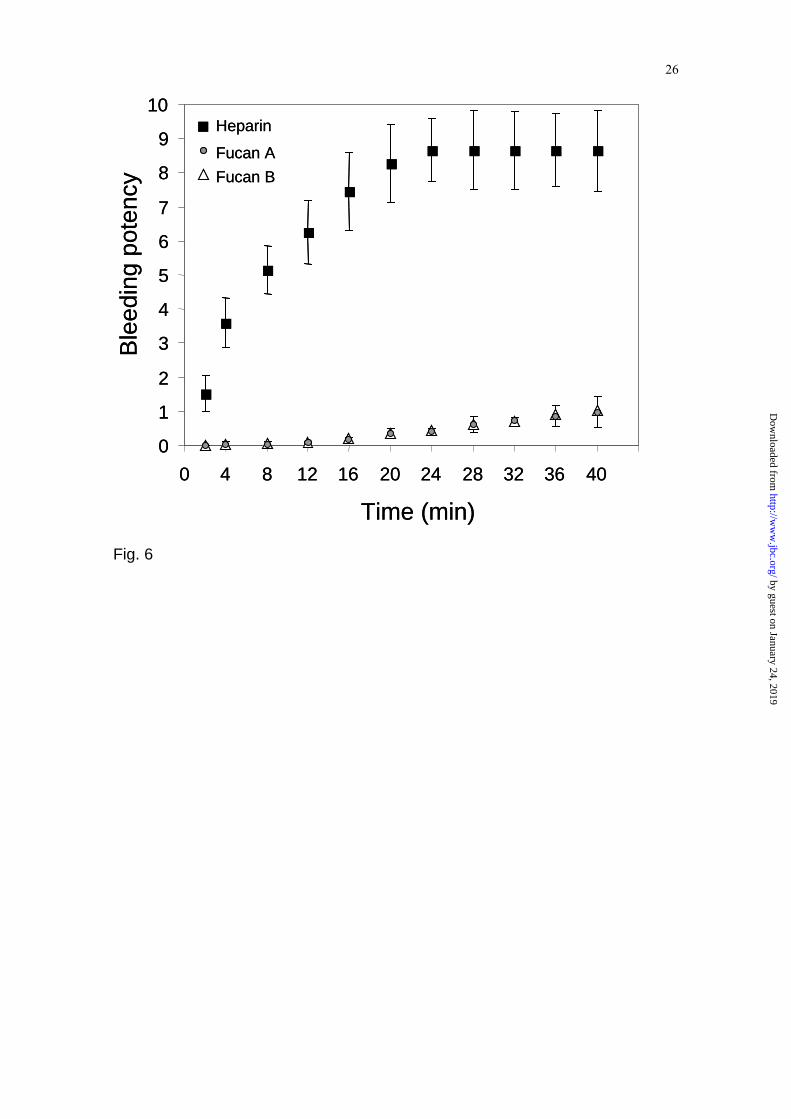
tests used (Table V). Fig. 6 shows that the polysaccharide has no hemorrhagic activity when compared to heparin. Similar results were obtained with fucan A from the same brown seaweed. “In vivo” antithrombotic activity ⎯ A dose response curve of the sulfated galactofucan in a venous model of thrombosis in rat is shown in Fig. 7A. No antithrombotic activity was found for all concentrations tested 1h after administration of the polysaccharide. Surprisingly, when the sulfated galactofucan was injected endovenously 24h before the ligature of the vena cavae, we observed a dose-dependent antithrombotic effect reaching saturation around 10mg/Kg of rat weight (Fig. 7B). Fig. 7C shows that this effect is also time-dependent reaching the maximum antithrombotic effect around 8h after administration of the sulfated galactofucan and remains for more than 24 hours. The antithrombotic activity was not detected with the desulfated fucan (data not shown). Sulfated galactofucan stimulates the synthesis of heparan sulfate by the endothelial cells ⎯ We have previously demonstrated that fucan A from S. schröederi was as effective as heparin in stimulating the synthesis of an antithrombotic heparan sulfate from endothelial cells (6, 36, 39). Thus, the effect of the present sulfated galactofucan was also investigated. The electrophoretic and enzymatic analysis of the sulfated glycosaminoglycans synthesized by endothelial cells showed that they are indeed heparan sulfate and chondroitin sulfate (data not shown). Nevertheless, the galactofucan specifically stimulated by four fold only the synthesis of heparan sulfate by endothelial cells in culture (Fig 8). In addition, this experiment also shows that the sulfated galactofucan is two fold more potent than heparin itself in stimulating the synthesis of the antithrombotic heparan sulfate. Desulfation of the polysaccharide abolishes this effect (data not shown).
This effect of the sulfated galactofucan could be related to cell proliferation leading to an apparent increase of heparan sulfate synthesis. In order to rule out this possibility the sulfated galactofucan was incubated with the endothelial cells during nine days. Thereafter, the cells were harvested and counted. Fig. 9A shows that
by guest on January 24, 2019http://w
ww
.jbc.org/D
ownloaded from

8
there are no differences between the number of cells in the presence or absence of the polysaccharide. Cell viability was 98% indicating an absence of cytotoxic activity. In addition, it was observed that the sulfated galactofucan did not affect thymidine incorporation in various phases of the cell cycle (Fig. 9B). We next focused on the effect of the conditioned media obtained from cells exposed or not to the sulfated galactofucan upon thrombin (FIIa) and FXa activities. Fig. 10 shows that conditioned media regardless of the presence of the polysaccharide are capable of inhibiting FXa activity. It thus indicate that the endothelial cells synthesize compound(s) that are related to this action. Furthermore the data also demonstrate that the media from cells exposed to the polysaccharide show higher inhibition of FXa activity when compared to the results of media obtained from cells not exposed to the sulfated galactofucan. Thus this result suggests that the polysaccharide enhances the synthesis of the compound(s) involved in this particular action. In addition, when galactofucan was added “in vitro” directly to the medium (negative control) no alteration in this inhibitory activity was observed (data not shown) (Fig. 10). On the other hand, this activity was totally abolished when the conditioned media were incubated with heparitinases (Fig. 10), thus indicating that the compound is heparan sulfate. Antithrombin had no influence on those effects (data not shown). Furthermore, no effect was observed when the conditioned media were incubated with thrombin both in the presence and absence of antithrombin (data not shown).
DISCUSSION
Structural features of the sulfated galactofucan ⎯ It has been previously shown the presence of sulfated fucans in different seaweeds such as Sargassum vulgare, Dictyota mertensis, Padina gimnospora (5), Dictyota menstrualis (8) and Sargassum stenophylum (45). We were able to show that the brown alga S. schröederi contains three main sulfated polysaccharides, named as fucan A, B and C according to their relative migration in agarose gel electrophoresis in 1,3-diaminopropane-acetate buffer (6). By a
combination of ion exchange chromatography and electrophoresis we have purified fucan A and proposed its structure after chemical analysis, methylation studies, NMR spectroscopy, and enzymatic degradation. Fucan A is a xylofucoglucuronan, with a molecular size of 21 kDa, containing a core oligosaccharide composed of 3-linked β-glucuronic acid and branches of 3-linked, mostly 4-sulfated α-fucose chains. The fucose units are also substituted at C-4 with chains of 4-linked β-xylose, which, in turn is also partially sulfated (6).
Here we report the purification of fucan B (now denominated as sulfated galactofucan) using the same methodology but with an additional fractionation step. Curiously, the present sulfated galactofucan contains galactose as its main constituent, which is absent in fucan A. The chemical analyses showed that the present fucan is indeed a sulfated xylogalactofucan. This leads to the conclusion that both polysaccharides, despite of being present in the same seaweed, show striking structural differences regarding their core oligosaccharides as well as the type of sugar residues, position of glycosidic linkages and sulfation sites. These differences certainly reflect in their pharmacological activities.
Since 1950s (46) several sulfated fucans containing xylose and galactose have been described but only a few with galactose as major component (45, 47, 48). The molecular weight of the present sulfated galactofucan (21.5 kDa) is similar to those encountered in other brown seaweed fucans (5, 48, 49) although in many cases products with values higher than 50 kDa were found (16, 48, 50, 51).
Structural features of several sulfated fucans have been investigated using a complex sequence of procedures for the extraction and purification of the polysaccharide (45, 50, 52, 53). Usually the fraction selected for biological studies do not represent the major polymer biosynthesized by the seaweed, but polysaccharides selected due to their high content of fucose or sulfate and potent anticoagulant activity. In most cases these homofucans have a α(1→3) linkage or a repeating structure of alternate α(1→3) and α(1→4) glycosidic linkages. The position of the sulfate groups was found to be mainly in position O-4 and/or O-2 (18).
by guest on January 24, 2019http://w
ww
.jbc.org/D
ownloaded from

9
Most of the difficulties for structural characterization of these polysaccharides arise from their heterogeneity, which give complex NMR spectra with broad signals hampering resolution. In fact, for these algal polysaccharides even high-field NMR is of limited value, and complete descriptions of their structures are not available at present (50, 54).
The use of gel electrophoresis in different buffer systems, as a criteria of purity of these compounds, allowed us to isolate different sulfated polysaccharides in high yields. This approach differs from the methodologies used by different authors whose criteria is based mainly in the enrichment of a specific sugar or a biological activity. Dissociation of the anticoagulant and antithrombotic activities ⎯ The sulfated galactofucan purified from the marine alga S. schröederi showed no anticoagulant activity on several “in vitro” assays. This was a surprising result considering the high sulfate content of the polysaccharide. Possibly, the presence of non-sulfate xylose units at the non-reducing terminal ends of the branches found in the polysaccharide may prevent its interaction with coagulation cofactors and their target proteases. Similar observation was reported for a variety of sulfated galactans from marine invertebrate. In this case, insertion of non-sulfated galactose residues as branched units abolished the anticoagulant effect of the polysaccharide (55).
When the sulfated galactofucan was tested on an experimental model of thrombosis in rat immediately after its intravenous administration it showed no antithrombotic activity. However, 8 h after its intravenous injection, and even after 24 h of administration, the sulfated galactofucan showed significant antithrombotic activity. This is unique since the antithrombotic polysaccharides tested so far like unfractionated heparin and its low-molecular-weight derivatives, the effect is more transient with a half life of 3 and 12 h, respectively. This observation raises interesting questions concerning the mechanism of antithrombotic action of the algal polysaccharide. Is it the polysaccharide itself that yield the antithrombotic effect? Is it a metabolic derivative from the algal polysaccharide responsible for the biological activity? Or
does the sulfated galactofucan induce metabolic modifications in the vessel walls, which ultimately prevent the formation of thrombus?
In order to clarify these aspects we tested the effect of the sulfated galactofucan on endothelial cells grown in culture. The polysaccharide did not stimulate proliferation of the cells, but induced the synthesis of a highly sulfated heparan sulfate, which may be in fact the antithrombotic agent. The sulfated galactofucan is two fold more potent than heparin. In addition this effect was only observed for heparan sulfate synthesis similar to the effect of heparin (4, 36, 39, 56). Since the synthesis of the heparan sulfate by endothelial cells in culture exposed to the fucan requires approximately 4-6 h for the full effect (results not shown) it explains the delay to detect the antithrombotic effect of the algal polysaccharide on the experimental model of thrombosis. It requires the synthesis and accumulation of the highly sulfated heparan sulfate on the vessel wall.
In the eighties, Colburn and Buonassisi (37) reported that rabbit aorta endothelial cells showed blood compatibility, i. e., the surface of these cells had antithrombotic activity. Using an antibody against heparan sulfate they neutralized the anti-clotting activity of endothelial cells culture medium, showing that this effect was at least in part heparan sulfate dependent. In the present work, using another methodology, we show that the conditionated medium from endothelial cells show anti-FXa activity. This effect is increased when the cells are exposed to sulfated galactofucan. This anti-FXa activity disappeared after heparitinases treatment, showing that this activity may be related to the heparan sulfate produced by endotelial cells. It seems that the effect does not need the presence of antithrombin, since no differences were observed in the presence or absence of AT. On the other hand, the media did not inhibit thombin activity, neither in the presence or absence antithrombin.
Possibly heparin prevents thrombosis due to its combined effect as an anticoagulant and an inducer of the synthesis of heparan sulfate by the endothelial cells. These two effects are difficult to distinguish. However, the algal sulfated galactofucan, which is devoid of anticoagulant action but has a potent action inducing the synthesis of
by guest on January 24, 2019http://w
ww
.jbc.org/D
ownloaded from
10
heparan sulfate by the endothelial cells, allowed us to emphasize this last mechanism involved in the prevention of thrombosis.
Finally, the findings that the sulfated galactofucan had no hemorrhagic activity, proliferative or cytotoxic actions make it an ideal candidate for further studies as an antithrombotic agent.
by guest on January 24, 2019http://w
ww
.jbc.org/D
ownloaded from

11
REFERENCES
1. National Vital Statistics Reports (2003) 51, 4-9 2. Moll, S., and Roberts, H. R. (2002) Sem. Hematol. 39, 145-157 3. Colliec, S., Fisher, A. M., Tapon-Bretaudier, J., Boisson-Vidal, C., Durand, P., and
Josefonvicz, J. (1991) Thromb. Res. 64, 143-154 4. Nader, H. B., Lopes, C. C., Rocha, H. A.O., Santos, E. A., and Dietrich, C.P. (2004) Curr.
Pharm. Des. 10, 951-966 5. Dietrich, C. P., Farias, G. G. M., Abreu, L. R. D., Leite, E. L., Silva, L. F., and Nader, H. B.
(1995) Plant Science 108, 143-153 6. Leite, E. L., Medeiros, M. G. L., Rocha, H. A. O., Farias, G. G. M., Silva, L. F., Chavante,
S. F., Dietrich, C. P., and Nader, H. B. (1998) Plant Science. 132, 215-228 7. Pereira, M. S., Mulloy, B., and Mourão, P. A. S. (1999) J. Biol. Chem. 274, 7656-7667 8. Albuquerque, I.R.L, Queiroz, K. C. S., Alves, L. G., Santos, E. A., Leite, E. L., and Rocha,
H. A.O. (2004) Braz. J. Med.Biol. Res. 37, 167-171 9. Chevolot, L., Foucault, A,. Chaubet, F., Kervarec, N., Sinquin, C., Fisher, A. M., and
Boisson-Vidal, C. (1999) Carbohydr. Res. 319,154-65 10. Shanmugam, M., and Mody, K.H. (2000) Current Science 79, 1672-1683 11. Boisson-Vidal C., Haroun, F., Ellouali, M., Blondin, C., Fischer, A. M., Agostini, A., and
Jozefonvicz, J. (1995) Drugs Fut. 20, 1237-1249 12. Haroun-Bouhedja, F., Lindenmeyer, F., Lu, H., Soria, C., Jozefonvicz, J., and Boisson-
Vidal C. (2002) Anticancer Res. 22, 2285-2292 13. Logeart, D., Prigent-Richard, S., Boisson-Vidal, C., Chaubet, F., Durand, P., Jozefonvicz,
J., and Letourneur, D. (1997) Eur. J. Cell Biol. 74,385-390. 14. Schaeffer, D. J., and Krylov, V. S. (2000) Ecotoxicol. Environ. Saf. 45, 208-227 15. Nagaoka M, Shibata, H., Kimura I., Hashimoto, S., Kimura, K., Makimo, T., Aiyama, R.,
Ueyama, S., and Yokokura, T. (1999) Glycoconj. J. 16, 19-26 16. Rocha, H.A.O., Franco, C.R.C., Trindade, E.S., Carvalho, L.C.M., Veiga, S.S., Leite, E..L.,
Dietrich, C..P., and Nader, H.B. (2001) Braz. J. Med. Biol. Res. 34, 621-626 17. Mulloy, B., Mourão, P. A. S., and Gray, E. (2000) J. Biotechnol. 77, 123-135 18. Dietrich, C. P., Tersariol, I. L. S., Toma, L., Moraes, C. T., Porcionatto, M. A., Oliveira, F.
W., and Nader, H. B (1998) Cell. Mol. Biol. 44, 417-429 19. Hoffman, D. L. (1989) Am. J. Med. 87, 23S-26S. 20. Nader, H. B., Porcionatto, M. A., Tersariol, I. L. S., Pinhal, M. A. S., Oliveira, F. W.,
Moraes, C. Dietrich, C.P. (1990) J. Biol. Chem. 265, 16807-16813 21. Dietrich, C. P., McDuffie N. M., and Sampaio, L. O. (1977) J. Chromatogr. 130, 299-304 22. Bianchini, P., Nader, H. B., Takahashi, H. K., Osima, B., Straus, A. H., and Dietrich, C. P.
(1980) J. Chromatogr. 196, 455-462 23. Dische, Z. (1962) Methods of Carbohydrate Chemistry, in: Whistler, R. L., and Wolfrom,
M. L. (Eds.) , Vol.1, Academic Press, New York, pp.501-503 24. Dische, Z. (1962) Methods of Carbohydrate Chemistry, in: Whistler, R. L., and Wolfrom,
M. L. (Eds.) , Vol.1, Academic Press, New York, pp. 484-488 25. Dische, Z. (1962) Methods of Carbohydrate Chemistry, in: Whistler, R. L., and Wolfrom,
M. L. (Eds.) , Vol.1, Academic Press, New York, pp. 497-501 26. Dubois, M., Gilles, K. A., Hamilton, J. K., Rebers, P. A., and Smith, F. (1956) Anal. Chem.
28, 250-25 27. Nader, H. B., and Dietrich, C. P. (1977) Anal. Biochem. 78, 112-118 28. Kozakai, M., and Yosizawa, Z. (1975) Anal. Biochem. 78, 425-429 29. Spector, J. (1978) Anal. Biochem. 86,142-143 30. Vilela-Silva, E. S. A., Castro, M. O., Valente, A. P., Biermann, H. C., and Mourão, P.A.S.
(2002) J. Biol. Chem.. 277, 379-387. 31. Patankar, M. S., Oehninger, L., Barnett, T, Williams, R. L., and Clark, G. F. (1993) J. Biol.
Chem. 268, 21770-21776 32. Kircher, H. W. (1960) Anal. Chem. 32, 1103-1106
by guest on January 24, 2019http://w
ww
.jbc.org/D
ownloaded from

12
33. Dietrich, C. P. , Paiva, J. F. , Castro, R. A. B., Chavante, S. F., Jeske, W., Fareed, J., Gorin, P. A. J., Mendes, A., and Nader, H. B. (1999) Biochim. Biophys. Acta 1428, 273-283
34. Reyers, I., Mussoni, A L., Donati, M B., and Gaetano, G. (1980) Thromb. Res. 18, 669-674 35. Dietrich, C. P., Shinjo, S. K., Moraes, F. A., Castro, R. A. B., Mendes, A., Gouvea, T. C.,
and Nader, H. B. (1999) Sem. Thromb. Hemost. 25, 43-50 36. Nader, H. B., Bounassisi, V., Colburn, P., and Dietrich, C. P. (1989) J. Cell. Physiol. 140,
305-310 37. Colburn, P., and Buonassisi, V. (1982) Biochem. Biophys. Res. Commun. 104, 220-227 38. Buonassisi, V., and Venter, J. C. (1976) Proc Natl Acad Sci USA 73, 1612-1616. 39. Pinhal, M. A. S., Walenga, J., Jeske, W., Hoppensteadt, D., Dietrich, C. P., Fareed J., and
Nader, H. B. (1994) Thromb. Res. 74, 143-153 40. Porcionatto, M. A., Moreira, C. R., Lotfi, C. F., Armelin, H. A., Dietrich, C. P., and Nader,
H. B. (1998) J. Cell Biochem.70, 563-72. 41. Campos, I. T. N., Silva, M. M., Azzolini, S. S., Souza, A. F., Sampaio, C. A. M., Fritz, H.,
and Tanaka, A. S. (2004) Arch. Bioch. Biophy. 425, 87-94 42. Maes, E., Florea, D., Delplace, F., Lemoine, J., Plancke, Y., and Strecker, G. (1997)
Glycoconj. J. 14, 127-146 43. Teleman, A., Lundqvist, J., Tjerneld, F., Stalbrand, H, and Dahlman, O. (2000) Carbohydr.
Res. 329, 807-815 44. Santos, J., Mulloy, B., and Mourão, P. A. S. (1991) Eur. J. Biochem 91, 669-677 45. Duarte, M. E. R., Cardoso, M. A. D., Cerezo, A. S., and Noseda, M. (2001) Carbohyd. Res.
333, 281-293 46. Kloareg, B., and Quatrano, R. S. (1988) Oceanogr. Mar. Biol. Ann. Ver. 26, 259-315 47. Medcalf, E. G., Whitmen, P., and Larsen, B. (1977) Carbohy. Res. 59, 531-536 48. Hussein, M. M. D., Magdel-Din, B., Abdel-Aziz, A, and Salem, H. M. I. (1980)
Phytochem. 19, 2133-2135 49. Ponce, M. A. N., Pujol, C. A., Damonte, E. B., Flores, M. L., and Stortz, C. A. (2003)
Carbohyd. Res. 338, 153-165 50. Berteau, O., and Mulloy, B. (2003) Glycobiology 13, 29R-40R 51. Preeprame, S., Hayashi, H., Lee, J., Sankawa, U., and Hayashi, T. (2001) Chem. Pharm.
Bull. 49, 484-485 52. Mourão, P. A. S.(2004) Curr. Pharm. Des. 10, 967-981 53. Chizhov, A. O., Dell, A., Morris, H. R., Haslam, S. M., Mcdowell, R. A., Shashkov, A. S.,
Nifant`Ev, N. E., Khatuntseva, E. A., and Usov, A. I. (1999) Carbohyd. Res. 320, 108-119 54. Marais, M. F., and Joseleau, J. P. (2001) Carbohydr Res. 336, 155-159 55. Farias, W.R.L., Valente, A.P., Pereira, M.S. and Mourão, P.A.S. (2000) J. Biol. Chem. 275,
29299-29307. 56. Pinhal, M. A., Trindade, E. S., Fareed. J., Dietrich, C. P., and Nader, H. B.(2001) Thromb
Res. 103, 35-45.
FOOTNOTES
*This work was supported by grants from FAPESP (Fundação de Amparo à Pesquisa do Estado de São Paulo), CNPq (Conselho Nacional de Desenvolvimento Científico e Tecnológico), FAPERJ (Fundação de Amparo à Pesquisa do Estado do Rio de Janeiro) and CAPES (Coordenação de Aperfeiçoamento de Pessoal de Nível Superior), Brazil
.
by guest on January 24, 2019http://w
ww
.jbc.org/D
ownloaded from

13
FIGURE LEGENDS Fig 1. Electrophoresis of sulfated polysaccharides from Spatoglossum schröederi. Electrophoresis in 0.05M diaminopropane acetate buffer, pH 9.0 of the fractions obtained by acetone precipitation of fraction 2.0M from Lewatite column (A.). Purified fucan obtained after Sephadex G-75 chromatography (lane 2) (B.). The purified fucan was subjected to electrophoresis in 0.06 M Tris-acetate buffer, pH 8.0 (C.), 0.05 M KCl-HCl buffer, pH 2.0, (D.) and discontinuous buffer 0.04M barium acetate, pH 4.0/ 0.05M diaminopropane acetate, pH 9.0 (E.). About 5µl-aliquots (50µg) of the fractions were applied in agarose gel (10x7.5 cm, 0.2 cm thick) prepared in different buffers and subjected to electrophoresis as previously descrribed (29, 30). The gels were then maintained in 0.1% cetyltrimethylammonium bromide for 4 h, dried and the polysaccharides stained with 0.1% toluidine blue in a solution containing 50% ethanol and 1% acid acetic in water for 15 min. The gels were then destained with the same solution lacking toluidine blue. St. - 10µg each of heparan sulfate (HS), dermatan sulfate (DS) and chondroitin sulfate (CS). Crude, polysaccharides extracted from S. schröederi; OR – origin. Fig. 2. 1H one-dimensional NMR spectrum of the sulfated galactofucan at 600 mHz. The spectrum was recorded at 60 oC for sample in D2O. α., α-H1; β, β-H1. Fig. 3. Strips from COSY, TOCSY and NOESY spectra of the sulfated galactofucan. A – Strips corresponding to the region of the α anomer signals. B – Strips corresponding to the region of the β anomer signals. The spectra were obtained as described under “Materials And Methods” with a mixing time of 80 ms for the COSY and TOCSY and 100 ms for the NOESY. Fig. 4. 1H/13C HMQC spectrum of the sulfated galactofucan from S. schröederi. The assignment was based on TOCSY and COSY spectra. Fig. 5. Proposed structure for the sulfated galactofucan from S. schröederi. Fig. 6. Hemorrhagic activity of the sulfated galactofucan. The compounds were applied topically (100 µg/mL) and the bleeding potency measured after 15 min following saline solution washing. Fucan A (reference 6); Fucan B, the sulfated galactofucan described in this paper. Fig. 7. Antithrombotic activity of the sulfated galactofucan is time- and dose dependent. A. – The sulfated galactofucan at the dose indicated was injected into the caudal vein of the rat and 5 minutes afterwards the vena cavae was ligated. After 2 hours the vena cavae was opened and the thrombi formed removed, dried and weighed. B. The experiment was performed as described in B except that the thrombi was removed and weighted after 24 hours of the injection. C. The experiment was performed as descrived above except that 10 ug of fucan was injected and the thrombi formed removed from the vena cavae and weighed at the indicated times. Fucan B, the sulfated galactofucan described in this paper. Fig. 8. Stimulation of the synthesis of heparan sulfate by endothelial cells exposed to different polysaccharides. Aorta endothelial cells were exposed to 100 µg/mL of the each polysaccharide and 150 µCi/ml of [35S]-inorganic sulfate in F-12 medium. After 24 hours the heparan sulfate and chondroitin sulfate synthesized by the cells and secreted to the medium were characterized and quantified as referred to in ”Materials And Methods”. SGAG, sulfated glycosaminoglycans, HS – heparan sulfate, CS – chondroitin sulfate, Fucan A (ref. 6), Fucan B, the sulfated galactofucan described in this paper.
by guest on January 24, 2019http://w
ww
.jbc.org/D
ownloaded from

14
Fig. 9. Proliferation of endothelial cells and thymidine incorporation in the presence of sulfated galactofucan. A. Growth arrested cells were released from G0 phase by addition of F-12 medium plus 10% FCS in the absence (control) or presence of the sulfated galactofucan (100 µg/ml). The initial cell number was 1x10⁵ cells/plate. After incubation at 37 °C in a 2.5% CO2 at the times indicated the cells (in triplicate plates) were harvested after pancreatin treatment and the viable cell number was determined. Values are mean ± SD of triplicate determinations. B. Endothelial cells were maintained in F12 medium supplemented with 10% FCS at 37 °C, 2.5% CO2. For the incorporation of [3H]-thymidine, quiescent cultures were incubated with [3H]-thymidine (0.25 µCi/ml) for the times indicated and the radioactivity determined by scintillation counting. Values are mean ± SD of four determinations. FCS, fetal calf serum; RAEC, Rabbit aorta endothelial cells; fucan, the sulfated galactofucan described in this paper.
Fig. 10. Conditioned media from endothelial cells exposed to the sulfated galactofucan inhibit factor Xa activity. Different volumes (10 to 100 µl) of endothelial cells conditioned media were pre-incubated with 20nM of FXa during 5 minutes (37 °C). After, 5 µl of 4 mM solution of the synthetic substrate (S2222) was added (37 °C) and the product formed continuously monitored at 405 nm during 3,000 sec using an ELISA reader. The results represent the average of three different experiments performed in duplicate. For details see Materials and Methods. (•) negative control (F-12 medium); (■) conditioned medium from cells not exposed to the sulfated galactofucan; ( ) conditioned medium from cells not exposed to the sulfated galactofucan after degradation with heparitinases; (▲) conditioned medium from cells exposed to the sulfated galactofucan; (∆) conditioned medium from cells exposed to the sulfated galactofucan after degradation with heparitinases. ANOVA test (*) p<0.05 and student t test (#) p<0.05.
by guest on January 24, 2019http://w
ww
.jbc.org/D
ownloaded from

15
TABLE I Chemical composition of acidic polysaccharides from S. schröederi
Obtained by acetone precipitation
Fraction (acetone volume)
Polysac-charides
(%*)
Molar Ratios to Fucose
Xylose Uronic acid
Galactose Sulfate
0.5 38.0 0.6 8.0 - 1.3 0.6 35.2 0.6 4.0 0.3 1.3 0.7 12.2 0.9 0.5 1.5 1.7 0.9 5.8 0.6 0.2 1.6 3.0 1.1 5.8 0.7 0.1 1.3 2.2 1.3 2.6 0.9 0.3 0.7 2.8 2.0 0.3 0.4 0.1 1.1 2.2 *Determinated by Phenol-H2SO4 reaction. The chemical composition of the sulfated galactofucan fraction is indicated by italic type.
by guest on January 24, 2019http://w
ww
.jbc.org/D
ownloaded from

16
TABLE II Chemical composition of acidic polysaccharides prepared by ion exchange chromatography
from the 2.0 volumes acetone fraction and from Sephadex G-75
Fraction (M NaCl)
Polysac-charides
(%)
Molar ratios to fucose
Xylose Uronic acid Galactose Sulfate 1.5 6 0.6 0.2 0.9 1.7 2.0 80 0.5 0.1 2.0 2.0 3.0 14 0.6 0.1 1.0 2.3 Gel filtration*
0.5 0.1 2.0 2.1
*Fraction 3.0M after Sephadex G-75 (see Methods). The chemical composition of the purified sulfated galactofucan fraction is indicated by italic type.
by guest on January 24, 2019http://w
ww
.jbc.org/D
ownloaded from

17
TABLE III Proton and carbon chemical shifts for α and β residues in Fucan B
Unit 1H Chemical shiftsa Ref. H1 H2 H3 H4 H5 H6
α 1-Fucose-4 (A) 5.31 3.95 4.52 4.28 4.31-4.44 1.23-1.34 *
α 1-Fucose-4 (B) 5.08 3.95 4.54 4.24 4.31-4.44 1.23-1.34 *
α 4 – Fucose – 1 5.09 3.95 4.12 3.98 4.53-4.59 1.33-1.43 29
α 4 – Fucose –3S - 1 - 3.95-4.15
4.65-4.80
4.3 - 1.3-1.4 10
β 4-Galactose-3S- 1 (C) 4.71 3.95 4.63 - - 3.76-3.82 *
β 4,2-Galactose-1 (D) 4.69 3.81 3.94 4.54 3.68 3.76-3.82 *
β 4-Galactose-1 (E) 4.58 3.70 4.10 4.35 - 3.76-3.82 *
β 4-Galactose-3S-1 (G) 4.44 3.83 4.55 - - 3.76-3.82 *
β 4 – Galactose – 1 4.50 3.63 3.79 4.20 3.76 3.70 38
β 4,2 – Galactose – 1 4.59 3.83 3.99 4.19 3.77 - 38
α 4 – Galactose – 3S- 1 5.20 4.17 4.70 4.50 4.35 3.90 39
β 4 –Xilose -1 (F) 4.57 3.37 3.66 3.72 4.14 and 3.41#
*
β 4 – xylose - 1 4.48 3.32 3.58 3.79 4.10 and 3.40#
40
Unit 13C Chemical shiftsa Ref. C1 C2 C3 C4 C5 C6
α 1-Fucose-4 (A) 101.83 70.72 79.33 81.72 69.32 16.30 *
α 1-Fucose-4 (B) 99.10 68.76 75.75 81.72 69.27 16.30 *
α 4 – Fucose – 1 103.03 71.30 70.97 83.90 69.96 13.00 29
α 4 – Fucose –3S - 1 ~101.00
- 78.00 ~82.00 69-73 18.50 10
β 4-Galactose-3S-1 (C) 105.07 75.92 71.28 82.91 - ~63.30 *
β 4,2-Galactose-1 (D) 105.00 76.13 75.88 82.23 72.00 ~63.30 *
β 4-Galactose-3S-1 (E) 106.47 71.15 82.57 - - ~63.30 *
β 4-Galactose-3S-1 (G) 106.27 75.97 79.39 - - ~63.30 *
β 4 – Galactose - 1 105.84 73.26 74.50 79.35 76.27 ~62.00 38
β 4,2 – Galactose – 1 103.48 80.25 73.91 78.39 75.57 62.08 38
α 4 – Galactose – 3S- 1 103.10 ~71.00
71.80 81.4 74.10 62.90 39
β Xylose (F) 106.09 75.85 74.43 76.71 65.01 *
β 4 – Xylose – 1 103.69 73.80 74.50 77.40 64.00 40 a Chemical shifts are referred to internal trimethilsililpropionic acid (0 ppm). # This residue has two H5. *Present paper. Values in italic type indicate positions bearing a sulfate ester, and those in boldface type indicate glycosylated positions.
by guest on January 24, 2019http://w
ww
.jbc.org/D
ownloaded from

18
TABLE IV
Methylation analysis of fucan and desulfated fucan
Glycosyl residue
Position of O-methyl group
Deduced position of substitution
Fucan (mol %)
Desulfated Fucan
(mol %) Xilosyl 2,3,4 Terminal 5.7 7.4 2,3 4 12.8 13.0 Fucosyl 2,3,4 Terminal - 3.8 2,3 4 2.5 25.2 2,4 3 2.9 2.8 2 3,4 26.0 - 0 2,3,4 1.7 - Galactosyl 2,3,4,6 Terminal 4.2 10.1 2,3,6 4 - 12.3 2,6 3,4 11.6 - 2,3 4,6 - 4.7 3,6 2,4 - 9.4 2 3,4,6 6.3 - 3/4 2,4,6 / 2,3,6 10.1 9.4 0 2,3,4,6 12.0 1.9
by guest on January 24, 2019http://w
ww
.jbc.org/D
ownloaded from

19
TABLE V Anticoagulant activity of fucan
COMPOUND ASSAYS Fucan (mg) PT (sec) APTT (sec) HEPTEST® (sec) TT (sec) 0 12.4 36.4 13.8 30.8 0.2 12.3 35.8 13.1 30.8 0.4 11.9 35.3 13.8 31.3 0.8 11.8 34.8 13.8 30.3 1.5 12.9 34.8 12.3 30.8 3.1 11.8 35.3 13.3 31.3 6.2 12.4 35.4 13.3 30.3 12.5 12.4 36.3 13.3 31.8 25 12.3 38.3 12.9 31.3 50 12.2 36.4 13.3 30.8 100 13.3 35.6 12.9 31.3 Heparin (0.1mg)
>80 >600 >120 >300
PT, prothrombin time; APTT, activated partial thromboplastin time; HEPTEST, heparin cofactor II time; TT, thrombin time
by guest on January 24, 2019http://w
ww
.jbc.org/D
ownloaded from

20
A CSDSHS
OR
crude 0.5 0.6 0.7 0.9 1.1 1.3 2.0 St.
A. B.
St. 1 2
CSDSHS
OR
E.
CSDSHS
OR
Fucan B St.
D.
CS/DSHS
OR
Fucan B St.
C.
CS/DSHS
OR
Fucan B St.
Fig.1
by guest on January 24, 2019http://w
ww
.jbc.org/D
ownloaded from


22
Fig. 3A
1H Chemical shift (ppm) 4.44.6 4.84.44.64.84.44.6 4.8
3.1
3.3
3.5
3.7
3.9
4.1
4.3
4.5
4.7
4.9
5.1
5.3
5.5
5.7
5.9
1 H C
hem
ical
shi
ft (p
pm)
COSY TOSCY NOESY
by guest on January 24, 2019http://w
ww
.jbc.org/D
ownloaded from

23
Fig. 3B
1H Chemical shift (ppm) 4.44.6 4.84.44.64.84.44.6 4.8
3.1
3.3
3.5
3.7
3.9
4.1
4.3
4.5
4.7
4.9
5.1
5.3
5.5
5.7
5.9
1 H C
hem
ical
shi
ft (p
pm)
COSY TOSCY NOESY
by guest on January 24, 2019http://w
ww
.jbc.org/D
ownloaded from

24
Fig. 4
1H Chemical shift (ppm)
13C
Che
mic
al s
hift
(ppm
)
5.4 5.2 5.0 4.8 4.6 4.4 4.2 4.0 3.8 3.6 3.4 3.2
115
110
105
100
95
90
85
80
75
70
65
60
55
50
by guest on January 24, 2019http://w
ww
.jbc.org/D
ownloaded from

25
Fig 5
OC
H2O
H OH
OO
SO
3-
OC
H2O
H
OO
CH
2OH O
H
OO
CH
2OH
OO
CH
2OH O
H
OO
CH
2OH
OO
H
OC
H3
OH
O
OC
H3
OH
O
OH
O
OH
OC
H2O
H OH
OO
CH 2
OH
O
OC
H3
OH
O
OH
OH
O
O
OH
OH
OH
O
O
OH
OH
OCH
2OH O
H
OO
CH
2OH
OO
H
OC
H3
OH
OCH
3
OH
O
O
O
OH
OH
OH
4
OH
OS
O3-
OS
O3-
OS
O3-
OSO
3-
OS
O3-
OSO
3-
OS
O3-
OS
O3-
OSO
3-
by guest on January 24, 2019http://w
ww
.jbc.org/D
ownloaded from
26
Fig. 6
0
1
2
3
4
5
6
7
8
9
10
0 4 8 12 16 20 24 28 32 36 40
Time (min)
Ble
edin
g po
tenc
yHeparin
Fucan AFucan B
0
1
2
3
4
5
6
7
8
9
10
0 4 8 12 16 20 24 28 32 36 40
Time (min)
Ble
edin
g po
tenc
yHeparin
Fucan AFucan B
by guest on January 24, 2019http://w
ww
.jbc.org/D
ownloaded from

27
Fig. 7
0
2
4
6
8
10
12
14
0 2 4 6 8 10 12 14 16 18 20
Fucan B (µg/g)
Thro
mbi
(mg)
A.
0
2
4
6
8
10
12
14
0 2 4 6 8 24
Time of exposure (hours)
Thro
mbi
(mg)
C.
0
2
4
6
8
10
12
14
0 2 4 6 8 10 15 20
Thro
mbi
(mg)
B.
Fucan B (µg/g)
0
2
4
6
8
10
12
14
0 2 4 6 8 10 12 14 16 18 20
Fucan B (µg/g)
Thro
mbi
(mg)
A.
0
2
4
6
8
10
12
14
0 2 4 6 8 10 12 14 16 18 20
Fucan B (µg/g)
Thro
mbi
(mg)
A.
0
2
4
6
8
10
12
14
0 2 4 6 8 24
Time of exposure (hours)
Thro
mbi
(mg)
C.
0
2
4
6
8
10
12
14
0 2 4 6 8 24
Time of exposure (hours)
Thro
mbi
(mg)
C.
0
2
4
6
8
10
12
14
0 2 4 6 8 10 15 20
Thro
mbi
(mg)
B.
Fucan B (µg/g)
0
2
4
6
8
10
12
14
0 2 4 6 8 10 15 20
Thro
mbi
(mg)
B.
Fucan B (µg/g)
by guest on January 24, 2019http://w
ww
.jbc.org/D
ownloaded from

28
Fig. 8
0
200
400
600
800
1000
1200
1400
Control Heparin Fucan A Fucan B
HS
CS
0
200
400
600
800
1000
1200
1400
Control Heparin Fucan A Fucan B
SG
AG
(cpm
/µg
of c
ell p
rote
in)
HS
CS
by guest on January 24, 2019http://w
ww
.jbc.org/D
ownloaded from

29
Fig. 9
B.
0
10000
20000
30000
40000
50000
60000
Time (hours)
RAEC + FCSRAEC + Fucan B + FCSRAECRAEC + Fucan B
[ H
]-Thy
mid
ine
inco
rpor
atio
n3
0 4 8 12 16 20 32 34
1
10
100
1000
Time (Days)
ControlFucan B
Cel
l num
ber(
x10
)5
1 2 3 4 5 6 7 8 9
A.
B.
0
10000
20000
30000
40000
50000
60000
Time (hours)
RAEC + FCSRAEC + Fucan B + FCSRAECRAEC + Fucan B
[ H
]-Thy
mid
ine
inco
rpor
atio
n3
0 4 8 12 16 20 32 34
B.
0
10000
20000
30000
40000
50000
60000
Time (hours)
RAEC + FCSRAEC + Fucan B + FCSRAECRAEC + Fucan B
[ H
]-Thy
mid
ine
inco
rpor
atio
n3
0 4 8 12 16 20 32 34 0
10000
20000
30000
40000
50000
60000
Time (hours)
RAEC + FCSRAEC + Fucan B + FCSRAECRAEC + Fucan B
RAEC + FCSRAEC + Fucan B + FCSRAECRAEC + Fucan B
[ H
]-Thy
mid
ine
inco
rpor
atio
n3 [ H
]-Thy
mid
ine
inco
rpor
atio
n3
0 4 8 12 16 20 32 34
1
10
100
1000
Time (Days)
ControlFucan B
Cel
l num
ber(
x10
)5
1 2 3 4 5 6 7 8 9
A.
1
10
100
1000
Time (Days)
ControlFucan BControlFucan B
Cel
l num
ber(
x10
)5C
ell n
umbe
r(x1
0 )5
1 2 3 4 5 6 7 8 9
A.
by guest on January 24, 2019http://w
ww
.jbc.org/D
ownloaded from

30
0 20 40 60 80 1000.0
0.3
0.6
0.9
1.2
*
*
*
*
*
**
###
#
Medium (µ l)
FXa
activ
ity r
elat
ive
toco
ntro
l
Fig. 10
by guest on January 24, 2019http://w
ww
.jbc.org/D
ownloaded from

Continuous measurement of the optical density at 405 nm for Factor Xa activity 1
0 s 15 s 30 s 45 s 60 s 74 s 89 s 105 s 120 s 134 s 149 s 164 s 179 sA1 0,059 0,059 0,059 0,059 0,06 0,06 0,06 0,06 0,06 0,06 0,06 0,06 0,06A2 0,074 0,075 0,077 0,08 0,081 0,083 0,085 0,086 0,088 0,089 0,091 0,093 0,095A3 0,073 0,074 0,075 0,077 0,077 0,078 0,08 0,081 0,082 0,083 0,084 0,085 0,086A4 0,083 0,085 0,086 0,088 0,089 0,09 0,091 0,092 0,093 0,094 0,095 0,097 0,098A5 0,08 0,082 0,083 0,085 0,085 0,086 0,087 0,087 0,088 0,089 0,09 0,09 0,091A6 0,086 0,087 0,089 0,09 0,092 0,092 0,092 0,093 0,095 0,096 0,098 0,1 0,101
B1 0,08 0,08 0,081 0,085 0,087 0,09 0,093 0,096 0,099 0,102 0,105 0,108 0,111B2 0,074 0,077 0,076 0,078 0,078 0,08 0,082 0,085 0,088 0,09 0,092 0,095 0,097B3 0,072 0,073 0,074 0,076 0,077 0,078 0,08 0,082 0,083 0,085 0,088 0,09 0,092B4 0,078 0,079 0,08 0,081 0,083 0,085 0,087 0,088 0,09 0,092 0,095 0,097 0,099B5 0,081 0,082 0,083 0,085 0,086 0,088 0,089 0,091 0,092 0,093 0,094 0,095 0,097B6 0,091 0,091 0,091 0,094 0,096 0,098 0,1 0,102 0,103 0,105 0,107 0,108 0,11
C1 0,087 0,088 0,092 0,094 0,097 0,1 0,103 0,106 0,109 0,113 0,117 0,121 0,125C2 0,078 0,08 0,082 0,083 0,086 0,088 0,091 0,092 0,095 0,097 0,1 0,102 0,105C3 0,074 0,074 0,074 0,075 0,077 0,078 0,08 0,082 0,084 0,085 0,087 0,089 0,091C4 0,08 0,082 0,085 0,087 0,088 0,089 0,091 0,092 0,093 0,095 0,097 0,099 0,1C5 0,088 0,09 0,092 0,094 0,094 0,096 0,097 0,099 0,101 0,104 0,106 0,108 0,111C6 0,089 0,091 0,093 0,093 0,095 0,097 0,099 0,102 0,105 0,107 0,109 0,111 0,113
D2 0,089 0,092 0,097 0,1 0,102 0,102 0,105 0,106 0,109 0,112 0,116 0,12 0,124D3 0,091 0,093 0,093 0,096 0,097 0,098 0,099 0,1 0,101 0,103 0,105 0,108 0,111D4 0,081 0,083 0,088 0,095 0,093 0,094 0,094 0,095 0,096 0,097 0,099 0,101 0,103D5 0,079 0,082 0,084 0,086 0,085 0,088 0,089 0,09 0,091 0,093 0,094 0,096 0,098D6 0,08 0,081 0,083 0,084 0,085 0,086 0,088 0,09 0,092 0,093 0,095 0,097 0,098
volume (µl)
A, B = 1 zero2 10
C, D = 3 204 405 806 100
conditioned medium from cells not exposed to the sulfated galactofucanduplicate experiment
duplicate experiment
conditioned medium from cells exposed to the sulfated galactofucan
by guest on January 24, 2019 http://www.jbc.org/ Downloaded from

Continuous measurement of the optical density at 405 nm for Factor Xa activity 2
195 s 210 s 224 s 239 s 255 s 270 s 285 s 299 s 315 s 329 s 344 s 359 s 375 s 390 s0,06 0,06 0,06 0,06 0,06 0,06 0,06 0,059 0,06 0,059 0,06 0,06 0,06 0,060,096 0,098 0,1 0,102 0,104 0,105 0,107 0,109 0,111 0,112 0,114 0,117 0,118 0,120,087 0,088 0,09 0,091 0,092 0,093 0,094 0,095 0,097 0,098 0,099 0,101 0,102 0,1030,099 0,1 0,102 0,103 0,104 0,105 0,107 0,108 0,109 0,11 0,111 0,113 0,114 0,1150,092 0,093 0,094 0,095 0,096 0,096 0,097 0,098 0,099 0,1 0,101 0,102 0,103 0,1030,102 0,104 0,105 0,107 0,108 0,109 0,111 0,112 0,113 0,114 0,115 0,116 0,117 0,118
0,114 0,117 0,12 0,123 0,125 0,128 0,13 0,133 0,135 0,138 0,141 0,143 0,146 0,1490,1 0,103 0,106 0,106 0,108 0,11 0,113 0,115 0,114 0,116 0,118 0,119 0,121 0,1270,094 0,097 0,099 0,101 0,103 0,104 0,106 0,108 0,109 0,111 0,112 0,113 0,115 0,1160,101 0,103 0,105 0,107 0,109 0,11 0,112 0,113 0,114 0,115 0,116 0,117 0,118 0,1190,098 0,099 0,1 0,102 0,103 0,105 0,106 0,107 0,108 0,11 0,111 0,113 0,114 0,1150,111 0,112 0,114 0,115 0,116 0,118 0,119 0,12 0,121 0,122 0,124 0,125 0,126 0,128
0,129 0,132 0,135 0,138 0,142 0,145 0,148 0,151 0,154 0,157 0,16 0,163 0,165 0,1680,107 0,11 0,113 0,116 0,118 0,121 0,123 0,126 0,128 0,131 0,133 0,135 0,137 0,1390,093 0,095 0,097 0,099 0,101 0,103 0,104 0,106 0,108 0,109 0,111 0,112 0,114 0,1150,103 0,105 0,107 0,109 0,111 0,113 0,115 0,117 0,119 0,121 0,123 0,126 0,128 0,1290,113 0,116 0,118 0,121 0,124 0,126 0,129 0,131 0,134 0,136 0,138 0,141 0,143 0,1440,115 0,118 0,119 0,121 0,124 0,126 0,127 0,129 0,131 0,133 0,135 0,137 0,139 0,141
0,128 0,133 0,137 0,141 0,145 0,149 0,153 0,156 0,159 0,162 0,166 0,169 0,172 0,1750,114 0,117 0,12 0,123 0,126 0,129 0,132 0,135 0,137 0,14 0,143 0,145 0,147 0,1490,106 0,109 0,112 0,115 0,118 0,121 0,123 0,126 0,128 0,13 0,133 0,135 0,137 0,1390,1 0,103 0,104 0,107 0,108 0,111 0,113 0,115 0,117 0,119 0,12 0,123 0,124 0,1260,1 0,102 0,104 0,106 0,107 0,109 0,111 0,113 0,114 0,116 0,118 0,12 0,121 0,123
volume (µl)
A, B = 1 zero2 10
C, D = 3 204 405 806 100
conditioned medium from cells exposed to the sulfated galactofucanduplicate experimentconditioned medium from cells not exposed to the sulfated galactofucanduplicate experiment
by guest on January 24, 2019 http://www.jbc.org/ Downloaded from

Continuous measurement of the optical density at 405 nm for Factor Xa activity 3
405 s 420 s 434 s 449 s 464 s 479 s 495 s 510 s 524 s 539 s 554 s 569 s 584 s 599 s0,059 0,059 0,059 0,059 0,059 0,059 0,059 0,059 0,059 0,059 0,059 0,059 0,059 0,0590,121 0,122 0,124 0,125 0,128 0,129 0,131 0,132 0,133 0,135 0,136 0,138 0,139 0,1410,103 0,104 0,105 0,106 0,108 0,109 0,11 0,111 0,112 0,113 0,114 0,115 0,115 0,1160,116 0,116 0,117 0,118 0,12 0,121 0,122 0,123 0,124 0,125 0,126 0,127 0,127 0,1280,103 0,104 0,105 0,105 0,106 0,107 0,108 0,108 0,109 0,11 0,11 0,111 0,111 0,1120,119 0,12 0,121 0,122 0,122 0,123 0,124 0,125 0,126 0,126 0,127 0,128 0,129 0,13
0,151 0,154 0,157 0,159 0,161 0,164 0,166 0,169 0,171 0,173 0,176 0,178 0,18 0,1830,123 0,126 0,127 0,127 0,129 0,131 0,132 0,133 0,135 0,138 0,139 0,139 0,141 0,1440,116 0,117 0,118 0,119 0,121 0,122 0,123 0,124 0,125 0,126 0,127 0,128 0,129 0,1310,12 0,121 0,122 0,123 0,124 0,125 0,126 0,127 0,129 0,13 0,131 0,132 0,133 0,1340,116 0,117 0,118 0,119 0,12 0,121 0,122 0,123 0,124 0,125 0,126 0,127 0,127 0,1280,129 0,13 0,131 0,132 0,134 0,134 0,136 0,137 0,138 0,139 0,14 0,141 0,142 0,143
0,171 0,174 0,177 0,18 0,182 0,185 0,188 0,191 0,194 0,196 0,199 0,202 0,204 0,2070,141 0,143 0,145 0,147 0,149 0,152 0,154 0,156 0,158 0,16 0,162 0,164 0,165 0,1670,117 0,118 0,119 0,121 0,123 0,124 0,126 0,128 0,13 0,131 0,133 0,135 0,137 0,1380,131 0,133 0,134 0,136 0,138 0,14 0,141 0,143 0,144 0,146 0,148 0,149 0,15 0,1520,145 0,147 0,148 0,149 0,151 0,152 0,153 0,155 0,156 0,157 0,158 0,159 0,161 0,1620,142 0,144 0,146 0,147 0,15 0,151 0,153 0,155 0,156 0,158 0,16 0,162 0,163 0,165
0,177 0,18 0,182 0,185 0,188 0,191 0,193 0,196 0,199 0,201 0,204 0,207 0,209 0,2120,15 0,152 0,153 0,155 0,158 0,16 0,162 0,164 0,166 0,168 0,17 0,173 0,175 0,1780,14 0,142 0,143 0,145 0,148 0,149 0,151 0,152 0,154 0,156 0,157 0,159 0,16 0,1620,127 0,129 0,13 0,132 0,134 0,135 0,137 0,138 0,139 0,14 0,141 0,143 0,144 0,1450,124 0,126 0,127 0,129 0,13 0,132 0,133 0,135 0,136 0,138 0,139 0,141 0,142 0,143
volume (µl)
A, B = 1 zero2 10
C, D = 3 204 405 806 100
conditioned medium from cells exposed to the sulfated galactofucanduplicate experimentconditioned medium from cells not exposed to the sulfated galactofucanduplicate experiment
by guest on January 24, 2019 http://www.jbc.org/ Downloaded from

Continuous measurement of the optical density at 405 nm for Factor Xa activity 4
614 s 629 s 645 s 660 s 675 s 690 s 705 s 719 s 734 s 749 s 764 s 779 s 795 s 810 s0,059 0,059 0,059 0,059 0,059 0,059 0,059 0,059 0,059 0,059 0,059 0,059 0,059 0,0590,142 0,144 0,145 0,147 0,148 0,15 0,151 0,153 0,155 0,156 0,157 0,159 0,16 0,1610,117 0,118 0,118 0,12 0,121 0,122 0,123 0,124 0,125 0,125 0,126 0,127 0,128 0,1290,129 0,13 0,13 0,132 0,133 0,134 0,135 0,135 0,136 0,137 0,138 0,139 0,14 0,140,112 0,113 0,113 0,114 0,115 0,115 0,116 0,116 0,117 0,117 0,118 0,118 0,119 0,1190,13 0,131 0,132 0,133 0,133 0,134 0,135 0,136 0,136 0,137 0,138 0,138 0,139 0,14
0,185 0,187 0,189 0,192 0,194 0,196 0,199 0,201 0,203 0,206 0,208 0,211 0,213 0,2150,143 0,145 0,146 0,149 0,151 0,151 0,152 0,157 0,155 0,157 0,16 0,159 0,161 0,1630,131 0,133 0,133 0,135 0,137 0,138 0,139 0,14 0,141 0,142 0,144 0,145 0,146 0,1470,135 0,136 0,137 0,138 0,139 0,14 0,141 0,142 0,142 0,143 0,144 0,145 0,146 0,1470,129 0,13 0,13 0,131 0,132 0,133 0,134 0,135 0,135 0,136 0,137 0,138 0,139 0,140,143 0,144 0,145 0,146 0,147 0,148 0,149 0,15 0,151 0,152 0,152 0,153 0,154 0,155
0,209 0,212 0,214 0,217 0,22 0,222 0,225 0,228 0,23 0,233 0,235 0,238 0,241 0,2430,169 0,171 0,173 0,175 0,177 0,179 0,181 0,183 0,185 0,186 0,188 0,19 0,192 0,1940,14 0,142 0,143 0,144 0,146 0,147 0,148 0,15 0,151 0,152 0,153 0,155 0,156 0,1570,153 0,155 0,156 0,158 0,159 0,161 0,162 0,164 0,165 0,167 0,168 0,17 0,172 0,1730,163 0,165 0,166 0,168 0,17 0,172 0,174 0,175 0,177 0,179 0,181 0,182 0,184 0,1860,166 0,168 0,169 0,172 0,173 0,175 0,177 0,178 0,18 0,182 0,183 0,185 0,187 0,188
0,214 0,217 0,219 0,223 0,225 0,228 0,231 0,233 0,236 0,239 0,241 0,244 0,247 0,2490,179 0,182 0,184 0,187 0,189 0,191 0,193 0,195 0,197 0,199 0,201 0,203 0,204 0,2060,163 0,166 0,166 0,169 0,171 0,173 0,174 0,176 0,178 0,182 0,181 0,183 0,184 0,1870,145 0,147 0,147 0,149 0,15 0,152 0,153 0,154 0,156 0,157 0,158 0,159 0,161 0,1620,145 0,146 0,148 0,149 0,151 0,152 0,153 0,155 0,157 0,158 0,16 0,161 0,162 0,164
volume (µl)
A, B = 1 zero2 10
C, D = 3 204 405 806 100
duplicate experiment
conditioned medium from cells exposed to the sulfated galactofucanduplicate experimentconditioned medium from cells not exposed to the sulfated galactofucan
by guest on January 24, 2019 http://www.jbc.org/ Downloaded from

Continuous measurement of the optical density at 405 nm for Factor Xa activity 5
824 s 839 s 855 s 869 s 884 s 900 s 915 s 930 s 944 s 959 s 974 s 989 s 1004 s 1020 s0,059 0,059 0,059 0,059 0,059 0,059 0,059 0,059 0,059 0,059 0,059 0,059 0,059 0,0590,163 0,164 0,165 0,167 0,168 0,17 0,17 0,172 0,173 0,175 0,176 0,178 0,179 0,180,13 0,13 0,131 0,132 0,133 0,134 0,134 0,135 0,136 0,137 0,138 0,139 0,139 0,140,141 0,142 0,143 0,143 0,144 0,145 0,145 0,146 0,147 0,148 0,148 0,149 0,15 0,150,119 0,12 0,12 0,121 0,121 0,122 0,122 0,122 0,123 0,123 0,124 0,124 0,124 0,1250,14 0,141 0,142 0,142 0,143 0,143 0,144 0,145 0,145 0,146 0,146 0,147 0,147 0,148
0,218 0,22 0,223 0,225 0,227 0,23 0,232 0,234 0,236 0,239 0,241 0,243 0,245 0,2470,163 0,166 0,166 0,167 0,169 0,17 0,171 0,173 0,174 0,176 0,177 0,178 0,18 0,1810,148 0,149 0,15 0,151 0,152 0,153 0,154 0,155 0,157 0,157 0,158 0,16 0,161 0,1620,148 0,149 0,15 0,151 0,151 0,152 0,153 0,154 0,155 0,156 0,156 0,157 0,158 0,1590,14 0,141 0,142 0,143 0,143 0,144 0,144 0,145 0,146 0,147 0,147 0,148 0,149 0,1490,156 0,157 0,158 0,158 0,159 0,16 0,16 0,161 0,162 0,163 0,164 0,164 0,165 0,166
0,246 0,248 0,251 0,254 0,256 0,259 0,261 0,264 0,266 0,269 0,271 0,274 0,276 0,2780,196 0,198 0,199 0,201 0,203 0,205 0,206 0,208 0,21 0,212 0,214 0,216 0,217 0,2190,159 0,16 0,162 0,163 0,165 0,166 0,168 0,169 0,171 0,172 0,174 0,176 0,177 0,1780,175 0,176 0,178 0,179 0,181 0,183 0,184 0,186 0,187 0,189 0,19 0,192 0,194 0,1950,187 0,189 0,19 0,192 0,193 0,195 0,196 0,198 0,199 0,201 0,202 0,204 0,205 0,2070,19 0,192 0,193 0,195 0,196 0,198 0,199 0,201 0,203 0,205 0,206 0,208 0,21 0,211
0,252 0,254 0,257 0,26 0,262 0,265 0,267 0,27 0,273 0,275 0,277 0,28 0,282 0,2850,208 0,21 0,212 0,213 0,215 0,217 0,218 0,221 0,223 0,225 0,226 0,229 0,23 0,2320,188 0,189 0,191 0,193 0,194 0,196 0,197 0,199 0,201 0,203 0,204 0,206 0,207 0,2090,164 0,165 0,166 0,168 0,169 0,171 0,171 0,173 0,175 0,176 0,177 0,178 0,179 0,1810,165 0,167 0,168 0,17 0,171 0,172 0,174 0,175 0,177 0,178 0,18 0,181 0,182 0,184
volume (µl)
A, B = 1 zero2 10
C, D = 3 204 405 806 100
conditioned medium from cells exposed to the sulfated galactofucanduplicate experimentconditioned medium from cells not exposed to the sulfated galactofucanduplicate experiment
by guest on January 24, 2019 http://www.jbc.org/ Downloaded from

Continuous measurement of the optical density at 405 nm for Factor Xa activity 6
1035 s 1050 s 1064 s 1079 s 1094 s 1109 s 1125 s 1139 s 1154 s 1170 s 1184 s 1200 s 1215 s 1230 s0,059 0,059 0,059 0,059 0,059 0,059 0,059 0,059 0,059 0,059 0,059 0,059 0,059 0,0590,181 0,183 0,184 0,185 0,186 0,188 0,189 0,19 0,191 0,193 0,194 0,195 0,196 0,1980,141 0,141 0,142 0,143 0,144 0,144 0,145 0,146 0,147 0,147 0,148 0,149 0,149 0,150,151 0,152 0,152 0,153 0,154 0,154 0,155 0,156 0,156 0,157 0,157 0,158 0,159 0,1590,125 0,125 0,126 0,126 0,126 0,127 0,127 0,127 0,128 0,128 0,128 0,129 0,129 0,1290,148 0,149 0,149 0,15 0,15 0,151 0,151 0,152 0,152 0,153 0,153 0,154 0,154 0,154
0,249 0,252 0,254 0,256 0,258 0,26 0,263 0,265 0,267 0,269 0,271 0,274 0,276 0,2780,182 0,183 0,185 0,186 0,187 0,188 0,19 0,191 0,192 0,193 0,194 0,196 0,197 0,1990,162 0,164 0,164 0,165 0,166 0,167 0,168 0,169 0,17 0,171 0,172 0,173 0,174 0,1750,159 0,16 0,161 0,162 0,162 0,163 0,164 0,164 0,165 0,166 0,167 0,167 0,168 0,1690,15 0,15 0,151 0,151 0,152 0,153 0,153 0,154 0,154 0,155 0,155 0,156 0,157 0,1570,166 0,167 0,168 0,168 0,169 0,17 0,17 0,171 0,171 0,172 0,172 0,173 0,174 0,174
0,281 0,283 0,286 0,288 0,291 0,293 0,296 0,298 0,301 0,303 0,305 0,308 0,31 0,3130,221 0,223 0,224 0,226 0,228 0,229 0,231 0,233 0,235 0,236 0,238 0,24 0,241 0,2430,18 0,181 0,182 0,184 0,185 0,187 0,188 0,189 0,191 0,192 0,193 0,195 0,196 0,1970,196 0,198 0,199 0,201 0,202 0,204 0,205 0,207 0,208 0,21 0,211 0,212 0,214 0,2150,208 0,21 0,211 0,213 0,214 0,216 0,217 0,219 0,22 0,222 0,223 0,224 0,226 0,2270,213 0,214 0,216 0,218 0,219 0,221 0,222 0,224 0,225 0,227 0,228 0,23 0,231 0,233
0,287 0,29 0,292 0,295 0,297 0,299 0,302 0,304 0,307 0,309 0,312 0,314 0,316 0,3190,234 0,236 0,238 0,24 0,242 0,244 0,246 0,248 0,25 0,252 0,254 0,256 0,257 0,2590,211 0,212 0,214 0,215 0,217 0,219 0,22 0,222 0,223 0,225 0,227 0,228 0,23 0,2320,182 0,183 0,185 0,186 0,187 0,188 0,19 0,191 0,192 0,193 0,194 0,196 0,197 0,1980,185 0,187 0,188 0,189 0,191 0,192 0,193 0,195 0,196 0,197 0,199 0,2 0,201 0,203
volume (µl)
A, B = 1 zero2 10
C, D = 3 204 405 806 100
conditioned medium from cells exposed to the sulfated galactofucanduplicate experimentconditioned medium from cells not exposed to the sulfated galactofucanduplicate experiment
by guest on January 24, 2019 http://www.jbc.org/ Downloaded from

Continuous measurement of the optical density at 405 nm for Factor Xa activity 7
1245 s 1260 s 1275 s 1290 s 1305 s 1320 s 1334 s 1350 s 1364 s 1379 s 1394 s 1409 s 1424 s 1439 s0,059 0,059 0,059 0,059 0,059 0,059 0,059 0,059 0,058 0,058 0,058 0,058 0,058 0,0580,199 0,2 0,201 0,202 0,204 0,205 0,206 0,207 0,208 0,209 0,21 0,212 0,213 0,2140,151 0,152 0,152 0,153 0,154 0,154 0,155 0,156 0,156 0,157 0,158 0,158 0,159 0,1590,16 0,16 0,161 0,161 0,162 0,163 0,163 0,164 0,164 0,165 0,165 0,166 0,166 0,1670,13 0,13 0,13 0,13 0,131 0,131 0,131 0,132 0,132 0,132 0,132 0,133 0,133 0,1330,155 0,155 0,156 0,156 0,157 0,157 0,158 0,158 0,158 0,159 0,159 0,159 0,16 0,16
0,28 0,282 0,284 0,286 0,288 0,29 0,292 0,294 0,297 0,299 0,301 0,303 0,305 0,3070,199 0,201 0,202 0,207 0,204 0,205 0,209 0,211 0,209 0,21 0,211 0,212 0,214 0,2150,176 0,176 0,177 0,178 0,179 0,18 0,181 0,182 0,183 0,183 0,184 0,185 0,186 0,1870,169 0,17 0,171 0,171 0,172 0,173 0,173 0,174 0,174 0,175 0,176 0,176 0,177 0,1770,158 0,158 0,159 0,159 0,16 0,16 0,161 0,161 0,162 0,162 0,162 0,163 0,163 0,1640,175 0,175 0,176 0,177 0,177 0,178 0,178 0,179 0,179 0,18 0,18 0,181 0,181 0,182
0,315 0,317 0,32 0,322 0,324 0,327 0,329 0,331 0,334 0,336 0,338 0,341 0,343 0,3450,244 0,246 0,248 0,249 0,251 0,252 0,254 0,256 0,257 0,259 0,26 0,262 0,264 0,2650,198 0,2 0,201 0,202 0,204 0,205 0,206 0,208 0,209 0,211 0,212 0,213 0,215 0,2160,217 0,218 0,22 0,221 0,222 0,224 0,225 0,227 0,228 0,23 0,231 0,232 0,234 0,2350,228 0,23 0,231 0,232 0,232 0,233 0,233 0,234 0,234 0,235 0,237 0,238 0,24 0,2420,234 0,236 0,237 0,239 0,24 0,242 0,243 0,245 0,246 0,248 0,249 0,251 0,252 0,254
0,321 0,324 0,326 0,328 0,331 0,333 0,335 0,338 0,34 0,342 0,344 0,347 0,349 0,3510,261 0,263 0,264 0,266 0,268 0,27 0,272 0,273 0,275 0,277 0,279 0,281 0,282 0,2840,233 0,234 0,236 0,237 0,239 0,241 0,242 0,243 0,245 0,246 0,248 0,249 0,251 0,2540,2 0,201 0,202 0,204 0,205 0,206 0,207 0,208 0,21 0,211 0,212 0,213 0,215 0,2160,204 0,205 0,207 0,208 0,209 0,211 0,212 0,213 0,215 0,216 0,217 0,218 0,22 0,221
volume (µl)
A, B = 1 zero2 10
C, D = 3 204 405 806 100
duplicate experiment
conditioned medium from cells exposed to the sulfated galactofucanduplicate experimentconditioned medium from cells not exposed to the sulfated galactofucan
by guest on January 24, 2019 http://www.jbc.org/ Downloaded from

Continuous measurement of the optical density at 405 nm for Factor Xa activity 8
1454 s 1469 s 1485 s 1500 s 1515 s 1530 s 1544 s 1559 s 1575 s 1590 s 1604 s 1619 s 1634 s 1649 s0,058 0,058 0,058 0,058 0,058 0,058 0,059 0,059 0,06 0,06 0,06 0,061 0,06 0,060,215 0,216 0,217 0,218 0,22 0,221 0,222 0,223 0,224 0,224 0,225 0,227 0,228 0,2290,16 0,161 0,161 0,162 0,163 0,163 0,164 0,165 0,165 0,165 0,166 0,167 0,168 0,1680,168 0,168 0,169 0,169 0,17 0,17 0,171 0,171 0,172 0,171 0,172 0,173 0,173 0,1740,133 0,134 0,134 0,134 0,134 0,135 0,135 0,135 0,135 0,135 0,135 0,136 0,136 0,1360,161 0,161 0,161 0,162 0,162 0,162 0,163 0,163 0,163 0,163 0,164 0,164 0,164 0,165
0,309 0,311 0,313 0,315 0,317 0,319 0,321 0,323 0,325 0,326 0,328 0,331 0,333 0,3340,216 0,217 0,218 0,219 0,22 0,221 0,223 0,224 0,225 0,225 0,226 0,228 0,229 0,230,187 0,188 0,189 0,19 0,191 0,191 0,192 0,193 0,194 0,194 0,195 0,196 0,197 0,1980,178 0,179 0,179 0,18 0,18 0,181 0,181 0,182 0,182 0,182 0,183 0,184 0,185 0,1850,164 0,165 0,165 0,165 0,166 0,166 0,167 0,167 0,167 0,167 0,168 0,168 0,169 0,1690,182 0,183 0,183 0,184 0,184 0,184 0,185 0,185 0,186 0,186 0,186 0,187 0,188 0,188
0,348 0,35 0,352 0,354 0,357 0,359 0,361 0,363 0,366 0,368 0,37 0,372 0,374 0,3770,267 0,268 0,27 0,271 0,273 0,274 0,276 0,277 0,279 0,28 0,281 0,283 0,285 0,2860,217 0,218 0,22 0,221 0,222 0,224 0,225 0,226 0,227 0,228 0,229 0,231 0,232 0,2330,237 0,238 0,24 0,241 0,242 0,244 0,245 0,246 0,247 0,248 0,25 0,252 0,253 0,2540,245 0,247 0,25 0,251 0,253 0,254 0,255 0,256 0,257 0,258 0,259 0,261 0,262 0,2630,255 0,257 0,258 0,259 0,261 0,262 0,264 0,265 0,267 0,267 0,269 0,271 0,272 0,274
0,354 0,355 0,358 0,36 0,363 0,365 0,367 0,369 0,371 0,373 0,375 0,378 0,38 0,3820,286 0,288 0,29 0,291 0,293 0,295 0,297 0,298 0,3 0,301 0,303 0,305 0,307 0,3090,254 0,257 0,257 0,26 0,26 0,261 0,264 0,264 0,266 0,266 0,268 0,27 0,272 0,2730,217 0,218 0,219 0,22 0,222 0,223 0,224 0,225 0,226 0,227 0,228 0,23 0,231 0,2320,222 0,223 0,225 0,226 0,227 0,229 0,23 0,231 0,232 0,233 0,235 0,236 0,237 0,239
volume (µl)
A, B = 1 zero2 10
C, D = 3 204 405 806 100
conditioned medium from cells exposed to the sulfated galactofucanduplicate experimentconditioned medium from cells not exposed to the sulfated galactofucanduplicate experiment
by guest on January 24, 2019 http://www.jbc.org/ Downloaded from

Continuous measurement of the optical density at 405 nm for Factor Xa activity 9
1665 s 1679 s 1694 s 1709 s 1724 s 1739 s 1754 s 1769 s 1784 s 1800 s 1815 s 1830 s 1844 s 1859 s0,059 0,058 0,058 0,057 0,057 0,056 0,056 0,056 0,056 0,056 0,056 0,056 0,056 0,0560,23 0,232 0,233 0,234 0,235 0,236 0,237 0,238 0,239 0,24 0,241 0,242 0,243 0,2440,169 0,17 0,17 0,171 0,171 0,172 0,173 0,173 0,174 0,174 0,175 0,175 0,176 0,1770,175 0,175 0,175 0,176 0,176 0,177 0,177 0,178 0,178 0,178 0,179 0,179 0,18 0,180,137 0,137 0,137 0,137 0,137 0,138 0,138 0,138 0,138 0,138 0,138 0,139 0,139 0,1390,165 0,165 0,166 0,166 0,166 0,166 0,167 0,167 0,167 0,168 0,168 0,168 0,168 0,169
0,336 0,338 0,34 0,342 0,344 0,346 0,347 0,349 0,351 0,353 0,355 0,357 0,359 0,3610,231 0,232 0,233 0,235 0,236 0,237 0,238 0,239 0,24 0,241 0,242 0,243 0,244 0,2450,199 0,199 0,2 0,201 0,202 0,203 0,203 0,204 0,205 0,206 0,206 0,207 0,208 0,2080,186 0,186 0,187 0,187 0,187 0,188 0,188 0,189 0,189 0,19 0,19 0,191 0,191 0,1920,17 0,17 0,17 0,171 0,171 0,171 0,172 0,172 0,172 0,173 0,173 0,173 0,174 0,1740,188 0,189 0,189 0,19 0,19 0,19 0,191 0,191 0,191 0,192 0,192 0,193 0,193 0,193
0,379 0,381 0,383 0,386 0,388 0,39 0,392 0,394 0,396 0,398 0,401 0,403 0,405 0,4070,288 0,289 0,291 0,292 0,294 0,295 0,296 0,298 0,299 0,3 0,302 0,303 0,304 0,3060,235 0,236 0,237 0,238 0,239 0,241 0,242 0,243 0,244 0,245 0,246 0,248 0,249 0,250,256 0,257 0,258 0,26 0,261 0,262 0,264 0,265 0,266 0,267 0,269 0,27 0,271 0,2730,265 0,266 0,267 0,269 0,27 0,272 0,273 0,275 0,276 0,277 0,279 0,28 0,281 0,2820,275 0,277 0,278 0,279 0,281 0,282 0,284 0,285 0,286 0,288 0,289 0,29 0,292 0,293
0,384 0,386 0,389 0,391 0,393 0,395 0,397 0,399 0,401 0,403 0,405 0,408 0,41 0,4110,311 0,312 0,314 0,315 0,317 0,319 0,321 0,322 0,324 0,326 0,327 0,329 0,331 0,3320,274 0,276 0,277 0,279 0,28 0,281 0,283 0,284 0,286 0,287 0,288 0,29 0,291 0,2920,233 0,234 0,235 0,237 0,238 0,239 0,24 0,241 0,242 0,243 0,245 0,246 0,247 0,2480,24 0,241 0,242 0,243 0,245 0,246 0,247 0,248 0,249 0,251 0,252 0,253 0,254 0,255
volume (µl)
A, B = 1 zero2 10
C, D = 3 204 405 806 100
duplicate experimentconditioned medium from cells not exposed to the sulfated galactofucanduplicate experiment
conditioned medium from cells exposed to the sulfated galactofucan
by guest on January 24, 2019 http://www.jbc.org/ Downloaded from

Continuous measurement of the optical density at 405 nm for Factor Xa activity 10
1875 s 1890 s 1904 s 1919 s 1934 s 1949 s 1964 s 1979 s 1994 s 2009 s 2024 s 2039 s 2054 s 2069 s0,056 0,056 0,056 0,056 0,056 0,056 0,056 0,056 0,056 0,056 0,056 0,056 0,056 0,0560,245 0,246 0,247 0,248 0,249 0,25 0,251 0,252 0,253 0,254 0,255 0,256 0,257 0,2580,177 0,178 0,178 0,179 0,179 0,18 0,18 0,181 0,182 0,182 0,183 0,183 0,184 0,1840,18 0,181 0,182 0,182 0,182 0,183 0,183 0,183 0,184 0,184 0,185 0,185 0,185 0,1860,139 0,139 0,14 0,14 0,14 0,14 0,14 0,14 0,14 0,141 0,141 0,141 0,141 0,1410,169 0,169 0,17 0,17 0,17 0,17 0,17 0,171 0,171 0,171 0,171 0,171 0,172 0,172
0,362 0,364 0,366 0,368 0,369 0,371 0,373 0,375 0,377 0,378 0,38 0,382 0,383 0,3850,246 0,247 0,248 0,249 0,25 0,251 0,252 0,253 0,254 0,255 0,256 0,257 0,258 0,2590,209 0,21 0,21 0,211 0,212 0,213 0,213 0,214 0,215 0,216 0,216 0,217 0,218 0,2180,192 0,192 0,193 0,193 0,194 0,194 0,195 0,195 0,196 0,196 0,196 0,197 0,197 0,1980,174 0,175 0,175 0,175 0,176 0,176 0,176 0,176 0,177 0,177 0,177 0,178 0,178 0,1780,194 0,194 0,194 0,195 0,195 0,195 0,196 0,196 0,196 0,197 0,197 0,197 0,198 0,198
0,409 0,411 0,413 0,415 0,417 0,419 0,421 0,423 0,425 0,428 0,429 0,431 0,434 0,4350,307 0,309 0,31 0,311 0,313 0,314 0,315 0,317 0,318 0,319 0,321 0,322 0,323 0,3240,251 0,252 0,253 0,255 0,256 0,257 0,258 0,26 0,261 0,262 0,263 0,264 0,265 0,2660,274 0,275 0,276 0,278 0,279 0,28 0,282 0,283 0,284 0,285 0,287 0,288 0,289 0,290,284 0,285 0,286 0,288 0,289 0,29 0,292 0,293 0,294 0,295 0,297 0,298 0,299 0,30,294 0,296 0,297 0,298 0,3 0,301 0,302 0,304 0,305 0,306 0,308 0,309 0,31 0,312
0,414 0,416 0,417 0,42 0,422 0,423 0,425 0,428 0,429 0,432 0,433 0,435 0,437 0,4390,334 0,335 0,337 0,339 0,34 0,342 0,343 0,345 0,347 0,349 0,35 0,352 0,353 0,3550,294 0,295 0,296 0,298 0,299 0,3 0,302 0,303 0,305 0,306 0,307 0,308 0,31 0,3110,249 0,25 0,251 0,252 0,254 0,255 0,256 0,257 0,258 0,259 0,26 0,261 0,262 0,2630,257 0,258 0,259 0,26 0,261 0,262 0,264 0,265 0,266 0,267 0,268 0,269 0,271 0,272
volume (µl)
A, B = 1 zero2 10
C, D = 3 204 405 806 100
conditioned medium from cells exposed to the sulfated galactofucanduplicate experimentconditioned medium from cells not exposed to the sulfated galactofucanduplicate experiment
by guest on January 24, 2019 http://www.jbc.org/ Downloaded from

Continuous measurement of the optical density at 405 nm for Factor Xa activity 11
2084 s 2099 s 2114 s 2129 s 2144 s 2160 s 2175 s 2190 s 2205 s 2220 s 2235 s 2250 s 2265 s 2279 s0,056 0,056 0,056 0,056 0,056 0,056 0,056 0,056 0,056 0,056 0,056 0,056 0,056 0,0570,259 0,26 0,26 0,261 0,262 0,263 0,264 0,264 0,266 0,267 0,268 0,269 0,27 0,2710,185 0,185 0,186 0,186 0,187 0,187 0,188 0,188 0,189 0,189 0,19 0,19 0,191 0,1920,186 0,186 0,187 0,187 0,187 0,188 0,188 0,188 0,189 0,189 0,19 0,19 0,19 0,1910,141 0,141 0,142 0,142 0,142 0,142 0,142 0,142 0,142 0,143 0,143 0,143 0,143 0,1430,172 0,172 0,173 0,173 0,173 0,173 0,173 0,173 0,174 0,174 0,174 0,174 0,174 0,174
0,387 0,388 0,39 0,392 0,394 0,395 0,397 0,398 0,4 0,402 0,404 0,405 0,407 0,4090,26 0,261 0,262 0,263 0,264 0,265 0,266 0,266 0,268 0,269 0,27 0,27 0,271 0,2720,219 0,22 0,22 0,221 0,222 0,222 0,223 0,223 0,224 0,225 0,226 0,226 0,227 0,2270,198 0,198 0,199 0,199 0,199 0,2 0,2 0,2 0,201 0,201 0,202 0,202 0,202 0,2030,178 0,179 0,179 0,179 0,179 0,18 0,18 0,18 0,18 0,181 0,181 0,181 0,181 0,1820,198 0,198 0,199 0,199 0,199 0,199 0,2 0,2 0,2 0,201 0,201 0,201 0,201 0,202
0,437 0,44 0,441 0,443 0,445 0,447 0,449 0,451 0,453 0,455 0,457 0,459 0,461 0,4630,326 0,327 0,328 0,329 0,331 0,332 0,333 0,334 0,335 0,337 0,338 0,339 0,34 0,3420,268 0,269 0,27 0,271 0,272 0,273 0,274 0,275 0,276 0,277 0,278 0,279 0,28 0,2820,292 0,293 0,294 0,295 0,297 0,298 0,299 0,299 0,301 0,303 0,304 0,305 0,306 0,3070,302 0,303 0,304 0,305 0,307 0,308 0,309 0,31 0,312 0,313 0,314 0,316 0,317 0,3180,313 0,314 0,316 0,317 0,318 0,32 0,321 0,321 0,323 0,325 0,326 0,327 0,328 0,329
0,441 0,443 0,445 0,447 0,449 0,451 0,453 0,454 0,456 0,458 0,46 0,462 0,464 0,4660,356 0,358 0,36 0,361 0,363 0,364 0,366 0,367 0,369 0,371 0,372 0,374 0,375 0,3770,313 0,314 0,315 0,316 0,318 0,319 0,32 0,321 0,323 0,324 0,325 0,327 0,328 0,3290,264 0,265 0,266 0,267 0,268 0,269 0,27 0,271 0,273 0,274 0,274 0,276 0,277 0,2780,273 0,274 0,275 0,276 0,277 0,278 0,28 0,281 0,282 0,283 0,284 0,285 0,286 0,287
volume (µl)
A, B = 1 zero2 10
C, D = 3 204 405 806 100
conditioned medium from cells exposed to the sulfated galactofucanduplicate experimentconditioned medium from cells not exposed to the sulfated galactofucanduplicate experiment
by guest on January 24, 2019 http://www.jbc.org/ Downloaded from

Continuous measurement of the optical density at 405 nm for Factor Xa activity 12
2294 s 2309 s 2324 s 2339 s 2354 s 2370 s 2385 s 2400 s 2414 s 2429 s 2445 s 2460 s 2475 s 2490 s0,056 0,056 0,057 0,056 0,057 0,056 0,057 0,057 0,056 0,057 0,057 0,056 0,057 0,0560,272 0,272 0,273 0,274 0,275 0,276 0,277 0,278 0,279 0,279 0,28 0,281 0,282 0,2830,192 0,192 0,193 0,193 0,194 0,194 0,195 0,195 0,196 0,196 0,197 0,197 0,198 0,1980,191 0,191 0,192 0,192 0,192 0,193 0,193 0,193 0,194 0,194 0,194 0,194 0,195 0,1950,143 0,143 0,144 0,144 0,144 0,144 0,144 0,144 0,144 0,144 0,144 0,144 0,145 0,1450,175 0,175 0,175 0,175 0,175 0,176 0,176 0,176 0,176 0,177 0,177 0,177 0,177 0,178
0,41 0,412 0,413 0,415 0,417 0,418 0,42 0,421 0,423 0,424 0,426 0,427 0,429 0,430,273 0,274 0,275 0,276 0,277 0,278 0,279 0,28 0,28 0,281 0,282 0,283 0,284 0,2850,228 0,229 0,229 0,23 0,231 0,231 0,232 0,232 0,233 0,234 0,234 0,235 0,235 0,2360,203 0,204 0,204 0,204 0,205 0,205 0,205 0,206 0,206 0,206 0,207 0,207 0,207 0,2070,182 0,182 0,182 0,183 0,183 0,183 0,183 0,183 0,184 0,184 0,184 0,184 0,184 0,1840,202 0,202 0,202 0,203 0,203 0,203 0,203 0,204 0,204 0,204 0,205 0,205 0,205 0,205
0,465 0,467 0,469 0,471 0,472 0,474 0,476 0,478 0,48 0,482 0,484 0,486 0,487 0,4890,343 0,344 0,345 0,346 0,347 0,348 0,35 0,351 0,352 0,353 0,354 0,355 0,356 0,3570,283 0,284 0,285 0,286 0,287 0,288 0,289 0,29 0,291 0,292 0,293 0,294 0,295 0,2960,308 0,31 0,311 0,312 0,313 0,314 0,316 0,317 0,318 0,319 0,32 0,321 0,322 0,3240,319 0,321 0,322 0,323 0,324 0,325 0,327 0,328 0,329 0,33 0,332 0,333 0,334 0,3350,331 0,332 0,333 0,335 0,336 0,337 0,338 0,34 0,341 0,342 0,343 0,344 0,346 0,347
0,467 0,469 0,471 0,473 0,475 0,477 0,478 0,48 0,482 0,484 0,486 0,487 0,489 0,4910,378 0,38 0,381 0,383 0,384 0,386 0,388 0,389 0,39 0,392 0,394 0,395 0,396 0,3980,33 0,332 0,333 0,334 0,335 0,337 0,338 0,339 0,34 0,342 0,343 0,344 0,345 0,3460,279 0,28 0,281 0,282 0,283 0,284 0,285 0,286 0,287 0,288 0,289 0,29 0,291 0,2920,288 0,289 0,29 0,292 0,293 0,294 0,295 0,296 0,297 0,298 0,299 0,3 0,301 0,302
volume (µl)
A, B = 1 zero2 10
C, D = 3 204 405 806 100
conditioned medium from cells exposed to the sulfated galactofucanduplicate experimentconditioned medium from cells not exposed to the sulfated galactofucanduplicate experiment
by guest on January 24, 2019 http://www.jbc.org/ Downloaded from

Continuous measurement of the optical density at 405 nm for Factor Xa activity 13
2505 s 2520 s 2534 s 2550 s 2565 s 2580 s 2594 s 2610 s 2625 s 2639 s 2654 s 2669 s 2684 s 2700 s0,057 0,056 0,057 0,057 0,056 0,057 0,057 0,057 0,057 0,057 0,057 0,057 0,057 0,0570,284 0,285 0,285 0,286 0,287 0,288 0,289 0,289 0,29 0,291 0,292 0,293 0,294 0,2940,199 0,199 0,2 0,2 0,2 0,201 0,201 0,202 0,202 0,203 0,203 0,204 0,204 0,2050,195 0,196 0,196 0,196 0,196 0,197 0,197 0,197 0,198 0,198 0,198 0,199 0,199 0,1990,145 0,145 0,145 0,145 0,145 0,145 0,145 0,146 0,146 0,146 0,146 0,146 0,146 0,1460,178 0,178 0,178 0,178 0,178 0,178 0,178 0,178 0,178 0,179 0,179 0,179 0,179 0,179
0,432 0,434 0,435 0,437 0,438 0,44 0,441 0,443 0,444 0,445 0,447 0,449 0,45 0,4510,286 0,287 0,287 0,288 0,289 0,29 0,291 0,292 0,293 0,293 0,294 0,295 0,296 0,2970,237 0,237 0,238 0,238 0,239 0,24 0,24 0,241 0,241 0,242 0,242 0,243 0,244 0,2440,208 0,208 0,208 0,209 0,209 0,209 0,21 0,21 0,21 0,21 0,211 0,211 0,211 0,2120,185 0,185 0,185 0,185 0,185 0,186 0,186 0,186 0,186 0,186 0,187 0,187 0,187 0,1870,205 0,206 0,206 0,206 0,206 0,207 0,207 0,207 0,207 0,207 0,208 0,208 0,208 0,208
0,491 0,493 0,495 0,496 0,498 0,5 0,502 0,504 0,506 0,507 0,509 0,511 0,512 0,5140,359 0,36 0,361 0,362 0,363 0,364 0,365 0,366 0,367 0,368 0,37 0,37 0,372 0,3730,297 0,298 0,299 0,3 0,301 0,302 0,303 0,304 0,305 0,306 0,307 0,307 0,309 0,310,325 0,326 0,327 0,328 0,329 0,33 0,331 0,333 0,334 0,335 0,336 0,337 0,338 0,3390,336 0,338 0,339 0,34 0,341 0,342 0,344 0,345 0,346 0,347 0,348 0,349 0,351 0,3520,348 0,349 0,35 0,352 0,353 0,354 0,355 0,357 0,358 0,359 0,36 0,361 0,362 0,363
0,492 0,494 0,496 0,498 0,499 0,501 0,503 0,504 0,506 0,508 0,509 0,511 0,513 0,5140,399 0,401 0,402 0,404 0,405 0,407 0,408 0,41 0,411 0,413 0,414 0,415 0,417 0,4180,348 0,349 0,35 0,351 0,352 0,354 0,355 0,356 0,357 0,358 0,359 0,361 0,362 0,3630,292 0,294 0,295 0,295 0,296 0,297 0,298 0,299 0,3 0,301 0,302 0,303 0,304 0,3050,303 0,304 0,305 0,306 0,307 0,309 0,31 0,311 0,312 0,313 0,314 0,315 0,316 0,317
volume (µl)
A, B = 1 zero2 10
C, D = 3 204 405 806 100
conditioned medium from cells exposed to the sulfated galactofucanduplicate experimentconditioned medium from cells not exposed to the sulfated galactofucanduplicate experiment
by guest on January 24, 2019 http://www.jbc.org/ Downloaded from

Continuous measurement of the optical density at 405 nm for Factor Xa activity 14
2715 s 2730 s 2745 s 2759 s 2774 s 2789 s 2804 s 2820 s 2835 s 2850 s 2865 s 2880 s 2894 s 2909 s0,057 0,057 0,057 0,057 0,057 0,057 0,057 0,057 0,057 0,057 0,057 0,056 0,057 0,0570,295 0,296 0,297 0,298 0,298 0,299 0,3 0,301 0,302 0,302 0,303 0,303 0,304 0,3050,205 0,206 0,206 0,206 0,207 0,207 0,208 0,208 0,209 0,209 0,21 0,209 0,21 0,2110,2 0,2 0,2 0,2 0,2 0,201 0,201 0,201 0,202 0,202 0,202 0,202 0,203 0,2030,146 0,146 0,146 0,146 0,147 0,147 0,147 0,147 0,147 0,147 0,147 0,147 0,147 0,1470,179 0,179 0,179 0,18 0,18 0,18 0,18 0,18 0,18 0,18 0,18 0,181 0,181 0,181
0,453 0,454 0,456 0,457 0,458 0,46 0,461 0,463 0,464 0,465 0,467 0,468 0,469 0,4710,298 0,299 0,3 0,3 0,301 0,302 0,303 0,304 0,304 0,305 0,306 0,306 0,307 0,3080,245 0,245 0,246 0,246 0,247 0,248 0,248 0,249 0,249 0,25 0,25 0,25 0,251 0,2520,212 0,212 0,213 0,213 0,213 0,213 0,213 0,214 0,214 0,214 0,215 0,214 0,215 0,2150,187 0,187 0,188 0,188 0,188 0,188 0,188 0,188 0,188 0,188 0,189 0,189 0,189 0,1890,208 0,208 0,209 0,209 0,209 0,209 0,209 0,21 0,21 0,21 0,21 0,21 0,21 0,211
0,516 0,518 0,519 0,521 0,523 0,525 0,526 0,528 0,53 0,531 0,533 0,535 0,536 0,5380,374 0,375 0,376 0,377 0,378 0,379 0,38 0,381 0,382 0,383 0,383 0,384 0,385 0,3860,31 0,311 0,312 0,313 0,314 0,315 0,317 0,317 0,318 0,319 0,32 0,321 0,322 0,3230,34 0,341 0,342 0,344 0,345 0,346 0,347 0,348 0,349 0,35 0,351 0,352 0,353 0,3540,353 0,354 0,355 0,356 0,357 0,359 0,36 0,361 0,362 0,363 0,364 0,365 0,366 0,3680,365 0,366 0,367 0,368 0,369 0,37 0,372 0,373 0,374 0,375 0,376 0,377 0,378 0,38
0,516 0,518 0,519 0,521 0,522 0,524 0,526 0,527 0,529 0,531 0,532 0,533 0,535 0,5370,42 0,421 0,422 0,424 0,425 0,427 0,428 0,43 0,431 0,432 0,434 0,434 0,436 0,4380,364 0,365 0,366 0,368 0,369 0,37 0,371 0,372 0,373 0,374 0,375 0,376 0,378 0,3790,306 0,307 0,308 0,309 0,31 0,311 0,312 0,313 0,314 0,314 0,315 0,316 0,317 0,3180,318 0,319 0,32 0,321 0,322 0,323 0,324 0,325 0,326 0,327 0,328 0,328 0,33 0,331
volume (µl)
A, B = 1 zero2 10
C, D = 3 204 405 806 100
conditioned medium from cells exposed to the sulfated galactofucanduplicate experimentconditioned medium from cells not exposed to the sulfated galactofucanduplicate experiment
by guest on January 24, 2019 http://www.jbc.org/ Downloaded from

Continuous measurement of the optical density at 405 nm for Factor Xa activity 15
2924 s 2939 s 2954 s 2969 s 2985 s0,057 0,057 0,057 0,057 0,057 A10,306 0,307 0,308 0,308 0,309 A20,211 0,212 0,212 0,213 0,213 A30,203 0,203 0,204 0,204 0,204 A40,147 0,148 0,148 0,148 0,148 A50,181 0,181 0,181 0,181 0,181 A6
0,472 0,473 0,475 0,476 0,478 B10,309 0,31 0,311 0,312 0,312 B20,253 0,253 0,254 0,254 0,255 B30,216 0,216 0,216 0,216 0,217 B40,189 0,189 0,19 0,19 0,19 B50,211 0,211 0,211 0,211 0,211 B6
0,54 0,541 0,543 0,544 0,546 C10,387 0,388 0,389 0,39 0,391 C20,324 0,325 0,326 0,326 0,327 C30,355 0,356 0,357 0,358 0,36 C40,369 0,37 0,371 0,372 0,373 C50,381 0,382 0,383 0,385 0,386 C6
0,538 0,54 0,541 0,543 0,544 D20,439 0,44 0,442 0,443 0,445 D30,38 0,381 0,382 0,383 0,384 D40,319 0,32 0,321 0,321 0,322 D50,331 0,332 0,333 0,334 0,335 D6
volume (µl)
A, B = 1 zero2 10
C, D = 3 204 405 806 100
duplicate experiment
conditioned medium from cells exposed to the sulfated galactofucanduplicate experimentconditioned medium from cells not exposed to the sulfated galactofucan
by guest on January 24, 2019 http://www.jbc.org/ Downloaded from

Helena B. Nader and Carl P. DietrichJ. S. Torquato, Silvio S. Veiga, Ana P. Valente, Paulo A. S. Mourao, Edda L. Leite,
Hugo A. O. Rocha, Fábio A. Moraes, Edvado S. Trindade, Célia R. C. Franco, Ricardoederi. An ideal antithrombotic agent?δalga spatoglossum schr
Structural and haemostatic activities of a sulfated galactofucan from the brown
published online September 20, 2005J. Biol. Chem.
10.1074/jbc.M501124200Access the most updated version of this article at doi:
Alerts:
When a correction for this article is posted•
When this article is cited•
to choose from all of JBC's e-mail alertsClick here
Supplemental material:
http://www.jbc.org/content/suppl/2005/09/22/M501124200.DC1
by guest on January 24, 2019http://w
ww
.jbc.org/D
ownloaded from