straight alzheimerdiseasehave a - pnas. nati. acad. sci. usa vol. 88, pp. 2288-2292, march 1991...
TRANSCRIPT
Proc. Nati. Acad. Sci. USAVol. 88, pp. 2288-2292, March 1991Medical Sciences
Straight and paired helical filaments in Alzheimer disease have acommon structural unit
(neuroflIriary tangles/neuropathdology/antibody labeling/electron microscopy/image processing)
R. A. CROWTHERMedical Research Council, Laboratory of Molecular Biology, Hills Road, Cambridge CB2 2QH, United Kingdom
Communicated by M. F. Perutz, December 20, 1990 (received for review November 12, 1990)
ABSTRACT The presence of abundant neurofibrillarytanles in certain areas of the brain constitutes one of thedefining pathological characteristics of Alzheimer disease. Thepredominant component of the tangle is an abnormal fibrousassembly known as the paired helical filament (PHF). The PHFis formed by a twisted double-helical ribbon of subunits thatgives rise to an image alternating in width between 8 am and20 am with a cross-over spacing of80 am. Also found in tanglesis the straight filament (SF), a different kind of abnormalfilament, about 15 am wide, that does not exhibit the markedmodulation in width shown by the PHF. It is reported hereinthat PHFs and SFs form hybrid filaments dplaying bothmorphologies, that PHFs and SFs share surface epitopes, andthat computed maps reveal a similar C-shaped morphologicalunit in PHFs and SFs, though differing in relative arrangementin the two types of filament. The observations imply that the SFis a structural variant of the PHF and establish a common unitof assembly for these two pathological filaments.
Various neurological diseases are accompanied by the for-mation of dense fibrous aggregates within particular classesof neuron in the brain. The abnormal filaments constitutingthese aggregates are morphologically distinct from any of thenormal components of the neuronal cytoskeleton. Little isknown about the identity of the molecules that form thesefilaments or about the reasons for their aberrant assembly.The paired helical filament (PHF), which constitutes theprincipal component of neurofibrillary tangles in Alzheimerdisease (1), is one of the most intensively studied of theseabnormal filaments. The characteristic modulated appear-ance of the PHF is generated by a double-helical stack ofmorphological units, each with a C-shaped cross-sectiondisplaying three domains (2, 3). As seen in the electronmicroscope, the PHF possesses a fuzzy coat that can bestripped off by Pronase to leave a Pronase-resistant core. Amonoclonal antibody (mAb 423) has been produced thatdecorates Pronase-stripped PHFs more strongly than un-stripped PHFs (4, 5). This antibody labels a protein fragmentextracted from PHF cores; sequencing and molecular cloninghave identified the fragment as the repeat region of micro-tubule-associated protein tau (4, 6). Earlier immunologicalstudies (7-11) showed that tau protein was associated withPHFs and some anti-tau antibodies decorate fuzzy PHFs butnot stripped PHFs (5), showing that Pronase removes partsof the tau molecule lying in the fuzzy coat.Also found in the neurofibrillary tangles of Alzheimer
disease (12, 13), as a minor class, is the so-called straightfilament (SF), a filament about 15 nm wide that in electronmicrographs does not exhibit the marked modulation in widthshown by the PHF. The term is somewhat of a misnomer asPHFs and SFs are equally straight with regard to the run of
the axis, but the name SF may have been prompted by theabsence of the prominently scalloped edges characteristic ofimages of PHFs. Antibody labeling of sectioned material andisolated filaments from Alzheimer patients has shown thatSFs share epitopes with PHFs (14). The characteristic neu-ropathology of Alzheimer disease is also observed in middle-aged patients with Down syndrome (15), and the SFs in Downsyndrome share epitopes with the PHFs of Alzheimer disease(16).
Filaments morphologically similar to Alzheimer PHFs andSFs have also been observed in Guam disease (17), inprogressive supranuclear palsy (18-20), in post-encephaliticParkinsonism (21), and in Hallervorden-Spatz disease (22).The SFs in Pick disease, a progressive senile dementia, shareepitopes with the Alzheimer PHF (23), as do those ofprogressive supranuclear palsy (24). Thus a range of neuro-degenerative diseases share common features in their fila-mentous pathology. However, most studies were carried outon sections of embedded tissue, in which it is difficult toobserve the detailed structure of the filaments. Before con-cluding that the various filaments are closely related, it isnecessary at the very least to analyze their fine structure inextracted preparations.
Electron micrographs shown herein of extracted SFs frompatients with Alzheimer disease clearly display their mor-phology and indicate that hybrid filaments, part PHF and partSF, can also occur. SFs and PHFs have similar stainingpatterns with several antibodies and behave in a similarmanner when treated with Pronase. Finally, cross-sections ofSFs and PHFs computed by image processing have similarlyshaped morphological units, though arranged differently inthe two types offilament. Combining these observations withthe fragmentation patterns of PHFs (2, 25) leads to theconclusion that PHFs and SFs represent different assembliesof an identical or closely related structural subunit.
MATERIALS AND METHODSFilament Preparation and Electron Microscopy. Prepara-
tions for electron microscopy were made as described (4, 5).Briefly, brains were obtained post-mortem from patients witha diagnosis of Alzheimer disease, histologically confirmed bythe presence of large numbers of plaques and tangles infrontal and temporal cortex. The tangle-enriched ifll fraction(4) used for microscopy was prepared by a series of differ-ential centrifugations in nondenaturing conditions but withthe inclusion of a Pronase digestion step. In some prepara-tions used for immunostaining, the Pronase digestion stepwas omitted. Pronase digestion removes a fuzzy coat fromthe surface of the filaments, leaving behind Pronase-resistantcores that retain the characteristic morphology. Preparationswere placed on carbon-coated 400-mesh grids and stainedwith 1% lithium phosphotungstate, and micrographs were
Abbreviations: PHF, paired helical filament; SF, straight filament;mAb, monoclonal antibody.
2288
The publication costs of this article were defrayed in part by page chargepayment. This article must therefore be hereby marked "advertisement"in accordance with 18 U.S.C. §1734 solely to indicate this fact.

Proc. Natl. Acad. Sci. USA 88 (1991) 2289
recorded at a nominal magnification of x45000 on a Philipsmodel EM301 microscope.Procedures for immunoelectron microscopy were as de-
scribed (5). The primary antibodies used were the mAbNOAL 6/66.423.2 (referred to as mAb 423), raised againstPHF core preparations (4), and the ICN 65-095 anti-tubulinpreparation that contains substantial anti-tau activity. Afterreaction with the appropriate secondary antibody (JanssenAuroprobe), the grids were stained with 1% lithium phos-photungstate.Image Processing. The micrographs were scanned by com-
puter-linked film densitometer (26) at a spacing correspond-ing to about 0.45 nm on the specimen. The images ofSFs werestraightened and reinterpolated to give constant axial spacingbetween repeated features by a modification of the methoddescribed for PHFs (3). The positions of the individualrepeats along the filament were located by cross-correlatinga short piece corresponding to a length of 40 nm around anarrow region of the filament with the whole image. The peakheights in the cross-correlation maps alternated in strength-,every second peak being higher than the intervening ones.When the short test patch was taken from the next narrowregion along the image, the sets of strong and weak peaksswapped over. This indicated that the axial repeat in SFs wasapproximately 160 nm, corresponding to twice the 80-nmspacing between neighboring narrow regions of the filament.By using the peak positions from the cross-correlation maps,the images of the filaments were reinterpolated to straightenthem and to produce repeats of constant axial length. Datacorresponding to layer lines n = 0 to n = 8 ofthe 160-nm axialrepeat and to a Fourier cutoff of 3 nm radially were extractedfrom the computed transforms of the three particles shown inFig. 1 Inset, averaged, and used to compute three-dimen-sional maps with standard programs for helical particles (27,28). Because of the low axial resolution, the computedcross-section represents an average of the structure in theaxial direction and so its appearance does not change signif-icantly through the three-dimensional map. However, thehelical twist means that the projection normal to the axis doesshow axial variation and produces an image comparable tothe original micrographs.
RESULTSMorphological Observations. Among the PHFs in nega-
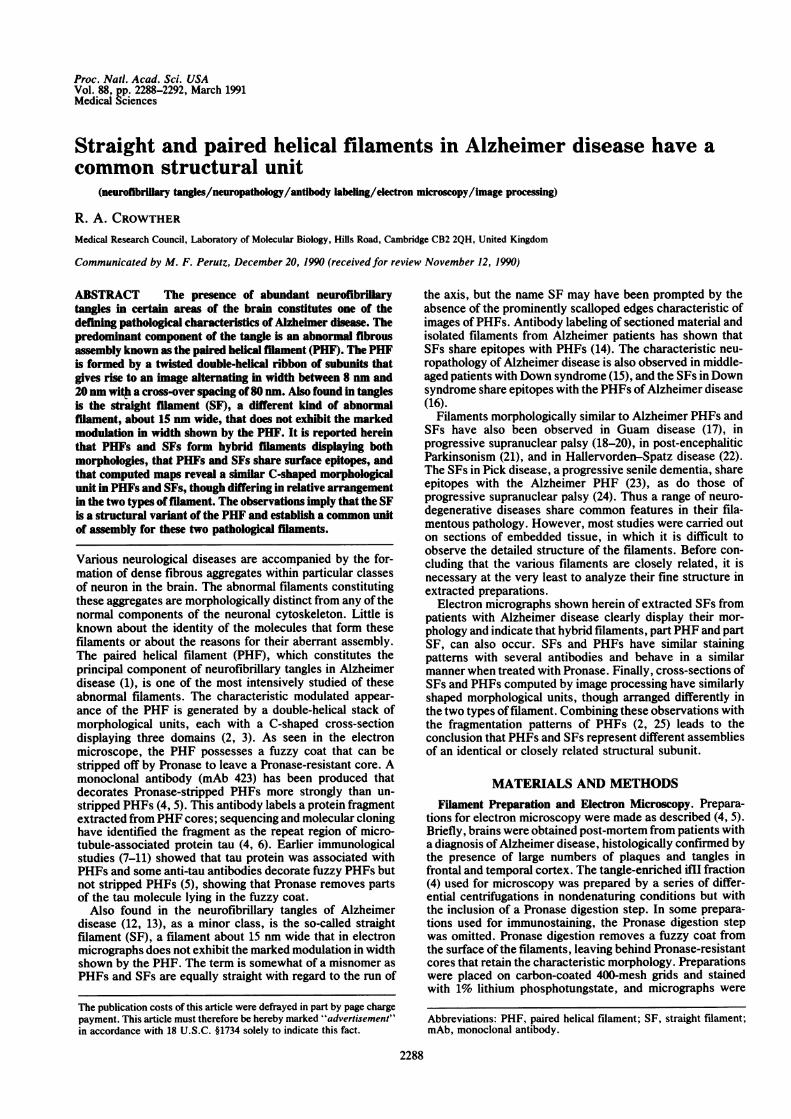
tively stained preparations of Alzheimer neurofibrillary tan-gles, clearly different filaments, corresponding to SFs, werefound (Fig. 1). It was hard to quantitate the overall frequencyof occurrence of SFs, but it could not amount to more thana few percent of PHFs. Often SFs appeared on the electronmicroscope grid in small groups, either on their own or amonglarger groups of PHFs, suggesting that they may have as-sembled in vivo within the same cell and remained togetherduring extraction of tangle fragments from the tissue. Com-paring the images of the two kinds of filaments, the modu-lation in width of SFs was much less marked than that ofPHFs; consequently, SFs showed a more uniform axialpattern of stain exclusion than PHFs. The average axialdistance between neighboring narrow portions of the SF wasabout 80 nm, as it was for the PHF, though in both filamentsthe pitch was variable. When scanning transmission electronmicroscope data were being collected on unstained PHFs (5),a small number of filaments identified as SFs gave the sameaverage mass per unit length as the PHFs, though theirnumbers were insufficient to obtain statistically reliable data.Fragmented SFs exhibited the same sharp transverse breaksand lack of fraying characteristic of PHFs (2).Most importantly, searches of negatively stained prepara-
tions of tangle fragments for SFs revealed a few examples ofhybrid filaments, which showed a sharp transition from a
FIG. 1. Abnormal filaments in Alzheimer disease. Negativelystained PHFs (P) and SFs (S) are most clearly distinguished byviewing the electron micrograph at a glancing angle along the axis ofeach filament: the PHF shows narrow deeply stain-embedded re-gions (crossovers) alternating with much wider shallowly stain-embedded regions, whereas the SF looks more uniformly bright withmuch less axial modulation in the pattern of staining. (Inset) SFs usedfor computing the average cross-section shown in Fig. 3a. (Bar = 100nm.)
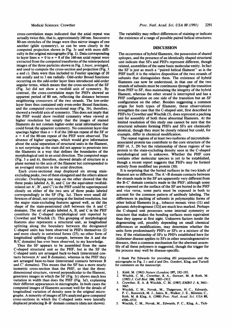
segment ofPHF into a segment of SF (Fig. 2 a and b). At thetransition point, one of the two strands of the PHF appearedinterrupted and the SF developed on the other strand. Thenicked appearance produced by the termination of one of thetwo strands of the PHF was seen particularly clearly in theimage of the hybrid filament shown in Fig. 2b. The hybridfilaments are clearly different from longitudinally split PHFs(2), in which one of the two strands is missing for part of thelength of the filament. The occurrence of such hybrid fia-ments, part PHF and part SF, implies a common structuralsubunit, assembled differently in PHFs and SFs.Antibody Labeling. Labeling with antibodies supports this
proposal of a common subunit. In preparations of filamentsthat were not treated with Pronase, both types of filamentwere labeled with an antiserum that contains antibodiesagainst the microtubule-associated protein tau (Fig. 2 c andd), whereas in Pronase-treated preparations neither filamenttype was labeled with this antibody. On the other hand, inpreparations treated with Pronase, both kinds of filamentwere labeled with mAb 423, which identifies a fragment oftauprotein in the Pronase-resistant core of the PHF (Fig. 2e) (4,5). Thus both types of filament display the same tau proteinepitopes and behave in a similar manner when treated withPronase.
Medical Sciences: Crowther

Proc. Natl. Acad. Sci. USA 88 (1991)
FIG. 2. (a and b) Examples of hybrid filaments. The arrows indicate the point of transition in structure from the PHF structure (below arrow)to the SF structure (above arrow). One of the two strands of the PHF appears to terminate and the SF develops on the other strand. (c andd) Filaments from an Alzheimer preparation that had not been Pronase-treated, showing decoration with an antibody with anti-tau activity (ICNanti-tubulin) visualized with electron-dense gold particles. The characteristic features of SF (c) and PHF (d) are clearly visible, despite thecovering of antibodies. (e) Filaments from a Pronase-treated preparation lightly decorated with mAb 423 showing PHFs (P) and an SF (S)equivalently labeled. (Bar = 100 nm.)
Image Processing. By using three-dimensional image re-construction, a cross-sectional density map of the SF wascomputed and compared with that computed (3) for the PHF(Fig. 3). To straighten and interpolate the filaments to con-
a
stant pitch, the individual repeats were located by cross-correlating a short piece ofthe filament with the whole image.Although at first glance features in the images of SFs ap-peared to repeat in an approximate manner every 80 nm, the
b
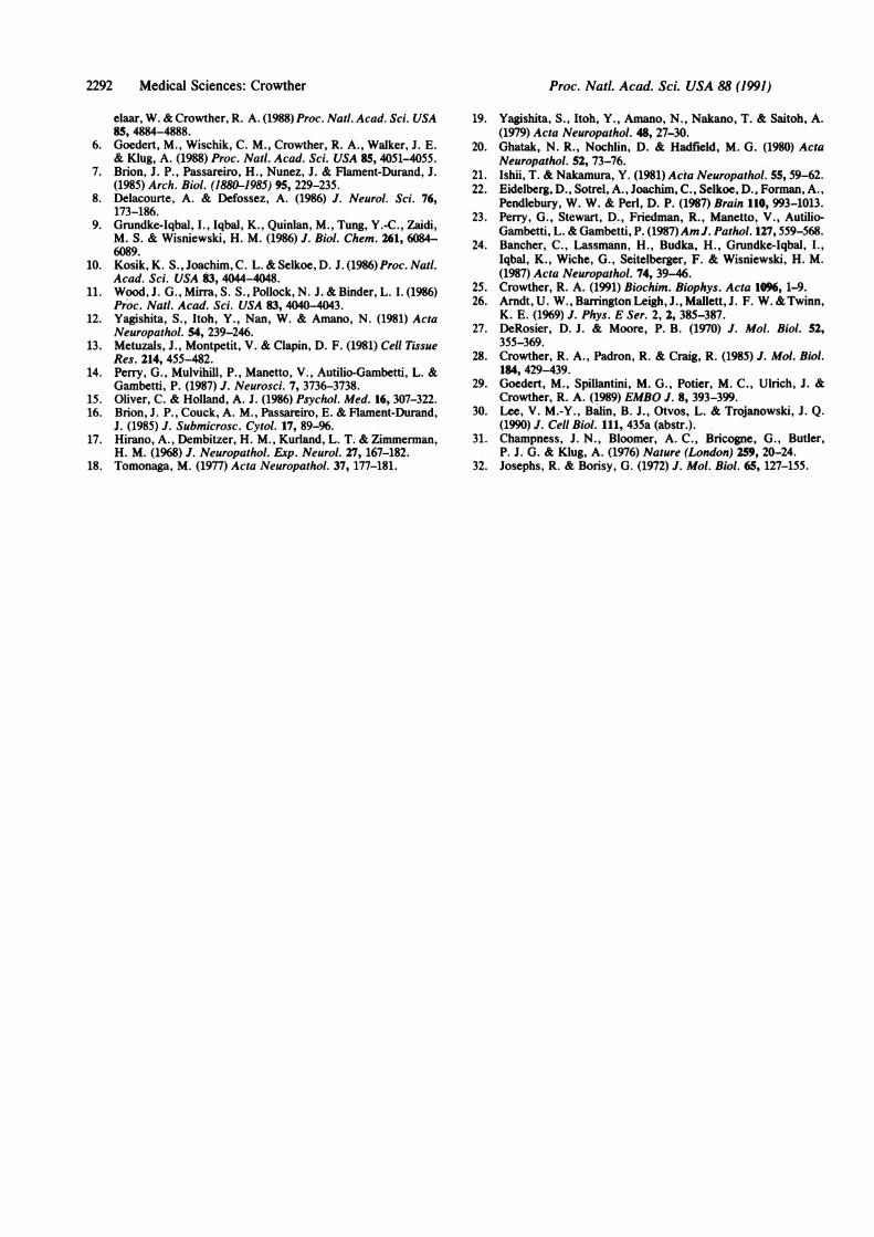
FIG. 3. Computed cross-section through the SF (a) compared with that found for the PHF (b) (3). The contour maps denote stain exclusion.Both cross-sections show two C-shaped morphological units, each displaying three peaks of density equivalently designated A, B, C and A',B', C' (see text). In the SF the C-shaped units are arranged back-to-back, whereas in the PHF they are arranged base-to-base. (Bar = 4 nm.)(c and d) Projections normal to the filament axis of the image reconstructions, demonstrating how the appearances of the SF and the PHF inelectron micrographs arise.
2290 Medical Sciences: Crowther
Proc. Natl. Acad. Sci. USA 88 (1991) 2291
cross-correlation maps indicated that the axial repeat wasactually twice this, that is, approximately 160 nm. Successive80-nm stretches of the image were thus mirror images of oneanother (glide symmetry), as can be seen clearly in thecomputed projection shown in Fig. 3c and with more diffi-culty in the original.micrographs (Fig. 1). Data correspondingto layer lines n = 0 to n = 8 of the 160-nm axial repeat wereextracted from the computed transforms of the reinterpolatedimages of the three particles shown in Fig. 1 Inset, averaged,and used to compute the cross-section and projection (Fig. 3a and c). Data were thus included to Fourier spacings of 20nm axially and to 3 nm radially. Odd-order Bessel functionsoccurring on the odd-order layer lines introduced odd-orderangular terms, which means that the cross-section of the SF(Fig. 3a) did not show a twofold axis of symmetry. Bycontrast, the cross-correlation maps for PHFs showed anapparent period of 80 nm, reflecting the distance betweenneighboring crossovers of the two strands. The low-orderlayer lines thus contained only even-order Bessel functions,and the computed cross-sectional map (Fig. 3b), therefore,showed a twofold axis of symmetry. This does not imply thatthe PHF would show twofold symmetry when viewed athigher resolution but simply that the images of stainedfilaments do not contain higher-order axial information thatcould break the apparent symmetry. No consistent layer-linespacings higher than n = 8 of the 160-nm repeat of the SF orn = 4 of the 80-nm repeat of the PHF were observed. Theabsence of higher spacings, which would give informationabout the axial separation of structural units in the filament,is not surprising as the stain did not appear to penetrate intothe filaments in a way that defines the boundaries of themorphological unit in an axial direction. The computed maps(Fig. 3 a and b), therefore, showed details of structure in aplane normal to the axis of the filament but corresponded toan averaged structure in the axial direction.Each cross-sectional map displayed six strong stain-
excluding peaks, two of them elongated and the others almostcircular. Overlaying one map on the other showed that thethree peaks labeled A, B, and C (or the equivalent twofold-related set A', B', and C') in the PHF could be superimposedclosely on either of the two sets of three peaks labeledcorrespondingly in the SF (Fig. 3a). There were small dif-ferences of detail, not surprising at the limited resolution, butthe major stain-excluding features agreed well, as did theshape of the stain-penetrated cleft between the A and Cdomains. The set of peaks labeled A, B, and C in the PHFconstitute the C-shaped morphological unit reported byCrowther and Wischik (3). This grouping of morphologicalfeatures also represents a structural unit, as longitudinalsplitting along the boundary between the designatedC-shaped units has been observed in PHFs themselves (2)and more clearly in untwisted forms (25); no other form oflongitudinal splitting (for example, between the A and theB/C domains) has ever been observed, to my knowledge.Thus the SF appears to be assembled from the same
C-shaped structural unit as the PHF, but in the SF theC-shaped units are arranged back-to-back (interstrand con-tacts between A' and B domains), whereas in the PHF theyare arranged base-to-base (interstrand contacts between Band C' domains). This means that the SF has a more nearlyisometric cross-section than the PHF, so that the three-dimensional structure, viewed perpendicular to the filament,generates images in which the SF (Fig. 3c) shows much lessvariation in width than does the PHF (Fig. 3d), explainingtheir different appearances in micrographs. In both cases thecomputed images of filaments account well for the details oflongitudinal variation of density seen in the original micro-graphs. A minority of images of SFs analyzed gave computedcross-sections in which the C-shaped units were laterallydisplaced producing B-B' domain contacts (data not shown).
The variability may reflect differences of staining or indicatethe existence of a range of possible paired helical structures.
DISCUSSIONThe occurrence of hybrid filaments, the possession of sharedepitopes, and the presence of an identically shaped structuralunit indicate that SFs and PHFs represent different, thoughrelated, assemblies of the same basic molecular entity. In factthe SF is just as much a "paired helical filanent" as is thePHF itself; it is the relative disposition of the two strands ofsubunits that distinguishes them. The existence of hybridfilaments can now be understood, in that one of the twostrands of subunits must be continuous through the transitionfrom PHF to SF, thus maintaining the integrity of the hybridfilament, whereas the other strand is interrupted and has aPHF configuration on one side of the transition but an SFconfiguration on the other. Besides suggesting a commonorigin for both types of filament, these observationsstrengthen the case that the C-shaped unit, first described inPHFs by Crowther and Wischik (3), does represent a packingunit for assembly of both these abnormal filaments. At thelimited resolution of this study one cannot be sure that thestructural subunits forming PHFs and SFs are chemicallyidentical, though they must be closely related but could, forexample, differ in chemical modification.The repeat regions of at least two isoforms of microtubule-
associated protein tau contribute to the core structure of thePHF (4, 5, 29) but the relationship of these regions of tauprotein to the stain-excluding density seen in the C-shapedmorphological unit is unknown. Whether the PHF corecontains other molecular species is yet to be established,though a recent report suggests that PHFs may be formedentirely from modified tau protein (30).
It is surprising that the buried surfaces in the two kinds offilament are so different. The A'-B domain contacts betweenthe strands made in the SF are apparently very different fromthe B-C' domain contacts made in the PHF; although someareas exposed on the surface of the SF are buried in the PHFand vice versa, some parts must be exposed in both toaccount for the common pattern of antibody labeling. Thedifferences in packing of subunits in polymorphic forms ofother helical filaments [e.g., tobacco mosaic virus (31) andglutamic dehydrogenase (32)] are much less marked. Perhapsthe C-shaped unit possesses some internal symmetry orstructure that makes the bonding surfaces more equivalentthan they appear at first sight. Unknown factors inside thedegenerating cell, possibly depending on small chemicaldifferences or modifications, may determine whether theunits form predominantly PHFs or SFs or a mixture of thetwo. If the relationship of SFs to PHFs established here forAlzheimer disease applies to SFs in other neurodegenerativediseases, then a common mechanism for the aberrant assem-bly of all these polymers is suggested, though the trigger forthe process may well be disease-specific.
I thank Pat Edwards for providing iflM preparations and themicrographs in Fig. 2 c and d and Drs. Goedert, KMug, and Turnellfor comments on the manuscript.
1. Kidd, M. (1963) Nature (London) 197, 192-193.2. Wischik, C. M., Crowther, R. A., Stewart, M. & Roth, M.
(1985) J. Cell Biol. 100, 1905-1912.3. Crowther, R. A. & Wischik, C. M. (1985) EMBO J. 4, 3661-
3665.4. Wischiki C. M., Novak, M., Th0gerson, H. C., Edwards,
P. C., Runswick, M. J., Jakes, R., Walker, J. E., Milstein, C.,Roth, M. & Klug, A. (1988) Proc. NatI. Acad. Sci. USA 85,4506-4510.
5. Wischik, C. M., Novak, M., Edwards, P. C., Klug, A., Tich-
Medical Sciences: Crowther
Proc. Natil. Acad. Sci. USA 88 (1991)
elaar, W. & Crowther, R. A. (1988) Proc. Nat!. Acad. Sci. USA85, 4884-4888.
6. Goedert, M., Wischik, C. M., Crowther, R. A., Walker, J. E.& Klug, A. (1988) Proc. Nat!. Acad. Sci. USA 85, 4051-4055.
7. Brion, J. P., Passareiro, H., Nunez, J. & Flament-Durand, J.(1985) Arch. Biol. (1880-1985) 95, 229-235.
8. Delacourte, A. & Defossez, A. (1986) J. Neurol. Sci. 76,173-186.
9. Grundke-Iqbal, I., Iqbal, K., Quinlan, M., Tung, Y.-C., Zaidi,M. S. & Wisniewski, H. M. (1986) J. Biol. Chem. 261, 6084-6089.
10. Kosik, K. S., Joachim, C. L. & Selkoe, D. J. (1986) Proc. Nat!.Acad. Sci. USA 83, 4044-4048.
11. Wood, J. G., Mirra, S. S., Pollock, N. J. & Binder, L. I. (1986)Proc. Nat!. Acad. Sci. USA 83, 4040-4043.
12. Yagishita, S., Itoh, Y., Nan, W. & Amano, N. (1981) ActaNeuropathol. 54, 239-246.
13. Metuzals, J., Montpetit, V. & Clapin, D. F. (1981) Cell TissueRes. 214, 455-482.
14. Perry, G., Mulvihill, P., Manetto, V., Autilio-Gambetti, L. &Gambetti, P. (1987) J. Neurosci. 7, 3736-3738.
15. Oliver, C. & Holland, A. J. (1986) Psychol. Med. 16, 307-322.16. Brion, J. P., Couck, A. M., Passareiro, E. & Flament-Durand,
J. (1985) J. Submicrosc. Cytol. 17, 89-96.17. Hirano, A., Dembitzer, H. M., Kurland, L. T. & Zimmerman,
H. M. (1968) J. Neuropathol. Exp. Neurol. 27, 167-182.18. Tomonaga, M. (1977) Acta Neuropathol. 37, 177-181.
19. Yagishita, S., Itoh, Y., Amano, N., Nakano, T. & Saitoh, A.(1979) Acta Neuropathol. 48, 27-30.
20. Ghatak, N. R., Nochlin, D. & Hadfield, M. G. (1980) ActaNeuropathol. 52, 73-76.
21. Ishii, T. & Nakamura, Y. (1981) Acta Neuropathol. 55, 59-62.22. Eidelberg, D., Sotrel, A., Joachim, C., Selkoe, D., Forman, A.,
Pendlebury, W. W. & Perl, D. P. (1987) Brain 110, 993-1013.23. Perry, G., Stewart, D., Friedman, R., Manetto, V., Autilio
Gambetti, L. & Gambetti, P. (1987)Am J. Patho!. 127, 559-568.24. Bancher, C., Lassmann, H., Budka, H., Grundke-Iqbal, I.,
Iqbal, K., Wiche, G., Seitelberger, F. & Wisniewski, H. M.(1987) Acta Neuropathol. 74, 39-46.
25. Crowther, R. A. (1991) Biochim. Biophys. Acta 1096, 1-9.26. Arndt, U. W., Barrington Leigh, J., Mallett, J. F. W. & Twinn,
K. E. (1969) J. Phys. E Ser. 2, 2, 385-387.27. DeRosier, D. J. & Moore, P. B. (1970) J. Mol. Biol. 52,
355-369.28. Crowther, R. A., Padron, R. & Craig, R. (1985) J. Mol. Biol.
184, 429-439.29. Goedert, M., Spillantini, M. G., Potier, M. C., Ulrich, J. &
Crowther, R. A. (1989) EMBO J. 8, 393-399.30. Lee, V. M.-Y., Balin, B. J., Otvos, L. & Trojanowski, J. Q.
(1990) J. Cell Biol. 111, 435a (abstr.).31. Champness, J. N., Bloomer, A. C., Bricogne, G., Butler,
P. J. G. & Klug, A. (1976) Nature (London) 259, 20-24.32. Josephs, R. & Borisy, G. (1972) J. Mol. Biol. 65, 127-155.
2292 Medical Sciences: Crowther