stimulative effects of hominis placental pharmacopuncture...
TRANSCRIPT
Orig
ina
lA
rticle
Tae-Keun Hong1, Jeehye Kim1, Juyoun Woo1, Ki-Tae Ha1, Myungsoo Joo1, Yoon-Bong Hahn2, Han-Sol Jeong1*
1 Division of Applied Medicine, Pusan National University School of Korean Medicine, Yangsan, Korea2 Department of BIN Fusion Technology, Chonbuk National University School of Semiconductor and Chemical Engineering, Jeonju, Korea
Key Words
Hominis Placenta; zinc oxide; pharmacopuncture; RAW264.7 cells
ISSN 2093-6966(Print), ISSN 2234-6856(Online) Journal of Pharmacopuncture 2012;15(3):013-018DOI: http://dx.doi.org/10.3831/KPI.2012.15.001
sed the uptake by RAW 264.7 cells. In addition, cellular struc-tural alterations were observed in groups treated with ZnONP HPPS.
CCoonncclluussiioonnss:: Neither ZnO NP nor HPPS activated RAW 264.7cells, which is likely due to a low cellular uptake. The ZnO NPHPPS, however, significantly activated NF-κB and up-regula-ted its dependent genes such as TNF-α, IL-1, and MCP-1.ZnO NP HPPS was also more easily taken into the RAW 264.7cells than either ZnO NP or HPPS.
1. Introduction
Nanotechnology is referred to as a science that makes anew system through which matter is manipulated on a nan-oscale [1], and great advances have been made in the field ofnanotechnology over the last decade. It is one of the mostadvancing technologies of the 21st century and is thought tooccupy a leading position in industry. Engineered nanomat-erials are already being used in various industrial fields suchas sporting goods, cosmetics, and electronics and will beincreasingly applied to medical fields such as diagnosis,cancer treatment, and drug delivery [2,3]. Regardless of the-se extensive applications, many concerns about the safetyand the potential risks of engineered nanomaterials associ-ated with human health have been raised [4].
Zinc oxid (ZnO) is one of the most exciting materials becau-se of its versatile properties such as its semiconducting,biosafe, and biocompatible natures. ZnO is also consideredto be a “GRAS”(generally recognized as safe) subst-ance bythe Food and Drug Administration (FDA). Because of these
Abstract
OObbjjeeccttiivveess:: The purpose of this study is to examine whetherHominis Placental pharmacopuncture solution (HPPS) combi-ned with zinc-oxide nanoparticles (ZnO NP) activates RAW264.7 cells.
MMeetthhooddss:: We soaked ZnO nanoparticles in the Hominis Plac-enta pharmacopuncture solution, thereby making a combinedform (ZnO NP HPPS). The effect of ZnO NP HPPS on the intra-cellular reactive oxygen species (ROS) production was measu-red by 2', 7'-dichlorofluorescin diacetate (DCFH-DA) assay.The effect of ZnO NP HPPS on NF-κB was measured by usinga luciferase assay. The effect of ZnO NP HPPS on the cytokineexpression was assessed by semi-quantitative reverse tran-scriptase polymerase chain reaction (RT-PCR). The cellularuptake of ZnO NP HPPS was measured by using a flowcytometric analysis, and cellular structural alterations wereanalyzed by using transmission electron microscopy (TEM).
RReessuullttss:: Neither the HPPS nor the ZnO NPs induced intracell-ular ROS production in RAW 264.7 cells. Neither of the mater-ials activated NF-κB or it’s dependent genes, such as TNF-α,IL-1, and MCP-1. However, ZnO NP HPPS, the combined formof ZnO NPs and HPPS, did induce the intracellular ROS prod-uction, as well as prominently activating NF-κB and it’s depe-ndent genes. Also, compared to ZnO NPs, it effectively increa-
Received: May 22, 2012 Accepted: Sept 10, 2012
This is an Open-Access article distributed under the terms of the Creative CommonsAttribution Non-Commercial License (http://creativecommons.org/licenses/by-nc/3.0/)which permits unrestricted noncommercial use, distribution, and reproduction in anymedium, provided the original work is properly cited.
This paper meets the requirements of KS X ISO 9706, ISO 9706-1994 and ANSI/NISOZ39.48-1992 (Permanence of Paper).
*Corresponding authorHan-Sol Jeong. Division of Applied Medicine, Pusan National University School of KoreanMedicine, Beomeo-ri, Mulgeum-eup, Yangsan, Gyeongnam 626-870, Korea. Tel: +82-51-510-8461 Fax: +82-51-510-8420 E-mail: [email protected]
ⓒ 2012 Korean Pharmacopuncture Institute http://www.journal.ac
Stimulative Effects of Hominis Placental
Pharmacopuncture Solution Combined
with Zinc-oxide Nanoparticles on RAW 264.7 Cells
- ZnO HPPS more easily stimulates RAW 264.7 cells -
013~18 정한솔 2012.9.28 10:9 AM 페이지13
Journal of Pharmacopuncture 2012;15(3):013~01814
properties, ZnO can be safely used in medical fields such ascancer therapy and drug delivery vehicles [5].
Hominis Placenta extracts have been used in traditional Kor-ean medicine to improve physiological function. Recently, therehave also been several reports that Hominis Placenta can beapplied to pathologic conditions such as wounds [6], cartilagedegradation [7], osteoporosis [8], nerve injury [9], and anemia [10].
In this study, we examined whether ZnO makes a differencewhen it is combined with Hominis Placenta pharmacopuncturesolution (HPPS). We made ZnO NP HPPS, a combined form ofZnO nanoparticles (NPs) and HPPS. Then, we compared the sti-mulatory effects of ZnO NP HPPS with those of ZnO NPs andHPPS. In this study, we found that ZnO NP HPPS, the combinedform of ZnO NPs and HPPS, had more ability to stimulate RAW264.7 cells, than ZnO NPs and HPPS alone.
2. Materials and methods
2.1. Preparation of Hominis Placenta pharmacopun-cture solutions (HPPS)
HPPS was prepared by using the following protocol providedby the Korean Pharmacopuncture Institute. Briefly, HominisPlacenta was hydrolyzed with HCL at 101°C, and the extractswere heated at 80-100 °C for 10 hrs, autoclaved, and filteredwith a 0.1 μm filter membrane. The final product was kept refr-igerated until usage.
2.2. Synthesis of ZnO nanoparticles (ZnO NP) ZnO NPs were synthesized by using a low-temperature soluti-
on method with zinc acetate dihydrate (ZnAc), hexamethy-lenetetramine (HMTA), and LiOH [11]. For synthesizing fluores-cein isothiocyanate (FITC)-conjugated ZnO NPs, a 3 ml FITC sol-ution (3 mM) was added to a solution of 0.1 M ZnAc and 0.05 MHMTA. Subsequently, a 1.0 M LiOH solution was slowly added tothe above solution to pH 10 and was refluxed at 90°C for 3 hrs.The precipitates were washed and then airdried. The structureand the crystallinity of the as-synthesized ZnO NPs were deter-mined by using transmission electron microscopy (TEM) and X-ray diffraction (XRD) in the range of 20-70°at an 8°/min scanningspeed.
2.3. Cell cultureThe RAW 264.7 cells were obtained from ATCC (American Type
Culture Collection, Rockville, MD, USA), were cultured in Dulbe-cco’s modi ed Eagle's medium (DMEM) containing L-glutamine(200 mg/L) supplemented with 10 % (v/v) heat-inactivated fetalbovine serum (FBS), 100 U/ml penicillin, and 100 μg/ml strepto-mycin, and were maintained in a humidified incubator at 37°Cwith 5% CO2 prior to the experiment.
2.4. Measurement of intracellular ROS generationCells, 1 x 10⁶/ml, were cultured under the same conditions as
in the flow cytometric analysis. ZnO NPs (1 μg/ml), ZnO NPHPPS and 10 uM 2', 7'-dichlorofluorescin diacetate (DCFH-DA)were added to cell plates. For 30 mins, the cells were maintai-ned under cell culture conditions. Then, reactive oxygen species(ROS) was measured by using a Victor3 system (Perkin Elmer,Waltham, MA, USA). H2O2 (1mM) was added to the cells to verifythe ROS experiment.
2.5. Reporter construct and luciferase assayTo measure the NF-κB transcriptional activity, we created an
NF-κB reporter construct, and stably transfected it into RAW264.7 cells. The reporter construct harbors four tandem copiesof a 36-base enhancer from the 5’HIV-long terminal repeat
(containing two NF-κB binding sites, GGGACTFTTTCC) placedupstream of the HSV minimal thymidine kinase promoter, whichwere cloned into pEGFPLuc. Then, the reporter construct wasintroduced into RAW 264.7 cells by transfection using lipof-ectamine, and the transfected cells were selected under G418(600 μg/ml). Candidate cell lines harboring the NF-κB reporterconstruct were tested for luciferase activity. The cell line used inthis study was cultured in DMEM medium containing 10% FBSand 1% penicillin/streptomycin antibiotics (PSA) at 37°C under a5% CO2 atmosphere. G418 was added as to a final 50 μg/mlconcentration to maintain the transfected cells. ZnO NPs or ZnONP HPPS was added to the cells at a concentration of 1 μg/ml,and the treated cells were incubated for 8 hrs. Cells were harv-ested by centrifugation at 13000 rpm and 4°C, and cell lysatewas prepared. Ten μl of the cell lysate was used for theluciferase activity per the protocol of the manufacturer(Promega, Madis-on, WI, USA). NF-κB activities were measuredby using an infini-te M200 (TECAN Group Ltd.).
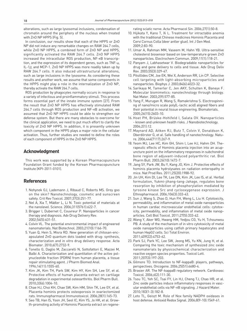
2.6. Isolation of total RNA from cells and RT-PCRWe isolated total RNA by using Trizol reagent through the
manufacturer’s instructions (Invitrogen, Carlslab, CA, USA).After the concentration of RNA had been determined by using aspectrophotometer, we made cDNA by using reverse-transcrip-tion from 1 μg of RNA obtained using M-MLV reverse transcrip-tase (Promega) and amplified using polymerase chain reaction(PCR) with specific primers (Table 1). The final PCR productswere separated on 1.5% agarose gel by electrophoresis. Imagesof the bands were captured and quantified by using Image J(Bio-Rad, Hercules, CA, USA).
2.7. Cellular uptake and FACS analysisRAW 264.7 cells (1 × 10⁶ cells/ml) were plated in a 96-well pl-
ate and were incubated in 5% CO2 at 37°C. After a 24 hrs incu-bation, the culture medium was replaced with 100 μl of freshmedium containing FITC- conjugated ZnO NPs or ZnO NP HPPSand was further incubated in 5% CO2 at 37°C for 2 and 4 hrs.After incubation, the cells were washed with PBS 3 times.Finally, the cells were resuspended in PBS for the flowcytometric analysis (FACS Canto II Analyzer, BD Bioscience, SanJose, CA, USA).
2.8. Transmission electron microscopy (TEM)Cells were cultured at a density of 1 × 10⁶ cells/ml in a cultu-
re medium, each medium containing samples at a concentra-tion of 100 μg/ml of ZnO NPs or ZnO NP HPPS. After 4 hrs, cellswere washed in PBS and were centrifuged at 1000 rpm for 5mins. Cell pallets were fixed in 2% osmium tetroxide and thendehydrated with propion aldehyde. Epoxy resins were added;
Primer Direction Sequence
MCP-1 Forward 5'-AGGTCCCTGTCATGCTTCTG-3'
Reverse 5'-TCTGGACCCATTCCTTCTTG-3'
IL-1β Forward 5'-GCCCATCCTCTGTGACTCAT-3'
Reverse 5'-AGGCCACAGGTATTTTGTCG-3'
TNF-α Forward 5’-CCAAACGATGTTGTACCCGA-3’
Reverse 5’-CAGTTGGAGGAGAGACGGTA-3’
GAPDH Forward 5'-AACTTTGGCATTGTGGAAGG-3'
Reverse 5'-ACACATTGGGGGTAGGAACA-3'
TTaabbllee 11 Sequences of the primers (for RT-PCR) used in the presentstudy
013~18 정한솔 2012.9.28 10:9 AM 페이지14

Journal of Pharmacopuncture 2012;15(3):013~018 15
then, they were sectioned for transmission microscopy (TEM)observations (HITACHI H-7600, Japan).
2.9. Statistical analysisAll data are expressed as means ± standard deviations of at
least three independent experiments. For comparison amonggroups, a one-way analysis of variance (ANOVA) test was used(with the assistance of Graph Pad Software, Inc., San Diego, CA).
3. Results
3.1. ZnO NP HPPS significantly induced intracellularROS production
To measure the effects of ZnO NP HPPS on the intracellularROS production, we treated RAW 264.7 cells with 1 μg/ml HPPS,ZnO NP, and ZnO NP HPPS for 30 mins. We observed that theamounts of intracellular ROS were higher in ZnO NP HPPStreated cells than HPPS and ZnO NP treated cells (Fig. 1). H2O2
was used as a positive control. Although HPPS or ZnO NP treat-ment alone induced a little of intracellular ROS, it was notstatistically significant.
3.2. ZnO NP HPPS significantly increased the NF-κBactivation
To evaluate the potential of ZnO NP HPPS to induce cellularactivation, we measured the activation of NF-κB transcriptionfactor. We treated the NF-κB reporter cell line with 1 μg/ml ofHPPS, ZnO NP, and ZnO NP HPPS in for 8 hrs, and then measu-red the NF-κB activity by luciferase assay. Either HPPS or ZnONP did not increase the luciferase activity but ZnO NP HPPSsignificantly increased the luciferase activity (p 0.01).
3.3. ZnO NP HPPS increased the expression of NF-κB dependent cytokine mRNA
To evaluate whether ZnO NP HPPS could modulate TNF-α, IL-1β, MCP-1 cytokine production, we performed semi-quantitiativeRT-PCR. Total RNA was extracted from the cells treated withHPPS, ZnO NP, and ZnO NP HPPS for 4 hrs at the concentrationof 1 μg/ml. In cases of TNF-α, IL-1β, MCP-1, no changes wereobs-erved in the HPPS and ZnO NP treated groups whencompared to the control, while significant increases of mRNAexpression, especially IL-1β, were identified in the ZnO NPHPPS treated groups (Fig. 3). The results clearly show that ZnONP HPPS affects production of MCP-1, TNF-α, IL-1βcytokines.
FFiigguurree 11 Effects of HPPS, ZnO NPs, and ZnO NP HPPS on theintracellular ROS production in RAW 264.7 cells. Cells weretreated with 1μg/ml HPPS, ZnO NPs, and ZnO NP HPPS for30 mins prior to assay. The y axis represents the relativefluorescence intensity and reflects the amount of ROS. ZnONP HPPS induces 25% and 15% more ROS than ZnO NPsand HPPS, respectively (**p 0.01).
FFiigguurree 22 Effects of HPPS, ZnO NPs, and ZnO NP HPPS on the NF-κBactivity. An NF-κB reporter cell line derived from RAW264.7 cells was treated with 1 μg/ml HPPS, ZnO NPs, orZnO HPPS for 8 hrs. HPPS and ZnO NPs did not increasethe luciferase activity. ZnO NP HPPS showed significantlyhigher activity than CTL and ZnO NP. Luciferase activitywas normalized to the amount of total protein in cell lysate.** Statistically significant difference as compared with thecontrol (**p 0.01).
013~18 정한솔 2012.9.28 10:9 AM 페이지15

Journal of Pharmacopuncture 2012;15(3):013~01816
3.4. Uptake of ZnO NP HPPS and ZnO NPs (by FACSanalysis)
To examine the cellular uptake and the permeation of the ZnONP HPPS through the cell membrane, we treated the RAW264.7 cells with 1 μg/ml of FITC-conjugated ZnO NP and ZnO NPHPPS for 2~4 hrs, and then performed flow cytometric anal-ysis. The cells treated without ZnO NP were used as negativecontrols. As shown in Fig. 4, there are almost negligible fluores-cence differences between ZnO NP and ZnO NP HPPS for 2 hrstreatment. However, a slight increase of fluorescence intensitywas detected in 4 hrs treatment, suggesting that the amount ofcellular uptake was increased.
3.5. ZnO NP HPPS caused structural alterations inRAW 264.7 cells
For the analysis of subcellular morphologic changes, ZnO NPand ZnO NP HPPS treated cells were examined by transmissionelectron microscopy (TEM). RAW 264.7 cells were treated witheither ZnO NP or ZnO NP HPPS for 4 hrs. There were no ultra-structural alterations in the cells treated with ZnO NP. Theintegrity of subcellular structures such as plasma and nuclearmembrane were normal (Fig. 5a, b). However, significantalterations were observed in the cells treated with ZnO NPHPPS. Variations in size and shape of the cells were prominent,and large inclusions were observed in the lysosome (Fig. 5c, d).
FFiigguurree 44 Uptake of ZnO NPs and ZnO NP HPPS in RAW 264.7 cells.The x axis represents the fluorescence intensity andreflects the amount of uptake, and the y coordinate showsthe number of cell counts. The region of high fluorescenceintensity (over 150,000) was assigned as A. A slight increasein the fluorescence intensity was observed especially in theZnO NP HPPS treatment groups for 4 hrs.
FFiigguurree 33 Effects of HPPS, ZnO NPs, and ZnO NP HPPS on the NF-κBdependent gene expression. RAW 264.7 cells were treatedwith 1 μg/ml HPPS, ZnO NPs, and ZnO HPPS for 4 hrs.Then, total RNA was extracted, and the expressions of NF-κB dependent genes, including TNF-α, IL-1β, MCP-1, wereanalyzed by semiquantitative RT-PCR. The relative expres-sion of each gene was determined by using a densitometricanalysis of each band (**p 0.01, ***p 0.001).
013~18 정한솔 2012.9.28 10:9 AM 페이지16

Journal of Pharmacopuncture 2012;15(3):013~018 17
4. Discussion
Nanotechnology is an engineering system that manipulatesmatter at a molecular scale. Engineered nanomaterials have al-ready been used in industry such as sports, cosmetics, and ele-ctronics. In the last decade, great advances have been made innanomedicinal field. Recent studies have focused on usingnanoparticles for imaging, tissue engineering, and drug delivery[12-15].
Delivering therapeutic agents into cells can be highly ineffici-ent because of diverse physicochemical properties of therapeu-tic agents. Recently there were many extensive investigations inthe field of drug delivery systems for the purpose of improvingand facilitating drug efficacy. Nanomaterials can modify thephysicochemical properties of a drug so to provide an opportun-ity for increasing uptake and interaction with target cells.Therefore, it is important to ensure that these agents do notcause any adverse effect. However, there are growing concernsabout the toxic effects of engineered nanomaterials on humanhealth because of their unusual physicochemical properties.There are some consensuses that deal with potential risks ass-ociated with inhalation of nanoparticles [16,17].
Nanoscale ZnO used in many applications in daily life is beli-eved to be nontoxic and biocompatible. Recently, attentions havefocused on the Zinc Oxide nanoparticles (ZnO NPs) because ofits own unique biosafe and biocompatible properties [5].
HPPS is one of the most widely used herb-acupunctural mate-rial to replenish vital essence and blood in Korean traditionalmedicine [18]. Several studies about therapeutic effectiveness ofHPPS were reported. HPPS has a protective effect against ost-eoporosis [8], growth-promoting activity on the nerve regenera-
tion [9], protective effect on radiation enteropathy [19], andcapacity to regulate bone resorption [20].
In this study, we examined whether ZnO NO HPPS, a combini-ng form of ZnO NP and HPPS, has any special effects overHPPS. Oxidative stress was regarded as a major cytotoxicstandard caused by nanomaterials [2]. Previous studies showedthat ZnO NP induces ROS production [21-23]. However, ourresults revealed that ZnO NP did not increase intracellular ROSproduction, but ZnO NP HPPS up-regulated the intracellularROS level (Fig. 1). It is well known that NF-κB is a transcriptionfactor which is involved in cellular response to stimuli such asstress, UV, ROS, and bacterial antigens. It is a key factor thatregulates genes responsible for the innate immunity [24,25].Tsou et al. reported that ZnO NP is related to NF-κB activation[26]. However, our results show that ZnO NP did not influencethe NF-κB activation. On the other hand, ZnO NP HPPS signific-antly activated the NF-κB transcription factor (Fig. 2) and itsdependent genes (Fig. 3). Among NF-κB dependent genes, IL-1βwas especially up-regulated by the treatment of ZnO NP HPPS(Fig. 3b). Our unpublished results indicate that a longertreatment of ZnO NP also increased the NF-κB activation (datanot shown). From these results, we have assumed that somecomponents in HPPS makes ZnO to easily activate RAW 264.7cells via NF-κB. As shown in Figure 4, increase of cellular upta-ke was observed in the ZnO NP HPPS treated group for 4 hrs. Toobserve whether ZnO NP HPPS can cause any subcellularchanges, we examined ultastructural alterations by usingtransmission electron microscopy (TEM) in the RAW 264.7 cellstreated with ZnO NP and ZnO NP HPPS for 4 hrs. The cells mai-ntained normal appearance when were treated with ZnO NP;however, the cells exhibited significant ultrastructural
FFiigguurree 55 Cellular structural changes of RAW 264.7 cells treated with ZnO NPs (a, b) and ZnO NP HPPS (c, d). (a) The nucleus contains amoderate quantity of nuclear membrane-associated condensed chromatin (‡). (b) The nuclear and mitochondrial membranes areclearly outlined (*). (c-d) Large inclusions are observed in the lysosome (▼). The arrow in (d) indicates complex inclusions.
013~18 정한솔 2012.9.28 10:9 AM 페이지17
Journal of Pharmacopuncture 2012;15(3):013~01818
alterations, such as large lysosomal inclusions, condensation ofchromatin around the periphery of the nucleus when treatedwith ZnO NP HPPS (Fig. 5).
In conclusion, our results show that each of the HPPS or ZnONP did not induce any remarkable changes on RAW 264.7 cells,while ZnO NP HPPS, a combined form of ZnO NP and HPPS,significantly activated the RAW 264.7 cells. ZnO NP HPPSincreased the intracellular ROS production, NF-κB transcrip-tion, and the expression of its dependent genes, such as TNF-α,IL-1β, and MCP-1. ZnO NP HPPS was more readily taken up byRAW 264.7 cells. It also induced ultrastructural alterations,such as large inclusions in the lysosome. As considering theseresults and another work, we assume that some components inthe HPPS might play a role in the internalization of ZnO NP,thereby activate the RAW 264.7 cells.
ROS production by phagocytes normally occurs in response toa variety of infectious and proinflammatory stimuli. This processforms essential part of the innate immune system [27]. Fromthe result that ZnO NP HPPS has effectively stimulated RAW264.7 cells through ROS production and NF-κB activation, weassumed that ZnO NP HPPS could be able to strengthen ourdefense system. But there are many obstacles to overcome forthe clinical application, we need to put much effort to clarify thetoxicity of ZnO NP HPPS. In addition, it is presently unknownwhich component in the HPPS plays a major role in the cellularactivation. Thus, further studies are needed to define the rolesof each component of HPPS in the ZnO NP HPPS.
Acknowledgment
This work was supported by a Korean PharmacopunctureFoundation Grant funded by the Korean PharmacopunctureInstitute (KPI-2011-0101).
References
1. Nohynek GJ, Lademann J, Ribaud C, Roberts MS. Grey gooon the skin? Nanotechnology, cosmetic and sunscreensafety. Crit Rev Toxicol. 2007;37(3):251-77.
2. Nel A, Xia T, Ma..dler L, Li N. Toxic potential of materials atthe nanolevel. Science. 2006;311(5761):622-7.
3. Brigger I, Dubernet C, Couvreur P. Nanoparticles in cancertherapy and diagnosis. Adv Drug Delivery Rev. 2002;54(5):631-51.
4. Colvin VL. The potential environmental impact of engineerednanomaterials. Nat Biotechnol. 2003;21(10):1166-70.
5. Yuan Q, Hein S, Misra RD. New generation of chitosan-enc-apsulated ZnO quantum dots loaded with drug: synthesis,characterization and in vitro drug delivery response. ActaBiomater. 2010;6(7):2732-9.
6. Tonello G, Daglio M, Zaccarelli N, Sottofattori E, Mazzei M,Balbi A. Characterization and quantitation of the active pol-ynucleotide fraction (PDRN) from human placenta, a tissuerepair stimulating agent. J Pharm Biomed Anal. 1996;14(11):1555-60.
7. Kim JK, Kim TH, Park SW, Kim HY, Kim SH, Lee SY, et al.Protective effects of human placenta extract on cartilagedegradation in experimental osteoarthritis. Biol Pharm Bull. 2010;33(6):1004-10.
8. Chae HJ, Choi KH, Chae SW, Kim HM, Shin TK, Lee GY, et al.Placenta hominis protects osteoporosis in ovariectomizedrats. Immunopharmacol Immunotoxicol. 2006;28(1):165-73.
9. Seo TB, Han IS, Yoon JH, Seol IC, Kim YS, Jo HK, et al. Grow-th-promoting activity of Hominis Placenta extract on regene-
rating sciatic nerve. Acta Pharmacol Sin. 2006;27(1):50-8.10. Hijikata Y, Kano T, Xi L. Treatment for intractable anemia
with the traditional Chinese medicines Hominis Placenta andCervi Cornus Colla (deer antler glue). Int J Gen Med. 2009;2:83-90.
11. Umar A, Rahman MM, Vaseem M, Hahn YB. Ultra-sensitivecholesterol biosensor based on low-temperature grown ZnOnanoparticles. Electrochem Commun. 2009;11(1):118-21.
12. Panyam J, Labhasetwar V. Biodegradable nanoparticles fordrug and gene delivery to cells and tissue. Adv Drug DelivRev. 2003;55(3):329-47.
13. Pitsillides CM, Joe EK, Wei X, Anderson RR, Lin CP. Selectivecell targeting with light-absorbing microparticles andnanoparticles. Biophys J. 2003;84(6):4023-32.
14. Sarikaya M, Tamerler C, Jen AKY, Schulten K, Baneyx F.Molecular biomimetics: nanotechnology through biology.Nat Mater. 2003;2(9):577-85.
15. Yang F, Murugan R, Wang S, Ramakrishna S. Electrospinni-ng of nano/micro scale poly(L-lactic acid) aligned fibers andtheir potential in neural tissue engineering. Biomaterials. 2005;26(15):2603-10.
16. Hoet PH, Bru..ske-Hohlfeld I, Salata OV. Nanoparticles - known and unknown health risks. J Nanobiotechnology. 2004;2(1):12.
17. Maynard AD, Aitken RJ, Butz T, Colvin V, Donaldson K,Oberdo..rster G, et al. Safe handling of nanotechnology. Natu-re. 2006;444(7117):267-9.
18. Yeom MJ, Lee HC, Kim GH, Shim I, Lee HJ, Hahm DH. The-rapeutic effects of Hominis placenta injection into an acup-uncture point on the inflammatory responses in subchondralbone region of adjuvant-induced polyarthritic rat. BiolPharm Bull. 2003;26(10):1472-7.
19. Jang SY, Park JW, Bu Y, Kang JO, Kim J. Protective effects ofhominis placenta hydrolysates on radiation enteropathy inmice. Nat Prod Res. 2011;25(20):1988-92.
20. Jin UH, Kim DI, Lee TK, Lee DN, Kim JK, Lee IS, et al. Herbalformulation, Yukmi-jihang-tang-Jahage, regulates boneresorption by inhibition of phosphorylation mediated bytyrosine kinase Src and cyclooxygenase expression. JEthnopharmacol. 2006;106(3):333-43.
21. Sun J, Wang S, Zhao D, Hun FH, Weng L, Liu H. Cytotoxicity,permeability, and inflammation of metal oxide nanoparticlesin human cardiac microvascular endothelial cells: cytotox-icity, permeability, and inflammation of metal oxide nanop-articles. Cell Biol Toxicol. 2011;27(5):333-42.
22. Wang Y, Aker WG, Hwang HM, Yedjou CG, Yu H, TchounwouPB. A study of the mechanism of in vitro cytotoxicity of metaloxide nanoparticles using catfish primary hepatocytes andhuman HepG2 cells. Sci Total Environ. 2011;409(22):4753-62.
23. Park SJ, Park YC, Lee SW, Jeong MS, Yu KN, Jung H, et al.Comparing the toxic mechanism of synthesized zinc oxidenanomaterials by physicochemical characterization andreactive oxygen species properties. Toxicol Lett. 2011;207(3):197-203.
24. Gilmore TD. Introduction to NF-kappaB: players, pathways,perspectives. Oncogene. 2006;25(51):6680-4.
25. Brasier AR. The NF-kappaB regulatory network. CardiovascToxicol. 2006;6(2):111-30.
26. Tsou TC, Yeh SC, Tsai FY, Lin HJ, Cheng TJ, Chao HR, et al.Zinc oxide particles induce inflammatory responses in vasc-ular endothelial cells via NF-κB signaling. J Hazard Mater. 2010;183(1-3):182-8.
27. Leto TL, Geiszt M. Role of Nox family NADPH oxidases inhost defense. Antioxid Redox Signal. 2006;8(9-10):1549-61.
013~18 정한솔 2012.9.28 10:9 AM 페이지18