states of consciousness_osch04_imageslideshow
TRANSCRIPT

PSYCHOLOGYChapter 4 STATE OF CONSCIOUSNESS
PowerPoint Image Slideshow

FIGURE 4.1
Sleep, which we all experience, is a quiet and mysterious pause in our daily lives. Two sleeping children are depicted in this 1895 oil painting titled Zwei schlafende Mädchen auf der Ofenbank, which translates as “two sleeping girls on the stove,” by Swiss painter Albert Anker.)

FIGURE 4.2
This chart illustrates the circadian change in body temperature over 28 hours in a group of eight young men. Body temperature rises throughout the waking day, peaking in the afternoon, and falls during sleep with the lowest point occurring during the very early morning hours.
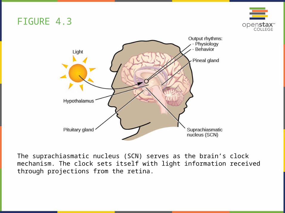
FIGURE 4.3
The suprachiasmatic nucleus (SCN) serves as the brain’s clock mechanism. The clock sets itself with light information received through projections from the retina.
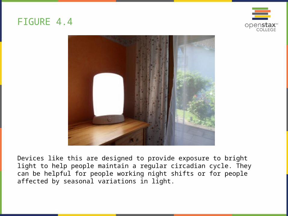
FIGURE 4.4
Devices like this are designed to provide exposure to bright light to help people maintain a regular circadian cycle. They can be helpful for people working night shifts or for people affected by seasonal variations in light.

FIGURE 4.5
This figure illustrates some of the negative consequences of sleep deprivation. While cognitive deficits may be the most obvious, many body systems are negatively impacted by lack of sleep. (credit: modification of work by Mikael Häggström)

FIGURE 4.6
This is a segment of a polysonograph (PSG), a recording of several physical variables during sleep. The x-axis shows passage of time in seconds; this record includes 30 seconds of data. The location of the sets of electrode that produced each signal is labeled on the y-axis. The red box encompasses EEG output, and the waveforms are characteristic of a specific stage of sleep. Other curves show other sleep-related data, such as body temperature, muscle activity, and heartbeat.

FIGURE 4.7
The pineal and pituitary glands secrete a number of hormones during sleep.
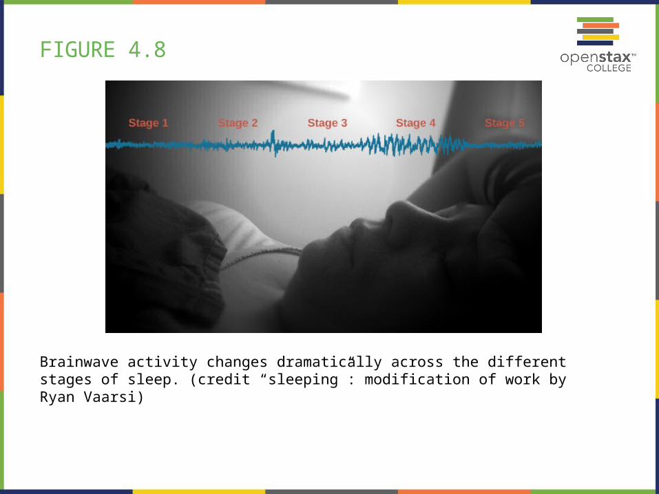
FIGURE 4.8
Brainwave activity changes dramatically across the different stages of sleep. (credit “sleeping”: modification of work by Ryan Vaarsi)

FIGURE 4.9
Brainwave activity changes dramatically across the different stages of sleep.
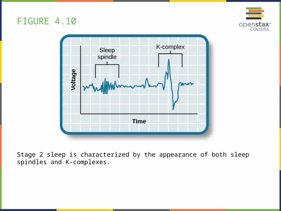
FIGURE 4.10
Stage 2 sleep is characterized by the appearance of both sleep spindles and K-complexes.

FIGURE 4.11
(a) Delta waves, which are low frequency and high amplitude, characterize
(b) low-wave stage 3 and stage 4 sleep.

FIGURE 4.12
(a) A period of rapid eye movement is marked by the short red line segment. The brain waves associated with REM sleep, outlined in the red box in (a), look very similar to those seen (b) during wakefulness.

FIGURE 4.13
A hypnogram is a diagram of the stages of sleep as they occur during a period of sleep. This hypnogram illustrates how an individual moves through the various stages of sleep.

FIGURE 4.14
(a) A typical CPAP device used in the treatment of sleep apnea is (b) affixed to the head with straps, and a mask that covers the nose and mouth.

FIGURE 4.15
The Safe to Sleep campaign educates the public about how to minimize risk factors associated with SIDS. This campaign is sponsored in part by the National Institute of Child Health and Human Development.

FIGURE 4.16
This figure illustrates various drug categories and overlap among them. (credit: modification of work by Derrick Snider)

FIGURE 4.17
The GABA-gated chloride (Cl-) channel is embedded in the cell membrane of certain neurons. The channel has multiple receptor sites where alcohol, barbiturates, and benzodiazepines bind to exert their effects. The binding of these molecules opens the chloride channel, allowing negatively-charged chloride ions (Cl-) into the neuron's cell body. Changing its charge in a negative direction pushes the neuron away from firing; thus, activating a GABA neuron has a quieting effect on the brain.

FIGURE 4.18
Crack rocks like these are smoked to achieve a high. Compared with other routes of administration, smoking a drug allows it to enter the brain more rapidly, which can often enhance the user’s experience. (credit: modification of work by U.S. Department of Justice)
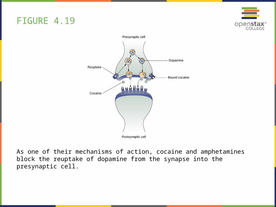
FIGURE 4.19
As one of their mechanisms of action, cocaine and amphetamines block the reuptake of dopamine from the synapse into the presynaptic cell.

FIGURE 4.20
(a) Common paraphernalia for heroin preparation and use are shown here in a needle exchange kit.
(b) Heroin is cooked on a spoon over a candle. (credit a: modification of work by Todd Huffman)

FIGURE 4.21
Psychedelic images like these are often associated with hallucinogenic compounds. (credit: modification of work by “new 1lluminati”/Flickr)

FIGURE 4.22
Medical marijuana shops are becoming more and more common in the United States.(credit: Laurie Avocado)

FIGURE 4.23
Popular portrayals of hypnosis have led to some widely-held misconceptions.

FIGURE 4.24
(a) This is a statue of a meditating Buddha, representing one of the many religious traditions of which meditation plays a part.
(b) People practicing meditation may experience an alternate state of consciousness. (credit a: modification of work by Jim Epler; credit b: modification of work by Caleb Roenigk)