stanford chronic pain management resident rotation syllabus
TRANSCRIPT

1 | P a g e S t a n f o r d C h r o n i c P a i n M a n a g e m e n t R e s i d e n t R o t a t i o n S y l l a b u s
Stanford Chronic Pain Management
Resident Rotation Syllabus Version 2009-10 Updated December 1, 2010
http://paincenter.stanford.edu
Contents
Pain Management Faculty .................................................................................................................................... 3
Volunteer Clinical Faculty .................................................................................................................................. 3
Goals & Objectives ............................................................................................................................................... 3
Patient Care ...................................................................................................................................................... 3
Medical Knowledge ........................................................................................................................................... 4
Practice-Based Learning & Improvement ......................................................................................................... 4
Interpersonal & Communication Skills .............................................................................................................. 5
Professionalism ................................................................................................................................................. 5
Systems-Based Practice ................................................................................................................................... 5
Procedural Rotation .......................................................................................................................................... 5
Patient Care ................................................................................................................................................... 6
Medical Knowledge ....................................................................................................................................... 6
Practice-Based Learning & Improvement ...................................................................................................... 6
Interpersonal & Communication Skills ........................................................................................................... 6
Professionalism ............................................................................................................................................. 7
Systems-Based Practice ............................................................................................................................... 7
Pathophysiology ................................................................................................................................................... 7
Cancer-related Pain .............................................................................................................................................. 7
Neuropathy ........................................................................................................................................................ 7
Metastatic cancer-induced bone pain ............................................................................................................... 8
Directly tumor-related pain ................................................................................................................................ 8
Chemical ........................................................................................................................................................... 8
Chronic Pain States .............................................................................................................................................. 8

2 | P a g e S t a n f o r d C h r o n i c P a i n M a n a g e m e n t R e s i d e n t R o t a t i o n S y l l a b u s
Acute and Chronic Neck and Low Back Pain .................................................................................................... 8
Neuropathic Pain States ................................................................................................................................... 8
Complex Regional Pain Syndromes, Types 1 & 2 ......................................................................................... 8
Postherpetic Neuralgia .................................................................................................................................. 9
Phantom Limb Pain ....................................................................................................................................... 9
Peripheral Neuropathies (e.g., Diabetic Neuropathy) .................................................................................. 10
Somatic Pain Conditions ................................................................................................................................. 10
Facet Arthropathy ........................................................................................................................................ 10
Myofascial Pain: .......................................................................................................................................... 19
Fibromyalgia ................................................................................................................................................ 20
Further Reading: .......................................................................................................................................... 31
Treatment ........................................................................................................................................................... 31
Cancer-Related Pain .......................................................................................................................................... 31
Cancer Bone Pain ........................................................................................................................................... 31
Spinal Anesthesia ........................................................................................................................................... 34
Neurolysis ....................................................................................................................................................... 37
World Health Organization (WHO) 3-step Analgesic Ladder (1984 ................................................................ 40
Chronic Pain (Non-Cancer Related) ................................................................................................................... 43
Opioid Analgesics – Incl. Tolerance & Addiction ............................................................................................. 43
Anticonvulsants ............................................................................................................................................... 43
Muscle Relaxants for Back Spasms ................................................................................................................ 43
Epidural Analgesia .......................................................................................................................................... 44
Peripheral Nerve Blocks for Chronic Pain ....................................................................................................... 48
Sympathetic Nerve Blocks .............................................................................................................................. 51
Transcutaneous Nerve Stimulation ................................................................................................................. 55
Spinal Cord Stimulation ................................................................................................................................... 56
Radiofrequency ............................................................................................................................................... 59
Other General Components ................................................................................................................................ 61
Physical Examination ...................................................................................................................................... 61
Outcomes in Pain Medicine ............................................................................................................................ 63
Radiation Risk and Interventional Pain Procedures ........................................................................................ 71

3 | P a g e S t a n f o r d C h r o n i c P a i n M a n a g e m e n t R e s i d e n t R o t a t i o n S y l l a b u s
Pain Management Faculty Sean Mackey, M.D., Ph.D., Chief, Division of Pain Management, Associate Professor Raymond Gaeta, M.D., Associate Professor, Medical Director, Pain Management Center Ravi Prasad, Ph.D., Clinical Assistant Professor, Assistant Director, Division of Pain Management Wendye Robbins, M.D., Clinical Assistant Professor Ian Carroll, M.D., Clinical Instructor Kale Wedemeyer, M.D., Clinical Instructor Vanila Singh, M.D., Clinical Assistant Professor Josh Kirz, Ph.D., Clinical Assistant Professor Tim Dawson, M.D., Clinical Assistant Professor Alpana Gowda, M.D., Clinical Instructor Meredith Barad, M.D., Clinical Instructor Mike Brook, M.D., Clinical Assistant Professor Michael Leong, M.D., Clinical Assistant Professor Einar Ottestad, M.D., Clinical Instructor Stephen Coleman, M.D., Clinical Assistant Professor Jiang-Ti Kong, M.D., Clinical Instructor
VOLUNTEER CLINICAL FACULTY William Brose, M.D. Steven Feinberg, M.D. John Massey, M.D. Annu Navani, M.D. Peter Abaci, M.D. Rebecca Posner, M.D.
Goals & Objectives
Sean Mackey, MD, PhD
The Pain Management Center clinic rotation is intended to provide opportunities for new patient evaluation and a continuity of care. The resident who is then supervised by one of the attending physicians evaluates patients with assorted chronic painful conditions. Clinic is held Monday through Friday during which exposure to the full range of chronic painful conditions is achieved. More complex cases are reviewed every Friday in an interdisciplinary case conference.
PATIENT CARE

4 | P a g e S t a n f o r d C h r o n i c P a i n M a n a g e m e n t R e s i d e n t R o t a t i o n S y l l a b u s
Goal: To gain an understanding of the domains of gathering history and physical information, making differential diagnoses, assimilation of clinical data from multidisciplinary team members for the purpose of creating an integrated treatment plan, and implementing the treatment plan.
Objectives: Upon completing this rotation, residents should understand
How to elicit a directed neurological history and perform a detailed neurological exam
Basic neuroimaging with an ability to identify significant findings
How to perform a comprehensive musculoskeletal examination with emphasis on both structure and function as it applies to diagnosing acute and chronic pain problems
How to assess for psychiatric and psychological comorbidities (e.g., chemical dependency issues, somatoform disorders, mood disturbances, personality disorders, etc.)
The role of psychological and psychiatric treatment
How to create and implement complex treatment plans across a variety of disciplines
The common obstacles that often interfere with successful implementation of treatment plans and how to overcome them
MEDICAL KNOWLEDGE Goal: To acquire a comprehensive understanding of the essential basic and applied medical and social sciences as they relate to management of pain conditions in an outpatient continuity of care clinic.
Objectives: Upon completing this rotation, residents should understand
The anatomy, physiology, and pharmacology of pain transmission and modulation
The role of opioid and non-opioid medications for treatment of pain conditions
The role of surgical and interventional treatment modalities for various pain conditions
The role of functional, vocational, and psychological treatment modalities for various pain conditions
The use of various strategies and techniques as a means of reducing the impact of pain on a patient’s life while concurrently improving functional capacity and quality of life
PRACTICE-BASED LEARNING & IMPROVEMENT Goal: To be able to perform self-assessments; retrieve, understand, and apply scientific evidence related to the practice of pain medicine; and make meaningful contributions to the education of others.
Objectives: Upon completing this rotation, residents should be able to
Identify their personal strengths and weaknesses and how this affects their practice

5 | P a g e S t a n f o r d C h r o n i c P a i n M a n a g e m e n t R e s i d e n t R o t a t i o n S y l l a b u s
Incorporate feedback from performance evaluations into daily practice
Utilize hospital and university information technology systems to optimize learning
Actively contribute to the education of patients, families, students, and other health professionals
INTERPERSONAL & COMMUNICATION SKILLS Goal: To be able to demonstrate strong interpersonal and communication (verbal and written) skills that result in effective exchange of information with patients, families, and other health professionals.
Objectives: Upon completing this rotation, residents should be able to
Provide concise yet comprehensive communication with other health professionals
Communicate effectively with patients, family members, and the general public across broad socio-economic and cultural domains
Work collaboratively with other physicians and health professionals from other disciplines
Maintain appropriate, timely, and accurate medical records
PROFESSIONALISM Goal: To strictly adhere to ethical principles in all aspects of practice.
Objectives: Upon completing this rotation, residents should be able to
Demonstrate compassion and respect for patients, families, and other health care professionals
Have a pervasive sense of personal integrity
Demonstrate sensitivity to individuals of diverse socio-economic and cultural backgrounds
SYSTEMS-BASED PRACTICE Goal: To understand how the practice of pain medicine is a part of a larger context of health care organization and to be able to utilize resources from the larger system to optimize patient care.
Objectives: Upon completing this rotation, residents should be able to
Be proficient with cost analysis and cost containment as it relates to patient care
Consistently employ quality assurance and improvement techniques in professional work
Work collaboratively with others to improve health care procedures and systems to positively impact patient care
PROCEDURAL ROTATION

6 | P a g e S t a n f o r d C h r o n i c P a i n M a n a g e m e n t R e s i d e n t R o t a t i o n S y l l a b u s
This aspect of the chronic pain rotation provides experience in the interventional management of pain including neural blockade, joint injection, and interventional spine therapies. Attention to the preoperative assessment for indications, alternative therapies, side effects and expected outcome are stressed.
PATIENT CARE Goal: To master the skills requisite to perform interventional pain management procedures.
Objectives: Upon completing this rotation, residents should understand
How to conduct a thorough preoperative evaluation
The appropriateness of various interventions among varying pain populations
The common complications that can occur with interventional procedures and how to address them
MEDICAL KNOWLEDGE Goal: To acquire a comprehensive understanding of the essential basic and applied medical and social sciences as they relate to interventional pain management procedures.
Objectives: Upon completing this rotation, residents should understand
The anatomy, physiology, and pharmacology of pain transmission and modulation
The safe use of fluoroscopy for the delivery of interventional blockade
The surgical concepts around the implantation of spinal cord stimulators and intrathecal medication delivery systems
PRACTICE-BASED LEARNING & IMPROVEMENT Goal: To be able to perform self-assessments; retrieve, understand, and apply scientific evidence related to the practice of pain medicine; and make meaningful contributions to the education of others.
Objectives: Upon completing this rotation, residents should be able to
Identify their personal strengths and weaknesses and how this affects their practice
Incorporate feedback from performance evaluations into daily practice
Utilize hospital and university information technology systems to optimize learning
Actively contribute to the education of patients, families, students, and other health professionals
INTERPERSONAL & COMMUNICATION SKILLS Goal: To be able to demonstrate strong interpersonal and communication (verbal and written) skills that result in effective exchange of information with patients, families, and other health professionals.

7 | P a g e S t a n f o r d C h r o n i c P a i n M a n a g e m e n t R e s i d e n t R o t a t i o n S y l l a b u s
Objectives: Upon completing this rotation, residents should be able to
Provide appropriate communication with OR nursing staff to ensure the highest quality of care for the patient
Communicate effectively with patients, family members, and the general public across broad socio-economic and cultural domains
Work collaboratively with other physicians and health professionals from other disciplines
Maintain appropriate, timely, and accurate medical records
PROFESSIONALISM Goal: To strictly adhere to ethical principles in all aspects of practice.
Objectives: Upon completing this rotation, residents should be able to
Demonstrate compassion and respect for patients, families, and other health care professionals
Have a pervasive sense of personal integrity
Demonstrate sensitivity to individuals of diverse socio-economic and cultural backgrounds
SYSTEMS-BASED PRACTICE Goal: To understand how the practice of pain medicine is a part of a larger context of health care organization and to be able to utilize resources from the larger system to optimize patient care.
Objectives: Upon completing this rotation, residents should be able to
Be proficient with cost analysis and cost containment as it relates to patient care
Consistently employ quality assurance and improvement techniques in professional work
Work collaboratively with others to improve health care procedures and systems to positively impact patient care
Pathophysiology
Cancer-related Pain Theresa Mallick-Searle, NP
NEUROPATHY Chemotherapy associated neuropathy arises due to different mechanisms, including disruption of tubulin function by chemotherapeutic agents w/release of cytokines, resulting degeneration of sensory neurons and sensitization of primary nociceptive afferents.

8 | P a g e S t a n f o r d C h r o n i c P a i n M a n a g e m e n t R e s i d e n t R o t a t i o n S y l l a b u s
Radiotherapy can cause tissue fibrosis with nerve compression and microvascular obstruction of the nerve.
Nervous tissue compression or lesion contributes to central sensitization.
METASTATIC CANCER-INDUCED BONE PAIN Injury or infiltration of sensory neurons that innervate the bone marrow cause pain.
Alterations in normal bone turnover occur, with loss of mechanisms that normally regulate the balance between osteoclast and osteoblast activity.
With advanced disease, the bone loses mechanical strength and is subject to osteolysis, pathological fracture, and microfractures.
Bony metastases can cause painful muscle spasm.
DIRECTLY TUMOR-RELATED PAIN Stretching of hollow visera, distortion of the capsule of solid organs, inflammation of the mucosa, and ischemia or necrosis activate visceral nociceptors, resulting in visceral pain.
Tumor infiltration in nerve plexuses and damage to nerve tissue can cause neuropathic pain.
Cancer cells can cause invasion of mechanically sensitive tissues (e.g visceral pain) or entrapment and injury of nerves (e.g. neuropathic pain).
CHEMICAL Tumors themselves secrete inflammatory and prohyperalgesic mediators.
Local and systemic inflammatory response, with production of pro-inflammatory cytokines, which facilitate pain transmission.
Tumors contain immune system cells that release factors including endothelin, prostaglandins, and TNF-α, which excite or sensitize peripheral nociceptive primary afferents.
Chronic Pain States
ACUTE AND CHRONIC NECK AND LOW BACK PAIN
Alpana Gowda, MD
Chou R, et al. Diagnosis and Treatment of Low Back Pain: A Joint Clinical Practice Guideline from the American College of Physicians and the American Pain Society. Annals of Internal Medicine. 2007;147:478-491. PDF
NEUROPATHIC PAIN STATES
COMPLEX REGIONAL PAIN SYNDROMES, TYPES 1 & 2 Sean Mackey, MD, PhD

9 | P a g e S t a n f o r d C h r o n i c P a i n M a n a g e m e n t R e s i d e n t R o t a t i o n S y l l a b u s
Mackey S, Feinberg S. Pharmacologic Therapies for Complex Regional Pain Syndrome. Curr Pain Headache Reports. 2007;11:38-43 PDF
Harden R, Bruehl S. Diagnosis of Complex Regional Pain Syndrome. Clin J Pain. 2006;22:415-419. PDF
POSTHERPETIC NEURALGIA Wendye Robbins, MD
Basbaum A, Julius D, Robbins W. Vanilloids. In: Bountra C, Munglani R, Schmidt W, eds. Pain: Current Understanding, Emerging Therapies, and Novel Approaches to Drug Discovery. New York, NY: Marcel Dekker; 2003:531-538. PDF
PHANTOM LIMB PAIN Meredith Barad, MD
Incidence 60-80% of patients experience phantom limb pain almost immediately after the loss of a limb.
Onset: 75% of phantom pain patient experience onset within the first week while 25% of in the weeks, and rarely in the years, following.
Duration: Usually fades in days to weeks, but in 30% of patients can persist for years.
Quality: Episodic pain with multiple daily events lasting seconds to minutes. The pain has been reported as stabbing, shooting, boring, burning, like the limb is stuck in a block of ice. This pain is not to be confused with stump pain which is often due to a scar neuroma and present in 10-25% of amputees.
Phantom Sensations: NOT PAIN. This is often described as the feeling that the limb is still present, usually more vivid in the most distal part of the extremity which may reflect the larger cortical representation of the more distal part of limb. The most common sensation is telescoping. In 50% of cases involving the upper limb, the arm will get progressively shorter until the patient is left with the hand alone dangling from the stump. These limbs can telescope sometimes at will. Paresthesias have also been reported.
Risk Factors: pre-amputation pain, phantom sensations.
Less frequent in children and adolescent amputees and patients with congenital limb defects. The phantom limb is not affected by the reason for surgery, the limb or the location on the limb of the amputation, sex, age, marital or socioeconomic status of the patient.
Position: The phantom usually occupies the habitual position – such as elbow partially flexed and forearm pronated. The phantom can occupy and unusual even painful position, that can sometimes be reset by movement of the stump.

10 | P a g e S t a n f o r d C h r o n i c P a i n M a n a g e m e n t R e s i d e n t R o t a t i o n S y l l a b u s
Treatment: There is a paucity of well-designed trials, but basically the same principles used for central neuropathic pain apply.
Medications: TCAs, Gabapentin, other AEDs, Opiates (consider Methadone), NMDA antagonists.
Procedures: SCS , DBS of the ventral caudal nucleus of the thalamus and MCS have all been shown to be effective in small numbers of patients Non-pharmacologic therapy: Mirror treatment, desensitization
Peri-operative management: Epidural (these patients have been shown to have considerably less pain at 6months) Nikolajsen et al. 1997. Consider preoperative gabapentin.
References: F. Cervero, T.S. Jensen, Editors. Handbook of Clinical Neurology, Vol. 81 (3rd series). Chapter 45. Pain. 2006
Nikolajsen L, Ilkjaer S, Christensen JH, Kroner K, Jensen TS. Randomised trial of epidural bupivacaine and morphine in prevention of stump and phantom pain in lower-limb amputation. Lancet 350: 1353–1357.
Katayama Y et al. Motor cortex stimulation for phantom limb pain: comprehensive therapy with spinal cord and thalamic stimulation. Stereotact Funct Neurosurg. 2001;77(1-4):159-62.
PERIPHERAL NEUROPATHIES (E.G., DIABETIC NEUROPATHY) Ian Carroll, MD, MS
Hocking G, Cousins M. Ketamine in Chronic Pain Management: An Evidence-Based Review. Anesth Analg. 2003;97:1730-9. PDF
SOMATIC PAIN CONDITIONS
FACET ARTHROPATHY Jiang-Ti Kong, MD
Anatomy and innervations The basic anatomical unit of the spine is consisted of three joints: the intervertebral disc, and the paired zygapophysial (or facet) joints (Fig 1a). The three joints function together to support and stabilize the spine and limit motion in all planes.[1] The upper lumbar facets (L2-3, L3-4) are in a more sagittal plane than coronal; thus protecting the spine from excessive axial rotation and lateral bending. The lower lumbar facets (L4-5, L5-S1) are just the opposite, with a more coronal orientation.
As people age, the overall orientation of the lumbar facets becomes more sagittal, the consequence of which is less protection from the shearing force caused by flexion and extension.[2] The orientation of

11 | P a g e S t a n f o r d C h r o n i c P a i n M a n a g e m e n t R e s i d e n t R o t a t i o n S y l l a b u s
the individual facet may also differ at the same spinal level, referred to as tropism. Tropism is positively associated with disc degeneration and herniation.[3] Furthermore, as the discs begin to degenerate and shrink in height, more loads are transmitted to the facet joints, eventually leading to their degeneration as well.
The zygapophysial joints are true synovial joints, consisted of the superior and the inferior articulating process (SAP and IAP), from the spinal level below and above; and the joint capsule.[4] The facet joints are innervated by medial branch nerves, which are branches of the dorsal primary rami of the spinal nerves (Fig 1b). Each medial branch sends out a proximal ascending and a distal descending branch. The former innervates the caudal aspect of the facet, while the latter innervates the superior and medial aspects of the facet at a level below. Hence, each facet joint is innervated by two medial branches, one from the same level, and one from a level above.
Clinical Presentation and Diagnosis Lumbar facet pain often refers to the low back, buttock, and thigh. Less commonly it may involve the groin and the flank (Fig 2), along with diffuse crampy leg pain and stiffness. Patients with facet arthropathy often display the following signs: 1) Paraspinal tenderness; 2) pain on facet joint loading maneuvers (axial rotation, lateral bending, flexion /extension); 3)Absence of signs of nerve root irritation; 4) Non-radiating back ,+/- hip, +/- buttock pain on straight leg raising.[4]
The diagnosis of lumbar facet arthropathy is not intuitive. Even though many signs and symptoms are ascribed to facet arthropathy, multiple large, randomized, well-controlled trials showed that NO historic or physical examination findings can reliably diagnose facet arthropathy.[5] Furthermore, the evidence in the literature does not support the use of any radiographic imaging (x-ray, CT, or MRI) to diagnose facet pain.[6] In fact, the agreed method to diagnose lumbar facet arthropathy is response to facet blocks: intra-articular or medial branch blocks (MBB). Although both blocks are frequently used, the evidence supporting the use of MBB is much stronger as judged by quality and number of clinical trials.[1]
Medial branch blocks are performed using fluoroscopy, with the x-ray beam 10-40 degrees ipslateral oblique.[7] The target is usually taken at the junction between the transverse process and the SAP (Fig 3). Half a milliliter to 1mL of local anesthetic plus 10-20mg of methylprednisolone is injected to each medial branch via a 22G or 25g needle. The pain relief from MBB comes in two phases: early phase which happens within ½ hour due to the local anesthetic, and a late phase which takes place within one week of the injection. The duration of the relief is often limited, roughly about one month. Given the high false positive rate of MBB (25-40%), two consecutive positive responses to MBB are required before proceeding to radiofrequency ablation (RFA) of the medial branches.
Treatment A multimodal approach to the treatment of facet arthropathy is recommended.[1] It involves physical therapy, medications, and psychological interventions. Tailored exercise programs combined with yoga have been shown to reduce pain and prevent relapses in patients with chronic low back pain. In terms of medications, NSAIDS, and the judicious use of antidepressants and muscle relaxants are advocated. Untreated psychiatric disorders including PTSD, depression, anxiety and substance abuse adversely

12 | P a g e S t a n f o r d C h r o n i c P a i n M a n a g e m e n t R e s i d e n t R o t a t i o n S y l l a b u s
affect the outcomes of patients with chronic low back pain. It is therefore critical to address these disorders while treating pain.
Evidence from literature supports radiofrequency ablation (RFA) of medial branches (MB) as the interventional treatment of choice for facet arthropathy.[1] The fluoroscopic technique for RFA is identical to that of MBB.[8] However, rather than injecting medications, it uses radiofrequency waves to denervate the medial branch. Before the ablation, the location of the needle is verified by sensory stimulation at 50Hz. If the probe is sufficiently close to the medial branch, the patient should feel the stimulation at less than 1V (some uses 0.5V). Next, motor stimulation at 2Hz is performed up to 2-2.5V, at which point only the multifidus (paraspinal) muscles are supposed to contract, and NOT the muscles in the girdles and /or limbs.
Two types of RFA are performed: continuous and pulsed. The continuous technique was the original technique used for RFA since the 1970’s. It works by heating tissues to 60-100oC, which causes near instantaneous coagulation necrosis of the medial branch, essentially a heat neurectomy. In contrast, the pulse RFA delivers bursts of radiofrequency waves at 42oC for 20ms with 480ms silence time in between, without causing neuronal death. It is thought that the pulsed RF works by using a shifting electromagnetic field around the medial branch to modify pain processing mechanisms at the spinal and supraspinal level. Direct head-to-head comparison of continuous vs. pulsed RF showed that both method work to reduce pain. However, there is significant difference in the duration of analgesia: the continuous RF results in up to one year of pain reduction, while the pulsed RF ≤ 4 months. The recommendation is thus continuous RF for lumbar medial branch ablation, and reserve pulsed RFA for neuropathic type of conditions mediated by peripheral nerves.[1]
Cautions and risks: radiofrequency ablation procedures are in general safe. The most common side effects from RFA is reactive flaring of pain right after the ablation procedure, which can be treated by injecting steroid via the needle immediately after ablation.[4] The administration of steroid itself carries certain risks, including suppression of the hypothalamic-pituitary-adrenal axis up to 4 weeks, impaired insulin sensitivity up to 1 week, as well as increased risk for infection. If appropriate sensory and motor tests are performed, the risk of injuring other nerves is minimal.
Cervical facet syndrome: The pathophysiology, presentation, diagnosis and treatment of cervical facet arthropathy largely mirror that of the lumbar facet disease, with the following exceptions: [4]
1. Presentation: The cervical facets refer pain to the occiput, neck, shoulder and upper arm (Fig 4). Typical signs of cervical facet disease include decreased range of motion in cervical spine, local tenderness over spinal process and affected facets, and pain upon extension and ipsilateral bending (i.e. facet loading maneuver). Of particular mention is the third occipital nerve, the medial branch of C3, which innervates the C2-3 facet. C2-3 arthropathy often contributes to the development of various headaches that involves the occiput.
2. Diagnosis: cervical facet joint injections are NOT recommended due to technical difficulties, the increased risk of injuring the vertebral arteries and exiting nerves, as well as the lack of positive clinical trials. Cervical MBB is the recommended technique for diagnosis and short term treatment of facet disease. It may be done via either an anterior-posterior or lateral approach (Fig 5a and 5b).

13 | P a g e S t a n f o r d C h r o n i c P a i n M a n a g e m e n t R e s i d e n t R o t a t i o n S y l l a b u s
3. Treatment: radiofrequency ablation is again the interventional treatment of choice. The efficacy of the traditional continuous RFA was shown to last between 9 and 12 months by two well controlled, randomized, prospective trials.
Conclusion Facet arthropathy is a common etiology that leads to axial neck and low back pain. It is diagnosed via medial branch blocks. The definitive interventional treatment is continuous RFA of the medial branches. It is also important to approach patients with facet disease via a multimodal treatment approach in addition to the invasive procedures to prevent relapses.
1. Cohen, S.P. and S.N. Raja, Pathogenesis, diagnosis, and treatment of lumbar zygapophysial (facet) joint pain. Anesthesiology, 2007. 106(3): p. 591-614.
2. Boden, S.D., et al., Orientation of the lumbar facet joints: association with degenerative disc disease. J Bone Joint Surg Am, 1996. 78(3): p. 403-11.
3. Vanharanta, H., et al., The relationship of facet tropism to degenerative disc disease. Spine (Phila Pa 1976), 1993. 18(8): p. 1000-5.
4. Clemans, R.R., Benzon, H.T., Facet Syndrome: Facet Joint Injections and Facet Nerve Blocks, in Essentials of Pain Medicine and Regional Anesthesia, S. Benzon, Molloy, Liu, Fishman, Editor. 2005. p. 341-347.
5. Revel, M., et al., Capacity of the clinical picture to characterize low back pain relieved by facet joint anesthesia. Proposed criteria to identify patients with painful facet joints. Spine (Phila Pa 1976), 1998. 23(18): p. 1972-6; discussion 1977.
6. Cohen, S.P., et al., Clinical predictors of success and failure for lumbar facet radiofrequency denervation. Clin J Pain, 2007. 23(1): p. 45-52.
7. Czervionke, L.F., Fenton, D.S., Facet Joint Injection and Medial Branch Block, in Image-Guided Spine Intervention, a.Z. Fenton, Editor. 2003, Saunders: Philadelphia, PA.
8. Fenton, D.S., Czervionke, L.F., Facet Denervation, in Image-Guided Spine Intervention, a.Z. Fenton, Editor. 2003, Saunders: Philadelphia, PA.

14 |
P a g e SS t a n f o r d C h r o n i c
Fig
Figure 1
c P a i n M
ure 1a. Ana
1b. Anatomy
M a n a g e m
atomy of Lum
y of Spinal M
e n t R e s
mbar Facets
Medial Branc
i d e n t R o
ches
o t a t i o n SS y l l a b u s

15 |
F
P a g e S
igure 2: Repatte
S t a n f o r d
ferral Pattererns extend
C h r o n i c
rns of Lumbafrom the da
Ad
c P a i n M
ar Facet Artharkest (low b
dapted from C
M a n a g e m
hropathy. In back) to the
Cohen and R
e n t R e s
descendinge lightest re
Raja 2007.
i d e n t R o
g order, the megions (flan
o t a t i o n S
most commok and foot)
S y l l a b u s
on referral .

16 |
Figujunctafter
P a g e S
ure 3. Radiotion betweenr aligning the
S t a n f o r d
ography for Ln the superioe endplates
C h r o n i c
Lumbar Medor articulatinat L4-5, and
c P a i n M
dial Branch Bng process (Sd oblique the
M a n a g e m
Blocks at LefSAP), and tr
e beam towa
e n t R e s
ft L4, L5, alaransverse pr
ard the left ab
i d e n t R o
a. Notice therocess (TP).bout 10-20 d
o t a t i o n S
e Target is a This pictur
degrees.
S y l l a b u s
at the re is taken

17 |
P a g e S
Figure 4.
S t a n f o r d
. Referral P
C h r o n i c
atterns for C
c P a i n M
Cervical Fac
M a n a g e m
et Arthropat
e n t R e s
thy, Adapted
i d e n t R o
d from www.w
o t a t i o n S
weblockpain
S y l l a b u s
n.com

18 |
P a g e S
Figur
F
S t a n f o r d
e 5a. Cervic
Figure 5b. C
C h r o n i c
cal MBB App
Cervical MBB
c P a i n M
proached by
B Approache
M a n a g e m
Anterior-Po
ed via Latera
e n t R e s
osterior Orien
al Orientatio
i d e n t R o
ntation of the
on of the Fluo
o t a t i o n S
e Fluoroscop
oroscope
S y l l a b u s
pe

19 | P a g e S t a n f o r d C h r o n i c P a i n M a n a g e m e n t R e s i d e n t R o t a t i o n S y l l a b u s
MYOFASCIAL PAIN: Jiang-Ti Kong, MD
Definition and Diagnosis: Myofacial pain (MP) is local and referred pain that arises from myofascial trigger points. Trigger points are defined as localized sensitive areas in muscles that contain palpable, taut bands of muscles.[1] Palpation of trigger points often reproduces the patient’s pain which often radiates and refers. Myofascial pain differs from fibromyalgia in location, pathogenesis and treatment (see chapter on Fibromyalgia).
MP is quite common and appears in up to 30% of patients who presents to orthopedic or general medical clinics. It tends to affect women slightly more often than man.
Differential Diagnosis: MP is a relatively common and benign disorder, which can be confused with other more serious local and systemic diseases.[1] It is therefore important to rule out these other disorders before making the diagnosis of myofascial pain. Local disorders that may mimic MP include arthritis, bursitis, fasciitis, tendinopathy, neuropathy, and referred visceral pain. Systemic disorders include endocrine disorders (hypothyroidism, Cushing’s disease and hyperparathyoroidism); infections (hepatitis C, lyme disease etc); and inflammatory disorders (RA, SLE, PMR, Sjogren’s and myositis).
Pathogenesis: Myofascial pain often results from acute injury, or, more commonly repetitive strain from abnormal posture and non-ergonomic body mechanics. These injuries can be acute or chronic. In some cases no obvious causes are found.
Although the details are not entirely clear, a generally accepted concept is that pathologic increase in acetylcholine release by motor endplates leads to prolonged muscle contraction, strain, and ischemia.[2] Ischemia in turn results in release of local vasoactive mediators which then further increase acetylcholine release, creating a positive feedback loop for myofacial pain. This model also explains why botulinum toxin (Botox), which irreversibly blocks the release of acetylcholine, is helpful in treating MP (see below).
Treatments: 1. Non-invasive physical modalities: The cornerstone of all treatment approaches to myofascial
pain is slow and sustained muscle stretching exercises.[1] Paradoxically, the contracted, painful muscle also tends to be weakened, hence requiring a strengthening program as well. The literature supports herapeutic exercises for the neck, low back and knee pain. Other modalities, such as TENS and relaxation therapy, are also useful if indicated
2. Injection therapies: Both trigger point injections and dry needling appear to be helpful for MP, although data suggests injection therapy results in more improvement than dry needling.[3] The medication used for injection, i.e. saline, lidocaine, bupivacaine, or botox did not seem to matter per several previous trials. However, recent data have shown that Botox may be more efficacious than the others.[4, 5]

20 | P a g e S t a n f o r d C h r o n i c P a i n M a n a g e m e n t R e s i d e n t R o t a t i o n S y l l a b u s
3. Medications: Few randomized controlled trials exist that evaluate the efficacy of pharmacotherapy for MP, partly because this order is more commonly treated by physical modalities and injections. Based on trials in patients with low back pain, arthritis and tension headaches, NSAIDS, tricyclic antidepressants (if insomnia and depression), and tizanidine may be helpful as adjuvants in treating MP.[1] There is some data supporting tramadol, which is not the author’s choice due to potential for addiction and interaction with other antidepressants (may cause seizure and serotonin syndrome).
Outcome summary: Myofascial pain deserves the attention of the physician because treatment outcome is often not ideal. For example, Cassisi et al found that patients with myofascial low back pain do worse than those with disc herniation.[6] They tend to harbor inaccurate beliefs about their pain condition and pessimism regarding their treatment. Therefore, a multidisciplinary program is often necessary to treat patients with MP, addressing emotional, ergonomic, rehabilitational and pharmacologic aspects of their disease.
1. Molloy, R., Myofascial Pain Syndrome, in Essentials of Pain Medicine and Regional Anesthesia, R. Benzon, Molloy, Liu, Fishman, Editor. 2005, Elsevier.
2. Rivner, M.H., The neurophysiology of myofascial pain syndrome. Curr Pain Headache Rep, 2001. 5(5): p. 432-40.
3. Ho, K.Y. and K.H. Tan, Botulinum toxin A for myofascial trigger point injection: a qualitative systematic review. Eur J Pain, 2007. 11(5): p. 519-27.
4. Colhado, O.C., M. Boeing, and L.B. Ortega, Botulinum toxin in pain treatment. Rev Bras Anestesiol, 2009. 59(3): p. 366-81.
5. Gul, K. and S.A. Onal, [Comparison of non-invasive and invasive techniques in the treatment of patients with myofascial pain syndrome.]. Agri, 2009. 21(3): p. 104-12.
6. Cassisi, J.E., et al., Pain, disability, and psychological functioning in chronic low back pain subgroups: myofascial versus herniated disc syndrome. Neurosurgery, 1993. 33(3): p. 379-85; discussion 385-6.
FIBROMYALGIA Jiang-Ti Kong, MD
I. Diagnosis and Epidemiology:
a. Diagnosis: The diagnosis of fibromyalgia (FM) is clinical and includes the follow two elements as required by the College of American Rheumatology:[1]
i. Chronic widespread pain (CWP) of at least 3 months’ duration, present above and below the diaphragm, on both sides of the body, plus axial pain.

21 | P a g e S t a n f o r d C h r o n i c P a i n M a n a g e m e n t R e s i d e n t R o t a t i o n S y l l a b u s
ii. Tenderness in at least 11/18 characteristic locations (Fig 1) when 4 kg/cm^2 is applied (roughly the pressure that results in the blanching of the nail bed).
iii. It differs from myofascial pain, which is local (Table 1)
b. Epidemiology: The prevalence of fibromyalgia is estimated to be between 0.5 to 5% of the population. There does not seem to be a race, national, or ethnic bias. However, it tends to occur in women between the ages of 20 and 50 years. The female to male ratio is 10:1.[1]
II. Associated Symptoms:
In addition to the CWP and tenderness, fibromyalgia is often associated with several symptoms including fatigue, sleep disturbance, mood disorders, and discognition.[2]
Several large patient survey studies from the US and Germany (all published in 2008) ranked fatigue at the top of the list of patient-perceived major manifestations of fibromyalgia (Table 2), second only to pain and stiffness. Non-restorative sleep (NRS) is the second major complaint of FM patients. It was initially thought secondary to alpha intrusion into the delta rhythm of sleep, which prevents the progression into the restorative stages 3 and 4 of Non-REM sleep (aka slow wave sleep). Mood disorders, particularly depression, anxiety, and PTSD are also present in up to 74% of patients with FM. Lastly, cognitive dysfunction (referred to as “fibro-fog” by some) is common in FM patients. It manifests in areas including working memory, concentration, motivation and other executive tasks. Some estimated this overall cognitive decline to be equivalent to 20 years of aging.
Other symptoms, such as subjective joint swelling, allodynia/hyperalgia, paresthesia without obvious EMG/NCS findings, are also common in FM patients.[2]
III. Coexisting pain disorders:
Patients with FM also tend to have other chronic pain syndromes such as: irritable bowel syndrome, interstitial cystitis, TMJ, and migraines which also involve central sensitization as part of their etiology (see below). Other associated syndromes include, but not limited to: restless leg syndrome, chronic fatigue syndrome and Raynaud phenomenon.[1]
IV. Pathophysiology:
Currently, we do not have sufficient knowledge to identify the exact mechanisms leading to fibromyalgia. However, mounting evidences have suggested abnormalities in pain processing, particularly the amplification of ascending nociceptive signals, and the reduction in descending pain inhibition are involved in the development of FM.[3, 4]
a. Ascending pathway: simply speaking, the ascending nociceptive pathway include primary afferent neuron whose cell body resides in the DRG, spinal secondary neurons, thalamic tertiary neurons, and finally cortical and subcortical neurons (Fig 2). In FM, abnormally high levels of pro-nociceptive mediators are found at the primary and spinal secondary neuron synapses,

22 | P a g e S t a n f o r d C h r o n i c P a i n M a n a g e m e n t R e s i d e n t R o t a t i o n S y l l a b u s
including substance P, NGF, glutamate and prostaglandin. Their presence leads to hyperexcitability of the primary afferent neurons and abnormal transformation of spinal secondary neurons to wide dynamic range neurons. These changes are then lead to the development of hyperalgesia and allodynia, respectively.
b. Descending Pathway: it refers to the neurocircuit connecting cortical structures (somatosensory and frontal cortex) to hypothalamus, the periaquaductal gray and other midbrain/pons structures (e.g. raphe magnus), which then arrives at and modifies the inputs from the synaptic junction between the spinal secondary and primary afferent neurons. Experimental evidences have shown the concentrations of biogenic amines (5HT, DA, NE) in the CSF are decreased in FM patients. These biogenic amines are considered major neurotransmitters responsible for descending pain inhibition.
c. Additional mechanisms: neuro-endocrine abnormalities, including growth hormone deficiency (present in 1/3 of FM patients), hypothyroidism, and decreased level of cortisol were also thought related to the pathogenesis of FM via a central process.[1] However, most of these deficiencies are mild and multiple small clinical trials with hormonal replacement did not demonstrate sustainable benefit.[5] The current recommendation is therefore: no hormonal treatment unless clinically significant deficiency is present.
V. Treatment:
The treatment of fibromyalgia needs to be comprehensive, addressing the myofascial, CNS, emotional and physical aspect of this complex disease. The three cornerstones FM thus include: medications, exercise, and psychological interventions.[1]
1. Medications.
a. The FDA approved drugs: Three drugs are currently approved by the FDA for fibromyalgia: pregabalin (lyrica), duloxetine (cymbalta), and milnacipran (savella).[6] Pregabalin primarily works on the inappropriately activated ascending nociceptive pathway while duloxetine and milnacipran increase the concentration of biogenic amines to enhance the descending inhibition. For details on the mechanism, indications, dosage and major side effects of these medications, please refer to Table 3a.
b. Other medications (Table 3b):
i. A single controlled trial showed efficacy of gabapentin (neurontin) in FM related pain, function and sleep disturbance. The median effective dose 1800mg per day.[6] Gabapentin is structurally similar to pregabalin; but pregabalin does not require a protein transporter to be absorbed, is analgesic at a lower dose, and easier to titrate.
ii. Tricyclic antidepressants (amitriptyline, nortriptyline, and desipramine) inhibits the reuptake of serotonin and norepinepherine at sypapses, also helps with restorative sleep. However, their use is often limited my anticholinergic side effects.

23 | P a g e S t a n f o r d C h r o n i c P a i n M a n a g e m e n t R e s i d e n t R o t a t i o n S y l l a b u s
iii. Cyclobenzaprine, a tricyclic muscle relaxant, and sodium oxybate, a GABA precursor, were also shown to be moderately helpful.
iv. NSAIDS and opioids were found NOT to be helpful for FM. Tramadol, however, was shown to be a moderately beneficial adjunct.
c. Typical algorithms:
i. First drug selection: is based on the patient’s symptoms, as well as cost. Often a second generation tri-cyclic such as desipramine or nortriptyline is selected if the patient has significant depression. Another choice would be gabapentin if sleep is a problem. Alternatively, lyrica, cymbalta or savella may be used if financially feasible.
ii. Dose escalation: most of the above drugs are not therapeutic at the initial dose and needs to be escalated. The patient should remain at the therapeutic target dose for at least 4 weeks before deciding the drug ineffective.
iii. Drug combinations: if the first drug does not work, one may choose to wean it off and try a second one; or adding a second to the first agent. Recent data showed that the combination of nortriptyline plus gabapentin is more effect than either alone for neuropathic pain. This may be applicable to FM.
iv. Novel agents: Naltrexone 4.5mg po qhs may be used by itself or in combination with another drug. It may decrease neuro-inflammation at the glial cell level and is efficacious in certain FM patients.[7]
2. Exercise Interventions: Exercise has been proven to be beneficial to patients with fibromyalgia in multiple randomized controlled trials.[8] However, due to their altered physiology in pain processing and abnormal response to endorphins, FM patients often have increase pain and thus poor tolerance with standardized exercise regimens. Their exercise program thus needs to be tailored. Below are recommendations from a recent review article:[8]
a. Goals: low-intensity non-repetitive, low-impact, individualized programs are preferred.
b. Examples: warm water-based gentle aerobics, mixed-type exercises; walking, stationary bikes, posture and flexibility (i.e. yoga, Qigong).
c. Recommended progression: 1) breath, posture, and relaxation training; 2) flexibility; 3) strength and balance; 4) aerobics (aquatics, walk, bike etc)
d. Addressing comorbidities: treat sleep and mood disorders; diet counseling for obesity, evaluate balance and kinesthetic training for those with autonomic instability.
e. Avoid: heavy weight, excessive repetition, eccentric muscle works. Realize the average 40-year-old patient who has FM has the physical stamina of and 80-year-old without FM.
3. Psychologic Interventions:[9]

24 | P a g e S t a n f o r d C h r o n i c P a i n M a n a g e m e n t R e s i d e n t R o t a t i o n S y l l a b u s
a. Interventions with definitive evidence for efficacy:
i. Patient education: involves explaining the neuropathophysiology of fibromyalgia; the biopsychosocial model of chronic pain; and de-stigmatization of FM.
ii. CBT: cognitive therapy aims at modifying maladaptive thoughts to change affect and behavior; while behavior therapy uses the operant behavior technique to reward adaptive behavior (e.g. pacing, graded exercise) and punish maladaptive behavior (e.g. secondary gain, catastrophizing). Relaxation techniques are often utilized in CBT.
iii. Biofeedback: multiple randomized controlled trials support the use of heart-rate-biofeedback; while more data is needed to support EMG- and EEG- biofeedback.
b. Interventions with equivocal evidence for efficacy: meditation alone, and Qigong. Hydrotherapy (i.e. warm spa) was found to be helpful only short term.
c. Interventions with minimal efficacy: acupuncture, massage.
VI. Conclusion:
Fibromyalgia is a clinically diagnosed disease with chronic wide-spread pain involving bilateral muscles and joints, both above and below the diaphragm, both peripheral and axial. Affected individuals often have additional symptoms including fatigue, non-restorative sleep, stiffness, mood and cognitive dysfunction. Disregulated pain perception and inhibition are indicated in its pathogenesis. Its treatment demands a multidisciplinary approach, including medications, tailored physical therapy, and psychological interventions.

25 |
P a g e SS t a n f o r d C h r o n i c
Fig
Ada
c P a i n M
gure 1: Fibro
apted from W
M a n a g e m
omyalgia Ten
Web Image f
e n t R e s
nder Points
from Google
i d e n t R o
e
o t a t i o n SS y l l a b u s

26 |
Figu
Asce
C: cifunicAdap
P a g e S
ure 2: Simpli
ending (trans
ingulated coculus. A: amypted from Ce
S t a n f o r d
ified Model o
smission) an
ortex, SS: soygdala, H: hecil Medicine
C h r o n i c
of
nd Descendi
matosensorhypothalamue on MD Co
c P a i n M
ing (modulat
ry cortex, F: us, PAG: peronsult
M a n a g e m
tion) Pathwa
frontal corteriaqueductal
e n t R e s
ays of Pain
ex. Right, pal gray, RVA:
i d e n t R o
ain modulatiorostroventro
o t a t i o n S
on via the doomedial med
S y l l a b u s
orsolateral dulla.

27 | P a g e S t a n f o r d C h r o n i c P a i n M a n a g e m e n t R e s i d e n t R o t a t i o n S y l l a b u s
Table 1: Myofascial Pain vs Fibromyalgia
Myofascial Pain Fibromyalgia
Location Local Widespread
Trigger point + -
Tender point - +
Severe fatigue Uncommon Common
Primary Treatment Stretching, PT, Multidisciplinary:
Trigger Point Injection Drugs, psych, tailored PT
1. Trigger point: taut point in muscle, the pressure upon which produces radiating pain
2. Tender point: sensitive points, where pressure produces localized pain

28 | P a g e S t a n f o r d C h r o n i c P a i n M a n a g e m e n t R e s i d e n t R o t a t i o n S y l l a b u s
Table 2: Adapted from Bennet 2009 Review Article
A comparison of the major patient-perceived manifestations of fibromyalgia
OMERACT 7 Patient Delphi NFA Survey DFV Survey
Pain or physical discomfort Morning stiffness Pain
Joint pain or aching Fatigue Fatigue
Fatigue or lack of energy Nonrestorative sleep Nonrestorative sleep
Poor sleep Pain Morning stiffness
Fibro-fog Forgetfulness Poor concentration
Stiffness Poor concentration Lack of energy
Disorganized thinking Difficulty falling asleep Low productivity
Difficulty with moving Muscle spasms Forgetfulness
Having to push yourself to accomplish things Anxiety Irritability
Problems with setting goals and completing tasks Depression Weather sensitivity
Tenderness to touch Headaches Feeling hands are swollen
Depression Anger Dizziness
Limitations in normal daily activities Restless legs Headaches
Poor memory Abdominal pain Visual disturbances

29 | P a g e S t a n f o r d C h r o n i c P a i n M a n a g e m e n t R e s i d e n t R o t a t i o n S y l l a b u s
OMERACT: Rigorous evaluations performed by the Outcome Measures in Rheumatology Clinical Trials (OMERACT) involving patient group interviews (Delphi groups).
NFA: Internet survey conducted by National fibromyalgia Association (NFA) on 2569 people diagnosed with fibromyalgia
DFV: Mail-in questionnaire survey conducted by the German Fibromyalgia Association (DFV) on 3996 patients.
Table 3: Common Medications Used to Treat Fibromyalgia
Table 3a: FDA approved Medications
Drug Name Duloxetine* Milnacipran* Pregabalin*
Trade Name Cymbalta Savella Lyrica
Mechanism SSNRI SSNRI Ca++ Ch. Blck
Also Treats: depression depression sleep
Side Effects N/V, SA N/V, SA WT gain, dizzinss
Starting Dose (once a day) 20-30mg 12.5mg 50-100mg
Dosing Frequency QD or BID BID BID or TID
Target Regimen 60mg QD 50mg BID 450mg / day

30 | P a g e S t a n f o r d C h r o n i c P a i n M a n a g e m e n t R e s i d e n t R o t a t i o n S y l l a b u s
Table 3b: Other Medications
Drug Name Gabapentin Tricyclic Antidepressants Naltrexone
Trade Name Neurontin Elavil, pamelor, norpramine
Mechanism Ca++ Ch. Blck Inhibits 5HT, DA, NE reuptake Glial cell modulator
Also Treats: sleep depression, sleep cannot use w opioid
Side Effects WT gain, dizzinss anticholinergic; lethal OD None
Starting Dose (once a day) 100-300mg 10-25mg 4.5mg
Dosing Frequency TID or QID QD or BID QHS
Target Regimen 1800mg / day 50-150mg /day 4.5mg QHS
1. Molloy, R.E., Fibromyalgia, in Essentials of Pain Medicine and Regional Anesthesia, R. Benzon, Molloy, Liu, Fishman, Editor. 2005, Elsevier.
2. Bennett, R.M., Clinical manifestations and diagnosis of fibromyalgia. Rheum Dis Clin North Am, 2009. 35(2): p. 215-32.
3. Russell, I.J. and A.A. Larson, Neurophysiopathogenesis of fibromyalgia syndrome: a unified hypothesis. Rheum Dis Clin North Am, 2009. 35(2): p. 421-35.
4. Staud, R., Abnormal pain modulation in patients with spatially distributed chronic pain: fibromyalgia. Rheum Dis Clin North Am, 2009. 35(2): p. 263-74.
5. Geenen, R., J.W. Jacobs, and J.W. Bijlsma, Evaluation and management of endocrine dysfunction in fibromyalgia. Rheum Dis Clin North Am, 2002. 28(2): p. 389-404.
6. Mease, P.J. and E.H. Choy, Pharmacotherapy of fibromyalgia. Rheum Dis Clin North Am, 2009. 35(2): p. 359-72.
7. Younger, J. and S. Mackey, Fibromyalgia symptoms are reduced by low-dose naltrexone: a pilot study. Pain Med, 2009. 10(4): p. 663-72.

31 | P a g e S t a n f o r d C h r o n i c P a i n M a n a g e m e n t R e s i d e n t R o t a t i o n S y l l a b u s
8. Jones, K.D. and G.L. Liptan, Exercise interventions in fibromyalgia: clinical applications from the evidence. Rheum Dis Clin North Am, 2009. 35(2): p. 373-91.
9. Hassett, A.L. and R.N. Gevirtz, Nonpharmacologic treatment for fibromyalgia: patient education, cognitive-behavioral therapy, relaxation techniques, and complementary and alternative medicine. Rheum Dis Clin North Am, 2009. 35(2): p. 393-407.
FURTHER READING: Gobel H, et al. Efficacy and safety of a single botulinum type A toxin complex treatment (Dysport_) for the relief of upper back myofascial pain syndrome: Results from a randomized double-blind placebo-controlled multicentre study. Pain. 2006;125:82-88. PDF
Borg-Stein J, Simons D. Myofascial Pain. Arch Phys Med Rehabil. 2002;83(S1):S40-S47. PDF
Crofford L, et al. Fibromyalgia relapse evaluation and efficacy for durability of meaningful relief (FREEDOM): A 6-month, double-blind, placebo-controlled trial with pregabalin. Pain. 2008;136:419-431. PDF
Russell I, et al. Efficacy and safety of duloxetine for treatment of fibromyalgia in patients with or without major depressive disorder: Results from a 6-month, randomized, double-blind, placebo-controlled, fixed-dose trial. Pain. 2008;136:432-444. PDF
Bennett R. Clinical Manifestations and Diagnosis of Fibromyalgia. Rheum Dis Clin N Am. 2009;35:215-232. PDF
Treatment
Cancer-Related Pain
CANCER BONE PAIN Stephanie Jones, MD
Michael Leong, MD
3 Key Points: 1. What are 3 of the 5 main areas of the body where cancer bone pain occurs?
2. What common pathologic fracture is treated by orthopedic surgery?
3. What are 3 oral or intravenous analgesic classes used to treat cancer bone pain?
Etiology: One of the most common causes of cancer-related pain

32 | P a g e S t a n f o r d C h r o n i c P a i n M a n a g e m e n t R e s i d e n t R o t a t i o n S y l l a b u s
Third most common metastatic site after lung and liver Bone fractures, hypercalcemia, and associated neurologic deficits affect quality of life
Tumor types: Primary bone tumors include: multiple myeloma, osteosarcoma, Ewing’s sarcoma Metastatic bone tumors include: breast, prostate, lung, thyroid, and kidney
Pathophysiology: Bone remodeling is modulated by osteoclasts (resporption) and osteoblasts (building) Skeletal imbalance and uncoupled resorption results in destruction of bone architecture and
subsequent weakness to connective tissue Osteoclast may be activated by tumor cells Prostaglandins may stimulate osteoclastic cells Different tumors have different presentations Lytic lesions are associated with Multiple Myeloma Pathologic fractures are associated with lung and renal carcinoma
Hypercalcemia associated with osteolytic metastases but extent of metastatic bone disease does not correlate with hypercalcemia
Additional Pain mechanisms: Many nerves are found in the periosteum Stretching of periosteum, mechanical stress of weakened bone and nerve entrapment Osteoclastic bone destruction may activate pain receptors
Clinical presentation: Osteolytic bone metastases present with bone pain, pathological fractures, hypercalcemia, or
rarely swelling / neurologic symptoms Most common presenting symptom is pain 50% 5 most involved areas (vertebrae, pelvis, ribs, femur, and skull) Spinal instability is due to bone loss of one or more vertebral bodies tending to collapse
resulting in kyphosis
Radiological Studies: radiography, scintigraphy, CT scan and MRI
Therapies: Radiotherapy: local bone pain; tumor shrinkage and inhibition of chemical pain mediators Single fraction of local or hemibody irradiation within 24 hours Rapid response suggests effect on chemical mediators of the inflammatory response Usually low dose (4 to 8 Grays) in 1 treatment or over course of one week Chemotherapy / Hormonal Therapy Concept is to deprive the tumor cells of growth stimulus induced by hormones, particularly in
breast, prostate, endometrial cancers
Chemotherapy regimen is disease specific and beyond the scope of this handbook
Patients can experience pain relief without showing objective tumor response
Orthopedic / Physiatric Approaches

33 | P a g e S t a n f o r d C h r o n i c P a i n M a n a g e m e n t R e s i d e n t R o t a t i o n S y l l a b u s
10-30% with bone metastases develop long bone fractures require orthopedic treatment – femur for stability and to promote ambulation
Conservative treament of bone fractures in axial skeleton is successful since bones have a better blood supply
Vertebroplasty (injection of methylmethacrylate) and Kyphoplasty (injection of methylmethacrylate into a balloon supported cavity)
Analgesics: Refer to WHO guidelines for first steps (see WHO section below) NSAIDSs treat the stimulation of free nervous ending of periosteal tissues; no conclusive
evidence that one is more effective than another Choline magnesium trisalicylate does not inhibit prostaglandin synthesis and has an analgesic
effect on malignant bone pain Celecoxib as a COX-2 inhibitor does not affect platelet aggregation but may have a class effect
for cardiovascular risk (although not clear with published data)
Corticosteroids are potent anti-inflammatories and decrease nociceptive input; duration of pain relief from systemic steroids is generally short
Calcitonin inhibits sodium and calcium reorption by real tubule and reduces osteoclastic bone resorption but short duration of action and poor efficacy for rapid development of tachyphylaxis; with EBM review effective for CRPS!
Bisphosphonates: Mechanism of inhibitory effect on bone resorption is unknown as well as pain relief
Clodronate – oral or intravenous o 1600 to 3200 mg / day or 300 to 600 mg / day o for i.v. 600 mg single dose or 300 mg qd for 5 days o slow infusion rate due to renal toxicity
Pamidronate o In Breast CA 30 to 50% reduction of pain o 60 mg i.v. every 2 weeks o monthly infusions of 90 mg infused over 2 hours provided protection of skeletal
complications and decrease of bone pain o Treatment well tolerated o Side-effects: transient low grade fever, nausea, myalgia, bone pain, mild infusion-site
reactions Zoledronic Acid
o Phase 3 studies in breast CA and multiple myeloma o i.v. 4 mg infused over 15 minutes every 3-4 weeks for 12 months o also effected in primary malignancies per unblended studies
Ibandronate o i.v. 6 mg and oral for breast CA o Acute phase reactions, gastrointestinal toxicity, renal toxicity, osteonecrosis of the jaw
(oral ulcerations expose underlying bone) – requires dental exam Radioisotopes:

34 | P a g e S t a n f o r d C h r o n i c P a i n M a n a g e m e n t R e s i d e n t R o t a t i o n S y l l a b u s
o Strontium-99 systemically administered to deliver radiation to the body; similar chemical structure to calcium
o Uptake is greatest in areas of marked osteoblastic activity, such as prostate o Invasive: per interventional and sympathetic blocks sections
High-dose Opioids Intraspinal analgesia: opioids vs Ziconotide
Surgical Ablative Percutaneous cervical cordotomy: interruption of ascending spinothalamic tract for unilateral bone pain below C5 dematome
Pituitary ablation: widely disseminated pain of bony metastatic origin and when primary tumor is hormonally responsive
Psychological Support: High incidence of depression and anxiety for patient and family
References: Gralow J, Tripathy D. Managing metastatic bone pain: the role of Bisphosphonates. Journal of Pain and Symptom Management 2007; 33:462-472.
Halvorson K, Sevcik M, et al. Intravenous ibandronate rapidly reduces pain, neurochemical indices of central sensitization, tumor burden, and skeletal destruction in a mouse model of bone cancer. Jour of Pain and Symp Management. 1008; 36: 289-303.
Mercadante S. Malignant bone pain: pathophysiology and treatment. Pain 1997; 69(1-2): 1-18.
SPINAL ANESTHESIA O. Jameson Stokes, MD
Anatomy Vertebral column and ligaments: The skin and subcutaneous tissues, the supraspinous and interspinous ligaments, and ligamentum flavum are all essential components1.
Meninges and spinal cord: The meninges form three connective tissue membranes that cover and protect the spinal cord. the dura mater is the outermost layer and consists of fibroelastic fibers. The inner layer of the dura is closely attached to the middle meningeal layer, the arachnoid mater. The arachnoid mater is composed of overlapping layers of epithelial cells connected by tight junctions, which allows this layer to function as the principal physiologic barrier to substances traversing in and out of the cerebrospinal fluid (CSF)2. The pia mater is the innermost meningeal layer and is composed of a thin layer of highly vascular connective tissue adherent to the spinal cord. In contrast to the arachnoid mater, the pia mater is fenestrated, which allows the spinal cord to communicate freely within the CSF. In most adults, the caudal tip of the spinal cord ends between L1 and L2.

35 |
P a g e S
PhysiolCerebrosCSF befolumbosacRecent svolume o
CerebroSolutionsSolutionsthan CSFCSF den
TechnicNeedle dneedles cneedles sless CSF
S t a n f o r d
logy of CSspinal Fluid Vore reachingcral CSF vo
studies utiliziof lumbosacr
ospinal flus that have ts that are deF are termednsity is only o
cal aspectdesign: Cuttcut through spread the f
F leakage co
C h r o n i c
SF: Volume: Loc
g their site oflumes can hng fast spinral CSF, with
uid densityhe same de
enser than Cd hypobaric. one of a num
ts of spinaing or non-cdural fibers,
fibers. In anompared to c
c P a i n M
cal anesthetf action with
have a signif-echo MRI dh a mean vo
y and barnsity as CSF
CSF are classCSF densit
mber of facto
al anesthescutting spina while non-c invitro inves
cutting need
M a n a g e m
ic solutions in the centraficant effect odemonstrateolume of 50±
icity: F have a basified as hypy is poorly p
ors influencin
sia: l needles ar
cutting (penstigation nonles of corres
e n t R e s
injected will al nervous syon the exten
e a wide varia±20 mL, but a
ricity of 1.00perbaric, whpredictive of ng the exten
re the two macil point like n-cutting nesponding siz
i d e n t R o
become diluystem. Thusnt and duratiability betwea range of 2
000 and are ereas solutiopeak block
nt of spinal a
ain categorieSprotte, Wh
eedles demoze4.
o t a t i o n S
uted in the vs, individual vion of spinal een individua28 to 81 mL3
classified isons that areheight, sugg
anesthesia.
es. Cutting (hitacre, Gertonstrated 2 to
S y l l a b u s
volume of thevariations in anesthesiaals in the .
obaric. e less dense gesting that
(Quincke) tie-Marx) o 3 times
e n .

36 |
P a g e S
FactorsBaricity: determinanesthetmean CSfunction 1.0015 c
Age, heigimportan
FactorsLocal aneagent. Inspinal loclocal anesame doconcentrto decrea
PhysiolCardiova
H-TB-T
Thermore
P- b-
S t a n f o r d
s influenciThe baricity ant. It is posic solution to
SF density inreliably as aan be expec
ght, weight, t or a reliabl
s influenciesthetic: Thecreasing thecal anesthetesthetic solutse. It is thou
ration in the ase below th
logic effecascular phys
Hypotension:Treatment: c
Bradycardia: Treatment: a
egulatory ph
PerioperativeDue to redislockade andTreatment: A
C h r o n i c
ng block hof local ane
ssible, by cho the dermatn patients, a a hypobaric scted to funct
and anatome predictor o
ng duratioe primary fae dose clearics. For a givtions usuallyught that theCSF and the
he minimally
cts of spiniology
due to decrcorrection ofblockage of
administratio
hysiology
e hypothermistribution of d decreased Active warm
c P a i n M
height: esthetic solutoosing the atomal segmelocal anesth
spinal anesthion reliably a
mic configuraof block heig
on of spinactor in deterly increasesven dose, hy produce ble lower cephe spinal nerveffective co
nal anesthe
rease in cardf decreased f sympatheticon of ephedr
ia core heat tothreshold fo
ming, warmed
M a n a g e m
tion relative appropriate bents that reqhetic solutionhetic. In conas hyperbari
tion of the sght6.
al anesthermining blocks duration of igher peak bocks of long
halad spreadve roots, whoncentration7
esia:
diac output avenous retu
c cardioacceine, atropine
o periphery cor vasoconstd fluids, cove
e n t R e s
to the patienbaricity and quire anesthn must have
ntrast, solutioic spinal ane
pine have no
esia: k duration isspinal anes
blocks tend tger duration d results in aich requires 7.
and systemicurn, SVR, anelerator fibere, or epineph
caused by vatriction and sering expose
i d e n t R o
nt position ispatient posiesia. Becau
e a density oons with a deesthetics5.
ot been sho
s the choice sthesia for alto regress fathan hyperb
a relatively hmore time f
c vascular rend CO. rs at T1 to Thrine.
asodilatationshivering ed skin, and
o t a t i o n S
s the most imtion, to "direse of the va
of less than 0ensity of gre
wn to be clin
of local anel the commo
aster. Thus, baric blocks uigher local afor the local
esistance
T5 levels
n from symp
d limiting blo
S y l l a b u s
mportant ect" the localriability in
0.9990 to eater than
nically
sthetic only used isobaric using the anesthetic anesthetic
pathetic
ck height

37 | P a g e S t a n f o r d C h r o n i c P a i n M a n a g e m e n t R e s i d e n t R o t a t i o n S y l l a b u s
Pulmonary physiology
Spinal blockade to even midthoracic levels has been shown to have minimal effect on inspiratory muscle function. In contrast, expiratory muscle function has been shown to decrease in proportion to the height of spinal blockade8.
Central nervous system physiology
Spinal anesthesia has been shown to have sedative effects in the absence of intravenous sedation. The proposed mechanism for this independent sedative effect is a decrease in reticular activating system activity due to interruption of ascending afferent sensory input to the brain. Clinically, the degree of sedation correlates with the level of peak block height9.
References 1 Brown DL: Spinal block. In Brown DL (ed): Atlas of Regional Anesthesia, ed 1. WB Saunders, Philadelphia, 1992, pp 267-281,
2 Hogan QH: Anatomy of the spinal anesthesia: some old and new findings. Reg Anesth Pain Med 23:340-343, 1998.
3 Hogan QH, Prost RF, Kulier A, et al: Magnetic resonance imaging of the cerebrospinal fluid volume and the influence of body habitus and abdominal pressure. Anesthesiology 84:1341-1349, 1996.
4 Halpern S, Preston R: Postdural puncture headache and spinal needle design. Meta-analyses. Anesthesiology 81:1376=1383, 1994.
5 Greene NM: Distribution of local anesthetic solutions within the subarachnoid space. Anesth Analg 64: 715-730, 1985.
6 Malinovsky JM, Renaud G, Le Corre P, et al: Intrathecal bupivacaine in humans: Influence of volume and baricity. Anesthesiology 91:1260-1266, 1999.
7 Kooger Infante NE, Van Gessel E, Forster A, et al: Extent of hyperbaric spinal anesthesia influences the duration of spinal block. Anesthesiology 92:1319-1323, 2000.
8 Salinas FV, Sueda LA, Liu SS: Physiology of spinal anesthesia and practical suggestions for successful spinal anesthesia. Best Pract Res Clin Anaesthesiol 17:289-303, 2003.
9 Gentili M, Huu PC, Enel D, et al: Sedation depends on the level of sensory block induced by spinal anesthesia. Br J Anaesth 81:970-971, 1998.
NEUROLYSIS Stephanie Jones, MD
Introduction Definition – intentional injury of a nerve by chemical, thermal, cryogenic, or surgical means (in order
to relieve pain) Used primarily for refractory cancer pain (less often for nonmalignant pain secondary to risks) Best used in conjunction with other techniques/medications for optimal outcomes (rarely effective
as the sole method of pain management)

38 | P a g e S t a n f o r d C h r o n i c P a i n M a n a g e m e n t R e s i d e n t R o t a t i o n S y l l a b u s
Chemical neurolysis – most commonly performed with 50 - 100% ethyl alcohol or phenol; alcohol is extremely noxious, therefore local anesthetic is strongly suggested prior to administration; phenol is painless and analgesic as well as neurolytic
Mechanisms o alcohol leads to the extraction of cholesterol and phospholipid from the neural membrane
and the precipitation of lipoproteins and mucoproteins o phenol 6-10% in water produces protein coagulation and necrosis of the neural structures
Radiofrequency thermocoagulation – may lower the risk of undesired deficits encountered with chemical ablation by better localization of the resulting lesion (untoward spread associated with injection techniques using chemical neurolytics; chemical neurolysis preferred for interventions that depend on disrupting a more diffuse neural network, ie. Celiac plexus, superior hypogastric plexus)
Cryoanalgesia – less commonly employed, less durable analgesia
Risks Postablative dysesthesias, neuritis, neurologic deficits, damage to nontargeted neural and non-
neural tissues, failure, non-permanence Unfortunately, neurolytic substances damage tissue indiscriminately; application may lead to
excessive, persistent neurologic injury; potential for undesired effects can be somewhat assessed with prognostic local anesthetic blocks
Aberrant spread with resultant tissue injury – vest avoided by verifying needle placement with imaging techniques (fluoro, CT, US), serial aspiration prior to injection, alert to paresthesias, appreciation of tissue compliance, electrical stimulation, and/or test doses with local anesthetic
Neurolytic agents predominantly affect neuronal axons, not the cell bodies; subsequently, pain relief is temporary due to axonal regeneration and neural plasticity; pain relief therefore averages 3 – 6 months in patients with stable disease
Patient selection Most often malignant pain Sometimes applicable to non-cancer pain in the presence of another advanced, irreversible, or
progressive illness Decisions should be carefully individualized Life expectancy – effects of neuroablation average 3 – 6 months, with duration of relief influenced
by incomplete neurolysis, new pain due to disease progression, and/or postdenervation neuropathic pain; therefore, optimal time to intervene is probably within 6 – 12 months of predicted death
When death is imminent – treatments associated with extensive preparation, discomfort, or recuperation should be avoided (ie. Intrathecal drug delivery system implantation); on the other hand, treatments with higher risks may be more liberally applied (ie. Intrathecal neurolysis)
Contraindications – absolute contraindications- inadequate informed consent; relative contraindications – local infection, bleeding diathesis, spinal cord compression, tumor spread at the injection site or when local tumor growth would prevent access to the neural target and make treatment effects unpredictable
Types of pain amenable to neurolysis

39 | P a g e S t a n f o r d C h r o n i c P a i n M a n a g e m e n t R e s i d e n t R o t a t i o n S y l l a b u s
Neurolysis most appropriate and effective for discrete, well-localized pain in a single area that the patient can reliably identify (when attempting to provide more extensive coverage, treatment more likely to fail and increased risk of undesired neurologic deficits)
Visceral pain – although visceral pain is often vague and broadly based, it is often amenable to sympathetic neurolysis (ie. Celiac plexus, superior hypogastric plexus)
Patients with vague pain (ie. “hurts everywhere” “can’t describe it”) or those whose complaints are inconsistent or change over time, are poor candidates for neurolysis
Somatic or visceral pain appears to respond more favorably than neuropathic pain
Specific neurolytic procedures Peripheral nerves
o Usually avoided secondary to higher incidence of neuritis (more common with alcohol than phenol)
o Risks of weakness due to mixed nerves (sensorimotor) o Risk of failure due to overlapping sensory distribution o Intercostal neurolysis – risk of pneumothorax; neighboring nerves need to be blocked for
overlapping innervation; risk of neuritis o Cranial nerves – alcohol, phenol, or radiofrequency ablation of the trigeminal nerve or its
branches, and occasionally 9th and 10th cranial nerves, are performed for refractory orofacial pain
Sympathetic nerves o Blockade to treat refractory visceral pain, sympathetically maintained pain, hyperhidrosis, and
peripheral ischemia – successful treatment associated with low incidence of new pain and no numbness or motor weakness
o Celiac plexus/splanchnic nerve neurolysis – neurolysis usually reserved for abdominal malignancy (ie. Pancreatic cancer, gastric cancer) due to risks of neurolysis
o Superior hypogastric plexus neurolysis – malignant visceral pelvic pain o Most lower-extremity malignant pain is somatically mediated, creating little role for lumbar
sympatholysis; upper extremity cancer pain rarely sympathetically-mediated and stellate ganglion neurolysis associated with brachial plexus injury (so usually avoided); lumbar sympatholysis has been shown to be effective in the treatment of ischemic lower extremity pain
Neuraxial neurolysis o Less common in contemporary practice o Intrathecal or epidural application of neurolytics only considered in advanced, irreversible disease
(usually cancer) secondary to the significance of the possible adverse outcomes o Anatomy - selectively interrupt sensory transmission while sparing motor function in the affected
area, possible due to the division between sensory and motor fibers of the spinal cord (the dorsal root carries sensory fibers and the ventral root carries motor and sympathetic fibers)
o Predictable, segmental sensory loss occurs by proper patient positioning, correct selection of the targeted level of injection, and choosing the appropriate neurolytic agent for intrathecal procedures based on baricity (alcohol is hypobaric and phenol is hyperbaric compared to the cerebrospinal fluid)

40 | P a g e S t a n f o r d C h r o n i c P a i n M a n a g e m e n t R e s i d e n t R o t a t i o n S y l l a b u s
o Intrathecal injection of absolute alcohol or phenol and glycerine, with respective hypobaricity and hyperbaricity, confer control over drug spread
o Especially useful for malignant chest wall pain, since dorsal targeted fibers are distant from those innervating the extremities and sphincters (lowers incidence of paralysis, incontinence)
o Studies suggest better outcomes for somatic pain than visceral pain
Unfortunately, there is a lack of controlled studies comparing different treatment techniques for a variety of clinical syndromes; this has been a barrier to the development of evidence-based algorithms when approaching treatment options in different pain syndromes; therefore, the decision to utilize neurolysis must be made after careful, individualized evaluation of each patient presenation, and fully informed consent is essential due to the significance of the adverse possible outcomes (ie. Paralysis, postablation neuritis)
References: Christo, Paul J. and Danesh Mazloomdoost “Interventional pain treatments for cancer pain.” Annals of the New York Academy of Sciences. 1138: 299–328 (2008).
De Leon-Casasola, Oscar A. Critical Evaluation of Chemical Neurolysis
of the Sympathetic Axis for Cancer Pain. Cancer Control. March/April 2000, Vol. 7, No.2
Mercadante, Sebastiano. “Celiac plexus block: a reappraisal.” Regional Anesthesia and Pain Medicine 23(1): 37-48, 1998
WORLD HEALTH ORGANIZATION (WHO) 3-STEP ANALGESIC LADDER (1984)
Theresa Mallick-Searle, NP
If pain occurs, there should be prompt oral administration of drugs in the following order: non-opiates; then, as necessary, mild opiates; then stronger opiates such as morphine, until patient is “free” of pain. To calm fears and anxiety, additional drugs, “adjuvants”, should be used.
To maintain freedom from pain, drugs should be given “by the clock”, that is q3-6hr, rather than “on demand”.
This 3-step approach of administering the right drug in the right dose at the right time is inexpensive and 80-90% effective (WHO); 69-100% effective (Jadad, JAMA 1995).
The Principles of the WHO Analgesic Ladder include:
o Using analgesics in adequate doses. o Titration for the individual patient. o Administration on a strictly regular basis to prevent pain not pro re nata (PRN), as pain occurs. o Developing a management plan for breakthrough pain. o Using oral route whenever possible. o Review and reassess analgesic requirements o Keep it simple.
Mild Pain Non-opiate (Tylenol, NSAIDS)

41 |
P a g e S
+/- Adjuvanxiolytic
Mild to Opiate/w
+/- Non-o
+/- Adjuv
ModeraOpiate/st
+/- Non-o
+/- Adjuv
S t a n f o r d
vants (anticocs)
Moderate weaker (long
opiate
vants
ate to Sevetronger
opiate
vants
C h r o n i c
onvulsants, a
Pain & short acti
ere Pain
c P a i n M
antidepressa
ng formulati
M a n a g e m
ants, corticos
ons; po, IM,
e n t R e s
steroids, dis
IV)
i d e n t R o
phosphonat
o t a t i o n S
tes (bisphos
S y l l a b u s
phonate),

42 |
P a g e S
Questio1. Ident2. Desc3. Acco
analg4. What
ReferenCaracenin Patien
Delaney Mechanis
Mantyh PCancer 2
Quigley C
Zech D, Gfor cance
S t a n f o r d
ons tify four causcribe the WHrding to the
gesic agent, t are adjuvan
nces i A, Martini Cts with Canc
A, Fleetwoosms and Ma
PW, Clohisy 2002;2:201-9
C. Role of O
Grond S, Lyer pain relief
C h r o n i c
ses of canceHO analgesic
WHO analgalone or in c
nts?
C, Zecca E, cer Pain: An
od-Walker SManagement.
DR, Koltzen9.
Opiates in Ca
ynch J, Hertef: a 10-year p
c P a i n M
er-related pac ladder. esic ladder, combination
Portenoy RK Internationa
M, Colvin LABr J Anaesth
nburg M, Hu
ancer Pain. B
el D, Lehmanprospective
M a n a g e m
in.
when wouldn?
K, et al. Breaal Survey. P
A, Fallon M. h 2008;101:
unt SP. Mole
BMJ 2005;33
nn K. Validastudy. Pain1
e n t R e s
d it be appro
atthrough PaPalliat Med 2
Translationa87-94.
ecular Mecha
31:825-9.
ation of World1995;63:65-
i d e n t R o
opriate to use
ain Characte004;18:177-
al Medicine:
anisms of Ca
d Health Org76.
o t a t i o n S
e an opiate a
eristics and S-83.
Cancer Pai
ancer Pain.
ganization G
S y l l a b u s
as an
Syndromes
in
Nat Rev
Guidelines

43 | P a g e S t a n f o r d C h r o n i c P a i n M a n a g e m e n t R e s i d e n t R o t a t i o n S y l l a b u s
Chronic Pain (Non-Cancer Related)
OPIOID ANALGESICS – INCL. TOLERANCE & ADDICTION Ballantyne J, Mao J. Opioid Therapy for Chronic Pain. NEJM. 2003;349:1943-53. PDF
ANTICONVULSANTS Stephen Coleman, MD
Backonja, M. Anticonvulsants (Antineuropathics) for Neuropathic Pain Syndromes. Clin J Pain. 2000;16:S67-S72. PDF
MUSCLE RELAXANTS FOR BACK SPASMS Michael Leong, MD
Acute musculoskeletal spasm of the back is a common disorder that causes localized pain, stiffness, reduced mobility, impaired activities of daily living, and sleep disturbances (Ralph et al., 2008).
Low back pain is the fifth most common reason to seek a physician (Chou et al., 2007; Chou & Huffman, 2007).
Health care costs attributable to low back pain in the U.S. were estimated at $26.3 billion in 1998 (Chou et al., 2007) and have certainly increased in the past decade.
Most episodes of acute low back pain are nonspecific (Cherkin et al., 1998). o Nonspecific back pain is defined as mechanical back pain, facet joint pain, osteoarthritis,
muscle sprains, and muscle spasms (Bratton, 1999). o Acute low back pain is likely caused by reflex spasms in the paraspinal muscles (Indahl et
al., 1995). o Typical pharmacologic treatments for acute low back pain are NSAIDS, acetaminophen, and
muscle relaxants. o Baclofen has demonstrated efficacy in treating muscle spasm associated with acute, painful
musculoskeletal conditions (Dapas 1985).
Muscle relaxants can be classified in general to having central versus spinal effects in reducing spasticity. Hence, Stanford’s Pain Center usually advocates using predominantly Baclofen or tizanidine modulating spinal polysynaptic reflexes rather than other more centrally acting agents. The following are a quick and noncomprehensive guide to some of the more common medications.
Baclofen (Lioresal) o Treatment of spastic movement from spinal cord injury, mutiple sclerosis, dystonias, or
trauma. o Mech of Action: modulates Gaba B receptors o Dosage: oral start at 5 to 10 mg TID; max recommended oral dose is 20 mg QID per
package insert but titrated to higher doses for SCI patients o Intrathecal: please refer to intrathecal section but for quick guide dosages between 2 to 20
mcg/hour are not uncommon for SCI o Side-effects: vomiting, weakness, drowsiness, visual hallucination and seizures at high
dosages, and withdrawal symptoms esp. intrathecal

44 | P a g e S t a n f o r d C h r o n i c P a i n M a n a g e m e n t R e s i d e n t R o t a t i o n S y l l a b u s
Tizanidine (Zanaflex) o Treatment of multiple sclerosis, spinal cord injury, spastic diplegia. o Mech of Action: alpha-2 agonist similar to clonidine with much weaker blood pressure
effects o Dosage: 2 to 4 mg tid starting at 2 mg night for spasms and sedation o Side-effects: sedation, 5% incidence of liver function test elevation, hallucination, withdrawal
effect if high doses due to rebound hypertension, tachycardia
Central Agents: Carisoprodol (Soma)
o Treatment of muscle spasms o Mech of Action: unknown central action; metabolized to Meprobamate – high abuse
potential o Dosage: 250 to 350 mg tid and qhs x 2 to 3 weeks o Side-effects: sedation at low doses, sedative / hypnotic effects at high doses, abuse
potential Cyclobenzaprine (Flexeril, Flexmid, Amrix)
o Treatment of pain and muscle spasms o Mech of Action: unknown central action but chemical structure is similar to 1st generation
TCA’s: amitriptyline, imipramine o Dosage: 5 to 10 mg tid; 15 to 30 mg qd ER o Side-effects: sedation, depression, headaches, severe dizziness, blurred vision
Metaxalone (Skelaxin) o Treatment of strains, sprains, muscle spasms o Mech of Action: unknown central action, general CNS depressant o Dosage: 400 to 800 mg tid to qid o Side-effects: nausea, vomiting, sedation, and CNS effects (headache, irritability)
Methocarbamol (Robaxin) o Treament of skeletal muscle spasms o Mech of Action: unknown central action, carbamate of guaifenesin o Dosage: 500 mg 1000 mg qid titrate to a maximum of 8000 mg per day; also available
IV/ IM o Side-effects: nausea, vomiting, sedation, and CNS effects (headache, irritability)
Other central agents include: chlorzoxazone (Parafon Forte), ophenadrine (Norflex) – anticholinergic to treat Parkinson’s disease, dantrolene, and benzodiazepines, such as diazepam (Valium).
Moreover, for spinal cord injury patients, other analgesics may be helpful such as antidepressants - TCA’s and antiepileptics – gabapentin and pregabalin (Lyrica) for pain and spasms.
Further Reading: Chou R, et al. Comparative Efficacy and Safety of Skeletal Muscle Relaxants for Spasticity and Musculoskeletal Conditions. J Pain Symptom Management. 2004;28(2):140-175. PDF
Van Tulder M, et al. Muscle Relaxants for Nonspecific Low Back Pain. Spine. 2003;28(17):1978-1992. PDF
EPIDURAL ANALGESIA

45 |
P a g e S
O. James
Epidural Tuohy neanestheswith the a
AnatomAnterior
Posterior
Lateral b
Superior
Caudal b
Layers Skin → Sflavum →
The deptpresent. in a study
SelectioThe guiddermatomconcentrmotor blo
S t a n f o r d
son Stokes,
anesthesia eedle and epsia for labor ability to exte
my boundaries:
r boundaries
boundaries: i
boundaries
boundaries: s
to pass thSubcutaneou→ Epidural sp
th from skin The most frey by Gutierre
on of intering principleme in the miration of locaock are desi
C h r o n i c
MD, MS has been uspidural catheanesthesia. end epidura
posterior lo
s: ligamentum
ntervertebra
: foramen m
sacral hiatus
hrough prius tissues →pace
to epidural sequently encez3.
rspace ande behind the ddle of the s
al anestheticred.
c P a i n M
sed since sheter in the 19Currently, thl analgesia i
ngitudinal lig
m flavum an
al foramina c
magnum whe
s and closed
or to ente→ Supraspino
space in aducountered d
d approacchoice of th
surgical incisc or opioid de
M a n a g e m
hortly after th940s led to ahis is a very into the post
gament
nd periosteum
containing ne
re spinal du
d by sacroco
ering the eous ligamen
ults is depenistance was
ch: he interspacesion. Followelivered at th
e n t R e s
he start of than interest inpopular tec
toperative pe
m of the lam
eural elemen
ra attaches
occygeal liga
pidural spnt → Interspi
ndent upon t5 cm, but th
e is to place wing this prinhe dermatom
i d e n t R o
e 20th centun caudal and
chnique for oeriod.
minae
nts
to cranial du
ament
pace2: nous ligame
he amount ohe distance
the epiduranciple will resmes where a
o t a t i o n S
ury1. The add lumbar epioperative ane
ura
ent → Ligam
of adipose tiranged from
al catheter clsult in maximanesthesia, a
S y l l a b u s
vent of the idural esthesia
mentum
ssue m 2.5 to 9 cm
osest to the mal analgesia, o
m
r

46 | P a g e S t a n f o r d C h r o n i c P a i n M a n a g e m e n t R e s i d e n t R o t a t i o n S y l l a b u s
Question. If the patient is undergoing gastrectomy with a T6-T11 surgical incision, where would you place the epidural catheter?
Answer. Ideally the catheter should be placed between T8 and T10.
There are two standard approaches to the epidural space: midline and paramedian.
The midline approach. This is the easiest, because the needle is inserted perpendicular to the skin and parallel to the spinous processes at that spinal level.
The paramedian approach. The target area when the needle is angled 10° to 15° from midline is larger than the target area for the midline approach. This approach is useful in elderly patients who may have osteoporotic changes narrowing the interspace or degenerative interspinous ligaments, which could produce a false loss of resistance to injection4. Also, in the thoracic region from T5-T10, the angulation of spinous processes require a very acute angle of insertion when the midline approach is attempted therefore requiring adaptation toward the paramedian approach.
Question. What is the largest interspace in the body?
Answer. The largest interspace in the body is the L5/S1 interspace. The paramedian approach to this interspace is called the "Taylor approach".
My advice on placing the epidural: Perform a complete H&P with review of pertinent lab and radiographic studies.
Consent the patient with an explanation of all possible complications.
Let the nurses know that you are placing an epidural. He/she should be near the bedside and actively involved in monitoring the patient during the procedure. This provides more help with on-the-fly positioning and can help you in case of complications (syncope, hypotension, arrhythmia, etc.)
Apply pulse oximetry and blood pressure cuff.
Raise the bed height
Choose an appropriate interspace and approach technique prior to prepping the patient.
Betadine requires drying, so Chlorohexidine is my first choice for skin prep.
The paramedian approach is favored in the thoracic region. The midline approach is favored in the lumbar region.
Loss of resistance technique can be accomplished with air or saline. I prefer air because the infiltration of saline into the tissues via a "false loss of resistance" will result in the distortion of field anatomy and decrease your ability to quickly determine malpositioning of the needle.
Never withdraw the catheter from the needle once it has passed the tip. This could shear the catheter tip, leaving it into the epidural space.

47 | P a g e S t a n f o r d C h r o n i c P a i n M a n a g e m e n t R e s i d e n t R o t a t i o n S y l l a b u s
Question. What would you do if a catheter tip sheared off when placing an epidural catheter?
Answer. No direct harm usually results from this since the materials used are non-irritating and "tissue-implantable". The patient should be informed of the foreign body present, but surgical removal is usually not required.
Without fluoroscopic guidance, the epidural catheter cannot reliably be directed once it leaves the tip of the catheter.
Advance the catheter about 5 cm into the epidural space. Lack of control in catheter placement may result in knotting or extension past the intended area of epidural analgesia. Catheters placed 3 cm or less into the epidural space are likely to be fall out. Monitor your depth!
A "test dose" will consist of 3 mL of 2% lidocaine with 5-10 micrograms of epinephrine. If the catheter rests intravascularly, an increase in heart rate of 15-20 beats per minute should be seen on the monitors. If the tip rests intrathecally, the test dose will result in a dense sensory and motor block, but is inadequate in dosage to induce a high spinal.
A "negative" test dose does not eliminate the possibility of intravascular or intrathecal placement. Elderly patients or those taking beta blocking medications may not display a significant heart rate increase from intravascular injection. "Negative" aspiration of the epidural catheter does not rule out intravascular or intrathecal placement. It may take 10 minutes or more for the full manifestations of an intrathecal test dose to be seen. Profound hypotension and bradycardia may be early signs. Constant vigilance is required whenever epidural analgesia is used.
Epidural Complications5: Headache
May be the result of dural puncture (1-2% incidence), usually self-limiting Treatment: Analgesia, Bed rest, Hydration, Caffeine, Blood patch
Backache
At insertion site, usually transient Treatment: Analgesics and reassurance, monitor for fever or neuro deficits
Sympathetic blockade
Hypotension, respiratory distress, bradycardia, unconsciousness, block higher than T4, numbness tingling in fingers or arms, Horner's syndrome Treatment: Hydration, vasopressors, resuscitation, stop infusion
Nerve damage
Rare and usually transient Treatment: Investigation and neurology consultation
Epidural Abscess

48 | P a g e S t a n f o r d C h r o n i c P a i n M a n a g e m e n t R e s i d e n t R o t a t i o n S y l l a b u s
Insidious and slow over hours to days, back pain and tenderness, fever and leukocytosis are usual, mental status changes, weakness over hours and days Diagnosis: MRI with gadolinum Treatment: Surgical decompression, medical treatment for mild or early cases
Epidural Hematoma
Acute and abrupt, from minutes to hours, back pain and tenderness, weakness progresses rapidly Diagnosis: MRI with gadolinum Treatment: Surgical decompression
1 Bromage PR: Epidural Analgesia. WB Saunders, Philadelphia, 1978, pp 1-4.
2 Hogan Q, Toth J: Epidural anatomy examined by cryomicrotome section: Influence of age, vertebral level, and disease. Reg Anesth 21: 395-406, 1996.
3 Gutierrez A: Anesthesia Extradural. Rev Cir Buenos Aires 1939, p 52.
4 Sharrock NE: Recording of, and an anatomical explanation for false positive loss of resistance during lumbar extradural anesthesia. Br J Anaesth 51:253-258, 1979.
5 Geibler RM, Scherer RV, Peters J. Incidence of neurologic complications related to thoracic epidural catheterization. Anesthesiology. 1997; 86:55-63.
Image. members.cox.net/injections/esi_principles.htm
PERIPHERAL NERVE BLOCKS FOR CHRONIC PAIN Einar Ottestad, MD
1. Trigeminal Ganglion Indication: Trigeminal neuralgia, diagnostic and ablation Anatomy: in Meckel's cave. Medial is cavernous sinus, superior is temporal lobe, posterior is
brainstem Function: Sensory - oral mucosa, gingiva, tooth pulp, anterior 2/3 tongue, V1/V2/V3 cutaneous
head/face Motor - muscles of mastication (V3) Autonomic - ciliary, sphenopalatine, otic, submaxillary ganglia Procedure: Fluoroguided. Entry 2 cm lateral to commisura labialis directed towards pupil. Target is
foramen ovale as seen on fluoro. Stim.
2. Maxillary nerve V2 Indication: Maxillofacial surgery, upper jaw pain Anatomy: V2. From gasserian ganglion it travels along cavernous sinus through foramen rotundum.
It extends to the superior aspect of the pterygopalatine fossa along inferior portion of the orbit and exits infraorbital foramen.
Function: Pure sensory 1. Intracranial group to medial cranial fossa dura mater

49 | P a g e S t a n f o r d C h r o n i c P a i n M a n a g e m e n t R e s i d e n t R o t a t i o n S y l l a b u s
2. Pterygopalatine group to temporal and lateral zygomatic area, and sphenopalatine branches to mucosa
3. Infraorbital canal group to incisors/canines/premolars, maxillary antrum, nasal cavity 4. Infraorbital face group to skin
Procedure: ID mandibular notch. Insert needle at inferior/posterior aspect of notch and advance until needle finds lateral pterygoid plate. Redirect anterior and superior into pterygopalatine fossa until stim.
3. Mandibular nerve V3 Indication: Mandible pain, tongue pain Anatomy: V3. Outside foramen ovale nerve traverses anterior and inferior deep in infratemporal
fossa anterior to middle meningeal artery, lateral to otic ganglion, medial to external pterygoid/masseter/ramus. Gives off Nervus spinosus to dura. Motor fibers to muscles of mastication. Sensory to buccal membranes, auriculotemporal, auricular, external meatal, articular, parotid, superficial temporal branches, lingual, inferior alveolar, mental nerves.
Function: Motor - muscles of mastication Sensory - jaw, temple, anterior 2/3 tongue, medial and superior of ear Procedure: Needle in mandibular notch, advance through infratemporal fossa, lateral pterygoid is
enpoint. Walk needle backward off plate until stim to jaw/tongue/ear.
4. Glossopharyngeal nerve Indication: glossopharyngeal neuralgia, head and neck cancer pain. Anatomy: CN IX. Exits jugular foramen near vagus, accessory nerve, jugular vein. Lies in groove
between jugular vein and carotid artery. Posterior to styloid process. Function: Sensory - posterior third of tongue sensation and taste, tonsils, mouth and pharynx,
middle ear Motor - stylopharyngeus muscle, elevates larynx Autonomics - from carotid sinus nerve (carotid sinus and body) blood pressure/respiration/pulse.
Parasympathetics to otic ganglion and parotid gland. Procedure: Fluoroscopy. Needle advanced perpedndicular to skin to contact styloid process, then
walked posteriorly. Negative aspiration then inject. Can stim.
5. Greater and lesser occipital nerves Indication: Occipital neuralgia, cancer pain, cervicogenic headache Anatomy: Greater occipital comes from dorsal primary ramus of C2 and some C3. Ascends in
posterior neck over dorsal surface of rectus capitis, then pierces semispinalis, runs deep to trapezius, becomes superficial below superior nuchal line. Lesser occipital nerve comes from ventral primary rami of C2 and C3. Passes superiorly along posterior border of SCM.
Function: GON - medial portion of scalp as far as vertex LON - lateral portion of posterior scalp and cranial surface of pinna of ear. Procedure: GON - Patient is seated and flexed forward. Note occipital produberance, mastoid, and
occipital artery. Artery usually 1/3 from protuberance to mastoid. Encounter bone and withdraw a few mm. Inject medial and lateral to artery. Can stim.
LON - Infiltrate posterior border of SCM at midpoint. Alternate technique is to medial to insertion of SCM at mastoid process. Aim needle cephalad and medial until skull contact, withdraw few mms, inject.
6. Intercostal nerve Indications: Chest wall pain from trauma, fractures, zoster, surgery, cancer, cehst tubes

50 | P a g e S t a n f o r d C h r o n i c P a i n M a n a g e m e n t R e s i d e n t R o t a t i o n S y l l a b u s
Anatomy: Arises from anterior division of thoracic paravertebral nerve. Can overlap from dermatome above and below.
4 branches. o unmyelinated postganglionic fibers of gray rami communicantes which interfaces with
sympathetic chain o Posterior cutaneous branch to paraspinal muscle and skin o lateral cutaneous division which arises in anterior axillary line to chest and abdominal wall o anterior cutaneous branch to midline chest and abdominal wall
Function: Intercostal muscles and cutaneous. Procedure: Prone, seatet, or lateral decubitus. Posterior axillary line. Insert needle to rib, then walk
inferior 2 more mm into intercostal groove. Inject. Can use fluoro or ultrasound to confirm position.
7. Suprascapular nerve Indications: Shoulder surgery or pain. Suprascapular nerve entrapment. Anatomy: Origin is C5 and C6 nerve roots, occassionally C4. Passes underneath coricoclavicular
ligament to suprascapular notch along with suprascapular artery and vein. Function: Sensory - 70% of shoulder joint Motor - supraspinatus (shoulder abduction) and infraspinatus (external rotation) Procedure: Palpate scapular spine all the way to acromion. ID a point on scapular spine 3-4 cm
medial to acromion, and insert needle 1 cm superior in the directon of the scapula. Walk superomedial or superolateral into notch. Stim, ultrasound, or fluoro can be helpful.
8. Ilioinguinal and iliohypogastric nerves Indications: Surgical anesthesia. Neuralgias. Anatomy: Both originate T12/L1, travel around inside ilium. At the ASIS they pierce the transversus
abdominis, and a bit more medially they pierce the internal oblique to lie between internal and external oblique. The ilioinguinal dives into superficial inguinal ring and travels to groin. Often overlap between these nerves and genitofemroal.
Function: Ilioinguinal is sensory to upper medial thigh and genitals. Iliohypogastric is sensory to pubis.
Procedure: Palpate ASIS. Draw line to umbilicus. 2 cm towards umbilicus on this line insert blunt needle. First pop is external to internal oblique. Give some local anesthetic in case the nerves have already become more superficial. Then insert to second pop which is between internal oblique and transversus abdominis. Give more local anesthetic. Can also stimulate in both layers to ID nerves or use ultrasound to see fascial planes.
9. Genitofemoral nerve Indications: Groin pain Anatomy: Originates L1/L2, travels through psoas major muscle, descends posterior to ureter,
divides above inguinal ligament. Femoral branch travels lateral to femoral artery to femoral triangle. Genital branch travels through inguinal canal/deep inguinal ring to spermatic cord/round ligament to scrotum/mons and labia majora.
Function: Sensory - genital area, overlap with ilioinguinal Motor: Cremaster Procedure: Femoral branch - SQ infiltration lateral to femoral artery. Genital branch - palpate pubic tubercle. Insert needle inferior to inguinal ligament and immediately
lateral to pubic tubercle.
10. Lateral femoral cutaneous nerve

51 | P a g e S t a n f o r d C h r o n i c P a i n M a n a g e m e n t R e s i d e n t R o t a t i o n S y l l a b u s
Indications: Meralgia paresthetica. Surgical procedures involving lateral thigh. Anatomy: Originates L2/L3. leaves psoas muscle to course medial to ASIS, under inguinal ligament
and beneath fascia lata then to lateral thigh Function: Sensory to lateral thigh from above knee to greater trochanter. Procedure: Palpate ASIS. 1 cm medial and 1 cm inferior, insert needle through first pop. Inject. Stim
or ultrasound can be used.
11. Pudental Nerve Indications: Genital, perineal, rectal pain. Anatomy: Origin at S2-S4. Passes inferiorly betwen piriformis and coccygeal muscles, leaves pelvis
with pudental vessels through greater sciatic foramen, passes around ischial spine, re-enters pelvis in lesser sciatic foramen. Travels in Alcock's canal near ichium then divides into inferior rectal nerve, perineal nerve, dorsal nerve of penis/clitoris.
Function: Sensory - perianal, posterior 2/3rd scrotum/labia, dorsum penis/clitoris Motor - anal sphincter, urogenital triangle muscles Procedure - Several choices.
1. Lithotomy position. Palpate ischial spine in vagina or rectum. Palpate ichial tuberosity. Insert needle 1-2 cm medial and posterior to ischial tuberosity and advance towards finger on ischial spine. inject.
2. Lithotomy. Insert stimuplex 1-2 cm medial and posterior to ichial tuberosity and direct perpendicular to skin. Elicit stim and inject.
3. Prone. Fluoro to ID ischial spine. Insert stimuplex needle through buttock to ischial spine. Walk medially without going deeper than spine to obtain stim. Inject.
4. Prone. Ultrasound to ID pudental artery and ischial spine. Insert needle medial to artery and inject.
References: Raj P et al. Interventional pain management: image-guided procedures. Saunders Elsevier 2008.
Peng P, et al. Ultrasound-guided interventional procedures for patients with chronic pelvic pain - a description of techniques and review of literature. Pain physician 2008; 11:215-224.
Questions: 1. What are the complications of trigeminal nerve blocks?
2. What are some interventions if diagnostic blocks provide short term pain relief?
SYMPATHETIC NERVE BLOCKS Stephanie Jones, MD
Sympathetic nervous system implicated in numerous pain syndromes:
Complex Regional Pain Syndrome – sympathetic blocks shown to have about 60% efficacy in the treatment of CRPS, effects somewhat better if applied early, and worse if delayed > 6 months
Lower extremity advanced ischemic disease caused by peripheral arteriosclerotic disease (other than from diabetic or small vessel disease) – responds well to lumbar sympathectomy, symptomatic improvement in 70% of cases

52 | P a g e S t a n f o r d C h r o n i c P a i n M a n a g e m e n t R e s i d e n t R o t a t i o n S y l l a b u s
Herpes Zoster/Post-herpetic neuralgia - No evidence exists from controlled prospective studies to support the use of sympathetic blocks in the prevention of post-herpetic neuralgia during acute HZ, but may be beneficial in pain relief during the acute phase of HZ
Sphenopalatine Ganglion Blockade Anatomy – sphenopalatine ganglion located in the pterygopalatine fossa (sphenopalatine fossa), posterior to the middle nasal conchae and anterior to the pterygoid canal; close to the maxillary nerve; sympathetic nerve passes through this ganglion to supply the sensory, vasomotor, and secretory fibers to the sphenopalatine, lacrimal, and nasal glands, and also to some of the sympathetic fibers along the cranial blood vessels
Indications – atypical facial pain, headaches
Technique –
1) simplest approach – advance two soft, cotton-tipped applicators soaked with viscous lidocaine through the nares, along the middle turbinate posteriorly; a second applicator is then applied superior and posterior to the first one and both are left in position for 30 minutes (some case studies support self-administered sphenopalatine block by patients with refractory facial pain/headaches)
2) fluoroscopic guidance – needle inserted between mandibular rami, under fluoroscopy, and under the zygoma, aiming at sphenopalatine fossa (paresthesia to maxillary nerve may occur); on AP view the needle tip should lie just adjacent to the lateral nasal cavity wall; contrast dye or radiofrequency stimulation can confirm position (stimulation should produce tingling in the nasal area and cavity); can then apply 1 ml of local for diagnostic blockade, neurolytic for neurolysis, or RFA can be performed
Complications – mechanical – trauma to maxillary nerve, intravascular injection, epistaxis, pain at site of injection, pharmacologic – intravascular injection, damage to maxillary nerve with neurolytic agent, seizure from IV injection
Cervicothoracic/Stellate Ganglion Block Anatomy – sympathetic flow to the head, neck, and upper extremities is derived from the upper 5 – 7 thoracic spinal segments; cell bodies located in the gray matter of the dorsolateral spinal cord; they exit with the anterior primary rami as white rami communicante; fiber ascends along the anterior lateral surface of the spinal column, to the three cervical sympathetic ganglia (superior, middle, and inferior cervical sympathetic ganglia); in 80% of individuals, the inferior cervical and the first thoracic sympathetic ganglia fuse to form the “stellate ganglion” (or cervicothoracic ganglion); cervicothoracic sympathetic ganglia supply the head, neck, and most of the upper extremity sympathetic flow; stellate ganglion resides anterolateral to the body of C7 at the neck of the 1st rib and is bordered medially by the longus coli muscle, laterally by the anterior scalene muscle, anteriorly by the subclavian artery, posteriorly by the prevertebral fascia, and inferiorly by the dome of the lung. *in 20% of people, nerves from the 2nd and 3rd thoracic sympathetic ganglia bypass the stellate ganglion and directly join the brachial plexus, called “Kuntz fibers,” explaining why stellate ganglion alone sometimes does not provide complete sympathetic block to the upper extremity
Indications – head and neck (if sympathetically-maintained), upper extremity and chest wall CRPS and other SMP (sympathetically-maintained pain), vascular insufficiency/vascular disorders of the upper extremities, head, and neck, including some vascular types of headaches (possibly migraines, cluster headaches)
Technique – classically use either C6 or C7 vertebra as landmark; after identification, either by fluoroscopy or palpation (C6 is lateral to cricoid), the C6 transverse process (Chassaignac’s process) is

53 | P a g e S t a n f o r d C h r o n i c P a i n M a n a g e m e n t R e s i d e n t R o t a t i o n S y l l a b u s
used as a landmark; at the junction of the body and transverse process of either C6 or C7, the periosteum is contacted using a 27-g short, beveled needle; carotid artery displaced laterally; once periosteum contacted, the needle withdrawn a few millimeters; after negative aspiration of CSF or blood, 1 ml dye can be injected for fluoroscopic approach, often approach is blind; 0.5 ml of local anesthetic is injected as a test dose – wait 2-3 minutes to monitor patient for signs of toxicity or spinal anesthesia, then the rest of the dose, 2 – 10 ml of local is injected, with incremental aspiration (carefully!); sympathetic blockade confirmed by Horner’s (ptosis, anhydrosis, miosis) – although may get Horner’s and still not complete upper extremity sympathetic blockade; Malmqvist et al. developed criteria comprising four elements of change believed necessary to confirm complete sympathetic block to the arm - (a) Horner's syndrome, (b) skin temperature rise to 34°C or higher, (c) greater than 50% increase in skin blood flow, and (d) complete abolition of skin resistance response. (Some studies reveal inadequacy of complete sympathetic blockade with the C6 approach)*
Complications – mechanical – pain, hematoma, pneumothorax, pneumomediastinum, esophageal injury, brachial plexus injury, vasovagal response; pharmacologic – horner’s (expected), spinal analgesia, brachial plexus and phrenic nerve block with difficulty breathing, recurrent laryngeal block with hoarseness (avoid bilateral stellates!), seizure secondary to intravascular injection
Contraindications – contralateral phrenic palsy, blood dyscrasia/coagulopathy, local infection/sepsis, patient refusal
Celiac Plexus/Splanchnic Nerve Block Anatomy – sympathetic supply to the abdominal viscera arises in the antero-lateral horn of the spinal cord; preganglionic fibers from the spinal segment T5 – T10 give rise to the greater splanchnic, T11 –T12 lesser splanchnic, T12 least splanchnic; these lie along the thoracic vertebrae and then join the celiac plexus; also receives some parasympathetic innervation from the vagal nerves; the celiac ganglion is a meshlike structure that lies in front of the aorta, retroperitoneally; measures 1 – 4.5 cm at the level of L1, from there postganglionic fibers supply the abdominal viscera; pain transmitted via the celiac plexus is primarily from the upper abdomen, including pancreas, diaphragm, liver, spleen, stomach, small bowel, ascending and proximal transverse colon, adrenal glands, kidneys, abdominal aorta, and mesentery
Technique – multiple approaches including posterior, anterior, and open (surgical); posterior approach can be retrocrural, transcrural, or transaortic, where the needle lies in front of the aorta, transcrural is celiac plexus block, retrocrural is splanchnic nerve block (*for the most part, we traditionally perform the retrocrural, splanchnic nerve block here at Stanford secondary to distorted anatomy of invasive tumors often limiting the transcrural approach); all provide sympathetic blockade, but splanchnic is better when tumor invasion distorts anatomy; posterior approach is the classic approach – patient is prone, either fluoroscopy or CT-guided (we perform with fluoroscopy); difference between transcrural and retrocrural is final needle position; if the needle tip ends up at the level of the T12 vertebra in the lower third of the anterior lateral area, then it is transcrural; if the needle is at the middle to upper third of the L1 vertebra, it is retrocrural; the needle is usually inserted at the edge of a triangle formed by the T12 rib, L1 transverse process, and tip of the T12 spinous process; needle directed according to retrocrural or transcrural approach; bilateral needles should be used; position confirmed by AP and lateral views with contrast spread; following confirmation of dye spread, either local anesthetic (8-15 ml) on each side, or alcohol or phenol for neurolysis; before lytic block, local should be injected first to ensure there is no intravascular, intrathecal, or epidural spread as confirmed by spinal analgesia – then inject the neurolytic agent

54 | P a g e S t a n f o r d C h r o n i c P a i n M a n a g e m e n t R e s i d e n t R o t a t i o n S y l l a b u s
Indications – abdominal visceral pain, including malignancy (gastric malignancy, pancreatic ca), acute/chronic pancreatitis, biliary sphincter disorder with pain, abdominal angina; (*here at Stanford, secondary to risks associated with neurolysis, we traditionally only perform neurolysis in cancer patients)
Complications – mechanical – injury to the blood vessels (AORTIC DISSECTION), retroperitoneal hemorrhage, injury to kidney, ureter, lung, pleura (pneumothorax, hemothorax); PARAPLEGIA secondary to intravascular/intrathecal injection or trauma to the blood supply to the anterior spinal artery (from the artery of adamkeiwicz); 1 /683 risk of serious adverse outcome (ie. Paraplegia) with neurolytic block; pharmacologic – hypotension and diarrhea secondary to sympathetic blockade, intravascular injection - seizures
Lumbar Sympathetic Block Anatomy – preganglionic flow to the lower extremities arises from the dorsolateral part of the spinal cord (lower thoracic and upper 2 lumbar segments); fibers synapse into the lumbar sympathetic ganglion, which is located in the anterior lateral surface of the L 2- L4 vertebrae, anterior to psoas, most postganglionic sympathetic fibers accompany nerve roots to the lower extremity
Technique – patient prone, with fluoroscopic guidance, avoiding the transverse process of L2 or L4, a 5 inch spinal needle is advanced so that the final position of the tip of the needle is in front of and just lateral to the L2 vertebral body (in the midfacetal line) or at the superior third of the L3 vertebral body (most individuals the sympathetic ganglia is located); usually needle entry point should lie 7 – 10 cm lateral to the spinous process of L2 vertebrae; injection of dye confirms spread anterior and lateral to the vertebral body on both AP and lateral views; to confirm sympathetic blockade, monitor temperature of lower extremity, use of 3 – 5 ml of local anesthetic (avoid larger volumes to avoid spread to somatic blockade and confuse the picture)
Indications – complex regional pain syndromes in lower extremities, vascular disorders of the lower extremity (ischemia), sympathetically maintained neuropathic pain of the lower extremities
Complications – mechanical – infection, trauma to the lumbar nerve and disc, intravascular, intrathecal, and epidural injection, hematuria from renal trauma; pharmacologic – intravascular or intrathecal injection of local or neurolytic, hypotension, paraplegia, genitofemoral neuralgia with neurolyiss; sympatholysis can be performed with RFA, neurolytic (careful to limit possible neurolytic spread to somatic nerves)
Superior Hypogastric Plexus Block Anatomy – plexus located retroperitoneally in the lower third of the 5th lumbar vertebral body and the upper 3rd of the sacrum in close proximity to the bifurcation of the common iliac vessel; supplied by the lumbar aortic and celiac sympathetic plexus; some parasympathetic contribution from ventral root of S2-S4; supplies the pelvic organs, sigmoid colon, rectum; communicates with the inferior hypogastric plexus
pain pathways - analgesia to the organs in the pelvis is possible because the afferent fibers innervating these structures travel in the sympathetic nerves, trunks, ganglia, and rami (therefore, a sympathectomy for visceral pain is analogous to a peripheral neurectomy or dorsal rhizotomy for somatic pain)
Indications – treatment of pelvic pain, including malignancy, endometriosis, and pelvic inflammatory disease/adhesions (*traditionally, neurolysis at Stanford usually only in malignancy secondary to risks associated with neurolysis)

55 | P a g e S t a n f o r d C h r o n i c P a i n M a n a g e m e n t R e s i d e n t R o t a t i o n S y l l a b u s
Technique – prone position on a pillow to reduce lumbar lordosis, xray beam turned 45 degrees posterolateral view at the level of the L5 vertebra; then cephalocaudal viw is used to avoid the iliac crest, with the view of the anterior lateral part of the L5 vertebra identified; gun barrel technique, needle inserted so that the tip lies in front of the vertebral body of L5 (usually need to bend needle tip to avoid TP of L4 or L5); position confirmed by AP and lateral views of contrast dye spread; then 3 – 10 ml local or neurolytic on each side
Complications – infection, trauma to nerves/disc, intrathecal/epidural injection, intravascular/intrathecal injection
Ganglion Impar (Ganglion of Walther) Block Anatomy – solitary retroperitoneal structure located at the level of the sacrococcygeal junction and marks the termination of the paravertebral sympathetic chain
Indications – sympathetically maintained perineal pain, visceral pain
Technique – transsacrococcygeal ligament - needle inserted through the ligament until it lies just a few millimeters in front of the curvature of the sacrum, confirmed by dye spread followed by local anesthetic or neurolytic
Complications – caudal/epidural injection, injury to rectum/periosteum, infection
References: Boas, Robert A. “Sympathetic nerve blocks: In search of a role.” Regional Anesthesia and Pain Medicine 23(3): 292-305, 1998.
Day, Miles. “Sympathetic Blocks: The evidence.” Pain Practice, Volume 8, Issue 2, 2008 98–109.
De Leon-Casasola, Oscar A. “Critical Evaluation of Chemical Neurolysis of the Sympathetic Axis for Cancer Pain.” Cancer Control. March/April 2000, Vol. 7, No.2.
Malmqvist EL, Bengtsson M, Sorensen J. “Efficacy of stellate ganglion block: A clinical study with bupivacaine.” Regional Anesthesia. 1992: 17: 340-347.
Mercadante, Sebastiano. “Celiac plexus block: a reappraisal.” Regional Anesthesia and Pain Medicine 23(1): 37-48, 1998.
TRANSCUTANEOUS NERVE STIMULATION Stephen Coleman, MD
Technique High frequency (>50 Hz) Stimulate at increasing amplitude until patient feels comfortable tingling. Low frequency (<10 Hz) Stimulate and increasing amplitude until patient feels tapping without motor contraction (low
intensity). Stimulate at increasing intensity to produce motor contraction yet below noxious level (high
intensity). Pulse duration and amplitude are variable. Theories of TENS

56 | P a g e S t a n f o r d C h r o n i c P a i n M a n a g e m e n t R e s i d e n t R o t a t i o n S y l l a b u s
Gate control theory Peripheral stimulate large afferent A-beta fibers inhibits nociceptive transmission in the
dorsal horn. This theory supports a segmental effect at the spinal cord level. Contrary data include reduction in TENS analgesia in spinalized animals and
antihyperalgesic effects outlast stimulation time by 8 to 24 hours. Neurotransmitter theory Blockage of adenosine receptors with caffeine significantly reduced effect of TENS Release of endogenous opioids and serotonin. Opioid agonists are increased in CSF after
TENS. Clinical Studies (inconclusive) Studies included heterogeneous populations Many studies were done without placebo TENS may have a placebo effect. Stimulation parameters (frequency, intensity, pulse duration) not standardized. Placement of electrodes not standardized Outcome measures not standardized.
Note: Sluka K, Walsh D. Transcutaneous Electrical Nerve Stimulation: Basic Science Mechanisms and Clinical Effectiveness. Journal of Pain. 2003;4(3):109-121. PDF
Johnson M, Martinson M. Efficacy of Electrical Nerve Stimulation for Chronic Musculoskeletal Pain. Pain. 2007;130:127-165. PDF
Questions 1. What are the variables controlled when using TENS? 2. Frequency, amplitude, pulse width, location, duration of treatment and frequency of treatment. 3. Clinical efficacy well documented in randomized clinical trials? What are some of the reasons that
may account for the lack of efficacy in clinical trials. 4. Describe theories to explain TENS effects
SPINAL CORD STIMULATION Einar Ottestad, MD
SCS is an adjustable, nondestructive, neuromodulatory procedure that delivers therapeutic doses of electrical current to the spinal cord for the management of neuropathic pain.
Indications: Failed back surgery syndrome Complex regional pain syndrome Peripheral neuropathy Phantom limb pain Postherpetic neuralgia Deafferentation pain Abdominal pain Pelvic pain Spinal stenosis

57 | P a g e S t a n f o r d C h r o n i c P a i n M a n a g e m e n t R e s i d e n t R o t a t i o n S y l l a b u s
Radiculopathy Anginal pain Peripheral vascular disease
Mechanism of action: General
o Gate control theory: Activation of A beta afferents inhibits transmission of C fibers and A delta fibers
o Antidromic action potentials pass caudally in dorsal column to activate spinal segmental mechanisms in dorsal horns
o Ascending action potentials to the brainstem activate descending inhibitory pathways o Changes to neurotransmitters such as release of serotonin, substance P, GABA within
dorsal horn Specific
o Angina: o Redistribution of myocardial flow o Inhibits activity of spinothalamic tracts evoked by cardiac sympathetic afferents o Less ST depression/EKG evidenced ischemia, less use of nitrates, less pain o More exercise capacity, QOL, time to angina o 5-year survival similar to CABG, less cost/complications o Decrease in NYHA grade o Does not conceal acute MI or true ischemia
PVD Increased blood flow Inhibition of sympathetic outflow Higher level of limb salvage More favorable ooutcome than arterial reconstruction in one study
Contraindications: Sepsis Coagulopathy Previous surgery obliterating spinal canal Local infection Psychological comorbidity
Anatomy: C2 – Occipital and jaw C2-4 – Shoulder C5-6 – Arm/hand Low cervical-high thoractic –Angina T5-7 – Abdominal pain T8-10 – Axial LBP, FBSS, radiculopathy Low thoracic high lumbar – Pelvic pain Sacral – Pelvic pain
Procedure: Percutaneous: Trial and implant Prone, local anesthetic, IV sedation

58 | P a g e S t a n f o r d C h r o n i c P a i n M a n a g e m e n t R e s i d e n t R o t a t i o n S y l l a b u s
Fluoroscopic guided entrance 2-3 levels below target LA, 14g Tuohy paramedian, LOR technique Live fluoro-guided advancement of lead to target level Lateral fluoroscopy to confirm dorsal vs. ventral epidural space Connect to programmer and confirm appropriate paresthesia Steri-strip/tegaderm; 3-5 days with external IPG
Implant: Prone, LA, IV sedation 3-4 cm vertical incision and dissection to spinous processes Tuohy, lead placement, confirmation of paresthesia Anchor lead to interspinous ligament Pocket creation on lateral buttock Lead extension connected Tunnel extension from back incision to pocket Connect extension to pocket and to IPG Close both incisions
Surgical: Laminotomy or laminectomy by ortho/neuro spine surgeon General anesthesia Paddle leads can be more effective
Complications: Migration ~ 11% Usually in first few days Fibrosis eventually secures lead (4th week) Infection ~ 3-5% CSF leak Seroma/hematoma
Results: 50-60% of patients obtain 50% pain relief. Goal is not NO pain.
With time, SCS might need to be reprogrammed or voltage increased as scar tissue forms.
Patient has PDA that can turn SCS on/off, change voltage, change programming
Rechargable IPGs available.
Questions: 1. Can a patient who is s/p thoraco-lumbar scoliosis surgery have a percutaneous SCS trial for L3
radiculopathy? 2. Can a S3 radiculopathy be covered by SCS? How? 3. Patient X is getting surveillance MRI for h/o brain meningioma s/p resection. Should she get a SCS
for right leg CRPS?
References:

59 | P a g e S t a n f o r d C h r o n i c P a i n M a n a g e m e n t R e s i d e n t R o t a t i o n S y l l a b u s
Falowski S, et al. Spinal cord stimulation: an update. Neurotherapeutics 2008; 5:86-99.
Taylor R, et al. Spinal cord stimulation in the treatment of refractory angina: systematic review and meta-analysis of randomized controlled trials. BMC cardiovascular disorders 2009; 9:13.
RADIOFREQUENCY Joshua Pal, MD
By utilizing a specialized RF lesion generator machine, and special needles, we can create defined, well localized, and predictable fields of radiofrequency energy around the tip of RF needles.
The goal is to apply these fields to sensory (not motor) nerves to reduce pain.
We can monitor the impedance and temperature at the needle tip to confirm that the correct amount of RF energy is being delivered to a relatively circumscribed field of tissue
Similar to nerve stimulator guidance in regional anesthesia, We optimize needle placement by trying to elicit sensory stimulation of the target nerve at around, but preferably below, 0.2-0.5 volts (at a stimulation frequency of 50 herz) and confirmIn order to avoid damaging a motor nerve, we confirm a lack of motor stimulation with final needle placement, usually below 2 volts .
RF energy delivered in "pulses" of 20msec bursts twice per second and is not typically painful (PulsedRF, or PRF) (eg, 40 degrees celsius for 60-120 seconds) to nerves does notdenature tissue or heat up tissue enough to cause a thermal lesion. The neurodestructive threshold has been thought to be 45 degrees celsius. PRF of peripheral nerves is sometimes performed with 45-60 volts at 2 herz for four minutes PRF has (controversially) been shown to "modulate" target neural structures and decrease nociceptive transmission (it is theorized that the PRF alters gene expression within the neural structure).
It is not typically painful. It is thought unlikely to damage nerves and therefore safer. Uses include PRF of the occipital nerves, illioinguinal nerves, DRGs, medial branch nerves, gasserian ganglion, peripheral nerves, facial nerves, intercostal nerves, etc
Continous RF energy of a higher temperature for a longer amount of time (eg, 80 degrees for 80 seconds) creates a "burn" that denatures tissue, including the target nerve It is typically painful, and often local anesthetic is administered through the needle to prevent pain with lesioning. Great care must be taken to ensure no adjacent structures are affected by the RF filed.Sensory and motor testing is often carrried out to confirm that adjacent neural structures are not close enough to the needle tip to be effected by the RF lesion.

60 |
P a g e S
Sometimsometime
Therapeuthe sacromusculatsupport fdevelopm
RF or PRnerve. Sonerves w
Medicare
Efficacy oimprovesclass 1, i
S t a n f o r d
mes PRF is pes the proce
utic use of coiliac joint, thture, and thefor gasserianment of anes
RF treatmentome centers
without the di
e may move
occurs in 1-2s. The eviden the lumba
C h r o n i c
performed firsedure is repe
ontinous RFhe medial bre loss of innen ganglion csthesia dolor
t is most oftes proceed striagnostic blo
to requiring
2 weeks, bunce level for
ar spine it is t
c P a i n M
st in the hopeated with co
F is limited toranch nerveservation to thontinous RFrosa).
en precededraight to conock (ie, at the
two diagnos
t may take ar efficacy of thought by s
M a n a g e m
pes that this ontinous RF
o primarily des that innervhis musculat
F for trigemin
d by positiventinous RF oe Palo Alto V
stic block tes
as long as 4 continous R
some bodies
e n t R e s
safer procedF.
enervating svate facet joiture seems
nal neuralgia
e diagnostic lof the cervicaVA).
sts
weeks, andRF for facet as to be class
i d e n t R o
dure will be
some of the nts (they alsto be well to
a (however a
local anesthal and lumbo
d pain is somarthropathy is 1, and there
o t a t i o n S
effective; if
posterior innso innervate olerated), ana side effect
hetic blocks oosacral med
metimes worsin the cervice are few stu
S y l l a b u s
it is not,
nervation of multifidus d there is is the
of the target ial branch
se before it al spine is udies

61 | P a g e S t a n f o r d C h r o n i c P a i n M a n a g e m e n t R e s i d e n t R o t a t i o n S y l l a b u s
looking at denervating the posterior aspect of the SIJ and posterior annulus of lumbar intervertebral disks with larger lesions using cooled-tip RF probes (Bayliss system).
It is interesting to note that MRI imaging, PET/CT, and physical exam are thought to poorly predict outcome of either PRF or continous RF.
Diagnostic blocks do predict efficacy.
Side effects:
In general these are very safe procedures with very rare adverse events (the exception may be RF of the gasserian ganglion, with the potential side effect of anesthesia dolorosa). This is part of the reason why at the VA we proceed straight to RF with no diagnostic blocks.
The worst-case scenario during the commonly performed RF of spinal medial branches is injury to the nerve root (this is why we confirm lack of dermatomal motor response with final needle placement).
Transient side effects include worsened pain, with a subtype being a mild-moderatley painful "neuritis" for a couple weeks, which is sometimes appreciated as a "sunburn" type feeling in the area.
Other General Components
PHYSICAL EXAMINATION Joshua Pal, MD
This section is designed to give you a rough sense of expectations for what to present and what to put into your note in regards to PE. It is not intended to replace review of a text that has good pictures and explanation of physical exam techniques, as well as a discussion of the relative predictive value of response to treatment (ie, both MRI imaging and P.E. tests for painful sacroiliac joint arthropathy and facet arthropathy are notoriously poor at predicting favorable response to diagnostic nerve block)
When you go into a room with a new evaluation, you are expected to perform the following physical exam elements, summarize them in your presentation, and document them in the chart if you are dictating.
Neuro Exam: (For a great visual and conceptual presentation of the neuro assessment, see the ASIA impairment scale, which may be copied freely per ASIA...we will include it at the end of this section) (For nomenclature, muscle stretch reflexes = "deep tendon" reflexes)
First, document the pt's sensation to ice at their most painful site. Reduction in sensation to cold is indicative of potentially a sensory neuropathy either at the nerve root or peripheral.
Lower back/LE pain: Examine the achilles and patellofemoral tendon reflexes and their symmetry, and L1-S1 dermatome sensation to light touch. Also check for pathological reflexes, including clonus (usually more than 3 beats is pathologic) and Babinski's (confirm no upgoing toes).

62 | P a g e S t a n f o r d C h r o n i c P a i n M a n a g e m e n t R e s i d e n t R o t a t i o n S y l l a b u s
For neck/UE pain: Examine the brachioradialis, biceps, and triceps tendon reflexes and their symmetry, and C5-T1 dermatomes sensation to light touch. Also check Hofman's sign, Spurling's manuever, and Lhermitte's sign. If they have hand or forearm pain, assess for CTS.
If you suspect myelopathy in the C/S, or central pain (ie post-stroke, MS) check all of the above. If you suspect diabetic peripheral neuropathy, assess at least position sense, and maybe vibration sense, of the great toes.
You may also wish to document whether the pt displays "pain behavior", as a means to convey your impression of the patient's presentation to the next person who reads you note. Note: these are not Waddel's signs, but are instead a subjective assessment of whether the pt is very vocal and colorful in their body language in conveying their suffering. Ask your attending their thoughts on this if you suspect your patient is displaying pain behaviour, and how this may or may not change management.
Document sedation, or lack of sedation, if on a relevant pharmacological regimen. Document affect and presence or absence of depression, anxiety, and risk factors for sleep apnea (anxiety/depression, sleep apnea, and hypothyroidism can exacerbate suffering as well as potentially nociception….testosterone deficiency can result from chronic high-dose opiate use, etc)
MSK Exam: LBP / LE pain/ hip pain/buttock pain/groin pain:
Depending on the pt's presentation, perform the following:
assess for piriformis syndrome perform FABER's and Patrick’s maneuvers externally rotate the hips, and perform scouring maneuvers with the pt supine pain with palpation over the superior aspect of the SIJs bilaterally, as well as the gluteus medius
muscles (they overlap, and it is sometimes difficult to determine whether its pain form the SIJ or with palpation of the gluteus medius)
relative tone of the lumbar paraspinals pain with lumbar facet loading maneuvers, pain with palpation of the lumbar facets (L3-L5 area),
or do both combined straight leg raises bilaterally, and note the degree at which pain radiating below the knee is
elicited, and whether its ipsilateral or contralateral (if greater than 45 degrees, not a true positive)
pain with palpation of the greater trochanteric bursae for groin pain, thigh flexion against resistance
Neck pain/Headache/UE pain/shoulder pain/pain between the shoulder blades
relative tone of the cervical paraspinals and trapezius pain with cervical facet loading maneuvers, pain with palpation of the lumbar facets (C5-T1
area), or do both combined assess for whether tapping over the proximal aspect of the occipital nerve evokes pain radiating
anteriorly (occipital neuralgia) assess for scapular winging (sometimes this is subtle if their most painful area is between their
scapulae and worse at the end of the day, but may be a clue for notalgia paresthetica)

63 | P a g e S t a n f o r d C h r o n i c P a i n M a n a g e m e n t R e s i d e n t R o t a t i o n S y l l a b u s
assess for whether the patient has tender points versus trigger points (trigger points refer pain when palpated)…these are often found in the trapezius or adjacent to the superomedial corner of the scapula, where many neck/shoulder muscles insert)
assess for external rotation of the shoulder (rule out frozen shoulder, which can develop secondarily)
When you go into a room with a return/follow-up patient, it is appropriate to perform a focused physical exam of the painful area. While learning chronic pain management, it is probably helpful to do at least this for your own learning. However, the attendings and fellows may not always do this because it is unlikely to change their management if the patient's verbal report is unchanged from the initial visit. Some believe that patients may have greater satisfaction with their visit if they are physically examined (ie, chiropractic care gets very high patient satisfaction scores even though it does not have very high pain reduction scores, which may be from a high "touch factor", which may also create a greater placebo response...but this is purely conjecture).
References: Elsevier/Saunders “AIDS TO THE EXAMINATION OF THE PERIPHERAL NERVOUS SYSTEM”
Rohen et al “Color Atlas of Anatomy” LWW
Joseph Cipriano “Photographic Manual of Regional Orthopaedic and Neurological Tests”
Renee Caillet series: low back pain, spine, etc.
OUTCOMES IN PAIN MEDICINE Sean Mackey, MD, PhD
Background Why is it important to evaluate outcomes in pain medicine? This question has become much more relevant in today’s environment of evidence based medicine (EBM). Soon – if in fact the time has not already arrived - we will no longer be able to say “In my experience….” and expect our colleagues, payers, and even patients to accept the most erudite explanation without responding with a demand for published evidence. Today we are expected to be able to reference multiple randomized controlled trials to support our decision for a particular test or therapy. Unfortunately, in the realm of Pain Medicine, the field is sorely lacking a large quantity of these studies. Nonetheless, it is incumbent on us to accurately measure what we can to so that we can address the following questions:
How do you know that you have helped a patient with chronic pain?
How do you know when a certain treatment is more appropriate than another for a specific disease?
How do you describe the unique benefits of the services you offer?
The purpose of this article is to review the multiple dimensions of pain and the current tools we have to measure that experience. The focus will be on self-administered tools that are readily available for a clinical environment.

64 | P a g e S t a n f o r d C h r o n i c P a i n M a n a g e m e n t R e s i d e n t R o t a t i o n S y l l a b u s
Pain – A Multidimensional Experience Pain has been defined by the International Association for the Study of Pain (IASP - 1979) as “an unpleasant sensory or emotional experience associated with actual or potential tissue damage, or described in terms of such damage.” This defines pain as a subjective experience; therefore, unlike many diseases such as hypertension or diabetes, there is no objective measurement for a patient’s pain. We must do our best to correlate objective data (physical exam findings, imaging results, lab tests) with the patient’s subjective reporting. Further complicating the problem is the fact that pain is often confused with the concept of nociception - the neural signals generated and transmitted to the spinal cord and brain in the face of stimuli that are potentially or actually tissue-damaging. Pain, in contrast, requires a functioning brain to process these nociceptive signals and translate them into a subjective experience.
This contrast is particularly important in chronic pain conditions where there is a lack of objective tissue damage but pain is still present. These neuropathic or non-nociceptive conditions are often the result of dysfunction or damage within the spinal cord, brainstem, or brain. It is this damage to the peripheral and central nervous system that warns us of actual or potential tissue damage. However, with neuropathic pain, these signals have become maladaptive in that they impart no beneficial survival value. Chronic pain becomes insidious in that it is often associated with depression, anxiety, decreased libido, impaired physical functioning, altered appetite, and sleep disturbances -- an overall reduction in the patient’s quality of life. It is clear that pain, both acute and chronic, can impact a patient in many domains that no single pain score can properly encompass. Therefore, to fully capture as much of the pain experience as is possible, it is necessary to characterize these domains in the greatest possible detail.
Qualities of a Good Outcome Instrument Some of the many attributes an instrument should possess to dependably capture a patient’s pain experience are listed below. Furthermore, when a clinician chooses an instrument to measure pain outcomes, he or she should provide literature support for the following essential features:
• Reliability—The instrument should be reliable. It should be stable over time. It should have test-retest reliability: when patient’s status does not change…provide similar score when the patient’s status does not change (test-retest reliability; Pearson’s r). If the scale is rated by clinicians, rather than patient self-reported, then clinicians observing the same patient should provide very similar scores
(inter-rater reliability; Cohen’s κ). If the scale contains multiple items, they should measure the same
construct and correlate with one another (internal reliability; commonly tested with Cronbach’s α).
• Validity—Second, the scale should be valid; it should measure what we think it measures. In
particular, the scale should agree with other indicators of pain (convergent validity) and be distinguishable from other related conditions such as anxiety and depression (discriminant validity).
• Other Factors—The reliability and validity of a scale depend upon several other factors: (1) The
scale should be feasible to administer and score, not burdensome for either patient or clinician, (2) it should be sensitive to change, so that the score reflects true improvement or deterioration in the patient’s status, and (3) the scale must accurately reflect the target patient population, so that scores do not tend to clump in a restricted area of the scale, or go off the charts.” When analyzed with statistical

65 | P a g e S t a n f o r d C h r o n i c P a i n M a n a g e m e n t R e s i d e n t R o t a t i o n S y l l a b u s
No pain Worst Pain Imaginable
techniques such as regression, outcomes measures should preferably be at least interval in scale (Table 2). All points should be equidistant, although this criterion is hard to meet, as measures of pain are ordinal. For example, at first glance, a 0 to 10 measure of pain seems to have 10 equally spaced intervals. However, in the patient’s experience, the “distance” between 0 and 1 is likely quite different from that between 9 and 10. Despite this issue, many pain measures are treated as having an interval scale, which allows the computation of means greater flexibility in choosing statistical tests. In general, this approach is acceptable, but the measure should have at least 5 distinct points.
Tools to Measure Pain Any tool used to measure pain should be consistent with the practice setting and provider, and patient needs. In a busy primary care setting, or even some subspecialty environments, it may be unreasonable to have the patient fill out multiple outcome forms if the facility lacks both sufficient staff to process the information and the infrastructure necessary to analyze and interpret the data. The instruments need to be reliable and valid for the population of interest. For instance, many practitioners combine general purpose instruments with disease specific surveys (e.g. Oswestry Disability Scale [1] for low back pain).
Self-Reported Unidimensional Instruments Historically, the methods and instruments used to measure pain assumed pain was a single quality that varied only in intensity. These methods have included visual analog scales, numerical rating scales, and verbal rating scales. These methods have been used effectively in hospital clinics and acute settings to provide useful information about pain and analgesia.
Visual Analog Scale The visual analog scale (VAS) is a relatively simple and efficient instrument to administer. It is typically a 10-cm line anchored at one end by the label “no pain” and the other end by a label such as “worst pain imaginable.” The patient marks the line to indicate their pain intensity, and the clinician measures the length of the line on a 101-point scale [2]. Slide rule-like devices are also available that make measurement easier [3]. The device has a line on the patient side and a numeric score on the clinician side.
The advantages of the VAS are that it has many - in fact an infinite number of - response categories; average group scores can be treated as ratio data [4] and there is good evidence for validity. The weaknesses and limitations are that it can be more time consuming than other instruments. Some patient populations, particularly elderly people, have difficulty using the VAS resulting in higher failure rates [2, 5].
Verbal and Numerical Rating Scales Verbal rating scales (VRS) consist of a series of verbal (categorical) descriptors ordered from least to most intense (i.e., none, mild, moderate, severe). The advantages of the VRS are that it is very easy to

66 | P a g e S t a n f o r d C h r o n i c P a i n M a n a g e m e n t R e s i d e n t R o t a t i o n S y l l a b u s
00 11 22 33 44 55 66 77 88 99 1100
No Pain Worst Pain Imaginable
administer and elderly patients may make fewer errors compared with a VAS [6]. Significant disadvantages are that it has relatively fewer response choices and the categorical nature of the instrument often requires more sophisticated statistical analysis techniques than measures made with the VAS.
The numerical rating scale (NRS) is the most commonly used unidimensional instrument. While variations exist, it typically consists of a rating scale from 0 to 10, with the left-most 0 meaning “no pain” and the right-most 10 meaning “worst pain imaginable.” Patients respond either orally or in writing with the number that best represents the intensity of their pain. A similar scale with 0 to 100 is also commonly used; however, the 0 to 10 version provides the same quality of information, while being more easily understood by patients. The NRS minimizes both patient and staff burden during data collection and computation. Patients are highly compliant when using this measure.
In busy clinical settings where there is an effort to minimize staff and patient burden during data collection, the simplest and most frequent approach is to use a numeric rating scale (NRS). There are typically an adequate number of pain response categories, and compliance with the measurement task is high. It can also, in contrast to the VAS, be administered via an interview. One of the primary weaknesses, however, is that the scores can not necessarily be treated as ratio data.
Self-Reported Multidimensional Instruments Except for the most simple and brief pain experiences, a more comprehensive assessment beyond the VAS, NRS or VRS is required. This assessment should include intensity and quality of the pain, interference with function, and effect on daily living and quality of life. Pain location is usually assessed by having the patient mark a body drawing. Measuring interference from pain will aid both clinical assessment as well as treatment efficacy. A patient may report a low pain score but also have low physical functioning. Similarly, if a patient reports favorable or effective pain relief from treatment, but no improvement in physical activity, then the report is either spurious or an incomplete indicator of the treatment effect if functional rehabilitation is one of the primary goals. A growing body of literature supports the importance of measuring physical functioning with several available instruments. These range from general purpose surveys (e.g. Brief Pain Inventory [7], Sickness Impact Profile [8], and West Haven-Yale Multidimensional Pain Inventory [9]) to condition specific measurements of function (e.g. Roland-Morris Questionnaire, Oswestry Questionnaire [1]). A sampling of these instruments are described below.
The McGill Pain Questionnaire The McGill pain questionnaire (MPQ) was developed by Melzack and Torgerson [10] to specify the qualities of pain and remains one of the more extensively tested multidimensional scales available. Pain is scaled in three dimensions: sensory, affective, and evaluative. The questionnaire consists of 20 sets of words that describe the quality of pain. Each set of words has from two to six words that vary in intensity for the quality of pain measured by the set. Multiple studies have supported the reliability and concurrent validity of the instrument [11]. It has been translated into multiple languages and has been

67 | P a g e S t a n f o r d C h r o n i c P a i n M a n a g e m e n t R e s i d e n t R o t a t i o n S y l l a b u s
used in more than 500 acute and chronic pain studies. It does take 5 to 15 minutes to complete, thereby putting an additional burden on the patient relative to the VAS or NRS.
As a clinical tool, the MPQ helps characterize the quality of the patient’s pain in both its affective and sensory effect. It has shown remarkable consistency in the words chosen by patients suffering from specific pain syndromes. The Short-Form McGill Pain Questionnaire (SF-MPQ) [12] was developed for research purposes and situations where the standard MPQ takes too long to administer, but the information derived from NRS and VAS are inadequate. It consists of 15 words from the sensory (n=11) and affective categories from the standard long form. The present pain intensity and VAS are also presented to provide overall assessment of pain intensity.
Brief Pain Inventory The Brief Pain Inventory (BPI) was developed Pain Research Group of the WHO Collaborating Centre for Symptom Evaluation in Cancer Care [7] with the goal of measuring both the intensity of pain (sensory dimension) and the interference of pain in the patient’s life (reactive dimension). The BPI has been used extensively for many pain syndromes (although most data pertain to cancer pain) and typically takes 5 to 15 minutes to complete. It has been validated in a number of countries and languages and demonstrates good sensitivity to treatment effects (mostly in pharmacological treatments).
West Haven-Yale Multidimensional Pain Inventory The West Haven-Yale Multidimensional Pain Inventory (WHYMPI) is an alternative to the BPI that has been used for a variety of pain conditions in multiple countries. It is composed of 56 items with three parts: (a) five dimensions covering the experience of pain and suffering, interference with family, social and work functioning; (b) the patient’s perception of the degree to which the spouse or significant other displays solicitous responses to pain behaviors; and (c) the degree to which the patient engages in common daily activities. Patients respond to the questions on a 7 point scale. The WHYMPI has been validated for multiple pain syndromes and is sensitive to treatment effects. It is broader in approach than the MPI and is a valuable assessment tool for both the clinician or researcher who wants to assess multiple dimensions of adaptation to chronic pain.
Medical Outcome Study 36-Item Short-Form Health Survey It is well recognized that functional limitations are only one aspect of overall perceived health, well-being and quality of life. The most widely used instrument to measure multiple dimensions of quality of life is the Medical Outcome Study 36-Item Short-Form Health Survey (SF-36) [13]. It consists of 8 subscales including: Physical Functioning; Role Limitations due to Physical Problems; Social Functioning; Bodily Pain; Role Limitations due to Emotional Problems; General Mental Health; Vitality; and General Health Perceptions. It was intended to be used for large heterogeneous patient populations, however it has been used for almost every disease condition imaginable. It is relatively easy to administer, taking approximately 10 minutes to complete. The scores can be compared to normative data for the U.S. adult population. While it is widely used, there are problems with its use as a sole chronic pain assessment tool. It only has two questions related to pain and there are concerns about its sensitivity to change and the usefulness of some of the scale scores.
Treatment Outcomes of Pain Survey

68 | P a g e S t a n f o r d C h r o n i c P a i n M a n a g e m e n t R e s i d e n t R o t a t i o n S y l l a b u s
The Treatment Outcomes of Pain Survey was meant to be an extension of the SF-36 specifically designed for patients with chronic pain [14, 15]. TOPS derived many of its questions from the SF-36, MPI, BPI, Fear Avoidance Beliefs Questionnaire (FABQ) [16], and Work Limitations Questionnaire [17]. It is a self-administered questionnaire (6 or 10 pages with 63 or 98 questions respectively) with the following core domains: Pain Symptoms; Functional Limitations; Perceived Family/Social Disability; Objective Family/Social Disability; Patient Satisfaction Outcomes; Fear Avoidance; Passive Coping; Solicitous Responses; Work Limitations; and Life Control. TOPS also captures demographics, patient satisfaction with treatment and substance abuse information. The scale scores have been found to be sensitive to change and it has been used in several large academic and private pain centers with good validity.
Measurement of Pain in Children Pain instruments used with children must be compatible with children’s rapidly developing cognitive abilities. The clinician must take care to ensure that the child can meaningfully respond to the scale and understands its intervals. Some researchers advocate calibrating the child’s ability to use the scale by having the clinician ask him or her to rate past painful experiences. For example, “Have you ever been stung by a bee? If so, where that pain would rate on the pain scale?” By testing a child’s response to several events with assumed differing intensities of pain, the clinician can assess the child’s understanding of the measure.
Many methods for assessing pain in children involve modifying adult scales. For example, the Colored Analog Scale (CAS) is simply a VAS with gradually increasing red coloring to indicate increasing intensity of pain. Many instruments are like a VRS or an NRS, but they replace the word descriptions with faces displaying increasingly negative expressions and emotions. An example is the Wong-Baker FACES Pain Rating Scale. A major disadvantage of the scales is difficulty separating pain from other sources of sadness, anxiety, and distress.
Assessment of Emotional Functioning There is a well established relationship between pain and emotional distress, and evidence of relative independence. Anxiety, fear, depression and anger all contribute to worsening pain and poorer treatment outcomes. Measurement of depression include instruments such as: Beck Depression Inventory [18], Hamilton Rating Scale for Depression [19], and Zung Self-Rating Depression Scale [20]. Anxiety measures include: State-Trait Anxiety Inventory, Pain Anxiety Symptoms Scale [21] and Fear-Avoidance Beliefs Questionnaire [16]. Finally, anger measures include: Buss Durkee Hostility Inventory [22], Anger Inventory and State-Trait Anger Expression Inventory. With appropriate staffing and resources, one or more of these instruments can provide valuable insight into the emotional functioning of the patient with chronic pain and may guide appropriate therapy.
Pain Measurement for Clinical Trials Design Representatives from academia, industry and government have been recently meeting to address the need of developing accurate pain measurements for clinical pain trials. They have developed the Initiative on Methods, Measurement, and Pain Assessment in Clinical Trials (IMMPACT) with two publications [23, 24] outlining their recommendations. They have recently identified 6 core outcome domains that should be considered when designing clinical trials. These core domains include: pain; physical functioning; emotional functioning; participant ratings of improvement; symptoms and adverse

69 | P a g e S t a n f o r d C h r o n i c P a i n M a n a g e m e n t R e s i d e n t R o t a t i o n S y l l a b u s
events; and participant disposition. They have recommended several specific outcome instruments to measure these domains, many of which have been summarized in this article.
Conclusion The measurement of pain remains a difficult problem, but there is considerable interest and research in theory and technological development. Most clinicians now recognize that pain is a multidimensional experience requiring assessment extending well beyond the simple 0 to 10 pain scores we frequently use in the acute setting. Sensory, emotional, functional, motivational, and cognitive aspects of our patients’ pain all deserve appropriate evaluation so that we can confidently and accurately answer the questions posed in the opening paragraphs of this article.
Given the multiple approaches and instruments related to assessing pain outcomes, deciding upon the correct tool for any given patient or study is difficult. Each scale described above represent a compromise between factors of sensitivity, statistical robustness, comprehensiveness, quickness, ease of use, etc. When you choose an instrument, be sure you know what critical construct it measures. In an acute pain context, for example, pain intensity may be the most important variable, whereas in the functional rehabilitation of a chronic pain patient, global physical activity or satisfaction with life may be more important variables than pain intensity.
It is tempting to administer an excessive number of instruments to tap into all possible aspects of pain. However, patient compliance may drop as the total questionnaire packet length increases in size; therefore, a trade-off exists between the quality and quantity of data. Even if a patient completes a packet, the patient’s responses may be made more hastily and less reliably in its latter parts. Moreover, a patient’s tolerance to questionnaire length can differ drastically depending on his or her level of interest in the questionnaire’s subject, reading level, the degree to which the pain condition is distracting, temperament, and degree of rapport with the administrator of the questionnaire. Fewer than 75 items (responded to in under 25 minutes) or about 5 pages should be acceptable to a majority of patients. This restriction often forces clinicians to make tough decisions regarding what measures are absolutely critical to the study. For measures completed every day, fewer than 5 minutes is preferable. One helpful way to balance data quality with feasibility is to administer quick, unidimensional measures daily and more comprehensive, multidisciplinary questionnaires every two weeks.
References 1. Roland, M. and J. Fairbank, The Roland-Morris Disability Questionnaire and the Oswestry Disability Questionnaire. Spine, 2000. 25(24): p. 3115-24.
2. Miller, M.D. and D.G. Ferris, Measurement of subjective phenomena in primary care research: the Visual Analogue Scale. Fam Pract Res J, 1993. 13(1): p. 15-24.
3. Choiniere, M. and R. Amsel, A visual analogue thermometer for measuring pain intensity. J Pain Symptom Manage, 1996. 11(5): p. 299-311.
4. Price, D.D., et al., The validation of visual analogue scales as ratio scale measures for chronic and experimental pain. Pain, 1983. 17(1): p. 45-56.

70 | P a g e S t a n f o r d C h r o n i c P a i n M a n a g e m e n t R e s i d e n t R o t a t i o n S y l l a b u s
5. Revill, S.I., et al., The reliability of a linear analogue for evaluating pain. Anaesthesia, 1976. 31(9): p. 1191-8.
6. Gagliese, L. and R. Melzack, Chronic pain in elderly people. Pain, 1997. 70(1): p. 3-14.
7. Cleeland, C.S. and K.M. Ryan, Pain assessment: global use of the Brief Pain Inventory. Ann Acad Med Singapore, 1994. 23(2): p. 129-38.
8. Follick, M.J., D.K. Ahern, and N. Laser-Wolston, Evaluation of a daily activity diary for chronic pain patients. Pain, 1984. 19(4): p. 373-82.
9. Kerns, R.D., D.C. Turk, and T.E. Rudy, The West Haven-Yale Multidimensional Pain Inventory (WHYMPI). Pain, 1985. 23(4): p. 345-56.
10. Melzack, R. and W.S. Torgerson, On the language of pain. Anesthesiology, 1971. 34(1): p. 50-9.
11. Chapman, C.R., et al., Pain measurement: an overview. Pain, 1985. 22(1): p. 1-31.
12. Melzack, R., The short-form McGill Pain Questionnaire. Pain, 1987. 30(2): p. 191-7.
13. McHorney, C.A., J.E. Ware, Jr., and A.E. Raczek, The MOS 36-Item Short-Form Health Survey (SF-36): II. Psychometric and clinical tests of validity in measuring physical and mental health constructs. Med Care, 1993. 31(3): p. 247-63.
14. Rogers, W.H., et al., Assessing individual outcomes during outpatient multidisciplinary chronic pain treatment by means of an augmented SF-36. Pain Med, 2000. 1(1): p. 44-54.
15. Rogers, W.H., et al., Using the "TOPS," an outcomes instrument for multidisciplinary outpatient pain treatment. Pain Med, 2000. 1(1): p. 55-67.
16. Waddell, G., et al., A Fear-Avoidance Beliefs Questionnaire (FABQ) and the role of fear-avoidance beliefs in chronic low back pain and disability. Pain, 1993. 52(2): p. 157-68.
17. Lerner, D., et al., The Work Limitations Questionnaire. Med Care, 2001. 39(1): p. 72-85.
18. Beck, A.T., et al., An inventory for measuring depression. Arch Gen Psychiatry, 1961. 4: p. 561-71.
19. Hamilton, M., A rating scale for depression. J Neurol Neurosurg Psychiatry, 1960. 23: p. 56-62.
20. Zung, W.W., A Self-Rating Depression Scale. Arch Gen Psychiatry, 1965. 12: p. 63-70.
21. McCracken, L.M., C. Zayfert, and R.T. Gross, The Pain Anxiety Symptoms Scale: development and validation of a scale to measure fear of pain. Pain, 1992. 50(1): p. 67-73.
22. Buss, A.H. and A. Durkee, An inventory for assessing different kinds of hostility. J Consult Psychol, 1957. 21(4): p. 343-9.

71 | P a g e S t a n f o r d C h r o n i c P a i n M a n a g e m e n t R e s i d e n t R o t a t i o n S y l l a b u s
23. Dworkin, R.H., et al., Core outcome measures for chronic pain clinical trials: IMMPACT recommendations. Pain, 2005. 113(1-2): p. 9-19.
24. Turk, D.C., et al., Core outcome domains for chronic pain clinical trials: IMMPACT recommendations. Pain, 2003. 106(3): p. 337-45.
RADIATION RISK AND INTERVENTIONAL PAIN PROCEDURES Michael Leong, MD
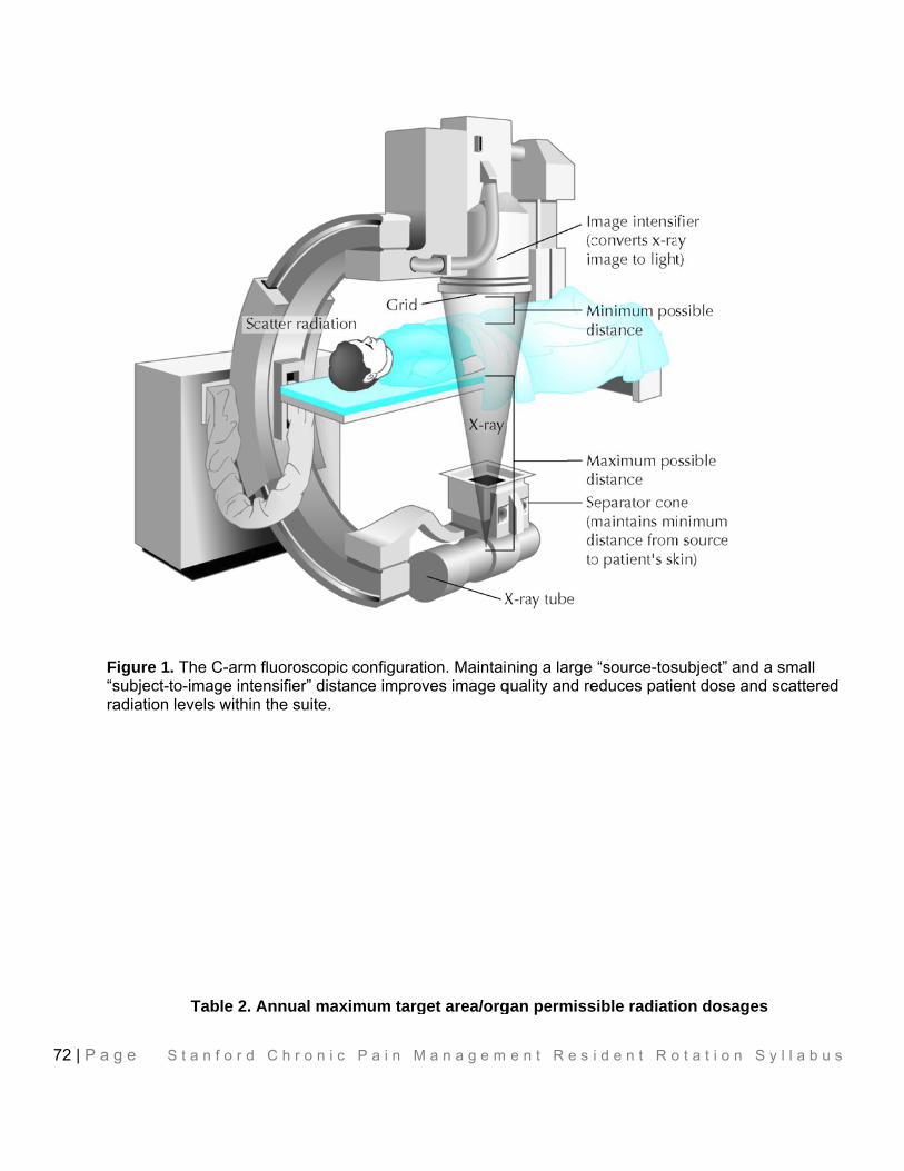
72 |
P a g e S
Figure 1“subject-radiation
S t a n f o r d
. The C-armto-image intlevels within
Table 2.
C h r o n i c
m fluoroscopiensifier” distn the suite.
Annual ma
c P a i n M
ic configurattance improv
ximum targ
M a n a g e m
tion. Maintaives image q
get area/org
e n t R e s
ning a largequality and re
gan permiss
i d e n t R o
e “source-toseduces patie
sible radiati
o t a t i o n S
subject” and ent dose and
ion dosages
S y l l a b u s
a small d scattered
s

73 | P a g e S t a n f o r d C h r o n i c P a i n M a n a g e m e n t R e s i d e n t R o t a t i o n S y l l a b u s
Area/organ Annual maximum permissible dose in
roentgen-equivalent man Thyroid 50
Extremities 50 Lens of the eye 15
Gonads 50 Whole body 5
Pregnant women 0.5
Fig 1 & Table 2 from: Broadman L, et al. Radiation risk management during fluoroscopy for interventional pain medicine physicians. Current Pain and Headache Reports. 2004;8:49-55. PDF
Table 3. Minimum Target Organ Pathologic Radiation Dose and Its Effects
Target Organ Dose*(rad) (Gy) Results Eye lens 200 (2) Cataract formation
Skin 500 (5) Erythema Skin 700 (7) Permanent alopecia
Whole body 200 to 700 (2 to 7) Death from infection due to hematopoetic failure
(4 to 6 weeks)
Whole body 700 to 5,000 (7 to 50) Death from GI failure (3
to 4 days)
Whole body 5,000 – 10,000 (50 to 100) Death from cerebral edema (1 to 2 days)
*Minimum dose that may produce the results (e.g., cataract, permanent alopecia).
From: Fishman S, et al. Radiation safety in pain medicine. Reg Anes and Pain Med. 2002;27:296-305. PDF
See also:
Botwin K, et al. Radiation exposure of a spinal interventionalist performing lumbar transforaminal epidural steroid injections. Arch Phys Med Rehab. 2002;83:697-701. PDF
Most interventional pain procedures utilize fluoroscopy for:
Visualization of boney structures and neural foramina Safety of patient and documentation of placement of block Reimbursement
Any practitioner performing the procedure needs to understand exposure and safety to self, other room personnel, and for the patient.
Physics:

74 | P a g e S t a n f o r d C h r o n i c P a i n M a n a g e m e n t R e s i d e n t R o t a t i o n S y l l a b u s
Time - duration exposed to radiation Distance – space between the invidual and x-ray source Exposure – quantity of radiation intensity measured in Roentgen ® or Coulomb / kg Radiation – energy absorbed by the individual in Radiation Absorbed Dose (Rad) or SI unit
(Gray) Radiation – causing cellular damage in Radiation equivalent man (REM) or Sievert (Sv) in SI
units; the later being a quality factor with variable impact on different tissues
Controlling exposure
Decrease time – use freeze frame feature and minimize beam on time Increase distance – a major source of radiation is the patient acting a conduit for scattered
radiation Inverse square law – the fall off of scatter intensity; doubling the distance produces ¼ exposure;
at a distance of 10 or more feet, irradiation will be negligible; radiation exposure is not just along the path of the beam but everywhere within the suite
Cross-table lateral – scatter of radiation is 2 to 3x at the entrance surface of x-ray tube then exit surface of image intensifier
Automated timer on the C-arm Fluoroscopy machines is a 5 minute limit
Shielding with lead: body, thyroid, eyes (min lead 0.35 mm attenuate scatter), surgical gloves, lead barriers; the patient – reproductive organs
Awareness of the room personnel: call “picture” to warn everyone in the room, including yourself that you are about to take an image and to move all unnecessary objects, such as your fingers out of the beam.
Collimation – making the image as small as possible and hence the area of exposure
Personal dosimeters
Certain procedures with multiple levels, such as radiofrequency of multiple cervical medial branches or spinal cord stimulation trials may require prolonged radiation exposure.