spy&go purification of spytag-proteins using pseudo
TRANSCRIPT

ARTICLE
Spy&Go purification of SpyTag-proteins usingpseudo-SpyCatcher to access an oligomerizationtoolboxIrsyad N.A. Khairil Anuar 1, Anusuya Banerjee1, Anthony H. Keeble 1, Alberto Carella1, Georgi I. Nikov 1 &
Mark Howarth 1
Peptide tags are a key resource, introducing minimal change while enabling a consistent
process to purify diverse proteins. However, peptide tags often provide minimal benefit post-
purification. We previously designed SpyTag, forming an irreversible bond with its protein
partner SpyCatcher. SpyTag provides an easy route to anchor, bridge or multimerize proteins.
Here we establish Spy&Go, enabling protein purification using SpyTag. Through rational
engineering we generated SpyDock, which captures SpyTag-fusions and allows efficient
elution. Spy&Go enabled sensitive purification of SpyTag-fusions from Escherichia coli,
giving superior purity than His-tag/nickel-nitrilotriacetic acid. Spy&Go allowed purification
of mammalian-expressed, N-terminal, C-terminal or internal SpyTag. As an oligomerization
toolbox, we established a panel of SpyCatcher-linked coiled coils, so SpyTag-fusions can
be dimerized, trimerized, tetramerized, pentamerized, hexamerized or heptamerized.
Assembling oligomers for Death Receptor 5 stimulation, we probed multivalency effects
on cancer cell death. Spy&Go, combined with simple oligomerization, should have broad
application for exploring multivalency in signaling.
https://doi.org/10.1038/s41467-019-09678-w OPEN
1 Department of Biochemistry, University of Oxford, South Parks Road, Oxford OX1 3QU, UK. Correspondence and requests for materials should be addressedto M.H. (email: [email protected])
NATURE COMMUNICATIONS | (2019) 10:1734 | https://doi.org/10.1038/s41467-019-09678-w |www.nature.com/naturecommunications 1
1234
5678
90():,;

Affinity chromatography is a central enabling technologyfor research and for producing therapeutics, vaccines, anddiagnostics1,2. However, a persistent problem with affinity
tags is paradoxically the tags themselves, since the tags often serveno purpose post-purification. Tags can inhibit crystallization3,interfere with protein interactions4, and produce an unhelpfulimmune response in vivo5. Tags may be removed by proteolysisbut this extra step is time-consuming, often inefficient andreduces overall yield6,7.
There are already a multitude of affinity tags1,8–11. However,each tag presents its own limitations. The most widely used,the His-tag, is not without faults. There are many examples ofHis-tagging disrupting protein solubility12, structure13,14, andfunction15,16. His-tag purification faces particular challenges fromleakage of metal ions into downstream assays17,18 and substantialimmunogenicity5. The four-amino acid C-tag is less immuno-genic but is only functional at the C-terminus8. Apart frompurification, it would be desirable to use peptide tags for assemblyor immobilization, but the low stability of peptide interactions isfrequently limiting19,20.
We previously developed a peptide-protein pair, SpyTagand SpyCatcher, that spontaneously forms a covalently-linkedcomplex21,22 (Fig. 1). We continued the progress by increasingthe rate of reaction via the evolved SpyTag002 and Spy-Catcher002 versions23. SpyTag technology has enabled diverseapplications including Plug-and-Display vaccine assembly24–26,multivalent activation of signaling27, modular antibody decora-tion28, and living or catalytic biomaterials29–32. Up until now, Spyproteins were nearly always purified via a His-tag, which requiredfurther tag cleavage for immunological applications24. Here, wereport the development of an affinity chromatography techniqueemploying SpyTag as a purification tag, by step-wise engineeringof the SpyCatcher protein partner (Fig. 1). In addition, to extendthe modular expansion of protein function, we establish a toolboxfor oligomerization of proteins of interest using SpyTag-mediatedcovalent reaction. These oligomers shed light on the nature ofmultivalent signaling of DR5 to induce cancer cell killing.
ResultsEstablishment of the Spy&Go purification system. As a first stepto establish Spy&Go purification, the formation of an isopeptidebond between SpyCatcher and SpyTag must be abrogated, tomake possible the elution of SpyTag-fusions. To generate a non-reactive “pseudo-SpyCatcher”, the activating glutamic acid resi-due in the CnaB2 triad (E77, Fig. 1) was mutated to aspartic acidto retain the charge, or to alanine, glycine, asparagine, glutamine,serine, threonine, or valine to remove any possibility of protondonation/acceptance33. We mixed SpyTag-MBP and the E77Xvariants at 25 °C for 24 h. E77D still showed a small amount ofreaction with SpyTag-MBP, but no trace of reaction was seenwith any other mutation (Fig. 2a).
To advance Spy&Go purification further, we hypothesized thatthe rate of association between SpyTag and the SpyCatchermoiety could be limiting. We have previously generatedSpyCatcher002 through phage display evolution for enhancedinteraction with SpyTag23. Here we additionally increased thenegative surface charge on SpyCatcher and reduced the flexibilityin one of the loops through proline substitution, generatingSpyCatcher2.1. When purified SpyTag-MBP was incubated withthe different E77X variants, SpyCatcher002 led to more efficientcapture and elution than SpyCatcher, while SpyCatcher2.1was more effective still (Fig. 2b). Comparing alternative E77mutations for SpyCatcher2.1, E77A led to recovery of the highestamount of SpyTag-MBP (Fig. 2b). Hence, this mutant was takenforward for subsequent development.
We aimed to attach SpyCatcher2.1 E77A site-specifically tosepharose beads, for maximum accessibility to SpyTag-fusions.A unique cysteine was introduced in SpyCatcher2.1 E77A at threepositions. Cysteine introduction sites were selected for efficientcoupling to iodoacetyl-activated (SulfoLink) beads as well as forminimal disruption to SpyTag/SpyCatcher2.1 binding, by havingthe cysteine substitutions distal from the CnaB2 reactive triad(Fig. 2c). Three mutations were examined: N-terminal cysteine(N-term Cys) preceding the coding sequence of SpyCatcher2.1,S49C and S55C34,35 (Fig. 2c). After immobilizing SpyCatcher2.1
SpyCatcher
E77 OH
OHO
OD117
SpyTag
K31
NH2
HN
O
SpyCatcher/SpyTagcomplex withisopeptide bond
Engineeringreactivity,effciency +anchoring
SpyDock
E77A
K31
HONH2
OD117
SpyTag
SpyDock/SpyTagcomplex with noisopeptide bond n = 180
Spy-nanostructures
n = 2, 3, 4, 5, 6, 7Oligomerization toolbox
n = 60
1) Spy&Go purification
SpyDock SpyTag-protein
Bea
d
Cell lysate
Wash Elute
2) Assembly using SpyCatcher platforms
Fig. 1 Overview of Spy&Go. SpyTag/SpyCatcher interact irreversibly by spontaneous isopeptide bond formation, promoted by E77. SpyTag purificationrequires the generation of reversible interaction with SpyDock in 3 steps: blocking reaction, enhancing efficiency of SpyTag binding and preciseimmobilization on resin. Spy&Go then enables purification of SpyTag-fusions, setting the stage for modular oligomerization or multimerization
ARTICLE NATURE COMMUNICATIONS | https://doi.org/10.1038/s41467-019-09678-w
2 NATURE COMMUNICATIONS | (2019) 10:1734 | https://doi.org/10.1038/s41467-019-09678-w |www.nature.com/naturecommunications
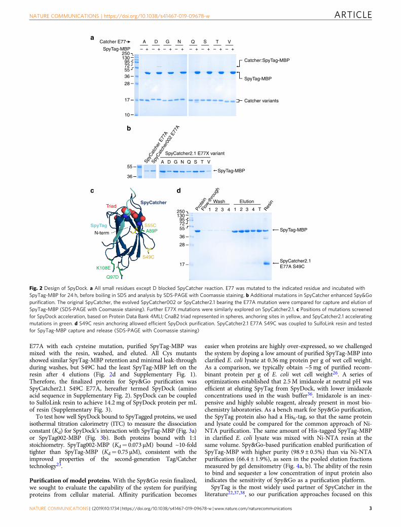
E77A with each cysteine mutation, purified SpyTag-MBP wasmixed with the resin, washed, and eluted. All Cys mutantsshowed similar SpyTag-MBP retention and minimal leak-throughduring washes, but S49C had the least SpyTag-MBP left on theresin after 4 elutions (Fig. 2d and Supplementary Fig. 1).Therefore, the finalized protein for Spy&Go purification wasSpyCatcher2.1 S49C E77A, hereafter termed SpyDock (aminoacid sequence in Supplementary Fig. 2). SpyDock can be coupledto SulfoLink resin to achieve 14.2 mg of SpyDock protein per mLof resin (Supplementary Fig. 3).
To test how well SpyDock bound to SpyTagged proteins, we usedisothermal titration calorimetry (ITC) to measure the dissociationconstant (Kd) for SpyDock’s interaction with SpyTag-MBP (Fig. 3a)or SpyTag002-MBP (Fig. 3b). Both proteins bound with 1:1stoichiometry. SpyTag002-MBP (Kd= 0.073 μM) bound ~10-foldtighter than SpyTag-MBP (Kd= 0.75 μM), consistent with theimproved properties of the second-generation Tag/Catchertechnology23.
Purification of model proteins. With the Spy&Go resin finalized,we sought to evaluate the capability of the system for purifyingproteins from cellular material. Affinity purification becomes
easier when proteins are highly over-expressed, so we challengedthe system by doping a low amount of purified SpyTag-MBP intoclarified E. coli lysate at 0.36 mg protein per g of wet cell weight.As a comparison, we typically obtain ~5mg of purified recom-binant protein per g of E. coli wet cell weight26. A series ofoptimizations established that 2.5 M imidazole at neutral pH wasefficient at eluting SpyTag from SpyDock, with lower imidazoleconcentrations used in the wash buffer36. Imidazole is an inex-pensive and highly soluble reagent, already present in most bio-chemistry laboratories. As a bench mark for Spy&Go purification,the SpyTag protein also had a His6-tag, so that the same proteinand lysate could be compared for the common approach of Ni-NTA purification. The same amount of His-tagged SpyTag-MBPin clarified E. coli lysate was mixed with Ni-NTA resin at thesame volume. Spy&Go-based purification enabled purification ofSpyTag-MBP with higher purity (98.9 ± 0.5%) than via Ni-NTApurification (66.4 ± 1.9%), as seen in the pooled elution fractionsmeasured by gel densitometry (Fig. 4a, b). The ability of the resinto bind and sequester a low concentration of input protein alsoindicates the sensitivity of Spy&Go as a purification platform.
SpyTag is the most widely used partner of SpyCatcher in theliterature22,37,38, so our purification approaches focused on this
Catcher E77
SpyTag-MBP25013095725536
28
17
10
Catcher:SpyTag-MBP
SpyTag-MBP
Catcher variants
A D G N Q S T V
++++++++– – – – – – – –
55
36SpyTag-MBP
A D G N Q S T V
SpyCatcher2.1 E77X variant
SpyC
atch
er E
77A
SpyC
atch
er00
2 E7
7A
250130957255
36
28
17
SpyTag-MBP
SpyCatcher2.1E77A S49C
Wash Elution
1 2 3 4 1 2 3 4 T Res
in
Prot
ein
Flow
-thro
ugh
SpyCatcher
S55CA89P
S49C
Q97D
K108E
N-term
SpyTag
Triad
a
b
c d
Fig. 2 Design of SpyDock. a All small residues except D blocked SpyCatcher reaction. E77 was mutated to the indicated residue and incubated withSpyTag-MBP for 24 h, before boiling in SDS and analysis by SDS-PAGE with Coomassie staining. b Additional mutations in SpyCatcher enhanced Spy&Gopurification. The original SpyCatcher, the evolved SpyCatcher002 or SpyCatcher2.1 bearing the E77A mutation were compared for capture and elution ofSpyTag-MBP (SDS-PAGE with Coomassie staining). Further E77X mutations were similarly explored on SpyCatcher2.1. c Positions of mutations screenedfor SpyDock acceleration, based on Protein Data Bank 4MLI; CnaB2 triad represented in spheres, anchoring sites in yellow, and SpyCatcher2.1 acceleratingmutations in green. d S49C resin anchoring allowed efficient SpyDock purification. SpyCatcher2.1 E77A S49C was coupled to SulfoLink resin and testedfor SpyTag-MBP capture and release (SDS-PAGE with Coomassie staining)
NATURE COMMUNICATIONS | https://doi.org/10.1038/s41467-019-09678-w ARTICLE
NATURE COMMUNICATIONS | (2019) 10:1734 | https://doi.org/10.1038/s41467-019-09678-w |www.nature.com/naturecommunications 3

tag version. However, we also validated that Spy&Go wasefficient for purification of a SpyTag002-fusion23 (SupplementaryFig. 4A).
Additionally, Spy&Go resin was still capable of purifyingSpyTag-MBP from bacterial lysate after storage of resin in 20%(v/v) ethanol for 12 weeks. Therefore Spy&Go resin showed goodstability, as long as microbial growth was inhibited (Supplemen-tary Fig. 4B).
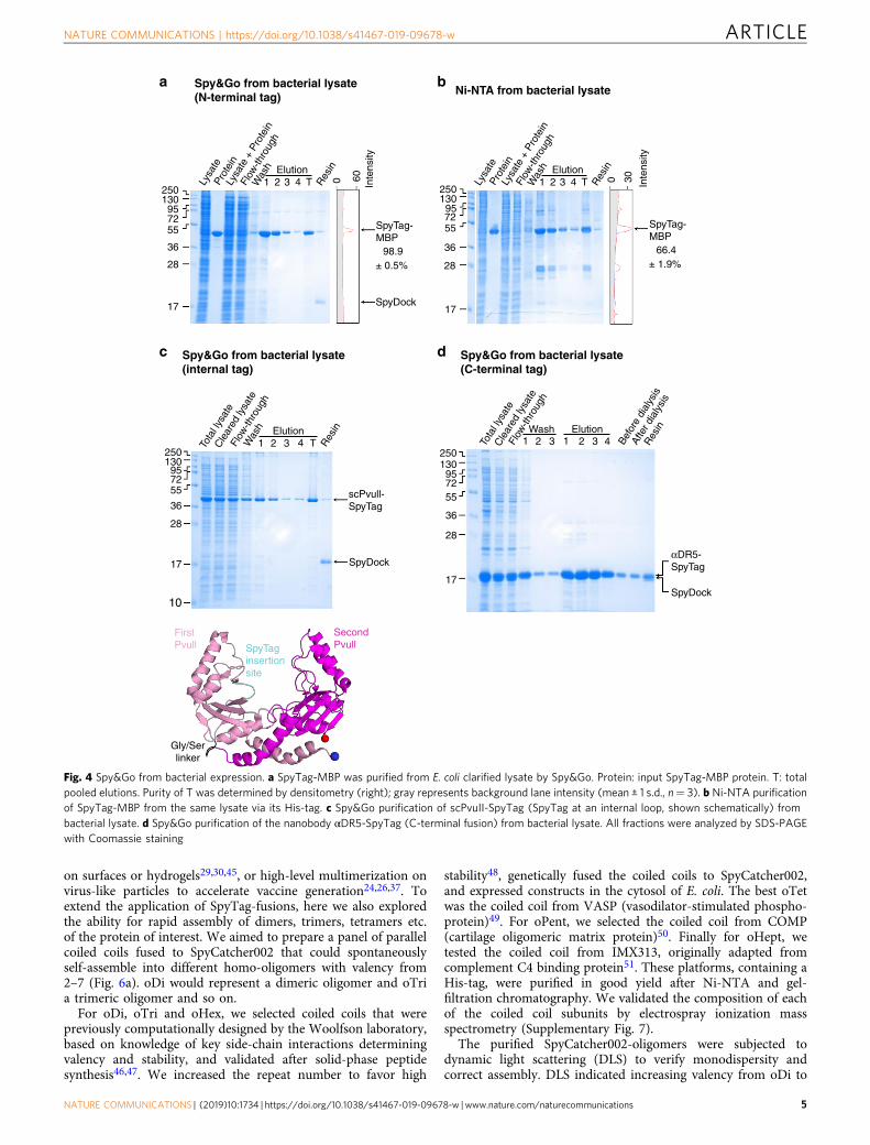
Spy&Go allowed purification with SpyTag at different sites.Some affinity tags can only be placed at either the N- or C-terminus, thus restricting experimental flexibility, especially if afunctional site of the protein of interest is close to a terminus1,8,39.To test capture of SpyTag inserted in the loop of a protein, wegenerated a single-chain dimer of the restriction enzyme PvuII.scPvuII-SpyTag was purified efficiently from soluble E. coliexpression using Spy&Go (Fig. 4c). To test capture of SpyTag atthe C-terminus of a protein, we expressed a nanobody to DeathReceptor 5 (DR5) (αDR5-SpyTag) in the cytosol of E. coli andshowed efficient purification using Spy&Go (Fig. 4d). A singleround of purification is not expected to achieve 100% purity andproteins may be subsequently polished using standard methodssuch as size-exclusion chromatography or ion-exchange chro-matography, as may assist subsequent applications.
To test purification of a different class of protein, the enhancedgreen fluorescent protein mClover3 was genetically fused withSpyTag at its N-terminus and expressed solubly in E. coli. Afterpurification and dialysis, the fluorescence of mClover3 wascomparable to the same protein purified via its His-tag usingNi-NTA (Supplementary Fig. 5), supporting that Spy&Gopurification maintained the functionality of purified proteins.From lysate purifications, the resin capacity ranged from 4–13mgof protein per mL of resin, depending on the location of theSpyTag and protein used.
We established effective regeneration of Spy&Go resin viastripping using 4M imidazole, guanidinium hydrochloride, andNaOH (Supplementary Fig. 6A). Following regeneration of theresin, the smaller protein αDR5-SpyTag was purified by Spy&Go,
while SpyTag-MBP (previously purified using the same resinbatch) was not detected (Supplementary Fig. 6B). Purification ofαDR5-SpyTag using new resin or regenerated resin gave similarresults (Supplementary Fig. 6B).
Spy&Go allowed purification from mammalian expression.Some affinity tags do not work well in particular cell expressionsystems, requiring extra materials or steps40. Transient mam-malian expression is becoming a dominant route for the pro-duction of many therapeutic and research proteins because of therapid pipeline, high degree of folding quality control, and thenative post-translational modifications (notably N-linked glyco-sylation)41. Epithelial cell adhesion molecule (EpCAM) is awidely used marker for capture of circulating tumor cells and itsadhesive interactions may affect metastasis42,43. We cloned thesoluble extracellular region of EpCAM with SpyTag and His-tagat the C-terminus and expressed this glycoprotein throughtransient transfection in HEK293T cells. After expression, thesame volume of supernatant from the cell culture was incubatedwith Spy&Go resin or Ni-NTA resin. EpCAM-SpyTag wasefficiently purified using Spy&Go, as with Ni-NTA (Fig. 5a, b).The heterogeneous gel mobility of EpCAM-SpyTag is expectedbecause of various glycoforms being secreted.
We previously expressed constructs with a His-tag, to allowbench-marking of our purification, but we also purified Cysteine-rich Protective Antigen (CyRPA) with a SpyTag but no His-tag.CyRPA from Plasmodium falciparum is a promising blood-stageantigen for malaria vaccination44. CyRPA-SpyTag was transientlyexpressed in Expi293HEK cells and efficiently purified bySpy&Go (Fig. 5c). Overall, we have shown that Spy&Go is aviable platform for the purification of proteins with SpyTag at theN-terminus, C-terminus or an internal site from either bacterialor mammalian expression systems.
Oligomeric assembly of SpyTag-fusions. An important featureof using SpyTag as the peptide tag for a protein of interest is thatSpyTag can then enable a range of subsequent bioconjugationreactions. For example, SpyTag allows irreversible immobilization
0.20.1
0–0.1–0.2–0.3–0.4–0.5–0.6–0.7
–1.0–0.9–0.8
0 10 20 30 40 50
Time (min)
60 70 80 0 10 20 30 40
Time (min)
50 60 70 80
0 0.4 0.8 1.2 1.6Molar ratio
2.0 0 0.4Molar ratio
0.8 1.2 1.6 2.0
SpyDock:SpyTag-MBP
N = 1.01 ± 0.01Kd = 0.75 ± 0.05 μM
SpyDock:SpyTag002-MBP
N = 1.00 ± 0.01Kd = 0.073 ± 0.013 μM
0–2–4–6–8
–10–12–14–16–18–20–22
0–2–4–6–8
–10–12–14–16–18–20–22
0.20.1
0–0.1–0.2–0.3–0.4–0.5–0.6–0.7–0.8–0.9–1.0
μcal
/s
μcal
/sΔH
(kc
al/m
ol)
ΔH (
kcal
/mol
)
a b
Fig. 3 Affinity of SpyDock for its targets. ITC binding isotherms for SpyDock binding at 25 °C to a SpyTag-MBP or b SpyTag002-MBP. Error bars representthe uncertainty in fit to the binding curve using a 1:1 binding model
ARTICLE NATURE COMMUNICATIONS | https://doi.org/10.1038/s41467-019-09678-w
4 NATURE COMMUNICATIONS | (2019) 10:1734 | https://doi.org/10.1038/s41467-019-09678-w |www.nature.com/naturecommunications
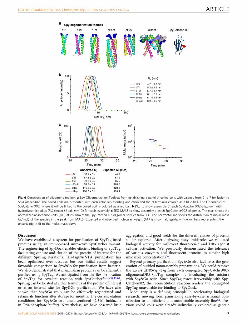
on surfaces or hydrogels29,30,45, or high-level multimerization onvirus-like particles to accelerate vaccine generation24,26,37. Toextend the application of SpyTag-fusions, here we also exploredthe ability for rapid assembly of dimers, trimers, tetramers etc.of the protein of interest. We aimed to prepare a panel of parallelcoiled coils fused to SpyCatcher002 that could spontaneouslyself-assemble into different homo-oligomers with valency from2–7 (Fig. 6a). oDi would represent a dimeric oligomer and oTria trimeric oligomer and so on.
For oDi, oTri and oHex, we selected coiled coils that werepreviously computationally designed by the Woolfson laboratory,based on knowledge of key side-chain interactions determiningvalency and stability, and validated after solid-phase peptidesynthesis46,47. We increased the repeat number to favor high
stability48, genetically fused the coiled coils to SpyCatcher002,and expressed constructs in the cytosol of E. coli. The best oTetwas the coiled coil from VASP (vasodilator-stimulated phospho-protein)49. For oPent, we selected the coiled coil from COMP(cartilage oligomeric matrix protein)50. Finally for oHept, wetested the coiled coil from IMX313, originally adapted fromcomplement C4 binding protein51. These platforms, containing aHis-tag, were purified in good yield after Ni-NTA and gel-filtration chromatography. We validated the composition of eachof the coiled coil subunits by electrospray ionization massspectrometry (Supplementary Fig. 7).
The purified SpyCatcher002-oligomers were subjected todynamic light scattering (DLS) to verify monodispersity andcorrect assembly. DLS indicated increasing valency from oDi to
Spy&Go from bacterial lysate(N-terminal tag)
250130957255
36
28
17
250
SpyTag-MBP
98.9± 0.5%
SpyTag-MBP
SpyDock
SpyDock
scPvull-SpyTag
αDR5-SpyTag
66.4± 1.9%
SpyDock
Inte
nsity
0 60
130957255
36
28
17
250130957255
36
28
17
10
FirstPvull
SecondPvull
Gly/Serlinker
SpyTaginsertionsite
2501 2 3
Elution
Res
in
Was
h
Res
in
Befo
re d
ialy
sis
Tota
l lys
ate
Cle
ared
lysa
te
Flow
-thro
ugh
Tota
l lys
ate
Lysa
teLy
sate
+ P
rote
in
Flow
-thro
ugh
Prot
ein
Cle
ared
lysa
te
Flow
-thro
ugh
Afte
r dia
lysi
s
4 1 2 31 2 3ElutionWash
4T
1 2 3Elution
Res
in
Was
h
4 T
130957255
36
28
17
Spy&Go from bacterial lysate(C-terminal tag)
Spy&Go from bacterial lysate(internal tag)
Ni-NTA from bacterial lysate
Inte
nsity
0 30Lysa
teLy
sate
+ P
rote
in
Flow
-thro
ugh
Prot
ein
1 2 3Elution
Res
in
Was
h
4 T
a b
c d
Fig. 4 Spy&Go from bacterial expression. a SpyTag-MBP was purified from E. coli clarified lysate by Spy&Go. Protein: input SpyTag-MBP protein. T: totalpooled elutions. Purity of T was determined by densitometry (right); gray represents background lane intensity (mean ± 1 s.d., n= 3). b Ni-NTA purificationof SpyTag-MBP from the same lysate via its His-tag. c Spy&Go purification of scPvuII-SpyTag (SpyTag at an internal loop, shown schematically) frombacterial lysate. d Spy&Go purification of the nanobody αDR5-SpyTag (C-terminal fusion) from bacterial lysate. All fractions were analyzed by SDS-PAGEwith Coomassie staining
NATURE COMMUNICATIONS | https://doi.org/10.1038/s41467-019-09678-w ARTICLE
NATURE COMMUNICATIONS | (2019) 10:1734 | https://doi.org/10.1038/s41467-019-09678-w |www.nature.com/naturecommunications 5

oHept based on the increasing hydrodynamic radius (Rh)(Fig. 6b). We further analyzed the oligomers by size-exclusionchromatography/multi-angle light scattering (SEC-MALS). SEC-MALS also analyzes uniformity but additionally characterizes themolecular weight of the assembly. The observed molecularweights were within 10% of the expected assembled molecularweights (Fig. 6c), suggesting successful assembly by each oligomerfrom oDi up to oHept.
In our hands, the previously designed tetrameric coiled coil,CC-Tet46, when fused to SpyCatcher002, had an observed massthat deviated substantially from the expected mass (Supplemen-tary Fig. 8). We also found that SpyCatcher002 fused to thedesigned CC-Hept47 gave satisfactory assembly (SupplementaryFig. 8), but expressed poorly and was prone to aggregate.Therefore the final Spy oligomerization toolbox was a mixture ofcomputationally designed or natural coiled coils (amino acidsequences shown in Supplementary Fig. 9).
Oligomeric assembly of a nanobody. DR5 is a pro-apoptoticreceptor overexpressed on many cancer cells. DR5 activationpromotes cell death in response to binding of TNF-relatedapoptosis-inducing ligand (TRAIL)52. DR5 targeting showspromise as a target for cancer therapy53. There has been extensiveinterest in the clustering of DR553–60, because dimeric anti-DR5 IgG typically causes weak activation of DR5 signaling53.Previous attempts at clustering focused on linear chains of anti-DR5 agonists. We explored the use of multivalent display of anagonist nanobody (αDR5), to evaluate the effect of increasingnanobody valency on triggering cancer cell death. Nanobodiesare a convenient monomeric scaffold, with the single chain
easily expressed in E. coli. αDR5 was fused genetically toSpyTag and was purified efficiently via Spy&Go (Fig. 4d). Wethen mixed this nanobody with the Spy-coiled coil platformsfor convenient modular preparation of dimers, trimers etc. up toheptamers.
αDR5-SpyTag was incubated with each SpyCatcher002-oligomer and efficient covalent reaction was confirmed by SDS-PAGE. Spy&Go also enabled us to remove excess αDR5-SpyTagfrom the mixture, by incubating with Spy&Go resin to recaptureunconjugated αDR5-SpyTag, leaving only oligomer:nanobodycomplex in the flow-through (Fig. 7a, b).
DR5 signal activation by the nanobody oligomeric series. Toassess the potency of these oligomeric SpyCatcher002 platforms,we chose a TRAIL-sensitive human breast cancer cell-line MDA-MB-23154. We tested a broad range of doses, from 0.1 pM–100nM for each SpyCatcher002-oligomer:αDR5-SpyTag and com-pared death induction after 24 h of treatment. While monomericunconjugated αDR5-SpyTag, dimeric, trimeric, and tetramericconjugates failed to display any cell killing within this range,we observed a clear stoichiometry-dependent dose-response forhigher-order αDR5-conjugates (Fig. 7c). All higher-order con-jugates (pentameric, hexameric and heptameric) showed potencyat sub-nanomolar concentrations. The pentameric complexresulted in killing of 50% of the cells (EC50) at 0.9 nM αDR5.Similarly, the hexamer gave a EC50 of 0.42 nM whereas theheptamer had high potency at 0.31 nM EC50 (Fig. 7c). As acontrol, we found that unconjugated SpyCatcher002-oligomerconstructs did not elicit any dose-dependent loss of cell viability(Fig. 7d).
a bSpy&Go from mammalian expression
c Spy&Go from mammalian expression
Ni-NTA from mammalian expression
Elution
1 225013095
EpCAM-SpyTag
CyRPA-SpyTag
SpyDock
EpCAM-SpyTag
SpyDock
725536
28
17
25013095725536
28
17
10
250130957255
36
28
17
3 4 5 6 TSupe
rnat
ant
Flow
-thro
ugh
Was
h
Res
in
Elution
1 2 3 4 TSupe
rnat
ant
Flow
-thro
ugh
Was
h
Res
in
Elution
1 2 3 4 5 6 TSupe
rnat
ant
Flow
-thro
ugh
Was
h
Res
in
Fig. 5 Spy&Go from mammalian expression. a HEK293T cells were transfected with the extracellular region of EpCAM fused to SpyTag and a His-tag(EpCAM-SpyTag). EpCAM-SpyTag was purified from the clarified cell supernatant using Spy&Go. Fractions were analyzed by SDS-PAGE with Coomassiestaining. T: total pooled elutions. Resin: resin post-elution. b Ni-NTA purification of EpCAM-SpyTag as in a. c Spy&Go purification of CyRPA-SpyTag fromExpi293HEK cells as in a
ARTICLE NATURE COMMUNICATIONS | https://doi.org/10.1038/s41467-019-09678-w
6 NATURE COMMUNICATIONS | (2019) 10:1734 | https://doi.org/10.1038/s41467-019-09678-w |www.nature.com/naturecommunications
DiscussionWe have established a system for purification of SpyTag-fusedproteins using an immobilized unreactive SpyCatcher variant.The engineering of SpyDock enables efficient binding of SpyTag,facilitating capture and elution of the protein of interest for thedifferent SpyTag iterations. His-tag/Ni-NTA purification hasbeen optimized over decades but our initial results suggestfavorable comparison to Spy&Go for purification from bacteria.We also demonstrated that mammalian proteins can be efficientlypurified using SpyTag. As anticipated from the flexible locationof SpyTag for covalent reaction with SpyCatcher21,37,38,45,61,SpyTag can be located at either terminus of the protein of interestor at an internal site for Spy&Go purification. We have alsoshown that Spy&Go resin can be effectively regenerated andretains its function after storage for months. The current elutionconditions for Spy&Go are unconventional (2.5 M imidazolein Tris-phosphate buffer). Nevertheless, we have observed little
aggregation and good yields for the different classes of proteinsso far explored. After dialyzing away imidazole, we validatedbiological activity for mClover3 fluorescence and DR5 agonistcellular activation. We previously demonstrated the toleranceof various enzymes and fluorescent proteins to similar highimidazole concentrations36.
Beyond primary purification, Spy&Go also facilitates the gen-eration of purified nanoassembly preparations. We could removethe excess αDR5-SpyTag from each conjugated SpyCatcher002-oligomer:αDR5-SpyTag complex by incubating the mixturewith Spy&Go resin. Since SpyTag reacts irreversibly with Spy-Catcher002, the reconstitution reaction renders the conjugatedSpyTag unavailable for binding to SpyDock.
Modularity is a defining principle in accelerating biologicalresearch, moving from painstaking case-by-case artisanal opti-mization to an efficient and automatable assembly-line62. Pre-vious coiled coils were already individually explored as genetic
a
b
c
Spy oligomerization toolbox
oDi
1.0
oDi 4.7 ± 1.8 nm5.2 ± 1.9 nm5.7 ± 1.7 nm6.1 ± 2.1 nm6.1 ± 1.9 nm6.9 ± 1.5 nm
37.1 ± 0.4 40.861.585.5
109.5
123.5159.4
57.4 ± 0.379.3 ± 0.299.9 ± 0.2
115.5 ± 0.2150.5 ± 0.1
oTrioTetoPent
oHexoHept
oDioTrioTetoPent
oHexoHept
0.5
Am
plitu
de (
inte
nsity
)
0.0
100 106
105
104
50
Rel
ativ
e A
U (
%)
Mol
ar m
ass
(g/m
ol)
020 25
Time (min)
30 35 20 25
Time (min)
30 35
1 10Size (Rh, nm)
Rh (nm)
Observed Mr Expected Mr (kDa)
100
oTri oTet oPent oHex oHept SpyCatcher002
Fig. 6 Construction of oligomeric toolbox. a Spy Oligomerization Toolbox from establishing a panel of coiled coils with valency from 2 to 7 for fusion toSpyCatcher002. The coiled coils are presented with each color representing one chain and the N-terminus colored as a blue ball. The C-terminus ofSpyCatcher002, where it will be linked to the coiled coil, is colored as a red ball. b DLS to show assembly of each SpyCatcher002-oligomer, withhydrodynamic radius (Rh) (mean ± 1 s.d., n= 10) for each assembly. c SEC-MALS to show assembly of each SpyCatcher002-oligomer. The peak shows thenormalized absorbance units (AU) at 280 nm of the SpyCatcher002-oligomer species from SEC. The horizontal line shows the distribution of molar mass(g/mol) of the species in the peak from MALS. Expected and observed molecular weight (Mr) is shown alongside, with error bars representing theuncertainty in fit to the molar mass curve
NATURE COMMUNICATIONS | https://doi.org/10.1038/s41467-019-09678-w ARTICLE
NATURE COMMUNICATIONS | (2019) 10:1734 | https://doi.org/10.1038/s41467-019-09678-w |www.nature.com/naturecommunications 7

fusion partners for proteins25,63–65, however, optimizationswould be needed for each distinct protein24,37,65. Examplesinclude heterogeneous populations of 5–7 oligomers instead of amonodisperse heptamer66, failed assembly of a trimer-fusionprotein67, and severe degradation of dimers in E. coli cytosol68,69.
The characterized Spy-based oligomerization toolbox canalleviate this problem, separating the expression/folding/post-translational modification of coiled coils and cargo protein beforeuniting via SpyTag/SpyCatcher. Spy&Go-purified αDR5-SpyTagreadily reacted to completion with multivalent SpyCatcher002-oligomer to form a panel of valency-defined platforms tounderstand the effect of higher valencies on cellular signaling andthe downstream apoptotic effect. Up until now, most multimericanti-DR5 or soluble TRAIL constructs consist of a lineararrangement linked by simple Gly-Ser linkers extending to 5
repeats55, 6 repeats56,57 and in one case 8 repeats57 of the agonistagainst DR553. However, the formation of a linear complex maynot mimic the clustering by the native TRAIL to DR5 receptor. A“combody” comprising a genetic fusion of the coiled coil COMPwith an agonistic αDR5 was previously made70, but direct fusionmay not be optimized for other proteins or coiled coil combi-nations, and no higher valencies were reported. We note that wehave characterized SpyCatcher002-coiled coil valency by DLS andSEC-MALS at micromolar concentrations. Both natural andsynthetic multimers may start to dissociate as one approaches lownanomolar or picomolar concentrations, where cells may stillrespond to DR5 clustering but bulk biophysical assays becomevery difficult46,47. In future work, single-molecule assays may bethe best way to understand molecular and cellular behavior in thislow concentration regime71,72.
100
c
b
a
d
75
50
25
0
100
2501309572
55
36
28
17
SpyCatcher002-
SpyTagSpyCatcher002-
oligomer:αDR5-
oligomer
Recaptured
Resin
SpyCatcher002-oligomer
[SpyCatcher002-oligomer]n
SpyDock
[SpyCatcher002-oligomer:αDR5-SpyTag]n
ExcessαDR5-SpyTag
Excess αDR5-SpyTagrecaptured
Purified[SpyCatcher002-oligomer: αDR5-SpyTag]n
αDR5-SpyTag
αDR5-SpyTag
75
50
25
00.1
[Protein monomer] (nM)
% V
iabi
lity
% V
iabi
lity
Building blocks
1 10 100 100010–4 10–3 10–2 0.1 1 10[αDR5] (nM)
αDR5 assemblies
αDR5
αDR5-SpyTag
oDi
oTrioTet
oPent
oHex
oHept
oDi
Reaction Recapture
Bea
d
–
–
–+
+ +
+
+
+
+
+
+
+
+
+
+
+
+
+ +
+ – + – + – + – +
+ + + + + + + +
+
oTri oTet oPent oHex oHept
100
Fig. 7 Oligomer panel tested for cancer cell killing. a Cartoon depicting depletion of free SpyTag-ligand using Spy&Go resin. b Coupling of SpyTag-fusion tocoiled coil series. αDR5-SpyTag was incubated with each SpyCatcher002-coiled coil and analyzed by SDS-PAGE with Coomassie staining. In Recapturedlanes, excess αDR5-SpyTag was removed from the coiled coil conjugate by an additional passage through Spy&Go resin. c Dose-response curve of MDA-MB-231 cell viability when treated with different concentrations of αDR5 conjugated to each SpyCatcher002-oligomer platforms. The line at 50% cellviability shows the cut-off for EC50 calculation. The x-axis is normalized to the concentration of αDR5 monomer. Error bars represent mean ±1 s.d., n= 2.d MDA-MB-231 viability upon incubation as in c with the building blocks of αDR5 alone or coiled coils alone. Error bars represent mean ± 1 s.d., n= 3
ARTICLE NATURE COMMUNICATIONS | https://doi.org/10.1038/s41467-019-09678-w
8 NATURE COMMUNICATIONS | (2019) 10:1734 | https://doi.org/10.1038/s41467-019-09678-w |www.nature.com/naturecommunications

We envision that the oligomerizing SpyCatcher002-coiled coilplatforms can advance the study of valency-dependence ondiverse cellular signaling processes73,74. By combining Spy&Gopurification with coiled coil nanoassembly, SpyTagging may helpto accelerate the exploration and exploitation of protein space.
MethodsPlasmids and cloning. Constructs were cloned by standard PCR methods andassembled using Gibson assembly. Inserts were verified by Sanger sequencing.pDEST14-SpyCatcher2.1 was derived from pDEST14-SpyCatcher00223 (GenBankMF974388 and Addgene plasmid ID 102827) with additional A89P, Q97D andK108E mutations (see below). pDEST14-SpyCatcher2.1 S49C E77A (SpyDock)(Supplementary Fig. 2, GenBank MK637462, Addgene plasmid ID 124618) has theorganization: His6, SpyCatcher2.1 with E77A S49C mutations, GSSGS. pDEST14-SpyCatcher2.1 E77X S49C has the same organization as SpyDock except with E77Xmutations instead, with X being D, G, N, Q, S, T, or V. pDEST14-SpyCatcher2.1E77A N-term Cys has the same organization as SpyDock except with a cysteine-anchoring mutation at the 6th amino acid residue preceding SpyCatcher2.1 E77Ainstead of S49C. pDEST14-SpyCatcher2.1 S55C E77A has the same organization asSpyDock except with a S55C mutation instead of S49C mutation. pET28a-SpyTag-MBP21 (Addgene plasmid ID 35050) and pET28a-SpyTag002-MBP23 (GenBankMF974389 and Addgene plasmid ID 102831) were as described. pENTR4-EpCAM-SpyTag (GenBank MK637463) was derived from pENTR4-EpCAM-SnoopTagJr30
(GenBank MH511516) with the organization: tissue plasminogen activator (tPA)secretion leader sequence, extracellular domain of human EpCAM protein(EpCAM; residue 24–265), (GSG)2, SpyTag, GEGS, His6. pENTR4-LPTOS-CyRPA-SpyTag26 (GenBank MH425516) was previously published. pET28a-αDR5-SpyTag (GenBank MK637464) was derived from pET28a-SnoopTag-αDR5-SpyTag (GenBank KU500643)54 with the organization: αDR5 (4E6 nanobody55),(GGGGS)2, SpyTag. pET28a-SpyTag-mClover3 has the organization: SpyTag,SGGGSG, mClover375, GSGSGS, His6. pET28a-scPvuII-SpyTag (GenBankMK637465) has the organization: His6, SSG, PvuII from Proteus vulgaris, GSG,TEV cleavage site, GGSG, SpyTag, GSGG, PvuII. SpyTag along with surroundingspacers was inserted into a loop of the first PvuII between amino acid residues 75and 76. PvuII constructs had the D58A mutation to block DNA cleavage.pDEST14-SpyCatcher002 was linked to coiled coil inserts consisting of oDi46
(GenBank MK637466, Addgene plasmid ID 124661), oTri46 (GenBank MK637467,Addgene plasmid ID 124662), oTet49 (GenBank MK637468, Addgene plasmid ID124663), oPent50 (GenBank MK637469, Addgene plasmid ID 124664), oHex47
(GenBank MK637470, Addgene plasmid ID 124670), oHept51 (GenBankMK637471, Addgene plasmid ID 124671), CC-Tet46 (GenBank MK637472) or CC-Hept47 (GenBank MK637473). The constructs have the organization: His6,DYDIPTT spacer, TEV cleavage site, SpyCatcher002, GSSGSGSGS, coiled coilinserts, GSGSG, C-tag. Coiled coil DNA was synthesized by IDT DNA Technol-ogies (Supplementary Fig. 9). oDi, oTri, oHex, CC-Tet, and CC-Hept have 5 heptadrepeats, instead of 4 repeats reported in the literature, to increase the stability ofcoiled coil assembly48.
SpyDock rational design. To complement the increase in positively chargedresidues of SpyTag002 compared to SpyTag, mutations Q97D and K108E weremade on SpyCatcher002 to increase the negatively charged residues, making theSpyDock precursor, SpyCatcher2.1, to improve the electrostatic interaction withthe positively-charged SpyTag or SpyTag002 (Fig. 2c). Residues Y83 and E85within the long A79-A89 loop in SpyCatcher make key interactions with residuesY9 and K10 of SpyTag in the SpyTag/SpyCatcher crystal structure76. These wild-type residues were also positively selected during directed evolution to produceSpyTag002, supporting the importance of the residues for rapid isopeptide bondformation23. Inclusion of prolines in turns and loops has previously been shown tostabilize proteins36,77–79. Hence, the A89P mutation was included to stabilize theA79-A89 loop (Fig. 2c). The E77 was targeted to nullify the isopeptide bondformation, with A, D, G, N, Q, S, T, and V mutations as candidates (Fig. 1). Thesemutations were postulated to enable better binding and retention of SpyTag toSpyDock during the binding and washing steps, along with easier elution.
Bacterial protein expression. pET28a-SpyTag-MBP, pET28a-SpyTag002-MBP,and pET28a-SpyTag-mClover3 were transformed into chemically competent E. coliBL21 (DE3) RIPL (Agilent Technologies). pET28a-scPvuII-SpyTag was trans-formed into T7 Express lysY/Iq (NEB). pDEST14-SpyCatcher2.1 S49C E77A(SpyDock), pDEST14-SpyCatcher2.1 S49C E77X variants, pDEST14-SpyCatcher2.1 E77A N-term Cys, pDEST14-SpyCatcher2.1 S55C E77A andpDEST14-SpyCatcher002-coiled coil fusions were transformed into chemically-competent E. coli C41 (DE3), a kind gift from Anthony Watts (University ofOxford). pET28a-αDR5-SpyTag was transformed into E. coli BL21 (DE3) RIPLcontaining a gene encoding phosphogluconolactonase, to degrade 6-phosphoglu-conolactone, which promotes protein gluconoylation80. The cells were plated on LBagar supplemented with 50 µg/mL kanamycin (pET28a) or 100 µg/mL ampicillin(pDEST14). For αDR5-SpyTag, 34 µg/mL chloramphenicol was added alongside
kanamycin throughout. The plates were incubated at 37 °C overnight until colonieswere observed.
Single colonies of pET28a-SpyTag-MBP, pET28a-SpyTag002-MBP, pET28a-SpyTag-mClover3, pET28a-scPvuII-SpyTag, and pDEST14-SpyDock, all variantsof pDEST14-SpyCatcher2.1 S49C E77X, pDEST14-SpyCatcher2.1 E77A N-termCys, pDEST14-SpyCatcher2.1 E77A S55C, and all variants of pDEST14-SpyCatcher002-coiled coils were picked and inoculated into 10 mL LB mediumsupplemented with 50 µg/mL kanamycin (pET28a) or 100 µg/mL ampicillin(pDEST14), incubated at 37 °C with shaking at 200 rpm for 16 h. The cultures werethen inoculated into 1 L LB supplemented with 50 µg/mL kanamycin (pET28a) or100 µg/mL ampicillin (pDEST14) and 0.8% (w/v) glucose (except for pDEST14-SpyCatcher002-coiled coils), incubated at 37 °C with shaking at 200 rpm until A600
0.5–0.6, when the cultures were induced with 0.42 mM isopropyl β-D-1-thiogalactopyranoside (IPTG) (Fluorochem). Cultures of pET28a-SpyTag-MBP,pET28a-SpyTag002-MBP, pET28a-SpyTag-mClover3, pET28a-scPvuII-SpyTag,and pDEST14-SpyDock, all variants of pDEST14-SpyCatcher2.1 S49C E77X,pDEST14-SpyCatcher2.1 E77A N-term Cys, and pDEST14-SpyCatcher2.1 E77AS55C were grown further for 4 h with shaking at 200 rpm at 30 °C. Cultures ofpDEST14-SpyCatcher002-oDi, pDEST14-SpyCatcher002-oTri, pDEST14-SpyCatcher002-oTet, pDEST14-SpyCatcher002-oPent, and pDEST14-SpyCatcher002-CC-Tet were grown further for 16 h with shaking at 200 rpm at22 °C. Cultures of pDEST14-SpyCatcher002-oHex, pDEST14-SpyCatcher002-oHept and pDEST14-SpyCatcher002-CC-Hept were grown for 4 h with shakingat 200 rpm at 37 °C.
A single colony of pET28a-αDR5-SpyTag was picked and inoculated into 1 Lauto-induction medium (AIMLB0205 from Formedium) supplemented with 50 µg/mL kanamycin and 34 µg/mL chloramphenicol, incubated at 30 °C with shaking at200 rpm for 24 h. All E. coli-expressed proteins were harvested by centrifugationat 4000×g for 15 min at 4 °C prior to purification.
Mammalian protein expression. EpCAM-SpyTag was expressed in adherentHEK293T cells. HEK293T cells were cultured in T175 adhesive culture flasks(Corning) with Dulbecco’s Modified Eagle’s Medium (DMEM) (Sigma-Aldrich)high glucose with 10% (v/v) Fetal Bovine Serum (Sigma-Aldrich), 2 mM L-gluta-mine, 100 U/mL penicillin, and 100 μg/mL streptomycin (Thermo Fisher Scientific)at 37 °C with 5% CO2. Before transfection, the cells were seeded into a T875 5-layerflask (Corning) and upon reaching 50% confluency, they were transferred intoserum-free media (DMEM, 2 mM glutamine, 50 U/mL penicillin, 25 mM HEPESadded) and mixed with 30 μg pENTR4-EpCAM-SpyTag plasmid per 7.5 mL ofmedia for each flask layer. After 15 min, 2.5 mL media containing 36 μg/mLpolyethyleneimine (Sigma-Aldrich) was added to each layer. 10 mL media con-taining 4.4 μM valproic acid (Sigma-Aldrich), 100 U/mL penicillin, and 100 μg/mLstreptomycin was added to each layer 16–20 h later. Cells were then incubated at37 °C with 5% CO2 for another 6 days.
CyRPA-SpyTag was expressed in suspension Expi293HEK cells (Thermo FisherScientific). Expi293HEK cells were cultured in Expi293 expression media (ThermoFisher Scientific) with 50 U/mL penicillin/streptomycin at 37 °C with 7% CO2
shaking at 110–125 rpm. Transient transfection of pENTR4-LPTOS-CyRPA-SpyTag was done using the ExpiFectamine 293 transfection kit (Thermo FisherScientific). Cells at a density of 2.5 × 10⁶ cells/mL were transfected with 2.7 μLExpiFectamine 293 Reagent per 1 μg of pENTR4-LPTOS-CyRPA-SpyTag plasmid.After 16–18 h, ExpiFectamine transfection enhancers (Thermo Fisher Scientific)were added and the cell supernatant was harvested 4 days post-transfection.
The cell supernatants were harvested by addition of cOmplete™, Mini, EDTA-free Protease Inhibitor Cocktail (Roche), centrifuged at 1000×g for 3 min, andfiltered through a 0.45 μm syringe filter to remove cell debris. 2.5% of 10×Ni-NTAor 10×TP buffer (250 mM orthophosphoric acid adjusted to pH 7.0 with Tris base)was added for pH adjustment before affinity chromatography purification.
Protein purification by Ni-NTA. Purifications were done at 4 °C throughout. AllE. coli-grown constructs (except αDR5-SpyTag, scPvuII-SpyTag, SpyTag-mClo-ver3, SpyCatcher002-oHex, SpyCatcher002-oHept and SpyCatcher002-CC-Hept)were resuspended in 1×Ni-NTA buffer (50 mM Tris-HCl, 300 mM NaCl pH 7.8;SpyDock along with all variants of SpyCatcher2.1 S49C E77X, SpyCatcher2.1 E77AN-term Cys, SpyCatcher2.1 E77A S55C and SpyCatcher002-oPent had additional10 mM 2-mercaptoethanol) with cOmplete™, Mini, EDTA-free Protease InhibitorCocktail and 1 mM phenylmethylsulfonyl fluoride (PMSF). Cells were lysed byaddition of 100 μg/mL lysozyme (Sigma-Aldrich) and 2 U/mL benzonase (Sigma-Aldrich). The lysate was rotated at 25 °C for 30 min and sonicated on ice for 4 × 1min with 1 min rest period at 50% duty cycle. Clarified cell lysates were centrifugedat 30,000 g for 30 min before incubation with Ni-NTA beads (Qiagen) on a rotaryshaker for 1 h. The lysate-bead mixture was added onto a polyprep gravity columnand washed with 20 packed resin volumes of Ni-NTA wash buffer (10 mM imi-dazole in Ni-NTA buffer, pH 7.8; SpyDock, all variants of SpyCatcher2.1 S49CE77X, SpyCatcher2.1 E77A N-term Cys, SpyCatcher2.1 E77A S55C andSpyCatcher002-oPent had 10 mM 2-mercaptoethanol for the first 10 packed resinvolumes; SpyCatcher002 coiled coil fusions used 75 mM imidazole in Ni-NTAbuffer) and eluted with Ni-NTA elution buffer (200 mM imidazole in Ni-NTAbuffer, pH 7.8; SpyCatcher002 coiled coil fusions used 350 mM imidazole in Ni-NTA buffer) at 4 °C. Elutions were monitored by A280 and stopped once A280 was
NATURE COMMUNICATIONS | https://doi.org/10.1038/s41467-019-09678-w ARTICLE
NATURE COMMUNICATIONS | (2019) 10:1734 | https://doi.org/10.1038/s41467-019-09678-w |www.nature.com/naturecommunications 9

<1.0. Proteins were dialyzed against 20 mM Tris-HCl pH 8.0 and concentrated, ifnecessary, using Vivaspin centrifugal concentrator 5 kDa cutoff (GE Healthcare).
SpyDock was further purified on a HiTrap Q HP anion exchangechromatography column (GE Healthcare) connected to an ÄKTA Pure 25 (GEHealthcare) fast protein liquid chromatography (FPLC) system at 4 °C. The proteinwas eluted with a linear gradient of 0.2–0.35M NaCl (in 10 mM Tris-HCl pH 8.0with 1 mM dithiothreitol) at a flow rate of 2 mL/min at 4 °C. Peak fractions wereverified by SDS-PAGE gels, dialyzed against 20 mM Tris-HCl pH 8.0, andconcentrated using Vivaspin centrifugal concentrator 5 kDa cutoff. Typical yieldfor SpyDock after Ni-NTA and ionic exchange chromatography is approximately28 mg/L culture.
All SpyCatcher002 coiled coil fusions were further purified by gel filtrationchromatography on a pre-equilibrated HiLoad 16/600 Superdex 200 pg columnconnected to ÄKTA Pure 25 FPLC system. The mobile phase was 50 mM Tris-HCl,150 mM NaCl pH 8.0 at a flow-rate of 1 mL/min at 4 °C with A280 monitoredthroughout. Fractions were collected corresponding to the size of the oligomericSpyCatcher002 coiled coil complex: SpyCatcher002-oDi (75–78 mL),SpyCatcher002-oTri (69–72 mL), SpyCatcher002-oTet (66–70 mL),SpyCatcher002-oPent (62–65 mL), SpyCatcher002-oHex (61–65 mL),SpyCatcher002-oHept (56–60 mL). All SpyCatcher002-coiled coil fusions had ayield of approximately 3 mg/L culture, following Ni-NTA and gel filtrationchromatography.
Protein purification by refolding. SpyCatcher002-oHex, SpyCatcher002-oHept,and SpyCatcher002-CC-Hept were found in inclusion bodies and so were purifiedby refolding. Cells were resuspended in 100 mM Tris-HCl pH 8.0 with cOmplete™,Mini, EDTA-free Protease Inhibitor Cocktail and 1 mM PMSF. Cells were lysedby addition of 100 μg/mL lysozyme and 2 U/mL benzonase and rotated at 25 °C for30 min. There was one freeze-thaw cycle from −80 °C to 25 °C, followed byaddition of 0.5% (v/v) Triton X-100 and sonication on ice for 4 × 1 min with 1 minrest period at 50% duty cycle. Clarified cell lysates were centrifuged at 30,000×g for30 min. The cell pellet was washed with 2 × PBS+ 0.1% (v/v) Triton X-100 andthen 2 × PBS, with centrifugation at 25,000×g for 20 min between steps. Theinclusion body pellet was resuspended in 8 M urea in 50 mM Tris-HCl pH 8.0 andrefolded by diluting 50-fold into refolding buffer of 0.1 M Tris-HCl pH 8.0, 0.4 ML-arginine and 0.1 M PMSF (for SpyCatcher002-oHept, 5 mM reduced glutathioneand 0.5 mM oxidized glutathione were present) for 40 h. The mixture was filteredthrough a 0.45-μm membrane before incubation with Ni-NTA beads on a rotaryshaker for 1 h. The lysate-bead mixture was added onto a polyprep gravity columnand washed with 20 resin volumes of Ni-NTA wash buffer (with 75 mM imidazole;SpyCatcher002-oHept had 10 mM 2-mercaptoethanol for the first 10 packed resinvolumes) and eluted with Ni-NTA elution buffer (with 350 mM imidazole) at 4 °C.Elutions were monitored by A280 and stopped once A280 was <1.0. Proteins weredialyzed against 20 mM Tris-HCl pH 8.0 and concentrated to suitable workingconcentrations, if needed, using Vivaspin centrifugal concentrator 5 kDa cutoff(GE Healthcare).
SpyCatcher2.1 E77A variants coupling to SulfoLink resin. Purified Spy-Catcher2.1 E77A with various cysteine anchoring residues (N-term Cys, S55C, andS49C) were coupled to SulfoLink Coupling Resin (Thermo Fisher Scientific)according to the manufacturer’s protocol at 25 °C. In short, 20 mg of protein forevery 1 mL of resin was reduced by 1 mM tris(2-carboxyethyl)phosphine (TCEP)(Fluorochem, UK) for 30 min, prior to mixing end-over-end with equilibrated resinfor 15 min and leaving to stand for 30 min covered by foil. Protein flow-throughwas aspirated and resin was washed with 10 resin volumes of coupling buffer (50mM Tris-HCl, 5 mM EDTA, pH 8.5). The resin was blocked with 50 mM L-cysteine-HCl in coupling buffer (MP Biomedicals), mixed end-over-end for 15 min,and left to stand for 30 min. The resin was then washed with 10 resin volumes of1M NaCl, and stored in 1xTris-phosphate (TP) buffer (25 mM orthophosphoricacid adjusted to pH 7.0 with Tris base), before adding 0.05% (w/v) NaN3 andstoring at 4 °C.
Isothermal titration calorimetry. SpyDock was trapped as a monomer byreduction with 2.5 mM TCEP in 50 mM Tris-HCl with 5 mM EDTA at pH 8.5 andby cysteine modification with 20 mM iodoacetamide in 50 mM Tris-HCl with 5mM EDTA at pH 8.5 for 30 min in the dark to produce carbamidomethylatedSpyDock. Excess TCEP and iodoacetamide were removed by dialysis. SpyTag-MBP, SpyTag002-MBP and the modified SpyDock were subsequently dialyzedtwice into 20 mM HEPES pH 7.0 with 150 mM NaCl. Experiments were carried outusing a Microcal PEAQ-isothermal titration calorimetry (ITC) calorimeter (Mal-vern) at 25 °C in 20 mM HEPES pH 7.0 with 150 mM NaCl. 20 μM SpyDock wasused in the cell and titrated with 20 injections of 210 μM SpyTag-MBP orSpyTag002-MBP in the syringe. Analyses were carried out using a 1:1 bindingmodel with the MicroCal PEAQ-ITC Analysis software version 1.1.0.1262.
Purification by Spy&Go. To determine the best cysteine anchoring site, 50 μLpacked resin with SpyCatcher2.1 E77A N-term Cys, S55C, or S49C was mixed with500 μL TP buffer containing 0.09 mg (10× lower than SpyCatcher2.1 E77A avail-able on resin) of Ni-NTA-purified SpyTag-MBP in an Eppendorf tube through
batch chromatography. The low amount of SpyTag-MBP introduced was to test thesensitivity of Spy&Go purification. The protein was mixed with the resin for 1 hwith tumbling at 4 °C. Standard Spy&Go batch chromatography purification is asfollows: the resin was washed 4 × with 10 resin volumes of TP buffer, with incu-bation at 4 °C shaking at 1,200 rpm for 3 min. Resin was then centrifuged at 4000×gfor 3 min at 4 °C. The protein was eluted with 4 × 1.5 resin volume of elution bufferETP (2.5 M imidazole in TP buffer).
For purification of SpyTag-MBP and SpyTag002-MBP from lysate, 0.09 mg ofthe Ni-NTA-purified proteins were added into ~0.25 g wet cell weight of inducedBL21 (DE3) RIPL cleared lysate dissolved in 500 μL TP buffer in an Eppendorf tubecontaining 50 μL packed Spy&Go resin. The protein-lysate was mixed with theresin for 1 h tumbling at 4 °C. Standard Spy&Go batch chromatographypurification was performed to purify the proteins but washed with wash bufferWTP (500 mM imidazole in TP buffer).
EpCAM-SpyTag was purified from the supernatant of HEK293T cells by mixing50 mL of the supernatant with 0.5 mL packed Spy&Go resin. CyRPA-SpyTag waspurified from the supernatant of Expi293HEK cells by mixing 3.5 mL of thesupernatant with 50 µL of packed Spy&Go resin. Both were rolling for 1 h at 4 °C.The mixture was then purified by standard gravity column chromatography: resinwas washed 4× with 10 resin volumes of TP buffer and eluted with 6 × 1 resinvolume of ETP buffer (4 × 1 resin volume for CyRPA-SpyTag). Typical yield forEpCAM-SpyTag after Spy&Go purification was approximately 2.9 mg/L cultureand for CyRPA-SpyTag was approximately 8.9 mg/L culture.
For comparison with Ni-NTA resin, the methods described were performedwith equivalent volume of packed Ni-NTA resin with the following changes:the cell pellet was resuspended in 50 mM Tris-HCl, 300 mM NaCl pH 7.8, thewash buffer was Ni-NTA wash buffer (30 mM imidazole for SpyTag-MBP, 10 mMimidazole for EpCAM-SpyTag) and elution was carried out with Ni-NTAelution buffer.
For purification of αDR5-SpyTag, the cell pellet from 1 L of culture wascentrifuged and resuspended in 10 mL TP buffer with 1 mM dithiothreitol,cOmplete™, Mini, EDTA-free Protease Inhibitor Cocktail and 1 mM PMSF. Cellswere lysed by addition of 100 μg/mL lysozyme and 2 U/mL benzonase rotated at25 °C for 30 min, and subsequently sonicated on ice for 4 × 1 min at 50% duty cycleand spun down at 30,000 g to remove cell debris. The cleared lysate was mixed with1 mL of Spy&Go resin rolling for 1 h at 4 °C. Standard gravity columnchromatography purification was performed to purify αDR5-SpyTag with washesusing WTP buffer.
For purification of scPvuII-SpyTag and SpyTag-mClover3, the cell pellet from0.25 L of culture was lysed with 5 mL BugBuster reagent per gram of wet cell weightin the presence of 100 µg/mL lysozyme and 2 U/mL benzonase. The sample wasincubated standing at room temperature for 20 min. After spinning down at30,000×g to remove cell debris, the cleared lysate was mixed with Spy&Go resinwith rolling for 1 h at 4 °C. Standard gravity column chromatography purificationwas performed using WTP buffer for washes and 4 × 1 resin volume for elution.
SpyTag-protein and SpyCatcher reconstitution reaction. To investigate anyformation of covalent bond, 5 μM SpyCatcher2.1 S49C with E77X (X being A, D,G, N, Q, S, T, or V) was reacted with 2 × molar excess of SpyTag-MBP (10 μM)at 25 °C for 24 h in TP buffer. Reaction was quenched with SDS-PAGE loadingbuffer [0.23 M Tris HCl pH 6.8, 24% (v/v) glycerol, 120 μM bromophenol blue,0.23 M SDS, 100 mM 2-mercaptoethanol] and heating at 95 °C for 5 min in aBio-Rad C1000 thermal cycler.
To form SpyCatcher002-oligomer:αDR5-SpyTag, 100 μM monomericconcentration of SpyCatcher002-oligomer was mixed with 300 μM αDR5-SpyTagin 200 μL total volume overnight at 25 °C in 50 mM Tris-HCl, 150 mM NaCl,pH 8.0. Excess αDR5-SpyTag was removed by recapturing using Spy&Go resinby incubating the reaction mixture with 50 μL Spy&Go resin for 2 h at roomtemperature with mixing end-over-end. The mixture was then loaded onto a MicroBio-Spin column (Bio-Rad) and the conjugated SpyCatcher002-oligomer:αDR5-SpyTag was recovered by centrifugation at 300 g for 1 min at 4 °C into amicrocentrifuge tube. This process was repeated once. The protein concentrationswere measured by Proteoquant BCA assay (Expedeon).
SDS-PAGE and protein purity quantification. SDS-PAGE was performed using16% Tris-glycine gels in an XCell SureLock system (Thermo Fisher Scientific).Samples were loaded with final concentration of 1x SDS-PAGE loading buffer. Forreduced samples, 100 mM 2-mercaptoethanol was added. SDS-PAGE gels were runat 190 V in 25 mM Tris-HCl, 192 mM glycine, 0.1% (w/v) SDS, pH 8.5. Gels werestained with InstantBlue Coomassie stain (Expedeon), destained with MilliQ water,and imaged using ChemiDoc XRS imager and analyzed with ImageLab (version6.0.1) (Bio-Rad). In ImageLab, low sensitivity band detection in the final elutedlane (T) was calculated and compared with the protein control lane (Protein) atbackground subtraction of disk size 2 mm. Percentage purity is defined as 100 ×[target protein Band % in lane T/target protein Band % in lane Protein].
Regeneration of Spy&Go resin. Lysate from bacterial expression of SpyTag-MBPwas purified using 0.5 mL Spy&Go resin as above. The resin was equilibrated with2 × 10 packed resin volumes of TP buffer before regeneration. Spy&Go resin was
ARTICLE NATURE COMMUNICATIONS | https://doi.org/10.1038/s41467-019-09678-w
10 NATURE COMMUNICATIONS | (2019) 10:1734 | https://doi.org/10.1038/s41467-019-09678-w |www.nature.com/naturecommunications

then regenerated at 25 °C by incubating with 3 × 10 packed resin volumes of 4Mimidazole in TP buffer pH 7.0 for 5 min each time, washing with 1 × TP buffer,incubating with 3 × 10 packed resin volumes of 6 M guanidine hydrochloride pH2.0 for 5 min each time, washing with 1 × TP buffer, incubating with 3 × 10 packedresin volumes of 0.1 M NaOH for 1 min each time, and washing with 2 × TP bufferbefore storage in 20% (v/v) ethanol in MilliQ water at 4 °C. Supernatant from eachstage was analyzed by SDS-PAGE with Coomassie staining. New resin, resin pre-regeneration and regenerated resin were also analyzed by boiling in SDS-PAGEloading buffer, followed by SDS-PAGE with Coomassie staining. Lysate frombacterial expression of αDR5-SpyTag was added to new resin or regenerated resinand purified using the standard Spy&Go protocol, before analysis by SDS-PAGEwith Coomassie staining. To test capture after storage, Spy&Go resin was stored in20% (v/v) ethanol for 12 weeks at at 4 °C, before purification of SpyTag-MBP frombacterial lysate following the standard protocol.
Mass spectrometry. An Agilent RapidFire 365 platform, coupled to an Agilent6550 Accurate-Mass Quadrupole Time-of-Flight (Q-TOF) mass spectrometer, wasused to perform intact protein mass spectrometry in positive ion mode with a jet-stream electrospray ion source (Agilent). Proteins with disulfide bonds werereduced with 2.5 mM TCEP in 50 mM Tris-HCl with 5 mM EDTA at pH 8.5 for 1h before treatment with formic acid. Protein samples at 10 μM in 50 μL volumewere prepared on a 384-well polypropylene plate (Greiner) and acidified to 1% (v/v) formic acid. Samples were aspirated under vacuum for 0.4 s on the RapidFiresampling platform and loaded onto a C4 solid-phase extraction cartridge. Fol-lowing washes with 0.1% (v/v) formic acid at 1.5 mL/min flow-rate for 5.5 s, thesamples were eluted to the mass spectrometer with deionized water containing 85%(v/v) acetonitrile and 0.1% (v/v) formic acid at 1.25 mL/min for 5.5 s. The cartridgewas equilibrated with deionized water for 0.5 s. Nitrogen drying gas for the ioni-zation source was operated at 13 L/min at 225 °C, with the jet stream sheath gas at aflow-rate of 12 L/min at 350 °C and the nozzle voltage at 1500 V. Data analysis wasdone using Mass Hunter Qualitative Analysis software version 7.0, with the proteinionization data deconvoluted using the maximum entropy algorithm. Predictedmass was calculated using ExPASy ProtParam, based on all disulfide bonds beingreduced and cleavage of the N-terminal formylmethionine.
SEC-MALS. Samples were prepared at 2 mg/mL in 100 μL in 50 mM Tris-HCl,150 mM NaCl pH 8.0 and injected into a Superdex 200 HR 10/30 column(GE Healthcare) at 25 °C at a flow-rate of 0.5 mL/min connected to a ShimadzuHPLC system comprising LC-20AD pump, SIL-20AC autosampler and SPD20AUV/Vis detector with 50 mM Tris-HCl, 150 mM NaCl pH 8.0 as running buffer.Light scattering was detected by a Wyatt Dawn HELEOS-II 8-angle light scatteringdetector and Wyatt Optilab rEX refractive index monitor. The resulting lightscattering, refractive index and UV traces were processed in ASTRA 6(Wyatt Technologies).
Dynamic light scattering. Samples were prepared at 1 mg/mL in 50 mM Tris-HCl,150 mM NaCl pH 8.0 and centrifuged for 30 min at 16,900×g at 4 °C to remove anyaggregates. 20 μL of each sample was loaded into a reusable quartz cuvette andmeasurements were taken at 20 °C using an Omnisizer (Viscotek) with 10 scans of10 s each. Data were analyzed using OmniSIZE 3.0 and the intensity distributionfrom the scans were plotted in Excel.
Fluorescence assay. A concentration gradient of SpyTag-mClover3 in PBS,purified by either Spy&Go or Ni-NTA, was prepared in triplicate from 15 μM to0.47 μM in a black, flat-bottom half-area 96-well plate (Corning). The protein wasexcited at λex=482 ± 16 nm and fluorescence intensity was detected at λem= 530 ±40 nm, at 40 flashes per well using a CLARIOstar plate reader (BMG Labtech) at30 °C. The fluorescence intensity was plotted using Microsoft Excel.
Cell killing assay. MDA-MB-231 cells from American Type Culture Collectionwere grown at 37 °C with 5% CO2 in Dulbecco’s Modified Eagle Medium (DMEM)(Life Technologies) containing 10% (v/v) Fetal Bovine Serum (Sigma-Aldrich)and 50 U/mL penicillin and 50 μg/mL streptomycin (Sigma-Aldrich). Cells werepassaged upon 70–80% confluency and for <3 months in total. MDA-MB-231 cellswere seeded into a 96-well plate at 40,000 cells per well in 100 µL DMEM con-taining 50 U/mL penicillin and 50 μg/mL streptomycin and 1% (v/v) FBS andincubated at 37 °C with 5% CO2 for 16 h. Each SpyCatcher002-oligomer:αDR5-SpyTag was prepared at 100 nM monomeric concentration and serial dilutionsthereof, in DMEM with antibiotics and 1% (v/v) FBS as above. For a negativecontrol, SpyTag-MBP (at 100 nM and serial dilutions) was added to cells. Cellswere washed with sterilized PBS twice before addition of the conjugates. The cellswere incubated with the protein samples at 37 °C with 5% CO2 for 24 h. Cellviability was tested by addition of 40 µL 0.15 mg/mL Resazurin (Alamar Blue)(Sigma-Aldrich) in PBS at 37 °C with 5% CO2 for 4 h and by measuring fluores-cence (λex 544 nm, λem 590 nm) using a SpectraMax3 plate reader with SoftMaxPro 5.4 software (Molecular Devices). The percentage of viable cells was calculatedas 100 × (signal of treated cells - signal without cells)/(signal untreated cells–signalwithout cells). The signal without cells was taken as the resazurin fluorescencein the absence of cells, whereas the signal of untreated cells came from the
fluorescence of resazurin with cells that were incubated with DMEM with anti-biotics and 1% (v/v) FBS.
Graphics and sequence analysis. Structures are shown from SpyTag/SpyCatcher(PDB ID 4MLI)76, oDi (PDB ID: 4DZM)46, oTri (PDB ID: 4DZL)46, oTet (PDBID: 1USE)49, oPent (PDB ID: 1VDF)50, oHex (PDB ID: 4PN9)47, oHept (2YF2)51,CC-Tet (3R4A)46, CC-Hept (4PNA)47, or PvuII (3KSK). PyMOL 2.0 was usedto visualize the structure of proteins based on PDB IDs. Amino acid sequencealignment was done with Clustal Omega.
Software. Microsoft Excel was used to plot DLS and fluorescence graphs.GraphPad Prism v7.0 was used to plot the SEC-MALS graph. OriginPro 2015 wasused to plot the cell killing assay and mass spectrometry graphs. MicroCal PEAQ-ITC Analysis Software version 1.1.0.1262 was used to plot ITC graphs.
Reporting summary. Further information on experimental design is available inthe Nature Research Reporting Summary linked to this article.
Data availabilityAmino acid sequences of SpyDock and SpyCatcher002-oligomers are available in theSupplementary Information. Sequences of other constructs are available in GenBank asdescribed above under “Plasmids and cloning”. Plasmids encoding the SpyDock andSpyCatcher002-oligomers were deposited in the Addgene repository (https://www.addgene.org/Mark_Howarth/). The source data for Fig. 2a, b, d, 3, 4, 5, 6b, c, 7b–d, andSupplementary Figs. 1, 3, 4, 5, 6, and 8b, c are provided as a Source Data File. Furtherinformation and request for resources and reagents should be directed to and will befulfilled by the Lead Contact, Mark Howarth ([email protected]).
Received: 14 December 2018 Accepted: 22 March 2019
References1. Kimple, M. E. & Sondek, J. Overview of affinity tags for protein purification.
Curr. Protoc. Protein Sci. 73, 9.9.1–9.9.19 (2004).2. Roque, A. C. A. & Lowe, C. R. Affinity chromatography: history, perspectives,
limitations and prospects. Methods Mol. Biol. 421, 1–21 (2008).3. Niedzialkowska, E. et al. Protein purification and crystallization artifacts: the
tale usually not told. Protein Sci. 25, 720–733 (2016).4. Zhao, X., Li, G. & Liang, S. Several affinity tags commonly used in
chromatographic purification. J. Anal. Methods Chem. 2013, 581093 (2013).5. Jin, J. et al. Accelerating the clinical development of protein-based vaccines for
malaria by efficient purification using a four amino acid C-terminal ‘C-tag’.Int. J. Parasitol. 47, 435–446 (2017).
6. Waugh, D. S. An overview of enzymatic reagents for the removal of affinitytags. Protein Expr. Purif. 80, 283–293 (2011).
7. Waugh, D. S. Making the most of affinity tags. Trends Biotechnol. 23, 316–320(2005).
8. De Genst, E. J. et al. Structure and properties of a complex of α-synucleinand a single-domain camelid antibody. J. Mol. Biol. 402, 326–343 (2010).
9. Braun, M. B. et al. Peptides in headlock - a novel high-affinity and versatilepeptide-binding nanobody for proteomics and microscopy. Sci. Rep. 6, 19211(2016).
10. Vassylyeva, M. N. et al. Efficient, ultra-high-affinity chromatography in aone-step purification of complex proteins. Proc. Natl Acad. Sci. USA 114,E5138–E5147 (2017).
11. Costa, S., Almeida, A., Castro, A. & Domingues, L. Fusion tags for proteinsolubility, purification, and immunogenicity in Escherichia coli: the novelFh8 system. Front. Microbiol. 5, 63 (2014).
12. Woestenenk, E. A., Hammarström, M., Van Den Berg, S., Härd, T. &Berglund, H. His tag effect on solubility of human proteins produced inEscherichia coli: a comparison between four expression vectors. J. Struct.Funct. Genomics 5, 217–229 (2004).
13. Wu, J. & Filutowicz, M. Hexahistidine (His6)-tag dependent proteindimerization: a cautionary tale. Acta Biochim. Pol. 46, 591–599 (1999).
14. Klose, J. et al. Hexa-histidin tag position influences disulfide structure but notbinding behavior of in vitro folded N-terminal domain of rat corticotropin-releasing factor receptor type 2a. Protein Sci. 13, 2470–2475 (2004).
15. Sabaty, M. et al. Detrimental effect of the 6 His C-terminal tag on YedYenzymatic activity and influence of the TAT signal sequence on YedYsynthesis. BMC Biochem. 14, 28 (2013).
16. Majorek, K. A., Kuhn, M. L., Chruszcz, M., Anderson, W. F. & Minor, W.Double trouble - buffer selection and his-tag presence may be responsiblefor nonreproducibility of biomedical experiments. Protein Sci. 23, 1359–1368(2014).
NATURE COMMUNICATIONS | https://doi.org/10.1038/s41467-019-09678-w ARTICLE
NATURE COMMUNICATIONS | (2019) 10:1734 | https://doi.org/10.1038/s41467-019-09678-w |www.nature.com/naturecommunications 11

17. Zhao, D. & Huang, Z. Effect of his-tag on expression, purification, andstructure of zinc finger protein, ZNF191(243–368). Bioinorg. Chem. Appl.2016, 1–6 (2016).
18. Chant, A., Kraemer-Pecore, C. M., Watkin, R. & Kneale, G. G. Attachment ofa histidine tag to the minimal zinc finger protein of the Aspergillus nidulansgene regulatory protein AreA causes a conformational change at the DNA-binding site. Protein Expr. Purif. 39, 152–159 (2005).
19. Unzueta, U. et al. Non-amyloidogenic peptide tags for the regulatableself-assembling of protein-only nanoparticles. Biomaterials 33, 8714–8722(2012).
20. Haglin, E. R., Briegel, A. & Thompson, L. K. Hijacking His-Tags to makefunctional multi-protein complexes. Biophys. J. 112, 360a (2017).
21. Zakeri, B. et al. Peptide tag forming a rapid covalent bond to a protein,through engineering a bacterial adhesin. Proc. Natl Acad. Sci. USA 109,E690–E697 (2012).
22. Sutherland, A. R., Alam, M. K. & Geyer, C. R. Post-translation assembly ofprotein parts into complex devices using SpyTag/SpyCatcher protein ligase.Chembiochem 20, 319–328 (2019).
23. Keeble, A. H. et al. Evolving accelerated amidation by SpyTag/SpyCatcherto analyze membrane dynamics. Angew. Chem. Int. Ed. 56, 16521–16525(2017).
24. Brune, K. D. et al. Plug-and-Display: decoration of Virus-Like Particles viaisopeptide bonds for modular immunization. Sci. Rep. 6, 19234 (2016).
25. Brune, K. D. et al. Dual Plug-and-Display synthetic assembly using orthogonalreactive proteins for twin antigen immunization. Bioconjug. Chem. 28,1544–1551 (2017).
26. Bruun, T. U. J., Andersson, A.-M. C., Draper, S. J. & Howarth, M. Engineeringa rugged nanoscaffold to enhance plug-and-display vaccination. ACS Nano 12,8855–8866 (2018).
27. Fairhead, M. et al. SpyAvidin hubs enable precise and ultrastable orthogonalnanoassembly. J. Am. Chem. Soc. 136, 12355–12363 (2014).
28. Moon, H., Bae, Y., Kim, H. & Kang, S. Plug-and-playable fluorescent cellimaging modular toolkits using the bacterial superglue, SpyTag/SpyCatcher.Chem. Commun. 52, 14051–14054 (2016).
29. Sun, F., Zhang, W.-B., Mahdavi, A., Arnold, F. H. & Tirrell, D. A. Synthesis ofbioactive protein hydrogels by genetically encoded SpyTag-SpyCatcherchemistry. Proc. Natl Acad. Sci. USA 111, 11269–11274 (2014).
30. Wieduwild, R. & Howarth, M. Assembling and decorating hyaluronanhydrogels with twin protein superglues to mimic cell-cell interactions.Biomaterials 180, 253–264 (2018).
31. Nguyen, P. Q., Botyanszki, Z., Tay, P. K. R. & Joshi, N. S. Programmablebiofilm-based materials from engineered curli nanofibres. Nat. Commun. 5,4945 (2014).
32. Peschke, T. et al. Self-assembling all-enzyme hydrogels for flow biocatalysis.Angew. Chem. Int. Ed. 57, 17028–17032 (2018).
33. Reddington, S. C. & Howarth, M. Secrets of a covalent interaction forbiomaterials and biotechnology: SpyTag and SpyCatcher. Curr. Opin. Chem.Biol. 29, 94–99 (2015).
34. Pessino, V., Citron, Y. R., Feng, S. & Huang, B. Covalent protein labeling bySpyTag–SpyCatcher in fixed cells for super-resolution microscopy.Chembiochem 18, 1492–1495 (2017).
35. Dovala, D., Sawyer, W. S., Rath, C. M. & Metzger, L. E. Rapid analysis ofprotein expression and solubility with the SpyTag-SpyCatcher system. ProteinExpr. Purif. 117, 44–51 (2016).
36. Buldun, C. M., Jean, J. X., Bedford, M. R. & Howarth, M. SnoopLigasecatalyzes peptide-peptide locking and enables solid-phase conjugate isolation.J. Am. Chem. Soc. 140, 3008–3018 (2018).
37. Brune, K. D. & Howarth, M. New routes and opportunities for modularconstruction of particulate vaccines: stick, click, and glue. Front. Immunol. 9,1432 (2018).
38. Keeble, A. H. & Howarth, M. Insider information on successful covalentprotein coupling with help from SpyBank. Methods Enzymol. 617, 443–461(2019).
39. Bass, R. B., Miller, A. S., Gloor, S. L. & Falke, J. J. The PICM chemicalscanning method for identifying domain-domain and protein-proteininterfaces: applications to the core signaling complex of E. coli chemotaxis.Methods Enzymol. 423, 3–24 (2007).
40. Schmidt, T. G. M. et al. Development of the Twin-Strep-tag® and itsapplication for purification of recombinant proteins from cell culturesupernatants. Protein Expr. Purif. 92, 54–61 (2013).
41. Seiradake, E., Zhao, Y., Lu, W., Aricescu, A. R. & Jones, E. Y. Production ofcell surface and secreted glycoproteins in mammalian cells. Methods Mol.Biol.1261, 115–127 (2015).
42. Jain, J., Veggiani, G. & Howarth, M. Cholesterol loading and ultrastableprotein interactions determine the level of tumor marker required for optimalisolation of cancer cells. Cancer Res. 73, 2310–2321 (2013).
43. Huang, L. et al. Functions of EpCAM in physiological processes and diseases(Review). Int. J. Mol. Med. 42, 1771–1785 (2018).
44. Dreyer, A. M. et al. Passive immunoprotection of plasmodium falciparum-infected mice designates the CyRPA as candidate malaria vaccine antigen.J. Immunol. 188, 6225–6237 (2012).
45. Zhang, W. B., Sun, F., Tirrell, D. A. & Arnold, F. H. Controllingmacromolecular topology with genetically encoded SpyTag-SpyCatcherchemistry. J. Am. Chem. Soc. 135, 13988–13997 (2013).
46. Fletcher, J. M. et al. A basis set of de novo coiled-Coil peptide oligomers forrational protein design and synthetic biology. ACS Synth. Biol. 1, 240–250(2012).
47. Thomson, A. R. et al. Computational design of water-soluble α-helical barrels.Science 346, 485–488 (2014).
48. Thomas, F., Boyle, A. L., Burton, A. J. & Woolfson, D. N. A set of de novodesigned parallel heterodimeric coiled coils with quantified dissociationconstants in the micromolar to sub-nanomolar regime. J. Am. Chem. Soc. 135,5161–5166 (2013).
49. Karin, K. et al. The VASP tetramerization domain is a right-handed coiled coilbased on a 15-residue repeat. Proc. Natl Acad. Sci. USA 101, 17027–17032(2004).
50. Malashkevich, V. N., Kammerer, R. A., Efimov, V. P., Schulthess, T. & Engel, J.The crystal structure of a five-stranded coiled coil in COMP: A prototype ionchannel? Science 274, 761–765 (1996).
51. Ogun, S. A., Dumon-Seignovert, L., Marchand, J. B., Holder, A. A. & Hill, F.The oligomerization domain of C4-binding protein (C4bp) acts as anadjuvant, and the fusion protein comprised of the 19-kilodalton merozoitesurface protein 1 fused with the murine C4bp domain protects mice againstmalaria. Infect. Immunol. 76, 3817–3823 (2008).
52. Bertsch, U., Röder, C., Kalthoff, H. & Trauzold, A. Compartmentalizationof TNF-related apoptosis-inducing ligand (TRAIL) death receptorfunctions: emerging role of nuclear TRAIL-R2. Cell Death Dis. 5, e1390–e1390(2014).
53. Dubuisson, A. & Micheau, O. Antibodies and derivatives targeting DR4 andDR5 for cancer therapy. Antibodies 6, 16 (2017).
54. Veggiani, G. et al. Programmable polyproteams built using twin peptidesuperglues. Proc. Natl Acad. Sci. USA 113, 1202–1207 (2016).
55. Huet, H. A. et al. Multivalent nanobodies targeting death receptor 5 elicitsuperior tumor cell killing through efficient caspase induction. MAbs 6,1560–1570 (2014).
56. Hutt, M. et al. Superior properties of Fc-comprising scTRAIL fusion proteins.Mol. Cancer Ther. 16, 2792–2802 (2017).
57. Swers, J. S. et al. Multivalent scaffold proteins as superagonists of TRAILreceptor 2-induced apoptosis. Mol. Cancer Ther. 12, 1235–1244 (2013).
58. Graves, J. D. et al. Apo2L/TRAIL and the death receptor 5 agonist antibodyAMG 655 cooperate to promote receptor clustering and antitumor activity.Cancer Cell 26, 177–189 (2014).
59. Gieffers, C. et al. APG350 induces superior clustering of TRAILreceptors and shows therapeutic antitumor efficacy independent ofcross-linking via Fc receptors. Mol. Cancer Ther. 12, 2735–2747(2013).
60. Bruenker, P. et al. RG7386, a novel tetravalent FAP-DR5 antibody, effectivelytriggers FAP-dependent, avidity-driven DR5 hyperclustering and tumor cellapoptosis. Mol. Cancer Ther. 15, 946–957 (2016).
61. Hagen, A., Sutter, M., Sloan, N. & Kerfeld, C. A. Programmed loading andrapid purification of engineered bacterial microcompartment shells. Nat.Commun. 9, 2881 (2018).
62. Way, J. C., Collins, J. J., Keasling, J. D. & Silver, P. A. Integrating biologicalredesign: where synthetic biology came from and where it needs to go. Cell157, 151–161 (2014).
63. Ross, J. F. et al. Decorating self-assembled peptide cages with proteins. ACSNano 11, 7901–7914 (2017).
64. Zhu, X. et al. COMBODY: one-domain antibody multimer with improvedavidity. Immunol. Cell Biol. 88, 667–675 (2010).
65. Müller, K. M., Arndt, K. M. & Alber, T. Protein fusions to coiled-coil domains.Methods Enzymol. 328, 261–282 (2000).
66. He, Y. G. et al. A novel C3d-containing oligomeric vaccine provides insightinto the viability of testing human C3d-based vaccines in mice.Immunobiology 223, 125–134 (2018).
67. Voulgaraki, D. et al. Multivalent recombinant proteins for probing functionsof leucocyte surface proteins such as the CD200 receptor. Immunology 115,337–346 (2005).
68. Arndt, K. M., Müller, K. M. & Plückthun, A. Helix-stabilized fv (hsfv)antibody fragments: substituting the constant domains of a fab fragmentfor a heterodimeric coiled-coil domain. J. Mol. Biol. 312, 221–228(2001).
69. Müller, K. M., Arndt, K. M. & Alber, T. Protein fusions to coiled-coil domains.Methods Enzymol. 328, 261–282 (2000).
70. Wang, W., He, W., Wang, L., Zhang, G. & Gao, B. Pentamerisation of a scFvdirected against TRAIL receptor 2 increases its antitumour efficacy. Immunol.Cell Biol. 91, 360–367 (2013).
ARTICLE NATURE COMMUNICATIONS | https://doi.org/10.1038/s41467-019-09678-w
12 NATURE COMMUNICATIONS | (2019) 10:1734 | https://doi.org/10.1038/s41467-019-09678-w |www.nature.com/naturecommunications
71. Miller, H., Zhou, Z., Shepherd, J., Wollman, A. J. M. & Leake, M. C. Single-molecule techniques in biophysics: a review of the progress in methods andapplications. Reports Prog. Phys. 81, 024601 (2018).
72. Günther, J. P., Börsch, M. & Fischer, P. Diffusion measurements of swimmingenzymes with fluorescence correlation spectroscopy. Acc. Chem. Res. 51,1911–1920 (2018).
73. Dubacheva, G. V., Curk, T., Auzély-Velty, R., Frenkel, D. & Richter, R. P.Designing multivalent probes for tunable superselective targeting. Proc. NatlAcad. Sci. USA 112, 5579–5584 (2015).
74. Kiessling, L. L., Gestwicki, J. E. & Strong, L. E. Synthetic multivalent ligands asprobes of signal transduction. Angew. Chem. Int. Ed. 45, 2348–2368 (2006).
75. Bajar, B. T. et al. Improving brightness and photostability of green and redfluorescent proteins for live cell imaging and FRET reporting. Sci. Rep. 6,20889 (2016).
76. Li, L., Fierer, J. O., Rapoport, T. A. & Howarth, M. Structural analysis andoptimization of the covalent association between SpyCatcher and a peptidetag. J. Mol. Biol. 426, 309–317 (2014).
77. Guruprasad, K. & Rajkumar, S. Beta-and gamma-turns in proteins revisited: anew set of amino acid turn-type dependent positional preferences andpotentials. J. Biosci. 25, 143–156 (2000).
78. Fu, H., Grimsley, G. R., Razvi, A., Scholtz, J. M. & Pace, C. N. Increasingprotein stability by improving Beta-turns. Proteins Struct. Funct. Bioinform.77, 491–498 (2009).
79. Yu, H., Yan, Y., Zhang, C. & Dalby, P. A. Two strategies to engineerflexible loops for improved enzyme thermostability. Sci. Rep. 7, 41212(2017).
80. Aon, J. C. et al. Suppressing posttranslational gluconoylation of heterologousproteins by metabolic engineering of Escherichia coli. Appl. Environ.Microbiol. 74, 950–958 (2008).
AcknowledgementsFunding was provided by Yayasan Khazanah, Oxford Centre for Islamic Studies, St.John’s College Oxford (I.N.A.K.A.), the European Research Council (ERC-2013-CoG615945-PeptidePadlock) (A.B., A.H.K., and M.H.), and the Biotechnology and BiologicalSciences Research Council (BBSRC Research Experience Placements) (G.I.N.). We thankDr. David Staunton of the University of Oxford Department of Biochemistry BiophysicalSuite for assistance. We acknowledge Dr. Anthony Tumber of the University of OxfordDepartment of Chemistry for assistance with MS, supported by the BBSRC (grant BB/R000344/1). We thank Dr. James F. Ross (Howarth laboratory), Dr. Anne-Marie C.Andersson (Howarth laboratory), and Dr. Karthik Rajasekar (David Sherratt laboratory)for advice.
Author contributionsI.N.A.K.A. performed all experiments except for cellular analysis and ITC. A.B.performed cellular analysis. A.H.K. performed ITC. A.H.K., A.C., and G.I.N. performedearly optimization of SpyDock reactivity. I.N.A.K.A. and M.H. designed the projectand wrote the manuscript. All authors approved the manuscript.
Additional informationSupplementary Information accompanies this paper at https://doi.org/10.1038/s41467-019-09678-w.
Competing interests: M.H., A.H.K., and A.C. are authors on a patent application bythe University of Oxford covering sequences for enhanced isopeptide bond formation(UK Intellectual Property Office 1706430.4). M.H. is an author on a granted patentcovering peptide tags forming spontaneous isopeptide bonds (EP2534484) and aSpyBiotech co-founder, shareholder, and consultant. M.H. and I.N.A.K.A. are authorson a patent application by the University of Oxford covering Spy&Go (UK IntellectualProperty Office 1819850.7). The remaining authors declare no competing interests.
Reprints and permission information is available online at http://npg.nature.com/reprintsandpermissions/
Journal peer review information: Nature Communications thanks Sebyung Kang andthe other anonymous reviewer(s) for their contribution to the peer review of this work.
Publisher’s note: Springer Nature remains neutral with regard to jurisdictional claims inpublished maps and institutional affiliations.
Open Access This article is licensed under a Creative CommonsAttribution 4.0 International License, which permits use, sharing,
adaptation, distribution and reproduction in any medium or format, as long as you giveappropriate credit to the original author(s) and the source, provide a link to the CreativeCommons license, and indicate if changes were made. The images or other third partymaterial in this article are included in the article’s Creative Commons license, unlessindicated otherwise in a credit line to the material. If material is not included in thearticle’s Creative Commons license and your intended use is not permitted by statutoryregulation or exceeds the permitted use, you will need to obtain permission directly fromthe copyright holder. To view a copy of this license, visit http://creativecommons.org/licenses/by/4.0/.
© The Author(s) 2019
NATURE COMMUNICATIONS | https://doi.org/10.1038/s41467-019-09678-w ARTICLE
NATURE COMMUNICATIONS | (2019) 10:1734 | https://doi.org/10.1038/s41467-019-09678-w |www.nature.com/naturecommunications 13
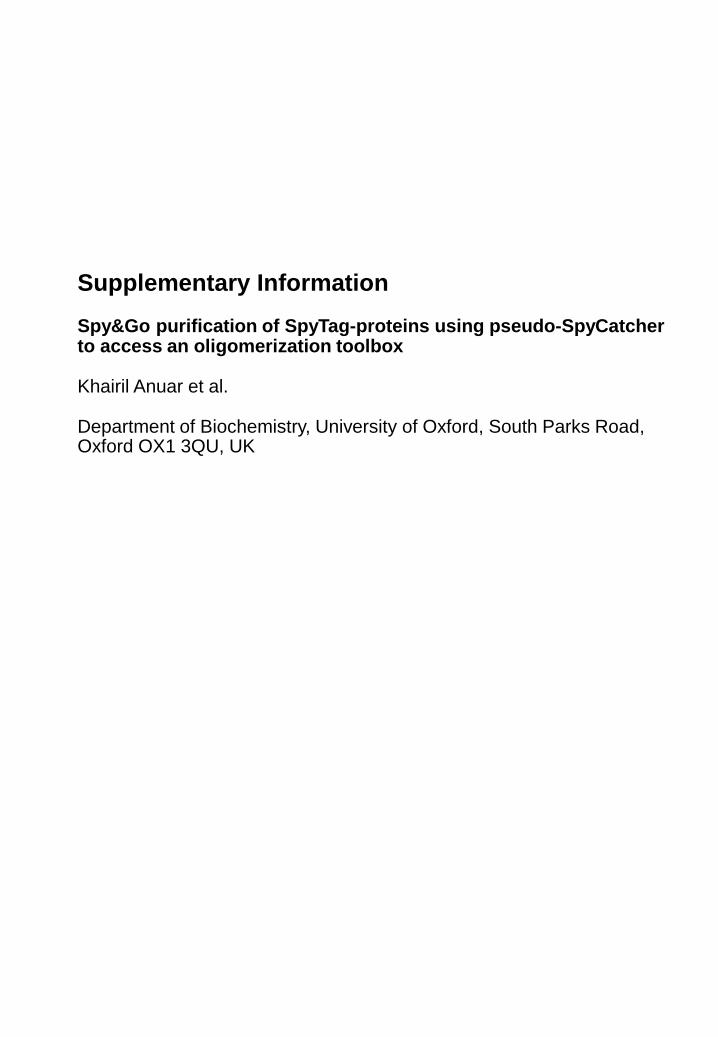
Supplementary Information
Spy&Go purification of SpyTag-proteins using pseudo-SpyCatcher to access an oligomerization toolbox
Khairil Anuar et al.
Department of Biochemistry, University of Oxford, South Parks Road, Oxford OX1 3QU, UK
957255
36
28
17
130250
N-terminal Cys
S55C
SpyTag-MBP
SpyCatcher2.1
E77A N-terminal Cys
SpyTag-MBP
SpyCatcher2.1
E77A S55C
A
B
Supplementary Figure 1. Comparison of cysteine-anchoring residues for SpyDock. Purified SpyTag-MBP was mixed with SpyDock anchored to resin by different cysteine-anchoring residues (A: N-terminal Cys, B: S55C). The sample was washed and eluted, before analysis by SDS-PAGE with Coomassie staining. Protein: input SpyTag-MBP protein. T: total pooled elutions.
1 2 3 4
Wash
1 2 3 4 T
Elution
957255
36
28
17
130250 1 2 3 4
Wash
1 2 3 4 T
Elution

110
SpyCatcher AMVDTLSGLSSEQGQSGDMTIEEDSATHIKFSKRDEDGKELAGATMELRDSSGKTIS
AMVTTLSGLSGEQGPSGDMTTEEDSATHIKFSKRDEDGRELAGATMELRDSSGKTIS
AMVTTLSGLSGEQGPSGDMTTEEDSATHIKFSKRDEDGRELAGATMELRDCSGKTIS
SpyCatcher002
SpyDock
SpyCatcher
SpyCatcher002
SpyDock
TWISDGQVKDFYLYPGKYTFVETAAPDGYEVATAITFTVNEQGQVTVNGKATKGDAHI*
TWISDGHVKDFYLYPGKYTFVETAAPDGYEVATAITFTVNEQGQVTVNGEATKGDAHT*
TWISDGHVKDFYLYPGKYTFVATAAPDGYEVATPITFTVNEDGQVTVNGEATEGDAHT*
SpyTag
SpyTag002
-AHIVMVDAYKPTK
VPTIVMVDAYKRYK
Supplementary Figure 2. Amino acid sequence alignment of SpyCatcher, SpyCatcher002 and SpyDock, numbered according to the PDB structure 2X5P. SpyTag and SpyTag002 are also aligned. Underlined green shows mutations to make SpyDock, red shows mutations to make SpyCatcher002, and cyan shows mutations to make SpyTag002.
1 50
60
10 20 30 40
70 80 90 100
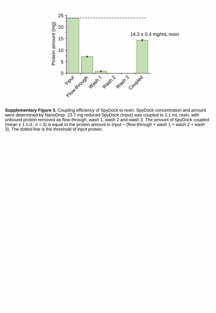
Supplementary Figure 3. Coupling efficiency of SpyDock to resin. SpyDock concentration and amount were determined by NanoDrop. 23.7 mg reduced SpyDock (Input) was coupled to 1.1 mL resin, with unbound protein removed as flow-through, wash 1, wash 2 and wash 3. The amount of SpyDock coupled (mean ± 1 s.d., n = 3) is equal to the protein amount in Input – (flow-through + wash 1 + wash 2 + wash 3). The dotted line is the threshold of input protein.
0
5
10
15
20
25
Pro
tein
am
ou
nt (m
g)
14.2 ± 0.4 mg/mL resin
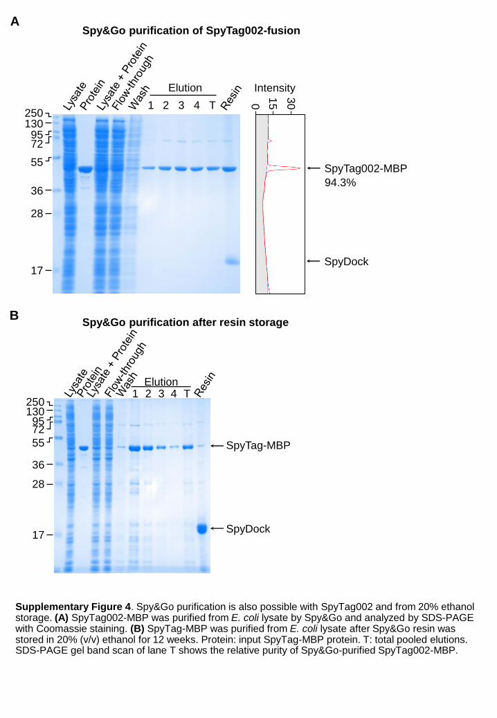
130
1 2 3 4 T
SpyTag002-MBP
SpyDock
Elution
0
15
30
Intensity
94.3%
Spy&Go purification of SpyTag002-fusion
Supplementary Figure 4. Spy&Go purification is also possible with SpyTag002 and from 20% ethanol storage. (A) SpyTag002-MBP was purified from E. coli lysate by Spy&Go and analyzed by SDS-PAGE with Coomassie staining. (B) SpyTag-MBP was purified from E. coli lysate after Spy&Go resin was stored in 20% (v/v) ethanol for 12 weeks. Protein: input SpyTag-MBP protein. T: total pooled elutions. SDS-PAGE gel band scan of lane T shows the relative purity of Spy&Go-purified SpyTag002-MBP.
9572
250
55
36
28
17
SpyTag-MBP
SpyDock
A
1 2 3 4 TElution
B
1309572
250
55
36
28
17
Spy&Go purification after resin storage

1 2 3 4 T
Elution
Supplementary Figure 5. Spy&Go purification of a fluorescent protein. (A) SpyTag-mClover3 was purified from E. coli lysate by Spy&Go and analyzed by SDS-PAGE with Coomassie staining. T: total pooled elutions. (B) Comparison of fluorescence of SpyTag-mClover3 purified by Spy&Go or Ni-NTA at different concentrations (mean ± 1 s.d., n = 3) (AU = arbitrary units).
A B
130
72
250
55
36
28
17
95
SpyTag-
mClover3
SpyDock
Flu
ore
sce
nce (
×1
00
0)
(AU
)
0.47
10
0
20
30
40
0.94 1.875 3.75 7.5 15
[SpyTag-mClover3] (µM)
Spy&Go
Ni-NTA
50
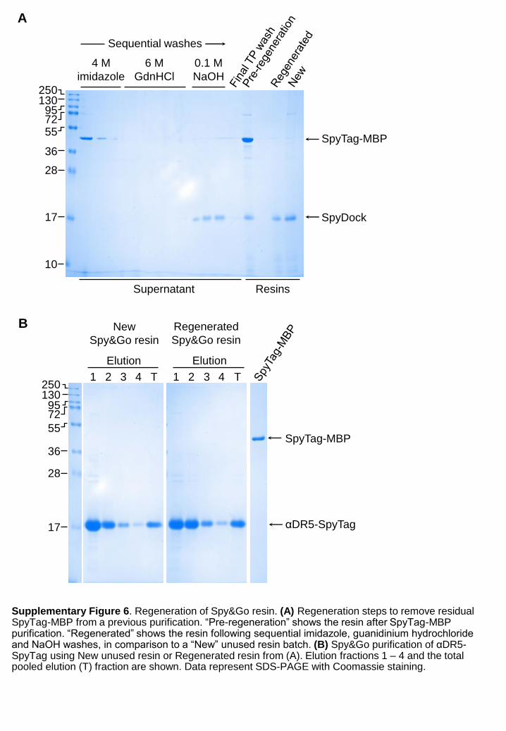
Supplementary Figure 6. Regeneration of Spy&Go resin. (A) Regeneration steps to remove residual SpyTag-MBP from a previous purification. “Pre-regeneration” shows the resin after SpyTag-MBP purification. “Regenerated” shows the resin following sequential imidazole, guanidinium hydrochloride and NaOH washes, in comparison to a “New” unused resin batch. (B) Spy&Go purification of αDR5-SpyTag using New unused resin or Regenerated resin from (A). Elution fractions 1 – 4 and the total pooled elution (T) fraction are shown. Data represent SDS-PAGE with Coomassie staining.
A
B
130
72
250
55
36
28
17
95
SpyDock
10
4 M
imidazole
6 M
GdnHCl
0.1 M
NaOH
Resins
SpyTag-MBP
130
72
250
55
36
28
17
95
αDR5-SpyTag
SpyTag-MBP
1 2 3 4 T
Elution
1 2 3 4 T
Elution
New
Spy&Go resin
Regenerated
Spy&Go resin
Supernatant
Sequential washes

10000 15000 20000 25000 30000
0
100000
200000
300000
400000
500000
Hept
Mass
Hept
10000 15000 20000 25000 30000
0.0
2.0x10⁵
4.0x10⁵
6.0x10⁵
8.0x10⁵
1.0x10⁶
Hex
Mass
Hex
10000 15000 20000 25000 30000
0.0
2.0x10⁴
4.0x10⁴
6.0x10⁴
8.0x10⁴
1.0x10⁵
1.2x10⁵
1.4x10⁵
1.6x10⁵
Pent
Mass
Pent
10000 15000 20000 25000 30000
0.0
2.0x10⁵
4.0x10⁵
6.0x10⁵
8.0x10⁵
1.0x10⁶
1.2x10⁶
1.4x10⁶
Tet
Mass
Tet
10000 15000 20000 25000 30000
0.0
2.0x10⁵
4.0x10⁵
6.0x10⁵
8.0x10⁵
1.0x10⁶
1.2x10⁶
1.4x10⁶
Tri
Mass
Tri
10000 15000 20000 25000 30000
0.0
2.0x10⁵
4.0x10⁵
6.0x10⁵
8.0x10⁵
1.0x10⁶
1.2x10⁶
1.4x10⁶
1.6x10⁶
Di
Mass
Di
30000250002000015000100000
0.2
0.4
0.6
0.8
1.0
1.2
1.4
1.6
30000250002000015000100000
0.2
0.4
0.6
0.8
1.0
1.2
1.4
30000250002000015000100000
0.2
0.4
0.6
0.8
1.0
1.2
1.4
30000250002000015000100000
0.2
0.4
0.6
0.8
1.0
1.2
1.4
1.6
30000250002000015000100000
0.2
0.4
0.6
0.8
1.0
30000250002000015000100000
1.0
2.0
3.0
4.0
5.0
Cou
nts
(x1
06)
Cou
nts
(x1
06)
Cou
nts
(x1
06)
Cou
nts
(x1
06)
Cou
nts
(x1
05)
Cou
nts
(x1
05)
Mass (Da)
Mass (Da)
Mass (Da)
Mass (Da)
Mass (Da)
Mass (Da)
Observed: 20,386
Predicted: 20,385
Observed: 21,373
Predicted: 21,372
Observed: 20,513
Predicted: 20,512
Observed: 21,891
Predicted: 21,891
Observed: 20,581
Predicted: 20,580
Observed: 22,766
Predicted: 22,765
SpyCatcher002-oDi SpyCatcher002-oTri
SpyCatcher002-oTet SpyCatcher002-oPent
SpyCatcher002-oHex SpyCatcher002-oHept
Supplementary Figure 7. Electrospray ionization mass spectra of SpyCatcher002-coiled coil subunits. The highest peak observed was compared to the predicted mass (Da).

CC-Tet
Observed Mr
C
81.562.8 ± 0.5
Expected Mr (kDa)
Rela
tive
AU
(%
)
20
50
100
0 104
25 30 35Time (min)
Mo
lar
Ma
ss (
g/m
ol)
106
105CC-Hept 140.0 ± 0.1 143.5
B
1.0
0.5
0.0
Am
plit
ude
(In
ten
sity)
1 10 100Size (Rh, nm)
CC-Tet
CC-Hept
5.4 ± 1.6 nm
6.5 ± 1.9 nm
ACC-Tet CC-Hept
Supplementary Figure 8. Characterization of alternative coiled-coils for the Spy oligomerization toolbox. (A) CC-Tet and CC-Hept were evaluated for oTet and oHept. The coiled coils are shown with each color representing one chain and the N-terminus as a blue ball. (B) DLS of SpyCatcher002-CC-Tet and SpyCatcher002-CC-Hept, with hydrodynamic radius (Rh) (mean ± 1 s.d., n = 10) for each assembly. (C) SEC-MALS of SpyCatcher002-CC-Tet and SpyCatcher002-CC-Hept. The peak shows the normalized absorbance units (AU) at 280 nm of the SpyCatcher002-oligomer from SEC. The horizontal line shows the distribution of molar mass (g/mol) in the peak from MALS. Observed and expected molecular weights (Mr) are shown alongside, with error bars representing the uncertainty in fit to the molar mass curve.
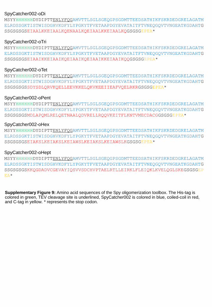
SpyCatcher002-oDi
MSYYHHHHHHDYDIPTTENLYFQGAMVTTLSGLSGEQGPSGDMTTEEDSATHIKFSKRDEDGRELAGATM
ELRDSSGKTISTWISDGHVKDFYLYPGKYTFVETAAPDGYEVATAITFTVNEQGQVTVNGEATKGDAHTG
SSGSGSGSEIAALKKEIAALKQENAALKQEIAALKKEIAALKQGSGSGEPEA*
SpyCatcher002-oTri
MSYYHHHHHHDYDIPTTENLYFQGAMVTTLSGLSGEQGPSGDMTTEEDSATHIKFSKRDEDGRELAGATM
ELRDSSGKTISTWISDGHVKDFYLYPGKYTFVETAAPDGYEVATAITFTVNEQGQVTVNGEATKGDAHTG
SSGSGSGSEIAAIKKEIAAIKQEIAAIKQEIAAIKKEIAAIKQQGSGSGEPEA*
SpyCatcher002-oTet
MSYYHHHHHHDYDIPTTENLYFQGAMVTTLSGLSGEQGPSGDMTTEEDSATHIKFSKRDEDGRELAGATM
ELRDSSGKTISTWISDGHVKDFYLYPGKYTFVETAAPDGYEVATAITFTVNEQGQVTVNGEATKGDAHTG
SSGSGSGSSDYSDLQRVKQELLEEVKKELQKVKEEIIEAFVQELRKRGSGSGEPEA*
SpyCatcher002-oPent
MSYYHHHHHHDYDIPTTENLYFQGAMVTTLSGLSGEQGPSGDMTTEEDSATHIKFSKRDEDGRELAGATM
ELRDSSGKTISTWISDGHVKDFYLYPGKYTFVETAAPDGYEVATAITFTVNEQGQVTVNGEATKGDAHTG
SSGSGSGSMDLAPQMLRELQETNAALQDVRELLRQQVKEITFLKNTVMECDACGGSGSGEPEA*
SpyCatcher002-oHex
MSYYHHHHHHDYDIPTTENLYFQGAMVTTLSGLSGEQGPSGDMTTEEDSATHIKFSKRDEDGRELAGATM
ELRDSSGKTISTWISDGHVKDFYLYPGKYTFVETAAPDGYEVATAITFTVNEQGQVTVNGEATKGDAHTG
SSGSGSGSEIAKSLKEIAKSLKEIAWSLKEIAKSLKEIAWSLKGSGSGEPEA*
SpyCatcher002-oHept
MSYYHHHHHHDYDIPTTENLYFQGAMVTTLSGLSGEQGPSGDMTTEEDSATHIKFSKRDEDGRELAGATM
ELRDSSGKTISTWISDGHVKDFYLYPGKYTFVETAAPDGYEVATAITFTVNEQGQVTVNGEATKGDAHTG
SSGSGSGSKKQGDADVCGEVAYIQSVVSDCHVPTAELRTLLEIRKLFLEIQKLKVELQGLSKEGSGSGEP
EA*
Supplementary Figure 9: Amino acid sequences of the Spy oligomerization toolbox. The His-tag is colored in green, TEV cleavage site is underlined, SpyCatcher002 is colored in blue, coiled-coil in red, and C-tag in yellow. * represents the stop codon.