spinal instability as defined by the three-column spine ... · pdf filespinal instability as...
TRANSCRIPT

Spinal Instability as Defined by the Three-column Spine Concept in Acute Spinal Trauma
FRANCIS DENIS, M.D., F.R.C.S.(C.), F.A.C.S.*
This article is a presentation of the concept of the three-column spine. The concept evolved from a retrospective review of 41 2 thoracolumbar spine injuries and observations on spinal instability. The posterior column consists of what Holdsworth de- scribed as the posterior ligamentous complex. The middle column includes the posterior longitudinal ligament, posterior annulus fibrosus, and posterior wall of the vertebral body. The anterior column consists of the anterior vertebral body, anterior an- nulus fibrosus, and anterior longitudinal ligament. Major spinal injuries are classified into four dif- ferent categories, all definable in terms of the degree of involvement of each of the three columns. Each type is defined also in terms of its pathomechanics, roentgenograms, and computerized axial tomo- grams, as well as in terms of its particular stability. The compression fracture is basically stress failure of the anterior column with an intact middle column. The burst fracture indicates failure under compres- sion of both the anterior and middle columns. The seat-belt-type spinal fracture is the result of failure of the posterior and middle columns under tension with an intact anterior hinge. In fracturedisloca- tions, the structure of all three columns fails from forces acting to various degrees from one or another direction.
Spinal instability was defined by Holds- worth6 as rupture of the posterior ligamentous
Assistant Professor of Orthopaedic Surgery, University of Minnesota, Orthopedic Surgeon in Charge of the Spine Service, St. Paul-Ramsey Medical Center, and Staff, Spine Service, Gillette Children’s Hospital.
Reprint requests to Francis Denis, M.D., 640 Jackson Street, St. Paul, MN 55101.
Received: November 21, 1983.
complex. This was confirmed by Roafs15 study of the mechanics of spinal injuries in which rupture of normal spinal ligaments could not be produced by hyperextension or hyperflexion. The implication was that rupture of the posterior ligamentous complex was not compatible with a stable compression fracture but was pathognomonic of instability initiated by either rotation or translation. Heuritsch and Bohler2 had an excellent intuitive un- derstanding of the pathomechanics of spinal fractures. They created drawings demonstrat- ing several cases of compression fractures seen in conjunction with disruption of the inter- spinous ligament.
According to Bohler,’ in 1932 Heuritsch made an accurate sketch of what was to be called, 16 years later, a Chance fra~ture.~ In spite of accumulating clinical
it has taken some recent biomechanic studies10.12.14.15 to demonstrate that subluxa- tion, dislocation, and simple instability appear only when the posterior longitudinal ligament and part of the disc are tom in conjunction with the posterior or the anterior ligamentous complex.
The past decade has shown the term “in- stability” to be a key word in therapeutic in- dications because it equates, in many cases, with a need for internal stabilization. The pur- pose of this paper is to introduce a classifi- cation based on the new concept of the three- column spine.s
8.10.17
65

Clinrlll Orthopeedics 66 Denis and Aaated Research
THE THREE-COLUMN SPINE
FIG. 1. Illustrations of the anterior, middle, and posterior columns. This and all succeeding figures are published with permission from Francis Denis: The three-column spine and its significance in the classification of acute thoracolumbar spinal injuries. Spine 8:817, 1983.
FIG. 2. Anterior compression fracture with dis- ruption of the inferior end-plate (Type C compres- sion fracture). Note the normal height of the pos- terior part of the vertebral body.
Recent biomechanic evidence',' ' . I 3 . l 4 shows that complete rupture of the posterior liga- mentous complex alone is not sufficient to establish instability (Fig. 1). Further biome- chanic data demonstrate that additional rup- ture of the posterior longitudinal ligament and posterior annulus fibrosus permits instability in flexion. Complete dislocation requires fur- ther disruption of the disc and stripping or disruption of the anterior longitudinal liga- ment. It appears logical, therefore, to separate the posterior longitudinal ligament, the pos- terior annulus fibrosus, and the posterior ver- tebral body into a third column independent of the two others, which plays its own role in the sequence of spinal injury.4
The posterior column remains essentially the same as described by Holdsworth.6 It is formed by the posterior bony complex (pos- terior arch) alternating with the posterior lig- amentous complex: supraspinous ligament, interspinous ligament, capsule, and ligamen- tum flavum. The middle column is formed by the posterior longitudinal ligament, pos- terior annulus fibrosus, and the posterior wall of the vertebral body. The anterior column is formed by the anterior longitudinal ligament, the anterior annulus fibrosus, and the anterior part of the vertebral body.
CLASSIFICATION OF SPINAL FRACTURES
The minor injuries represented by fractures of transverse processes, articular processes, pars interarticularis, and spinous processes in- volve only a part of the posterior column and do not lead to acute instability. The more sig- nificant spinal injuries are classified into four different categories.
COMPRESSION FRACTURES
Definition. The compression fracture is a failure under compression of the anterior col- umn (Fig. 2). The middle column is intact and acts as a hinge. The more severe the

Number 189 October, 1984 SDinal lnstabilitv 67
compression fracture, the more likely it will be to present, in addition to the anterior wedg- ing, a partial failure of the posterior column, indicating the tension forces at that level. The fact that the middle column is intact is of major importance because it prevents the fracture from subluxation or compression of the neural elements by fragmentation and re- tropulsion of the fragment of the posterior wall into the canal. Both of these instances are neurologic threats encountered in fracture- dislocations for the former and in burst frac- tures for the latter.
Roentgenographic characteristics. The lat- eral film shows a normal posterior body cortex and a normal height of the posterior vertebral body (intact middle column). There is no sub- luxation of the body above or the body below. The interspinous distance of the compressed segment is increased in proportions that are geometrically expected from the angulation at that level. The anteroposterior film shows the lateral wedging in lateral compression fractures (Fig. 3).
CA T scan characteristics. Computerized axial tomography is rarely indicated in compression fractures, but when done it will demonstrate an intact vertebral ring (intact middle column). There is no retropulsion of bone into the canal (Fig. 4).
FIG. 3. Lateral compression fracture.
BURST FRACTURES
Definition. The burst fracture results from failure under axial load of both the anterior and the middle columns originating at the level of one or both end-plates of the same vertebra.
Roentgenographic characteristics. The lat- eral roentgenogram demonstrates a fracture of the posterior wall cortex, loss of height of the posterior vertebral body, and tilting and retropulsion of the fragment of bone into the canal of either or both end-plates (compression failure of the middle column) (Fig. 5) . The anteroposterior roentgenogram demonstrates the pathognomonic increase Of the intew- diculate distance, the vertical laminar fracture, and the splaying of the posterior joints (Fig.
FIG. 4. Computerized axial tomogram of a compression fracture. Note anterior end-plate frac- ture and totally intact posterior wall of the vertebral body.

Clinical OnhOpaedlcs 68 Denis and Related Research
L4, and LS). The majority of burst fractures involve only one plate (the superior one in most cases). For this reason, five different types of burst fractures are described (Fig. 8). Type A: Fracture of both end-plates due to pure axial load. The bone is retropulsed into the canal at the level of both discs adjacent to the comminuted vertebra. Type B: Fracture of the superior end-plate. This is the most common burst fracture. I t is encountered mainly at the thoracolumbar junction and its mechanism is a combination of axial load with flexion. De- compression should be performed at the upper level only when indicated. Type C: Fracture of the inferior end-plate. This fracture pattern is rare. The mechanism of injury also appears to be axial load and flexion. Type D: Burst rotation. This fracture could be misdiagnosed as a fracture-dislocation because of the rota- tional component of injury. It presents, how- ever, all the pathognomonic signs of burst
FIG. 5 . Lateral tomogram of a burst fracture (Type B) showing severe disruption of superior end- plate, loss of height of posterior vertebral body. and fracture of posterior wall of body. The arrow shows the large fragment retropulsed into the canal.
6) . The latter two signs are another expression of the increase of the interpediculate distance leading to the splay of the entire posterior arch. The vertical fracture of the lamina may not be seen at the time of surgery because in most cases it is a greenstick fracture of the anterior cortex of the lamina with an intact posterior cortex. The operator should decorticate the posterior arch very carefully.
CAT scan characteristics. The vertebral ring is fractured both anteriorly and posteriorly. This fragment of bone retropulsed from the vertebral body is sequestrated in the spinal canal and locked in position by the posterior arch (Fig. 7).
Classification of burst fractures. The burst fracture described by Holdsworth with com- minution of the entire vertebra and without kYPhosis involves both end-Plates and 1s mainly localized in the low lumbar region (L3,
FIG. 6. tornogram of a burst fracture. Note the increased interpediculate distance (34 mm) and the vertical laminar fracture.

Number 189 October, 1984 Spinal Instability 69
FIG. 7. Computerized axial tomogram of a burst fracture. Note the large fragment of bone retro- pulsed from the posterior wall.
fractures, with increase of the interpediculate distance, comminution of the vertebral body, vertical fracture of the lamina, retropulsion of bone into the canal, and loss of posterior height. Computerized axial tomography as well as myelography may identify the large
FIG. 8. Classification of burst fractures: Types A, B, and C are mainly di- agnosed on lateral roent- genograms; their antero- posterior roentgenograms reveal the basic pathog- nomonic features seen in Figs. 5-6. Types D and E are diagnosed on an- teroposterior roentgeno- grams. The lateral film of a Type D looks like a Type A, whereas the lat- eral film of a Type E may look like Type A, B, or C.
A
fragment of bone occluding the canal. The mechanism of this injury is a combination of axial load and rotation. Type E: Burst lateral flexion. This type of fracture differs from the lateral compression fracture in that it presents an increase of the interpediculate distance on
B
D
C
E

70 Denis Clinlcal Onhopaedocs
and Related Research
anteroposterior roentgenogram. The lateral film will disclose the retropulsion of bone from the posterior wall into the canal. Computer- ized axial tomography again identifies the ex- truded fragment and shows it to be somewhat more lateralized as compared with the other types of burst fractures.
SEAT-BELT-TYPE INJURIES
Dejnition. These injuries represent a failure of both the posterior and middle columns un- der tension forces generated by flexion with its axis placed in the anterior column. The anterior part of the anterior column may par- tially fail under compression but will not lose its role as a hinge. This type of injury will be unstable in flexion and will not present with association of subluxation, which indicates that the anterior hinge is also disrupted and that fracture dislocation is present.
Roentgenographiccharacteristics. A pathog- nomonic sign of this type of injury is the hor- izontal split of the transverse processes as well as of the pedicles. There may be a horizontal fracture of the spinous process or of the pars intra-articularis, or also, in some cases, an in- crease of the interspinous distance with a min- imal spinous process avulsion. The height of the posterior vertebral body is increased or there may be an increase of the disc space posteriorly at the level of the injury.
CAT scan characteristics. CAT scan does not provide additional information for this type of injury because the horizontal cuts are often parallel to the plane of injury. Coned- down views or lateral tomograms are more useful in terms of identifying the precise level of the fracture.
Subtypes of seat-belt-type injuries. These injuries are divided into one- and two-level lesions (Fig. 9). One-level lesions may present as a simple Chance fracture going through bone or as a ligamentous disruption starting at the level of the supraspinous ligament and proceeding to the anterior part of the disc. Two-level lesions are comparable to the con-
dition presented in hangman’s fracture in which the middle column may rupture either through the bone or through the disc.
FR ACTURE-DISLOCATIONS
Dejnition. This is the most unstable of in- juries and presents with failure of all three columns under compression. tension, rotation, or shear.
Roentgenographic Characteristics. Its pa- thognomonic sign is the subluxation or dis- location seen on anteroposterior or lateral roentgenograms. Some indirect signs may suggest this type of injury in the presence of multiple rib fractures, multiple transverse process fractures, fracture of a unilateral ar- ticular process, slight increased height of a disc, or minimal vertebral body offset.
Siihtjpes c?efructure-dislocations. There are three major mechanisms in fracture-disloca- tions: flexion rotation, shear, and flexion dis- traction.
Flexion-rotation fracture-dislocation (Fig. 10). This injury has been described by Holds- worth6 and also by Roaf.’’ There is usually a complete rupture of the posterior and middle columns under tension and rotation. The an- terior column may fail in rotation or some- times in varying combinations of compression and rotation. The failure at the level of the middle and anterior columns may occur through the vertebral body or purely through the disc. Roentgenographic characteristics, the pathognomonic sign of the fracture-disloca- tion, will be the subluxation or dislocation of a vertebral segment on another one. There is frequently an increase of the interspinous dis- tance and a displaced fracture of a superior articular process on one side, indicating ro- tational failure of the posterior column. Mul- tiple transverse process fractures and multiple rib fractures are frequent. A slight amount of rotation between the segment above and the segment below may be observed.
computerized axial tomography in flexion rotation. This may demonstrate the occlusion

Number 189 October. 1984 Spinal Instability 71
FIG. 9. (Upper Left) One-level seat-belt-type injury through the ligaments. (Upper Right) One-level seat-belt-type injury through bone (Chance fracture). (Lower Left) Two-level seat-belt-type injury through ligaments at the level of the middle column. (Lower Right) Two-level seat-belt-type injury through ligaments at the level of the middle column.
of the canal resulting from the offset of one vertebra on another (Fig. 11). It will occa- sionally show jumped facets not identifiable on plain roentgenograms. It may also show a fragment of bone retropulsed into the canal,
indicating a burst fracture. However, a sig- nificant difference between the burst fracture fragment and the fragment seen in fracture- dislocations is that the burst fracture is covered in the former by an intact posterior longitu-

Clinical Onhopaedi and Reiated Research 72 Denis
A
FIG. 10. (Upper Left) Lateral diagram of a fracturedislocation of the flexion-rotation type through the disc. Note the superior articular process fracture on one side only. (Upper Right) Anteroposterior diagram of a fracturedislocation of the flexion-rotation type through the disc. Note the fracture of the left superior articular surface. (Lower Left) Lateral diagram of a fracturedislocation of the flexion-rotation type through bone (slice fracture). (Lower Right) Anteroposterior diagram of a fracture-dislocation of the flexion- rotation type through bone (slice fracture). Note the difference in rotation between both spinal segments.
dinal ligament, whereas this same structure is tom in the latter. The implication of this is twofold: first, in terms of stability of the injury,
and second, in terms of reduction of the re- tropulsed fragment by ligamentotaxis.
Shear type of fracture-dislocation. This in-

Number 189 October, 1984 Spinal Instability 73
FIG. 1 I . Computerized axial tomogram of an L2- L3 lumbar fracture-dis- location of the flexion-ro- tation type. Note the fracture of the right su- perior articular process of L3 and the 80% neural canal obstruction result- ing from the malalign- ment.
jury results from an extension type of mech- anism in which the anterior longitudinal lig- ament is disrupted. The disc is first tom an- teriorly to posteriorly until the continued shearing force translates the upper segment on top of the inferior segment, or vice versa. ( 1 ) In the posteroanterior shear subtype (Fig. 12), the segment above is sheared off forward on top of the segment below. The posterior arch of the last one or two vertebrae of the upper segment is usually fractured in the translation, leaving a floating posterior arch behind. The frequency of dural tear and com- plete paraplegia is very high in this type of fracture. (2) In the anteroposterior shear, the segment above shears off on the segment below in a posterior direction. Its posterior arch has nothing to clear during its posterior displace- ment; therefore, no free-floating laminae exist (Figs. 13 and 14).
Fracturedislocation of the flexiondistrac- tion type. This injury resembles the seat-belt type of injury with disruption of both the pos- tenor and middle columns under tension. However, in addition, it presents tear of the anterior annulus fibrosus, allowing stripping of the anterior longitudinal ligament during subluxation or dislocation (Fig. 15).
Roentgenographic characteristics. This is a symmetrical type of injury with frequent hor- izontal split of the transverse process pedicle and spinous process. This injury may be sub-
FIG. 12. Lateral diagram of a posteroanterior shear injury. Note the intact anterior vertebral bod- ies. The spinous process or entire posterior arch may be fractured by the same mechanism, leaving a “floating lamina” behind.
74 Denis Clinical onhopaedlcs and Related Research
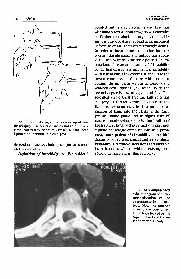
FIG. 13. Lateral diagram of an anteroposterior shear injury. The posterior arches and anterior ver- tebral bodies may be entirely intact, but the three ligamentous columns are disrupted.
divided into the seat-belt-type injuries in one- and two-level types.
Definition of instability. As Whitesides"
pointed out, a stable spine is one that can withstand stress without progressive deformity or further neurologic damage. An unstable spine is thus one that may lead to an increased deformity or an increased neurologic deficit. In order to incorporate that notion into the present classification, the author has subdi- vided instability into the three potential com- binations of these complications. ( 1) Instability of the first degree is a mechanical instability with risk of chronic kyphosis. It applies to the severe compression fracture with posterior column disruption as well as to some of the seat-belt-type injuries. (2) Instability of the second degree is a neurologic instability. The so-called stable burst fracture falls into this category as further vertical collapse of the fractured vertebra may lead to more retro- pulsion of bone into the canal in the early post-traumatic phase and to higher risks of post-traumatic spinal stenosis after healing of the fracture. Both of these situations may pre- cipitate neurologic complications in a previ- ously intact patient. (3) Instability of the third degree is both a mechanical and a neurologic instability. Fracture-dislocations and unstable burst fractures with or without existing neu- rologic damage are in this category.
FIG. 14. Computerized axial tomogram of a frac- ture-dislocation of the anteroposterior shear type. Note the anterior aspect of the superior ver- tebral body locked on the superior facets of the in- ferior vertebral body.

Number 189 October. 1984 Spinal Instability 75
DISCUSSION
Nicoll” reported a classification of dorsal and lumbar spinal injuries based on four main types: anterior wedge fracture, lateral wedge fracture, fracture-dislocation, and isolated fracture of the neural arch. This classification may be confusing because under the heading “Neural Arch Fractures” the author includes Chance fractures with chronic spondylolis- thesis as well as traumatic spondylolisthesis. Nicoll believed that rotation was responsible for these neural arch fractures and that the lateral wedge fracture was due to flexion ro- tation; both of these mechanisms were for- merly attributed to fracture-dislocations by most authors.
Olof Perey ” subjected one-level spinal mo- tion segments consisting of two vertebrae and the intervening disc to a strong force of axial load with a fast rate of loading. The amount of stress used was established at a right angle to the cross-sectional dimension. Disc spaces were visualized by diskography. Four exper- imental series were made in which the max- imum forces were calculated to be 1050, 1250, and 1350 kiloponds during approximately 0.06 seconds. A total of 76 experiments were performed, which demonstrated how vertebral end-plate fractures occurred experimentally. Only two cases of double end-plate disruption were observed (two out of 24 experiments done with two-level spinal motion segments as opposed to frequent rupture of both end- plates encountered in the clinical series). It should be noted also that the changes at the level of the posterior arch were not mentioned.
Roaf” demonstrated that discs, joints, and ligaments were rather resistant to distraction, flexion, and extension but were very vulner- able to rotation and horizontal shearing forces. His experimental work suggested that rupture of the anterior longitudinal ligament in hy- perextension was impossible and that neural arches fractured first. When the spine was ro- tated in extension, the anterior longitudinal ligament easily ruptured; therefore, the so- called hyperextension injury was actually a
FIG. IS. Lateral diagram of a fracturedislocation of the flexion-distraction type. The posterior, mid- dle, and anterior ligamentous columns are dis- rupted, but the anterior longitudinal ligament is intact and strips off the vertebral body below.
rotation-extension injury. Similarly, Roaf was unable to experimentally reproduce rupture of the posterior ligamentous complex as ob- served clinically in severe compression frac- tures. It appears that his results were greatly influenced by the limited degree of freedom of his biomechanic testing apparatus, which lacked, in particular, the essential versatility of being able to combine predetermined vec- tors of forces. Smith and Kaufer16 reported 24 lumbar spine injuries sustained by persons wearing a lap seat belt who were involved in motorcycle accidents. Twenty of these patients presented a specific pattern of lumbar spine injury: a transverse type of lumbar fracture believed to be extremely rare in unbelted in- dividuals. The authors emphasized the risk of abdominal contusions associated with the spinal injury and characterized the disruption as osseous, ligamentous, or both. There was little or no decrease in anterior vertical height of the involved vertebral body. Most disrup tion occurred between the first and third lum- bar vertebrae. It was assumed that the axis of flexion of the spine during injury was at the level of the lap belt pressing over a thick layer

76 Denis Chnlcal Orthopedics
and Related Research
of tissue separating it from the spine. The im- plications of such an access are that the spine is submitted to pure distraction forces. This assumption may be challenged for two rea- sons. Firstly, the lap belt acts as a fulcrum that becomes the access of flexion only if and when the bending strength of the "spinal beam" under consideration is nil at the point of application of the fulcrum. Secondly, in- direct evidence of this was demonstrated by Gordon Armstrong, who pointed out that in the 15 Chance fractures in the present series there was a slight vertical shortening of the anterior vertebral body, demonstrating post fucto that the instantaneous axis of flexion was somewhere in the anterior column at the time of injury (unpublished data). It should be added also that an instantaneous axis of flexion is dynamic, not static, and moves dur- ing the sequence of rupture from somewhere in the middle column to somewhere in the anterior column as the ligaments or bony parts rupture posteriorly to anteriorly. Panjabi ef uf.," in an individual study conducted to es- tablish the thresholds of thoracic spine sta- bility, demonstrated that under flexion loads the thoracic functional spinal unit is on the verge of instability when all ligaments posterior to and including the posterior half of the discs are cut. Nagel el uL9 tested five fresh human cadavers to determine range-of-motion mea- surements between the first and second lumbar vertebrae after progressive disruption of the motion segment. Their study showed that an anterior flexion of 20" or a lateral flexion of 10" seen on a routine roentgenogram indi- cated that all posterior ligaments and at least part of the annulus fibrosus must be disrupted.
1 .
2.
3.
4.
5 .
6.
7.
8.
9.
10
II
I 2
13
14.
15.
16.
17.
18.
REFERENCES Bedbrook. G. M.: Stability of spinal fractures and fracture dislocations. Paraplegia 9:23. 197 I . Bohler. L.: The Treatment of Fracture. ed. 5. New York, Grune & Stratton. 1956. pp. 323-340. Chance, C . Q.: Note on a type of flexion fracture of the spine. Br. J. Radiol. 21:452. 1948. Denis. F.: The three column spine and its significance in the classification of acute thoracolurnbar spinal injuries. Spine 8317 . 1983. Denis. F., and Armstrong. G. W. D.: Compression fractures versus burst fractures in the lumbar and thoracic spine. J . Bone Joint Surg. 63B(3):462, 1981. Holdsworth. F. W.: Fractures, dislocations and frac- ture-dislocations of the spine. J. Bone Joint Surg. 52A(R):1534. 1970. Howorth. M. B.: Fracture of the spine. .Am. J. Surg. 92:573. 1956. Kaufer. H.. and Hayes. J. T.: Lumbar fracture dis- location. J . Bone Joint Surg. 48A:712. 1966. Nagel, D. A,. Koogle. T. A,. Piziali, R. L., and Perkash, 1.: Stability of the upper lumbar spine following pro- gressive disruptions and the application of the indi- vidual internal and external fixation devices. J. Bone Joint Surg. 63A( I ):62. I98 I . Nicoll. E. A.: Fractures of the dorsolumbar spine. J. Bone Joint Surg. 31B:376. 1949. Panjabi. M. M.. Hausfeld, J. N.. and White, A. A,: A biomechanical study of the ligamentous stability of the thoracic spine in man. Acta Orthop. Scand. 52315. 1981. Perey. 0.: Fracture ofthe vertebral end plate in lumbar spine. Acta Orthop. Scand. (Supp1.)25: I , 1957. Purcell. G. A,, Markolf. K. L.. and Dawson, E. G.: Twelfth thoracic-first lumbar vertical mechanical stability of fractures after Hanington rod instrumen- tation. J. Bone Joint Surg. 63A(l):71, 1981. Reuber. M.. Schultz, A,. Denis. F., and Spencer, D.: Bulging of lumbar intervertebral discs. J. Biomech. Eng. 104:187. 1982. Roaf. R.: A study of the mechanics of spinal injuries. J. Bone Joint Surg. 42B(4):810, 1960. Smith, W. S.. and Kaufer, H.: Patterns and mecha- nisms of lumbar injuries associated with lap seat belts. J. Bone Joint Surg. 51A:239. 1969. Stauffer. E. S.. and Neil. J. L.: Biomechanical analysis of structural stability of internal fixation in fractures of the thoracolumbar spine. Clin. Orthop. I 12: 159, 1975. Whitesides. T. E.: Traumatic kyphosis of the thora- columbar spine. Clin. Orthop. I28:78, 1977.