spinal cord injury
TRANSCRIPT
ABC OFSPINAL CORD
INJURY: Fourth edition
BMJ Books

ABC OFSPINAL CORD INJURY

ABC OFSPINAL CORD INJURY
Fourth edition
Edited by
DAVID GRUNDYHonorary Consultant in Spinal Injuries,
The Duke of CornwallSpinal Treatment Centre,
Salisbury District Hospital, UK
ANDREW SWAINClinical Director, Emergency Department,
MidCentral Health, Palmerston Hospital North,New Zealand
© BMJ Books 2002BMJ Books is an imprint of the BMJ Publishing Group
BMJ Publishing Group 1986, 1993, 1996
All rights reserved. No part of this publication may be reproduced,stored in a retrieval system, or transmitted, in any form or by anymeans, electronic, mechanical, photocopying, recording and/orotherwise, without the prior written permission of the publishers.
First published 1986Reprinted 1989Reprinted 1990Reprinted 1991
Second edition 1993Reprinted 1994
Third edition 1996Reprinted 2000
Fourth edition 2002
by the BMJ Publishing Group, BMA House, Tavistock Square,London WC1H 9JR
British Library Cataloguing in Publication DataA catalogue record for this book is available from the British Library
ISBN 0-7279-1518-5
Typeset by Newgen Imaging Systems (P) Ltd., Chennai, IndiaPrinted in Malaysia by Times Offset
Cover image: Lumbar spine. Coloured x ray of four lumbarvertebrae of the human spine, seen in antero-posterior view.
Reproduced with permission from Science Photo Library.
v
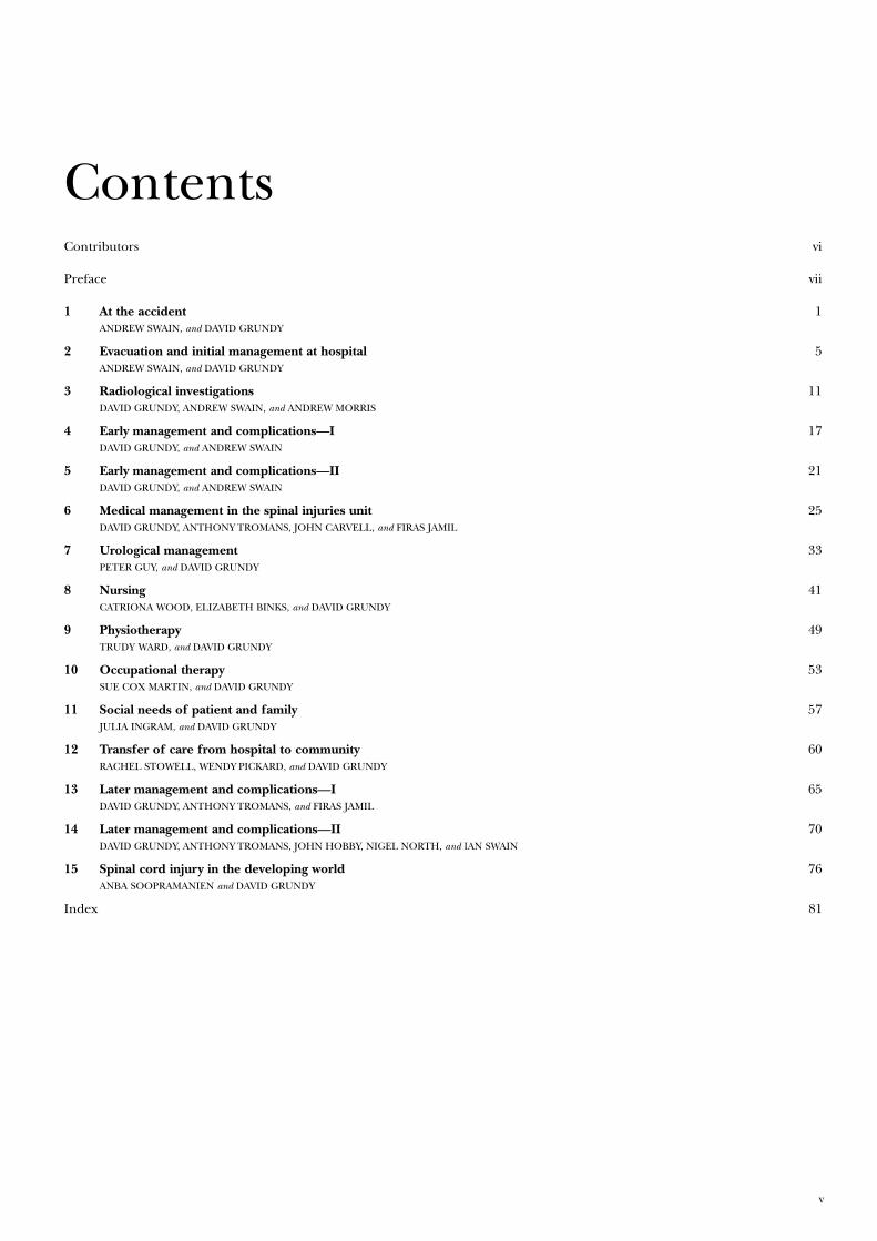
ContentsContributors vi
Preface vii
1 At the accident 1ANDREW SWAIN, and DAVID GRUNDY
2 Evacuation and initial management at hospital 5ANDREW SWAIN, and DAVID GRUNDY
3 Radiological investigations 11DAVID GRUNDY, ANDREW SWAIN, and ANDREW MORRIS
4 Early management and complications—I 17DAVID GRUNDY, and ANDREW SWAIN
5 Early management and complications—II 21DAVID GRUNDY, and ANDREW SWAIN
6 Medical management in the spinal injuries unit 25DAVID GRUNDY, ANTHONY TROMANS, JOHN CARVELL, and FIRAS JAMIL
7 Urological management 33PETER GUY, and DAVID GRUNDY
8 Nursing 41CATRIONA WOOD, ELIZABETH BINKS, and DAVID GRUNDY
9 Physiotherapy 49TRUDY WARD, and DAVID GRUNDY
10 Occupational therapy 53SUE COX MARTIN, and DAVID GRUNDY
11 Social needs of patient and family 57JULIA INGRAM, and DAVID GRUNDY
12 Transfer of care from hospital to community 60RACHEL STOWELL, WENDY PICKARD, and DAVID GRUNDY
13 Later management and complications—I 65DAVID GRUNDY, ANTHONY TROMANS, and FIRAS JAMIL
14 Later management and complications—II 70DAVID GRUNDY, ANTHONY TROMANS, JOHN HOBBY, NIGEL NORTH, and IAN SWAIN
15 Spinal cord injury in the developing world 76ANBA SOOPRAMANIEN and DAVID GRUNDY
Index 81
vi
ContributorsElizabeth BinksSenior Sister, The Duke of Cornwall Spinal Treatment Centre,Salisbury District Hospital
John CarvellConsultant Orthopaedic Surgeon, Salisbury District Hospital
Sue Cox MartinSenior Occupational Therapist, The Duke of Cornwall SpinalTreatment Centre, Salisbury District Hospital
Peter GuyConsultant Urologist, Salisbury District Hospital
John HobbyConsultant Plastic Surgeon, Salisbury District Hospital
Julia IngramSocial Worker, The Duke of Cornwall Spinal Treatment Centre,Salisbury District Hospital
Firas JamilConsultant in Spinal Injuries, The Duke of Cornwall SpinalTreatment Centre, Salisbury District Hospital
Andrew MorrisConsultant Radiologist, Salisbury District Hospital
Nigel NorthConsultant Clinical Psychologist, The Duke of Cornwall SpinalTreatment Centre, Salisbury District Hospital
Wendy PickardPressure Nurse Specialist, The Duke of Cornwall SpinalTreatment Centre, Salisbury District Hospital
Anba SoopramanienConsultant in Spinal Injuries, The Duke of Cornwall SpinalTreatment Centre, Salisbury District Hospital
Rachel StowellCommunity Liaison Sister, The Duke of Cornwall SpinalTreatment Centre, Salisbury District Hospital
Ian SwainProfessor of Medical Physics and Bioengineering, SalisburyDistrict Hospital
Anthony TromansConsultant in Spinal Injuries, The Duke of Cornwall SpinalTreatment Centre, Salisbury District Hospital
Trudy WardTherapy Manager, The Duke of Cornwall Spinal TreatmentCentre, Salisbury District Hospital
Catriona WoodSenior Clinical Nurse, The Duke of Cornwall Spinal TreatmentCentre, Salisbury District Hospital
The fourth edition of the ABC of Spinal Cord Injury, although now redesigned in the current ABC style, has the same goals asprevious editions. It assumes spinal cord injury to be the underlying condition, and it must be remembered that a slightly differentapproach is used for trauma patients in whom spinal column injury cannot be excluded but cord damage is not suspected.
This ABC aims to present in as clear a way as possible the correct management of patients with acute spinal cord injury, step bystep, through all the phases of care and rehabilitation until eventual return to the community.
The book discusses how to move the injured patient from the scene of the accident, in conformity with pre-hospital techniquesused by ambulance services in developed countries, and it incorporates refinements in advanced trauma life support (ATLS)which have developed over the past decade.
The text explains how to assess the patient, using updated information on the classification and neurological assessment ofspinal cord injury.
There is a greater emphasis in making the correct diagnosis of spinal injury and established cord injury—unfortunately,litigation due to missed diagnosis is not uncommon. The pitfalls in diagnosis are identified, and by following the step by stepapproach described, failure to diagnose these serious injuries should therefore be minimised.
Patients with an acute spinal cord injury often have associated injuries, and the principles involved in managing these injuriesare also discussed.
The later chapters follow the patient through the various stages of rehabilitation, and describe the specialised nursing,physiotherapy and occupational therapy required. They also discuss the social and psychological support needed for many of thesepatients in helping both patient and family adjust to what is often a lifetime of disability. Where applicable, the newer surgicaladvances, including the use of implants which can result in enhanced independence and mobility, are described.
Later complications and their management are discussed, and for the first time there is a chapter on the special challenges ofmanaging spinal cord injuries in developing countries, where the incidence is higher and financial resources poorer than in thedeveloped world.
David GrundyAndrew Swain
vii
Preface

1
1 At the accidentAndrew Swain, David Grundy
Spinal cord injury is a mortal condition and has beenrecognised as such since antiquity. In about 2500 BC, in theEdwin Smith papyrus, an unknown Egyptian physicianaccurately described the clinical features of traumatictetraplegia (quadriplegia) and revealed an awareness of theawful prognosis with the chilling advice: “an ailment not to betreated”. That view prevailed until the early years of thiscentury. In the First World War 90% of patients who suffereda spinal cord injury died within one year of wounding and onlyabout 1% survived more than 20 years. Fortunately, the visionof a few pioneers—Guttmann in the United Kingdom togetherwith Munro and Bors in the United States—has greatlyimproved the outlook for those with spinal cord injury,although the mortality associated with tetraplegia was still 35%in the 1960s. The better understanding and management ofspinal cord injury have led to a reduction in mortality and ahigher incidence of incomplete spinal cord damage in thosewho survive. Ideal management now demands immediateevacuation from the scene of the accident to a centre whereintensive care of the patient can be undertaken in liaison with aspecialist in spinal cord injuries.
At present the annual incidence of spinal cord injurywithin the United Kingdom is about 10 to 15 per million of thepopulation. In recent years there has been an increase in theproportion of injuries to the cervical spinal cord, and this isnow the most common indication for admission to a spinalinjuries unit.
Only about 5% of spinal cord injuries occur in children,mainly following road trauma or falls from a height greaterthan their own, but they sustain a complete cord injury morefrequently than adults.
Although the effect of the initial trauma is irreversible, thespinal cord is at risk from further injury by injudicious earlymanagement. The emergency services must avoid suchcomplications in unconscious patients by being aware of thepossibility of spinal cord injury from the nature of the accident,and in conscious patients by suspecting the diagnosis from thehistory and basic examination. If such an injury is suspected thepatient must be handled correctly from the outset.
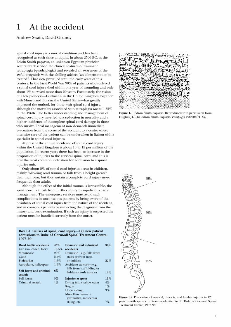
Figure 1.1 Edwin Smith papyrus. Reproduced with permission fromHughes JT. The Edwin Smith Papyrus. Paraplegia 1988:26:71–82.
1
1
2
23456789
10
40%
45%
1112
1
2
3
4
5
15%
34567
Figure 1.2 Proportion of cervical, thoracic, and lumbar injuries in 126patients with spinal cord trauma admitted to the Duke of Cornwall SpinalTreatment Centre, 1997–99.
Box 1.1 Causes of spinal cord injury—126 new patientadmissions to Duke of Cornwall Spinal Treatment Centre,1997–99
Road traffic accidents 45% Domestic and industrial 34%Car, van, coach, lorry 16.5% accidents Motorcycle 20% Domestic—e.g. falls down Cycle 5.5% stairs or from treesPedestrian 1.5% or ladders 22%Aeroplane, helicopter 1.5% Accidents at work—e.g.
Self harm and criminal 6%falls from scaffolding or
assaultladders, crush injuries 12%
Self harm 5% Injuries at sport 15%Criminal assault 1% Diving into shallow water 4%
Rugby 1%Horse riding 3%Miscellaneous—e.g. gymnastics, motocross,skiing, etc, 7%
Management at the scene of theaccident
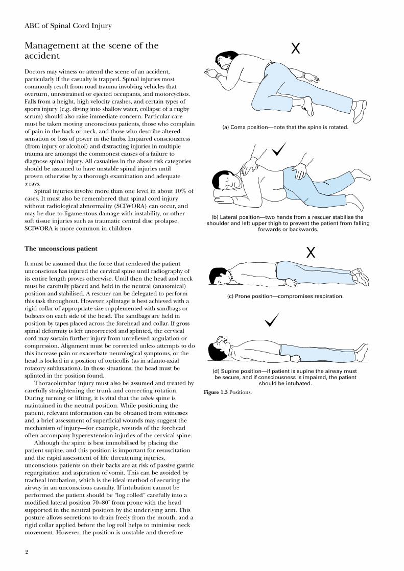
Doctors may witness or attend the scene of an accident,particularly if the casualty is trapped. Spinal injuries mostcommonly result from road trauma involving vehicles thatoverturn, unrestrained or ejected occupants, and motorcyclists.Falls from a height, high velocity crashes, and certain types ofsports injury (e.g. diving into shallow water, collapse of a rugbyscrum) should also raise immediate concern. Particular caremust be taken moving unconscious patients, those who complainof pain in the back or neck, and those who describe alteredsensation or loss of power in the limbs. Impaired consciousness(from injury or alcohol) and distracting injuries in multipletrauma are amongst the commonest causes of a failure todiagnose spinal injury. All casualties in the above risk categoriesshould be assumed to have unstable spinal injuries untilproven otherwise by a thorough examination and adequatex rays.
Spinal injuries involve more than one level in about 10% ofcases. It must also be remembered that spinal cord injurywithout radiological abnormality (SCIWORA) can occur, andmay be due to ligamentous damage with instability, or othersoft tissue injuries such as traumatic central disc prolapse.SCIWORA is more common in children.
The unconscious patient
It must be assumed that the force that rendered the patientunconscious has injured the cervical spine until radiography ofits entire length proves otherwise. Until then the head and neckmust be carefully placed and held in the neutral (anatomical)position and stabilised. A rescuer can be delegated to performthis task throughout. However, splintage is best achieved with arigid collar of appropriate size supplemented with sandbags orbolsters on each side of the head. The sandbags are held inposition by tapes placed across the forehead and collar. If grossspinal deformity is left uncorrected and splinted, the cervicalcord may sustain further injury from unrelieved angulation orcompression. Alignment must be corrected unless attempts to dothis increase pain or exacerbate neurological symptoms, or thehead is locked in a position of torticollis (as in atlanto-axialrotatory subluxation). In these situations, the head must besplinted in the position found.
Thoracolumbar injury must also be assumed and treated bycarefully straightening the trunk and correcting rotation.During turning or lifting, it is vital that the whole spine ismaintained in the neutral position. While positioning thepatient, relevant information can be obtained from witnessesand a brief assessment of superficial wounds may suggest themechanism of injury—for example, wounds of the foreheadoften accompany hyperextension injuries of the cervical spine.
Although the spine is best immobilised by placing thepatient supine, and this position is important for resuscitationand the rapid assessment of life threatening injuries,unconscious patients on their backs are at risk of passive gastricregurgitation and aspiration of vomit. This can be avoided bytracheal intubation, which is the ideal method of securing theairway in an unconscious casualty. If intubation cannot beperformed the patient should be “log rolled” carefully into amodified lateral position 70–80˚ from prone with the headsupported in the neutral position by the underlying arm. Thisposture allows secretions to drain freely from the mouth, and arigid collar applied before the log roll helps to minimise neckmovement. However, the position is unstable and therefore
ABC of Spinal Cord Injury
2
(d) Supine position—if patient is supine the airway must be secure, and if consciousness is impaired, the patient
should be intubated.
(c) Prone position—compromises respiration.
(a) Coma position—note that the spine is rotated.
(b) Lateral position—two hands from a rescuer stabilise the shoulder and left upper thigh to prevent the patient from falling
forwards or backwards.
Figure 1.3 Positions.
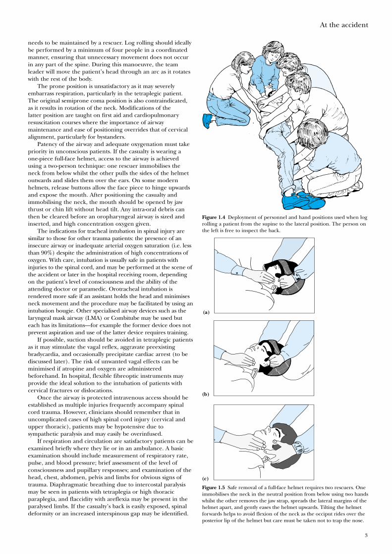
needs to be maintained by a rescuer. Log rolling should ideallybe performed by a minimum of four people in a coordinatedmanner, ensuring that unnecessary movement does not occurin any part of the spine. During this manoeuvre, the teamleader will move the patient’s head through an arc as it rotateswith the rest of the body.
The prone position is unsatisfactory as it may severelyembarrass respiration, particularly in the tetraplegic patient.The original semiprone coma position is also contraindicated,as it results in rotation of the neck. Modifications of thelatter position are taught on first aid and cardiopulmonaryresuscitation courses where the importance of airwaymaintenance and ease of positioning overrides that of cervicalalignment, particularly for bystanders.
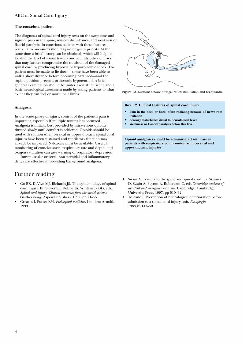
Patency of the airway and adequate oxygenation must takepriority in unconscious patients. If the casualty is wearing aone-piece full-face helmet, access to the airway is achievedusing a two-person technique: one rescuer immobilises theneck from below whilst the other pulls the sides of the helmetoutwards and slides them over the ears. On some modernhelmets, release buttons allow the face piece to hinge upwardsand expose the mouth. After positioning the casualty andimmobilising the neck, the mouth should be opened by jawthrust or chin lift without head tilt. Any intra-oral debris canthen be cleared before an oropharyngeal airway is sized andinserted, and high concentration oxygen given.
The indications for tracheal intubation in spinal injury aresimilar to those for other trauma patients: the presence of aninsecure airway or inadequate arterial oxygen saturation (i.e. lessthan 90%) despite the administration of high concentrations ofoxygen. With care, intubation is usually safe in patients withinjuries to the spinal cord, and may be performed at the scene ofthe accident or later in the hospital receiving room, dependingon the patient’s level of consciousness and the ability of theattending doctor or paramedic. Orotracheal intubation isrendered more safe if an assistant holds the head and minimisesneck movement and the procedure may be facilitated by using anintubation bougie. Other specialised airway devices such as thelaryngeal mask airway (LMA) or Combitube may be used buteach has its limitations—for example the former device does notprevent aspiration and use of the latter device requires training.
If possible, suction should be avoided in tetraplegic patientsas it may stimulate the vagal reflex, aggravate preexistingbradycardia, and occasionally precipitate cardiac arrest (to bediscussed later). The risk of unwanted vagal effects can beminimised if atropine and oxygen are administeredbeforehand. In hospital, flexible fibreoptic instruments mayprovide the ideal solution to the intubation of patients withcervical fractures or dislocations.
Once the airway is protected intravenous access should beestablished as multiple injuries frequently accompany spinalcord trauma. However, clinicians should remember that inuncomplicated cases of high spinal cord injury (cervical andupper thoracic), patients may be hypotensive due tosympathetic paralysis and may easily be overinfused.
If respiration and circulation are satisfactory patients can beexamined briefly where they lie or in an ambulance. A basicexamination should include measurement of respiratory rate,pulse, and blood pressure; brief assessment of the level ofconsciousness and pupillary responses; and examination of thehead, chest, abdomen, pelvis and limbs for obvious signs oftrauma. Diaphragmatic breathing due to intercostal paralysismay be seen in patients with tetraplegia or high thoracicparaplegia, and flaccidity with areflexia may be present in theparalysed limbs. If the casualty’s back is easily exposed, spinaldeformity or an increased interspinous gap may be identified.
At the accident
3
Figure 1.4 Deployment of personnel and hand positions used when logrolling a patient from the supine to the lateral position. The person onthe left is free to inspect the back.
Figure 1.5 Safe removal of a full-face helmet requires two rescuers. Oneimmobilises the neck in the neutral position from below using two handswhilst the other removes the jaw strap, spreads the lateral margins of thehelmet apart, and gently eases the helmet upwards. Tilting the helmetforwards helps to avoid flexion of the neck as the occiput rides over theposterior lip of the helmet but care must be taken not to trap the nose.
(a)
(b)
(c)
The conscious patient
The diagnosis of spinal cord injury rests on the symptoms andsigns of pain in the spine, sensory disturbance, and weakness orflaccid paralysis. In conscious patients with these featuresresuscitative measures should again be given priority. At thesame time a brief history can be obtained, which will help tolocalise the level of spinal trauma and identify other injuriesthat may further compromise the nutrition of the damagedspinal cord by producing hypoxia or hypovolaemic shock. Thepatient must be made to lie down—some have been able towalk a short distance before becoming paralysed—and thesupine position prevents orthostatic hypotension. A briefgeneral examination should be undertaken at the scene and abasic neurological assessment made by asking patients to whatextent they can feel or move their limbs.
Analgesia
In the acute phase of injury, control of the patient’s pain isimportant, especially if multiple trauma has occurred.Analgesia is initially best provided by intravenous opioidstitrated slowly until comfort is achieved. Opioids should beused with caution when cervical or upper thoracic spinal cordinjuries have been sustained and ventilatory function mayalready be impaired. Naloxone must be available. Carefulmonitoring of consciousness, respiratory rate and depth, andoxygen saturation can give warning of respiratory depression.
Intramuscular or rectal non-steroidal anti-inflammatorydrugs are effective in providing background analgesia.
Further reading
• Go BK, DeVivo MJ, Richards JS. The epidemiology of spinalcord injury. In: Stover SL, DeLisa JA, Whiteneck GG, eds.Spinal cord injury. Clinical outcomes from the model systems.Gaithersburg: Aspen Publishers, 1995, pp 21–55
• Greaves I, Porter KM. Prehospital medicine. London: Arnold,1999
ABC of Spinal Cord Injury
4
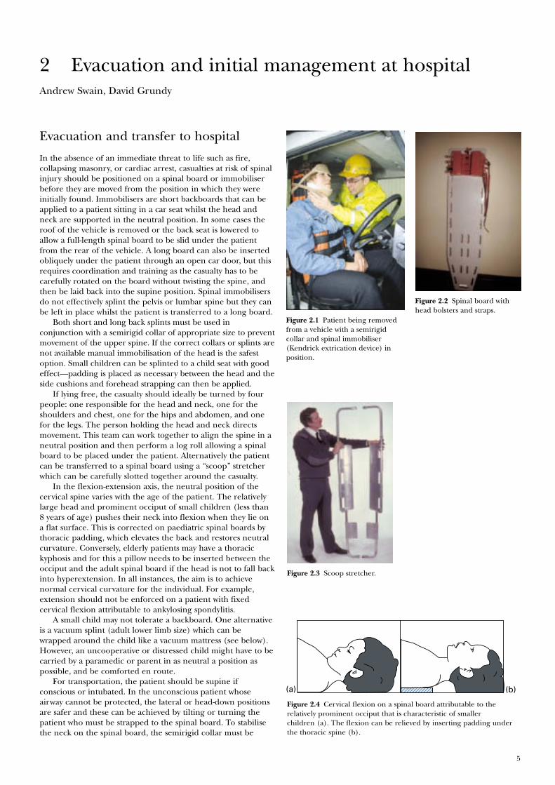
Figure 1.6 Suction: beware of vagal reflex stimulation and bradycardia.
Box 1.2 Clinical features of spinal cord injury
• Pain in the neck or back, often radiating because of nerve rootirritation
• Sensory disturbance distal to neurological level• Weakness or flaccid paralysis below this level
Opioid analgesics should be administered with care inpatients with respiratory compromise from cervical andupper thoracic injuries
• Swain A. Trauma to the spine and spinal cord. In: SkinnerD, Swain A, Peyton R, Robertson C, eds.Cambridge textbook ofaccident and emergency medicine. Cambridge: CambridgeUniversity Press, 1997, pp 510–32
• Toscano J. Prevention of neurological deterioration beforeadmission to a spinal cord injury unit. Paraplegia1988;26:143–50
5
Andrew Swain, David Grundy
Evacuation and transfer to hospital
In the absence of an immediate threat to life such as fire,collapsing masonry, or cardiac arrest, casualties at risk of spinalinjury should be positioned on a spinal board or immobiliserbefore they are moved from the position in which they wereinitially found. Immobilisers are short backboards that can beapplied to a patient sitting in a car seat whilst the head andneck are supported in the neutral position. In some cases theroof of the vehicle is removed or the back seat is lowered toallow a full-length spinal board to be slid under the patientfrom the rear of the vehicle. A long board can also be insertedobliquely under the patient through an open car door, but thisrequires coordination and training as the casualty has to becarefully rotated on the board without twisting the spine, andthen be laid back into the supine position. Spinal immobilisersdo not effectively splint the pelvis or lumbar spine but they canbe left in place whilst the patient is transferred to a long board.
Both short and long back splints must be used inconjunction with a semirigid collar of appropriate size to preventmovement of the upper spine. If the correct collars or splints arenot available manual immobilisation of the head is the safestoption. Small children can be splinted to a child seat with goodeffect—padding is placed as necessary between the head and theside cushions and forehead strapping can then be applied.
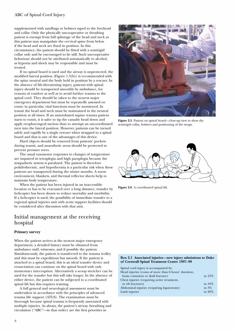
If lying free, the casualty should ideally be turned by fourpeople: one responsible for the head and neck, one for theshoulders and chest, one for the hips and abdomen, and onefor the legs. The person holding the head and neck directsmovement. This team can work together to align the spine in aneutral position and then perform a log roll allowing a spinalboard to be placed under the patient. Alternatively the patientcan be transferred to a spinal board using a “scoop” stretcherwhich can be carefully slotted together around the casualty.
In the flexion-extension axis, the neutral position of thecervical spine varies with the age of the patient. The relativelylarge head and prominent occiput of small children (less than8 years of age) pushes their neck into flexion when they lie ona flat surface. This is corrected on paediatric spinal boards bythoracic padding, which elevates the back and restores neutralcurvature. Conversely, elderly patients may have a thoracickyphosis and for this a pillow needs to be inserted between theocciput and the adult spinal board if the head is not to fall backinto hyperextension. In all instances, the aim is to achievenormal cervical curvature for the individual. For example,extension should not be enforced on a patient with fixedcervical flexion attributable to ankylosing spondylitis.
A small child may not tolerate a backboard. One alternativeis a vacuum splint (adult lower limb size) which can bewrapped around the child like a vacuum mattress (see below).However, an uncooperative or distressed child might have to becarried by a paramedic or parent in as neutral a position aspossible, and be comforted en route.
For transportation, the patient should be supine ifconscious or intubated. In the unconscious patient whoseairway cannot be protected, the lateral or head-down positionsare safer and these can be achieved by tilting or turning thepatient who must be strapped to the spinal board. To stabilisethe neck on the spinal board, the semirigid collar must be
2 Evacuation and initial management at hospital
Figure 2.1 Patient being removedfrom a vehicle with a semirigidcollar and spinal immobiliser(Kendrick extrication device) inposition.
Figure 2.2 Spinal board withhead bolsters and straps.
Figure 2.3 Scoop stretcher.
(b)(a)
Figure 2.4 Cervical flexion on a spinal board attributable to therelatively prominent occiput that is characteristic of smallerchildren (a). The flexion can be relieved by inserting padding underthe thoracic spine (b).
ABC of Spinal Cord Injury
6
supplemented with sandbags or bolsters taped to the foreheadand collar. Only the physically uncooperative or thrashingpatient is exempt from full splintage of the head and neck asthis patient may manipulate the cervical spine from belowif the head and neck are fixed in position. In thiscircumstance, the patient should be fitted with a semirigidcollar only and be encouraged to lie still. Such uncooperativebehaviour should not be attributed automatically to alcohol,as hypoxia and shock may be responsible and must betreated.
If no spinal board is used and the airway is unprotected, themodified lateral position (Figure 1.3(b)) is recommended withthe spine neutral and the body held in position by a rescuer. Inthe absence of life-threatening injury, patients with spinalinjury should be transported smoothly by ambulance, forreasons of comfort as well as to avoid further trauma to thespinal cord. They should be taken to the nearest majoremergency department but must be repeatedly assessed enroute; in particular, vital functions must be monitored. Intransit the head and neck must be maintained in the neutralposition at all times. If an unintubated supine trauma patientstarts to vomit, it is safer to tip the casualty head down andapply oropharyngeal suction than to attempt an uncoordinatedturn into the lateral position. However, patients can be turnedsafely and rapidly by a single rescuer when strapped to a spinalboard and that is one of the advantages of this device.
Hard objects should be removed from patients’ pocketsduring transit, and anaesthetic areas should be protected toprevent pressure sores.
The usual vasomotor responses to changes of temperatureare impaired in tetraplegia and high paraplegia because thesympathetic system is paralysed. The patient is thereforepoikilothermic, and hypothermia is a particular risk when thesepatients are transported during the winter months. A warmenvironment, blankets, and thermal reflector sheets help tomaintain body temperature.
When the patient has been injured in an inaccessiblelocation or has to be evacuated over a long distance, transfer byhelicopter has been shown to reduce mortality and morbidity.If a helicopter is used, the possibility of immediate transfer to aregional spinal injuries unit with acute support facilities shouldbe considered after discussion with that unit.
Initial management at the receivinghospital
Primary survey
When the patient arrives at the nearest major emergencydepartment, a detailed history must be obtained fromambulance staff, witnesses, and if possible the patient.Simultaneously, the patient is transferred to the trauma trolleyand this must be expeditious but smooth. If the patient isattached to a spinal board, this is an ideal transfer device andresuscitation can continue on the spinal board with onlymomentary interruption. Alternatively a scoop stretcher can beused for the transfer but this will take longer. In the absence ofeither device, the patient can be subjected to a coordinatedspinal lift but this requires training.
A full general and neurological assessment must beundertaken in accordance with the principles of advancedtrauma life support (ATLS). The examination must bethorough because spinal trauma is frequently associated withmultiple injuries. As always, the patient’s airway, breathing andcirculation (“ABC”—in that order) are the first priorities in
Figure 2.5 Patient on spinal board—close-up view to show thesemirigid collar, bolsters and positioning of the straps.
Figure 2.6 A coordinated spinal lift.
Box 2.1 Associated injuries—new injury admissions to Dukeof Cornwall Spinal Treatment Centre 1997–99
Spinal cord injury is accompanied by:Head injuries (coma of more than 6 hours’ duration,brain contusion or skull fracture) in 12%
Chest injuries (requiring active treatment,or rib fractures) in 19%
Abdominal injuries (requiring laparotomy) in 3%Limb injuries in 20%
Evacuation and initial management at hospital
7
resuscitation from trauma. If not already secure, the cervicalspine is immobilised in the neutral position as the airway isassessed. Following attention to the ABC, a central nervoussystem assessment is undertaken and any clothing is removed.This sequence constitutes the primary survey of ATLS. Thespinal injury itself can directly affect the airway (for exampleby producing a retropharyngeal haematoma or trachealdeviation) as well as the respiratory and circulatory systems(see chapter 4).
Secondary survey
Once the immediately life-threatening injuries have beenaddressed, the secondary (head to toe) survey that followsallows other serious injuries to be identified. Areas that are notbeing examined should be covered and kept warm, and bodytemperature should be monitored. In the supine position, thecervical and lumbar lordoses may be palpated by sliding a handunder the patient. A more comprehensive examination is madeduring the log roll. Unless there is an urgent need to inspectthe back, the log roll is normally undertaken near the end ofthe secondary survey by a team of four led by the person whoholds the patient’s head. If neurological symptoms or signs arepresent, a senior doctor should be present and a partial roll toabout 45˚ may be sufficient. A doctor who is not involved withthe log roll must examine the back for specific signs of injuryincluding local bruising or deformity of the spine (e.g.a gibbus or an increased interspinous gap) and vertebraltenderness. The whole length of the spine must be palpated,as about 10% of patients with an unstable spinal injury haveanother spinal injury at a different level. Priapism anddiaphragmatic breathing invariably indicate a high spinal cordlesion. The presence of warm and well-perfused peripheries ina hypotensive patient should always raise the possibility ofneurogenic shock attributable to spinal cord injury in the
L
L
S
S
T
T
C
C
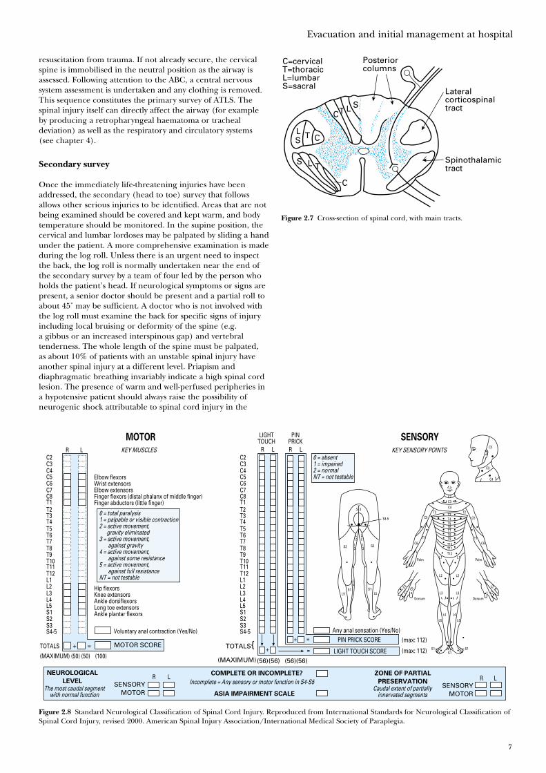
Spinothalamictract
Lateralcorticospinaltract
Posteriorcolumns
CT L S
C=cervicalT=thoracicL=lumbar S=sacral
Figure 2.7 Cross-section of spinal cord, with main tracts.
+ = MOTOR SCORE
NEUROLOGICAL
LEVEL
COMPLETE OR INCOMPLETE? ZONE OF PARTIAL
PRESERVATIONSENSORY
MOTORASIA IMPAIRMENT SCALE
SENSORYMOTOR
TOTALS{(MAXIMUM)(56)(56) (56)(56)
+ = LIGHT TOUCH SCORE
+ = PIN PRICK SCORE (max: 112)
(max: 112)
R L
Any anal sensation (Yes/No)
KEY MUSCLES
0 = total paralysis1 = palpable or visible contraction2 = active movement, gravity eliminated3 = active movement, against gravity4 = active movement, against some resistance5 = active movement, against full resistanceNT = not testable
The most caudal segmentwith normal function
Incomplete = Any sensory or motor function in S4-S5Caudal extent of partially
innervated segments
0 = absent1 = impaired2 = normalNT = not testable
KEY SENSORY POINTS
Elbow flexorsWrist extensorsElbow extensorsFinger flexors (distal phalanx of middle finger)Finger abductors (little finger)
Hip flexorsKnee extensorsAnkle dorsiflexorsLong toe extensorsAnkle plantar flexors
Voluntary anal contraction (Yes/No)
R L R
LIGHTTOUCHMOTOR SENSORYPIN
PRICKL R L
C2C3C4C5C6C7C8T1T2T3T4T5T6T7T8T9T10T11T12L1L2L3L4L5S1S2S3S4-5
C2C3C4C5C6C7C8T1T2T3T4T5T6T7T8T9T10T11T12L1L2L3L4L5S1S2S3S4-5
TOTALS
(MAXIMUM) (50) (50) (100)
C2
C3
C4
C2C3
C4T2
C5T2
C5
T1T1C6C6
T3T4T5T6T7T8T9T10T11T12
L1L1
L2
L5 L5S1S1
S2
L2
L2
L3
L3S2
S4-5
S 3
L4
L2
L3
DorsumDorsum
Palm
C7 C7
C3 C3
C6
Palm
L4
L5
S1S1S1
L5
L4
L3
R L
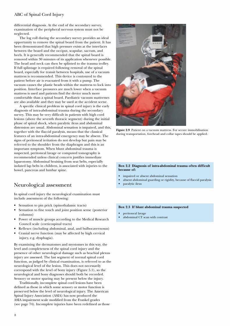
Figure 2.8 Standard Neurological Classification of Spinal Cord Injury. Reproduced from International Standards for Neurological Classification ofSpinal Cord Injury, revised 2000. American Spinal Injury Association/International Medical Society of Paraplegia.
ABC of Spinal Cord Injury
8
differential diagnosis. At the end of the secondary survey,examination of the peripheral nervous system must not beneglected.
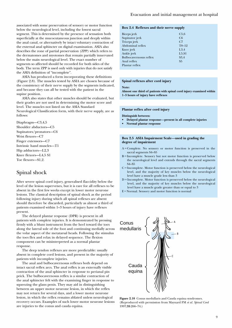
The log roll during the secondary survey provides an idealopportunity to remove the spinal board from the patient. It hasbeen demonstrated that high pressure exists at the interfacesbetween the board and the occiput, scapulae, sacrum, andheels. It is generally recommended that the spinal board isremoved within 30 minutes of its application whenever possible.The head and neck can then be splinted to the trauma trolley.If full splintage is required following removal of the spinalboard, especially for transit between hospitals, use of a vacuummattress is recommended. This device is contoured to thepatient before air is evacuated from it with a pump. Thevacuum causes the plastic beads within the mattress to lock intoposition. Interface pressures are much lower when a vacuummattress is used and patients find the device much morecomfortable than a spinal board. Paediatric vacuum mattressesare also available and they may be used at the accident scene.
A specific clinical problem in spinal cord injury is the earlydiagnosis of intra-abdominal trauma during the secondarysurvey. This may be very difficult in patients with high cordlesions (above the seventh thoracic segment) during the initialphase of spinal shock, when paralytic ileus and abdominaldistension are usual. Abdominal sensation is impaired, and this,together with the flaccid paralysis, means that the classicalfeatures of an intra-abdominal emergency may be absent. Thesigns of peritoneal irritation do not develop but pain may bereferred to the shoulder from the diaphragm and this is animportant symptom. When blunt abdominal trauma issuspected, peritoneal lavage or computed tomography isrecommended unless clinical concern justifies immediatelaparotomy. Abdominal bruising from seat belts, especiallyisolated lap belts in children, is associated with injuries to thebowel, pancreas and lumbar spine.
Neurological assessment
In spinal cord injury the neurological examination mustinclude assessment of the following:
• Sensation to pin prick (spinothalamic tracts)• Sensation to fine touch and joint position sense (posterior
columns) • Power of muscle groups according to the Medical Research
Council scale (corticospinal tracts)• Reflexes (including abdominal, anal, and bulbocavernosus)• Cranial nerve function (may be affected by high cervical
injury, e.g. dysphagia).
By examining the dermatomes and myotomes in this way, thelevel and completeness of the spinal cord injury and thepresence of other neurological damage such as brachial plexusinjury are assessed. The last segment of normal spinal cordfunction, as judged by clinical examination, is referred to as theneurological level of the lesion. This does not necessarilycorrespond with the level of bony injury (Figure 5.1), so theneurological and bony diagnoses should both be recorded.Sensory or motor sparing may be present below the injury.
Traditionally, incomplete spinal cord lesions have beendefined as those in which some sensory or motor function ispreserved below the level of neurological injury. The AmericanSpinal Injury Association (ASIA) has now produced theASIA impairment scale modified from the Frankel grades(see page 74). Incomplete injuries have been redefined as those
Figure 2.9 Patient on a vacuum mattress. For secure immobilisationduring transportation, forehead and collar tapes should be applied.
Box 2.2 Diagnosis of intra-abdominal trauma often difficultbecause of:
• impaired or absent abdominal sensation• absent abdominal guarding or rigidity, because of flaccid paralysis• paralytic ileus
Box 2.3 If blunt abdominal trauma suspected
• peritoneal lavage• abdominal CT scan with contrast
Evacuation and initial management at hospital
9
associated with some preservation of sensory or motor functionbelow the neurological level, including the lowest sacralsegment. This is determined by the presence of sensation bothsuperficially at the mucocutaneous junction and deeply withinthe anal canal, or alternatively by intact voluntary contraction ofthe external anal sphincter on digital examination. ASIA alsodescribes the zone of partial preservation (ZPP) which refers tothe dermatomes and myotomes that remain partially innervatedbelow the main neurological level. The exact number ofsegments so affected should be recorded for both sides of thebody. The term ZPP is used only with injuries that do not satisfythe ASIA definition of “incomplete”.
ASIA has produced a form incorporating these definitions(Figure 2.8). The muscles tested by ASIA are chosen because ofthe consistency of their nerve supply by the segments indicated,and because they can all be tested with the patient in thesupine position.
ASIA also states that other muscles should be evaluated, buttheir grades are not used in determining the motor score andlevel. The muscles not listed on the ASIA StandardNeurological Classification form, with their nerve supply, are asfollows:
Diaphragm—C3,4,5Shoulder abductors—C5Supinators/pronators—C6Wrist flexors—C7Finger extensors—C7Intrinsic hand muscles—T1Hip adductors—L2,3Knee flexors—L4,5 S1Toe flexors—S1,2.
Spinal shock
After severe spinal cord injury, generalised flaccidity below thelevel of the lesion supervenes, but it is rare for all reflexes to beabsent in the first few weeks except in lower motor neuronelesions. The classical description of spinal shock as the periodfollowing injury during which all spinal reflexes are absentshould therefore be discarded, particularly as almost a third ofpatients examined within 1–3 hours of injury have reflexespresent.
The delayed plantar response (DPR) is present in allpatients with complete injuries. It is demonstrated by pressingfirmly with a blunt instrument from the heel toward the toesalong the lateral sole of the foot and continuing medially acrossthe volar aspect of the metatarsal heads. Following the stimulusthe toes flex and relax in delayed sequence. The flexioncomponent can be misinterpreted as a normal plantarresponse.
The deep tendon reflexes are more predictable: usuallyabsent in complete cord lesions, and present in the majority ofpatients with incomplete injuries.
The anal and bulbocavernosus reflexes both depend onintact sacral reflex arcs. The anal reflex is an externally visiblecontraction of the anal sphincter in response to perianal pinprick. The bulbocavernosus reflex is a similar contraction ofthe anal sphincter felt with the examining finger in response tosqueezing the glans penis. They may aid in distinguishingbetween an upper motor neurone lesion, in which the reflexmay not return for several days, and a lower motor neuronelesion, in which the reflex remains ablated unless neurologicalrecovery occurs. Examples of such lower motor neurone lesionsare injuries to the conus and cauda equina.
Box 2.4 Reflexes and their nerve supply
Biceps jerk C5,6Supinator jerk C6Triceps jerk C7Abdominal reflex T8–12Knee jerk L3,4Ankle jerk L5,S1Bulbocavernosus reflex S3,4Anal reflex S5Plantar reflex
Box 2.5 ASIA Impairment Scale—used in grading thedegree of impairment
A�Complete. No sensory or motor function is preserved in thesacral segments S4–S5
B�Incomplete. Sensory but not motor function is preserved belowthe neurological level and extends through the sacral segmentsS4–S5
C�Incomplete. Motor function is preserved below the neurologicallevel, and the majority of key muscles below the neurologicallevel have a muscle grade less than 3
D�Incomplete. Motor function is preserved below the neurologicallevel, and the majority of key muscles below the neurologicallevel have a muscle grade greater than or equal to 3
E�Normal. Sensory and motor function is normal
Spinal reflexes after cord injury
Note:Almost one third of patients with spinal cord injury examined within1–3 hours of injury have reflexes
Plantar reflex after cord injury
Distinguish between:• Delayed plantar response—present in all complete injuries• Normal plantar response
Conusmedullaris
Caudaequina
A
B
C
Figure 2.10 Conus medullaris and Cauda equina syndromes.(Reproduced with permission from Maynard FM et al. Spinal Cord1997;35:266–74.)
ABC of Spinal Cord Injury
10
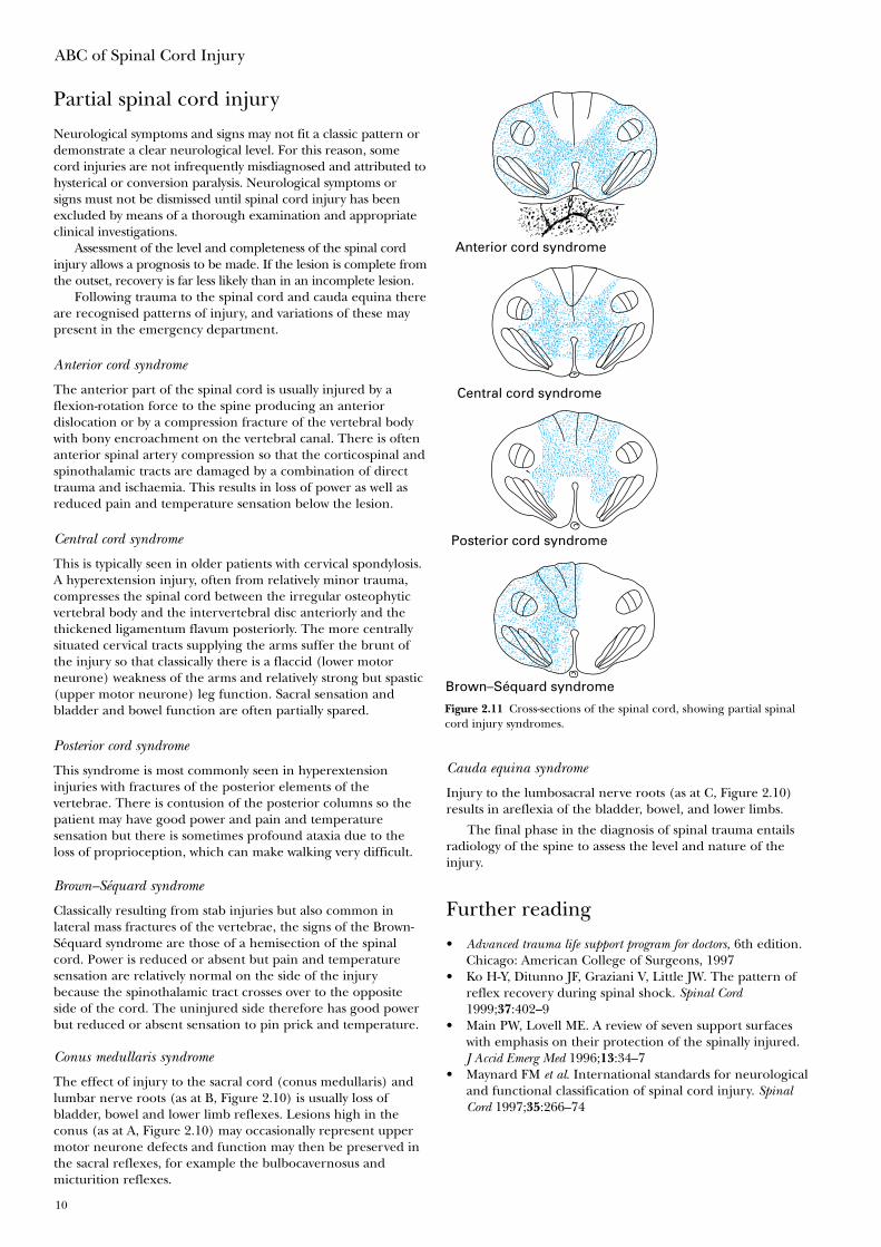
Partial spinal cord injury
Neurological symptoms and signs may not fit a classic pattern ordemonstrate a clear neurological level. For this reason, somecord injuries are not infrequently misdiagnosed and attributed tohysterical or conversion paralysis. Neurological symptoms orsigns must not be dismissed until spinal cord injury has beenexcluded by means of a thorough examination and appropriateclinical investigations.
Assessment of the level and completeness of the spinal cordinjury allows a prognosis to be made. If the lesion is complete fromthe outset, recovery is far less likely than in an incomplete lesion.
Following trauma to the spinal cord and cauda equina thereare recognised patterns of injury, and variations of these maypresent in the emergency department.
Anterior cord syndrome
The anterior part of the spinal cord is usually injured by aflexion-rotation force to the spine producing an anteriordislocation or by a compression fracture of the vertebral bodywith bony encroachment on the vertebral canal. There is oftenanterior spinal artery compression so that the corticospinal andspinothalamic tracts are damaged by a combination of directtrauma and ischaemia. This results in loss of power as well asreduced pain and temperature sensation below the lesion.
Central cord syndrome
This is typically seen in older patients with cervical spondylosis.A hyperextension injury, often from relatively minor trauma,compresses the spinal cord between the irregular osteophyticvertebral body and the intervertebral disc anteriorly and thethickened ligamentum flavum posteriorly. The more centrallysituated cervical tracts supplying the arms suffer the brunt ofthe injury so that classically there is a flaccid (lower motorneurone) weakness of the arms and relatively strong but spastic(upper motor neurone) leg function. Sacral sensation andbladder and bowel function are often partially spared.
Posterior cord syndrome
This syndrome is most commonly seen in hyperextensioninjuries with fractures of the posterior elements of thevertebrae. There is contusion of the posterior columns so thepatient may have good power and pain and temperaturesensation but there is sometimes profound ataxia due to theloss of proprioception, which can make walking very difficult.
Brown–Séquard syndrome
Classically resulting from stab injuries but also common inlateral mass fractures of the vertebrae, the signs of the Brown-Séquard syndrome are those of a hemisection of the spinalcord. Power is reduced or absent but pain and temperaturesensation are relatively normal on the side of the injurybecause the spinothalamic tract crosses over to the oppositeside of the cord. The uninjured side therefore has good powerbut reduced or absent sensation to pin prick and temperature.
Conus medullaris syndrome
The effect of injury to the sacral cord (conus medullaris) andlumbar nerve roots (as at B, Figure 2.10) is usually loss ofbladder, bowel and lower limb reflexes. Lesions high in theconus (as at A, Figure 2.10) may occasionally represent uppermotor neurone defects and function may then be preserved inthe sacral reflexes, for example the bulbocavernosus andmicturition reflexes.
Anterior cord syndrome
Posterior cord syndrome
Central cord syndrome
Brown–Séquard syndrome
Figure 2.11 Cross-sections of the spinal cord, showing partial spinalcord injury syndromes.
Cauda equina syndrome
Injury to the lumbosacral nerve roots (as at C, Figure 2.10)results in areflexia of the bladder, bowel, and lower limbs.
The final phase in the diagnosis of spinal trauma entailsradiology of the spine to assess the level and nature of theinjury.
Further reading
• Advanced trauma life support program for doctors, 6th edition.Chicago: American College of Surgeons, 1997
• Ko H-Y, Ditunno JF, Graziani V, Little JW. The pattern ofreflex recovery during spinal shock. Spinal Cord1999;37:402–9
• Main PW, Lovell ME. A review of seven support surfaceswith emphasis on their protection of the spinally injured.J Accid Emerg Med 1996;13:34–7
• Maynard FM et al. International standards for neurologicaland functional classification of spinal cord injury. SpinalCord 1997;35:266–74
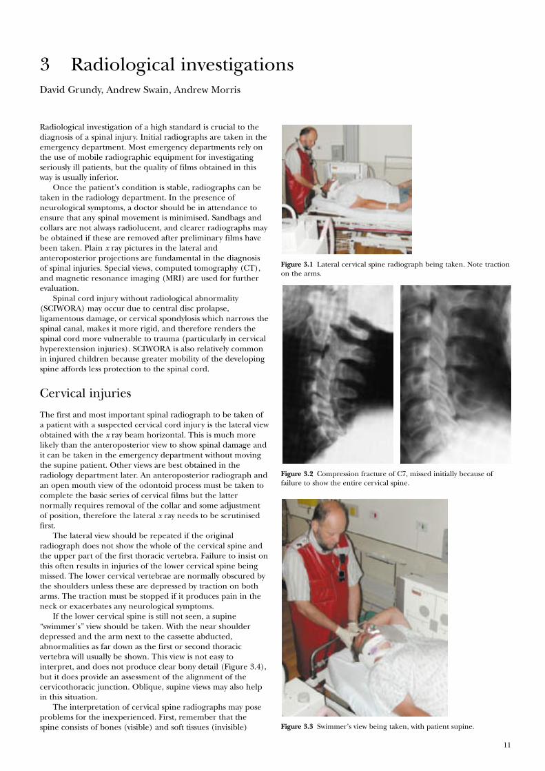
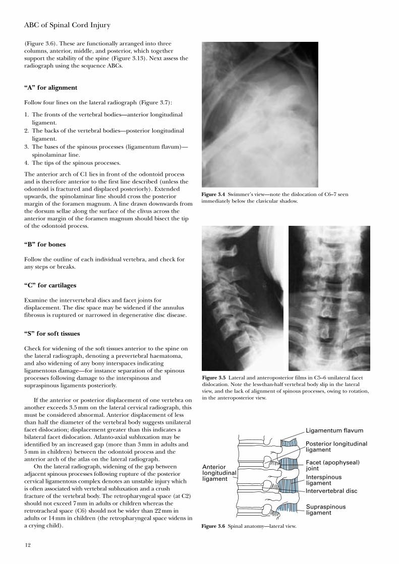
Figure 3.2 Compression fracture of C7, missed initially because offailure to show the entire cervical spine.
11
David Grundy, Andrew Swain, Andrew Morris
Radiological investigation of a high standard is crucial to thediagnosis of a spinal injury. Initial radiographs are taken in theemergency department. Most emergency departments rely onthe use of mobile radiographic equipment for investigatingseriously ill patients, but the quality of films obtained in thisway is usually inferior.
Once the patient’s condition is stable, radiographs can betaken in the radiology department. In the presence ofneurological symptoms, a doctor should be in attendance toensure that any spinal movement is minimised. Sandbags andcollars are not always radiolucent, and clearer radiographs maybe obtained if these are removed after preliminary films havebeen taken. Plain x ray pictures in the lateral andanteroposterior projections are fundamental in the diagnosisof spinal injuries. Special views, computed tomography (CT),and magnetic resonance imaging (MRI) are used for furtherevaluation.
Spinal cord injury without radiological abnormality(SCIWORA) may occur due to central disc prolapse,ligamentous damage, or cervical spondylosis which narrows thespinal canal, makes it more rigid, and therefore renders thespinal cord more vulnerable to trauma (particularly in cervicalhyperextension injuries). SCIWORA is also relatively commonin injured children because greater mobility of the developingspine affords less protection to the spinal cord.
Cervical injuries
The first and most important spinal radiograph to be taken ofa patient with a suspected cervical cord injury is the lateral viewobtained with the x ray beam horizontal. This is much morelikely than the anteroposterior view to show spinal damage andit can be taken in the emergency department without movingthe supine patient. Other views are best obtained in theradiology department later. An anteroposterior radiograph andan open mouth view of the odontoid process must be taken tocomplete the basic series of cervical films but the latternormally requires removal of the collar and some adjustmentof position, therefore the lateral x ray needs to be scrutinisedfirst.
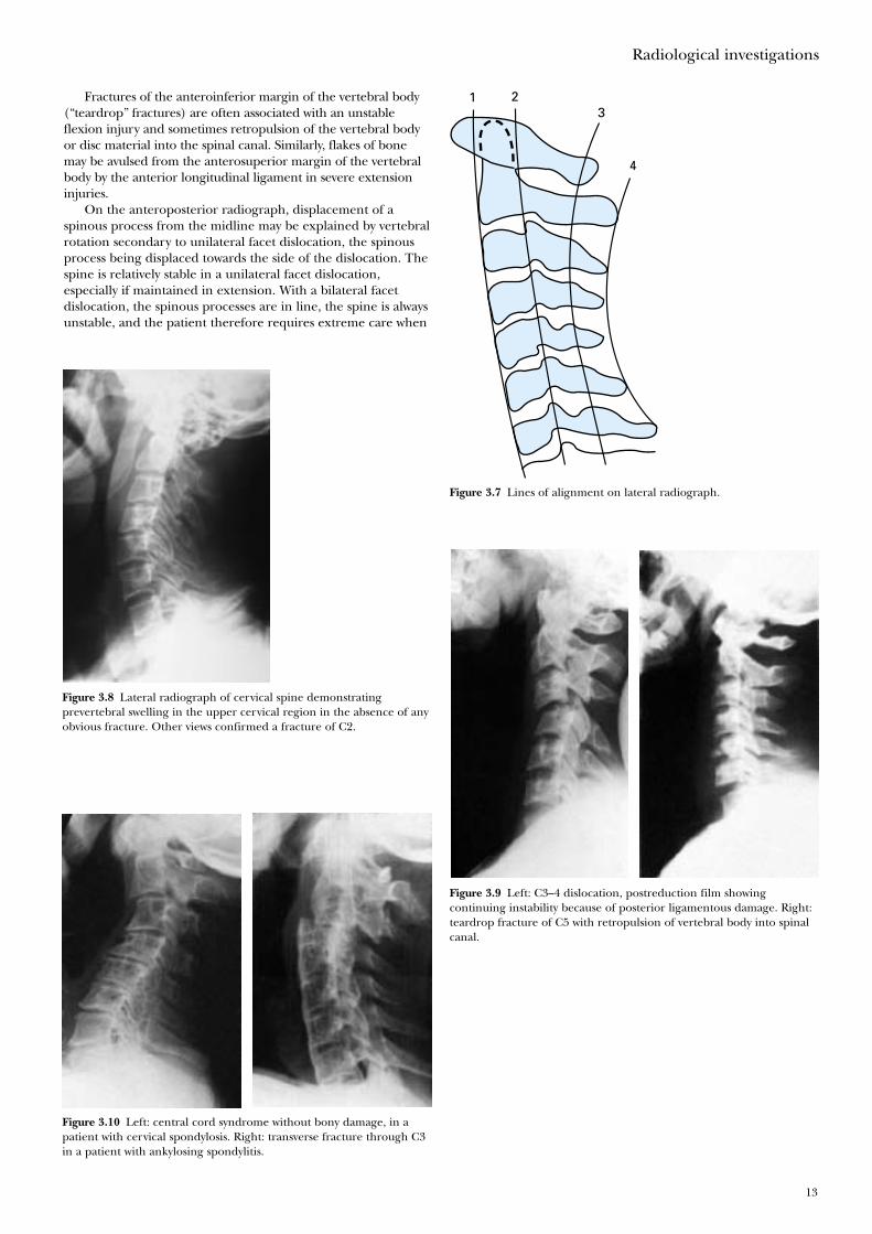
The lateral view should be repeated if the originalradiograph does not show the whole of the cervical spine andthe upper part of the first thoracic vertebra. Failure to insist onthis often results in injuries of the lower cervical spine beingmissed. The lower cervical vertebrae are normally obscured bythe shoulders unless these are depressed by traction on botharms. The traction must be stopped if it produces pain in theneck or exacerbates any neurological symptoms.
If the lower cervical spine is still not seen, a supine“swimmer’s” view should be taken. With the near shoulderdepressed and the arm next to the cassette abducted,abnormalities as far down as the first or second thoracicvertebra will usually be shown. This view is not easy tointerpret, and does not produce clear bony detail (Figure 3.4),but it does provide an assessment of the alignment of thecervicothoracic junction. Oblique, supine views may also helpin this situation.
The interpretation of cervical spine radiographs may poseproblems for the inexperienced. First, remember that thespine consists of bones (visible) and soft tissues (invisible)
3 Radiological investigations
Figure 3.1 Lateral cervical spine radiograph being taken. Note tractionon the arms.
Figure 3.3 Swimmer’s view being taken, with patient supine.
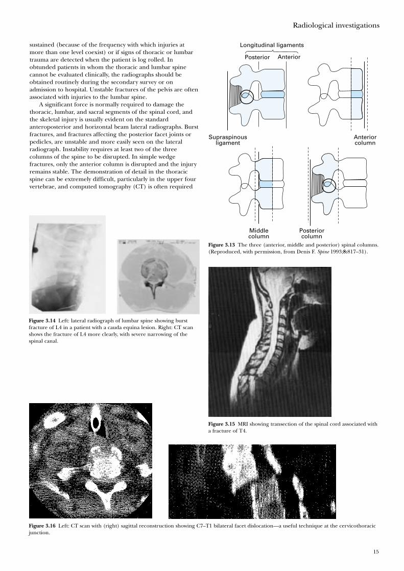
(Figure 3.6). These are functionally arranged into threecolumns, anterior, middle, and posterior, which togethersupport the stability of the spine (Figure 3.13). Next assess theradiograph using the sequence ABCs.
“A” for alignment
Follow four lines on the lateral radiograph (Figure 3.7):
1. The fronts of the vertebral bodies—anterior longitudinalligament.
2. The backs of the vertebral bodies—posterior longitudinalligament.
3. The bases of the spinous processes (ligamentum flavum)—spinolaminar line.
4. The tips of the spinous processes.
The anterior arch of C1 lies in front of the odontoid processand is therefore anterior to the first line described (unless theodontoid is fractured and displaced posteriorly). Extendedupwards, the spinolaminar line should cross the posteriormargin of the foramen magnum. A line drawn downwards fromthe dorsum sellae along the surface of the clivus across theanterior margin of the foramen magnum should bisect the tipof the odontoid process.
“B” for bones
Follow the outline of each individual vertebra, and check forany steps or breaks.
“C” for cartilages
Examine the intervertebral discs and facet joints fordisplacement. The disc space may be widened if the annulusfibrosus is ruptured or narrowed in degenerative disc disease.
“S” for soft tissues
Check for widening of the soft tissues anterior to the spine onthe lateral radiograph, denoting a prevertebral haematoma,and also widening of any bony interspaces indicatingligamentous damage—for instance separation of the spinousprocesses following damage to the interspinous andsupraspinous ligaments posteriorly.
If the anterior or posterior displacement of one vertebra onanother exceeds 3.5 mm on the lateral cervical radiograph, thismust be considered abnormal. Anterior displacement of lessthan half the diameter of the vertebral body suggests unilateralfacet dislocation; displacement greater than this indicates abilateral facet dislocation. Atlanto-axial subluxation may beidentified by an increased gap (more than 3 mm in adults and5 mm in children) between the odontoid process and theanterior arch of the atlas on the lateral radiograph.
On the lateral radiograph, widening of the gap betweenadjacent spinous processes following rupture of the posteriorcervical ligamentous complex denotes an unstable injury which is often associated with vertebral subluxation and a crush fracture of the vertebral body. The retropharyngeal space (at C2)should not exceed 7 mm in adults or children whereas theretrotracheal space (C6) should not be wider than 22 mm inadults or 14 mm in children (the retropharyngeal space widens ina crying child).
ABC of Spinal Cord Injury
12
Anteriorlongitudinalligament
Ligamentum flavum
Facet (apophyseal)jointInterspinousligamentIntervertebral disc
Supraspinousligament
Posterior longitudinalligament
Figure 3.6 Spinal anatomy—lateral view.
Figure 3.4 Swimmer’s view—note the dislocation of C6–7 seenimmediately below the clavicular shadow.
Figure 3.5 Lateral and anteroposterior films in C5–6 unilateral facetdislocation. Note the less-than-half vertebral body slip in the lateralview, and the lack of alignment of spinous processes, owing to rotation,in the anteroposterior view.
Fractures of the anteroinferior margin of the vertebral body(“teardrop” fractures) are often associated with an unstableflexion injury and sometimes retropulsion of the vertebral bodyor disc material into the spinal canal. Similarly, flakes of bonemay be avulsed from the anterosuperior margin of the vertebralbody by the anterior longitudinal ligament in severe extensioninjuries.
On the anteroposterior radiograph, displacement of aspinous process from the midline may be explained by vertebralrotation secondary to unilateral facet dislocation, the spinousprocess being displaced towards the side of the dislocation. Thespine is relatively stable in a unilateral facet dislocation,especially if maintained in extension. With a bilateral facetdislocation, the spinous processes are in line, the spine is alwaysunstable, and the patient therefore requires extreme care when
Radiological investigations
13
4
321
Figure 3.7 Lines of alignment on lateral radiograph.
Figure 3.8 Lateral radiograph of cervical spine demonstratingprevertebral swelling in the upper cervical region in the absence of anyobvious fracture. Other views confirmed a fracture of C2.
Figure 3.9 Left: C3–4 dislocation, postreduction film showingcontinuing instability because of posterior ligamentous damage. Right:teardrop fracture of C5 with retropulsion of vertebral body into spinalcanal.
Figure 3.10 Left: central cord syndrome without bony damage, in apatient with cervical spondylosis. Right: transverse fracture through C3in a patient with ankylosing spondylitis.
being handled. The anteroposterior cervical radiograph alsoprovides an opportunity to examine the upper thoracicvertebrae and first to third ribs: severe trauma is required toinjure these structures.
Oblique radiographs are not routinely obtained, but theydo help to confirm the presence of subluxation or dislocationand indicate whether the right or left facets (apophysealjoints), or both, are affected. They may elucidate abnormalitiesat the cervicothoracic junction and some authoritiesrecommend them as part of a five-view cervical spine series.
The 45˚ supine oblique view shows the intervertebralforamina and the facets but a better view for the facets is onetaken with the patient log rolled 22.5˚ from the horizontal.
Flexion and extension views of the cervical spine may betaken if the patient has no neurological symptoms or signs andinitial radiographs are normal but an unstable (ligamentous)injury is nevertheless suspected from the mechanism of injury,severe pain, or radiological signs of ligamentous injury. Toobtain these radiographs, flexion and extension of the wholeneck must be performed as far as the patient can tolerateunder the supervision of an experienced doctor. Movementmust cease if neurological symptoms are precipitated.
If there is any doubt about the integrity of the cervicalspine on plain radiographs, CT should be performed. Thisprovides much greater detail of the bony structures and willshow the extent of encroachment on the spinal canal byvertebral displacement or bone fragments. It is particularlyuseful in assessing the cervicothoracic junction, the uppercervical spine and any suspected fracture or misalignment.Helical (or spiral) CT is now more available. It allows for afaster examination and also clearer reconstructed images inthe sagittal and coronal planes. Many patients with majortrauma will require CT of their head, chest or abdomen, and itis often appropriate to scan any suspicious or poorly seen areaof their spine at the same time rather than struggle withfurther plain films.
MRI gives information about the spinal cord and soft tissuesand will reveal the cause of cord compression, whether frombone, prolapsed discs, ligamentous damage, or intraspinalhaematomas. It will also show the extent of cord damage andoedema which is of some prognostic value. Although an acutetraumatic disc prolapse may be associated with bony injury, itcan also occur with normal radiographs, and in these patients itis vital that an urgent MRI scan is obtained. These scans canalso be used to demonstrate spinal instability, particularly in thepresence of normal radiographs. MRI has supersededmyelography, both in the quality of images obtained and insafety for the patient, allowing decisions to be made without theneed for invasive imaging modalities. Its use may be limited byits availability and the difficulty in monitoring the acutelyinjured patient within the scanner.
Pathological changes in the spine—for example, ankylosingspondylitis or rheumatoid arthritis—may predispose to bonydamage after relatively minor trauma and in these patientsfurther radiological investigation and imaging must bethorough.
Thoracic and lumbar injuries
The thoracic spine is often demonstrated well on theanteroposterior chest radiograph that forms part of thestandard series of views requested in major trauma. This x raymay be the first to reveal an injury to the thoracic spine.Radiographs of the thoracic and lumbar spine must bespecifically requested if a cervical spine injury has been
ABC of Spinal Cord Injury
14
Figure 3.12 Patient with fracture of T5 with widening of mediastinumdue to a prevertebral haematoma, initially diagnosed as traumaticdissection of the aorta, for which he underwent aortography.
Figure 3.11 The 22.5˚ oblique view of the right facet joints (left) showsclearly the facet dislocation at the C5–6 level, less obvious in the 45˚oblique view (right), which, however, shows a malalignment of theintervertebral foramina.
Box 3.1 Indications for thoracic and lumbar radiographs
• Major trauma• Impaired consciousness• Distracting injury• Physical signs of thoracic or lumbar trauma• Pelvic fractures• Altered peripheral neurology
sustained (because of the frequency with which injuries atmore than one level coexist) or if signs of thoracic or lumbartrauma are detected when the patient is log rolled. Inobtunded patients in whom the thoracic and lumbar spinecannot be evaluated clinically, the radiographs should beobtained routinely during the secondary survey or onadmission to hospital. Unstable fractures of the pelvis are oftenassociated with injuries to the lumbar spine.
A significant force is normally required to damage thethoracic, lumbar, and sacral segments of the spinal cord, andthe skeletal injury is usually evident on the standardanteroposterior and horizontal beam lateral radiographs. Burstfractures, and fractures affecting the posterior facet joints orpedicles, are unstable and more easily seen on the lateralradiograph. Instability requires at least two of the threecolumns of the spine to be disrupted. In simple wedgefractures, only the anterior column is disrupted and the injuryremains stable. The demonstration of detail in the thoracicspine can be extremely difficult, particularly in the upper fourvertebrae, and computed tomography (CT) is often required
Radiological investigations
15
Longitudinal ligaments
Supraspinousligament
Anteriorcolumn
Middlecolumn
Posteriorcolumn
Posterior Anterior
Figure 3.13 The three (anterior, middle and posterior) spinal columns.(Reproduced, with permission, from Denis F. Spine 1993;8:817–31).
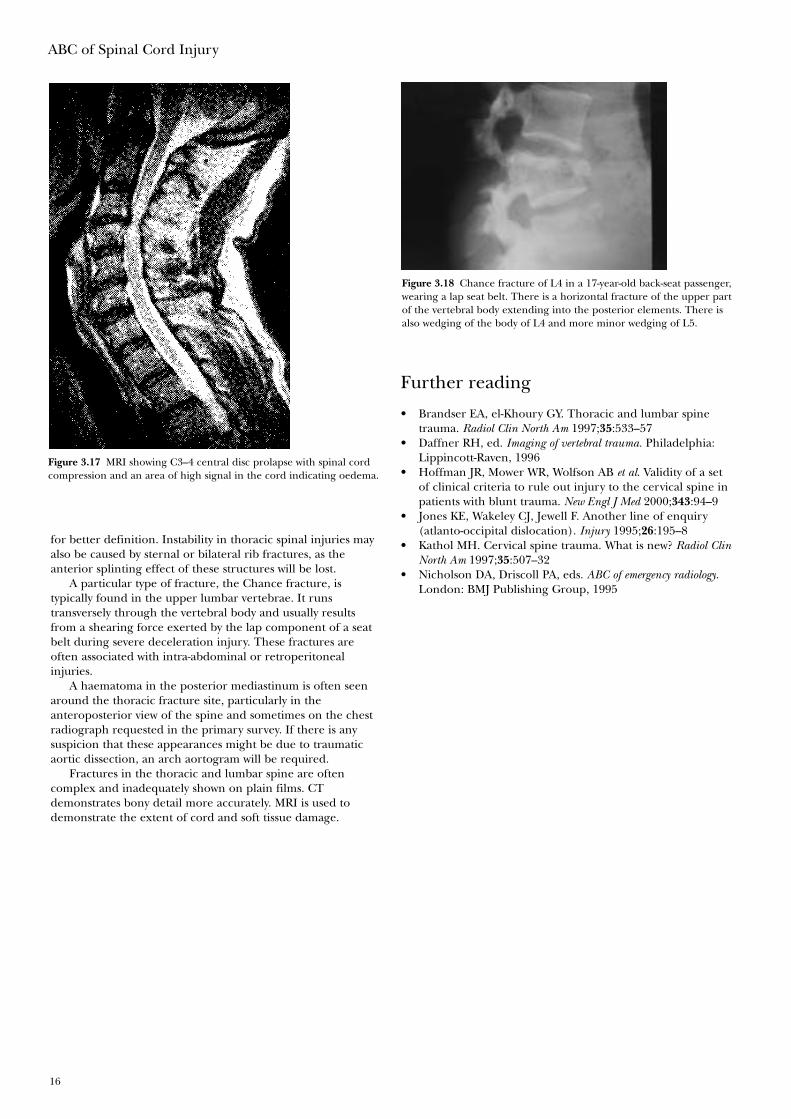
Figure 3.14 Left: lateral radiograph of lumbar spine showing burstfracture of L4 in a patient with a cauda equina lesion. Right: CT scanshows the fracture of L4 more clearly, with severe narrowing of thespinal canal.
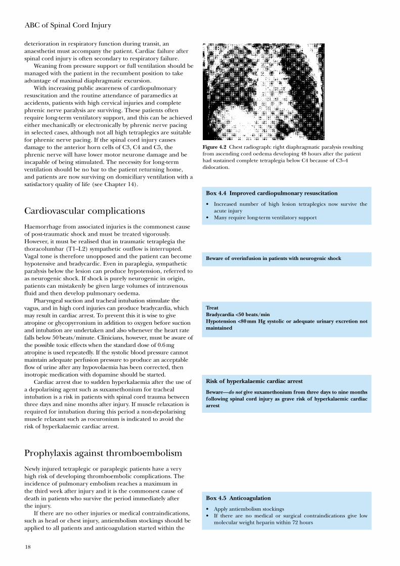
Figure 3.16 Left: CT scan with (right) sagittal reconstruction showing C7–T1 bilateral facet dislocation—a useful technique at the cervicothoracicjunction.
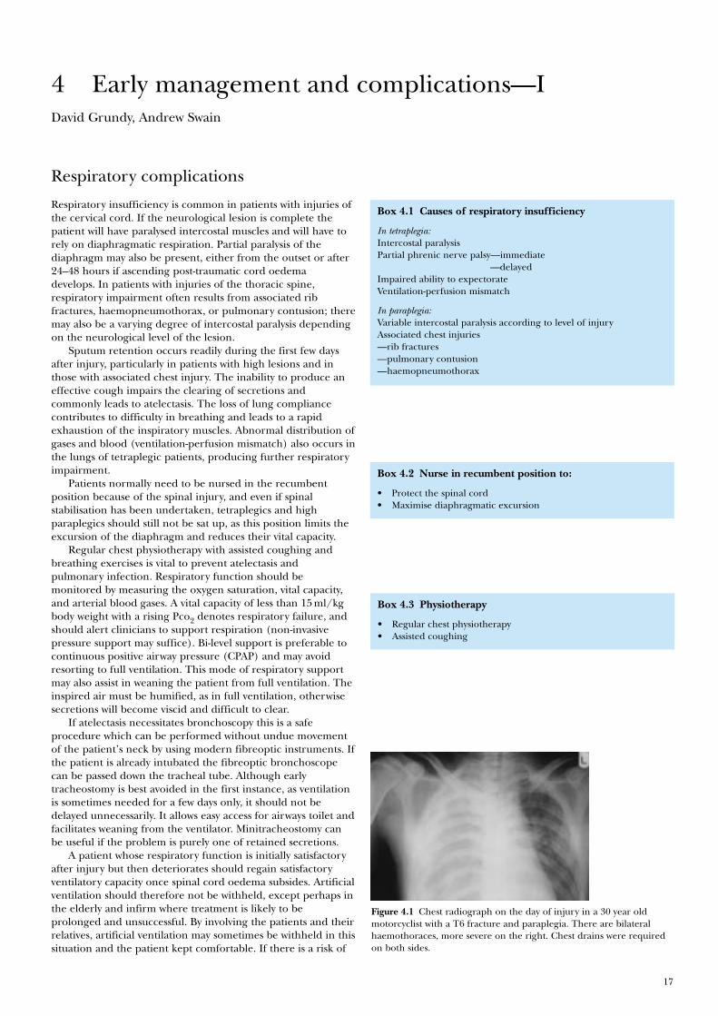
Figure 3.15 MRI showing transection of the spinal cord associated witha fracture of T4.
for better definition. Instability in thoracic spinal injuries mayalso be caused by sternal or bilateral rib fractures, as theanterior splinting effect of these structures will be lost.
A particular type of fracture, the Chance fracture, istypically found in the upper lumbar vertebrae. It runstransversely through the vertebral body and usually resultsfrom a shearing force exerted by the lap component of a seatbelt during severe deceleration injury. These fractures areoften associated with intra-abdominal or retroperitonealinjuries.
A haematoma in the posterior mediastinum is often seenaround the thoracic fracture site, particularly in theanteroposterior view of the spine and sometimes on the chestradiograph requested in the primary survey. If there is anysuspicion that these appearances might be due to traumaticaortic dissection, an arch aortogram will be required.
Fractures in the thoracic and lumbar spine are oftencomplex and inadequately shown on plain films. CTdemonstrates bony detail more accurately. MRI is used todemonstrate the extent of cord and soft tissue damage.
ABC of Spinal Cord Injury
16
Figure 3.18 Chance fracture of L4 in a 17-year-old back-seat passenger,wearing a lap seat belt. There is a horizontal fracture of the upper partof the vertebral body extending into the posterior elements. There isalso wedging of the body of L4 and more minor wedging of L5.
Figure 3.17 MRI showing C3–4 central disc prolapse with spinal cordcompression and an area of high signal in the cord indicating oedema.
Further reading
• Brandser EA, el-Khoury GY. Thoracic and lumbar spinetrauma. Radiol Clin North Am 1997;35:533–57
• Daffner RH, ed. Imaging of vertebral trauma. Philadelphia:Lippincott-Raven, 1996
• Hoffman JR, Mower WR, Wolfson AB et al. Validity of a setof clinical criteria to rule out injury to the cervical spine inpatients with blunt trauma. New Engl J Med 2000;343:94–9
• Jones KE, Wakeley CJ, Jewell F. Another line of enquiry(atlanto-occipital dislocation). Injury 1995;26:195–8
• Kathol MH. Cervical spine trauma. What is new? Radiol ClinNorth Am 1997;35:507–32
• Nicholson DA, Driscoll PA, eds. ABC of emergency radiology.London: BMJ Publishing Group, 1995
17
David Grundy, Andrew Swain
Respiratory complications
Respiratory insufficiency is common in patients with injuries ofthe cervical cord. If the neurological lesion is complete thepatient will have paralysed intercostal muscles and will have torely on diaphragmatic respiration. Partial paralysis of thediaphragm may also be present, either from the outset or after24–48 hours if ascending post-traumatic cord oedemadevelops. In patients with injuries of the thoracic spine,respiratory impairment often results from associated ribfractures, haemopneumothorax, or pulmonary contusion; theremay also be a varying degree of intercostal paralysis dependingon the neurological level of the lesion.
Sputum retention occurs readily during the first few daysafter injury, particularly in patients with high lesions and inthose with associated chest injury. The inability to produce aneffective cough impairs the clearing of secretions andcommonly leads to atelectasis. The loss of lung compliancecontributes to difficulty in breathing and leads to a rapidexhaustion of the inspiratory muscles. Abnormal distribution ofgases and blood (ventilation-perfusion mismatch) also occurs inthe lungs of tetraplegic patients, producing further respiratoryimpairment.
Patients normally need to be nursed in the recumbentposition because of the spinal injury, and even if spinalstabilisation has been undertaken, tetraplegics and highparaplegics should still not be sat up, as this position limits theexcursion of the diaphragm and reduces their vital capacity.
Regular chest physiotherapy with assisted coughing andbreathing exercises is vital to prevent atelectasis andpulmonary infection. Respiratory function should bemonitored by measuring the oxygen saturation, vital capacity,and arterial blood gases. A vital capacity of less than 15 ml/kgbody weight with a rising Pco2 denotes respiratory failure, andshould alert clinicians to support respiration (non-invasivepressure support may suffice). Bi-level support is preferable tocontinuous positive airway pressure (CPAP) and may avoidresorting to full ventilation. This mode of respiratory supportmay also assist in weaning the patient from full ventilation. Theinspired air must be humified, as in full ventilation, otherwisesecretions will become viscid and difficult to clear.
If atelectasis necessitates bronchoscopy this is a safeprocedure which can be performed without undue movementof the patient’s neck by using modern fibreoptic instruments. Ifthe patient is already intubated the fibreoptic bronchoscopecan be passed down the tracheal tube. Although earlytracheostomy is best avoided in the first instance, as ventilationis sometimes needed for a few days only, it should not bedelayed unnecessarily. It allows easy access for airways toilet andfacilitates weaning from the ventilator. Minitracheostomy canbe useful if the problem is purely one of retained secretions.
A patient whose respiratory function is initially satisfactoryafter injury but then deteriorates should regain satisfactoryventilatory capacity once spinal cord oedema subsides. Artificialventilation should therefore not be withheld, except perhaps inthe elderly and infirm where treatment is likely to beprolonged and unsuccessful. By involving the patients and theirrelatives, artificial ventilation may sometimes be withheld in thissituation and the patient kept comfortable. If there is a risk of
4 Early management and complications—I
Box 4.1 Causes of respiratory insufficiency
In tetraplegia:Intercostal paralysisPartial phrenic nerve palsy—immediate
—delayedImpaired ability to expectorateVentilation-perfusion mismatch
In paraplegia:Variable intercostal paralysis according to level of injuryAssociated chest injuries—rib fractures—pulmonary contusion—haemopneumothorax
Figure 4.1 Chest radiograph on the day of injury in a 30 year oldmotorcyclist with a T6 fracture and paraplegia. There are bilateralhaemothoraces, more severe on the right. Chest drains were requiredon both sides.
Box 4.2 Nurse in recumbent position to:
• Protect the spinal cord• Maximise diaphragmatic excursion
Box 4.3 Physiotherapy
• Regular chest physiotherapy• Assisted coughing
deterioration in respiratory function during transit, ananaesthetist must accompany the patient. Cardiac failure afterspinal cord injury is often secondary to respiratory failure.
Weaning from pressure support or full ventilation should bemanaged with the patient in the recumbent position to takeadvantage of maximal diaphragmatic excursion.
With increasing public awareness of cardiopulmonaryresuscitation and the routine attendance of paramedics ataccidents, patients with high cervical injuries and completephrenic nerve paralysis are surviving. These patients oftenrequire long-term ventilatory support, and this can be achievedeither mechanically or electronically by phrenic nerve pacingin selected cases, although not all high tetraplegics are suitablefor phrenic nerve pacing. If the spinal cord injury causesdamage to the anterior horn cells of C3, C4 and C5, thephrenic nerve will have lower motor neurone damage and beincapable of being stimulated. The necessity for long-termventilation should be no bar to the patient returning home,and patients are now surviving on domiciliary ventilation with asatisfactory quality of life (see Chapter 14).
Cardiovascular complications
Haemorrhage from associated injuries is the commonest causeof post-traumatic shock and must be treated vigorously.However, it must be realised that in traumatic tetraplegia thethoracolumbar (T1–L2) sympathetic outflow is interrupted.Vagal tone is therefore unopposed and the patient can becomehypotensive and bradycardic. Even in paraplegia, sympatheticparalysis below the lesion can produce hypotension, referred toas neurogenic shock. If shock is purely neurogenic in origin,patients can mistakenly be given large volumes of intravenousfluid and then develop pulmonary oedema.
Pharyngeal suction and tracheal intubation stimulate thevagus, and in high cord injuries can produce bradycardia, whichmay result in cardiac arrest. To prevent this it is wise to giveatropine or glycopyrronium in addition to oxygen before suctionand intubation are undertaken and also whenever the heart ratefalls below 50 beats/minute. Clinicians, however, must be aware ofthe possible toxic effects when the standard dose of 0.6 mgatropine is used repeatedly. If the systolic blood pressure cannotmaintain adequate perfusion pressure to produce an acceptableflow of urine after any hypovolaemia has been corrected, theninotropic medication with dopamine should be started.
Cardiac arrest due to sudden hyperkalaemia after the use ofa depolarising agent such as suxamethonium for trachealintubation is a risk in patients with spinal cord trauma betweenthree days and nine months after injury. If muscle relaxation isrequired for intubation during this period a non-depolarisingmuscle relaxant such as rocuronium is indicated to avoid therisk of hyperkalaemic cardiac arrest.
Prophylaxis against thromboembolism
Newly injured tetraplegic or paraplegic patients have a veryhigh risk of developing thromboembolic complications. Theincidence of pulmonary embolism reaches a maximum in the third week after injury and it is the commonest cause ofdeath in patients who survive the period immediately after the injury.
If there are no other injuries or medical contraindications,such as head or chest injury, antiembolism stockings should beapplied to all patients and anticoagulation started within the
ABC of Spinal Cord Injury
18
Box 4.5 Anticoagulation
• Apply antiembolism stockings• If there are no medical or surgical contraindications give low
molecular weight heparin within 72 hours
Beware of overinfusion in patients with neurogenic shock
TreatBradycardia <50 beats/minHypotension <80 mm Hg systolic or adequate urinary excretion notmaintained
Box 4.4 Improved cardiopulmonary resuscitation
• Increased number of high lesion tetraplegics now survive theacute injury
• Many require long-term ventilatory support
Risk of hyperkalaemic cardiac arrest
Beware—do not give suxamethonium from three days to nine monthsfollowing spinal cord injury as grave risk of hyperkalaemic cardiacarrest
Figure 4.2 Chest radiograph: right diaphragmatic paralysis resultingfrom ascending cord oedema developing 48 hours after the patienthad sustained complete tetraplegia below C4 because of C3–4dislocation.
first 72 hours of the accident. Low molecular weight heparinfor 8–12 weeks is usually preferred to warfarin.
Initial bladder management
After a severe spinal cord injury the bladder is initiallyacontractile, and untreated the patient will develop acuteretention. The volume of urine in the bladder should never beallowed to exceed 500 ml because overstretching the detrusormuscle can delay the return of bladder function. If the patientis transferred to a spinal injuries unit within a few hours afterinjury it may be possible to defer catheterisation until then, butif the patient drank a large volume of fluid before injury this isunwise. In these circumstances, and in patients withmultiple injuries, the safest course is to pass a small bore(12–14 Ch) 10 ml balloon silicone Foley catheter.
The gastrointestinal tract
The patient should receive intravenous fluids for at least thefirst 48 hours, as paralytic ileus usually accompanies a severespinal injury. A nasogastric tube is passed and oral fluids areforbidden until normal bowel sounds return. If paralytic ileusbecomes prolonged the abdominal distension splints thediaphragm and, particularly in tetraplegic patients, this mayprecipitate a respiratory crisis if not relieved by nasogastricaspiration. If a tetraplegic patient vomits, gastric contents areeasily aspirated because the patient cannot cough effectively.Ileus may also be precipitated by an excessive lumbar lordosis iftoo bulky a lumbar pillow is used for thoracolumbar injuries.
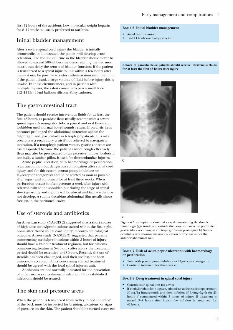
Acute peptic ulceration, with haemorrhage or perforation,is an uncommon but dangerous complication after spinal cordinjury, and for this reason proton pump inhibitors orH2-receptor antagonists should be started as soon as possibleafter injury and continued for at least three weeks. Whenperforation occurs it often presents a week after injury withreferred pain to the shoulder, but during the stage of spinalshock guarding and rigidity will be absent and tachycardia maynot develop. A supine decubitus abdominal film usually showsfree gas in the peritoneal cavity.
Use of steroids and antibiotics
An American study (NASCIS 2) suggested that a short courseof high-dose methylprednisolone started within the first eighthours after closed spinal cord injury improves neurologicaloutcome. A later study (NASCIS 3) suggested that patientscommencing methylprednisolone within 3 hours of injuryshould have a 24-hour treatment regimen, but for patientscommencing treatment 3–8 hours after injury the treatmentperiod should be extended to 48 hours. Recently the use ofsteroids has been challenged, and their use has not beenuniversally accepted. Policy concerning steroid treatmentshould be agreed with the local spinal injuries unit.
Antibiotics are not normally indicated for the preventionof either urinary or pulmonary infection. Only establishedinfections should be treated.
The skin and pressure areas
When the patient is transferred from trolley to bed the wholeof the back must be inspected for bruising, abrasions, or signsof pressure on the skin. The patient should be turned every two
Early management and complications—I
19
Box 4.8 Drug treatment in spinal cord injury
• Consult your spinal unit for advice• If methylprednisolone is given, administer at the earliest opportunity:
30 mg/kg intravenously and then infusion of 5.4 mg/kg/h for 23hours if commenced within 3 hours of injury. If treatment isstarted 3–8 hours after injury, the infusion is continued for47 hours.
Beware of paralytic ileus: patients should receive intravenous fluidsfor at least the first 48 hours after injury
Box 4.6 Initial bladder management
• Avoid overdistension• 12–14 Ch silicone Foley catheter
Box 4.7 Risk of acute peptic ulceration with haemorrhageor perforation
• Treat with proton pump inhibitor or H2-receptor antagonist• Continue treatment for three weeks
Figure 4.3 a) Supine abdominal x ray demonstrating the doublelumen sign (gas inside and outside the bowel) in an acute perforatedgastric ulcer occurring in a tetraplegic 5 days post-injury. b) Supinedecubitus view showing massive collection of free gas under theanterior abdominal wall.
(b)
(a)
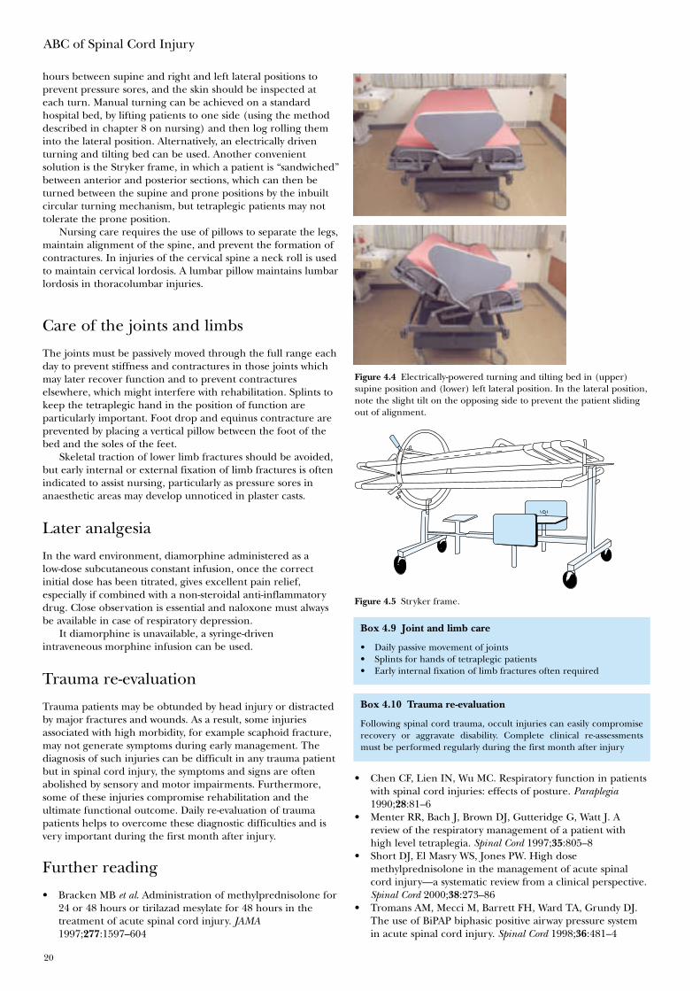
hours between supine and right and left lateral positions toprevent pressure sores, and the skin should be inspected ateach turn. Manual turning can be achieved on a standardhospital bed, by lifting patients to one side (using the methoddescribed in chapter 8 on nursing) and then log rolling theminto the lateral position. Alternatively, an electrically driventurning and tilting bed can be used. Another convenientsolution is the Stryker frame, in which a patient is “sandwiched”between anterior and posterior sections, which can then beturned between the supine and prone positions by the inbuiltcircular turning mechanism, but tetraplegic patients may nottolerate the prone position.
Nursing care requires the use of pillows to separate the legs,maintain alignment of the spine, and prevent the formation ofcontractures. In injuries of the cervical spine a neck roll is usedto maintain cervical lordosis. A lumbar pillow maintains lumbarlordosis in thoracolumbar injuries.
Care of the joints and limbs
The joints must be passively moved through the full range eachday to prevent stiffness and contractures in those joints whichmay later recover function and to prevent contractureselsewhere, which might interfere with rehabilitation. Splints tokeep the tetraplegic hand in the position of function areparticularly important. Foot drop and equinus contracture areprevented by placing a vertical pillow between the foot of thebed and the soles of the feet.
Skeletal traction of lower limb fractures should be avoided,but early internal or external fixation of limb fractures is oftenindicated to assist nursing, particularly as pressure sores inanaesthetic areas may develop unnoticed in plaster casts.
Later analgesia
In the ward environment, diamorphine administered as alow-dose subcutaneous constant infusion, once the correctinitial dose has been titrated, gives excellent pain relief,especially if combined with a non-steroidal anti-inflammatorydrug. Close observation is essential and naloxone must alwaysbe available in case of respiratory depression.
It diamorphine is unavailable, a syringe-drivenintraveneous morphine infusion can be used.
Trauma re-evaluation
Trauma patients may be obtunded by head injury or distractedby major fractures and wounds. As a result, some injuriesassociated with high morbidity, for example scaphoid fracture,may not generate symptoms during early management. Thediagnosis of such injuries can be difficult in any trauma patientbut in spinal cord injury, the symptoms and signs are oftenabolished by sensory and motor impairments. Furthermore,some of these injuries compromise rehabilitation and theultimate functional outcome. Daily re-evaluation of traumapatients helps to overcome these diagnostic difficulties and isvery important during the first month after injury.
Further reading
• Bracken MB et al. Administration of methylprednisolone for24 or 48 hours or tirilazad mesylate for 48 hours in thetreatment of acute spinal cord injury. JAMA1997;277:1597–604
ABC of Spinal Cord Injury
20
Figure 4.4 Electrically-powered turning and tilting bed in (upper)supine position and (lower) left lateral position. In the lateral position,note the slight tilt on the opposing side to prevent the patient slidingout of alignment.
Figure 4.5 Stryker frame.
Box 4.9 Joint and limb care
• Daily passive movement of joints• Splints for hands of tetraplegic patients• Early internal fixation of limb fractures often required
Box 4.10 Trauma re-evaluation
Following spinal cord trauma, occult injuries can easily compromiserecovery or aggravate disability. Complete clinical re-assessmentsmust be performed regularly during the first month after injury
• Chen CF, Lien IN, Wu MC. Respiratory function in patientswith spinal cord injuries: effects of posture. Paraplegia1990;28:81–6
• Menter RR, Bach J, Brown DJ, Gutteridge G, Watt J. Areview of the respiratory management of a patient withhigh level tetraplegia. Spinal Cord 1997;35:805–8
• Short DJ, El Masry WS, Jones PW. High dosemethylprednisolone in the management of acute spinalcord injury—a systematic review from a clinical perspective.Spinal Cord 2000;38:273–86
• Tromans AM, Mecci M, Barrett FH, Ward TA, Grundy DJ.The use of BiPAP biphasic positive airway pressure systemin acute spinal cord injury. Spinal Cord 1998;36:481–4
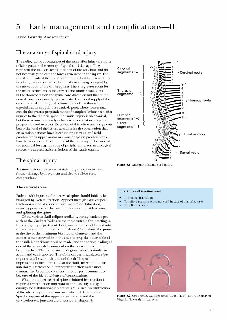
21
David Grundy, Andrew Swain
The anatomy of spinal cord injury
The radiographic appearances of the spine after injury are not areliable guide to the severity of spinal cord damage. Theyrepresent the final or “recoil” position of the vertebrae and donot necessarily indicate the forces generated in the injury. Thespinal cord ends at the lower border of the first lumbar vertebrain adults, the remainder of the spinal canal being occupied bythe nerve roots of the cauda equina. There is greater room forthe neural structures in the cervical and lumbar canals, butin the thoracic region the spinal cord diameter and that of theneural canal more nearly approximate. The blood supply of thecervical spinal cord is good, whereas that of the thoracic cord,especially at its midpoint, is relatively poor. These factors mayexplain the greater preponderance of complete lesions seen afterinjuries to the thoracic spine. The initial injury is mechanical,but there is usually an early ischaemic lesion that may rapidlyprogress to cord necrosis. Extension of this, often many segmentsbelow the level of the lesion, accounts for the observation thaton occasion patients have lower motor neurone or flaccidparalysis when upper motor neurone or spastic paralysis wouldhave been expected from the site of the bony injury. Because ofthe potential for regeneration of peripheral nerves, neurologicalrecovery is unpredictable in lesions of the cauda equina.
The spinal injury
Treatment should be aimed at stabilising the spine to avoidfurther damage by movement and also to relieve cordcompression.
The cervical spine
Patients with injuries of the cervical spine should initially bemanaged by skeletal traction. Applied through skull calipers,traction is aimed at reducing any fracture or dislocation,relieving pressure on the cord in the case of burst fractures,and splinting the spine.
Of the various skull calipers available, spring-loaded typessuch as the Gardner-Wells are the most suitable for inserting inthe emergency department. Local anaesthetic is infiltrated intothe scalp down to the periosteum about 2.5 cm above the pinnaat the site of the maximum bitemporal diameter, and thecaliper is then screwed into the scalp to grip the outer table ofthe skull. No incisions need be made, and the spring loading ofone of the screws determines when the correct tension hasbeen reached. The University of Virginia caliper is similar inaction and easily applied. The Cone caliper is satisfactory butrequires small scalp incisions and the drilling of 1 mmimpressions in the outer table of the skull. Insertion too faranteriorly interferes with temporalis function and causestrismus. The Crutchfield caliper is no longer recommendedbecause of the high incidence of complications.
When the upper cervical spine is injured less traction isrequired for reduction and stabilisation. Usually 1–2 kg isenough for stabilisation; if more weight is used overdistractionat the site of injury may cause neurological deterioration.Specific injuries of the upper cervical spine and thecervicothoracic junction are discussed in chapter 6.
5 Early management and complications—II
Lumbar segments 1–5
Thoracic segments 1–12
Cervical segments 1–8 Cervical roots
1
C123456
7T123
45
67
89
101112L1
234
5S1
23
4
5
1
23456712345678910
11
12 12
11
109876543 2 1
1
2
3
4
5
5
4
3
2
1
11
2345
23
45
2345678
Thoracic roots
Lumbar roots
Sacral roots
Sacral segments 1–5
Figure 5.1 Anatomy of spinal cord injury.
Figure 5.2 Cone (left), Gardner-Wells (upper right), and University ofVirginia (lower right) calipers.
Box 5.1 Skull traction used
• To reduce dislocation• To relieve pressure on spinal cord in case of burst fractures• To splint the spine
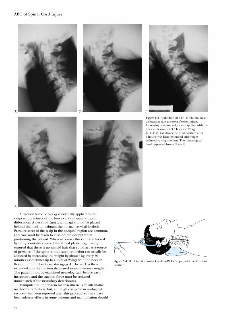
ABC of Spinal Cord Injury
22
A traction force of 3–5 kg is normally applied to thecalipers in fractures of the lower cervical spine withoutdislocation. A neck roll (not a sandbag) should be placedbehind the neck to maintain the normal cervical lordosis.Pressure sores of the scalp in the occipital region are common,and care must be taken to cushion the occiput whenpositioning the patient. When necessary this can be achievedby using a suitably covered fluid-filled plastic bag, havingensured that there is no matted hair that could act as a sourceof pressure. If the spine is dislocated reduction can usually beachieved by increasing the weight by about 4 kg every 30minutes (sometimes up to a total of 25 kg) with the neck inflexion until the facets are disengaged. The neck is thenextended and the traction decreased to maintenance weight.The patient must be examined neurologically before eachincrement, and the traction force must be reducedimmediately if the neurology deteriorates.
Manipulation under general anaesthesia is an alternativemethod of reduction, but, although complete neurologicalrecovery has been reported after this procedure, there havebeen adverse effects in some patients and manipulation should
Figure 5.3 Reduction of a C4–5 bilateral facetdislocation due to severe flexion injury.Increasing traction weight was applied with theneck in flexion for 3.5 hours to 25 kg((1)–(4)). (5) shows the final position after4 hours with head extended and weightreduced to 4 kg traction. The neurologicallevel improved from C5 to C6.
Figure 5.4 Skull traction using Gardner-Wells caliper, with neck roll inposition.
(4) (5)
(3)(1) (2)
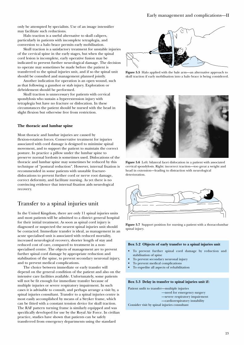
Early management and complications—II
23
only be attempted by specialists. Use of an image intensifiermay facilitate such reductions.
Halo traction is a useful alternative to skull calipers,particularly in patients with incomplete tetraplegia, andconversion to a halo brace permits early mobilisation.
Skull traction is a satisfactory treatment for unstable injuriesof the cervical spine in the early stages, but when the spinalcord lesion is incomplete, early operative fusion may beindicated to prevent further neurological damage. The decisionto operate may sometimes be made before the patient istransferred to the spinal injuries unit, and if so the spinal unitshould be consulted and management planned jointly.
Another indication for operation is an open wound, suchas that following a gunshot or stab injury. Exploration ordebridement should be performed.
Skull traction is unnecessary for patients with cervicalspondylosis who sustain a hyperextension injury withtetraplegia but have no fracture or dislocation. In thesecircumstances the patient should be nursed with the head inslight flexion but otherwise free from restriction.
The thoracic and lumbar spine
Most thoracic and lumbar injuries are caused byflexion-rotation forces. Conservative treatment for injuriesassociated with cord damage is designed to minimise spinalmovement, and to support the patient to maintain the correctposture. In practice a pillow under the lumbar spine topreserve normal lordosis is sometimes used. Dislocations of thethoracic and lumbar spine may sometimes be reduced by thistechnique of “postural reduction”. However, internal fixation isrecommended in some patients with unstable fracture-dislocations to prevent further cord or nerve root damage,correct deformity, and facilitate nursing. As yet there is noconvincing evidence that internal fixation aids neurologicalrecovery.
Transfer to a spinal injuries unit
In the United Kingdom, there are only 11 spinal injuries unitsand most patients will be admitted to a district general hospitalfor their initial treatment. As soon as spinal cord injury isdiagnosed or suspected the nearest spinal injuries unit shouldbe contacted. Immediate transfer is ideal, as management in anacute specialised unit is associated with reduced mortality,increased neurological recovery, shorter length of stay andreduced cost of care, compared to treatment in a non-specialised centre. The objects of management are to preventfurther spinal cord damage by appropriate reduction andstabilisation of the spine, to prevent secondary neuronal injury,and to prevent medical complications.
The choice between immediate or early transfer willdepend on the general condition of the patient and also on theintensive care facilities available. Unfortunately, some patientswill not be fit enough for immediate transfer because ofmultiple injuries or severe respiratory impairment. In suchcases it is advisable to consult, and perhaps arrange a visit by, aspinal injuries consultant. Transfer to a spinal injuries centre ismost easily accomplished by means of a Stryker frame, whichcan be fitted with a constant tension device for skull traction.The RAF pattern turning frame is similarly equipped and wasspecifically developed for use by the Royal Air Force. In civilianpractice, studies have shown that patients can be safelytransferred from emergency departments using the standard
Figure 5.5 Halo applied with the bale arm—an alternative approach toskull traction if early mobilisation into a halo brace is being considered.
Figure 5.6 Left: bilateral facet dislocation in a patient with associatedcervical spondylosis. Right: incorrect traction—too great a weight andhead in extension—leading to distraction with neurologicaldeterioration.
Figure 5.7 Support position for nursing a patient with a thoracolumbarspinal injury.
Box 5.2 Objects of early transfer to a spinal injuries unit
• To prevent further spinal cord damage by reduction andstabilisation of spine
• To prevent secondary neuronal injury• To prevent medical complications• To expedite all aspects of rehabilitation
Box 5.3 Delay in transfer to spinal injuries unit if:
Patient unfit to transfer—multiple injuries—need for emergency surgery—severe respiratory impairment—cardiorespiratory instability
Consider visit by spinal injuries consultant
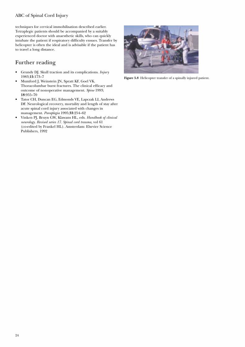
ABC of Spinal Cord Injury
24
techniques for cervical immobilisation described earlier.Tetraplegic patients should be accompanied by a suitablyexperienced doctor with anaesthetic skills, who can quicklyintubate the patient if respiratory difficulty ensues. Transfer byhelicopter is often the ideal and is advisable if the patient hasto travel a long distance.
Further reading
• Grundy DJ. Skull traction and its complications. Injury1983;15:173–7
• Mumford J, Weinstein JN, Spratt KF, Goel VK.Thoracolumbar burst fractures. The clinical efficacy andoutcome of nonoperative management. Spine 1993;18:955–70
• Tator CH, Duncan EG, Edmonds VE, Lapczak LI, AndrewsDF. Neurological recovery, mortality and length of stay afteracute spinal cord injury associated with changes inmanagement. Paraplegia 1995;33:254–62
• Vinken PJ, Bruyn GW, Klawans HL, eds. Handbook of clinicalneurology. Revised series 17. Spinal cord trauma, vol 61(co-edited by Frankel HL). Amsterdam: Elsevier SciencePublishers, 1992
Figure 5.8 Helicopter transfer of a spinally injured patient.
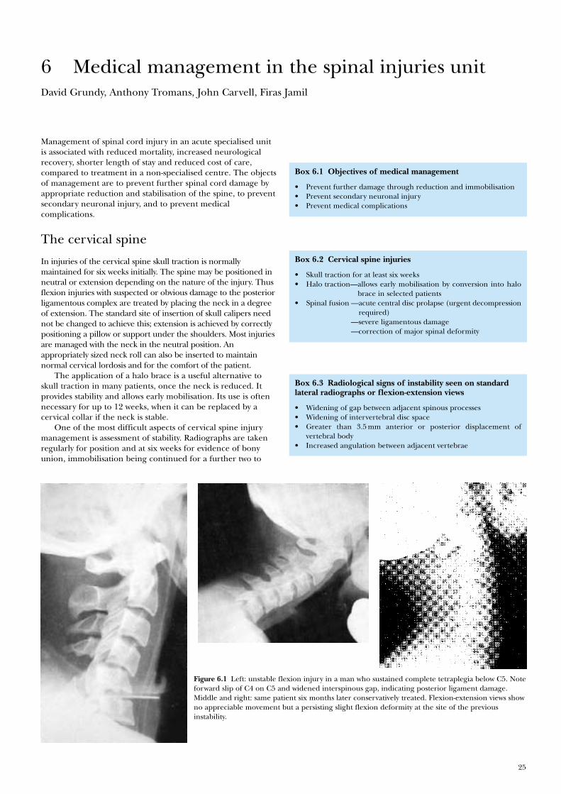
25
Management of spinal cord injury in an acute specialised unitis associated with reduced mortality, increased neurologicalrecovery, shorter length of stay and reduced cost of care,compared to treatment in a non-specialised centre. The objectsof management are to prevent further spinal cord damage byappropriate reduction and stabilisation of the spine, to preventsecondary neuronal injury, and to prevent medicalcomplications.
The cervical spine
In injuries of the cervical spine skull traction is normallymaintained for six weeks initially. The spine may be positioned inneutral or extension depending on the nature of the injury. Thusflexion injuries with suspected or obvious damage to the posteriorligamentous complex are treated by placing the neck in a degreeof extension. The standard site of insertion of skull calipers neednot be changed to achieve this; extension is achieved by correctlypositioning a pillow or support under the shoulders. Most injuriesare managed with the neck in the neutral position. Anappropriately sized neck roll can also be inserted to maintainnormal cervical lordosis and for the comfort of the patient.
The application of a halo brace is a useful alternative toskull traction in many patients, once the neck is reduced. Itprovides stability and allows early mobilisation. Its use is oftennecessary for up to 12 weeks, when it can be replaced by acervical collar if the neck is stable.
One of the most difficult aspects of cervical spine injurymanagement is assessment of stability. Radiographs are takenregularly for position and at six weeks for evidence of bonyunion, immobilisation being continued for a further two to
6 Medical management in the spinal injuries unitDavid Grundy, Anthony Tromans, John Carvell, Firas Jamil
Figure 6.1 Left: unstable flexion injury in a man who sustained complete tetraplegia below C5. Noteforward slip of C4 on C5 and widened interspinous gap, indicating posterior ligament damage.Middle and right: same patient six months later conservatively treated. Flexion-extension views showno appreciable movement but a persisting slight flexion deformity at the site of the previousinstability.
Box 6.1 Objectives of medical management
• Prevent further damage through reduction and immobilisation• Prevent secondary neuronal injury• Prevent medical complications
Box 6.2 Cervical spine injuries
• Skull traction for at least six weeks• Halo traction—allows early mobilisation by conversion into halo
brace in selected patients• Spinal fusion —acute central disc prolapse (urgent decompression
required)—severe ligamentous damage—correction of major spinal deformity
Box 6.3 Radiological signs of instability seen on standardlateral radiographs or flexion-extension views
• Widening of gap between adjacent spinous processes• Widening of intervertebral disc space• Greater than 3.5 mm anterior or posterior displacement of
vertebral body• Increased angulation between adjacent vertebrae
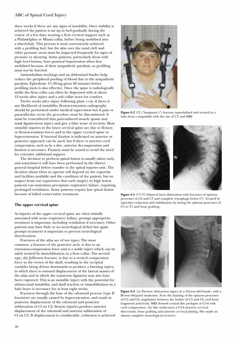
ABC of Spinal Cord Injury
26
three weeks if there are any signs of instability. Once stability isachieved the patient is sat up in bed gradually during thecourse of a few days, wearing a firm cervical support such asa Philadelphia or Miami collar, before being mobilised intoa wheelchair. This process is most conveniently achievedwith a profiling bed, but the skin over the natal cleft andother pressure areas must be inspected frequently for signs ofpressure or shearing. Some patients, particularly those withhigh level lesions, have postural hypotension when firstmobilised because of their sympathetic paralysis, so profilingmust not be hurried.
Antiembolism stockings and an abdominal binder helpreduce the peripheral pooling of blood due to the sympatheticparalysis. Ephedrine 15–30 mg given 20 minutes beforeprofiling starts is also effective. Once the spine is radiologicallystable the firm collar can often be dispensed with at about12 weeks after injury and a soft collar worn for comfort.
Twelve weeks after injury following plain x ray, if there isany likelihood of instability, flexion-extension radiographyshould be performed under medical supervision but if pain orparaesthesiae occur the procedure must be discontinued. Itmust be remembered that pain-induced muscle spasm maymask ligamentous injury and give a false sense of security. Mostunstable injuries in the lower cervical spine are due to flexionor flexion-rotation forces and in the upper cervical spine tohyperextension. If internal fixation is indicated an anterior orposterior approach can be used, but if there is anterior cordcompression, such as by a disc, anterior decompression andfixation is necessary. Fixation must be sound to avoid the needfor extensive additional support.
The decision to perform spinal fusion is usually taken early,and sometimes it will have been performed in the districtgeneral hospital before transfer to the spinal injuries unit. Thedecision about when to operate will depend on the expertiseand facilities available and the condition of the patient, but wesuspect from our experience that early surgery in high lesionpatients can sometimes precipitate respiratory failure, requiringprolonged ventilation. Some patients require late spinal fusionbecause of failed conservative treatment.
The upper cervical spine
As injuries of the upper cervical spine are often initiallyassociated with acute respiratory failure, prompt appropriatetreatment is important, including ventilation if necessary. Otherpatients may have little or no neurological deficit but againprompt treatment is important to prevent neurologicaldeterioration.
Fractures of the atlas are of two types. The mostcommon, a fracture of the posterior arch, is due to anextension-compression force and is a stable injury which can besafely treated by immobilisation in a firm collar. The secondtype, the Jefferson fracture, is due to a vertical compressionforce to the vertex of the skull, resulting in the occipitalcondyles being driven downwards to produce a bursting injury,in which there is outward displacement of the lateral masses ofthe atlas and in which the transverse ligament may also havebeen ruptured. This is an unstable injury with the potential foratlanto-axial instability, and skull traction or immobilisation in ahalo brace is necessary for at least eight weeks.
Fractures through the base of the odontoid process (type IIfractures) are usually caused by hyperextension, and result inposterior displacement of the odontoid and posteriorsubluxation of Cl on C2; flexion injuries produce anteriordisplacement of the odontoid and anterior subluxation ofCl on C2. If displacement is considerable, reduction is achieved
Figure 6.2 C2 (“hangman’s”) fracture immobilised and treated in ahalo brace compatible with the use of CT and MRI.
Figure 6.3 C7–T1 bilateral facet dislocation with fractures of spinousprocesses of C6 and C7 and complete tetraplegia below C7. Treated byoperative reduction and stabilisation by wiring the spinous processes ofC5 to T1 and bone grafting.
Figure 6.4 (a) Flexion/distraction injury in a 42-year-old female, with aBrown–Séquard syndrome. Note the fanning of the spinous processesof C5 and C6, angulation between the bodies of C5 and C6, and bonyfragments anteriorly. MRI showed central disc prolapse at C5-6 withcord compression. (b) She underwent a C5-6 anterior cervicaldiscectomy, bone grafting and anterior cervical plating. She made analmost complete neurological recovery.
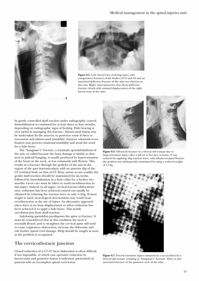
Medical management in the spinal injuries unit
27
by gentle controlled skull traction under radiographic control.Immobilisation is continued for at least three to four months,depending on radiographic signs of healing. Halo bracing isvery useful in managing this fracture. Atlanto-axial fusion maybe undertaken by the anterior or posterior route if there isnon-union and atlanto-axial instability. Anterior odontoid screwfixation may prevent rotational instability and avoid the needfor a halo brace.
The “hangman’s” fracture, a traumatic spondylolisthesis ofthe axis, so called because the bony damage is similar to thatseen in judicial hanging, is usually produced by hyperextensionof the head on the neck, or less commonly with flexion. Thisresults in a fracture through the pedicles of the axis in theregion of the pars interarticularis, with an anterior slip of theC2 vertebral body on that of C3. Bony union occurs readily, butgentle skull traction should be maintained for six weeks,followed by immobilisation in a firm collar for a further twomonths. Great care must be taken to avoid overdistraction inthis injury. Indeed, in all upper cervical fracture-dislocationsonce reduction has been achieved control can usually beobtained by reducing the traction force to only 1–2 kg. If moreweight is used, neurological deterioration may result fromoverdistraction at the site of injury. An alternative approachwhen there is no bony displacement or when reduction hasbeen achieved is to apply a halo brace. This avoidsoverdistraction from skull traction.
Ankylosing spondylitis predisposes the spine to fracture. Itmust be remembered that in this condition the neck isnormally flexed, and to straighten the cervical spine will tendto cause respiratory obstruction, increase the deformity andrisk further spinal cord damage. Help should be sought as soonas the problem is recognised.
The cervicothoracic junction
Closed reduction of a C7–T1 facet dislocation is often difficultif not impossible, in which case operative reduction byfacetectomy and posterior fusion is indicated, particularly inpatients with an incomplete spinal cord lesion.
Figure 6.5 Left: lateral view of diving injury withcompression fractures of the bodies of C5 and C6 and anassociated Jefferson fracture of the atlas not obvious onthis view. Right: anteroposterior view shows Jeffersonfracture clearly with outward displacement of the rightlateral mass of the atlas.
Figure 6.6 Odontoid fracture in a 64-year-old woman due tohyperextension injury after a fall on to her face at home. It wasreduced by applying 4 kg traction force, with atlanto-occipital flexion;the position was subsequently maintained by using a reduced weightof 1.5 kg.
Figure 6.7 Forced extension injury sustained in a car accident by a22-year-old woman, resulting in “hangman’s” fracture. There is alsoassociated fracture of the posterior arch of the atlas.
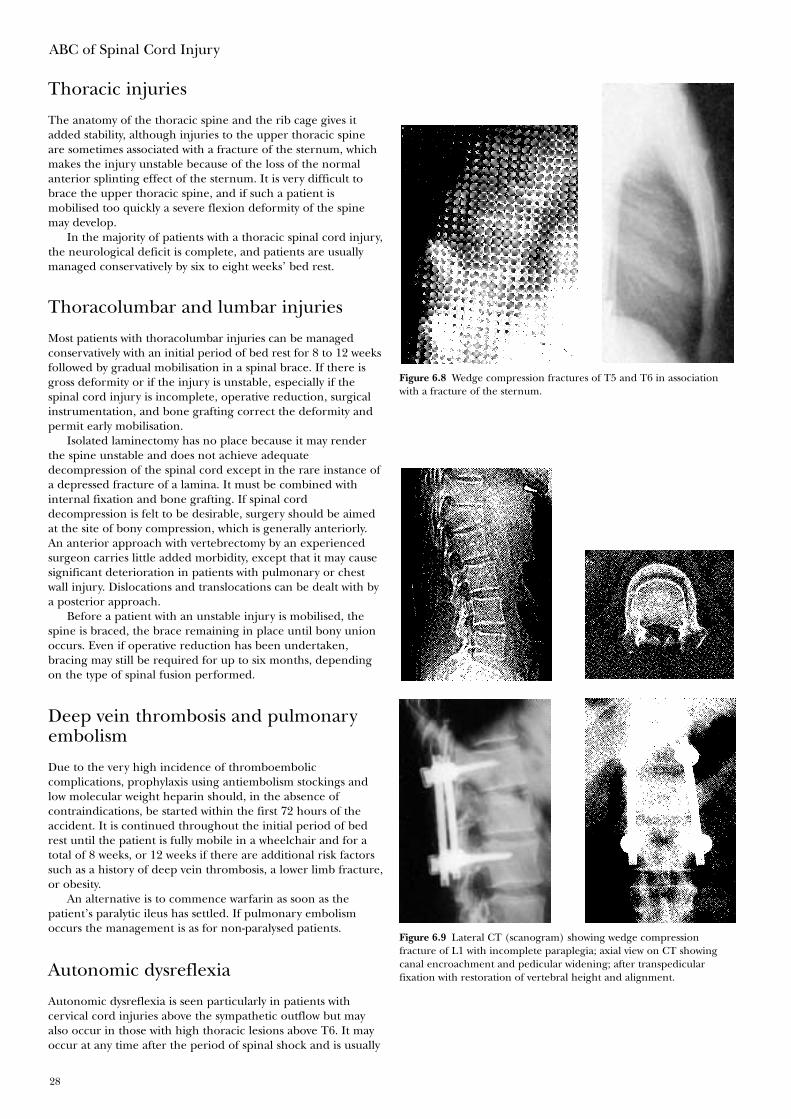
ABC of Spinal Cord Injury
28
Thoracic injuries
The anatomy of the thoracic spine and the rib cage gives itadded stability, although injuries to the upper thoracic spineare sometimes associated with a fracture of the sternum, whichmakes the injury unstable because of the loss of the normalanterior splinting effect of the sternum. It is very difficult tobrace the upper thoracic spine, and if such a patient ismobilised too quickly a severe flexion deformity of the spinemay develop.
In the majority of patients with a thoracic spinal cord injury,the neurological deficit is complete, and patients are usuallymanaged conservatively by six to eight weeks’ bed rest.
Thoracolumbar and lumbar injuries
Most patients with thoracolumbar injuries can be managedconservatively with an initial period of bed rest for 8 to 12 weeksfollowed by gradual mobilisation in a spinal brace. If there isgross deformity or if the injury is unstable, especially if thespinal cord injury is incomplete, operative reduction, surgicalinstrumentation, and bone grafting correct the deformity andpermit early mobilisation.
Isolated laminectomy has no place because it may renderthe spine unstable and does not achieve adequatedecompression of the spinal cord except in the rare instance ofa depressed fracture of a lamina. It must be combined withinternal fixation and bone grafting. If spinal corddecompression is felt to be desirable, surgery should be aimedat the site of bony compression, which is generally anteriorly.An anterior approach with vertebrectomy by an experiencedsurgeon carries little added morbidity, except that it may causesignificant deterioration in patients with pulmonary or chestwall injury. Dislocations and translocations can be dealt with bya posterior approach.
Before a patient with an unstable injury is mobilised, thespine is braced, the brace remaining in place until bony unionoccurs. Even if operative reduction has been undertaken,bracing may still be required for up to six months, dependingon the type of spinal fusion performed.
Deep vein thrombosis and pulmonaryembolism
Due to the very high incidence of thromboemboliccomplications, prophylaxis using antiembolism stockings andlow molecular weight heparin should, in the absence ofcontraindications, be started within the first 72 hours of theaccident. It is continued throughout the initial period of bedrest until the patient is fully mobile in a wheelchair and for atotal of 8 weeks, or 12 weeks if there are additional risk factorssuch as a history of deep vein thrombosis, a lower limb fracture,or obesity.
An alternative is to commence warfarin as soon as thepatient’s paralytic ileus has settled. If pulmonary embolismoccurs the management is as for non-paralysed patients.
Autonomic dysreflexia
Autonomic dysreflexia is seen particularly in patients withcervical cord injuries above the sympathetic outflow but mayalso occur in those with high thoracic lesions above T6. It mayoccur at any time after the period of spinal shock and is usually
Figure 6.9 Lateral CT (scanogram) showing wedge compressionfracture of L1 with incomplete paraplegia; axial view on CT showingcanal encroachment and pedicular widening; after transpedicularfixation with restoration of vertebral height and alignment.
Figure 6.8 Wedge compression fractures of T5 and T6 in associationwith a fracture of the sternum.
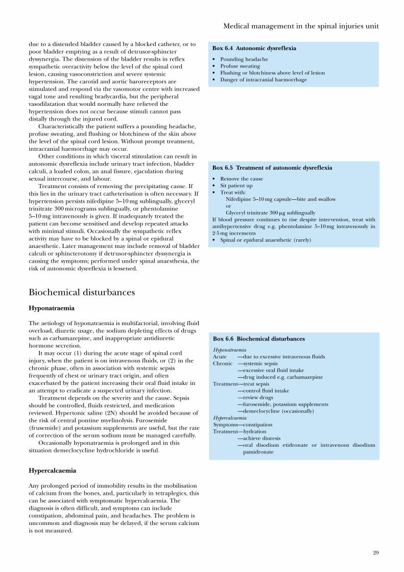
Medical management in the spinal injuries unit
29
due to a distended bladder caused by a blocked catheter, or topoor bladder emptying as a result of detrusor-sphincterdyssynergia. The distension of the bladder results in reflexsympathetic overactivity below the level of the spinal cordlesion, causing vasoconstriction and severe systemichypertension. The carotid and aortic baroreceptors arestimulated and respond via the vasomotor centre with increasedvagal tone and resulting bradycardia, but the peripheralvasodilatation that would normally have relieved thehypertension does not occur because stimuli cannot passdistally through the injured cord.
Characteristically the patient suffers a pounding headache,profuse sweating, and flushing or blotchiness of the skin abovethe level of the spinal cord lesion. Without prompt treatment,intracranial haemorrhage may occur.
Other conditions in which visceral stimulation can result inautonomic dysreflexia include urinary tract infection, bladdercalculi, a loaded colon, an anal fissure, ejaculation duringsexual intercourse, and labour.
Treatment consists of removing the precipitating cause. Ifthis lies in the urinary tract catheterisation is often necessary. Ifhypertension persists nifedipine 5–10 mg sublingually, glyceryltrinitrate 300 micrograms sublingually, or phentolamine5–10 mg intravenously is given. If inadequately treated thepatient can become sensitised and develop repeated attackswith minimal stimuli. Occasionally the sympathetic reflexactivity may have to be blocked by a spinal or epiduralanaesthetic. Later management may include removal of bladdercalculi or sphincterotomy if detrusor-sphincter dyssynergia iscausing the symptoms; performed under spinal anaesthesia, therisk of autonomic dysreflexia is lessened.
Biochemical disturbances
Hyponatraemia
The aetiology of hyponatraemia is multifactorial, involving fluidoverload, diuretic usage, the sodium depleting effects of drugssuch as carbamazepine, and inappropriate antidiuretichormone secretion.
It may occur (1) during the acute stage of spinal cordinjury, when the patient is on intravenous fluids, or (2) in thechronic phase, often in association with systemic sepsisfrequently of chest or urinary tract origin, and oftenexacerbated by the patient increasing their oral fluid intake inan attempt to eradicate a suspected urinary infection.
Treatment depends on the severity and the cause. Sepsisshould be controlled, fluids restricted, and medicationreviewed. Hypertonic saline (2N) should be avoided because ofthe risk of central pontine myelinolysis. Furosemide(frusemide) and potassium supplements are useful, but the rateof correction of the serum sodium must be managed carefully.
Occasionally hyponatraemia is prolonged and in thissituation demeclocycline hydrochloride is useful.
Hypercalcaemia
Any prolonged period of immobility results in the mobilisationof calcium from the bones, and, particularly in tetraplegics, thiscan be associated with symptomatic hypercalcaemia. Thediagnosis is often difficult, and symptoms can includeconstipation, abdominal pain, and headaches. The problem isuncommon and diagnosis may be delayed, if the serum calciumis not measured.
Box 6.6 Biochemical disturbances
HyponatraemiaAcute —due to excessive intravenous fluidsChronic —systemic sepsis
—excessive oral fluid intake—drug induced e.g. carbamazepine
Treatment—treat sepsis—control fluid intake—review drugs—furosemide, potassium supplements—demeclocycline (occasionally)
HypercalcaemiaSymptoms—constipationTreatment—hydration
—achieve diuresis—oral disodium etidronate or intravenous disodium
pamidronate
Box 6.4 Autonomic dysreflexia
• Pounding headache• Profuse sweating• Flushing or blotchiness above level of lesion• Danger of intracranial haemorrhage
Box 6.5 Treatment of autonomic dysreflexia
• Remove the cause• Sit patient up• Treat with:
Nifedipine 5–10 mg capsule—bite and swalloworGlyceryl trinitrate 300 �g sublingually
If blood pressure continues to rise despite intervention, treat withantihypertensive drug e.g. phentolamine 5–10 mg intravenously in2.5 mg increments• Spinal or epidural anaesthetic (rarely)
ABC of Spinal Cord Injury
30
Treatment involves hydration, achieving a diuresis (with afluid load and furosemide (frusemide)), and the use of oralsodium etidronate or intravenous disodium pamidronate. Oncethe patient is fully mobile the problem usually resolves.
Para-articular heterotopic ossification
After injury to the spinal cord new bone is often laid down inthe soft tissues around paralysed joints, particularly the hip andknee. The cause is unknown, although local trauma has beensuggested. It usually presents with erythema, induration, orswelling near a joint. There is pronounced osteoblastic activity,but the new bone formed does not mature for at least18 months. This has an important bearing on treatment in thatif excision of heterotopic bone is required because of grossrestriction of movement or bony ankylosis of a joint, surgery isbest delayed for at least 18 months—until the new bone ismature. Earlier surgical intervention may provoke further newbone formation, thus compounding the original condition.Treatment with disodium etidronate suppresses themineralisation of osteoid tissue and may reverse up to half ofearly lesions when used for 3–6 months, and non-steroidalanti-inflammatory drugs are also used to prevent theprogression of this complication. Postoperative radiotherapymay halt the recurrence of the problem if early surgicalintervention has to be performed.
Spasticity
Spasticity is seen only in patients with upper motor neuronelesions of the cord whose intact spinal reflex arcs below thelevel of the lesion are isolated from higher centres. It usuallyincreases in severity during the first few weeks after injury, afterthe period of spinal shock. In incomplete lesions it is oftenmore pronounced and can be severe enough to preventpatients with good power in the legs from walking. Patients withsevere spasticity and imbalance of opposing muscle groups havea tendency to develop contractures. It is important to realisethat once a contracture occurs spasticity is increased and avicious circle is established with further deformity resulting.Although excessive spasticity may hamper patients’ activities oreven throw them out of their wheelchairs or make walkingimpossible, spasticity may have advantages. It maintains musclebulk and possibly bone density, and improves venous return.
Treatment of severe spasticity is indicated if it interfereswith activities of daily living, and is initially directed atremoving any obvious precipitating cause. An irritative lesion inthe paralysed part, such as a pressure sore, urinary tractinfection or calculus, anal fissure, infected ingrowing toenail, orfracture, tends to increase spasticity.
Passive stretching of spastic muscles and regular standingare helpful in relieving spasticity and preventing contractures.The drugs most commonly used to decrease spasticity arebaclofen and tizanidine, which act at spinal level, anddantrolene sodium, which acts directly on skeletal muscle.Although diazepam relieves spasticity, its sedative action andhabit-forming tendency limit its usefulness. With these drugs,the liver function tests need to be closely monitored.
If spasticity is localised it can be relieved by interrupting thenerve supply to the muscles affected by neurectomy after adiagnostic block with a long-acting local anaesthetic(bupivacaine). For example, in patients with severe hipadductor spasticity obturator neurectomy is effective.Alternatively, motor point injections, initially with bupivacaine,followed by either 6% aqueous phenol or 45% ethyl alcohol for
Figure 6.10 Heterotopic ossification in the right hip.
Box 6.7 Factors that aggravate spasticity
• Urinary tract infection or calculus• Infected ingrowing toenail• Pressure sores• Anal fissure• Fracture• Contractures
Box 6.8 Management of spasticity
• Treat factors that aggravate spasticity• Improve comfort/posture• Manage pain• Passively stretch spastic muscles• Regular standing• Oral therapy—baclofen
—tizanidine—dantrolene—diazepam
• Botulinum toxin• Motor point injections• Implanted drug delivery system for administration of intrathecal
baclofen
If the above fail, are contraindicated, or are unavailable:• tendon release and/or neurectomy, and other orthopaedic
procedures• intrathecal block (rarely used)—6% aqueous phenol
—absolute alcohol
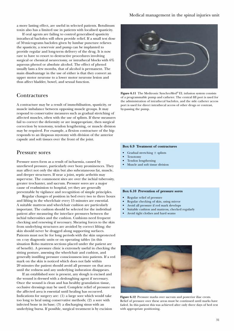
Medical management in the spinal injuries unit
31
a more lasting effect, are useful in selected patients. Botulinumtoxin also has a limited use in patients with localised spasticity.
If oral agents are failing to control generalised spasticityintrathecal baclofen will often provide relief. If a small test doseof 50 micrograms baclofen given by lumbar puncture relievesthe spasticity, a reservoir and pump can be implanted toprovide regular and long-term delivery of the drug. It is nowrare to have to resort to destructive procedures involvingsurgical or chemical neurectomy, or intrathecal blocks with 6%aqueous phenol or absolute alcohol. The effect of phenolusually lasts a few months, that of alcohol is permanent. Themain disadvantage in the use of either is that they convert anupper motor neurone to a lower motor neurone lesion andthus affect bladder, bowel, and sexual function.
Contractures
A contracture may be a result of immobilisation, spasticity, ormuscle imbalance between opposing muscle groups. It mayrespond to conservative measures such as gradual stretching ofaffected muscles, often with the use of splints. If these measuresfail to correct the deformity or are inappropriate, then surgicalcorrection by tenotomy, tendon lengthening, or muscle divisionmay be required. For example, a flexion contracture of the hipresponds to an iliopsoas myotomy with division of the anteriorcapsule and soft tissues over the front of the joint.
Pressure sores
Pressure sores form as a result of ischaemia, caused byunrelieved pressure, particularly over bony prominences. Theymay affect not only the skin but also subcutaneous fat, muscle,and deeper structures. If near a joint, septic arthritis maysupervene. The commonest sites are over the ischial tuberosity,greater trochanter, and sacrum. Pressure sores are a majorcause of readmission to hospital, yet they are generallypreventable by vigilance and recognition of simple principles.
Regular changes of position in bed every two to three hoursand lifting in the wheelchair every 15 minutes are essential.A suitable mattress and wheelchair cushion are particularlyimportant. The cushion should be selected for the individualpatient after measuring the interface pressures between theischial tuberosities and the cushion. Cushions need frequentchecking and renewing if necessary. Shearing forces to the skinfrom underlying structures are avoided by correct lifting; theskin should never be dragged along supporting surfaces.Patients must not lie for long periods with the skin unprotectedon x ray diagnostic units or on operating tables (in thissituation Roho mattress sections placed under the patient areof benefit). A pressure clinic is extremely useful in checking thesitting posture, assessing the wheelchair and cushion, andgenerally instilling pressure consciousness into patients. If a redmark on the skin is noticed which does not fade within20 minutes the patient should avoid all pressure on that areauntil the redness and any underlying induration disappears.
If an established sore is present, any slough is excised andthe wound is dressed with a desloughing agent if necessary.Once the wound is clean and has healthy granulation tissue,occlusive dressings may be used. Complete relief of pressure onthe affected area is essential until healing has occurred.Indications for surgery are: (1) a large sore which would taketoo long to heal using conservative methods; (2) a sore withinfected bone in its base; (3) a discharging sinus with anunderlying bursa. If possible, surgical treatment is by excision
Figure 6.12 Pressure marks over sacrum and posterior iliac crests.Relief of pressure over these areas must be continued until marks havefaded. In this patient this was achieved after only three days of bed restwith appropriate positioning.
Box 6.9 Treatment of contractures
• Gradual stretching � splints• Tenotomy• Tendon lengthening• Muscle and soft tissue division
Box 6.10 Prevention of pressure sores
• Regular relief of pressure• Regular checking of skin, using mirror• Avoid all pressure if red mark develops• Suitable cushion and mattress, checked regularly• Avoid tight clothes and hard seams
Figure 6.11 The Medtronic SynchroMed® EL infusion system consistsof a programmable pump and catheter. The central fill port is used forthe administration of intrathecal baclofen, and the side catheter accessport is used for direct intrathecal access of other drugs or contrast, by-passing the pump.
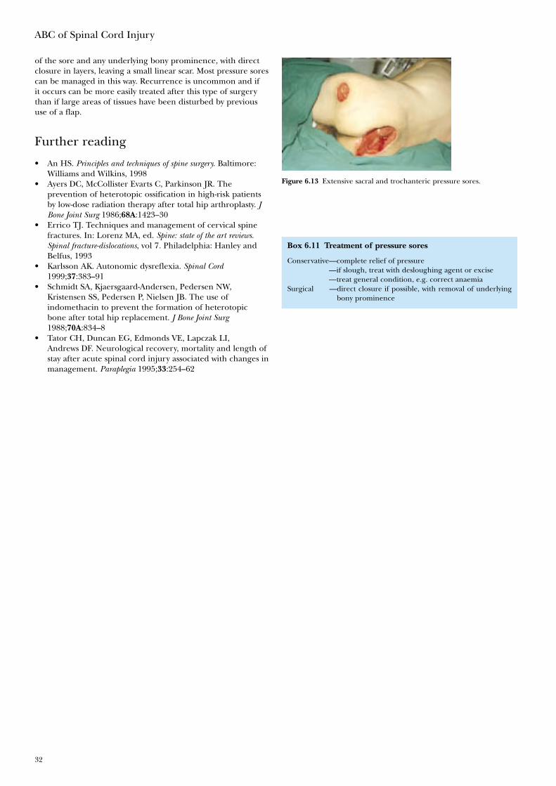
ABC of Spinal Cord Injury
32
of the sore and any underlying bony prominence, with directclosure in layers, leaving a small linear scar. Most pressure sorescan be managed in this way. Recurrence is uncommon and ifit occurs can be more easily treated after this type of surgerythan if large areas of tissues have been disturbed by previoususe of a flap.
Further reading
• An HS. Principles and techniques of spine surgery. Baltimore:Williams and Wilkins, 1998
• Ayers DC, McCollister Evarts C, Parkinson JR. Theprevention of heterotopic ossification in high-risk patientsby low-dose radiation therapy after total hip arthroplasty. JBone Joint Surg 1986;68A:1423–30
• Errico TJ. Techniques and management of cervical spinefractures. In: Lorenz MA, ed. Spine: state of the art reviews.Spinal fracture-dislocations, vol 7. Philadelphia: Hanley andBelfus, 1993
• Karlsson AK. Autonomic dysreflexia. Spinal Cord1999;37:383–91
• Schmidt SA, Kjaersgaard-Andersen, Pedersen NW,Kristensen SS, Pedersen P, Nielsen JB. The use ofindomethacin to prevent the formation of heterotopicbone after total hip replacement. J Bone Joint Surg1988;70A:834–8
• Tator CH, Duncan EG, Edmonds VE, Lapczak LI,Andrews DF. Neurological recovery, mortality and length ofstay after acute spinal cord injury associated with changes inmanagement. Paraplegia 1995;33:254–62
Figure 6.13 Extensive sacral and trochanteric pressure sores.
Box 6.11 Treatment of pressure sores
Conservative—complete relief of pressure—if slough, treat with desloughing agent or excise—treat general condition, e.g. correct anaemia
Surgical —direct closure if possible, with removal of underlyingbony prominence
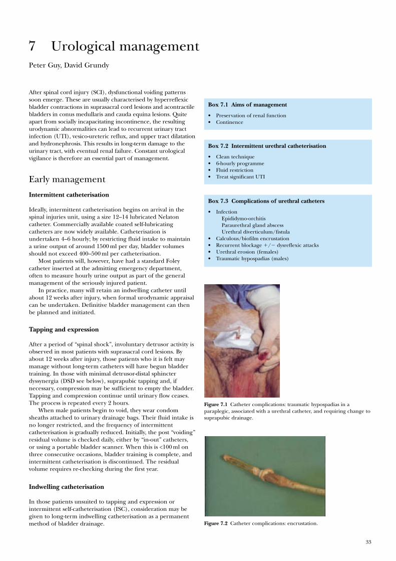
33
Peter Guy, David Grundy
After spinal cord injury (SCI), dysfunctional voiding patternssoon emerge. These are usually characterised by hyperreflexicbladder contractions in suprasacral cord lesions and acontractilebladders in conus medullaris and cauda equina lesions. Quiteapart from socially incapacitating incontinence, the resultingurodynamic abnormalities can lead to recurrent urinary tractinfection (UTI), vesico-ureteric reflux, and upper tract dilatationand hydronephrosis. This results in long-term damage to theurinary tract, with eventual renal failure. Constant urologicalvigilance is therefore an essential part of management.
Early management
Intermittent catheterisation
Ideally, intermittent catheterisation begins on arrival in thespinal injuries unit, using a size 12–14 lubricated Nelatoncatheter. Commercially available coated self-lubricatingcatheters are now widely available. Catheterisation isundertaken 4–6 hourly; by restricting fluid intake to maintaina urine output of around 1500 ml per day, bladder volumesshould not exceed 400–500 ml per catheterisation.
Most patients will, however, have had a standard Foleycatheter inserted at the admitting emergency department,often to measure hourly urine output as part of the generalmanagement of the seriously injured patient.
In practice, many will retain an indwelling catheter untilabout 12 weeks after injury, when formal urodynamic appraisalcan be undertaken. Definitive bladder management can thenbe planned and initiated.
Tapping and expression
After a period of “spinal shock”, involuntary detrusor activity isobserved in most patients with suprasacral cord lesions. Byabout 12 weeks after injury, those patients who it is felt maymanage without long-term catheters will have begun bladdertraining. In those with minimal detrusor-distal sphincterdyssynergia (DSD see below), suprapubic tapping and, ifnecessary, compression may be sufficient to empty the bladder.Tapping and compression continue until urinary flow ceases.The process is repeated every 2 hours.
When male patients begin to void, they wear condomsheaths attached to urinary drainage bags. Their fluid intake isno longer restricted, and the frequency of intermittentcatheterisation is gradually reduced. Initially, the post “voiding”residual volume is checked daily, either by “in-out” catheters,or using a portable bladder scanner. When this is <100 ml onthree consecutive occasions, bladder training is complete, andintermittent catheterisation is discontinued. The residualvolume requires re-checking during the first year.
Indwelling catheterisation
In those patients unsuited to tapping and expression orintermittent self-catheterisation (ISC), consideration may begiven to long-term indwelling catheterisation as a permanentmethod of bladder drainage.
7 Urological management
Box 7.1 Aims of management
• Preservation of renal function• Continence
Box 7.2 Intermittent urethral catheterisation
• Clean technique• 6-hourly programme• Fluid restriction• Treat significant UTI
Box 7.3 Complications of urethral catheters
• InfectionEpididymo-orchitisParaurethral gland abscessUrethral diverticulum/fistula
• Calculous/biofilm encrustation• Recurrent blockage �/� dysreflexic attacks• Urethral erosion (females)• Traumatic hypospadias (males)
Figure 7.2 Catheter complications: encrustation.
Figure 7.1 Catheter complications: traumatic hypospadias in aparaplegic, associated with a urethral catheter, and requiring change tosuprapubic drainage.
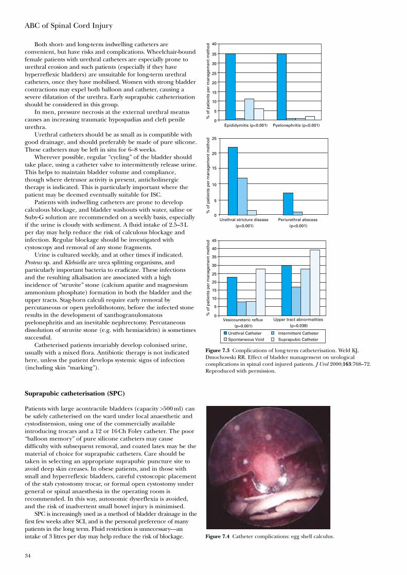
ABC of Spinal Cord Injury
34
Both short- and long-term indwelling catheters areconvenient, but have risks and complications. Wheelchair-boundfemale patients with urethral catheters are especially prone tourethral erosion and such patients (especially if they havehyperreflexic bladders) are unsuitable for long-term urethralcatheters, once they have mobilised. Women with strong bladdercontractions may expel both balloon and catheter, causing asevere dilatation of the urethra. Early suprapubic catheterisationshould be considered in this group.
In men, pressure necrosis at the external urethral meatuscauses an increasing traumatic hypospadias and cleft penileurethra.
Urethral catheters should be as small as is compatible withgood drainage, and should preferably be made of pure silicone.These catheters may be left in situ for 6–8 weeks.
Wherever possible, regular “cycling” of the bladder shouldtake place, using a catheter valve to intermittently release urine.This helps to maintain bladder volume and compliance,though where detrusor activity is present, anticholinergictherapy is indicated. This is particularly important where thepatient may be deemed eventually suitable for ISC.
Patients with indwelling catheters are prone to developcalculous blockage, and bladder washouts with water, saline orSuby-G solution are recommended on a weekly basis, especiallyif the urine is cloudy with sediment. A fluid intake of 2.5–3 Lper day may help reduce the risk of calculous blockage andinfection. Regular blockage should be investigated withcystoscopy and removal of any stone fragments.
Urine is cultured weekly, and at other times if indicated.Proteus sp. and Klebsiella are urea splitting organisms, andparticularly important bacteria to eradicate. These infectionsand the resulting alkalisation are associated with a highincidence of “struvite” stone (calcium apatite and magnesiumammonium phosphate) formation in both the bladder and theupper tracts. Stag-horn calculi require early removal bypercutaneous or open pyelolithotomy, before the infected stoneresults in the development of xanthogranulomatouspyelonephritis and an inevitable nephrectomy. Percutaneousdissolution of struvite stone (e.g. with hemiacidrin) is sometimessuccessful.
Catheterised patients invariably develop colonised urine,usually with a mixed flora. Antibiotic therapy is not indicatedhere, unless the patient develops systemic signs of infection(including skin “marking”).
Suprapubic catheterisation (SPC)
Patients with large acontractile bladders (capacity >500 ml) canbe safely catheterised on the ward under local anaesthetic andcystodistension, using one of the commercially availableintroducing trocars and a 12 or 16 Ch Foley catheter. The poor“balloon memory” of pure silicone catheters may causedifficulty with subsequent removal, and coated latex may be thematerial of choice for suprapubic catheters. Care should betaken in selecting an appropriate suprapubic puncture site toavoid deep skin creases. In obese patients, and in those withsmall and hyperreflexic bladders, careful cystoscopic placementof the stab cystostomy trocar, or formal open cystostomy undergeneral or spinal anaesthesia in the operating room isrecommended. In this way, autonomic dysreflexia is avoided,and the risk of inadvertent small bowel injury is minimised.
SPC is increasingly used as a method of bladder drainage in thefirst few weeks after SCI, and is the personal preference of manypatients in the long term. Fluid restriction is unnecessary—anintake of 3 litres per day may help reduce the risk of blockage.
40
35
30
25
20
15
10
5
0Epididymitis (p<0.001) Pyelonephritis (p<0.001)
% o
f p
atie
nts
per
man
agem
ent
met
ho
d
40
45
35
30
25
20
15
10
5
0Vesicoureteric reflux
(p=0.001)
Intermittent Catheter
% o
f p
atie
nts
per
man
agem
ent
met
ho
d
25
20
15
10
5
0Urethral stricture disease
(p<0.001)Periurethral abscess
(p<0.001)
Upper tract abnormalities (p=0.038)
% o
f p
atie
nts
per
man
agem
ent
met
ho
d
Urethral Catheter
Spontaneous Void Suprapubic Catheter
Figure 7.3 Complications of long-term catheterisation. Weld KJ,Dmochowski RR. Effect of bladder management on urologicalcomplications in spinal cord injured patients. J Urol 2000;163:768–72.Reproduced with permission.
Figure 7.4 Catheter complications: egg shell calculus.
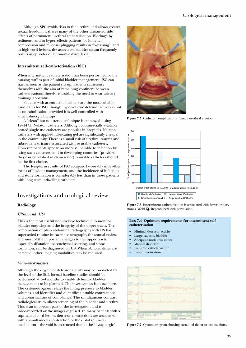
Urological management
35
Although SPC avoids risks to the urethra and allows greatersexual freedom, it shares many of the other unwanted sideeffects of permanent urethral catheterisation. Blockage bysediment, and in hyperreflexic patients, by lumenalcompression and mucosal plugging results in “bypassing”, andin high cord lesions, the associated bladder spasm frequentlyresults in episodes of autonomic dysreflexia.
Intermittent self-catheterisation (ISC)
When intermittent catheterisation has been performed by thenursing staff as part of initial bladder management, ISC canstart as soon as the patient sits up. Patients catheterisethemselves with the aim of remaining continent betweencatheterisations, therefore avoiding the need to wear urinarydrainage apparatus.
Patients with acontractile bladders are the most suitablecandidates for ISC, though hyperreflexic detrusor activity is nota contraindication provided it is well controlled withanticholinergic therapy.
A “clean” but not sterile technique is employed, using12–14 Ch Nelaton catheters. Although commercially availablecoated single use catheters are popular in hospitals, Nelatoncatheters with applied lubricating gel are significantly cheaperin the community. There is a small risk of urethral trauma andsubsequent stricture associated with re-usable catheters.However, patients appear no more vulnerable to infection byusing such catheters, and in developing countries (providedthey can be washed in clean water) re-usable catheters shouldbe the first choice.
The long-term results of ISC compare favourably with otherforms of bladder management, and the incidence of infectionand stone formation is considerably less than in those patientswith long-term indwelling catheters.
Investigations and urological review
Radiology
Ultrasound (US)
This is the most useful non-invasive technique to monitorbladder emptying and the integrity of the upper tracts. Thecombination of plain abdominal radiography with US hassuperseded routine intravenous urography for annual review,and most of the important changes to the upper tracts,especially dilatation, parenchymal scarring, and stoneformation, can be diagnosed on US. When abnormalities aredetected, other imaging modalities may be required.
Video-urodynamics
Although the degree of detrusor activity may be predicted bythe level of the SCI, formal baseline studies should beperformed at 3–4 months to enable definitive bladdermanagement to be planned. The investigation is in two parts.The cystometrogram relates the filling pressure to bladdervolumes, and identifies and quantifies unstable contractionsand abnormalities of compliance. The simultaneous contrastradiological study allows screening of the bladder and urethra.This is an important part of the investigation and isvideo-recorded or the images digitised. In many patients with asuprasacral cord lesion, detrusor contractions are associatedwith a simultaneous contraction of the distal sphinctermechanism—the void is obstructed due to the “dyssynergic”
Figure 7.5 Catheter complications: female urethral erosion.
60
50
40
20
20
10
0
Intermittent Catheter
% o
f p
atie
nts
per
man
agem
ent
met
ho
d
Urethral Catheter
Spontaneous Void Suprapubic Catheter
Upper tract stone (p<0.001) Bladder stone (p<0.001)
Figure 7.6 Intermittent catheterisation is associated with fewer urinarystones. Weld KJ. Reproduced with permission.
Figure 7.7 Cystometrogram showing sustained detrusor contractions.
Box 7.4 Optimum requirements for intermittent self-catheterisation
• Minimal detrusor activity• Large capacity bladder• Adequate outlet resistance• Manual dexterity• Pain-free catheterisation• Patient motivation
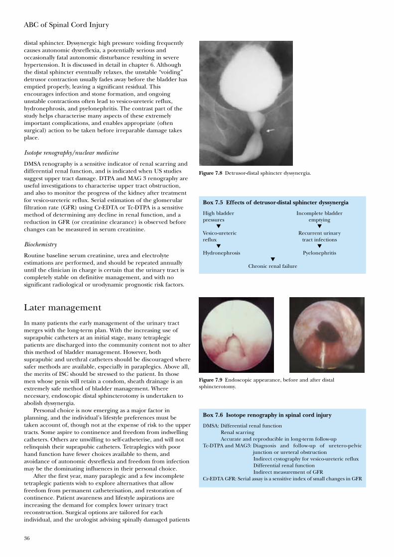
ABC of Spinal Cord Injury
36
distal sphincter. Dyssynergic high pressure voiding frequentlycauses autonomic dysreflexia, a potentially serious andoccasionally fatal autonomic disturbance resulting in severehypertension. It is discussed in detail in chapter 6. Althoughthe distal sphincter eventually relaxes, the unstable “voiding”detrusor contraction usually fades away before the bladder hasemptied properly, leaving a significant residual. Thisencourages infection and stone formation, and ongoingunstable contractions often lead to vesico-ureteric reflux,hydronephrosis, and pyelonephritis. The contrast part of thestudy helps characterise many aspects of these extremelyimportant complications, and enables appropriate (oftensurgical) action to be taken before irreparable damage takesplace.
Isotope renography/nuclear medicine
DMSA renography is a sensitive indicator of renal scarring anddifferential renal function, and is indicated when US studiessuggest upper tract damage. DTPA and MAG 3 renography areuseful investigations to characterise upper tract obstruction,and also to monitor the progress of the kidney after treatmentfor vesico-ureteric reflux. Serial estimation of the glomerularfiltration rate (GFR) using Cr-EDTA or Tc-DTPA is a sensitivemethod of determining any decline in renal function, and areduction in GFR (or creatinine clearance) is observed beforechanges can be measured in serum creatinine.
Biochemistry
Routine baseline serum creatinine, urea and electrolyteestimations are performed, and should be repeated annuallyuntil the clinician in charge is certain that the urinary tract iscompletely stable on definitive management, and with nosignificant radiological or urodynamic prognostic risk factors.
Later management
In many patients the early management of the urinary tractmerges with the long-term plan. With the increasing use ofsuprapubic catheters at an initial stage, many tetraplegicpatients are discharged into the community content not to alterthis method of bladder management. However, bothsuprapubic and urethral catheters should be discouraged wheresafer methods are available, especially in paraplegics. Above all,the merits of ISC should be stressed to the patient. In thosemen whose penis will retain a condom, sheath drainage is anextremely safe method of bladder management. Wherenecessary, endoscopic distal sphincterotomy is undertaken toabolish dyssynergia.
Personal choice is now emerging as a major factor inplanning, and the individual’s lifestyle preferences must betaken account of, though not at the expense of risk to the uppertracts. Some aspire to continence and freedom from indwellingcatheters. Others are unwilling to self-catheterise, and will notrelinquish their suprapubic catheters. Tetraplegics with poorhand function have fewer choices available to them, andavoidance of autonomic dysreflexia and freedom from infectionmay be the dominating influences in their personal choice.
After the first year, many paraplegic and a few incompletetetraplegic patients wish to explore alternatives that allowfreedom from permanent catheterisation, and restoration ofcontinence. Patient awareness and lifestyle aspirations areincreasing the demand for complex lower urinary tractreconstruction. Surgical options are tailored for eachindividual, and the urologist advising spinally damaged patients
Figure 7.8 Detrusor-distal sphincter dyssynergia.
Box 7.5 Effects of detrusor-distal sphincter dyssynergia
High bladder Incomplete bladderpressures emptying
▼ ▼
Vesico-ureteric Recurrent urinaryreflux tract infections
▼ ▼
Hydronephrosis Pyelonephritis▼
Chronic renal failure
Figure 7.9 Endoscopic appearance, before and after distalsphincterotomy.
Box 7.6 Isotope renography in spinal cord injury
DMSA: Differential renal functionRenal scarringAccurate and reproducible in long-term follow-up
Tc-DTPA and MAG3: Diagnosis and follow-up of uretero-pelvicjunction or ureteral obstructionIndirect cystography for vesico-ureteric refluxDifferential renal functionIndirect measurement of GFR
Cr-EDTA GFR: Serial assay is a sensitive index of small changes in GFR
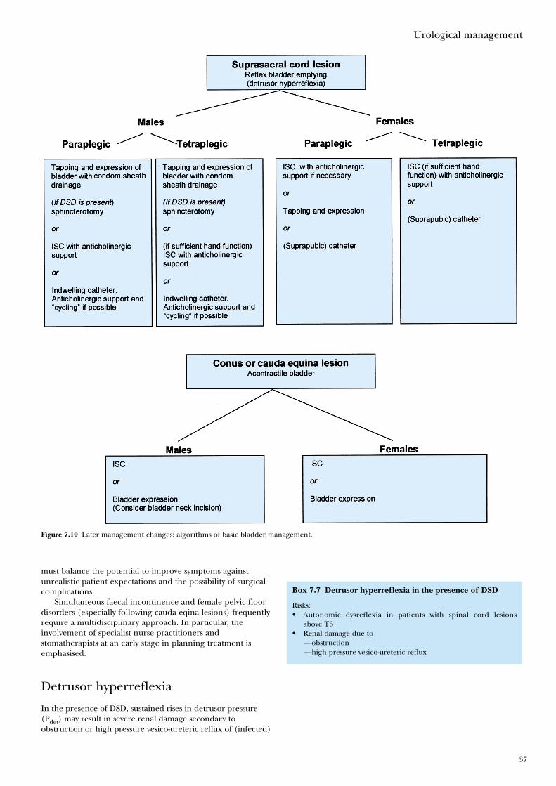
Urological management
37
Box 7.7 Detrusor hyperreflexia in the presence of DSD
Risks:• Autonomic dysreflexia in patients with spinal cord lesions
above T6• Renal damage due to
—obstruction—high pressure vesico-ureteric reflux
must balance the potential to improve symptoms againstunrealistic patient expectations and the possibility of surgicalcomplications.
Simultaneous faecal incontinence and female pelvic floordisorders (especially following cauda eqina lesions) frequentlyrequire a multidisciplinary approach. In particular, theinvolvement of specialist nurse practitioners andstomatherapists at an early stage in planning treatment isemphasised.
Detrusor hyperreflexia
In the presence of DSD, sustained rises in detrusor pressure(Pdet) may result in severe renal damage secondary toobstruction or high pressure vesico-ureteric reflux of (infected)
Figure 7.10 Later management changes: algorithms of basic bladder management.
ABC of Spinal Cord Injury
38
urine. Continence and voiding are secondary considerations inthese cases (nearly always males), where renal preservation is ofthe utmost priority. Recurrent suprapubic catheter blockage iscommon, even in the absence of calculous debris, and may resultfrom catheter shaft compression by grossly unstable bladdercontractions and mucosal plugging. Significant rises in detrusorpressure may occur even in the presence of an indwellingcatheter on free drainage, and an association between theseunstable contractions and upper tract scarring has recently beenconfirmed. Suprapubic catheters (SPC) should be cycled on atleast two occasions each day, and simultaneous anticholinergictherapy should be used. Some men may opt for distalendoscopic sphincterotomy or stenting and condom drainagerather than SPC. Stents are less reliable in SCI patients than inthose with outflow obstruction associated with prostaticenlargement. Others may be obese or suffer penile retraction,and condom sheath drainage may be impossible.
The maintenance of continence is of vital importance topersonal morale, and for the preservation of intact perinealand buttock skin. We briefly examine some of the many surgicalprocedures available to restore continence and to facilitateself-catheterisation.
Augmentation cystoplasty
Where conventional medical treatment with anticholinergictherapy has failed, and if indwelling catheters are to be avoided,the bladder must be deafferentated or bisected and augmentedwith a patch of bowel to provide a bladder of sufficientcompliance and volume to safely accommodate a socially usefulvolume of urine. In female patients, DSD is very unusual, andsevere incontinence rather than upper tract protection is themain indication for augmentation. After augmentation, inabilityto void is the rule rather than the exception, and the patientmust demonstrate the willingness and ability to self-catheterisebefore surgery can be contemplated.
Even after augmentation, anticholinergic therapy may berequired to make the patient completely dry. Cystitis may be arecurrent problem after enterocystoplasty, and there remains along-term theoretical risk of neoplastic transformation in theenteric patch, especially if this is colon. Nitrosamine productionassociated with UTI has been implicated in this process.
For those who cannot access their own urethra (wheelchair-bound females being an especially important group), thesimultaneous provision of a self-catheterising abdominal stoma(Mitrofanoff) may be an integral part of the initial surgery (seebelow).
Neuromodulation and sacral anterior rootstimulation (SARS)
In patients with complete suprasacral cord lesions, functionalelectrical stimulation of the anterior nerve roots of S2, S3 andS4 is very successful in completely emptying the paralysedbladder. Assisted defaecation, and in the male, implant-inducederections may be coincidental advantages of the implant. Thedevice most commonly in use is the Finetech-Brindleystimulator; the anterior roots of S2, S3 and S4 are stimulatedvia a receiver block implanted under the skin, and a posteriorrhizotomy is performed simultaneously. This cures reflexincontinence, improves bladder compliance and diminishesDSD, and thus ensures that neither the use of the implant noroverfilling of the bladder will trigger autonomic dysreflexia.Reflex erections and ejaculation will be lost with posterior
Box 7.9 Detrusor hyperreflexia: surgical management
• Augmentation cystoplasty• Sacral anterior root stimulator (SARS)• Posterior rhizotomy• Urinary diversion
Figure 7.11 Illustration of the two halves of the bisected bladder(“clammed”) prior to augmentation.
Figure 7.12 Components of the Finetech-Brindley SARS.
Box 7.8 Detrusor hyperreflexia: medical management
• Anticholinergic treatmentOxybutyninTolterodinePropiverine HCIFlavoxatePropantheline
• Intravesical therapy (experimental)CapsaicinResiniferatoxin
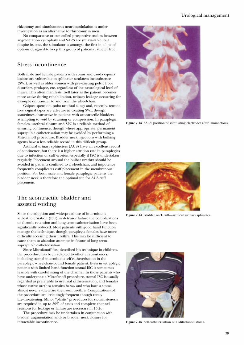
Urological management
39
rhizotomy, and simultaneous neuromodulation is underinvestigation as an alternative to rhizotomy in men.
No comparative or controlled prospective studies betweenaugmentation cystoplasty and SARS are yet available, butdespite its cost, the stimulator is amongst the first in a line ofoptions designed to keep this group of patients catheter free.
Stress incontinence
Both male and female patients with conus and cauda equinalesions are vulnerable to sphincter weakness incontinence(SWI), as well as older women with pre-existing pelvic floordisorders, prolapse, etc. regardless of the neurological level ofinjury. This often manifests itself later as the patient becomesmore active during rehabilitation, urinary leakage occurring forexample on transfer to and from the wheelchair.
Colposuspension, pubo-urethral slings and, recently, tensionfree vaginal tapes are effective in treating SWI, thoughsometimes obstructive in patients with acontractile bladdersattempting to void by straining or compression. In paraplegicfemales, urethral closure and SPC is a reliable method ofensuring continence, though where appropriate, permanentsuprapubic catheterisation may be avoided by performing aMitrofanoff procedure. Bladder neck injections with bulkingagents have a less reliable record in this difficult group.
Artificial urinary sphincters (AUS) have an excellent recordof continence, but there is a higher attrition rate in paraplegicsdue to infection or cuff erosion, especially if ISC is undertakenregularly. Placement around the bulbar urethra should beavoided in patients confined to a wheelchair, and impotencefrequently complicates cuff placement in the membranousposition. For both male and female paraplegic patients thebladder neck is therefore the optimal site for AUS cuffplacement.
The acontractile bladder andassisted voiding
Since the adoption and widespread use of intermittentself-catheterisation (ISC) in detrusor failure the complicationsof chronic retention and long-term catheterisation have beensignificantly reduced. Most patients with good hand functionmanage the technique, though paraplegic females have moredifficulty accessing their urethra. This may be sufficient tocause them to abandon attempts in favour of long-termsuprapubic catheterisation.
Since Mitrofanoff first described his technique in children,the procedure has been adapted to other circumstances,including stomal intermittent self-catheterisation in theparaplegic wheelchair-bound female patient. Even in tetraplegicpatients with limited hand function stomal ISC is sometimesfeasible with careful siting of the channel. In those patients whohave undergone a Mitrofanoff procedure, stomal ISC is usuallyregarded as preferable to urethral catheterisation, and femaleswhose native urethra remains in situ and who have a stomaalmost never catheterise their own urethra. Complications ofthe procedure are irritatingly frequent though rarelylife-threatening. Minor “plastic” procedures for stomal stenosisare required in up to 30% of cases and complete channelrevisions for leakage or failure are necessary in 15%.
The procedure may be undertaken in conjunction withbladder augmentation and/or bladder neck closure forintractable incontinence.
Figure 7.13 SARS: position of stimulating electrodes after laminectomy.
Figure 7.14 Bladder neck cuff—artificial urinary sphincter.
Figure 7.15 Self-catheterisation of a Mitrofanoff stoma.
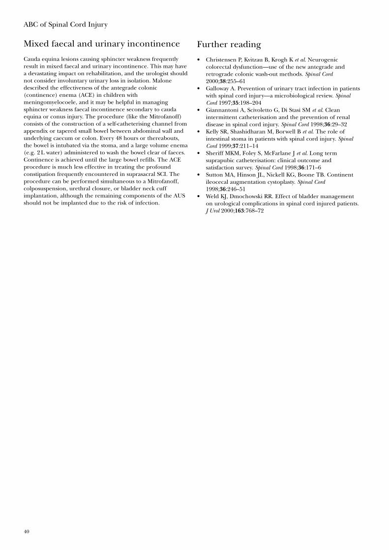
ABC of Spinal Cord Injury
40
Mixed faecal and urinary incontinence
Cauda equina lesions causing sphincter weakness frequentlyresult in mixed faecal and urinary incontinence. This may havea devastating impact on rehabilitation, and the urologist shouldnot consider involuntary urinary loss in isolation. Malonedescribed the effectiveness of the antegrade colonic(continence) enema (ACE) in children withmeningomyelocoele, and it may be helpful in managingsphincter weakness faecal incontinence secondary to caudaequina or conus injury. The procedure (like the Mitrofanoff)consists of the construction of a self-catheterising channel fromappendix or tapered small bowel between abdominal wall andunderlying caecum or colon. Every 48 hours or thereabouts,the bowel is intubated via the stoma, and a large volume enema(e.g. 2 L water) administered to wash the bowel clear of faeces.Continence is achieved until the large bowel refills. The ACEprocedure is much less effective in treating the profoundconstipation frequently encountered in suprasacral SCI. Theprocedure can be performed simultaneous to a Mitrofanoff,colposuspension, urethral closure, or bladder neck cuffimplantation, although the remaining components of the AUSshould not be implanted due to the risk of infection.
Further reading
• Christensen P, Kvitzau B, Krogh K et al. Neurogeniccolorectal dysfunction—use of the new antegrade andretrograde colonic wash-out methods. Spinal Cord2000;38:255–61
• Galloway A. Prevention of urinary tract infection in patientswith spinal cord injury—a microbiological review. SpinalCord 1997;35:198–204
• Giannantoni A, Scivoletto G, Di Stasi SM et al. Cleanintermittent catheterisation and the prevention of renaldisease in spinal cord injury. Spinal Cord 1998;36:29–32
• Kelly SR, Shashidharan M, Borwell B et al. The role ofintestinal stoma in patients with spinal cord injury. SpinalCord 1999;37:211–14
• Sheriff MKM, Foley S, McFarlane J et al. Long termsuprapubic catheterisation: clinical outcome andsatisfaction survey. Spinal Cord 1998;36:171–6
• Sutton MA, Hinson JL, Nickell KG, Boone TB. Continentileocecal augmentation cystoplasty. Spinal Cord1998;36:246–51
• Weld KJ, Dmochowski RR. Effect of bladder managementon urological complications in spinal cord injured patients.J Urol 2000;163:768–72
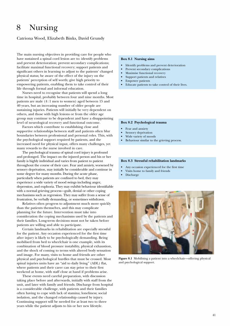
41
Catriona Wood, Elizabeth Binks, David Grundy
The main nursing objectives in providing care for people whohave sustained a spinal cord lesion are to: identify problemsand prevent deterioration; prevent secondary complications;facilitate maximal functional recovery; support patients andsignificant others in learning to adjust to the patients’ changedphysical status; be aware of the effect of the injury on thepatients’ perception of self worth; give high priority toempowering patients, enabling them to take control of theirlife through formal and informal education.
Nurses need to recognise that patients will spend a longtime in hospital, probably between four and nine months. Mostpatients are male (4 : 1 men to women) aged between 15 and40 years, but an increasing number of older people aresustaining injuries. Patients will initially be very dependent onothers, and those with high lesions or from the older agegroup may continue to be dependent and have a disappointinglevel of neurological recovery and functional outcome.
Factors which contribute to establishing close andsupportive relationships between staff and patients often blurboundaries between professional and personal roles. This, withthe psychological support required by patients, and theincreased need for physical input, offers many challenges, yetmany rewards to the nurse involved in care.
The psychological trauma of spinal cord injury is profoundand prolonged. The impact on the injured person and his or herfamily is highly individual and varies from patient to patientthroughout the course of their care. Fear and anxiety, worsened bysensory deprivation, may initially be considerable and continue insome degree for many months. During the acute phase,particularly when patients are confined to bed, they mayexperience a wide variety of mood swings including anger,depression, and euphoria. They may exhibit behaviour identifiablewith a normal grieving process—guilt, denial or other copingmechanisms such as regression. They may suffer from a sense offrustration, be verbally demanding, or sometimes withdrawn.
Relatives often progress to adjustment much more quicklythan the patients themselves, and this may complicateplanning for the future. Intervention must take intoconsideration the coping mechanisms used by the patients andtheir families. Long-term decisions must not be taken beforepatients are willing and able to participate.
Certain landmarks in rehabilitation are especially stressfulfor the patient. Any occasion experienced for the first timeafter injury is likely to be psychologically demanding. Beingmobilised from bed to wheelchair is one example, with itscombination of blood pressure instability, physical exhaustion,and the shock of coming to terms with altered body sensationand image. For many, visits to home and friends are otherphysical and psychological hurdles that must be crossed. Mostspinal injuries units have an “aid to daily living” (ADL) flat,where patients and their carer can stay prior to their firstweekend at home, with staff close at hand if problems arise.
These events need careful preparation, with discussiontaking place before and afterwards, initially with staff from theunit, and later with family and friends. Discharge from hospitalis a considerable challenge, with patients and their familiesoften having to cope with lack of stamina; loneliness; socialisolation, and the changed relationship caused by injury.Continuing support will be needed for at least two to threeyears while the patient adjusts to his or her new lifestyle.
8 Nursing
Box 8.1 Nursing aims
• Identify problems and prevent deterioration• Prevent secondary complications• Maximise functional recovery• Support patients and relatives• Empower patients• Educate patients to take control of their lives.
Box 8.2 Psychological trauma
• Fear and anxiety• Sensory deprivation• Wide variety of moods• Behaviour similar to the grieving process.
Box 8.3 Stressful rehabilitation landmarks
• Any occasion experienced for the first time• Visits home to family and friends• Discharge
Figure 8.1 Mobilising a patient into a wheelchair—offering physicaland psychological support.
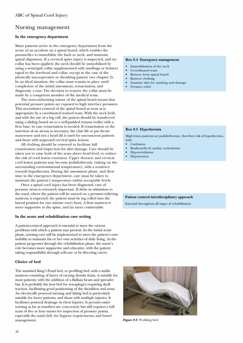
ABC of Spinal Cord Injury
42
Nursing management
In the emergency department
Many patients arrive in the emergency department from thescene of an accident on a spinal board, which enables theparamedics to immobilise the back or neck, and maintainspinal alignment. If a cervical spine injury is suspected, and nocollar has been applied, the neck should be immobilised byusing a semirigid collar supplemented with sandbags or bolsterstaped to the forehead and collar, except in the case of thephysically uncooperative or thrashing patient (see chapter 2).In an ideal situation, the collar must remain in place untilcompletion of the initial assessment, resuscitation, anddiagnostic x rays. The decision to remove the collar must bemade by a competent member of the medical team.
The non-conforming nature of the spinal board means thatpotential pressure points are exposed to high interface pressures.This necessitates removal of the spinal board as soon as isappropriate by a coordinated trained team. With the neck held,and with the use of a log roll, the patient should be transferredusing a sliding board on to a well-padded trauma trolley with afirm base, in case resuscitation is needed. If resuscitation or theinsertion of an airway is necessary, the chin lift or jaw thrustmanoeuvre and not a head tilt is used for unconscious patientsand those with suspected cervical spine lesions.
All clothing should be removed to facilitate fullexamination and inspection for skin damage. Care should betaken not to raise both of the arms above head level, to reducethe risk of cord lesion extension. Upper thoracic and cervicalcord lesion patients may become poikilothermic (taking on thesurrounding environmental temperature), with a tendencytowards hypothermia. During the assessment phase, and theirtime in the emergency department, care must be taken tomaintain the patient’s temperature within acceptable levels.
Once a spinal cord injury has been diagnosed, care ofpressure areas is extremely important. If delay in admission tothe ward, where the patient will be nursed on a pressure-relievingmattress, is expected, the patient must be log rolled into thelateral position for one minute every hour. A firm mattress ismore supportive to the spine, and far more comfortable.
In the acute and rehabilitation care setting
A patient-centred approach is essential to meet the variousproblems with which a patient may present. In the initial acutephase, nursing care will be implemented to meet the patient’s owninability to maintain his or her own activities of daily living. As thepatient progresses through the rehabilitation phase, the nurse’srole becomes more supportive and educative, with the patienttaking responsibility through self-care or by directing carers.
Choice of bed
The standard King’s Fund bed, or profiling bed, with a stablemattress consisting of layers of varying density foam, is suitable formost patients, with the addition of a Balkan beam and spreaderbar. It is probably the best bed for tetraplegics requiring skulltraction, facilitating good positioning of the shoulders and arms.An electrically powered turning and tilting bed is particularlysuitable for heavy patients, and those with multiple injuries. Itfacilitates postural drainage in chest injuries. It permits easiernursing as far as numbers are concerned, but still requires a fullteam of five or four nurses for inspection of pressure points,especially the natal cleft, for hygiene requirements, and bowelmanagement. Figure 8.2 Profiling bed.
Box 8.4 Emergency management
• Immobilisation of the neck• Co-ordinated team• Remove from spinal board• Remove clothing• Examine skin for marking and damage• Pressure relief
Box 8.5 Hypothermia
High lesion patients are poikilothermic, therefore risk of hypothermia,with:• Confusion• Bradycardia ➪ cardiac arrhythmias• Hypoventilation• Hypotension
Patient centred interdisciplinary approach
Essential throughout all stages of rehabilitation
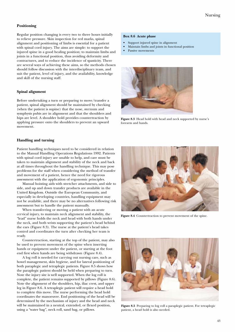
Nursing
43
Positioning
Regular position changing is every two to three hours initiallyto relieve pressure. Skin inspection for red marks, spinalalignment and positioning of limbs is essential for a patientwith spinal cord injury. The aims are simple: to support theinjured spine in a good healing position; to maintain limbs andjoints in a functional position, thus avoiding deformity andcontractures, and to reduce the incidence of spasticity. Thereare several ways of achieving these aims, so the methods chosenshould follow discussion with the interdisciplinary team, andsuit the patient, level of injury, and the availability, knowledgeand skill of the nursing staff.
Spinal alignment
Before undertaking a turn or preparing to move/transfer apatient, spinal alignment should be maintained by checking(when the patient is supine) that the nose, sternum andsymphysis pubis are in alignment and that the shoulders andhips are level. A shoulder hold provides countertraction byapplying pressure onto the shoulders to prevent an upwardmovement.
Handling and turning
Patient handling techniques need to be considered in relationto the Manual Handling Operations Regulations 1992. Patientswith spinal cord injury are unable to help, and care must betaken to maintain alignment and stability of the neck and backat all times throughout the handling technique. This may poseproblems for the staff when considering the method of transferand movement of a patient, hence the need for rigorousassessment with the application of ergonomic principles.
Manual hoisting aids with stretcher attachments, and side toside, and up and down transfer products are available in theUnited Kingdom. Outside the European Community, andespecially in developing countries, handling equipment maynot be available, and there may be no alternatives following riskassessment but to handle the patient manually.
When transferring or moving a patient with an acutecervical injury, to maintain neck alignment and stability, the“lead” nurse holds the neck and head with both hands underthe neck, and both wrists supporting the patient’s head behindthe ears (Figure 8.3). The nurse at the patient’s head takescontrol and coordinates the turn after checking her team isready.
Countertraction, starting at the top of the patient, may alsobe used to prevent movement of the spine when insertinghands or equipment under the patient, or starting at the footend first when hands are being withdrawn (Figure 8.4).
A log roll is needed for carrying out nursing care, such asbowel management, skin hygiene, and for lateral positioning ofboth paraplegic and tetraplegic patients. Figure 8.5 shows howthe paraplegic patient should be held when preparing to turn.Note the injury site is well supported. When the log roll iscomplete, the patient remains supported by pillows (Figure 8.6).Note the alignment of the shoulders, hip, iliac crest, and upperleg in Figure 8.6. A tetraplegic patient will require a head holdto complete this move. The nurse performing the head holdcoordinates the manoeuvre. End positioning of the head will bedetermined by the mechanism of injury and the head and neckwill be maintained in a neutral, extended, or flexed position,using a “water bag”, neck roll, sand bag, or pillows.
Box 8.6 Acute phase
• Support injured spine in alignment• Maintain limbs and joints in functional position• Passive movements
Figure 8.3 Head hold with head and neck supported by nurse’sforearm and hands.
Figure 8.5 Preparing to log roll a paraplegic patient. For tetraplegicpatient, a head hold is also needed.
Figure 8.4 Countertraction to prevent movement of the spine.
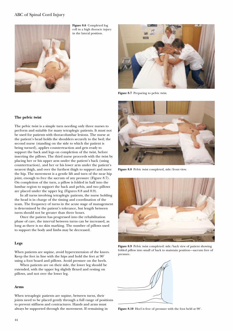
ABC of Spinal Cord Injury
44
The pelvic twist
The pelvic twist is a simple turn needing only three nurses toperform and suitable for many tetraplegic patients. It must notbe used for patients with thoracolumbar lesions. The nurse atthe patient’s head holds the shoulders securely to the bed; thesecond nurse (standing on the side to which the patient isbeing turned), applies countertraction and gets ready tosupport the back and legs on completion of the twist, beforeinserting the pillows. The third nurse proceeds with the twist byplacing her or his upper arm under the patient’s back (usingcountertraction), and her or his lower arm under the patient’snearest thigh, and over the furthest thigh to support and movethe hip. The movement is a gentle lift and turn of the near hipjoint, enough to free the sacrum of any pressure (Figure 8.7).On completion of the turn, a pillow is folded in half into thelumbar region to support the back and pelvis, and two pillowsare placed under the upper leg (Figures 8.8 and 8.9).
In all turns involving tetraplegic patients, the nurse holdingthe head is in charge of the timing and coordination of theteam. The frequency of turns in the acute stage of managementis determined by the patient’s tolerance, but length betweenturns should not be greater than three hours.
Once the patient has progressed into the rehabilitationphase of care, the interval between turns can be increased, aslong as there is no skin marking. The number of pillows usedto support the body and limbs may be decreased.
Legs
When patients are supine, avoid hyperextension of the knees.Keep the feet in line with the hips and hold the feet at 90˚using a foot board and pillows. Avoid pressure on the heels.
When patients are on their side, the lower leg should beextended, with the upper leg slightly flexed and resting onpillows, and not over the lower leg.
Arms
When tetraplegic patients are supine, between turns, theirjoints need to be placed gently through a full range of positionsto prevent stiffness and contractures. Hands and arms mustalways be supported through the movement. If remaining in
Figure 8.6 Completed logroll in a high thoracic injuryin the lateral position.
Figure 8.7 Preparing to pelvic twist.
Figure 8.8 Pelvic twist completed, side/front view.
Figure 8.9 Pelvic twist completed: side/back view of patient showingfolded pillow into small of back to maintain position—sacrum free ofpressure.
Figure 8.10 Heel is free of pressure with the foot held at 90˚.
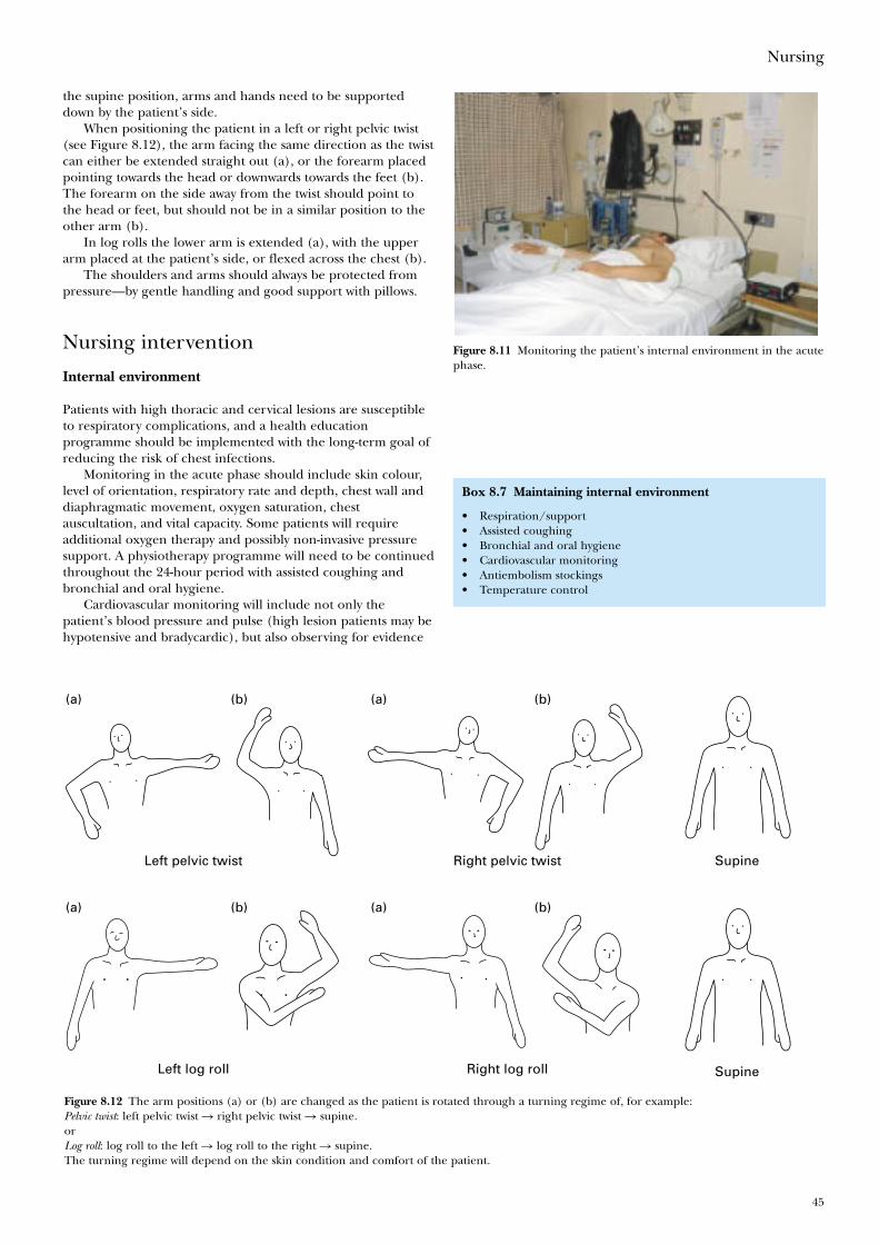
Nursing
45
the supine position, arms and hands need to be supporteddown by the patient’s side.
When positioning the patient in a left or right pelvic twist(see Figure 8.12), the arm facing the same direction as the twistcan either be extended straight out (a), or the forearm placedpointing towards the head or downwards towards the feet (b).The forearm on the side away from the twist should point tothe head or feet, but should not be in a similar position to theother arm (b).
In log rolls the lower arm is extended (a), with the upperarm placed at the patient’s side, or flexed across the chest (b).
The shoulders and arms should always be protected frompressure—by gentle handling and good support with pillows.
Nursing intervention
Internal environment
Patients with high thoracic and cervical lesions are susceptibleto respiratory complications, and a health educationprogramme should be implemented with the long-term goal ofreducing the risk of chest infections.
Monitoring in the acute phase should include skin colour,level of orientation, respiratory rate and depth, chest wall anddiaphragmatic movement, oxygen saturation, chestauscultation, and vital capacity. Some patients will requireadditional oxygen therapy and possibly non-invasive pressuresupport. A physiotherapy programme will need to be continuedthroughout the 24-hour period with assisted coughing andbronchial and oral hygiene.
Cardiovascular monitoring will include not only thepatient’s blood pressure and pulse (high lesion patients may behypotensive and bradycardic), but also observing for evidence
Box 8.7 Maintaining internal environment
• Respiration/support• Assisted coughing• Bronchial and oral hygiene• Cardiovascular monitoring• Antiembolism stockings• Temperature control
Figure 8.12 The arm positions (a) or (b) are changed as the patient is rotated through a turning regime of, for example:Pelvic twist: left pelvic twist � right pelvic twist � supine.orLog roll: log roll to the left � log roll to the right � supine.The turning regime will depend on the skin condition and comfort of the patient.
Left log roll
(a) (b)
Right log roll
(a) (b)
Left pelvic twist
(a) (b)
Right pelvic twist Supine
Supine
(a) (b)
Figure 8.11 Monitoring the patient’s internal environment in the acutephase.
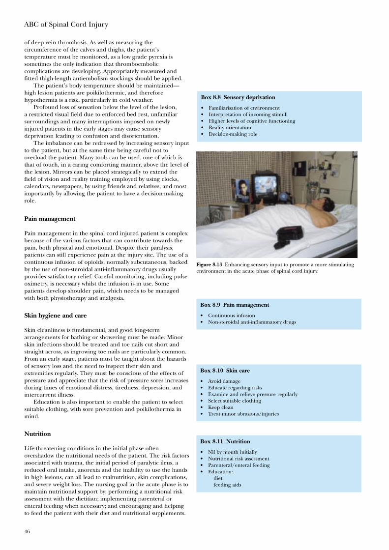
ABC of Spinal Cord Injury
46
of deep vein thrombosis. As well as measuring thecircumference of the calves and thighs, the patient’stemperature must be monitored, as a low grade pyrexia issometimes the only indication that thromboemboliccomplications are developing. Appropriately measured andfitted thigh-length antiembolism stockings should be applied.
The patient’s body temperature should be maintained—high lesion patients are poikilothermic, and thereforehypothermia is a risk, particularly in cold weather.
Profound loss of sensation below the level of the lesion,a restricted visual field due to enforced bed rest, unfamiliarsurroundings and many interruptions imposed on newlyinjured patients in the early stages may cause sensorydeprivation leading to confusion and disorientation.
The imbalance can be redressed by increasing sensory inputto the patient, but at the same time being careful not tooverload the patient. Many tools can be used, one of which isthat of touch, in a caring comforting manner, above the level ofthe lesion. Mirrors can be placed strategically to extend thefield of vision and reality training employed by using clocks,calendars, newspapers, by using friends and relatives, and mostimportantly by allowing the patient to have a decision-makingrole.
Pain management
Pain management in the spinal cord injured patient is complexbecause of the various factors that can contribute towards thepain, both physical and emotional. Despite their paralysis,patients can still experience pain at the injury site. The use of acontinuous infusion of opioids, normally subcutaneous, backedby the use of non-steroidal anti-inflammatory drugs usuallyprovides satisfactory relief. Careful monitoring, including pulseoximetry, is necessary whilst the infusion is in use. Somepatients develop shoulder pain, which needs to be managedwith both physiotherapy and analgesia.
Skin hygiene and care
Skin cleanliness is fundamental, and good long-termarrangements for bathing or showering must be made. Minorskin infections should be treated and toe nails cut short andstraight across, as ingrowing toe nails are particularly common.From an early stage, patients must be taught about the hazardsof sensory loss and the need to inspect their skin andextremities regularly. They must be conscious of the effects ofpressure and appreciate that the risk of pressure sores increasesduring times of emotional distress, tiredness, depression, andintercurrent illness.
Education is also important to enable the patient to selectsuitable clothing, with sore prevention and poikilothermia inmind.
Nutrition
Life-threatening conditions in the initial phase oftenovershadow the nutritional needs of the patient. The risk factorsassociated with trauma, the initial period of paralytic ileus, areduced oral intake, anorexia and the inability to use the handsin high lesions, can all lead to malnutrition, skin complications,and severe weight loss. The nursing goal in the acute phase is tomaintain nutritional support by: performing a nutritional riskassessment with the dietitian; implementing parenteral orenteral feeding when necessary; and encouraging and helpingto feed the patient with their diet and nutritional supplements.
Box 8.10 Skin care
• Avoid damage• Educate regarding risks• Examine and relieve pressure regularly• Select suitable clothing• Keep clean• Treat minor abrasions/injuries
Box 8.11 Nutrition
• Nil by mouth initially• Nutritional risk assessment• Parenteral/enteral feeding• Education:
dietfeeding aids
Box 8.8 Sensory deprivation
• Familiarisation of environment• Interpretation of incoming stimuli• Higher levels of cognitive functioning• Reality orientation• Decision-making role
Box 8.9 Pain management
• Continuous infusion• Non-steroidal anti-inflammatory drugs
Figure 8.13 Enhancing sensory input to promote a more stimulatingenvironment in the acute phase of spinal cord injury.
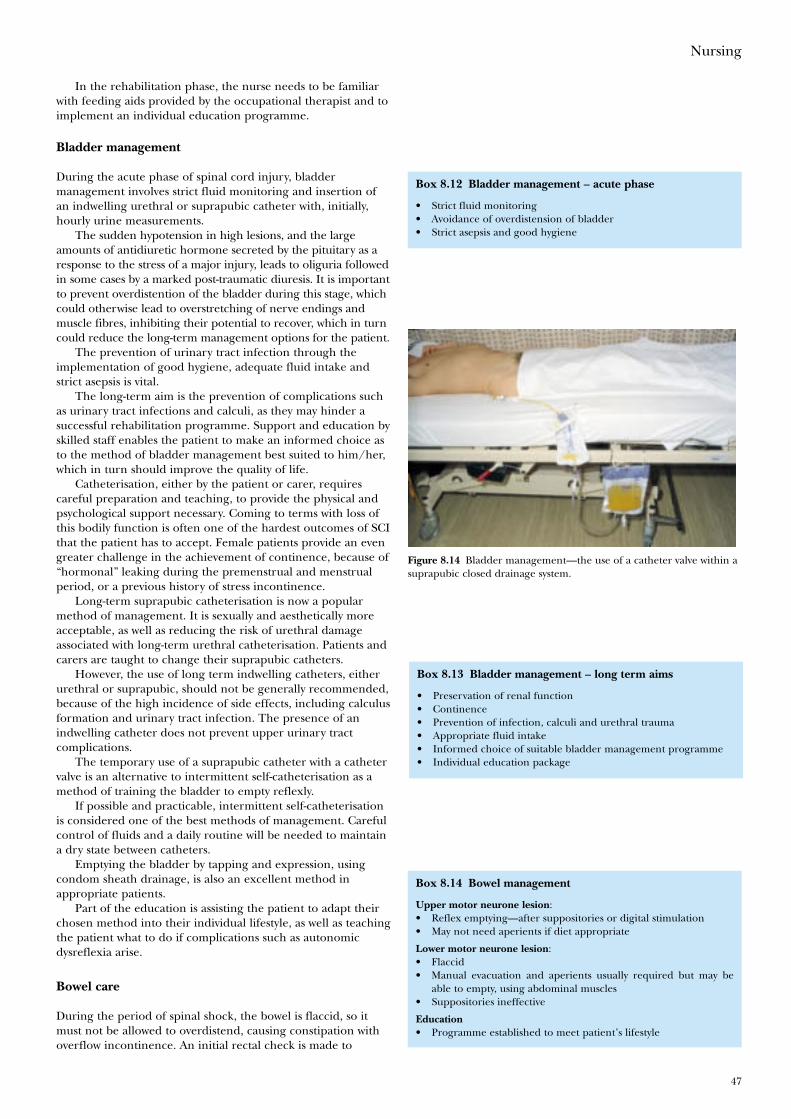
Nursing
47
In the rehabilitation phase, the nurse needs to be familiarwith feeding aids provided by the occupational therapist and toimplement an individual education programme.
Bladder management
During the acute phase of spinal cord injury, bladdermanagement involves strict fluid monitoring and insertion ofan indwelling urethral or suprapubic catheter with, initially,hourly urine measurements.
The sudden hypotension in high lesions, and the largeamounts of antidiuretic hormone secreted by the pituitary as aresponse to the stress of a major injury, leads to oliguria followedin some cases by a marked post-traumatic diuresis. It is importantto prevent overdistention of the bladder during this stage, whichcould otherwise lead to overstretching of nerve endings andmuscle fibres, inhibiting their potential to recover, which in turncould reduce the long-term management options for the patient.
The prevention of urinary tract infection through theimplementation of good hygiene, adequate fluid intake andstrict asepsis is vital.
The long-term aim is the prevention of complications suchas urinary tract infections and calculi, as they may hinder asuccessful rehabilitation programme. Support and education byskilled staff enables the patient to make an informed choice asto the method of bladder management best suited to him/her,which in turn should improve the quality of life.
Catheterisation, either by the patient or carer, requirescareful preparation and teaching, to provide the physical andpsychological support necessary. Coming to terms with loss ofthis bodily function is often one of the hardest outcomes of SCIthat the patient has to accept. Female patients provide an evengreater challenge in the achievement of continence, because of“hormonal” leaking during the premenstrual and menstrualperiod, or a previous history of stress incontinence.
Long-term suprapubic catheterisation is now a popularmethod of management. It is sexually and aesthetically moreacceptable, as well as reducing the risk of urethral damageassociated with long-term urethral catheterisation. Patients andcarers are taught to change their suprapubic catheters.
However, the use of long term indwelling catheters, eitherurethral or suprapubic, should not be generally recommended,because of the high incidence of side effects, including calculusformation and urinary tract infection. The presence of anindwelling catheter does not prevent upper urinary tractcomplications.
The temporary use of a suprapubic catheter with a cathetervalve is an alternative to intermittent self-catheterisation as amethod of training the bladder to empty reflexly.
If possible and practicable, intermittent self-catheterisationis considered one of the best methods of management. Carefulcontrol of fluids and a daily routine will be needed to maintaina dry state between catheters.
Emptying the bladder by tapping and expression, usingcondom sheath drainage, is also an excellent method inappropriate patients.
Part of the education is assisting the patient to adapt theirchosen method into their individual lifestyle, as well as teachingthe patient what to do if complications such as autonomicdysreflexia arise.
Bowel care
During the period of spinal shock, the bowel is flaccid, so itmust not be allowed to overdistend, causing constipation withoverflow incontinence. An initial rectal check is made to
Box 8.12 Bladder management – acute phase
• Strict fluid monitoring• Avoidance of overdistension of bladder• Strict asepsis and good hygiene
Box 8.13 Bladder management – long term aims
• Preservation of renal function• Continence• Prevention of infection, calculi and urethral trauma• Appropriate fluid intake• Informed choice of suitable bladder management programme• Individual education package
Box 8.14 Bowel management
Upper motor neurone lesion:• Reflex emptying—after suppositories or digital stimulation• May not need aperients if diet appropriate
Lower motor neurone lesion:• Flaccid• Manual evacuation and aperients usually required but may be
able to empty, using abdominal muscles• Suppositories ineffective
Education• Programme established to meet patient’s lifestyle
Figure 8.14 Bladder management—the use of a catheter valve within asuprapubic closed drainage system.
ABC of Spinal Cord Injury
48
ascertain whether faeces are present; if they are, they should bemanually evacuated. Manual evacuation thereafter will continuedaily or on alternate days. Very little bowel activity will beexpected for the first two or three days. Evacuation should beperformed using plenty of lubricant, and with only one glovedfinger inserted into the anus. Trauma, including anal stretchingand a split natal cleft, is possible if insufficient care is taken.Suppositories to lubricate or stimulate, possibly with the use ofaperients, may be required to achieve an evacuation after theinitial period of spinal shock. The patient is taught to have anadequate fibre diet, and a high fluid intake to help preventconstipation. The use of aperients is kept to a minimum,especially in a patient with reflex bowel activity. The use ofnatural laxatives is encouraged in the diet.
Slowly a routine is established that will fit into the patient’sfuture lifestyle. Bowel management may be performed daily, oron alternate days, depending upon the individual’s bowelpattern. The patient is encouraged to sit on a specially paddedshower chair, so that bowel care can be performed over thetoilet, followed by a shower. Sometimes this is not possible dueto poor balance or preferred choice, and bowel managementcontinues on the patient’s bed.
Where possible, to promote independent care patients aretaught manual evacuation if the bowel is flaccid, orsuppositories are inserted and/or digital stimulation performedif they have a reflex bowel. If the patient is unable to carry outthis activity, carers or district nurses are taught.
Sexuality
Following a spinal cord injury patients need to redefinesexuality and all that it encompasses. Nurses have to recognisewhen patients are ready to discuss sexuality, and respondappropriately as this need is not always verbalised in the earlystages, and often manifests itself only in indirect questions orsexual innuendo. Discussion should be encouraged and shouldinclude dispelling myths, exploring the patient’s new sexualstatus, looking at alternative methods, identifying practicalproblems; and advising on how to deal with them. Referral tomedical staff, sexual health specialists and other agencies forfurther information and management, should be offered asappropriate.
Further reading
• Addison R, Smith M. Digital rectal stimulation and manualremoval of faeces. Guidance for nurses. London: Royal Collegeof Nursing, 2000
• Harrison P. The first 48 hours. London: Spinal InjuriesAssociation, 2000
• Harrison P. HDU/ICU. Managing spinal injury: critical care.London: Spinal Injuries Association, 2000
• Leyson JFJ. Sexual rehabilitation of the spinal cord injuredpatient. Clifton, New Jersey: Humana Press, 1991
• Zejdlik CM. Management of spinal cord injury, 2nd edition.Boston: Jones and Bartlett Publishers, 1992
Box 8.17 Sexuality
• Acknowledge the need to discuss sexuality• Redefine sexuality• Dispel myths• Explore opportunity and alternatives• Advice to overcome practical difficulties
Box 8.15 Bowel management assessment
Consider• Level of injury• Pre-injury bowel pattern• Diet/fluid intake• What previously helped defaecation• Hand function• Balance/brace• Psychological state
Box 8.16 Aims of bowel management
• Achieve regular bowel emptying by production of a formed stoolat a chosen time and place
• Avoid leaks or unplanned emptying• Avoid constipation and other complications• Try to complete bowel care in 30–60 minutes• Be as practical as possible
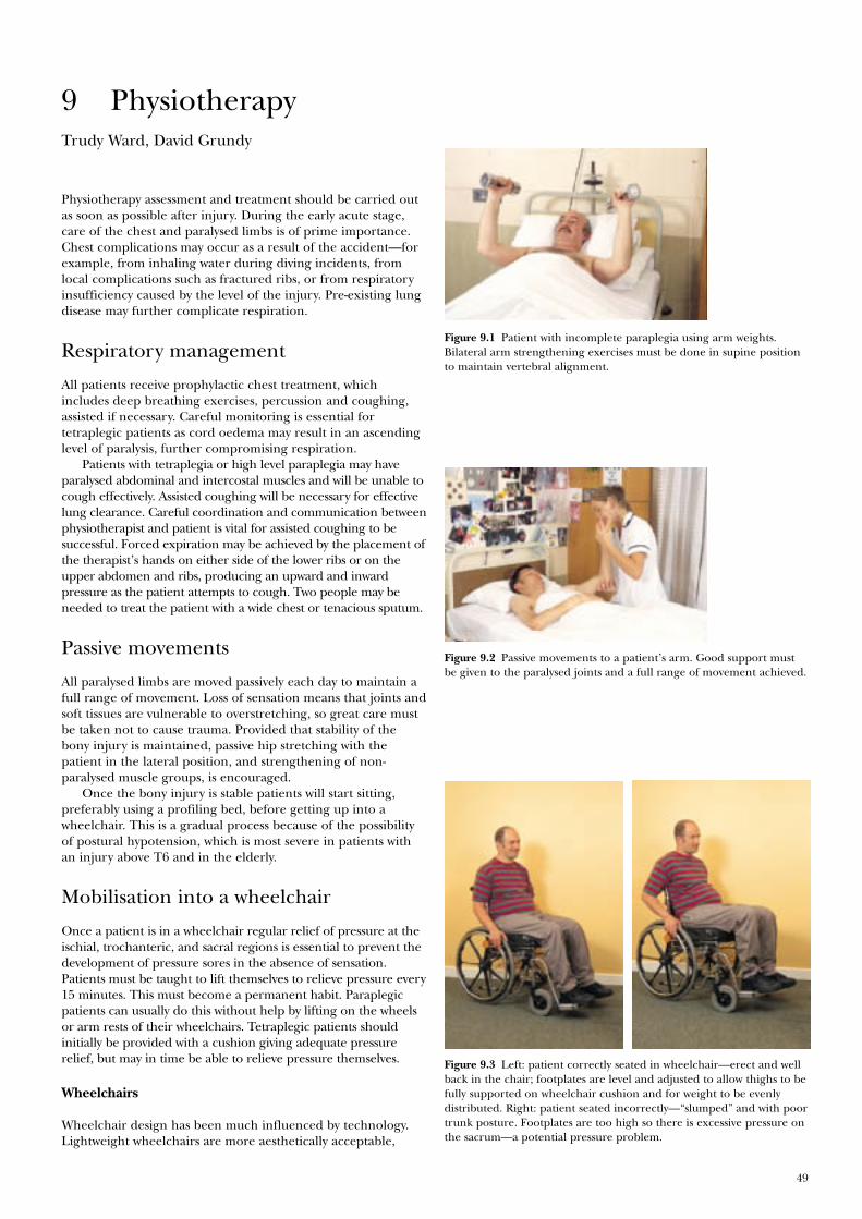
49
Figure 9.1 Patient with incomplete paraplegia using arm weights.Bilateral arm strengthening exercises must be done in supine positionto maintain vertebral alignment.
Figure 9.2 Passive movements to a patient’s arm. Good support mustbe given to the paralysed joints and a full range of movement achieved.
Figure 9.3 Left: patient correctly seated in wheelchair—erect and wellback in the chair; footplates are level and adjusted to allow thighs to befully supported on wheelchair cushion and for weight to be evenlydistributed. Right: patient seated incorrectly—“slumped” and with poortrunk posture. Footplates are too high so there is excessive pressure onthe sacrum—a potential pressure problem.
Trudy Ward, David Grundy
Physiotherapy assessment and treatment should be carried outas soon as possible after injury. During the early acute stage,care of the chest and paralysed limbs is of prime importance.Chest complications may occur as a result of the accident—forexample, from inhaling water during diving incidents, fromlocal complications such as fractured ribs, or from respiratoryinsufficiency caused by the level of the injury. Pre-existing lungdisease may further complicate respiration.
Respiratory management
All patients receive prophylactic chest treatment, whichincludes deep breathing exercises, percussion and coughing,assisted if necessary. Careful monitoring is essential fortetraplegic patients as cord oedema may result in an ascendinglevel of paralysis, further compromising respiration.
Patients with tetraplegia or high level paraplegia may haveparalysed abdominal and intercostal muscles and will be unable tocough effectively. Assisted coughing will be necessary for effectivelung clearance. Careful coordination and communication betweenphysiotherapist and patient is vital for assisted coughing to besuccessful. Forced expiration may be achieved by the placement ofthe therapist’s hands on either side of the lower ribs or on theupper abdomen and ribs, producing an upward and inwardpressure as the patient attempts to cough. Two people may beneeded to treat the patient with a wide chest or tenacious sputum.
Passive movements
All paralysed limbs are moved passively each day to maintain afull range of movement. Loss of sensation means that joints andsoft tissues are vulnerable to overstretching, so great care mustbe taken not to cause trauma. Provided that stability of thebony injury is maintained, passive hip stretching with thepatient in the lateral position, and strengthening of non-paralysed muscle groups, is encouraged.
Once the bony injury is stable patients will start sitting,preferably using a profiling bed, before getting up into awheelchair. This is a gradual process because of the possibilityof postural hypotension, which is most severe in patients withan injury above T6 and in the elderly.
Mobilisation into a wheelchair
Once a patient is in a wheelchair regular relief of pressure at theischial, trochanteric, and sacral regions is essential to prevent thedevelopment of pressure sores in the absence of sensation.Patients must be taught to lift themselves to relieve pressure every15 minutes. This must become a permanent habit. Paraplegicpatients can usually do this without help by lifting on the wheelsor arm rests of their wheelchairs. Tetraplegic patients shouldinitially be provided with a cushion giving adequate pressurerelief, but may in time be able to relieve pressure themselves.
Wheelchairs
Wheelchair design has been much influenced by technology.Lightweight wheelchairs are more aesthetically acceptable,
9 Physiotherapy
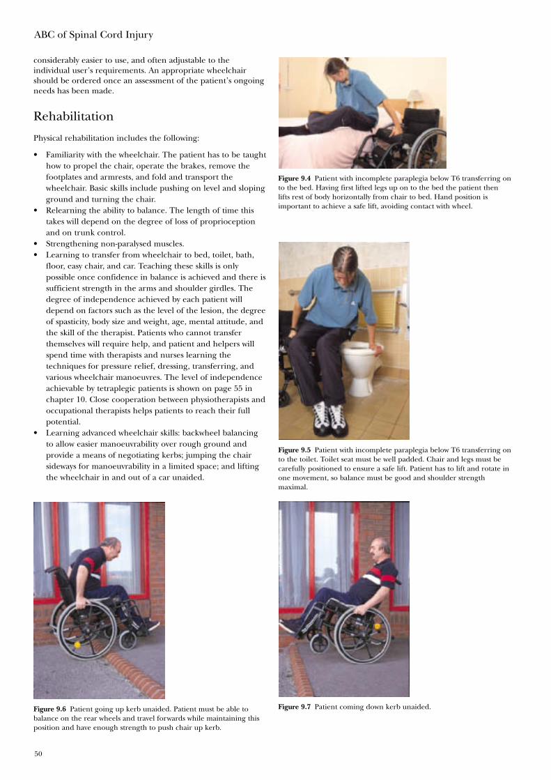
ABC of Spinal Cord Injury
50
considerably easier to use, and often adjustable to theindividual user’s requirements. An appropriate wheelchairshould be ordered once an assessment of the patient’s ongoingneeds has been made.
Rehabilitation
Physical rehabilitation includes the following:
• Familiarity with the wheelchair. The patient has to be taughthow to propel the chair, operate the brakes, remove thefootplates and armrests, and fold and transport thewheelchair. Basic skills include pushing on level and slopingground and turning the chair.
• Relearning the ability to balance. The length of time thistakes will depend on the degree of loss of proprioceptionand on trunk control.
• Strengthening non-paralysed muscles.• Learning to transfer from wheelchair to bed, toilet, bath,
floor, easy chair, and car. Teaching these skills is onlypossible once confidence in balance is achieved and there issufficient strength in the arms and shoulder girdles. Thedegree of independence achieved by each patient willdepend on factors such as the level of the lesion, the degreeof spasticity, body size and weight, age, mental attitude, andthe skill of the therapist. Patients who cannot transferthemselves will require help, and patient and helpers willspend time with therapists and nurses learning thetechniques for pressure relief, dressing, transferring, andvarious wheelchair manoeuvres. The level of independenceachievable by tetraplegic patients is shown on page 55 inchapter 10. Close cooperation between physiotherapists andoccupational therapists helps patients to reach their fullpotential.
• Learning advanced wheelchair skills: backwheel balancingto allow easier manoeuvrability over rough ground andprovide a means of negotiating kerbs; jumping the chairsideways for manoeuvrability in a limited space; and liftingthe wheelchair in and out of a car unaided.
Figure 9.4 Patient with incomplete paraplegia below T6 transferring onto the bed. Having first lifted legs up on to the bed the patient thenlifts rest of body horizontally from chair to bed. Hand position isimportant to achieve a safe lift, avoiding contact with wheel.
Figure 9.5 Patient with incomplete paraplegia below T6 transferring onto the toilet. Toilet seat must be well padded. Chair and legs must becarefully positioned to ensure a safe lift. Patient has to lift and rotate inone movement, so balance must be good and shoulder strengthmaximal.
Figure 9.6 Patient going up kerb unaided. Patient must be able tobalance on the rear wheels and travel forwards while maintaining thisposition and have enough strength to push chair up kerb.
Figure 9.7 Patient coming down kerb unaided.
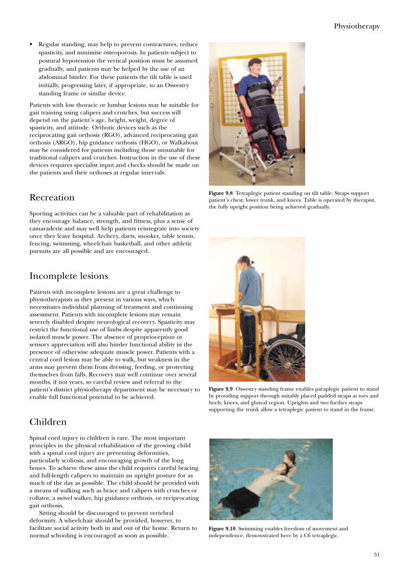
Physiotherapy
51
• Regular standing, may help to prevent contractures, reducespasticity, and minimise osteoporosis. In patients subject topostural hypotension the vertical position must be assumedgradually, and patients may be helped by the use of anabdominal binder. For these patients the tilt table is usedinitially, progressing later, if appropriate, to an Oswestrystanding frame or similar device.
Patients with low thoracic or lumbar lesions may be suitable forgait training using calipers and crutches, but success willdepend on the patient’s age, height, weight, degree ofspasticity, and attitude. Orthotic devices such as thereciprocating gait orthosis (RGO), advanced reciprocating gaitorthosis (ARGO), hip guidance orthosis (HGO), or Walkaboutmay be considered for patients including those unsuitable fortraditional calipers and crutches. Instruction in the use of thesedevices requires specialist input and checks should be made onthe patients and their orthoses at regular intervals.
Recreation
Sporting activities can be a valuable part of rehabilitation asthey encourage balance, strength, and fitness, plus a sense ofcamaraderie and may well help patients reintegrate into societyonce they leave hospital. Archery, darts, snooker, table tennis,fencing, swimming, wheelchair basketball, and other athleticpursuits are all possible and are encouraged.
Incomplete lesions
Patients with incomplete lesions are a great challenge tophysiotherapists as they present in various ways, whichnecessitates individual planning of treatment and continuingassessment. Patients with incomplete lesions may remainseverely disabled despite neurological recovery. Spasticity mayrestrict the functional use of limbs despite apparently goodisolated muscle power. The absence of proprioception orsensory appreciation will also hinder functional ability in thepresence of otherwise adequate muscle power. Patients with acentral cord lesion may be able to walk, but weakness in thearms may prevent them from dressing, feeding, or protectingthemselves from falls. Recovery may well continue over severalmonths, if not years, so careful review and referral to thepatient’s district physiotherapy department may be necessary toenable full functional potential to be achieved.
Children
Spinal cord injury in children is rare. The most importantprinciples in the physical rehabilitation of the growing childwith a spinal cord injury are preventing deformities,particularly scoliosis, and encouraging growth of the longbones. To achieve these aims the child requires careful bracingand full-length calipers to maintain an upright posture for asmuch of the day as possible. The child should be provided witha means of walking such as brace and calipers with crutches orrollator, a swivel walker, hip guidance orthosis, or reciprocatinggait orthosis.
Sitting should be discouraged to prevent vertebraldeformity. A wheelchair should be provided, however, tofacilitate social activity both in and out of the home. Return tonormal schooling is encouraged as soon as possible.
Figure 9.8 Tetraplegic patient standing on tilt table. Straps supportpatient’s chest, lower trunk, and knees. Table is operated by therapist,the fully upright position being achieved gradually.
Figure 9.9 Oswestry standing frame enables paraplegic patient to standby providing support through suitably placed padded straps at toes andheels, knees, and gluteal region. Uprights and two further strapssupporting the trunk allow a tetraplegic patient to stand in the frame.
Figure 9.10 Swimming enables freedom of movement andindependence, demonstrated here by a C6 tetraplegic.
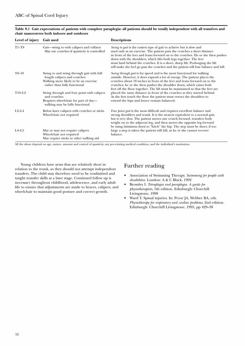
ABC of Spinal Cord Injury
52
Young children have arms that are relatively short inrelation to the trunk, so they should not attempt independenttransfers. The child may therefore need to be readmitted andtaught transfer skills at a later stage. Continued follow up isnecessary throughout childhood, adolescence, and early adultlife to ensure that adjustments are made to braces, calipers, andwheelchair to maintain good posture and correct growth.
Table 9.1 Gait expectations of patients with complete paraplegia: all patients should be totally independent with all transfers andchair manoeuvres both indoors and outdoors
Level of injury Gait used Descriptions
T1–T8 Gait—swing to with calipers and rollator Swing to gait is the easiest type of gait to achieve but is slow andMay use crutches if spasticity is controlled used only as an exercise. The patient puts the crutches a short distance
in front of the feet and leans forward on to the crutches. He or she then pushesdown with the shoulders, which lifts both legs together. The feetmust land behind the crutches. It is a short, sharp lift. Prolonging the liftwill make the feel go past the crutches and the patient will lose balance and fall.
T8–10 Swing to and swing through gait with full Swing through gait is for speed and is the most functional for walkinglength calipers and crutches outside. However, it does expend a lot of energy. The patient places the
Walking more likely to be an exercise crutches about 18 inches in front of the feet and leans forward on to therather than fully functional crutches; he or she then pushes the shoulder down, which raises both
feet off the floor together. The lift must be maintained so that the feet areT10–L2 Swing through and four point with calipers placed the same distance in front of the crutches as they started behind.
and crutches As the feet touch the floor the patient must retract the shoulders toRequires wheelchair for part of day— extend the hips and hence remain balanced.walking may be fully functional
L2–L4 Below knee calipers with crutches or sticks Four point gait is the most difficult and requires excellent balance andWheelchair not required strong shoulders and trunk. It is the nearest equivalent to a normal gait,
but is very slow. The patient moves one crutch forward, transfers bodyweight on to the adjacent leg, and then moves the opposite leg forwardby using latissimus dorsi to “hitch” the hip. The step must be short; if too
L4–L5 May or may not require calipers large a step is taken the patient will fall, as he or she cannot recoverWheelchair not required balance.May require sticks or other walking aid
All the above depend on age, stature, amount and control of spasticity, any pre-existing medical condition, and the individual’s motivation.
Further reading
• Association of Swimming Therapy. Swimming for people withdisabilities. London: A & C Black, 1992
• Bromley I. Tetraplegia and paraplegia. A guide forphysiotherapists, 5th edition. Edinburgh: ChurchillLivingstone, 1998
• Ward T. Spinal injuries. In: Pryor JA, Webber BA, eds.Physiotherapy for respiratory and cardiac problems, 2nd edition.Edinburgh: Churchill Livingstone, 1995, pp 429–38
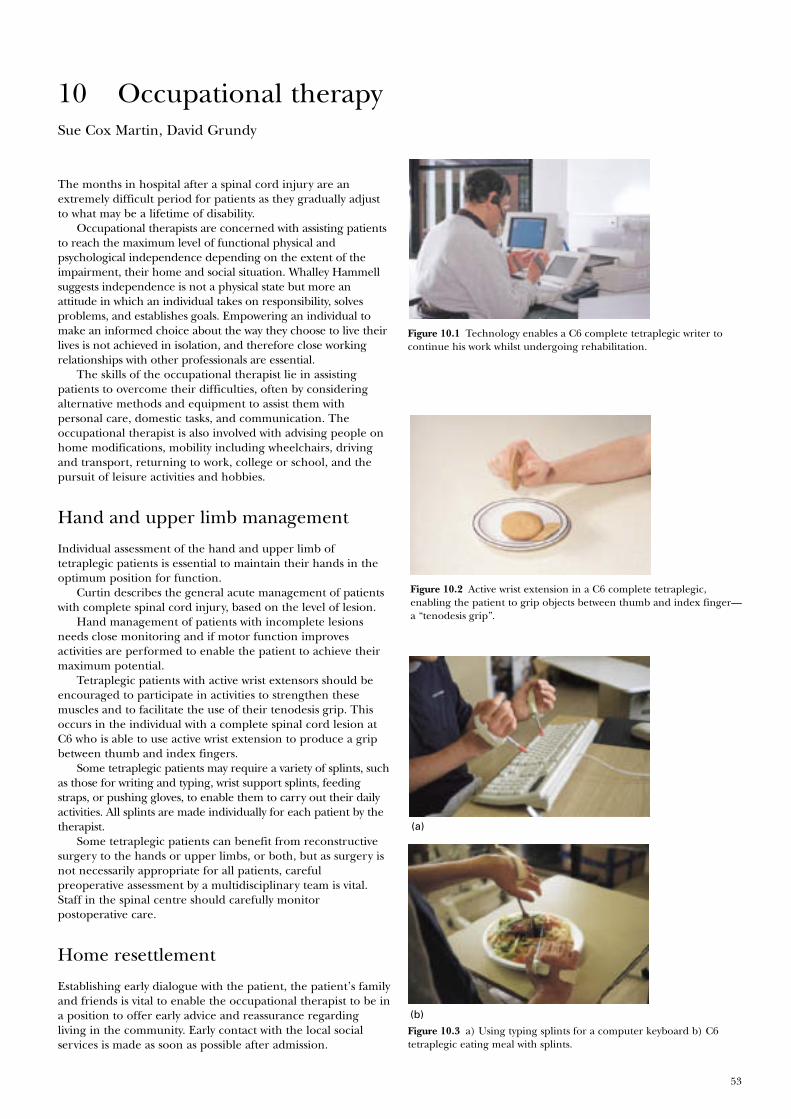
53
Figure 10.1 Technology enables a C6 complete tetraplegic writer tocontinue his work whilst undergoing rehabilitation.
Figure 10.2 Active wrist extension in a C6 complete tetraplegic,enabling the patient to grip objects between thumb and index finger—a “tenodesis grip”.
Sue Cox Martin, David Grundy
The months in hospital after a spinal cord injury are anextremely difficult period for patients as they gradually adjustto what may be a lifetime of disability.
Occupational therapists are concerned with assisting patientsto reach the maximum level of functional physical andpsychological independence depending on the extent of theimpairment, their home and social situation. Whalley Hammellsuggests independence is not a physical state but more anattitude in which an individual takes on responsibility, solvesproblems, and establishes goals. Empowering an individual tomake an informed choice about the way they choose to live theirlives is not achieved in isolation, and therefore close workingrelationships with other professionals are essential.
The skills of the occupational therapist lie in assistingpatients to overcome their difficulties, often by consideringalternative methods and equipment to assist them withpersonal care, domestic tasks, and communication. Theoccupational therapist is also involved with advising people onhome modifications, mobility including wheelchairs, drivingand transport, returning to work, college or school, and thepursuit of leisure activities and hobbies.
Hand and upper limb management
Individual assessment of the hand and upper limb oftetraplegic patients is essential to maintain their hands in theoptimum position for function.
Curtin describes the general acute management of patientswith complete spinal cord injury, based on the level of lesion.
Hand management of patients with incomplete lesionsneeds close monitoring and if motor function improvesactivities are performed to enable the patient to achieve theirmaximum potential.
Tetraplegic patients with active wrist extensors should beencouraged to participate in activities to strengthen thesemuscles and to facilitate the use of their tenodesis grip. Thisoccurs in the individual with a complete spinal cord lesion atC6 who is able to use active wrist extension to produce a gripbetween thumb and index fingers.
Some tetraplegic patients may require a variety of splints, suchas those for writing and typing, wrist support splints, feedingstraps, or pushing gloves, to enable them to carry out their dailyactivities. All splints are made individually for each patient by thetherapist.
Some tetraplegic patients can benefit from reconstructivesurgery to the hands or upper limbs, or both, but as surgery isnot necessarily appropriate for all patients, carefulpreoperative assessment by a multidisciplinary team is vital.Staff in the spinal centre should carefully monitorpostoperative care.
Home resettlement
Establishing early dialogue with the patient, the patient’s familyand friends is vital to enable the occupational therapist to be ina position to offer early advice and reassurance regardingliving in the community. Early contact with the local socialservices is made as soon as possible after admission.
10 Occupational therapy
(a)
(b)
Figure 10.3 a) Using typing splints for a computer keyboard b) C6tetraplegic eating meal with splints.
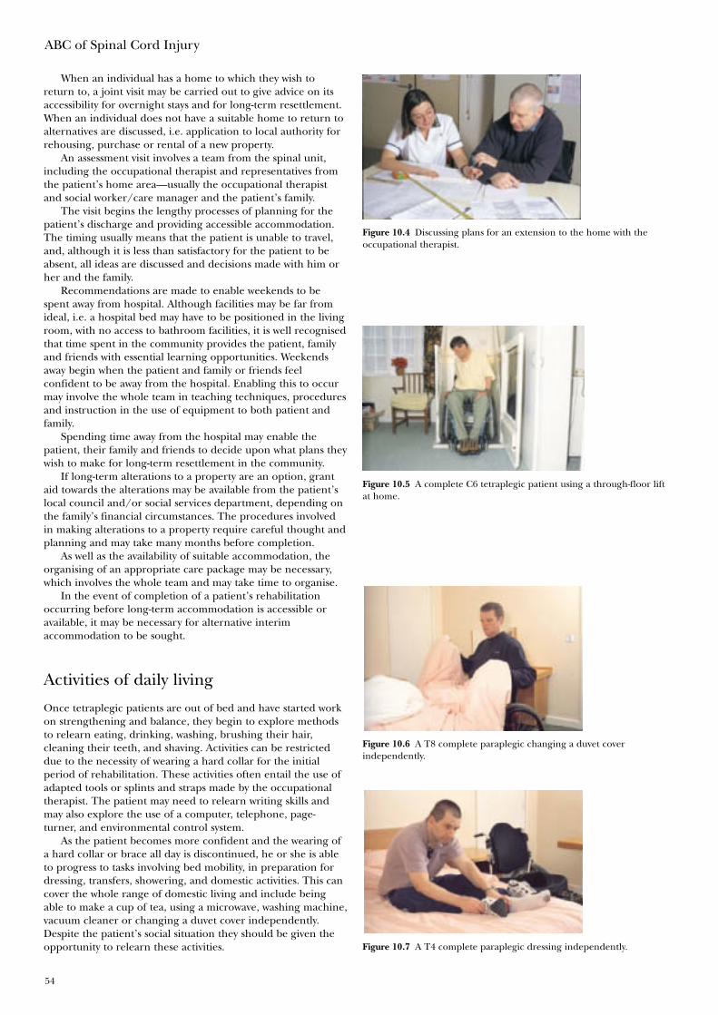
ABC of Spinal Cord Injury
54
When an individual has a home to which they wish toreturn to, a joint visit may be carried out to give advice on itsaccessibility for overnight stays and for long-term resettlement.When an individual does not have a suitable home to return toalternatives are discussed, i.e. application to local authority forrehousing, purchase or rental of a new property.
An assessment visit involves a team from the spinal unit,including the occupational therapist and representatives fromthe patient’s home area—usually the occupational therapistand social worker/care manager and the patient’s family.
The visit begins the lengthy processes of planning for thepatient’s discharge and providing accessible accommodation.The timing usually means that the patient is unable to travel,and, although it is less than satisfactory for the patient to beabsent, all ideas are discussed and decisions made with him orher and the family.
Recommendations are made to enable weekends to bespent away from hospital. Although facilities may be far fromideal, i.e. a hospital bed may have to be positioned in the livingroom, with no access to bathroom facilities, it is well recognisedthat time spent in the community provides the patient, familyand friends with essential learning opportunities. Weekendsaway begin when the patient and family or friends feelconfident to be away from the hospital. Enabling this to occurmay involve the whole team in teaching techniques, proceduresand instruction in the use of equipment to both patient andfamily.
Spending time away from the hospital may enable thepatient, their family and friends to decide upon what plans theywish to make for long-term resettlement in the community.
If long-term alterations to a property are an option, grantaid towards the alterations may be available from the patient’slocal council and/or social services department, depending onthe family’s financial circumstances. The procedures involvedin making alterations to a property require careful thought andplanning and may take many months before completion.
As well as the availability of suitable accommodation, theorganising of an appropriate care package may be necessary,which involves the whole team and may take time to organise.
In the event of completion of a patient’s rehabilitationoccurring before long-term accommodation is accessible oravailable, it may be necessary for alternative interimaccommodation to be sought.
Activities of daily living
Once tetraplegic patients are out of bed and have started workon strengthening and balance, they begin to explore methodsto relearn eating, drinking, washing, brushing their hair,cleaning their teeth, and shaving. Activities can be restricteddue to the necessity of wearing a hard collar for the initialperiod of rehabilitation. These activities often entail the use ofadapted tools or splints and straps made by the occupationaltherapist. The patient may need to relearn writing skills andmay also explore the use of a computer, telephone, page-turner, and environmental control system.
As the patient becomes more confident and the wearing ofa hard collar or brace all day is discontinued, he or she is ableto progress to tasks involving bed mobility, in preparation fordressing, transfers, showering, and domestic activities. This cancover the whole range of domestic living and include beingable to make a cup of tea, using a microwave, washing machine,vacuum cleaner or changing a duvet cover independently.Despite the patient’s social situation they should be given theopportunity to relearn these activities.
Figure 10.4 Discussing plans for an extension to the home with theoccupational therapist.
Figure 10.6 A T8 complete paraplegic changing a duvet coverindependently.
Figure 10.7 A T4 complete paraplegic dressing independently.
Figure 10.5 A complete C6 tetraplegic patient using a through-floor liftat home.
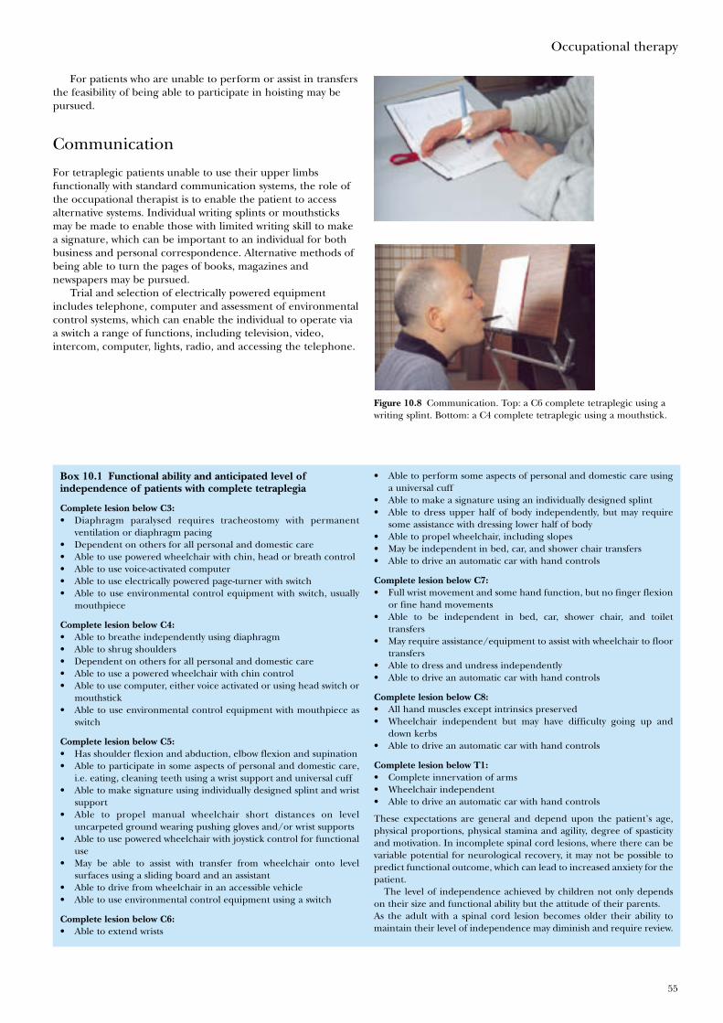
Occupational therapy
55
For patients who are unable to perform or assist in transfersthe feasibility of being able to participate in hoisting may bepursued.
Communication
For tetraplegic patients unable to use their upper limbsfunctionally with standard communication systems, the role ofthe occupational therapist is to enable the patient to accessalternative systems. Individual writing splints or mouthsticksmay be made to enable those with limited writing skill to makea signature, which can be important to an individual for bothbusiness and personal correspondence. Alternative methods ofbeing able to turn the pages of books, magazines andnewspapers may be pursued.
Trial and selection of electrically powered equipmentincludes telephone, computer and assessment of environmentalcontrol systems, which can enable the individual to operate viaa switch a range of functions, including television, video,intercom, computer, lights, radio, and accessing the telephone.
Figure 10.8 Communication. Top: a C6 complete tetraplegic using awriting splint. Bottom: a C4 complete tetraplegic using a mouthstick.
Box 10.1 Functional ability and anticipated level ofindependence of patients with complete tetraplegia
Complete lesion below C3:• Diaphragm paralysed requires tracheostomy with permanent
ventilation or diaphragm pacing• Dependent on others for all personal and domestic care• Able to use powered wheelchair with chin, head or breath control• Able to use voice-activated computer• Able to use electrically powered page-turner with switch• Able to use environmental control equipment with switch, usually
mouthpiece
Complete lesion below C4:• Able to breathe independently using diaphragm• Able to shrug shoulders• Dependent on others for all personal and domestic care• Able to use a powered wheelchair with chin control• Able to use computer, either voice activated or using head switch or
mouthstick• Able to use environmental control equipment with mouthpiece as
switch
Complete lesion below C5:• Has shoulder flexion and abduction, elbow flexion and supination• Able to participate in some aspects of personal and domestic care,
i.e. eating, cleaning teeth using a wrist support and universal cuff• Able to make signature using individually designed splint and wrist
support• Able to propel manual wheelchair short distances on level
uncarpeted ground wearing pushing gloves and/or wrist supports• Able to use powered wheelchair with joystick control for functional
use• May be able to assist with transfer from wheelchair onto level
surfaces using a sliding board and an assistant• Able to drive from wheelchair in an accessible vehicle• Able to use environmental control equipment using a switch
Complete lesion below C6:• Able to extend wrists
• Able to perform some aspects of personal and domestic care usinga universal cuff
• Able to make a signature using an individually designed splint• Able to dress upper half of body independently, but may require
some assistance with dressing lower half of body• Able to propel wheelchair, including slopes• May be independent in bed, car, and shower chair transfers• Able to drive an automatic car with hand controls
Complete lesion below C7:• Full wrist movement and some hand function, but no finger flexion
or fine hand movements• Able to be independent in bed, car, shower chair, and toilet
transfers• May require assistance/equipment to assist with wheelchair to floor
transfers• Able to dress and undress independently• Able to drive an automatic car with hand controls
Complete lesion below C8:• All hand muscles except intrinsics preserved• Wheelchair independent but may have difficulty going up and
down kerbs• Able to drive an automatic car with hand controls
Complete lesion below T1:• Complete innervation of arms• Wheelchair independent• Able to drive an automatic car with hand controls
These expectations are general and depend upon the patient’s age,physical proportions, physical stamina and agility, degree of spasticityand motivation. In incomplete spinal cord lesions, where there can bevariable potential for neurological recovery, it may not be possible topredict functional outcome, which can lead to increased anxiety for thepatient.
The level of independence achieved by children not only dependson their size and functional ability but the attitude of their parents.As the adult with a spinal cord lesion becomes older their ability tomaintain their level of independence may diminish and require review.
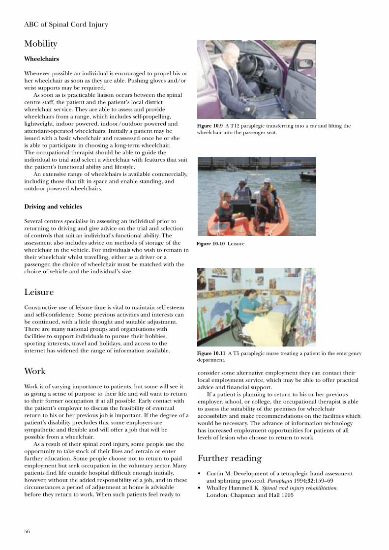
ABC of Spinal Cord Injury
56
Mobility
Wheelchairs
Whenever possible an individual is encouraged to propel his orher wheelchair as soon as they are able. Pushing gloves and/orwrist supports may be required.
As soon as is practicable liaison occurs between the spinalcentre staff, the patient and the patient’s local districtwheelchair service. They are able to assess and providewheelchairs from a range, which includes self-propelling,lightweight, indoor powered, indoor/outdoor powered andattendant-operated wheelchairs. Initially a patient may beissued with a basic wheelchair and reassessed once he or sheis able to participate in choosing a long-term wheelchair.The occupational therapist should be able to guide theindividual to trial and select a wheelchair with features that suitthe patient’s functional ability and lifestyle.
An extensive range of wheelchairs is available commercially,including those that tilt in space and enable standing, andoutdoor powered wheelchairs.
Driving and vehicles
Several centres specialise in assessing an individual prior toreturning to driving and give advice on the trial and selectionof controls that suit an individual’s functional ability. Theassessment also includes advice on methods of storage of thewheelchair in the vehicle. For individuals who wish to remain intheir wheelchair whilst travelling, either as a driver or apassenger, the choice of wheelchair must be matched with thechoice of vehicle and the individual’s size.
Leisure
Constructive use of leisure time is vital to maintain self-esteemand self-confidence. Some previous activities and interests canbe continued, with a little thought and suitable adjustment.There are many national groups and organisations withfacilities to support individuals to pursue their hobbies,sporting interests, travel and holidays, and access to theinternet has widened the range of information available.
Work
Work is of varying importance to patients, but some will see itas giving a sense of purpose to their life and will want to returnto their former occupation if at all possible. Early contact withthe patient’s employer to discuss the feasibility of eventualreturn to his or her previous job is important. If the degree of apatient’s disability precludes this, some employers aresympathetic and flexible and will offer a job that will bepossible from a wheelchair.
As a result of their spinal cord injury, some people use theopportunity to take stock of their lives and retrain or enterfurther education. Some people choose not to return to paidemployment but seek occupation in the voluntary sector. Manypatients find life outside hospital difficult enough initially,however, without the added responsibility of a job, and in thesecircumstances a period of adjustment at home is advisablebefore they return to work. When such patients feel ready to
Figure 10.9 A T12 paraplegic transferring into a car and lifting thewheelchair into the passenger seat.
Figure 10.11 A T5 paraplegic nurse treating a patient in the emergencydepartment.
consider some alternative employment they can contact theirlocal employment service, which may be able to offer practicaladvice and financial support.
If a patient is planning to return to his or her previousemployer, school, or college, the occupational therapist is ableto assess the suitability of the premises for wheelchairaccessibility and make recommendations on the facilities whichwould be necessary. The advance of information technologyhas increased employment opportunities for patients of alllevels of lesion who choose to return to work.
Further reading
• Curtin M. Development of a tetraplegic hand assessmentand splinting protocol. Paraplegia 1994;32:159–69
• Whalley Hammell K. Spinal cord injury rehabilitation.London: Chapman and Hall 1995
Figure 10.10 Leisure.
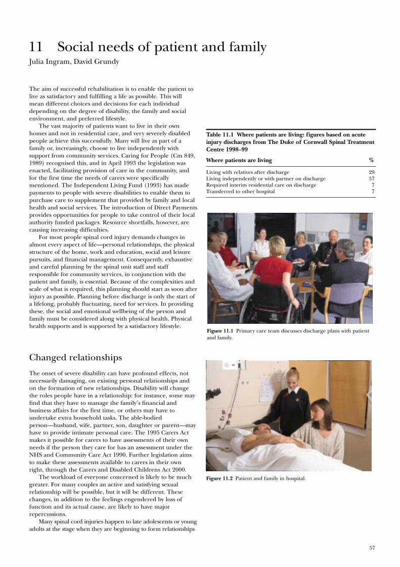
57
Figure 11.2 Patient and family in hospital.
Figure 11.1 Primary care team discusses discharge plans with patientand family.
Julia Ingram, David Grundy
The aim of successful rehabilitation is to enable the patient tolive as satisfactory and fulfilling a life as possible. This willmean different choices and decisions for each individualdepending on the degree of disability, the family and socialenvironment, and preferred lifestyle.
The vast majority of patients want to live in their ownhomes and not in residential care, and very severely disabledpeople achieve this successfully. Many will live as part of afamily or, increasingly, choose to live independently withsupport from community services. Caring for People (Cm 849,1989) recognised this, and in April 1993 the legislation wasenacted, facilitating provision of care in the community, andfor the first time the needs of carers were specificallymentioned. The Independent Living Fund (1993) has madepayments to people with severe disabilities to enable them topurchase care to supplement that provided by family and localhealth and social services. The introduction of Direct Paymentsprovides opportunities for people to take control of their localauthority funded packages. Resource shortfalls, however, arecausing increasing difficulties.
For most people spinal cord injury demands changes inalmost every aspect of life—personal relationships, the physicalstructure of the home, work and education, social and leisurepursuits, and financial management. Consequently, exhaustiveand careful planning by the spinal unit staff and staffresponsible for community services, in conjunction with thepatient and family, is essential. Because of the complexities andscale of what is required, this planning should start as soon afterinjury as possible. Planning before discharge is only the start ofa lifelong, probably fluctuating, need for services. In providingthese, the social and emotional wellbeing of the person andfamily must be considered along with physical health. Physicalhealth supports and is supported by a satisfactory lifestyle.
Changed relationships
The onset of severe disability can have profound effects, notnecessarily damaging, on existing personal relationships andon the formation of new relationships. Disability will changethe roles people have in a relationship: for instance, some mayfind that they have to manage the family’s financial andbusiness affairs for the first time, or others may have toundertake extra household tasks. The able-bodiedperson—husband, wife, partner, son, daughter or parent—mayhave to provide intimate personal care. The 1995 Carers Actmakes it possible for carers to have assessments of their ownneeds if the person they care for has an assessment under theNHS and Community Care Act 1990. Further legislation aimsto make these assessments available to carers in their ownright, through the Carers and Disabled Childrens Act 2000.
The workload of everyone concerned is likely to be muchgreater. For many couples an active and satisfying sexualrelationship will be possible, but it will be different. Thesechanges, in addition to the feelings engendered by loss offunction and its actual cause, are likely to have majorrepercussions.
Many spinal cord injuries happen to late adolescents or youngadults at the stage when they are beginning to form relationships
11 Social needs of patient and family
Table 11.1 Where patients are living: figures based on acuteinjury discharges from The Duke of Cornwall Spinal TreatmentCentre 1998–99
Where patients are living %
Living with relatives after discharge 29Living independently or with partner on discharge 57Required interim residential care on discharge 7Transferred to other hospital 7
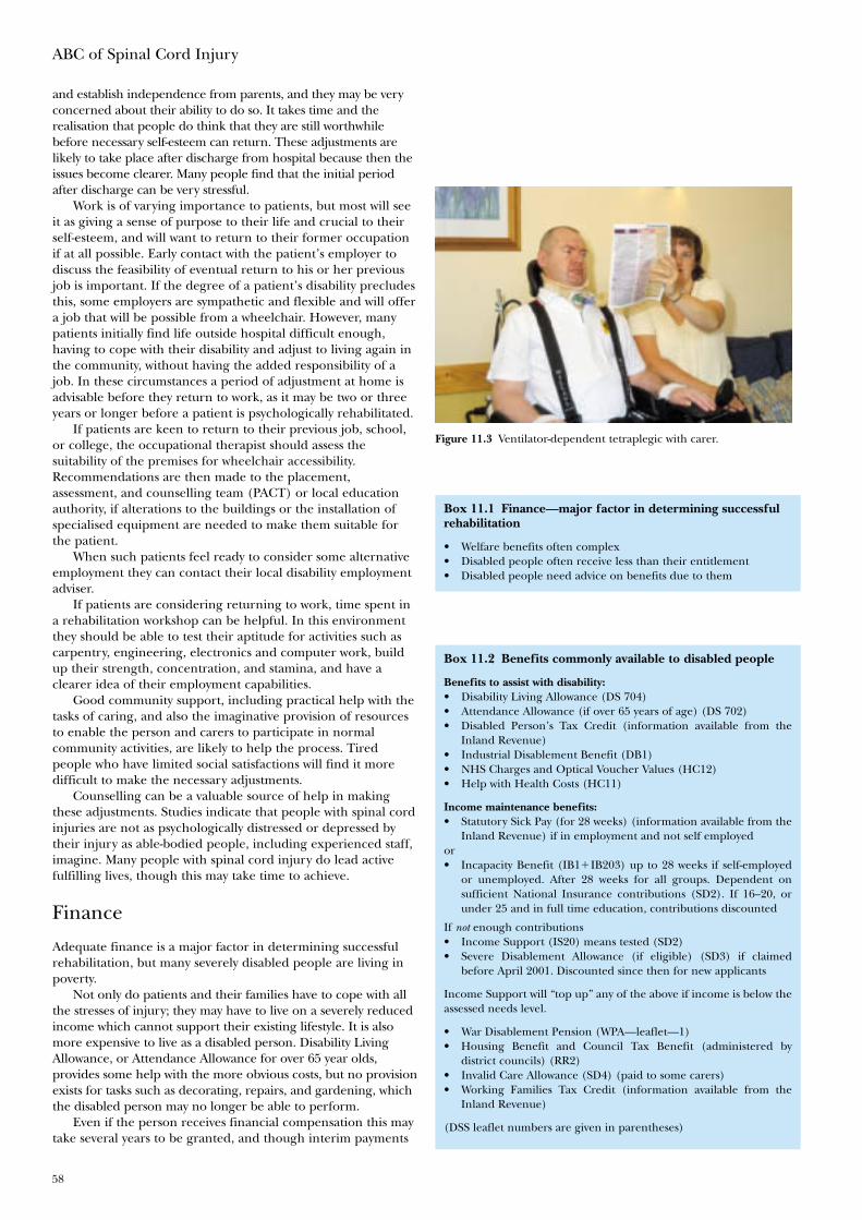
ABC of Spinal Cord Injury
58
and establish independence from parents, and they may be veryconcerned about their ability to do so. It takes time and therealisation that people do think that they are still worthwhilebefore necessary self-esteem can return. These adjustments arelikely to take place after discharge from hospital because then theissues become clearer. Many people find that the initial periodafter discharge can be very stressful.
Work is of varying importance to patients, but most will seeit as giving a sense of purpose to their life and crucial to theirself-esteem, and will want to return to their former occupationif at all possible. Early contact with the patient’s employer todiscuss the feasibility of eventual return to his or her previousjob is important. If the degree of a patient’s disability precludesthis, some employers are sympathetic and flexible and will offera job that will be possible from a wheelchair. However, manypatients initially find life outside hospital difficult enough,having to cope with their disability and adjust to living again inthe community, without having the added responsibility of ajob. In these circumstances a period of adjustment at home isadvisable before they return to work, as it may be two or threeyears or longer before a patient is psychologically rehabilitated.
If patients are keen to return to their previous job, school,or college, the occupational therapist should assess thesuitability of the premises for wheelchair accessibility.Recommendations are then made to the placement,assessment, and counselling team (PACT) or local educationauthority, if alterations to the buildings or the installation ofspecialised equipment are needed to make them suitable forthe patient.
When such patients feel ready to consider some alternativeemployment they can contact their local disability employmentadviser.
If patients are considering returning to work, time spent ina rehabilitation workshop can be helpful. In this environmentthey should be able to test their aptitude for activities such ascarpentry, engineering, electronics and computer work, buildup their strength, concentration, and stamina, and have aclearer idea of their employment capabilities.
Good community support, including practical help with thetasks of caring, and also the imaginative provision of resourcesto enable the person and carers to participate in normalcommunity activities, are likely to help the process. Tiredpeople who have limited social satisfactions will find it moredifficult to make the necessary adjustments.
Counselling can be a valuable source of help in makingthese adjustments. Studies indicate that people with spinal cordinjuries are not as psychologically distressed or depressed bytheir injury as able-bodied people, including experienced staff,imagine. Many people with spinal cord injury do lead activefulfilling lives, though this may take time to achieve.
Finance
Adequate finance is a major factor in determining successfulrehabilitation, but many severely disabled people are living inpoverty.
Not only do patients and their families have to cope with allthe stresses of injury; they may have to live on a severely reducedincome which cannot support their existing lifestyle. It is alsomore expensive to live as a disabled person. Disability LivingAllowance, or Attendance Allowance for over 65 year olds,provides some help with the more obvious costs, but no provisionexists for tasks such as decorating, repairs, and gardening, whichthe disabled person may no longer be able to perform.
Even if the person receives financial compensation this maytake several years to be granted, and though interim payments
Box 11.2 Benefits commonly available to disabled people
Benefits to assist with disability:• Disability Living Allowance (DS 704)• Attendance Allowance (if over 65 years of age) (DS 702)• Disabled Person’s Tax Credit (information available from the
Inland Revenue)• Industrial Disablement Benefit (DB1)• NHS Charges and Optical Voucher Values (HC12)• Help with Health Costs (HC11)
Income maintenance benefits:• Statutory Sick Pay (for 28 weeks) (information available from the
Inland Revenue) if in employment and not self employedor• Incapacity Benefit (IB1�IB203) up to 28 weeks if self-employed
or unemployed. After 28 weeks for all groups. Dependent onsufficient National Insurance contributions (SD2). If 16–20, orunder 25 and in full time education, contributions discounted
If not enough contributions• Income Support (IS20) means tested (SD2)• Severe Disablement Allowance (if eligible) (SD3) if claimed
before April 2001. Discounted since then for new applicants
Income Support will “top up” any of the above if income is below theassessed needs level.
• War Disablement Pension (WPA—leaflet—1)• Housing Benefit and Council Tax Benefit (administered by
district councils) (RR2)• Invalid Care Allowance (SD4) (paid to some carers)• Working Families Tax Credit (information available from the
Inland Revenue)
(DSS leaflet numbers are given in parentheses)
Box 11.1 Finance—major factor in determining successfulrehabilitation
• Welfare benefits often complex• Disabled people often receive less than their entitlement• Disabled people need advice on benefits due to them
Figure 11.3 Ventilator-dependent tetraplegic with carer.
Social needs of patient and family
59
can be made, in some circumstances they are not alwaysavailable at the time of discharge from hospital, when there islikely to be major expenditure on either moving or adaptingthe present house.
Because of the interruption in, or possible loss of, earningcapacity many people will be dependent for long periods onwelfare benefits administered by the Department of SocialSecurity. These are complex, and various studies have shownthat many disabled people are receiving less than theirentitlement, sometimes by quite substantial amounts. It istherefore important for those working with disabled people tobe aware that they may be underclaiming benefits and to advisethem accordingly.
Adapting homes
Most houses are unsuitable for wheelchairs unless adapted.Disabled Facilities Grants may be available to assist with thecost, but for many people help is limited because mortgagerepayments are not taken into account in the financialassessment. Housing presents a continuing problem because,though patients may return to an adapted house or berehoused from hospital, they may well want to change house inthe future, especially as spinal cord injuries typically occur inyoung people who would normally move house several times. Adisabled person may have difficulty in finding a suitable house,and there can be time restrictions on further provision ofgrants for adaptations. There are also mandatory anddiscretionary limitations on grants which may be madeavailable to assist in the adaptation of a property. Many peoplefind the discrepancy between local authorities in theirinterpretation of the legislation around this frustrating. Manycannot afford to buy a house and will depend on councilhousing, housing association property, or privately rentedproperty, all of which are in short supply. Consequently, anymove can be difficult to achieve and has to be planned wellahead. The services of community occupational therapists,housing departments, and social workers may be required.
A considerable number of statutory services are concernedwith providing services for disabled people. Voluntaryorganisations also provide important resources. They can act aspressure and self-help groups, and organisations of disabledpeople have the knowledge and understanding born ofpersonal experience. There are many such organisations, ofwhich the Spinal Injuries Association is particularly relevant.
To mobilise and coordinate these services, which often varyin what they can provide in different geographical areas, is amajor undertaking. Too often disabled people fail to receive aservice that would be of benefit or they may feel overwhelmedand not in control of their own lives, with consequent damageto morale and health. Disabled people and their familiesshould have access to full information about the servicesavailable and be enabled to make their own decisions aboutwhat they need. The 1998 White Paper Modernising SocialServices sets out government objectives for more partnershipworking, joint funding and uniformity of charging policiesacross local authorities which should make services moreaccessible to patients and with greater parity.
Table 11.2 Adapting homes—where patients go: figures basedon acute injury discharges from The Duke of Cornwall SpinalTreatment Centre 1998–99
Where patients go %
Able to return to own home with adaptations 55Had to move to live with relatives 11Required rehousing provided by District Councilor Housing Association 29
Required rehousing, patient or family bought property 5
Box 11.4 Information and advice on benefits
• Department of Social Security (local office or DSS) BenefitEnquiry Line. Tel: 0800 882200
• Citizens Advice Bureau• DIAL (Disabled Information Advice Line) (Name of town)—A
voluntary organisation operating in some areas• Disability Rights Handbook (Price £11.50 post free); published
annually by the Disability Alliance Educational & ResearchAssociation, Universal House, 88–94 Wentworth Street, LondonE1 7SA. Tel: 020 77247 8776.
• Spinal Injuries Association—76 St James’s Lane, London N103DF. Tel: 020 8444 2121 or 0800 980 0501. www.spinal.co.uk.email: [email protected]
Table 11.3 Employment—what patients do: figures based onacute injury discharges from The Duke of Cornwall SpinalTreatment Centre 1998–99
Employment—what patients do %
In work or job left open 30In education or training 10No employment on discharge, but previously employed 38No employment on discharge—not employed when admitted 22
Box 11.3 Typical adaptations funded either privately or inpart by a Disabled Facilities Grant
• Ramped access to external doors• Widening of internal doors• Level access parking, with carport/garage• Level access shower• Toilet with access for shower chair• Accessible light switches, sockets, door locks• Accessible kitchen and facilities• Patio area in the garden• Thermostatically controlled heating system• Through-floor lift or stair lift• Internal ramps
60
Figure 12.1 Top: areas of skin most at risk of effects of pressure.Bottom: skin pressure elevator to measure the pressure between theskin and supporting surface.
Figure 12.2 Force sensing array: pressure mapping can be used toassess pressure distribution, which helps to prevent skin breakdown.
Rachel Stowell, Wendy Pickard, David Grundy
Discharge from hospital is a complicated process for bothparaplegic and tetraplegic patients. Physical care is a majorconcern, and here the difference between levels of injury isprofound. People with paraplegia usually become self caring;those with low tetraplegia, especially if young, may also achieveindependence, but those with high tetraplegia may requirehelp with their physical needs.
Achievement of good care depends largely on educatingthe patients, their families, and the community staff. Patientsshould be expert at understanding and, as far as possible,caring for their own bodies. They need to be able to recognisepotential problems and either deal with them themselves orknow where to seek advice. Much time is spent in teaching theimportance of good skin, bladder and bowel care, as long-termproblems in these areas are common.
Education of patients
Skin care
Patients are taught how to use a mirror to check their pressureareas regularly, the stages of development of pressure sores,and what to do should a pressure mark occur. Wheelchairs andcushions are best assessed in a pressure clinic. If patientscannot lift themselves in their chair they need a cushion thatallows them to sit in their wheelchair all day without resultingin a red mark on their skin. Weight, height, the degree ofsensation and mobility, age, posture, motivation, and thequality of the skin all affect the type of cushion needed. Allwheelchair cushions have a limited life and need regularchecking to provide a reliable degree of assistance inprevention of sores.
Clothes made of natural fibres are preferable because manypatients sweat excessively; clothing should not be tightotherwise there is risk of skin damage resulting in pressuresores. It should also be noted that hard seams and pocketswhich cross over the ischial tuberosities, trochanters, or coccyxmay cause pressure marks on the skin. It is recommended thatshoes should generally be one size larger than previously wornbecause of a tendency of the feet to swell during the day.
The choice of a suitable mattress is also important. Manypressure-relieving mattresses are now available, so all patientsshould be able to sleep for at least eight hours without beingturned, and without resulting in a red mark on their skin. Thepatient should be assessed individually to ascertain theappropriate mattress for their long-term needs. Patients areencouraged to contact the pressure clinic for information andadvice regarding any aspect of their care. In addition, thecommunity liaison staff while visiting the patient in thecommunity are able to reinforce educational aspects.
Bladder care
Patients are taught the most effective method of bladderemptying (see chapter 7). Although men with high tetraplegiacan often tap over the bladder to induce a detrusorcontraction, they may require help to apply a sheath and to fita leg bag. In women with high tetraplegia the bladder is often
12 Transfer of care from hospital to community
Box 12.1 People need to develop skills in:
• Understanding and caring for their own bodies• Recognising potential problems• Dealing with problems or learning where to go for advice
Transfer of care from hospital to community
61
best managed by a suprapubic catheter. It is best to avoidlong-term urethral catheters as these may damage the bladderneck and urethra. Patients whose bladder emptying methodinvolves an indwelling catheter are taught to regularly use acatheter valve (which can be opened and closed), to maintainbladder volume and compliance. Intermittent clean self-catheterisation is the preferred method for most women andmen with paraplegia and some with low tetraplegia. It isparticularly successful in patients with an acontractile bladder.
Long-term bladder management
Urinary tract infections are common but may be reduced byadequate hydration and often by urinary acidification. In generalantibiotics are given only when an infection causes systemicsymptoms. If recurrent urinary tract infections occur the patientshould be investigated for underlying causes such as stones in theurinary tract or poor bladder emptying. Various surgicalinterventions are available in selected patients (see chapter 7).
Bowel care
Patients with tetraplegia and paraplegia who have an uppermotor neurone cord lesion generally have reflex bowel activity,and evacuation can usually be produced by inserting glycerinesuppositories or by anal digital stimulation, or both. Patientswith high tetraplegia generally have poor balance and have tobe hoisted on to a padded shower chair which can be wheeledover a toilet. This is more acceptable than being hoisteddirectly on to a toilet, which is generally less stable. In somecircumstances bowel evacuation may need to take place on thebed with the patient in the left lateral position.
Patients with low paraplegia and a lower motor neuronecord lesion generally have a flaccid bowel, so will need toevacuate their bowel manually or by straining using theabdominal muscles, or by a combination of the two methods.Patients who carry out manual evacuation are advised to keeptheir stools slightly constipated to ease removal. They should beable to transfer themselves onto a toilet, and the seat should bepadded to prevent pressure sores from developing due toprolonged sitting. In practice most patients evacuate theirbowels daily or on alternate days. When possible, the timing andfrequency of bowel evacuation should be made to fit in with theperson’s lifestyle. Help can be requested from the district nurse.Patients are advised to maintain their bowel regime and to avoidstrong oral and rectal stimulant laxatives and enemas. Furthereducational principles are described in chapter 8 on nursing.
Long-term bowel management
A change in diet or lifestyle can affect bowel management, andpatients are advised to change only one aspect of their bowelcare at a time to minimise potential problems. Long-termoptions which can address chronic bowel managementproblems include colonic irrigation via the rectum, or throughan abdominal stoma (an antegrade colonic enema), or a stoma,such as a colostomy.
Autonomic dysreflexia
Autonomic dysreflexia is commonly associated with bladder orbowel problems, particularly overdistension. By the time ofdischarge from hospital, patients should be fully aware of thesigns and symptoms of autonomic dysreflexia and be able todirect people to help find and remove the cause (see chapter 6).
Box 12.2 Long-term bladder management
• Intermittent self-catheterisation is preferred method for thosewith acontractile bladders
• Condom sheath drainage in contractile bladders• If indwelling catheter, suprapubic catheter is preferable to
urethral catheter, to avoid urethral damage. Use catheter valve tomaintain bladder compliance and capacity
Box 12.3 Urinary tract infections
• Treat with antibiotics only if systemic symptoms present• Adequate hydration and urinary acidification• Investigate recurrent infections
Box 12.4 Bowel
• Upper motor neurone cord lesion reflex emptying—responds to suppositories and anal digitalstimulation
• Lower motor neurone cord lesionflaccid bowel with emptying by manual evacuation
Box 12.6 Autonomic dysreflexia
High lesion patients must:• be aware of the signs and symptoms• be able to direct care.
Box 12.5 Long-term bowel management
• Daily or on alternate days• Maintain consistent bowel regime• Avoid strong laxatives and enemas• Should fit in with patient’s lifestyle• Change in diet or lifestyle can affect bowel management• Only change one aspect of bowel care at a time, to minimise
potential problems
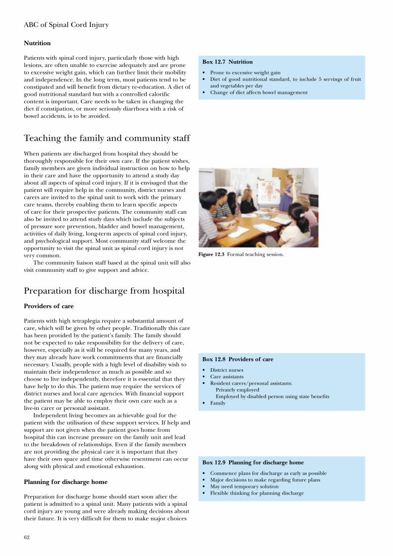
ABC of Spinal Cord Injury
62
Nutrition
Patients with spinal cord injury, particularly those with highlesions, are often unable to exercise adequately and are proneto excessive weight gain, which can further limit their mobilityand independence. In the long term, most patients tend to beconstipated and will benefit from dietary re-education. A diet ofgood nutritional standard but with a controlled calorificcontent is important. Care needs to be taken in changing thediet if constipation, or more seriously diarrhoea with a risk ofbowel accidents, is to be avoided.
Teaching the family and community staff
When patients are discharged from hospital they should bethoroughly responsible for their own care. If the patient wishes,family members are given individual instruction on how to helpin their care and have the opportunity to attend a study dayabout all aspects of spinal cord injury. If it is envisaged that thepatient will require help in the community, district nurses andcarers are invited to the spinal unit to work with the primarycare teams, thereby enabling them to learn specific aspectsof care for their prospective patients. The community staff canalso be invited to attend study days which include the subjectsof pressure sore prevention, bladder and bowel management,activities of daily living, long-term aspects of spinal cord injury,and psychological support. Most community staff welcome theopportunity to visit the spinal unit as spinal cord injury is notvery common.
The community liaison staff based at the spinal unit will alsovisit community staff to give support and advice.
Preparation for discharge from hospital
Providers of care
Patients with high tetraplegia require a substantial amount ofcare, which will be given by other people. Traditionally this carehas been provided by the patient’s family. The family shouldnot be expected to take responsibility for the delivery of care,however, especially as it will be required for many years, andthey may already have work commitments that are financiallynecessary. Usually, people with a high level of disability wish tomaintain their independence as much as possible and sochoose to live independently, therefore it is essential that theyhave help to do this. The patient may require the services ofdistrict nurses and local care agencies. With financial supportthe patient may be able to employ their own care such as alive-in carer or personal assistant.
Independent living becomes an achievable goal for thepatient with the utilisation of these support services. If help andsupport are not given when the patient goes home fromhospital this can increase pressure on the family unit and leadto the breakdown of relationships. Even if the family membersare not providing the physical care it is important that theyhave their own space and time otherwise resentment can occuralong with physical and emotional exhaustion.
Planning for discharge home
Preparation for discharge home should start soon after thepatient is admitted to a spinal unit. Many patients with a spinalcord injury are young and were already making decisions abouttheir future. It is very difficult for them to make major choices
Figure 12.3 Formal teaching session.
Box 12.8 Providers of care
• District nurses• Care assistants• Resident carers/personal assistants:
Privately employedEmployed by disabled person using state benefits
• Family
Box 12.7 Nutrition
• Prone to excessive weight gain• Diet of good nutritional standard, to include 5 servings of fruit
and vegetables per day• Change of diet affects bowel management
Box 12.9 Planning for discharge home
• Commence plans for discharge as early as possible• Major decisions to make regarding future plans• May need temporary solution• Flexible thinking for planning discharge
Transfer of care from hospital to community
63
of where to live and with whom and to decide who may be ableto help them with their care. It is sometimes necessary to have atemporary solution, and when they have had more time toadjust to their injury a more permanent solution can be found.It is therefore essential that this initial decision should allow acertain amount of flexibility.
Planning for independent living
Many patients with tetraplegia choose to live independently,and initially statutory care facilities in the patient’s home maybe used. These services may not be able to meet fully the careneeds of someone with a high level of injury so they may needto be supplemented. Many people will prefer to employ apersonal assistant to live in to help with personal care and dailyliving activities. This allows people to take control of their livesbut requires them to develop skills in interviewing, financialmanagement, and teaching. When financial compensation forthe injury is not available staff from the spinal unit contact thepatient’s local social services and health care trusts for financialsupport through community care assessment, the IndependentLiving Fund (ILF), and direct payment scheme. Initially,patients choosing this option often require additional supportfrom the spinal unit for advice in relation to, for instance,advertising for carers and interviewing. Patients quickly becometotally responsible for their own care on leaving the spinal unit.
Planning for interim care
When their homes have not been adapted for wheelchair usebefore the patients’ discharge from the spinal unit, interim caremay be necessary and can help to act as a bridge between theprotection of a spinal injuries unit and the reality of everydayliving with a disability.
Easing transfer from hospital tocommunity
The support of the district nursing service is invaluable ineasing the transfer from the spinal unit to the community.Before discharge patients will have some weekends at home.Difficulties experienced by them and their families can bediscussed with the district nurse and care agency, who canassess the situation and contact the spinal unit if advice isrequired. The families are often reassured to know that there isan effective link between the spinal unit and the community. Tomaintain this link the initial discharge plan may require adistrict nurse each morning to provide personal care with helpfrom a care assistant.
The early weeks at home
The early weeks after discharge can be both physically andemotionally exhausting for all concerned. The patients arefaced with the harsh reality of everyday living. However well apatient’s community care is planned, problems may still arise.For this reason all patients are visited by the community liaisonstaff from the spinal unit usually within 6–8 weeks of dischargeand at other times as required at the request of the patient,family, carers, district nurse, or general practitioner.Community liaison staff will meet with community staff and visitpatients together to educate and further ease the transfer ofcare from the spinal unit to home.
Box 12.12 Transfer from hospital to community eased by:
• Single and joint visits from:District nurse (once a day initially)Care assistantCommunity liaison staff from spinal unit (within 6–8 weeksafter discharge from hospital, thereafter on request)
Figure 12.4 Patient in a hoist.
Box 12.10 Skills required for independent living
• Interviewing, employing and training staff• Managing personal finances• Effective communication skills—assertiveness
—telephone skills
Box 12.11 Planning for interim care
Acts as a bridge between:• protection of a spinal injuries unit• reality of everyday living with a disability
Aim for independent living
To become totally responsible for their own care on discharge fromspinal unit
ABC of Spinal Cord Injury
64
Travel and holidays
When considering travelling and holidays it is important thatthis is well planned to ensure there is no risk to the patient’shealth. There are a few specific areas that will need to beaddressed.
Skin care
If there is some uncertainty about the condition of the mattressthe patient will be sleeping on while away from home, it is wisefor the patient to take an overlay mattress in the luggage.
Bladder care
If the patient normally manages his/her bladder byintermittent self-catheterisation and is going on a long journey,such as a long flight, it may be more practicable to have anindwelling urethral catheter for the duration of the flight, dueto difficult access to toilet facilities. If the patient is travelling toa hot country fluid intake should be increased and cathetersadjusted accordingly.
Bowel care
Different food, particularly spicy dishes, may affect a patient’sbowel habit. Patients are advised to adjust their bowel regimeaccordingly, and are taught how to carry out bowel care on abed, should toilets be inaccessible.
Cushions
When travelling on a plane, patients are advised to keep theircushions with them and not to allow them to be stored in thehold with the wheelchair, as they can easily get lost. It may benecessary for patients to sit on their cushions whilst on a planeto aid pressure relief, particularly on long journeys.
Patients should seek advice from their spinal unit, or anassociation such as the Spinal Injuries Association, prior totravelling.
Follow-up
Patients are followed up as outpatients by their spinal unit. Thisconsists of regular outpatient appointments, which normallyinclude a yearly renal ultrasound and abdominal x ray. Duringthese appointments it is important that the patient has access toa multidisciplinary team who can provide ongoing assessmentof the patient’s health care needs, and minimise the incidenceof potential problems. It is also important that patients haveaccess to telephone advice and community visits from a spinalunit, and that they are aware of information resources availableto them, such as the Spinal Injuries Association.
Further reading
• Addison R, Smith M. Digital rectal stimulation and manualremoval of faeces. Guidance for nurses. London: Royal Collegeof Nursing, 2000
• Fowler CJ, ed. Neurology of bladder, bowel and sexualdysfunction. Oxford: Butterworth Heinemann, 1999
• Moving ‘further’ forward—the guide to living with spinal cordinjury. London: Spinal Injuries Association, 1999. [Providesa wealth of information for spinal cord injured patients andtheir carers]
Box 12.13 Travel and holidays
• Plan well ahead• Ascertain condition of mattress—use an overlay• Air flights—indwelling urethral catheter may be needed
—keep wheelchair cushion during flight and sit on it ifpossible
• Increase fluid intake in hot climates• Spicy food may affect bowel management• Bowel care may need to be carried out on a bed• Seek advice from the spinal unit prior to travelling abroad
Box 12.14 Follow-up
• Regular outpatient clinic appointments• Regular renal ultrasound and abdominal x ray• Access to a multidisciplinary team• Telephone support and advice from spinal unit• Information resources—Spinal Injuries Association
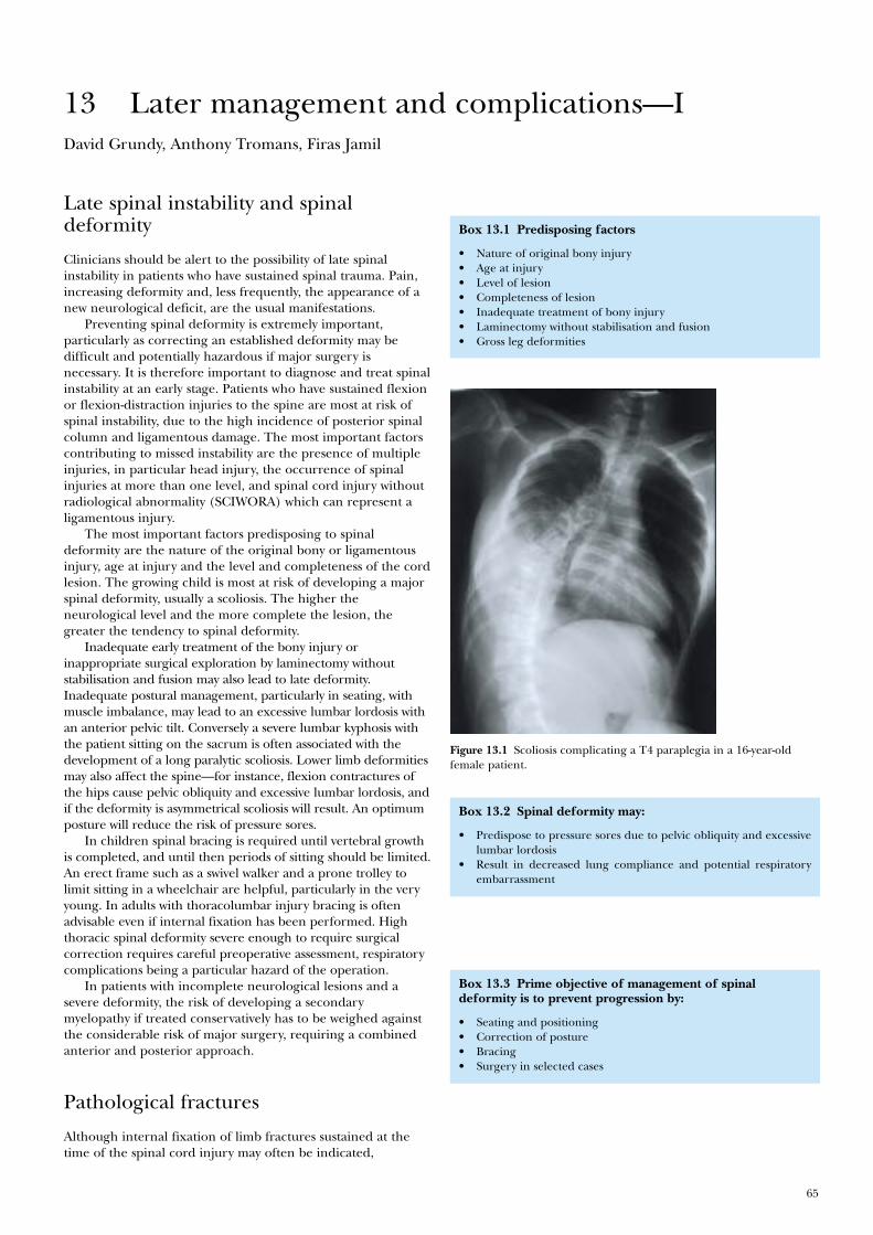
65
Figure 13.1 Scoliosis complicating a T4 paraplegia in a 16-year-oldfemale patient.
David Grundy, Anthony Tromans, Firas Jamil
Late spinal instability and spinaldeformity
Clinicians should be alert to the possibility of late spinalinstability in patients who have sustained spinal trauma. Pain,increasing deformity and, less frequently, the appearance of anew neurological deficit, are the usual manifestations.
Preventing spinal deformity is extremely important,particularly as correcting an established deformity may bedifficult and potentially hazardous if major surgery isnecessary. It is therefore important to diagnose and treat spinalinstability at an early stage. Patients who have sustained flexionor flexion-distraction injuries to the spine are most at risk ofspinal instability, due to the high incidence of posterior spinalcolumn and ligamentous damage. The most important factorscontributing to missed instability are the presence of multipleinjuries, in particular head injury, the occurrence of spinalinjuries at more than one level, and spinal cord injury withoutradiological abnormality (SCIWORA) which can represent aligamentous injury.
The most important factors predisposing to spinaldeformity are the nature of the original bony or ligamentousinjury, age at injury and the level and completeness of the cordlesion. The growing child is most at risk of developing a majorspinal deformity, usually a scoliosis. The higher theneurological level and the more complete the lesion, thegreater the tendency to spinal deformity.
Inadequate early treatment of the bony injury orinappropriate surgical exploration by laminectomy withoutstabilisation and fusion may also lead to late deformity.Inadequate postural management, particularly in seating, withmuscle imbalance, may lead to an excessive lumbar lordosis withan anterior pelvic tilt. Conversely a severe lumbar kyphosis withthe patient sitting on the sacrum is often associated with thedevelopment of a long paralytic scoliosis. Lower limb deformitiesmay also affect the spine—for instance, flexion contractures ofthe hips cause pelvic obliquity and excessive lumbar lordosis, andif the deformity is asymmetrical scoliosis will result. An optimumposture will reduce the risk of pressure sores.
In children spinal bracing is required until vertebral growthis completed, and until then periods of sitting should be limited.An erect frame such as a swivel walker and a prone trolley tolimit sitting in a wheelchair are helpful, particularly in the veryyoung. In adults with thoracolumbar injury bracing is oftenadvisable even if internal fixation has been performed. Highthoracic spinal deformity severe enough to require surgicalcorrection requires careful preoperative assessment, respiratorycomplications being a particular hazard of the operation.
In patients with incomplete neurological lesions and asevere deformity, the risk of developing a secondarymyelopathy if treated conservatively has to be weighed againstthe considerable risk of major surgery, requiring a combinedanterior and posterior approach.
Pathological fractures
Although internal fixation of limb fractures sustained at thetime of the spinal cord injury may often be indicated,
13 Later management and complications—I
Box 13.1 Predisposing factors
• Nature of original bony injury• Age at injury• Level of lesion• Completeness of lesion• Inadequate treatment of bony injury• Laminectomy without stabilisation and fusion• Gross leg deformities
Box 13.2 Spinal deformity may:
• Predispose to pressure sores due to pelvic obliquity and excessivelumbar lordosis
• Result in decreased lung compliance and potential respiratoryembarrassment
Box 13.3 Prime objective of management of spinaldeformity is to prevent progression by:
• Seating and positioning• Correction of posture• Bracing• Surgery in selected cases
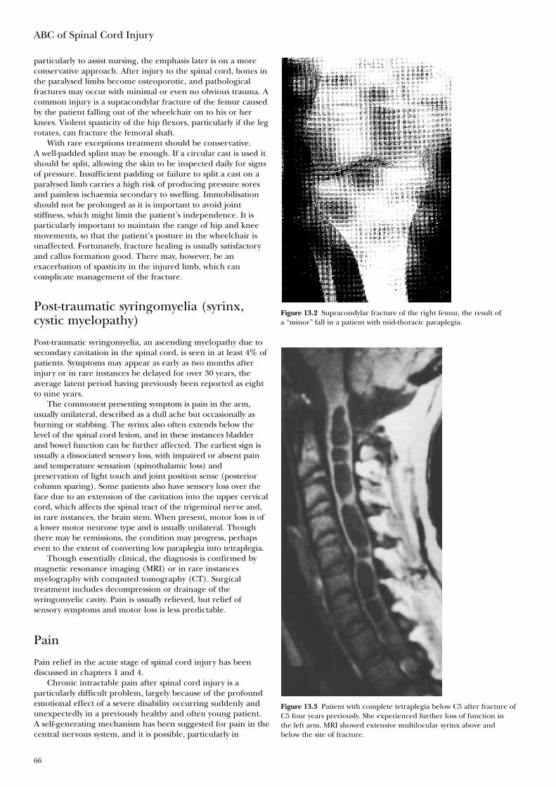
ABC of Spinal Cord Injury
66
particularly to assist nursing, the emphasis later is on a moreconservative approach. After injury to the spinal cord, bones inthe paralysed limbs become osteoporotic, and pathologicalfractures may occur with minimal or even no obvious trauma. Acommon injury is a supracondylar fracture of the femur causedby the patient falling out of the wheelchair on to his or herknees. Violent spasticity of the hip flexors, particularly if the legrotates, can fracture the femoral shaft.
With rare exceptions treatment should be conservative.A well-padded splint may be enough. If a circular cast is used itshould be split, allowing the skin to be inspected daily for signsof pressure. Insufficient padding or failure to split a cast on aparalysed limb carries a high risk of producing pressure soresand painless ischaemia secondary to swelling. Immobilisationshould not be prolonged as it is important to avoid jointstiffness, which might limit the patient’s independence. It isparticularly important to maintain the range of hip and kneemovements, so that the patient’s posture in the wheelchair isunaffected. Fortunately, fracture healing is usually satisfactoryand callus formation good. There may, however, be anexacerbation of spasticity in the injured limb, which cancomplicate management of the fracture.
Post-traumatic syringomyelia (syrinx,cystic myelopathy)
Post-traumatic syringomyelia, an ascending myelopathy due tosecondary cavitation in the spinal cord, is seen in at least 4% ofpatients. Symptoms may appear as early as two months afterinjury or in rare instances be delayed for over 30 years, theaverage latent period having previously been reported as eightto nine years.
The commonest presenting symptom is pain in the arm,usually unilateral, described as a dull ache but occasionally asburning or stabbing. The syrinx also often extends below thelevel of the spinal cord lesion, and in these instances bladderand bowel function can be further affected. The earliest sign isusually a dissociated sensory loss, with impaired or absent painand temperature sensation (spinothalamic loss) andpreservation of light touch and joint position sense (posteriorcolumn sparing). Some patients also have sensory loss over theface due to an extension of the cavitation into the upper cervicalcord, which affects the spinal tract of the trigeminal nerve and,in rare instances, the brain stem. When present, motor loss is ofa lower motor neurone type and is usually unilateral. Thoughthere may be remissions, the condition may progress, perhapseven to the extent of converting low paraplegia into tetraplegia.
Though essentially clinical, the diagnosis is confirmed bymagnetic resonance imaging (MRI) or in rare instancesmyelography with computed tomography (CT). Surgicaltreatment includes decompression or drainage of thesyringomyelic cavity. Pain is usually relieved, but relief ofsensory symptoms and motor loss is less predictable.
Pain
Pain relief in the acute stage of spinal cord injury has beendiscussed in chapters 1 and 4.
Chronic intractable pain after spinal cord injury is aparticularly difficult problem, largely because of the profoundemotional effect of a severe disability occurring suddenly andunexpectedly in a previously healthy and often young patient.A self-generating mechanism has been suggested for pain in thecentral nervous system, and it is possible, particularly in
Figure 13.2 Supracondylar fracture of the right femur, the result ofa “minor” fall in a patient with mid-thoracic paraplegia.
Figure 13.3 Patient with complete tetraplegia below C5 after fracture ofC5 four years previously. She experienced further loss of function inthe left arm. MRI showed extensive multilocular syrinx above andbelow the site of fracture.
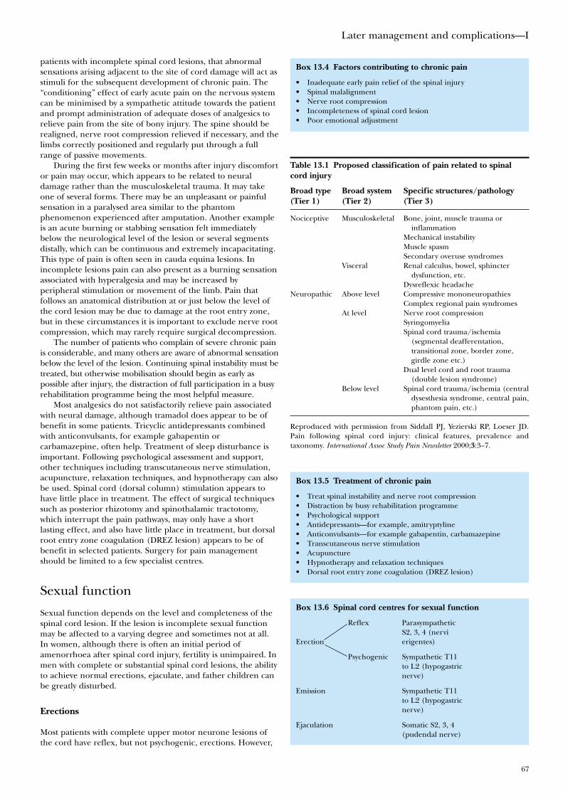
Later management and complications—I
67
patients with incomplete spinal cord lesions, that abnormalsensations arising adjacent to the site of cord damage will act asstimuli for the subsequent development of chronic pain. The“conditioning” effect of early acute pain on the nervous systemcan be minimised by a sympathetic attitude towards the patientand prompt administration of adequate doses of analgesics torelieve pain from the site of bony injury. The spine should berealigned, nerve root compression relieved if necessary, and thelimbs correctly positioned and regularly put through a fullrange of passive movements.
During the first few weeks or months after injury discomfortor pain may occur, which appears to be related to neuraldamage rather than the musculoskeletal trauma. It may takeone of several forms. There may be an unpleasant or painfulsensation in a paralysed area similar to the phantomphenomenon experienced after amputation. Another exampleis an acute burning or stabbing sensation felt immediatelybelow the neurological level of the lesion or several segmentsdistally, which can be continuous and extremely incapacitating.This type of pain is often seen in cauda equina lesions. Inincomplete lesions pain can also present as a burning sensationassociated with hyperalgesia and may be increased byperipheral stimulation or movement of the limb. Pain thatfollows an anatomical distribution at or just below the level ofthe cord lesion may be due to damage at the root entry zone,but in these circumstances it is important to exclude nerve rootcompression, which may rarely require surgical decompression.
The number of patients who complain of severe chronic painis considerable, and many others are aware of abnormal sensationbelow the level of the lesion. Continuing spinal instability must betreated, but otherwise mobilisation should begin as early aspossible after injury, the distraction of full participation in a busyrehabilitation programme being the most helpful measure.
Most analgesics do not satisfactorily relieve pain associatedwith neural damage, although tramadol does appear to be ofbenefit in some patients. Tricyclic antidepressants combinedwith anticonvulsants, for example gabapentin orcarbamazepine, often help. Treatment of sleep disturbance isimportant. Following psychological assessment and support,other techniques including transcutaneous nerve stimulation,acupuncture, relaxation techniques, and hypnotherapy can alsobe used. Spinal cord (dorsal column) stimulation appears tohave little place in treatment. The effect of surgical techniquessuch as posterior rhizotomy and spinothalamic tractotomy,which interrupt the pain pathways, may only have a shortlasting effect, and also have little place in treatment, but dorsalroot entry zone coagulation (DREZ lesion) appears to be ofbenefit in selected patients. Surgery for pain managementshould be limited to a few specialist centres.
Sexual function
Sexual function depends on the level and completeness of thespinal cord lesion. If the lesion is incomplete sexual functionmay be affected to a varying degree and sometimes not at all.In women, although there is often an initial period ofamenorrhoea after spinal cord injury, fertility is unimpaired. Inmen with complete or substantial spinal cord lesions, the abilityto achieve normal erections, ejaculate, and father children canbe greatly disturbed.
Erections
Most patients with complete upper motor neurone lesions ofthe cord have reflex, but not psychogenic, erections. However,
Box 13.4 Factors contributing to chronic pain
• Inadequate early pain relief of the spinal injury• Spinal malalignment• Nerve root compression• Incompleteness of spinal cord lesion• Poor emotional adjustment
Box 13.5 Treatment of chronic pain
• Treat spinal instability and nerve root compression• Distraction by busy rehabilitation programme• Psychological support• Antidepressants—for example, amitryptyline• Anticonvulsants—for example gabapentin, carbamazepine• Transcutaneous nerve stimulation• Acupuncture• Hypnotherapy and relaxation techniques• Dorsal root entry zone coagulation (DREZ lesion)
Box 13.6 Spinal cord centres for sexual function
Reflex ParasympatheticS2, 3, 4 (nervi
Erection erigentes)
Psychogenic Sympathetic T11to L2 (hypogastricnerve)
Emission Sympathetic T11to L2 (hypogastricnerve)
Ejaculation Somatic S2, 3, 4(pudendal nerve)
Table 13.1 Proposed classification of pain related to spinalcord injury
Broad type Broad system Specific structures/pathology(Tier 1) (Tier 2) (Tier 3)
Nociceptive Musculoskeletal Bone, joint, muscle trauma orinflammation
Mechanical instabilityMuscle spasmSecondary overuse syndromes
Visceral Renal calculus, bowel, sphincterdysfunction, etc.
Dysreflexic headacheNeuropathic Above level Compressive mononeuropathies
Complex regional pain syndromesAt level Nerve root compression
SyringomyeliaSpinal cord trauma/ischemia
(segmental deafferentation,transitional zone, border zone,girdle zone etc.)
Dual level cord and root trauma(double lesion syndrome)
Below level Spinal cord trauma/ischemia (centraldysesthesia syndrome, central pain,phantom pain, etc.)
Reproduced with permission from Siddall PJ, Yezierski RP, Loeser JD.Pain following spinal cord injury: clinical features, prevalence andtaxonomy. International Assoc Study Pain Newsletter 2000;3:3–7.
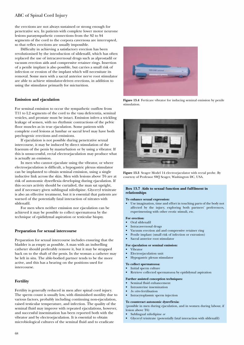
ABC of Spinal Cord Injury
68
the erections are not always sustained or strong enough forpenetrative sex. In patients with complete lower motor neuronelesions parasympathetic connections from the S2 to S4segments of the cord to the corpora cavernosa are interrupted,so that reflex erections are usually impossible.
Difficulty in achieving a satisfactory erection has beenrevolutionised by the introduction of sildenafil, which has oftenreplaced the use of intracavernosal drugs such as alprostadil orvacuum erection aids and compressive retainer rings. Insertionof a penile implant is also possible, but carries a small risk ofinfection or erosion of the implant which will necessitate itsremoval. Some men with a sacral anterior nerve root stimulatorare able to achieve stimulator-driven erections, in addition tousing the stimulator primarily for micturition.
Emission and ejaculation
For seminal emission to occur the sympathetic outflow fromT11 to L2 segments of the cord to the vasa deferentia, seminalvesicles, and prostate must be intact. Emission infers a tricklingleakage of semen, with no rhythmic contractions of the pelvicfloor muscles as in true ejaculation. Some patients withcomplete cord lesions at lumbar or sacral level may have bothpsychogenic erections and emissions.
If ejaculation is not possible during penetrative sexualintercourse, it may be induced by direct stimulation of thefraenum of the penis by masturbation or by using a vibrator. Ifthis is unsuccessful, rectal electroejaculation may produce whatis actually an emission.
In men who cannot ejaculate using the vibrator, or whereelectroejaculation is difficult, a hypogastric plexus stimulatorcan be implanted to obtain seminal emission, using a singleinductive link across the skin. Men with lesions above T6 are atrisk of autonomic dysreflexia developing during ejaculation. Ifthis occurs activity should be curtailed, the man sat upright,and if necessary given sublingual nifedipine. Glyceryl trinitrateis also an effective treatment, but it is essential that patients arewarned of the potentially fatal interaction of nitrates withsildenafil.
For men when neither emission nor ejaculation can beachieved it may be possible to collect spermatozoa by thetechnique of epididymal aspiration or testicular biopsy.
Preparation for sexual intercourse
Preparation for sexual intercourse includes ensuring that thebladder is as empty as possible. A man with an indwellingcatheter should preferably remove it, but it may be strappedback on to the shaft of the penis. In the woman a catheter maybe left in situ. The able-bodied partner tends to be the moreactive, and this has a bearing on the positions used forintercourse.
Fertility
Fertility is generally reduced in men after spinal cord injury.The sperm count is usually low, with diminished motility due tovarious factors, probably including continuing non-ejaculation,raised testicular temperature, and infection. The quality of theseminal fluid may improve with repeated ejaculations, however,and successful insemination has been reported both with thevibrator and by electroejaculation. It is essential to obtainmicrobiological cultures of the seminal fluid and to eradicate
Figure 13.4 Ferticare vibrator for inducing seminal emission by penilestimulation.
Figure 13.5 Seager Model 14 electroejaculator with rectal probe. Bycourtesy of Professor SWJ Seager, Washington DC, USA.
Box 13.7 Aids to sexual function and fulfilment inrelationships
To enhance sexual expression:• Use imagination, time and effort in touching parts of the body not
affected by the injury, exploring both partners’ preferences,experimenting with other erotic stimuli, etc.
For erection:• Oral sildenafil• Intracavernosal drugs• Vacuum erection aid and compressive retainer ring• Penile implant (small risk of infection or extrusion)• Sacral anterior root stimulator
For ejaculation or seminal emission:• Vibrator• Electroejaculation unit• Hypogastric plexus stimulator
To collect spermatozoa:• Initial sperm culture• Retrieve collected spermatozoa by epididymal aspiration
Further assisted conception techniques:• Seminal fluid enhancement• Intrauterine insemination• In vitro fertilisation• Intracytoplasmic sperm injection
To counteract autonomic dysreflexia:(possible in men during ejaculation, and in women during labour, iflesion above T6)• Sublingual nifedipine or• Glyceryl trinitrate (potentially fatal interaction with sildenafil)
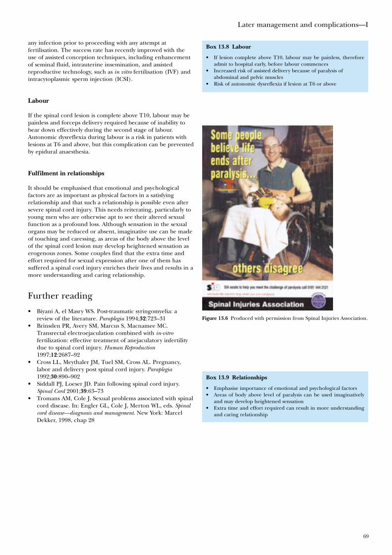
Later management and complications—I
69
any infection prior to proceeding with any attempt atfertilisation. The success rate has recently improved with theuse of assisted conception techniques, including enhancementof seminal fluid, intrauterine insemination, and assistedreproductive technology, such as in vitro fertilisation (IVF) andintracytoplasmic sperm injection (ICSI).
Labour
If the spinal cord lesion is complete above T10, labour may bepainless and forceps delivery required because of inability tobear down effectively during the second stage of labour.Autonomic dysreflexia during labour is a risk in patients withlesions at T6 and above, but this complication can be preventedby epidural anaesthesia.
Fulfilment in relationships
It should be emphasised that emotional and psychologicalfactors are as important as physical factors in a satisfyingrelationship and that such a relationship is possible even aftersevere spinal cord injury. This needs reiterating, particularly toyoung men who are otherwise apt to see their altered sexualfunction as a profound loss. Although sensation in the sexualorgans may be reduced or absent, imaginative use can be madeof touching and caressing, as areas of the body above the levelof the spinal cord lesion may develop heightened sensation aserogenous zones. Some couples find that the extra time andeffort required for sexual expression after one of them hassuffered a spinal cord injury enriches their lives and results in amore understanding and caring relationship.
Further reading
• Biyani A, el Masry WS. Post-traumatic syringomyelia: areview of the literature. Paraplegia 1994;32:723–31
• Brinsden PR, Avery SM, Marcus S, Macnamee MC.Transrectal electroejaculation combined with in-vitrofertilization: effective treatment of anejaculatory infertilitydue to spinal cord injury. Human Reproduction1997;12:2687–92
• Cross LL, Meythaler JM, Tuel SM, Cross AL. Pregnancy,labor and delivery post spinal cord injury. Paraplegia1992;30:890–902
• Siddall PJ, Loeser JD. Pain following spinal cord injury.Spinal Cord 2001;39:63–73
• Tromans AM, Cole J. Sexual problems associated with spinalcord disease. In: Engler GL, Cole J, Merton WL, eds. Spinalcord disease—diagnosis and management. New York: MarcelDekker, 1998, chap 28
Box 13.8 Labour
• If lesion complete above T10, labour may be painless, thereforeadmit to hospital early, before labour commences
• Increased risk of assisted delivery because of paralysis ofabdominal and pelvic muscles
• Risk of autonomic dysreflexia if lesion at T6 or above
Box 13.9 Relationships
• Emphasise importance of emotional and psychological factors• Areas of body above level of paralysis can be used imaginatively
and may develop heightened sensation• Extra time and effort required can result in more understanding
and caring relationship
Figure 13.6 Produced with permission from Spinal Injuries Association.
70
Later respiratory management ofhigh tetraplegia
As a result of improved first aid at the scenes of accidents anincreasing number of patients are surviving with neurologicallesions above C4, who have therefore lost diaphragmaticfunction and can no longer breathe. These patients requirelong-term ventilatory support. Modern portable ventilators thatuse a 12-volt battery can be mounted on a wheelchair, allowingthe patient a degree of freedom and independence. Speech ispossible with an uncuffed tracheostomy tube around which aircan escape to the larynx.
In a small number of these patients the anterior horn cellsof the phrenic nerve are spared and it may be possible toimplant a phrenic nerve stimulator to achieve ventilation. Theadvantages of electrophrenic respiration are that it is morephysiological than positive pressure ventilation and it gives thepatient more freedom, the equipment being much lighter thana mechanical ventilator. The disadvantages are that stimulationoften cannot be sustained for 24 hours, a rest period overnightis necessary on a mechanical ventilator, and the implant isexpensive. The long-term ventilator-dependent patient needs24-hour care by a team of carers competent to undertakeendotracheal suction, but not necessarily including aqualified nurse.
Psychological factors
In the acute stage of spinal cord injury an individual mayexperience a range of emotions such as numbness, despair,fear, hope, and anger. This emotional turmoil is often chaoticand disorganised. It may be further complicated by theenforced period of bed rest during which a state of sensorydeprivation ensues. Following this early period anxiety anddepression may become apparent in approximately one thirdof individuals. The frustrations associated with the physicallimitations of such a severe injury are compounded by the factthat most patients are young and before injury led active lives,often expressing themselves mainly through physical activities.The sudden inability to continue in this manner and the needto lead a more ordered life can mean a very difficult andprolonged period of adjustment. Failure to recognise that thisprocess can continue for as long as two or three years maydamage the process of rehabilitation and the patient’s ultimateresettlement. The patient needs time to come to terms with hisor her new status and to make decisions about the futurewithout undue pressure.
Detection of these psychological problems is vital in orderto make appropriate referral. Psychological support andtherapy has been shown to be very effective in improvingmood and also later adjustment in individuals with spinal cordinjury. Other psychological problems that may be presentfollowing injury include post-traumatic stress disorder in whichan individual continually relives their accident and markedproblems with memory, concentration, and problem solving.
Detection of these problems and the provision ofpsychological therapy will enable an individual to overcome
14 Later management and complications—IIDavid Grundy, Anthony Tromans, John Hobby, Nigel North, Ian Swain
Box 14.2 Chronic ventilation
• Electrophrenic respiration (diaphragmatic pacing)• Mechanical “domiciliary” ventilation
Advantages of electrophrenic respiration• More physiological than positive pressure ventilation• More portable than positive pressure ventilation
Drawbacks of electrophrenic respiration• Often stimulation cannot be sustained, necessitating rest period
overnight on mechanical ventilator• More expensive
Box 14.3 Psychological factors
Acute stage:• Initial stress reaction• Sensory deprivation
Later stages:• Anxiety and depression• Post-traumatic stress disorder• Cognitive problems• Detecting psychological problems• Appropriate referral• Family• Psychological support and therapy
Box 14.1 High tetraplegia
• Improved first aid has increased number of high tetraplegicssurviving scene of accident
• If lesion above C4, diaphragmatic function lost
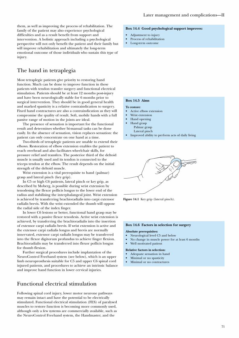
Later management and complications—II
71
them, as well as improving the process of rehabilitation. Thefamily of the patient may also experience psychologicaldifficulties and as a result benefit from support andintervention. A holistic approach including a psychologicalperspective will not only benefit the patient and their family butwill improve rehabilitation and ultimately the long-termemotional outcome of those individuals who sustain this type ofinjury.
The hand in tetraplegia
Most tetraplegic patients give priority to restoring handfunction. Much can be done to improve function in thesepatients with tendon transfer surgery and functional electricalstimulation. Patients should be at least 12 months post-injuryand have been neurologically stable for 6 months prior tosurgical intervention. They should be in good general healthand marked spasticity is a relative contraindication to surgery.Fixed hand contractures are also a contraindication as they willcompromise the quality of result. Soft, mobile hands with a fullpassive range of motion in the joints are ideal.
The presence of sensation is important for the functionalresult and determines whether bi-manual tasks can be doneeasily. In the absence of sensation, vision replaces sensation: thepatient can only concentrate on one hand at a time.
Two-thirds of tetraplegic patients are unable to extend theirelbows. Restoration of elbow extension enables the patient toreach overhead and also facilitates wheelchair skills, forpressure relief and transfers. The posterior third of the deltoidmuscle is usually used and its tendon is connected to thetriceps tendon at the elbow. The result depends on the initialstrength of the deltoid muscle.
Wrist extension is a vital prerequisite to hand (palmar)grasp and lateral pinch (key grip).
In C5 or high C6 patients, lateral pinch or key grip, asdescribed by Moberg, is possible during wrist extension bytenodesing the flexor pollicis longus to the lower end of theradius and stabilising the interphalangeal joint. Wrist extensionis achieved by transferring brachioradialis into carpi extensorradialis brevis. With the wrist extended the thumb will opposethe radial side of the index finger.
In lower C6 lesions or better, functional hand grasp may berestored with a passive flexor tenodesis. Active wrist extension isachieved, by transferring the brachioradialis into the insertionof extensor carpi radialis brevis. If wrist extension is active andthe extensor carpi radialis longus and brevis are normallyinnervated, extensor carpi radialis longus may be transferredinto the flexor digitorum profundus to achieve finger flexion.Brachioradialis may be transferred into flexor pollicis longusfor thumb flexion.
Further surgical procedures include implantation of theNeuroControl Freehand system (see below), which is an upperlimb neuroprosthesis suitable for C5 and upper C6 spinal cordinjured patients, and procedures to achieve an intrinsic balanceand improve hand function in lower cervical injuries.
Functional electrical stimulation
Following spinal cord injury, lower motor neurone pathwaysmay remain intact and have the potential to be electricallystimulated. Functional electrical stimulation (FES) of paralysedmuscles to restore function is becoming more commonly used,although only a few systems are commercially available, such asthe NeuroControl Freehand system, the Handmaster, and the
Box 14.5 Aims
To restore:• Active elbow extension• Wrist extension• Hand opening• Hand grasp
Palmar graspLateral pinch
• Improved ability to perform acts of daily living
Figure 14.1 Key grip (lateral pinch).
Box 14.6 Factors in selection for surgery
Absolute prerequisites:• Neurological level C5 and below• No change in muscle power for at least 6 months• Well motivated patient
Relative factors in selection:• Adequate sensation in hand• Minimal or no spasticity• Minimal or no contractures
Box 14.4 Good psychological support improves:
• Adjustment to injury• Process of rehabilitation• Long-term outcome
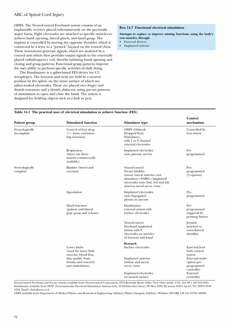
ABC of Spinal Cord Injury
72
ODFS. The NeuroControl Freehand system consists of animplantable receiver placed subcutaneously on the pectoralismajor fascia. Eight electrodes are attached to specific muscles toachieve hand opening, lateral pinch, and hand grasp. Theimplant is controlled by moving the opposite shoulder, which isconnected by a lever to a “joystick” located on the central chest.These movements generate signals, which are analysed by acontrol unit which then provides output signals to the externallyplaced radiofrequency coil, thereby initiating hand opening andclosing and grasp patterns. Functional grasp patterns improvethe user ability to perform specific activities of daily living.
The Handmaster is a splint-based FES device for C5tetraplegics. The forearm and wrist are held in a neutralposition by the splint, on the inner surface of which aresaline-soaked electrodes. These are placed over finger andthumb extensors and a thumb abductor, using pre-set patternsof stimulation to open and close the hand. The system isdesigned for holding objects such as a fork or pen.
Table 14.1 The practical uses of electrical stimulation to achieve function (FES)
ControlPatient group Stimulated function Stimulator type mechanism
Neurologically Control of foot drop ODFS (Odstock Controlled byincomplete �\� knee extension, Dropped Foot foot switch
hip extension Stimulator),with 1 or 2 channelexternal electrodes
Respiration Implanted electrodes Pre-(there are three onto phrenic nerves programmedsystems commerciallyavailable)
Neurologically Bladder (bowel and NeuroControl Pre-complete erection) Vocare bladder programmed
system (sacral anterior root (3 options)stimulator—SARS)—implanted electrodes onto 2nd, 3rd and 4th anterior sacral nerve roots
Ejaculation Implanted electrodes Pre-onto hypogastric programmedplexus on sacrum
Hand function Handmaster Pre-(palmar and lateral external system with programmedgrip, grasp and release) surface electrodes triggered by
pressing button
NeuroControl JoystickFreehand implanted attached tosystem with 8 contralateralelectrodes on muscles shoulderof forearm and hand
ResearchLower limbs Surface electrodes External feed(used for lower limb back controlexercise, blood flow, systemskin quality, bone Implanted anterior External multi-density and research lumbar and sacral option pre-into ambulation) nerve roots programmed
controllerImplanted electrodes Externalon muscle surface controller
NeuroControl Freehand and Vocare systems available from NeuroControl Corporation, 8333 Rockside Road, Valley View, Ohio 44125, USA. Tel: 00 1 216 912 0101.Handmaster available from NESS (Neuromuscular Electrical Stimulation Systems Ltd), 19 Ha-Haroshet Street, PO Box 2500, Ra’anana 43654, Israel. Tel: 00972 97485738. Email: [email protected] available from Department of Medical Physics and Biomedical Engineering, Salisbury District Hospital, Salisbury, Wiltshire SP2 8BJ, UK Tel: 01722 429065.
Box 14.7 Functional electrical stimulation
Attempts to replace or improve missing functions, using the body’sown muscles, through:• External devices• Implanted systems
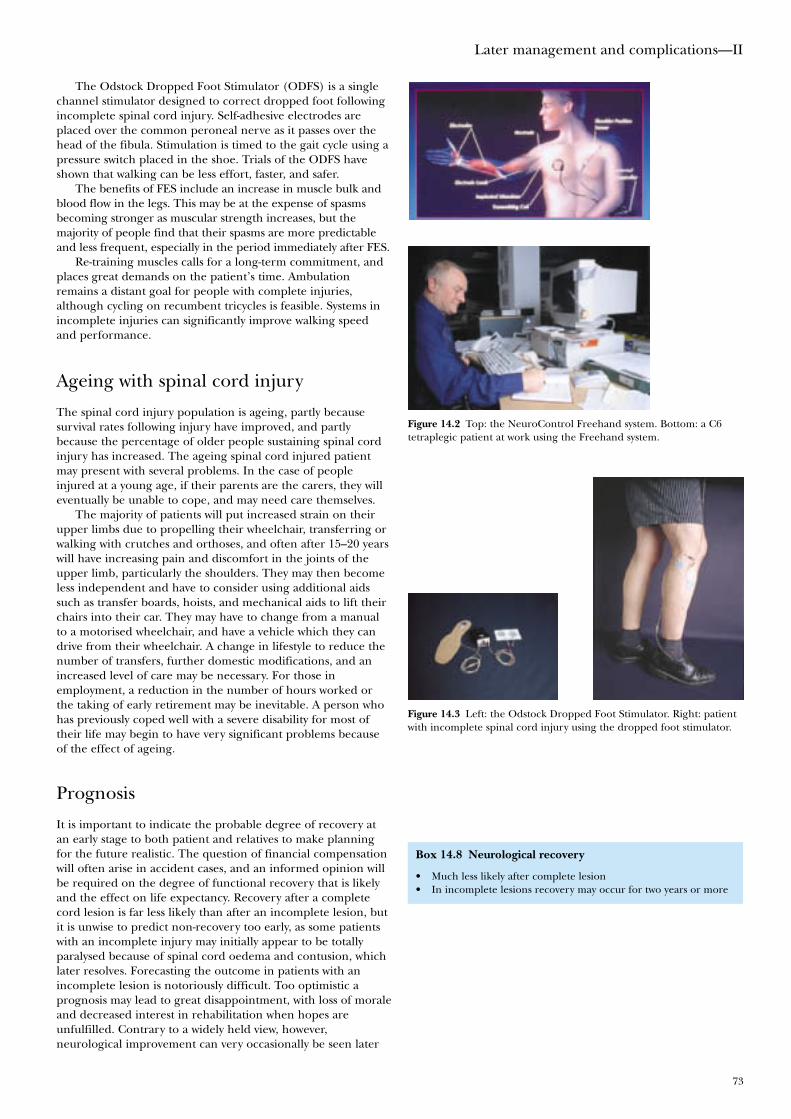
Later management and complications—II
73
The Odstock Dropped Foot Stimulator (ODFS) is a singlechannel stimulator designed to correct dropped foot followingincomplete spinal cord injury. Self-adhesive electrodes areplaced over the common peroneal nerve as it passes over thehead of the fibula. Stimulation is timed to the gait cycle using apressure switch placed in the shoe. Trials of the ODFS haveshown that walking can be less effort, faster, and safer.
The benefits of FES include an increase in muscle bulk andblood flow in the legs. This may be at the expense of spasmsbecoming stronger as muscular strength increases, but themajority of people find that their spasms are more predictableand less frequent, especially in the period immediately after FES.
Re-training muscles calls for a long-term commitment, andplaces great demands on the patient’s time. Ambulationremains a distant goal for people with complete injuries,although cycling on recumbent tricycles is feasible. Systems inincomplete injuries can significantly improve walking speedand performance.
Ageing with spinal cord injury
The spinal cord injury population is ageing, partly becausesurvival rates following injury have improved, and partlybecause the percentage of older people sustaining spinal cordinjury has increased. The ageing spinal cord injured patientmay present with several problems. In the case of peopleinjured at a young age, if their parents are the carers, they willeventually be unable to cope, and may need care themselves.
The majority of patients will put increased strain on theirupper limbs due to propelling their wheelchair, transferring orwalking with crutches and orthoses, and often after 15–20 yearswill have increasing pain and discomfort in the joints of theupper limb, particularly the shoulders. They may then becomeless independent and have to consider using additional aidssuch as transfer boards, hoists, and mechanical aids to lift theirchairs into their car. They may have to change from a manualto a motorised wheelchair, and have a vehicle which they candrive from their wheelchair. A change in lifestyle to reduce thenumber of transfers, further domestic modifications, and anincreased level of care may be necessary. For those inemployment, a reduction in the number of hours worked orthe taking of early retirement may be inevitable. A person whohas previously coped well with a severe disability for most oftheir life may begin to have very significant problems becauseof the effect of ageing.
Prognosis
It is important to indicate the probable degree of recovery atan early stage to both patient and relatives to make planningfor the future realistic. The question of financial compensationwill often arise in accident cases, and an informed opinion willbe required on the degree of functional recovery that is likelyand the effect on life expectancy. Recovery after a completecord lesion is far less likely than after an incomplete lesion, butit is unwise to predict non-recovery too early, as some patientswith an incomplete injury may initially appear to be totallyparalysed because of spinal cord oedema and contusion, whichlater resolves. Forecasting the outcome in patients with anincomplete lesion is notoriously difficult. Too optimistic aprognosis may lead to great disappointment, with loss of moraleand decreased interest in rehabilitation when hopes areunfulfilled. Contrary to a widely held view, however,neurological improvement can very occasionally be seen later
Figure 14.2 Top: the NeuroControl Freehand system. Bottom: a C6tetraplegic patient at work using the Freehand system.
Figure 14.3 Left: the Odstock Dropped Foot Stimulator. Right: patientwith incomplete spinal cord injury using the dropped foot stimulator.
Box 14.8 Neurological recovery
• Much less likely after complete lesion• In incomplete lesions recovery may occur for two years or more
ABC of Spinal Cord Injury
74
than two years after injury, not only with nerve root and caudaequina lesions but also with cord injuries.
Mortality in acutely injured patients managed in a spinalinjuries unit is now less than 5%. Death within the first few daysis likely to be from respiratory failure, particularly in hightetraplegia. The presence of multiple injuries, age, andprevious health of the patient all play a part. In patientssurviving the period immediately after injury pulmonaryembolism is still the commonest cause of death in the acutephase.
With the modern management of spinal cord injury,particularly improvements in the management of the urinarytract and pressure sore prevention, life expectancy hasimproved over recent years; as a consequence pathologiesexperienced by the general population such as atherosclerosisand its complications, and malignancy, are now major causes oflate death, as well as respiratory causes, particularly intetraplegic patients.
Great progress has been made in the care of patients withspinal cord injuries since the 1940s, when spinal injuries unitswere first established. There has been a remarkable decrease incomplications by using the multidisciplinary approach providedby such units, yet some patients are still denied referral. Unlesscomplete recovery occurs, patients should have lifelonghospital outpatient follow up but with emphasis on continuingcare and support in the community.
Although it is right to be optimistic about the future ofthese patients, their injuries can make a devastating change totheir lives. In many cases the injuries need not have happened.For example, a high proportion of road traffic accidents iscaused by alcohol consumption, high speeds, and dangerousdriving, motorcyclists being particularly vulnerable. Ignoranceof the danger of diving into shallow water results in manyinjuries to the cervical spine. Failure to take simple precautionsin the home, such as ensuring that stairs are adequately lit atnight for the elderly, may result in falls with cervicalhyperextension injuries. Carelessness in contact sports can leadto serious injury. Recognition of this fact has led responsibleauthorities such as the Rugby Football Union to modify the lawsof the game and issue advice on how it can be made safer, but
Table 14.2 Life expectancy in years for people with spinal cord injuries who survive at least one yearafter injury, according to current age and neurological category (Frankel grades—see box below)
Current C1–C4 C5–C8 T1–S5age (Frankel grade (Frankel grade (Frankel grade (Frankel(years) Normal* A, B, C) A, B, C) A, B, C) grade D)
5 70.8 45.0 52.0 59.5 63.010 65.9 40.5 47.3 53.7 58.215 61.0 36.1 42.6 49.0 53.420 56.3 32.8 38.6 44.8 49.025 51.6 29.9 34.7 40.8 44.730 46.9 26.8 30.7 36.7 40.535 42.2 23.7 27.0 32.7 36.140 37.6 20.9 23.6 28.8 31.745 33.0 18.4 20.4 25.1 27.550 28.6 15.5 17.0 21.2 23.455 24.4 12.8 13.8 17.3 19.560 20.5 11.0 11.2 13.8 15.965 16.9 8.8 8.8 10.9 13.270 13.6 6.6 6.6 8.3 10.475 10.7 4.7 4.7 6.1 8.080 8.1 3.1 3.1 4.2 6.1
*Normal values are from 1988 United States life tables for the general population.Taken from DeVivo MJ, Stover SL, Long-term survival and causes of death.In: Stover SL, et al. eds., Spinal cord injury. Clinical outcomes from the model systems. Gaithersburg: Aspen Publishers, 1995.
Box 14.9 Frankel grades
A “Complete”—total motor and sensory lossB “Sensory only”—sensory sparingC “Motor useless”—motor sparing of no functional valueD “Motor useful”—motor sparing of functional valueE “Recovery”—no functional deficit
From Frankel HL, Hancock DO, Hyslop G, et al. The value of posturalreduction in the initial management of closed injuries of the spine withparaplegia and tetraplegia. Paraplegia 1969;7:179–92
Box 14.10 Many injuries are preventable
• Road traffic accidents associated with alcohol consumption anddangerous driving
• Diving into shallow water, resulting in tetraplegia• Contact sports, e.g., rugby• Some injuries are made worse by mishandling
Later management and complications—II
75
much more could be done in other aspects of accidentprevention, for instance in horse riding.
Finally, those who work with patients with spinal cordinjuries are often impressed by the surprisingly high quality oflife possible after injury. Many achieve a remarkable degree ofindependence, earn their own living, choose to marry, havechildren, and participate fully in family life. They may indeedhave special qualities because they have successfully come toterms with their disability, and many will make a valuablecontribution to society.
Further reading
• Brindley GS. The first 500 patients with sacral anterior rootstimulator implants: general description. Paraplegia1994;32:795–805
• Glass C. Spinal cord injury: impact and coping. Leicester: BPSBooks, 1999
• North NT. The psychological effects of spinal cord injury: areview. Spinal Cord 1999;37:671–79
• Stover SL, DeLisa JA, Whiteneck GC, eds. Spinal cord injury.Clinical outcomes from the model systems. Gaithersburg: AspenPublishers, 1995
• Whiteneck GC, Charlifue SW, Gerhart KA, Lammertse DP et al., eds. Aging with spinal cord injury. New York: Demos,1993
• Functional electrical stimulation: sources of information:<www.salisburyfes.com> FES clinical service and research atSalisbury District Hospital. Good links to other sites.<www.fes.cwru.edu> General FES information
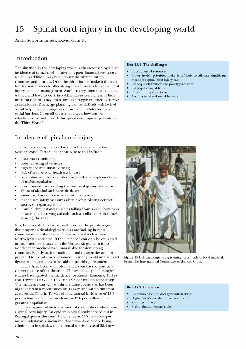
76
Anba Soopramanien, David Grundy
Introduction
The situation in the developing world is characterised by a highincidence of spinal cord injuries and poor financial resources,which, in addition, may be unevenly distributed withincountries and districts. Other health priorities make it difficultfor decision makers to allocate significant means for spinal cordinjury care and management. Staff are very often inadequatelytrained and have to work in a difficult environment with littlefinancial reward. They often have to struggle in order to surviveas individuals. Discharge planning can be difficult with lack ofsocial help, poor housing conditions, and architectural andsocial barriers. Given all these challenges, how can weeffectively care and provide for spinal cord injured patients inthe Third World?
Incidence of spinal cord injury
The incidence of spinal cord injury is higher than in thewestern world. Factors that contribute to this include:
• poor road conditions• poor servicing of vehicles• high speed and unsafe driving• lack of seat belts or headrests in cars• corruption and bribery interfering with the implementation
of traffic regulations• overcrowded cars, shifting the centre of gravity of the cars• abuse of alcohol and narcotic drugs• widespread use of firearms in certain cultures• inadequate safety measures when diving, playing contact
sports, or repairing roofs• unusual circumstances such as falling from a cart, from trees
or accidents involving animals such as collisions with camelscrossing the road.
It is, however, difficult to know the size of the problem giventhat proper epidemiological studies are lacking in mostcountries except the United States, where data has beenrelatively well collected. If the incidence can only be estimatedin countries like France and the United Kingdom, it is nowonder that precise data is unavailable for developingcountries. Rightly so, international funding agencies are notprepared to spend scarce resources in trying to obtain the exactfigures when stress has to be laid on providing treatment.
There have been attempts in a few countries to portray aclearer picture of the situation. The available epidemiologicalstudies have quoted the incidence for Russia, Romania, Turkeyand Taiwan as 29.7, 30, 12.7 and 18.8 per million respectively.The incidence can vary within the same country as has beenhighlighted in a recent study on Turkey, and within differentage groups. Thus in Taiwan with an annual incidence of 18.8per million people, the incidence is 47.6 per million for thegeriatric population.
These figures relate to the survival rate of those who sustaina spinal cord injury. An epidemiological study carried out inPortugal quotes the annual incidence as 57.8 new cases permillion inhabitants, including those who died before beingadmitted to hospital, with an annual survival rate of 25.4 new
15 Spinal cord injury in the developing world
Figure 15.1 A paraplegic using training steps made of local material.From The International Committee of the Red Cross.
Box 15.1 The challenges
• Poor financial resources• Other health priorities make it difficult to allocate significant
means for spinal cord injury care• Inadequately trained and poorly paid staff• Inadequate social help• Poor housing conditions• Architectural and social barriers
Box 15.2 Incidence
• Epidemiological studies generally lacking• Higher incidence than in western world• Mostly paraplegic• Predominantly young males
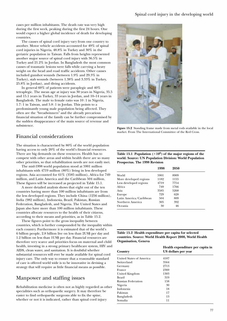
Spinal cord injury in the developing world
77
cases per million inhabitants. The death rate was very highduring the first week, peaking during the first 24 hours. Onewould expect a higher global incidence of death for developingcountries.
The causes of spinal cord injury vary from one country toanother. Motor vehicle accidents accounted for 49% of spinalcord injuries in Nigeria, 48.8% in Turkey and 30% in thegeriatric population in Taiwan. Falls from heights representedanother major source of spinal cord injury with 36.5% inTurkey and 21.2% in Jordan. In Bangladesh the most commoncauses of traumatic lesions were falls while carrying a heavyweight on the head and road traffic accidents. Other causesincluded gunshot wounds (between 1.9% and 29.3% inTurkey), stab wounds (between 1.38% and 3.33% in Turkey,25.8% in Jordan), and diving accidents.
In general 60% of patients were paraplegic and 40%tetraplegic. The mean age at injury was 30 years in Nigeria, 35.5and 15.1 years in Turkey, 33 years in Jordan, and 10–14 years inBangladesh. The male to female ratio was 10 : 1 in Nigeria,1.7 : 1 in Taiwan, and 5.8 : 1 in Jordan. This points to apredominantly young male population being affected. Theyoften are the “breadwinners” and the already precariousfinancial situation of the family can be further compromised bythe sudden disappearance of the main source of revenue andsubsistence.
Financial considerations
The situation is characterised by 80% of the world populationhaving access to only 20% of the world’s financial resources.There are big demands on these resources. Health has tocompete with other areas and within health there are so manyother priorities, so that rehabilitation needs are not easily met.
The mid-1998 world population stood at 5901 millioninhabitants with 4719 million (80%) living in less developedregions. Asia accounted for 61% (3585 million), Africa for 749million, and Latin America and the Caribbean 504 million.These figures will be increased as projected in Table 15.1.
A more detailed analysis shows that eight out of the tencountries having more than 100 million inhabitants are fromthe less developed regions. They include China (1256 million),India (982 million), Indonesia, Brazil, Pakistan, RussianFederation, Bangladesh, and Nigeria. The United States andJapan also have more than 100 million inhabitants. Thesecountries allocate resources to the health of their citizens,according to their means and priorities, as in Table 15.2.
These figures point to the gross inequality betweencountries, which is further compounded by the inequality withineach country. Furthermore it is estimated that of the world’s6 billion people, 2.8 billion live on less than 2US$ per day and1.2 billion on less than 1US$ per day. Financial resources aretherefore very scarce and priorities focus on maternal and childhealth, investing in a strong primary healthcare system, HIV andAIDS, clean water, and sanitation. It is doubtful whethersubstantial resources will ever be made available for spinal cordinjury care. The only way to ensure that a reasonable standardof care is offered world wide is to be innovative in devising astrategy that will require as little financial means as possible.
Manpower and staffing issues
Rehabilitation medicine is often not as highly regarded as otherspecialities such as orthopaedic surgery. It may therefore beeasier to find orthopaedic surgeons able to fix the spine,whether or not it is indicated, rather than spinal cord injury
Figure 15.2 Standing frame made from metal rods available in the localmarket. From The International Committee of the Red Cross.
Table 15.1 Population (�106) of the major regions of theworld. Source: UN Population Division: World PopulationProspectus. The 1998 Revision
1998 2050
World 5901 8909More developed regions 1182 1155Less developed regions 4719 7754Africa 749 1766Asia 3585 5268Europe 729 628Latin America/Caribbean 504 809Northern America 305 392Oceania 30 46
Table 15.2 Health expenditure per capita for selectedcountries. Source: World Health Report 2000, World HealthOrganisation, Geneva
Health expenditure per capita inCountry US dollars per year
United States of America 4187Switzerland 3564Germany 2713France 2369United Kingdom 1303Brazil 319Russian Federation 158Nigeria 30Indonesia 18Pakistan 17Bangladesh 13Somalia 11
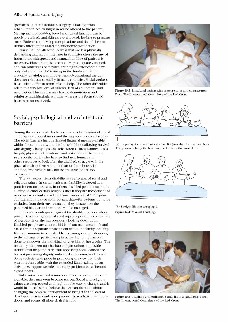
ABC of Spinal Cord Injury
78
specialists. In many instances, surgery is isolated fromrehabilitation, which might never be offered to the patient.Management of bladder, bowel and sexual function can bepoorly organised, and skin care overlooked, leading to pressuresores. Patients can develop complications and die of chest orurinary infection or untreated autonomic dysfunction.
Nurses will be attracted to areas that are less physicallydemanding and labour intensive in countries where the use ofhoists is not widespread and manual handling of patients isnecessary. Physiotherapists are not always adequately trained,and can sometimes be physical training instructors who haveonly had a few months’ training in the fundamentals ofanatomy, physiology, and movement. Occupational therapydoes not exist as a speciality in many countries. Social workershave little to offer in terms of state help. The other difficultiesrelate to a very low level of salaries, lack of equipment, andmedication. This in turn may lead to demotivation andreinforce individualistic attitudes, whereas the focus shouldhave been on teamwork.
Social, psychological and architecturalbarriers
Among the major obstacles to successful rehabilitation of spinalcord injury are social issues and the way society views disability.The social barriers include limited financial means availablewithin the community, and the household not allowing survivalwith dignity; changing social roles when a “breadwinner” loseshis job, physical independence and status within the family;stress on the family who have to find new human andother resources to look after the disabled; struggle with thephysical environment within and around the house. Inaddition, wheelchairs may not be available, or are tooexpensive.
The way society views disability is a reflection of social andreligious values. In certain cultures, disability is viewed as apunishment for past sins. In others, disabled people may not beallowed to enter certain religious sites if they are incontinent ofurine or faeces and considered “unclean or soiled”. Religiousconsiderations may be so important that—for patients not to beexcluded from their environment—they dictate how theparalysed bladder and/or bowel will be managed.
Prejudice is widespread against the disabled person, who ispitied. By acquiring a spinal cord injury, a person becomes partof a group he or she was previously looking down upon.Disabled people are at times hidden from mainstream life andcared for in a separate environment within the family dwelling.It is not common to see a disabled person going out shopping,to the cinema, or participating in active life. Little has beendone to empower the individual or give him or her a voice. Thetendency has been for charitable organisations to provideinstitutional help and care, thus appeasing social conscience,but not promoting dignity, individual expression, and choice.Some societies take pride in promoting the view that theirsystem is acceptable, with the extended family taking up anactive new, supportive role, but many problems exist “behindclosed doors”.
Substantial financial resources are not expected to becomeavailable; they may even become scarcer. Social and religiousvalues are deep-rooted and might not be easy to change, and itwould be unrealistic to believe that we can do much aboutchanging the physical environment to bring it to the level ofdeveloped societies with wide pavements, roads, streets, slopes,doors, and rooms all wheelchair friendly.
Figure 15.3 Emaciated patient with pressure sores and contractures.From The International Committee of the Red Cross.
Figure 15.4 Manual handling.
Figure 15.5 Teaching a co-ordinated spinal lift in a paraplegic. FromThe International Committee of the Red Cross.
(a) Preparing for a coordinated spinal lift (straight lift) in a tetraplegic.The person holding the head and neck directs the procedure.
(b) Straight lift in a tetraplegic.
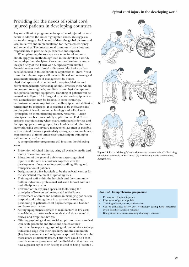
Spinal cord injury in the developing world
79
Providing for the needs of spinal cordinjured patients in developing countries
Any rehabilitation programme for spinal cord injured patientsneeds to address the issues highlighted above. We suggest anational strategy to look at and address the global picture, andlocal initiatives and implementation for increased efficiencyand ownership. The international community has a duty andresponsibility to provide help, expertise and support.
When planning the strategy, care must be taken not toblindly apply the methodology used in the developed world,but to adapt the principles of treatment to take into accountthe specificity of the Third World, especially the limitedfinancial means and cultural differences. Much of what hasbeen addressed in this book will be applicable to Third Worldcountries: relevant topics will include clinical and neurologicalassessment; principles of management by nurses,physiotherapists and occupational therapists; bladder andbowel management; home adaptations. However, there will beno powered turning beds, and little or no physiotherapy andoccupational therapy equipment. Handling of patients will bemanual as in Figure 15.4. Surgical expertise and equipment aswell as medication may be lacking. In some countries,enthusiasm to create sophisticated, well-equipped rehabilitationcentres may be misplaced. It is essential to be innovative anduse the principles of low-cost technology and self-reliance(principally on local, including human, resources). Theseprinciples have been successfully applied in two Red Crossprojects: manufacturing wheelchairs, orthopaedic devices andtherapy equipment using pipes, bicycle wheels and other localmaterials; using conservative management as often as possibleto treat spinal fractures, particularly as surgery is so much moreexpensive and at times unnecessary; investing in training ofstaff and relatives/carers.
A comprehensive programme will focus on the followingareas:
• Prevention of spinal injuries, using all available media andmodes of communication.
• Education of the general public on suspecting spinalinjuries at the sites of accidents, together with thedevelopment of means to improve handling, lifting andtransportation of patients.
• Designation of a few hospitals to be the referral centres forthe specialised treatment of spinal injuries.
• Training of staff within the hospitals and the communityboth in individual, professional skills and to work within amultidisciplinary team.
• Provision of the required specialist tools, using theprinciples of low-cost technology and self-reliance.
• Involvement of carers and relatives in managing patients inhospital, and training them in areas such as turning,positioning of patients, chest physiotherapy, and bladderand bowel evacuation.
• Setting up appliance services to manufacture at low cost:wheelchairs, orthoses such as cervical and thoracolumbarbraces, and drop-foot devices.
• Offering psychological and social support to patients to dealwith acute problems and those anticipated at theirdischarge. Incorporating psychological interventions to helpindividuals cope with their disability, and the community(key family members and religious or spiritual leaders) to bemore aware of disability issues. Thus there could be a shifttowards more empowerment of the disabled so that they canhave a greater say in their destiny instead of being “assisted”.
(1)
(2)
(3)
Figure 15.6 (1) “Mekong” Cambodia wooden wheelchair. (2) Teachingwheelchair assembly in Sri Lanka. (3) Two locally made wheelchairs,Bangladesh.
Box 15.3 Comprehensive programme
• Prevention of spinal injuries• Education of general public• Training of staff, carers, and relatives• Use of principles of low-cost technology (using local materials
when possible) and self-reliance• Being innovative in overcoming discharge barriers
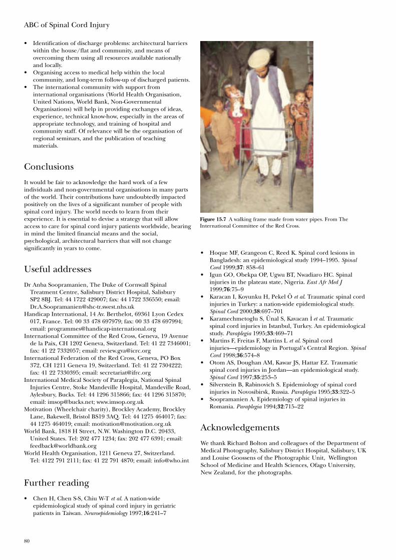
ABC of Spinal Cord Injury
80
• Identification of discharge problems: architectural barrierswithin the house/flat and community, and means ofovercoming them using all resources available nationallyand locally.
• Organising access to medical help within the localcommunity, and long-term follow-up of discharged patients.
• The international community with support frominternational organisations (World Health Organisation,United Nations, World Bank, Non-GovernmentalOrganisations) will help in providing exchanges of ideas,experience, technical know-how, especially in the areas ofappropriate technology, and training of hospital andcommunity staff. Of relevance will be the organisation ofregional seminars, and the publication of teachingmaterials.
Conclusions
It would be fair to acknowledge the hard work of a fewindividuals and non-governmental organisations in many partsof the world. Their contributions have undoubtedly impactedpositively on the lives of a significant number of people withspinal cord injury. The world needs to learn from theirexperience. It is essential to devise a strategy that will allowaccess to care for spinal cord injury patients worldwide, bearingin mind the limited financial means and the social,psychological, architectural barriers that will not changesignificantly in years to come.
Useful addresses
Dr Anba Soopramanien, The Duke of Cornwall SpinalTreatment Centre, Salisbury District Hospital, SalisburySP2 8BJ. Tel: 44 1722 429007; fax: 44 1722 336550; email:[email protected]
Handicap International, 14 Av. Berthelot, 69361 Lyon Cedex017, France. Tel: 00 33 478 697979; fax: 00 33 478 697994;email: [email protected]
International Committee of the Red Cross, Geneva, 19 Avenuede la Paix, CH 1202 Geneva, Switzerland. Tel: 41 22 7346001;fax: 41 22 7332057; email: [email protected]
International Federation of the Red Cross, Geneva, PO Box372, CH 1211 Geneva 19, Switzerland. Tel: 41 22 7304222;fax: 41 22 7330395; email: [email protected]
International Medical Society of Paraplegia, National SpinalInjuries Centre, Stoke Mandeville Hospital, Mandeville Road,Aylesbury, Bucks. Tel: 44 1296 315866; fax: 44 1296 315870;email: [email protected]; www.imsop.org.uk
Motivation (Wheelchair charity), Brockley Academy, BrockleyLane, Bakewell, Bristol BS19 3AQ. Tel: 44 1275 464017; fax:44 1275 464019; email: [email protected]
World Bank, 1818 H Street, N.W. Washington D.C. 20433,United States. Tel: 202 477 1234; fax: 202 477 6391; email:[email protected]
World Health Organisation, 1211 Geneva 27, Switzerland.Tel: 4122 791 2111; fax: 41 22 791 4870; email: [email protected]
Further reading
• Chen H, Chen S-S, Chiu W-T et al. A nation-wideepidemiological study of spinal cord injury in geriatricpatients in Taiwan. Neuroepidemiology 1997;16:241–7
Figure 15.7 A walking frame made from water pipes. From TheInternational Committee of the Red Cross.
• Hoque MF, Grangeon C, Reed K. Spinal cord lesions inBangladesh: an epidemiological study 1994–1995. SpinalCord 1999;37: 858–61
• Igun GO, Obekpa OP, Ugwu BT, Nwadiaro HC. Spinalinjuries in the plateau state, Nigeria. East Afr Med J1999;76:75–9
• Karacan I, Koyunku H, Pekel Ö et al. Traumatic spinal cordinjuries in Turkey: a nation-wide epidemiological study.Spinal Cord 2000;38:697–701
• Karamechmetoglu S, Ünal S, Kavacan I̊ et al. Traumaticspinal cord injuries in Istanbul, Turkey. An epidemiologicalstudy. Paraplegia 1995;33:469–71
• Martins F, Freitas F, Martins L et al. Spinal cordinjuries—epidemiology in Portugal’s Central Region. SpinalCord 1998;36:574–8
• Otom AS, Doughan AM, Kawar JS, Hattar EZ. Traumaticspinal cord injuries in Jordan—an epidemiological study.Spinal Cord 1997;35:253–5
• Silverstein B, Rabinovich S. Epidemiology of spinal cordinjuries in Novosibirsk, Russia. Paraplegia 1995;33:322–5
• Soopramanien A. Epidemiology of spinal injuries inRomania. Paraplegia 1994;32:715–22
Acknowledgements
We thank Richard Bolton and colleagues of the Department ofMedical Photography, Salisbury District Hospital, Salisbury, UKand Louise Goossens of the Photographic Unit, WellingtonSchool of Medicine and Health Sciences, Ofago University,New Zealand, for the photographs.

abdominal binder 26, 51abdominal bruising 8abdominal distension 8abdominal injuries incidence 6abdominal stoma 38, 61accidents 1
prevention 74–5acupuncture 67adolescents with spinal injury 57–8advanced reciprocating gait orthosis 51advanced trauma life support (ATLS) 6–7age at injury 77ageing with spinal cord injury 73aid to daily living (ADL) 41, 53–4airway, breathing and circulation (ABC) assessment 6–7alcohol therapy 30alignment of radiological views 12alprostadil therapy 68ambulances 6American Spinal Injury Association impairment scale 8, 8anal canal sensation 9anal digital stimulation 61anal reflex 9anal sphincter examination 9anal stretching 48analgesia 4, 20, 67anatomy of spinal cord injury 21, 22ankylosing spondylitis
fracture and 27radiography 13, 14transporting patient with 5
antegrade colonic enema (ACE) 40, 61anterior cord compression 26anterior cord syndrome 10, 10anterior spinal cord artery compression 10anteroposterior views, radiological 12, 13, 15antibiotic therapy 19anticholinergic therapy 38anticoagulation for embolism prevention 18–19anticonvulsant therapy 67anxiety 70aortic dissection 16apophyseal joint subluxation/dislocation 14areflexia 3arms
flaccidity 10management 53 positioning 44–5pain 66passive movements 49surgery 53weights for strengthening 49
artificial urinary sphincters (AUS) 39, 39artificial ventilation 17–18assault 1
assessment of patient during transport 6 primary survey 6–7 secondary survey 7–8visit 54
ataxia 10atelectasis 17Attendance Allowance 58, 58atlanto-axial fusion 27atlanto-axial instability 26 atlanto-axial subluxation 2, 12 atlas fracture 26, 27atropine
at scene of injury 3for cardiac arrest prevention 18
augmentation cystoplasty 38autonomic dysreflexia 28–9, 36, 61, 68, 69
back splints 5baclofen therapy 30, 31balancing 50Balkan beam 42beds 20, 20, 26, 42
transferring to 50behaviour problems 41bi-level support 17biochemical disturbances 29–30, 36bladder
acontractile 34, 39, 61augmentation cystoplasty 38calculus 47
incidence 35care, on holiday 64
patient education 60–1clammed 38contractions, unstable 36cycling 34distension 29management 47
basic algorithms 37initial 19, 47long-term 47, 61
neck injections 39overdistention 47spasm 35system stimulation 72tapping 60–1
see also tapping and expressionblood supply to cervical cord 21board see spinal boardbody temperature
during transport 6 in hospital 7, 46
bolster 2, 5, 6, 42bony diagnosis 8
IndexPage numbers in bold refer to figures; those in italic refer to tables or boxed material

Index
82
bony formation 30bony radiography 12botulinum toxin therapy 31bowel
care 47–8, 47, 48 on holiday 64patient education 61–2
flaccid 61bracing 28, 51, 54
children 65bradycardia 18
from suction 3bronchoscopy 17Brown–Sèquard syndrome 10, 10, 26bruising 7, 8bulbocavernosus reflex 9bupivacaine therapy 30burst fractures 15, 15bursting injury 26
calculus 34, 34, 36calipers 51carbamazepine therapy 29, 67cardiac arrest from suction 3cardiac failure 18cardiovascular complications 18cardiovascular monitoring 45–6care
ageing and 73transfer from hospital to community 60–4
carers 62Carers Act 57Carers and Disabled Children’s Act 57Caring for People 57cartilage radiography 12catheter
encrustation 33valve 61
catheterisation 29, 47complications 33, 34, 35indwelling 33–4, 36 initial 19intermittent 33 intermittent self- 34, 36, 47, 61, 64patient education 61sexual intercourse and 68shaft compression 36suprapubic 34–5, 36, 47, 47, 61
cauda equina lesion 21bladder management 37, 39, 40pain 67
cauda equine syndrome 9causes of spinal cord injury 1, 77central cord syndrome 10, 10, 13central nervous system assessment 7cervical cord blood supply 21cervical flexion 5cervical injuries 11–14, 25
hyperextension 2incidence 1management 21–3, 25–7upper 26–7
cervical lordosis palpation 7cervical spondylosis 10, 13, 23, 23cervicothoracic junction injury
management 27radiography 14
Chance fracture 16, 16chest
infections 45injuries 6physiotherapy 17treatment, prophylactic 39
children antegrade colonic enema 40bracing 65flexion-extension axis 5incidence of spinal cord injury 1mattresses 8physiotherapy 51–2SCIWORA 11scoliosis 65, 65splints 5
chin lift manoeuvre 42classification of spinal cord injury 7clinical features of spinal cord injury 4clothes
patient education 60removal 42
collars 2, 5, 5, 6, 11, 26, 42, 54colonic irrigation 61colostomy 61colposuspension 39coma position 2, 3Combitube 3communication 55community care 60–4, 74Community Care Act 57community liaison staff 60, 63
education 62community support 58complications 17–24, 65–75compression fracture 11, 27compressive retainer rings 68computed tomography 14, 15, 16condom sheaths drainage 33, 36, 38, 47Cone caliper 21, 21confusion 46conscious patient management at scene of
injury 4consciousness, loss of 2constipation 62continence
achievement 47maintenance importance 38
contractures 78management 30, 31prevention 20
conus lesion, bladder management 37, 39, 40conus medullaris syndrome 9conversion paralysis 10coping mechanisms 41cornus medullaris syndrome 10coughing, assisted 49Council Tax Benefit 58counselling 58countertraction 43, 43cranial nerve function assessment 8creatinine monitoring 36Cr-EDTA GFR 36Crutchfield caliper 21cushions 31, 60, 64cystitis 38cystometrogram 35–6, 35
cystoplasty, augmentation 38cystotomy 34
dantrolene therapy 30deep tendon reflexes 9deep vein thrombosis 28
monitoring 46delayed plantar response (DPR) 9Department of Social Security 59dependency 41depression 70dermatome assessment 8, 9detrusor activity, hyperreflexic 35, 37–9detrusor pressure rises 36detrusor-distal sphincter dyssynergia 29, 33, 35, 36
effects 36developing world, spinal cord injury incidence 76–7DIAL 59diamorphine analgesia 20diaphragmatic breathing 3, 8, 17diaphragmatic paralysis 18diazepam therapy 30diet see nutritionDirect Payments 57Disability Living Allowance 58, 58Disability Rights Handbook 59disability, attitudes to 78Disabled Person’s Tax Credit 58disc prolapse 14, 16, 26disorientation 46district nursing service 63diuresis 30, 47diving accidents 77DMSA renography 36, 36dopamine therapy 18dorsal root entry zone coagulation (DREZ) 67double lumen sign 19dressing 54dressings for sores 31driving 56dysreflexia see autonomic dysreflexia
eating splints 53education
family and community staff 62patients 60–2
egg shell calculus 34ejaculation 68
aids 68stimulation 72
elbow extension 71electrical stimulation 71–2
for hand 71–2 practical uses 72
electrolyte monitoring 36emergency department nursing 42emotional problems 70employment see workenteral feeding 46enterocystoplasty 38ephedrine therapy 26epididymitis 34equinus contracture 20equipment for communication 55erections 67–8
implant-induced 38etidronate therapy 30
Index
83
evacuation and transfer to hospital 5–6extension injury 27extension view, radiological 14
facet joints dislocation 12, 13–14, 14, 15, 22, 23, 26
closed reduction 27fractures affecting 14
faecal evacuation 48faecal incontinence 37, 40 falls 2, 77femoral supracondylar fracture 66, 66Ferticare vibrator 68fertility 68–9fibreoptic instruments 3financial aspects 58–9
compensation 63issues in developing world 77, 78
Finetech-Brindley SARS 38, 38flaccid bowel 61flaccid paralysis 21flaccidity 3, 8, 9, 10flexion contractures 31, 65flexion injury 25, 65
radiography 25flexion view, radiological 14flexion-distraction injury 26, 65flexion-extension axis 5flexion-extension views 25, 26flexion-rotation forces 10, 26flexor tenodesis, passive 71floor lift 54fluids
intake on holiday 64restriction 34
flushing 29Foley catheter 33, 34follow-up 64foot board 44foot drop 20force sensing array 60fracture
femoral supracondylar 66, 66pathological 65–6
frames 65Frankel grades 8, 74functional ability in tetraplegia 55furosemide therapy 29, 30
gabapentin therapy 67gait training 51, 52Gardner-Wells caliper 21, 21, 22gastric regurgitation, passive 2gastric ulcer 19gastrointestinal tract management, initial 19gender ratio in spinal cord injury 77gibbus 7glans penis squeezing 9glomerular filtration rate 36glycerine suppositories 61glyceryl trinitrate therapy 29glycopyrronium for cardiac arrest prevention 18grant aid 54gunshot wounds 77
H2-receptor antagonist for peptic ulcer prevention 19haemathoraces 17

haematomaposterior mediastinum 16prevertebral 14
haemorrhage 18halo traction 23, 23, 25, 26hand
management 53positioning 45 surgery 53tetraplegic 71
handling patient 43Handmaster 71–2hangman’s fracture 26, 27, 27head
holding 43, 43injuries incidence 6splinting 8
headache 29health expenditure in various countries 77heel pressure 44, 44helicopter transport 6, 24, 24helmet removal 3, 3Help with Health Costs 58heparin for embolism prevention 19, 28hip
flexion contracture 31, 65flexor spasticity 66guidance orthosis 51
history of patient 6of spinal cord injury 1
hoisting aids 43, 63holidays 64home
discharge to 63extensions and adaptations 54, 58, 59, 59resettlement 53–4
horizontal beam lateral views, radiological 15hospital
discharge from 41, 54, 58, 62–3evacuation and transfer to 5–6initial management in 6–8
Housing Benefit 58humidification of inspired air 17hydronephrosis 36hyperalgesia 67hypercalcaemia 29–30hyperextension injury of cervical spine 2, 10, 23, 26hyperkalaemia 18hyperreflexic detrusor activity 35, 37–9hypnotherapy 67hypogastric plexus stimulator 68hyponatraemia 29hypospadias 33, 34hypotension 18
orthostatic 4postural 26
hypothermia 6, 42, 42, 46hysterical paralysis 10
iliac crest sores 31iliopsoas myotomy 31immobilisation 2immobiliser 5, 5, 7Incapacity Benefit 58incidence of spinal cord injury 1, 77
Index
84
Income Support 58incontinence 39, 40independence 54, 58, 62–3, 75Independent Living Fund 57Industrial Disablement Benefit 58injuries, associated 6institutional help barriers in developing
world 78intercostal paralysis 3interim care provision 63intermittent self-catheterisation 34, 36, 47
on holiday 64optimum requirements 35patient education 61 stoma for 38
internal environment maintaining 45monitoring 45–6, 45
interspinous gap 7intra-abdominal trauma 8intravenous access 3intravenous fluids 19Invalid Care Allowance 58ischial tuberosity sores 31isotope renography, urological 36
jaw thrust manoeuvre 42Jefferson fracture 26, 27joints
ageing and 73care 20
Kendrick extrication device 5kerb manoeuvring 50key grip 71, 71King’s Fund bed 42knees hyperextension 44kyphosis
lumbar 65transporting patient with 5
labour 69laminectomy, isolated 28laryngeal mask airway (LMA) 3lateral position 2, 6lateral views, radiological 11, 11, 13, 15laxatives 48leg
deformity 65positioning 44spasticity 10stimulation 72
leisure see holidays; recreation; sportslife expectancy 74, 74lifting patient 6, 78ligamentous injury 26limb
care 20 fracture fixation 20injuries 6see also arm; leg
log rolling 2, 3, 5, 7, 8, 20, 42, 43, 43, 44, 45, 45long board 5lordosis
excessive 65palpation 7

lower motor neurone lesions 9 paralysis 21
lumbar injury 14–16, 23incidence 1management 28
lumbar kyphosis 65lumbar lordosis
excessive 65ileus from 19palpation 7
lumbar pillow 20lung compliance loss 17
MAG3 renography 36, 36magnetic resonance imaging 14. 15, 16, 16malnutrition 46management
at scene of accident 2–4early 17–24 initial 6–8 later 65–75
manipulation under anaesthetic 22–3manpower problems in developing world 77–8mattresses 8, 8, 31, 42, 60, 64medical management in spinal injuries unit 25–32Medtronic SynchroMed® EL infusion system 31Mekong Cambodia wheelchair 79methylprednisolone therapy 19Miami collar 26minitracheostomy 17Mitrofanoff stoma 38, 39, 39mobile radiographic equipment 11mobilisation 41, 49–50, 67mobility 56Modernising Social Services 59mood swings 41morphine analgesia 20mortality 1, 74, 77motor loss 66motor muscles 7motor neurone lesions 9
see mainly lower or upper motor neurone lesionsmotor point injections 30mouthsticks 55, 55moving patients at scene of accident 2mucocutaneous junction sensation assessment 9muscle
assessment 9division 31passive stretching 30, 31, 49, 49power assessment 8spasm, pain-induced 26
myelopathycystic 66secondary 65
myotome assessment 8, 9
naloxone therapy 4, 20nasograstric aspiration 19natal cleft, split 48neck
flexion-extension 14, 25immobilisation 42roll 20, 22, 22, 25splinting 8stabilisation 5–6
Index
85
Nelaton catheter 33, 35nerve supply to reflexes 9neurectomy 31
obturator 30NeuroControl Freehand 71, 72, 73NeuroControl Vocare bladder system 72neurogenic shock 7–8neurological assessment 4
initial 6–7, 8–9Neurological Classification of Spinal Cord Injury 7neurological level of lesion 8neuromodulation 38–9neutral position 2, 6, 7NHS Charges and Optical Voucher Values 58nifedipine therapy 29nitrosamine production 38non-steroidal anti-inflammatories 4, 20, 30, 46nuclear medicine, urological 36nurses in developing world 78nursing 41–8
aims 41intervention 45–8management 42–5 positions 17
nutrition 46–7, 48patient education 62
oblique views, radiological 14obturator neurectomy 30occupational therapy 53–6
in developing world 78odontoid
process fracture 26–7, 27screw fixation 27
Odstock Dropped Foot Stimulator 72, 73, 73oliguria 47opioid therapy 4, 46oropharyngeal suction during transport 6orotracheal intubation 3orthotic devices 51osteoporosis 66Oswestry standing frame 51, 51outpatient appointements 64oxygen therapy 3, 18, 45
paediatric see childrenpain 66–7
ageing and 73arm 66classification 67-induced muscle spasm 26management 46shoulder 8, 46treatment 67
pamidronate therapy 30para-articular heterotopic ossification 30paralytic ileus 8, 19paraplegia
artificial urinary sphincters 39bladder management 37, 39developing world 77gait expectations 52high thoracic, diaphragmatic breathing 3hypotension 18independence 60log rolling 42

paraplegia – Continuedmobilising into wheelchair 49respiratory insufficiency causes 17respiratory monitoring 49response to temperature changes 6urological management 36
passive movement 30, 31, 49, 49, 67pathological fractures 65–6patient
education 60–2examination at scene of injury 3, 4handling in developing world 79
pelvic floor disorders 37pelvic fracture 15pelvic tilt, anterior 65pelvic twist 44, 44, 45penile urethra, cleft 34peptic ulcer prevention 19peritoneal irritation 8periurethral abscess 34personal assistant 63phantom pain 67phenol therapy 30phentolamine theapy 29Philadelphia collar 26phrenic nerve pacing 18phrenic nerve stimulator 70physiotherapy 49–52
chest 17in developing world 78programme 45
pillows 5, 20, 23, 44, 45pin prick sensation 8, 9placement, assessment and counselling team (PACT) 58plantar response 9poikilothermia 6, 42, 46populations of various countries 77positioning patient 6position changing 31, 43posterior cord syndrome 10, 10post-traumatic stress disorder 70post-traumatic syringomyelia 66, 66postural hypotension 26postural management, inadequate 65postural reduction 23potassium supplements 29prejudice regarding disability 78pressure areas 19–20, 42
inspection 42patient education 60, 60
pressure mapping 60pressure sores 78
management 31–2prevention 6, 8, 31, 65risk 46scalp 22from wheelchairs 49
prevertebral haematoma 14priapism 7primary survey 6–7profiling bed 26, 42, 42prognosis 1, 10, 73–5prone position 2, 3prone trolley 65proton pump inhibitor for peptic ulcer prevention 19psychological barriers and spinal cord injury 78psychological factors 70–1
Index
86
psychological trauma 41 rehabilitatinon 58
pubo-urethral slings 39pulmonary embolism 18, 28pulmonary oedema 18 pyelolithotomy, open 34pyelonephritis 34, 34, 36
radiological investigations 11–16 cervical injury 25–6 signs 25urological 35–6
RAF pattern turning frame 23reciprocating gait orthosis 51recreation 51, 56rectal ejaculation 68recumbent position 17reflex assessment 8, 9, 9rehabilitation 41, 42, 50–1
programmes in developing world 79workshop 58
relations, education 62relationships 41, 57–8
fulfilment 69relaxation techniques 67religious attitudes to disability 78renography 36, 36respiratory complications 17–18 , 45respiratory depression 4respiratory failure 26
mortality and 74respiratory function monitoring 17respiratory insufficiency causes 17respiratory management 49
in tetraplegia 70retropharyngeal haematoma 7retropharyngeal space 12rheumatoid arthritis radiography 14rhizotomy, posterior 38–9, 67road traffic accidents 1, 2, 74, 77rocuronium muscle relaxant 18Roho mattress 31rollator 51root entry zone damage 67
sacral anterior root stimulation (SARS) 38–9, 39sacral reflexes 10sacral sores 31, 31, 32saline therapy 29sandbags 2, 6, 11, 42scalp pressure sores 22scaphoid fracture 20scoliosis 51, 65, 65
long paralytic 65scoop stretcher 5, 5, 6Seager electroejaculator 68seat belt injury 8, 16secondary survey 7–8self-harm 1seminal emission 68semiprone coma position 2, 3sensation assessment 8sensory loss 66, 71sensory points 7
input 46, 46Severe Disablement Allowance 58

sexual function 67–8, 67aids 68
sexual intercourse, preparation for 68sexual relationships 57sexuality 48shoes, patient education 60shoulder
hold 43pain 8, 46, 73
sildenafil therapy 68sitting, children 51skin
blotching 29care 46
on holiday 64independent 60
complications 46hygiene 46infections 46inspection 43management, initial 19–20
skull calipers 21, 25traction 20, 21–3, 22
sleep disturbance 67social barriers and spinal cord injury 78social needs of patient and family 57–9social workers in developing world 78sodium level monitoring 29soft tissue radiography 12spasticity 51
exacerbation 66management 30–1
speech 70sperm count 68sphincterotomy 36
endoscopic 36, 36, 38spinal alignment 43spinal board 5, 5, 6, 6, 8
removal 42spinal column anatomy 15spinal cord
centre for sexual function 67cross-section 7hemisection 10injury (general only), anatomy 21, 22
pain 67 partial 10, 51 prognosis 73–4without radiological abnormality (SCIWORA) 2, 11, 65
stimulation 67spinal cord artery compression 10Spinal Injuries Association 59, 59spinal injuries unit
community visits from 64medical management in 25–32transfer to 6, 23–4
spine anatomy 12deformity 2
examination for 7late 65 prevention 65
fusion 26instability, late 65shock 9stabilisation 21–3, 26
Index
87
spinothalamic tractotomy 67spinous process radiography 12, 13, 26splints 2, 5, 8, 20, 31, 53, 53spondylolisthesis of axis 27sports injury 1, 2, 51, 74–5spreader bar 42sputum retention 17stab injury 10, 77staffing issues in developing world 77–8stag-horn calculus 34standing 51, 51
frame 77Statutory Sick Pay 58stenting 38sternum fracture 28steroid therapy 19stockings for embolism prevention 18–19, 26, 28, 46stomal stenosis 39straps during transport 6strategy planning in developing world 79stress incontinence 39stretchers 5, 5struvite calculus 34Stryker frame 20, 20, 23study days 62suction 3, 4supine position 2, 7suprapubic catheterisation 34–5, 36, 47, 47
blockage 38 patient education 61
suprasacral cord lesion, bladder management 37survival 1, 18suxamethonium therapy 18sweating 29swimmer’s view, radiological 11, 11, 12swimming 51swivel walker 51, 65syringomyelia, post-traumatic 66, 66syrinx 66, 66
tapping and expression 33, 47Tc-DTPA renography 36teardrop fracture 13, 13temperature changes
in A&E 42during transport 6
tendon lengthening 31reflexes 9transfer surgery 71
tenodesis of flexor pollicis longa 71grip 53
tenotomy 31tension-free vaginal tapes 39tetraplegia
accompanying patient to unit 24arm positioning 44bladder management 37, 39developing world 77diaphragmatic breathing 3hand 20, 53, 71hyperextension injury 23independence 60mobilising into wheelchair 49pelvic twist 44respiratory insufficiency causes 17

Index
88
tetraplegia – Continuedrespiratory management, later 70respiratory monitoring 49response to temperature changes 6suction contraindication 3sympathetic outflow interruption 18syrinx 66turning patient 20, 43, 43urological management 36
thoracic deformity 65thoracic injury 14–16, 23
incidence 1management 28
thoracic kyphosis, transporting patient with 5thoracolumbar injury
bracing 65initial management 2management 28pelvic twist contraindication 44support position 23
thromboembolism prophylaxis 18–19tilt table 51, 51tizanidine therapy 30toe nail care 46toilet transfer 50, 61torticollis 2touch sensation 46tracheal deviation 7tracheal intubation
2–3 indications 3tracheostomy 17, 70traction see skull tractiontractotomy, spinothalamic 67tramodol therapy 67transcutaneous nerve stimulation67transfer
ageing and 73skills 50, 52, 56
transporting to hospital 5–6trauma trolley 6, 8trauma re-evaluation 20travel 64tricyclic antidepressant therapy 67trismus 21trochanteric sores 31, 32trolley 65turning frame 23–4turning patient 5, 20, 43–5typing splints 53
ultrasound, urological 35unconscious patient 2–3
transporting to hospital 5–6University of Virginia caliper 21, 21upper motor neurone lesions 9upper urinary tract abnormalities 34uretheral catheterisation see catheterisationurethral closure 39urethral erosion in female 35urethral stricture disease 34urinary drainage bags 33urinary infection 34urinary sphincters, artificial 39urinary tract
catheterisation see catheterisation infections 61
prevention 47
reconstruction 36–7urine
culture 34expression 33, 47flow 18volume 19
urological management 33–40 early 33–5 investigations and review 35–6later 36–7
vacuum erection aids 68vacuum mattress 8, 8vacuum splint 5vehicles 56ventilatory support 18, 70vertebral fracture 12, 13
anteroinferior margin 13burst 15, 15compression 11, 28mass 10
vertebral radiography 12vertebral subluxation 12, 14vertebrectomy 28vesicoureteric reflux 34
reflux 36video-urodynamics 35–6vital capacity 17voiding
assisted 39dysfunctional patterns 32
voluntary organisations 59vomit aspiration 2vomiting during transport 6
walking frame 80War Disablement Pension 58warfarin for embolism prevention 19, 28weaning from ventilation 18wedge compression 28weekend stays at home 54, 63weight loss 46welfare benefits 58, 59wheelchair 56
accessibility 58ageing and 73children 51design 49–50in developing world 79familiarity with 50lifting from, to prevention sores 31mobilising into 41, 49–50 patient education 60seating position 49skills, 50, 50transfer from 50
work 56, 58, 59Working Families Tax Credit 58wound
forehead 2open 23
wrist extension 53, 53, 71writing splints 55, 55
xanthogranulomatous pyelonephritis 34
zone of partial preservation (ZPP) 9