spikes, synchrony, and attentive learning by laminar thalamocortical
TRANSCRIPT

B R A I N R E S E A R C H 1 2 1 8 ( 2 0 0 8 ) 2 7 8 – 3 1 2
ava i l ab l e a t www.sc i enced i r ec t . com
www.e l sev i e r. com/ loca te /b ra in res
Research Report
Spikes, synchrony, and attentive learning by laminarthalamocortical circuits
Stephen Grossberg⁎, Massimiliano Versace1
Department of Cognitive and Neural Systems, Center for Adaptive Systems, Center of Excellence for Learning in Education, Science, andTechnology, Boston University, 677 Beacon Street, Boston, MA 02215, USA
A R T I C L E I N F O
⁎ Corresponding author.Department of Cognit617 353 7755.
E-mail addresses: [email protected], steve@cnURL: http://cns.bu.edu/~steve (S. Grossber
1 Authors in alphabetical order. MV was suNational Science Foundation (NSF SBE-03543the National Science Foundation (NSF SBE-03
0006-8993/$ – see front matter © 2008 Elsevidoi:10.1016/j.brainres.2008.04.024
A B S T R A C T
Article history:Accepted 4 April 2008Available online 22 April 2008
This article develops the Synchronous Matching Adaptive Resonance Theory (SMART)neural model to explain how the brain may coordinate multiple levels of thalamocorticaland corticocortical processing to rapidly learn, and stably remember, important informationabout a changing world. The model clarifies how bottom-up and top-down processes worktogether to realize this goal, notably how processes of learning, expectation, attention,resonance, and synchrony are coordinated. The model hereby clarifies, for the first time,how the following levels of brain organization coexist to realize cognitive processingproperties that regulate fast learning and stable memory of brain representations: single-cell properties, such as spiking dynamics, spike-timing-dependent plasticity (STDP), andacetylcholine modulation; detailed laminar thalamic and cortical circuit designs and theirinteractions; aggregate cell recordings, such as current source densities and local fieldpotentials; and single-cell and large-scale inter-areal oscillations in the gamma and betafrequency domains. In particular, the model predicts how laminar circuits of multiplecortical areas interact with primary and higher-order specific thalamic nuclei andnonspecific thalamic nuclei to carry out attentive visual learning and informationprocessing. The model simulates how synchronization of neuronal spiking occurs withinand across brain regions, and triggers STDP. Matches between bottom-up adaptively filteredinput patterns and learned top-down expectations cause gamma oscillations that supportattention, resonance, learning, and consciousness. Mismatches inhibit learning whilecausing beta oscillations during reset and hypothesis testing operations that are initiated inthe deeper cortical layers. The generality of learned recognition codes is controlled by avigilance process mediated by acetylcholine.
© 2008 Elsevier B.V. All rights reserved.
Keywords:AttentionLearningSTDPBottom-up filterTop-down expectationMatchPredictionMismatchLGNPulvinarV1V2SpikesGamma oscillationsBeta oscillationsSynchronizationLocal field potentialsMismatch negativityAcetylcholineCortical layersAdaptive Resonance Theory
ive andNeural Systems, BostonUniversity, 677 Beacon Street, Boston,MA 02215, USA. Fax: +1
s.bu.edu (S. Grossberg), [email protected] (M. Versace).g), http://cns.bu.edu/~versace (M. Versace).pported in part by the Air Force Office of Scientific Research (AFOSR F49620-01-1-0397), the78), and the Office of Naval Research (ONR N00014-01-1-0624). SG was supported in part by54378) and the Office of Naval Research (ONR N00014-01-1-0624).
er B.V. All rights reserved.
279B R A I N R E S E A R C H 1 2 1 8 ( 2 0 0 8 ) 2 7 8 – 3 1 2
1. Introduction
1.1. The link between learning, expectation, attention,resonance, and synchrony
This article proposes how the brain coordinates multiplelevels of thalamocortical and corticocortical processing torapidly learn, and stably remember, important informationabout the world. The Synchronous Matching Adaptive Reso-nance Theory (SMART) model that is presented here showshow bottom-up and top-down pathways work together toaccomplish this goal by coordinating processes of learning,expectation, attention, resonance, and synchrony. In particu-lar, SMART explains how attentive learning requirementsare realized by detailed brain circuits, notably the layeredorganization of cells in neocortical circuits and how theyinteract with first-order (e.g., the lateral geniculate nucleus,LGN) and higher-order (e.g., the pulvinar nucleus, PULV;Sherman and Guillery, 2001; Shipp, 2003), and nonspecificthalamic nuclei (van Der Werf et al., 2002).
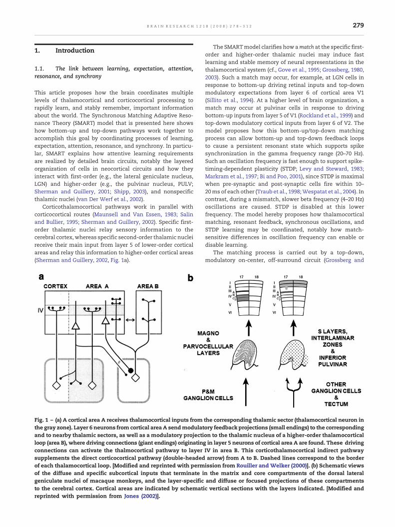
Corticothalamocortical pathways work in parallel withcorticocortical routes (Maunsell and Van Essen, 1983; Salinand Bullier, 1995; Sherman and Guillery, 2002). Specific first-order thalamic nuclei relay sensory information to thecerebral cortex, whereas specific second-order thalamic nucleireceive their main input from layer 5 of lower-order corticalareas and relay this information to higher-order cortical areas(Sherman and Guillery, 2002, Fig. 1a).
Fig. 1 – (a) A cortical area A receives thalamocortical inputs fromthe gray zone). Layer 6 neurons from cortical area A sendmodulatand to nearby thalamic sectors, as well as a modulatory projectiloop (area B), where driving connections (giant endings) originatingconnections can activate the thalmocortical pathway to layersupplements the direct corticocortical pathway (double-headedof each thalamocortical loop. [Modified and reprinted with permof the diffuse and specific subcortical inputs that terminate igeniculate nuclei of macaque monkeys, and the layer-specificto the cerebral cortex. Cortical areas are indicated by schematireprinted with permission from Jones (2002)].
The SMARTmodel clarifies how amatch at the specific first-order and higher-order thalamic nuclei may induce fastlearning and stable memory of neural representations in thethalamocortical system (cf., Gove et al., 1995; Grossberg, 1980,2003). Such a match may occur, for example, at LGN cells inresponse to bottom-up driving retinal inputs and top-downmodulatory expectations from layer 6 of cortical area V1(Sillito et al., 1994). At a higher level of brain organization, amatch may occur at pulvinar cells in response to drivingbottom-up inputs from layer 5 of V1 (Rockland et al., 1999) andtop-down modulatory cortical inputs from layer 6 of V2. Themodel proposes how this bottom-up/top-down matchingprocess can allow bottom-up and top-down feedback loopsto cause a persistent resonant state which supports spikesynchronization in the gamma frequency range (20–70 Hz).Such an oscillation frequency is fast enough to support spike-timing-dependent plasticity (STDP; Levy and Steward, 1983;Markram et al., 1997; Bi and Poo, 2001), since STDP is maximalwhen pre-synaptic and post-synaptic cells fire within 10–20ms of each other (Traub et al., 1998;Wespatat et al., 2004). Incontrast, during a mismatch, slower beta frequency (4–20 Hz)oscillations are caused. STDP is disabled at this lowerfrequency. The model hereby proposes how thalamocorticalmatching, resonant feedback, synchronous oscillations, andSTDP learning may be coordinated, notably how match-sensitive differences in oscillation frequency can enable ordisable learning.
The matching process is carried out by a top-down,modulatory on-center, off-surround circuit (Grossberg and
the corresponding thalamic sector (thalamocortical neuron inory feedback projections (small endings) to the correspondingon to the thalamic nucleus of a higher-order thalamocorticalin layer 5 neurons of cortical area A are found. These driving
IV in area B. This corticothalamocortical indirect pathwayarrow) from A to B. Dashed lines correspond to the borderission from Rouiller and Welker (2000)]. (b) Schematic viewsn the matrix and core compartments of the dorsal lateraland diffuse or focused projections of these compartmentsc vertical sections with the layers indicated. [Modified and
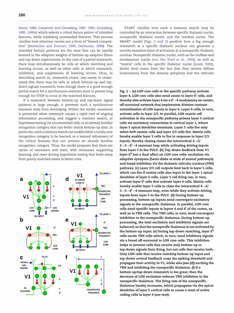
Fig. 2 – (a) LGN core cells in the specific pathway activatelayer 4. LGN core cells also send axons to layer 6I cells, andthereby also activate layer 4 via a 6I→4modulatory on-center,off-surround network that implements divisive contrastnormalization of LGN inputs in layer 4. Layer 4 cells, in turn,activate cells in layer 2/3. In parallel, LGN matrix cellactivation in the nonspecific pathway primes layer 5 corticalcells via excitatory connections to cortical layer 1, wherelayer 5 apical dendrites terminate. Layer 5 cells fire onlywhen both matrix cells and layer 2/3 cells fire. Matrix cellshereby enable layer 5 cells to fire in response to layer 2/3inputs, thereby closing closes the intracortical 4→2/3→5→6I→4 resonant loop while activating driving inputsfrom layer 5 to the PULV. (b) Top-down feedback from V1layer 6II has a dual effect on LGN core cells: excitation viaadaptive synapses (hemi-disks at ends of axonal pathways)
280 B R A I N R E S E A R C H 1 2 1 8 ( 2 0 0 8 ) 2 7 8 – 3 1 2
Stone, 1986; Carpenter and Grossberg, 1987, 1991; Grossberg,1995, 1999a) which selects a critical feature pattern of attendedfeatures, while inhibiting unattended features. This processclarifies how attention carries out a form of “biased competi-tion” (Desimone and Duncan, 1995; Desimone, 1998). Theattended feature patterns are the ones that can be rapidlylearned in the adaptive weights of bottom-up adaptive filtersand top-down expectations. In the case of a partial mismatch,there may simultaneously be cells at which matching andlearning occurs, as well as other cells at which mismatch,inhibition, and suppression of learning occurs. Thus, indescribing match vs. mismatch states, one needs to under-stand that there may be cells at which bottom-up and top-down signals mismatch, even though there is a good enoughpartial match for a synchronous resonant state to persist longenough for STDP to occur at the matched features.
If a mismatch between bottom-up and top-down signalpatterns is large enough, it prevents such a synchronousresonant state from developing. Within the model, resonanceis prevented when mismatch causes a rapid reset of ongoinginformation processing, and triggers a memory search, orhypothesis testing, foruncommittedcells, or analready familiarrecognition category that can better match bottom-up data. Inparticular, suchamemory searchcanenable either a totallynewrecognition category to be learned, or a learned refinement ofthe critical features that can activate an already familiarrecognition category. Thus, the model proposes that there arecycles of resonance and reset, with resonance supportinglearning, and reset driving hypothesis testing that leads awayfrom poorly matched states to better ones.
START clarifies how such a memory search may becontrolled by an interaction between specific thalamic nuclei,nonspecific thalamic nuclei, and the cerebral cortex. TheSMART model (Figs. 2 and 3) predicts how a big enoughmismatch at a specific thalamic nucleus can generate anovelty-sensitive burst of activation at a nonspecific thalamicnucleus. Nonspecific thalamic nuclei, such as the midline andintralaminar nuclei (van Der Werf et al., 2002), as well as“matrix” cells in the specific thalamic nuclei (Jones, 2002),derive their name from the fact that they receive diffuseinnervations from the sensory periphery and the reticular
and broad inhibition via the thalamic reticular nucleus (TRN)pathway. (c) Layer 2/3 cell outputs feed back to layer 5 cells,which can fire if matrix cells also input to the layer 1 apicaldendrites of layer 5 cells. Layer 5 cell firing can, in turn,activate layer 6I cells that activate layer 4 cells. Matrix cellshereby enable layer 5 cells to close the intracortical 4→2/3→5→6I→4 resonant loop, even while they activate drivinginputs from layer 5 to the PULV. (d) During bottom-upprocessing, bottom-up inputs send convergent excitatorysignals to the nonspecific thalamus. In parallel, LGN corecells send specific inputs to layers 4 and 6I of the cortex, aswell as to TRN cells. The TRN cells, in turn, send convergentinhibition to the nonspecific thalamus. During bottom-upprocessing, the total excitatory and inhibitory signals arebalanced, so that the nonspecific thalamus is not activated bythe bottom-up input. (e) During top-down matching, layer 6II
cells excite TRN cells which, in turn, send inhibitory signalsvia a broad off-surround to LGN core cells. This inhibitionhelps to prevent cells that receive only bottom-up ortop-down signals from firing, but not cells that receive both.Only LGN cells that receive matching bottom-up input andtop-down cortical feedback cross the spiking threshold andpropagate their activity to V1, while also (see (d)) exciting theTRN and inhibiting the nonspecific thalamus. (f) If abottom-up/top-down mismatch is too great, then thedecrease of LGN excitation reduces TRN inhibition to thenonspecific thalamus. The firing rate of the nonspecificthalamus hereby increases, which propagates via the apicaldendrites of layer 5 cortical cells to cause a reset of activecoding cells in layer 4 (see text).

Fig. 3 – (a) Layer 5 of V1 provides a driving bottom-up input to the pulvinar (PULV), which is matched against top-down signalsfrom layer 6II of V2. This circuit is homologous to the bottom-up driving input from the retina to the LGN, which is matchedagainst top-down signals from layer 6II of V1 (see Fig. 2). Layer 5 of V1 also excites PULVmatrix cells, which provide nonspecificpriming input to layer 5 cells in V2. (b) Layer 6II of V2 also provides top-down corticocortical feedback to layer 4 of V1 vialayer 1 apical dendrites of layer 5 cells that project to layer 6I and then to 4 via a modulatory on-center, off-surround circuit. (c)The entire SMARTmodel circuit includes thalamic nuclei and laminar cortical circuits. The thalamus is subdivided into specificfirst-order and second-order nuclei, nonspecific nucleus, and thalamic reticular nucleus (TRN). The first-order thalamicmatrix cells (1 cell population at each specific thalamic nucleus, shown as an open ring) provide nonspecific excitatory primingto layer 1 in response to bottom-up input, priming layer 5 cells and allowing them to respond to layer 2/3 input. This allowslayer 5 to close the intracortical loop and activate the PULV. V1 layer 4 receives inputs from two parallel bottom-upthalamocortical pathways: a direct LGN→4 excitatory input, and a 6I→4 modulatory on-center, off-surround network thatcontrast-normalizes the pattern of layer 4 activation via the recurrent 4→2/3→5→6I→4 loop. V1 activates the bottom-up V1→V2corticocortical pathways from V1 layer 2/3 to V2 layers 6I and 4, as well as the bottom-up corticothalamocortical pathway fromV1 layer 5 to the PULV, which projects to V2 layers 6I and 4. In V2, as in V1, the layer 6I→4 pathway provides divisive contrastnormalization to V2 layer 4 cells. Corticocortical feedback from V2 layer 6II reaches V1 layer 1, where it activates apicaldendrites of layer 5 cells. Layer 5 cells, in turn, activate the modulatory 6I→4 pathway in V1, which projects a V1 top-downexpectation to the LGN. TRN cells of the two thalamic sectors are linked via gap junctions, which synchronize activation acrossthe two thalamocortical sectors when processing bottom-up stimuli. The nonspecific thalamic nucleus receives convergentbottom-up excitatory input from specific thalamic nuclei and inhibition from the TRN, and projects to layer 1 of the laminarcortical circuit, where it regulates mismatch-activated reset and hypothesis testing in the cortical circuit (see text).Corticocortical feedback connections from layer 6II of the higher cortical area terminate in layer 1 of the lower cortical area,whereas corticothalamic feedback from layer 6II terminates in its specific thalamus and on the TRN. This corticothalamicfeedback is matched against bottom-up input in the specific thalamus.
281B R A I N R E S E A R C H 1 2 1 8 ( 2 0 0 8 ) 2 7 8 – 3 1 2

Fig. 4 – Search for a recognition code within an ART learningcircuit: (a) The input pattern I is instated across the featuredetectors at processing stage F1 as a short term memory(STM) activity pattern X. Input I also nonspecifically activatesthe orienting system with a gain that is called vigilance (ρ);that is, all the input pathways converge with gain ρ onto theorienting system and try to activate it. STM pattern X isrepresented by the hatched pattern across F1. Pattern X bothinhibits the orienting system and generates the outputpattern S. Pattern S ismultiplied by learned adaptiveweights,also called long-termmemory (LTM) traces. These LTM-gatedsignals are added at F2 cells to form the input pattern T, whichactivates the STM pattern Y across the recognition categoriescoded at level F2. (b) Pattern Y generates the top-down outputpattern U which is multiplied by top-down LTM traces and
282 B R A I N R E S E A R C H 1 2 1 8 ( 2 0 0 8 ) 2 7 8 – 3 1 2
formation, and project diffusely to the superficial layers of thecerebral cortex (Fig. 1b).
In particular, the nonspecific thalamic nuclei are predictedto generate reset signals in the form of novelty-sensitivebursts of activation during mismatch episodes. Such a burstis broadcast nonspecifically to the superficial layers of thecerebral cortex, notably layer 1. The nonspecific burst issensed by dendrites in layer 1 of cortical layer 5 cells. Themodel explains how the burst leads to a reset event bypropagating from layer 1 dendrites via their layer 5 cells tolayer 6 and then on to layer 4, shutting down previously activecells there, and thereby enabling a different pattern ofactivation to take hold in layer 4. This reset event causes aslower beta oscillation frequency in the model. Thus the resetevent prevents learning of poorly matched bottom-up andtop-down information, both by inhibiting the active learnedcategorical representations whose top-down expectations ledto the mismatch, and also by creating a slower oscillationfrequency to which STDP is insensitive. The details of how thisworks will be described below.
As noted above, the SMART model predicts that the resetevent is expressed in the deeper layers of cerebral cortex,such as layers 4 to 6, and may thereby initiate slower betaoscillations in these layers. The more superficial corticallayers (e.g., layers 2/3) may, in contrast, express fastergamma oscillations. The model supports its proposal abouthow match-sensitive differences in oscillation frequency canenable or disable learning by quantitatively simulating dataabout single-cell biophysics, pharmacology, and neurophy-siology; laminar neuroanatomy; aggregate cell recordings,such as current source densities and local field potentials;large-scale oscillations at beta and gamma frequencies; and
functionally links them all to requirements about how toachieve fast attentive learning and stable memory. It is alsosuggested below how to directly test this prediction.
Many authors have examined synchronous oscillationswithin and across brain regions as one way in which be-haviorally significant brain states are organized (Engel et al.,2001). Aggregate and single-cell recordings from multiplethalamic and cortical levels of mammals have shown high-frequency and low-frequency rhythmic synchronous activitycorrelated with cognitive, perceptual and behavioral tasks. Inaddition, large-scale neuronal population models have beenproposed tomodel oscillatory dynamics (Bazhenov et al., 1998;Lumer et al., 1997; Destexhe et al., 1999; Siegel et al., 2000).However, these models do not link brain spikes, oscillations,
added at F1 cells to form a prototype pattern V that encodesthe learned expectation of the active F2 nodes. Such aprototype represents the set of commonly shared features inall the input patterns capable of activating Y. If VmismatchesI at F1, then a new STM activity pattern X* is selected at F1. X*is represented by the hatched pattern. It consists of thefeatures of I that are confirmed byV. Mismatched features areinhibited. The inactivated cells corresponding tounconfirmed features of X are unhatched. The reduction intotal STMactivitywhich occurswhenX is transformed intoX*causes a decrease in the total inhibition from F1 to theorienting system. (c) If inhibition decreases sufficiently, theorienting system releases a nonspecific arousal wave to F2;that is, a wave of activation that equally activates all F2 cells.This wave instantiates the intuition that “novel events arearousing”. This arousalwave resets theSTMpatternY at F2 byinhibiting Y. (d) After Y is inhibited, its top-down prototypesignal is eliminated, and X can be reinstated at F1. The priorreset event maintains inhibition of Y during the search cycle.As a result,X can activate a different STMpatternY at F2. If thetop-down prototype due to this new Y pattern alsomismatches I at F1, then the search for an appropriate F2category continues until a better-matching one is selected.Such a search cycle represents a type of non-stationaryhypothesis testing. When search ends, an attentiveresonance develops and learning of the attended data isinitiated. [Adapted with permission from Carpenter andGrossberg (1993).]

283B R A I N R E S E A R C H 1 2 1 8 ( 2 0 0 8 ) 2 7 8 – 3 1 2
and self-stabilizing STDP with the brain states that subserveattentive cognitive information processing.
1.2. ART, LAMINART and SMART
The SMART model fills this gap. It clarifies data about howbottom-up processing and learned tuning of adaptive filters ismodulated by top-down attentive learned expectations thatembody predictions or hypotheses that focus attention onexpected bottom-up stimuli (Salin and Bullier, 1995; Engelet al., 2001; Gao and Suga, 1998; Krupa et al., 1999; Desimone,1998; Ahissar and Hochstein, 2002; Herrmann et al., 2004).These data support predictions of Adaptive ResonanceTheory, or ART (Grossberg, 1980, 2003; Carpenter and Gross-berg, 1987, 1991, 1993; Carpenter et al., 1991) that top-downexpectations regulate predictive coding and matching andthereby help to focus attention, synchronize and gain-modulate attended feature representations, and trigger fastlearning that is dynamically buffered against catastrophicforgetting. The goal of achieving fast stable learning withoutcatastrophic forgetting is often summarized as the stability–plasticity dilemma (Grossberg, 1980). The stability–plasticitydilemma must be solved by every brain system that needs torapidly, yet stably, learn about the flood of signals thatsubserves even the most ordinary experiences. If the brain'sdesign is parsimonious, then we should expect to find similarprinciples operating in all the brain systems that can stablylearn an accumulating knowledge base in response to chan-ging conditions throughout life.
ART has predicted that some fundamental properties ofhuman and animal perception and cognition are part of thebrain's solution of the stability–plasticity dilemma. In parti-cular, humans are intentional beings who learn expectationsabout the world and make predictions about what is about tohappen. Humans are also attentional beings who focus proces-sing resources upon a restricted amount of incoming informa-tion at any time. Why are we both intentional and attentionalbeings, and are these two types of processes related? Thestability–plasticity dilemma and its solution using resonantstates provides a unifying framework for understanding theseissues.
In particular, ART predicted that there is an intimateconnection between the mechanisms that enable us to learnquickly and stably about a changing world, and the mechan-isms that enable us to learn expectations about such a world,test hypotheses about it, and focus attention upon informa-tion that we find interesting. ART also proposes that, in orderto solve the stability–plasticity dilemma, only resonant statescan drive rapid new learning, which gave the theory its name.
Table 1 – ART operations and their new implementation in the SMART circuitry
ART operations SMART implementation
Arousal Intralaminar/midline nonspecific thalamic nuclei projections to layer 5 apical dendrites in layer 1Reset and search Layer 5 → 6I → 4 and layer 6I → 4 habituative synapses respond to arousal increases with graded reset of
previously active cellsVigilance regulation Intralaminar/midline nonspecific thalamic nuclei → Nucleus basalis of Meynert → Layer 5Resonance/learning enabled Gamma (γ) oscillationsReset/learning disabled Beta (β) oscillations
Fig. 4 illustrates these ART ideas in a simple two-level examplewhose anatomical, physiological, and pharmacological sub-strates are clarified by SMART. Here, a bottom-up inputpattern, or vector, I activates a pattern X of activity acrossthe feature detectors of the first processing stage F1. Forexample, a visual scene may be represented by the featurescomprising its boundary and surface representations (Cao andGrossberg, 2005; Grossberg, 1994; Grossberg and Yazdan-bakhsh, 2005). This feature pattern represents the relativeimportance of different features in the inputs pattern I. InFig. 4a, the pattern peaks represent more active feature de-tector cells, the troughs less activated feature detectors. Thisfeature pattern sends signals S through an adaptive filter tothe second level F2 at which a compressed representation Y(also called a recognition category, or a symbol) is activated inresponse to the distributed input T. Input T is computed bymultiplying the signal vector S by amatrix of adaptive weightsthat can be altered through learning. The representation Y iscompressed by competitive interactions across F2 that allowonly a small subset of its most strongly activated cells toremain active in response to T. The pattern Y in the figureindicates that a small number of category cells may beactivated to different degrees. These category cells, in turn,send top-down signals U to F1 (Fig. 4b). The vector U isconverted into the top-down expectation V by being multi-plied by another matrix of adaptive weights. When V isreceived by F1, a matching process takes place between theinput vector I and Vwhich selects that subset X⁎ of F1 featuresthat were “expected” by the active F2 category Y. The set ofthese selected features is the emerging “attentional focus”.
If the top-down expectation is close enough to the bottom-up input pattern, then the pattern X⁎ of attended featuresreactivates the category Y which, in turn, reactivates X⁎. Thenetwork hereby locks into a resonant state through a positivefeedback loop that dynamically links, or binds, the attendedfeatures across X⁎ with their category, or symbol, Y. Figs. 4cand d shows how such an ART circuit can search for a novel orbetter marching recognition category if there is not a goodenough match.
This match-based learning process is the foundation of thestability of learned memories, both bottom-up categories andtop-down expectations, in an ART model. Match-basedlearning allows memories to change only when input fromthe external world is close enough to internal expectations, orwhen something completely new occurs. This feature makesART systems well suited to problems that require onlinelearning of large and evolving databases. For example, ARTsystems have been applied in fields ranging from technologi-cal solutions in industrial design and manufacturing, to the

Fig. 5 –The large shaded gray arrows in the figure indicate the SMART pathways involved in the generation of the (a) AROUSALburst, (b) RESET, (c) SEARCH, and (d), VIGILANCE control. See text for details.
284 B R A I N R E S E A R C H 1 2 1 8 ( 2 0 0 8 ) 2 7 8 – 3 1 2
control of mobile robots, to remote sensing land coverclassification (see Carpenter et al., 2005 for a review).
Reconciling distributed and symbolic representations usingresonance. ART models also clarify fundamental issues con-cerning symbol grounding, in addition to intentional andattentional aspects of primate cognition. The individualfeatures at F1 have no meaning on their own, just like thepixels in a picture are meaningless one-by-one. The bottom-up category, or symbol, in F2 is sensitive to the globalpatterning of these features, but it cannot represent the“contents” of the experience, including their conscious qualia,due to the very fact that a category is a compressed, or“symbolic” representation. The bottom-up/top-down reso-nance between these two types of information converts thepattern of attended features into a coherent context-sensitivestate that is linked to its category through feedback. It is thiscoherent state, that joins together distributed features andsymbolic categories, that can enter consciousness. ART
predicts that all conscious states are resonant states. In particular,such a resonance binds spatially distributed features intoeither a synchronous equilibrium or oscillation, until it isdynamically reset. Such synchronous states were predicted inthe 1970′s in the articles which introduced ART (see Grossberg,1999b, 2003 for data reviews). The SMART model simulatesfiner properties of synchronous oscillations and their reset inthe form of gamma and beta oscillations.
Recent ART models, called LAMINART, began to show howART predictions may be embodied in laminar cortical circuits(Grossberg, 1999a, 2003; Raizada and Grossberg, 2003). TheseLAMINART models unify properties of visual development,learning, perceptual grouping, attention, and 3D vision. Theydid not, however, incorporate spiking dynamics, higher-orderspecific thalamic nuclei and nonspecific thalamic nuclei,control mechanisms for regulating resonance vs. reset, orpharmacological modulation of learning. The SMART modelgoes beyond ART and LAMINART models by showing how

Table 2 – Major simulated anatomical pathways
Model connections Type Functional interpretation References
First-order thalamic relaycells → Layer 4 cells V1
D Primary thalamic relay cells drive layer 4. Blasdel and Lund (1983)
First-order thalamic relaycells → Layer 6I cells V1
D Primary thalamic relay cells prime layer 4 via the6 → 4 modulatory circuit.
Blasdel and Lund (1983) for LGN → 6; LGNinput to 6 is weak (Callaway, 1998,page 56); Layer 5 projects to 6 [Note 1]
First-order thalamic relaycells → TRN
D Recurrent inhibition to primary and secondarythalamic relay cells.
Sherman and Guillery (2001); Jones (2002)
TRN → First-order thalamicrelay cells
I Off-surround to primary and secondary thalamicrelay cells, synchronization of thalamic relay cells.
Pinault and Deschenes (1998); Shermanand Guillery (2001)
TRN → TRN I Normalization of inhibition. Jones (2002); Sohal and Huguenard (2003)TRN → TRN GJ Synchronize TRN and thalamic relay cells. Landisman et al. (2002)TRN → Nonspecific thalamic cells I Inhibition of nonspecific thalamic cells, participates
in the reset mechanism.Kolmac and Mitrofanis (1997); van derWerf et al. (2002)
Nonspecific thalamic cells → Layer5 cells V1
M To 5 through apical dendrites in 1, participates inthe reset mechanism.
van der Werf et al. (2002)
Layer 4 cells V1 → Layer 4inhibitory interneurons V1
D Lateral inhibition in layer 4. Markram et al. (2004)
Layer 4 inhibitory interneuronsV1 → Layer 4 cells V1
I Lateral inhibition in layer 4. Markram et al. (2004)
Layer 4 inhibitory cells V1 → Layer4 inhibitory interneurons V1
I Normalization of inhibition in layer 4. Ahmed et al. (1997), Markram et al. (2004)
Layer 4 cells V1 → Layer 2/3 cells V1 D Feedforward driving output from 4 to 2/3. Fitzpatrick at al. (1985); Callaway andWiser (1996)
Layer 2/3 cells V1 → Layer 2/3cells V1
D Recurrent connections (grouping) in 2/3. Bosking et al. (1997); Schmidt et al. (1997);Raizada and Grossberg (2003)
Layer 2/3 cells V1 → Layer2/3 inhibitory interneurons V1
D Avoid outward spreading (bipole) in 2/3. McGuire et al. (1991); Raizada andGrossberg (2003)
Layer 2/3 inhibitory cells V1 → Layer2/3 inhibitory interneurons V1
I Normalization of inhibition. Tamas et al. (1998); Raizada andGrossberg (2003)
Layer 2/3 cells V1 → Layer 4 cells V2 D Feedforward output from cortical Area A tocortical Area B.
Van Essen et al. (1986)
Layer 2/3 cells V1 → Layer 6II
cells V2D Feedforward output from cortical Area A to
cortical Area B.Van Essen et al. (1986)
Layer 2/3 cells V1 → Layer 5 cells V1 D Conveys layer 2/3 output to layer 5. Callaway and Wiser (1996)Layer 2/3 cells V1 → Layer 6II
cells V1D Conveys layer 2/3 output to layer 6II. Callaway (1998)
Layer 5 cells V1 → Pulvinar D Feedforward connections from cortical Area A tocortical Area B through secondary thalamicrelay neurons.
Sherman and Guillery (2001).
Layer 5 cells V1 → Layer 6I cells V1 D Delivers corticocortical feedback to the 6I → 4circuit from higher cortical areas, sensed at theapical dendrites of 5 branching in 1.
Callaway (1998); Callaway and Wiser(1996, “class B” cells) [Note 2]
Layer 6I cells V1 → Layer 4 cells V1 M On-center to 4. Mediated by habituative synapses. Stratford et al. (1996); Callaway (1998);Raizada and Grossberg (2003)
Layer 6I cells V1 → Layer 4inhibitory interneurons V1
D Off-surround to 4. McGuire et al. (1984); Ahmed et al. (1997);Callaway (1998);
Layer 6II cells V1 → First-orderthalamic relay cells
M On-center to primary thalamic relay cells. Sillito et al. (1994); Callaway (1998);
Layer 6II cells V1 → TRN D Off-surround to primary thalamic relay cellsmediated by thalamic TRN.
Guillery and Harting (2003); Shermanand Guillery (2001)
Layer 6II cells V2 → Layer 5 cellsV1 → Layer 6II cells V1→ Layer 4cells V1
M Intercortical feedback from 6II Area B to 1 area A,where it synapses on layer 5 cells apical dendritesbranching in 1, resulting in subliminar priming oflayer 4 cells via 5 → 6I→ 4 on-center/off-surroundcircuit.
Rockland and Virga (1989); Salin andBullier (1995)
Abbreviations: TRN=thalamic reticularnucleus;D=drivingconnections;M=modulatory connections; I=inhibitory connections;GJ=gap junctions. [Note1]:Callaway (1998) subdivides cortical layer6neurons in3classes:Class I:project to4C, also receive input fromLGN,andproject toLGN;Class IIa:dendritesin layer 6, receive projections from2/3, project back to 2/3withmodulatory connections;Class IIb: dendrites in 5, project exclusively to deep layers (5 and6) and claustrum. In the model, these populations are clustered in 2 classes, layer 6I and 6II, which provide feedback to thalamic relay cells andlayer 4, respectively. [Note 2]: Callaway (1998) subdivides Layer 5 neurons in 3 classes: Class A: dendrites in 5, axons from 2/3, project back to 2/3with modulatory connections; Class B: dendrites in 5, axons from 2/3, project laterally to 5 and the pulvinar; Class C: dendrites in 1, project to SC.In the model, layer 5 neurons receive input from 2/3 (Classes A and B), as well modulatory input from nonspecific thalamic nucleus (Class C,apical dendrites in layer 1), and provides output to 6I and second-order thalamic nuclei.
285B R A I N R E S E A R C H 1 2 1 8 ( 2 0 0 8 ) 2 7 8 – 3 1 2
286 B R A I N R E S E A R C H 1 2 1 8 ( 2 0 0 8 ) 2 7 8 – 3 1 2
these properties naturally coexist in the LAMINART framework.In particular, SMART explains and simulates how laminarcortical circuits may interact with specific primary and higher-order thalamic nuclei and nonspecific thalamic nuclei tocontrolmatch vs.mismatch processes that regulate recognitionlearning and dynamically buffer learned memories againstcatastrophic forgetting; how spiking dynamics are incorporatedinto synchronous oscillations whose oscillation frequenciescan provide an additional degree of freedom for controllingcognitively-mediated operations such as matching and fastlearning; and how acetylcholine-based processes may embodypredicted properties of vigilance control that regulate thegenerality of learned recognition categories in a way that issensitive to changing environmental statistics, using onlylocally computed signals in the network. Table 1 illustratesthe new ART operations implemented in the SMART circuitry.Fig. 5 depicts anatomical pathways that are predicted tosubserve the arousal, reset, search, and vigilance operations.
What is vigilance and why is it needed? It is not enough tojust regulate the stability of learned memories in a changingworld. Survival requires that a human or animal learn tocorrectly discriminate, recognize, and predict importantobjects and events. An effective learner must be sensitiveto changing environmental statistics and feedback thatdetermine how specific or general learned knowledge mustbe to control and predict the external environment. Howdoes the brain determine how specific (concrete) or general(abstract) a learned recognition category should be in agiven situation? If matches trigger learning, then a flexible,situationally-sensitive, criterion of matching is needed tocontrol specific vs. general learning. Such a criterion has beencalled vigilance (Carpenter and Grossberg, 1987, 1991), corre-sponding to the intuition that higher vigilance enables finerdiscriminations to be made. In all ART models, includingSMART, high vigilance triggers reset and search for a newcategory when even small mismatches occur, thereby lead-ing to concrete learning. Low vigilance allows even coarsematches to trigger resonance, and to thereby learn abstractcategories that respond to many input variations. What isnew in SMART is the prediction that neuromodulation byacetylcholine (ACh) may regulate the level of vigilancethrough time.
1.3. Specific and nonspecific interactions control attention,learning, reset, and memory search
The remainder of this section specifies in greater detail howSMART model circuits work. SMART clarifies how retinalinputs activate the thalamus, and from there, the cortex,through two separate pathways, a specific pathway targetingmiddle cortical layers (LGN core cells to layers 4 and 6I cells, asubdivision of layer 6, see Table 2), and a nonspecific pathwaytargeting superficial layers (LGN matrix cells and nonspecificthalamic nucleus to layer 1 of V1). These two pathways aretreated separately due to the different functional roles thatwere outlined in the previous section.
1.3.1. The specific pathwayThe SMART specific pathway includes both specific first-orderand second-order thalamic nuclei projecting to the middle
layers of the cerebral cortex (Jones, 2002). Specific thalamicnuclei are often divided into first-order relays, such as theLGN, which receive inputs from the sensory periphery, andsecond-order relays, which receive their main inputs from thecerebral cortex (Sherman and Guillery, 2002). Although thelargest part of the thalamus consists of second-order relays,themost widely studied structures are the first-order thalamicnuclei. As a consequence, thalamic nuclei are usually seen asrelay stations of information from the sensory periphery tothe cerebral cortex. This picture is misleading. For instance inthe LGN, a first-order relay nucleus, the retina contributes only5–10% of the total afferents (Sherman and Guillery, 2001). Thepulvinar (PULV), one of the largest second-order thalamicnuclei, receives only minimal afferents from the sensoryperiphery. Most of its inputs originate from the cerebral cortexand the superior colliculus (SC). The LGN receives a massivecortical projection from V1 cortical layer 6, and the PULVreceives afferents from layers 5 and 6 of several cortical areas(Rockland, 1998; Wang et al., 2002; Shipp, 2003).
Driving vs. modulatory pathways: Round-large vs. round-smallterminals. In the primate, synaptic terminals in the thalamuscan be roughly subdivided into two classes (Rockland, 1996;Sherman and Guillery, 2001): (a) round-large (RL) synapses,such as retinogeniculate synapses. These synapses are be-lieved to be driving; (b) round-small (RS) terminations, such asthe corticothalamic synapses from V1 layer 6 to the LGN.These synapses are believed to be modulatory. Terminationsarising from layer 5 to a second-order thalamic nucleus aresimilar to retinogeniculate RL synapses, or driving, connec-tions, often found in more proximal segments of the den-drites. This dual pattern of connectivity seems to be constantacross species (Rouiller and Welker, 2000). A functional cor-relate of the distinction between RL and RS synapses is that,whereas lesioning a cortical area that innervates the thalamusthrough layer 6 alone does not change the receptive fieldproperty of the thalamic cell, lesioning an area that innervatesthe thalamus through layer 5 does abolish the receptive field ofthe cell (in, for example, areas 17, 18 and 19; Sherman andGuillery, 2002; Soares et al., 2004). In addition, the observedreceptive fields in the PULV resemble those of complex cells invisual cortex (binocular and direction selective).
In the SMART specific pathway, LGN core cells are driven bybottom-up sensory inputs and excite both layer 4 and layer 6I
(Fig. 2a). Layer 6I, in turn, contrast-normalizes layer 4 cellactivities in response to bottom-up input patterns (Grossberg,1980; Heeger, 1992; Douglas et al., 1995) via a modulatory on-center, driving off-surround network (Carpenter and Gross-berg, 1987; Grossberg, 1980, 2003) whose off-surround ismediated by layer 4 inhibitory interneurons (Grieve and Sillito,1991). The direct pathway from LGN to layer 4 enables thecortex to fire despite the modulatory nature of the on-centerfrom layer 6I to 4. The on-center off-surround of theLGN→6I→4 pathway biases the emergence of orientationsensitivity in layer 4 cells that spike after the arrival of theLGN input within the STDP learning window (see Section 2.1).
Top-down matching, attention, and learning. Top-down feed-back pathways coexist with bottom-up pathways in the brain.SMART proposes that top-down feedback from layer 6II of V1to the LGN controls attention and plasticity in both thebottom-up adaptive filter pathways from LGN to V1 and in

287B R A I N R E S E A R C H 1 2 1 8 ( 2 0 0 8 ) 2 7 8 – 3 1 2
the top-down expectation pathways (Fig. 2b). As in previousART models, SMART corticothalamic feedback is realized by atop-down, modulatory on-center, driving off-surround circuitwhose on-center helps to create an attentional focus thatselects, enhances, and synchronizes behaviorally relevant,bottom-up sensory inputs (match), and whose off-surroundsuppresses inputs that are irrelevant (mismatch).
The processing that goes on between LGN and V1 hashomologs in the processing by PULV and V2, and beyond.Bottom-up driving inputs to higher-order specific thalamicnuclei, such as the PULV, arise from layer 5 of V1, as indicatedin Fig. 3a (Salin and Bullier, 1995; Callaway, 1998). Top-downfeedback from layer 6II (see Table 2) of V2 to PULV can matchthe bottom-up input pattern from V1 layer 5 in a mannersimilar to how top-down feedback from layer 6II of V1matchesretinal input in the LGN (Figs. 3a and 2a, respectively).
Accumulating experimental evidence supports the ARTprediction (Carpenter and Grossberg, 1987; Grossberg 1980,1999a, 2003) that that top-down attentional signals aremediated by a modulatory on-center, off-surround network.Both V2→V1 feedback (Bullier et al., 1988) and V1→LGNfeedback (Sillito et al., 1994) possess this structure. A similarmodulatory on-center, off-surround architecture has beenobserved in feedback interactions from auditory cortex to themedial geniculate nucleus (MGN) and the inferior colliculus(IC) (Zhang et al., 2004; Gao and Suga, 1998). Consistent withthe ART prediction of the role of attention in controlling adultplasticity, Gao and Suga (1998) found that acoustic stimulicaused plastic changes in the IC of bats only when the ICreceived top-down feedback from auditory cortex. Moreover,plasticity is enhanced with behaviorally relevant auditorystimuli, consistent with the ART proposal that top-downfeedback allows matched, and therefore attended, criticalfeature patterns to be learned,while suppressingmismatched,and thus unattended, features. Nicolelis and colleagues haveshown that cortical feedback also controls thalamic plasticityin the somatosensory system (Krupa et al., 1999).
ART also predicted that matching synchronizes the firingpatterns of cells coding matched stimuli and thereby facil-itates fast stable learning (cf., Engel et al., 2001; Fries et al.,2001; Grossberg, 1976, 1980, 1999a; Pollen, 1999; Usrey, 2002).SMART further develops that proposal to include spikingneurons and the role of the higher-order specific andnonspecific thalamic nuclei.
SMART clarifies how the thalamic reticular nucleus (TRN)mediates the off-surround that helps to select thalamic cellsduring the matching process (Fig. 2b). The TRN forms a shellaround the lateral and dorsal portions of the thalamus, lying inthe axonal path connecting the specific and nonspecific thala-mus and the cortex (Guillery and Harting, 2003). Afferents to theTRN are mainly branches of bottom-up axons from a specificthalamus to its target cortex, or branchesof top-downaxons fromcortical layer 6 to its specific thalamic nucleus. Notably, the TRNdoes not receive projections from layer 5. The TRN has a ratheruniform local structure. TRN cells are GABAergic, and arereciprocally linked both by chemical inhibitory projections andby electrical synapses (Landisman et al., 2002). Top-downinhibitory feedback from the TRN to specific thalamic nucleihelps to balance top-down cortical layer 6 excitatory signals attheir shared target cells (Figs. 2b and 3a), and thereby enables the
excitatory signals to have only amodulatory effect on these cells(GuilleryandHarting, 2003)when theseare theonlyactive inputs.In addition to projecting to first-order and higher-order specificthalamicnuclei (Guillery andHarting, 2003), theTRNalsoprojectsto the nonspecific intralaminar and midline thalamic nuclei(KolmacandMitrofanis, 1997); seeFigs. 2eand3c.TRNprojectionsto the intralaminar andmidline nuclei aremore diffuse than thereticular projection to the specific dorsal thalamic nuclei. TheTRN is known to influence a number of important brainprocesses. In particular, it influences the sleep/wake cycle(Steriade et al., 1993), the efficacy of thalamic inputs to the cortex(Nicolelis andFanselow, 2002; Swadlowet al., 2002), andattention(Sherman and Guillery, 2001). The current article focuses on thelatter two processes, which are clearly relevant to the sleep/wakecycle, while suggesting an additional role for the TRN insuppressing unmatched features in recognition and learning.
Both V1 layer 2/3 and PULV inputs are required to fullyactivate the SMART V2 area. V1 layer 2/3 and PULV can driveV2 layer 4, which in turns activates V2 layers 2/3, 5 and 6II,whose axons project to the PULV, where V1 input from layer 5is attentively matched against the layer 6II feedback (Figs. 3aand c). Layer 5 of V1 excites thematrix cells (see below), whoseinput is necessary for V2 layer 5 cells to close the intracorticalresonant loop in V2 that is capable of driving fast self-stabilizing learning in V2; see Section 1.3.2.
V2 layer 6II can also influence attentive top-down cortico-cortical feedback to layer 4 of V1 via layer 1 apical dendrites oflayer 5 cells that project to layer 6I and then to 4 via amodulatory on-center, off-surround circuit (Fig. 3b). SeeSection 2.2 for simulation results.
In summary, the SMART specific pathway is responsible forattentively matching bottom-up and top-down information inthe specific thalamus, and creating attentive and synchronousresonant states that can support fast stable learning ofbottom-up oriented filters and top-down oriented modulatoryexpectations. When the specific pathway interacts with thenonspecific pathway, it can also experience reset andmemorysearch for better-matching filters and expectations, as thefollowing section clarifies.
1.3.2. The nonspecific pathwayThe thalamic nonspecific pathway includes both the “matrix”cells in the specific thalamic nuclei (Fig. 2a; Jones, 2002) andthe nonspecific thalamic nuclei (Figs. 2d–f). Both pathwaysproject to the superficial layers of the cerebral cortex. The termnonspecific, as opposed to specific, thalamic nuclei (both first-order and second-order nuclei), refers to the midline thalamicand the intralaminar nuclei. The term nonspecific derives fromthree characteristics of these nuclei, namely: (1) their diffuseinnervation from pontine, medullary and mesencephalicreticular formation; (2) the signature of their stimulation inthe cortical mantle (somnolence for low-frequency stimula-tion, arousal for high-frequency); and (3) the anatomicalobservation that they project to cerebral cortex in a fairlyuniform fashion (van Der Werf et al., 2002). Most of thenonspecific thalamic nuclei are characterized by a high degreeof convergent cortical input, widespread projections to largeportions of neocortical layer 1, inhibition from the thalamicreticular nucleus (TRN), and strong neuromodulatory inputfrom several brainstem centers (van Der Werf et al., 2002).

288 B R A I N R E S E A R C H 1 2 1 8 ( 2 0 0 8 ) 2 7 8 – 3 1 2
Neuropsychological and neurological evidence has demon-strated the importance of the intralaminar and midline nucleifor cortical functioning (Llinas and Pare, 1991; Llinas et al.,2002). Midline lesions of the thalamus affect general cognition,resulting in lethargy or coma (Facon et al., 1958) or unilateralhemineglect (Heilman et al., 1993), despite the fact that thespecific sensory stimuli are relayed to the cortex.
Core vs. matrix cells. Recent studies have shed additionallight on the dichotomy between specific and nonspecificthalamic nuclei, showing how the distinction between pat-terns of cortical termination (superficial vs. deep layers) notonly characterizes cells between nuclei, but also cells withinspecific thalamic nuclei. Cytological studies on thalamocor-tical relay cells in monkeys conducted with the use ofimmunoreactivity for the calcium binding proteins parvalbu-min and calbindin have shown a “core” of parvalbumin-richcells projecting to the middle layers of their cortical targets,surrounded by a “matrix” of calbindin-rich cells projecting tothe superficial layers (Jones, 2002). This matrix extends to allspecific thalamic nuclei irrespective of nuclear borders, anddiffers from the core also by the nature of its input.
Core cells receive subcortical afferents that are highlyordered topographically, and a similarly ordered pattern ismaintained at the site of cortical terminations of core cellaxons at layer 4 (Jones 2002). Matrix cells receive subcortical
Fig. 6 – (a) STDP curves obtained by varying the time interval betms for five gating functions: grey (no gating), blue (dual OR gating(dual AND gating),modified fromGorchetchnikov et al. (2005b). FoGorchetchnikov et al. (2005a). (b) Presentation of a horizontal barbottom-up synaptic weights of LGN→layer 4 synapses (postsyna6II→LGN weights change by adapting to the BU input shape (preconnections from V2 layer 6II to the layer 1 apical dendrites of laactive (dual AND gating). Episodes of asynchronous activities canDual AND gating prevents learning when only V2 layer 6II cells a
input which tends to terminate in multiple thalamic nuclei,show a less precise stimulus–response relationship, havereceptive fields that are not easily definable, and project tosuperficial cortical layers. For instance, in the medial genicu-late complex, core cells receive tonotopically-ordered inputsfrom the central nucleus of the inferior colliculus, represent-ing the most direct ascending pathway from the cochlea.Matrix cells are instead innervated by a less direct auditorypathway which ascends in the midbrain tegmentum andterminates diffusely inmost of the nuclei that form part of themedial geniculate complex. Similar patterns of terminationsare repeated in somatosensory and visual sections of thethalamus (ventral posterior complex and dorsal lateralgeniculate nucleus, respectively). The results of Jones (2002)suggest that a functional microarticulation similar to the oneobserved in the specific and nonspecific thalamic nuclei maybe mirrored by the core/matrix cell dichotomy in the specificthalamic nuclei. Both the matrix cells and nonspecificthalamic cells in the nonspecific pathway terminate on apicaldendrites of layer 5 cells, mirroring the anatomical andfunctional similarities between matrix cells in specific nucleiand nonspecific thalamic cells (Jones, 2002).
Priming vs. reset. As noted above, in the SMART model, thematrix cells in the nonspecific pathway provide a priminginput that allows the cortical hierarchy to fully process a
ween presynaptic and postsynaptic spikes between [−30, 30]), red (presynaptic gating), green (postsynaptic gating), yellowr a discussion of all gating functions, seeMethods section andto a untrained thalamocortical circuit causes changes in theptic gating, 100 ms episode). (c) At the same time, TD layersynaptic gating). (d) Top-down synaptic weights at theyer 5 cells change during learning when layer 6II feedback isoccur at learnable synaptic stages in different cortical areas.re active and no activity is present in V1 layer 5.
289B R A I N R E S E A R C H 1 2 1 8 ( 2 0 0 8 ) 2 7 8 – 3 1 2
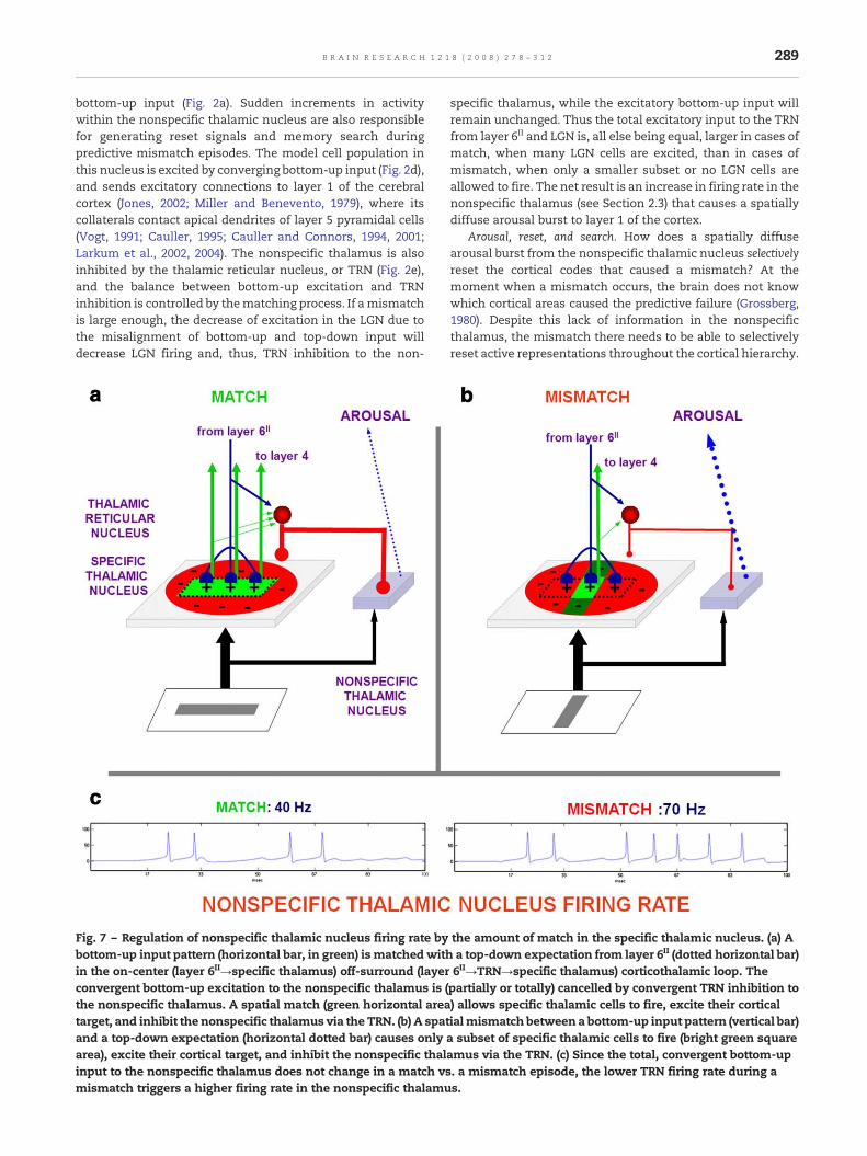
bottom-up input (Fig. 2a). Sudden increments in activitywithin the nonspecific thalamic nucleus are also responsiblefor generating reset signals and memory search duringpredictive mismatch episodes. The model cell population inthis nucleus is excited by converging bottom-up input (Fig. 2d),and sends excitatory connections to layer 1 of the cerebralcortex (Jones, 2002; Miller and Benevento, 1979), where itscollaterals contact apical dendrites of layer 5 pyramidal cells(Vogt, 1991; Cauller, 1995; Cauller and Connors, 1994, 2001;Larkum et al., 2002, 2004). The nonspecific thalamus is alsoinhibited by the thalamic reticular nucleus, or TRN (Fig. 2e),and the balance between bottom-up excitation and TRNinhibition is controlled by thematching process. If amismatchis large enough, the decrease of excitation in the LGN due tothe misalignment of bottom-up and top-down input willdecrease LGN firing and, thus, TRN inhibition to the non-
Fig. 7 – Regulation of nonspecific thalamic nucleus firing rate bybottom-up input pattern (horizontal bar, in green) is matchedwithin the on-center (layer 6II→specific thalamus) off-surround (layerconvergent bottom-up excitation to the nonspecific thalamus is (the nonspecific thalamus. A spatial match (green horizontal areatarget, and inhibit the nonspecific thalamus via the TRN. (b) A spatand a top-down expectation (horizontal dotted bar) causes onlyarea), excite their cortical target, and inhibit the nonspecific thalinput to the nonspecific thalamus does not change in a match vsmismatch triggers a higher firing rate in the nonspecific thalamu
specific thalamus, while the excitatory bottom-up input willremain unchanged. Thus the total excitatory input to the TRNfrom layer 6II and LGN is, all else being equal, larger in cases ofmatch, when many LGN cells are excited, than in cases ofmismatch, when only a smaller subset or no LGN cells areallowed to fire. The net result is an increase in firing rate in thenonspecific thalamus (see Section 2.3) that causes a spatiallydiffuse arousal burst to layer 1 of the cortex.
Arousal, reset, and search. How does a spatially diffusearousal burst from the nonspecific thalamic nucleus selectivelyreset the cortical codes that caused a mismatch? At themoment when a mismatch occurs, the brain does not knowwhich cortical areas caused the predictive failure (Grossberg,1980). Despite this lack of information in the nonspecificthalamus, the mismatch there needs to be able to selectivelyreset active representations throughout the cortical hierarchy.
the amount of match in the specific thalamic nucleus. (a) Aa top-down expectation from layer 6II (dotted horizontal bar)6II→TRN→specific thalamus) corticothalamic loop. Thepartially or totally) cancelled by convergent TRN inhibition to) allows specific thalamic cells to fire, excite their corticalialmismatch between a bottom-up input pattern (vertical bar)a subset of specific thalamic cells to fire (bright green squareamus via the TRN. (c) Since the total, convergent bottom-up. a mismatch episode, the lower TRN firing rate during as.

290 B R A I N R E S E A R C H 1 2 1 8 ( 2 0 0 8 ) 2 7 8 – 3 1 2
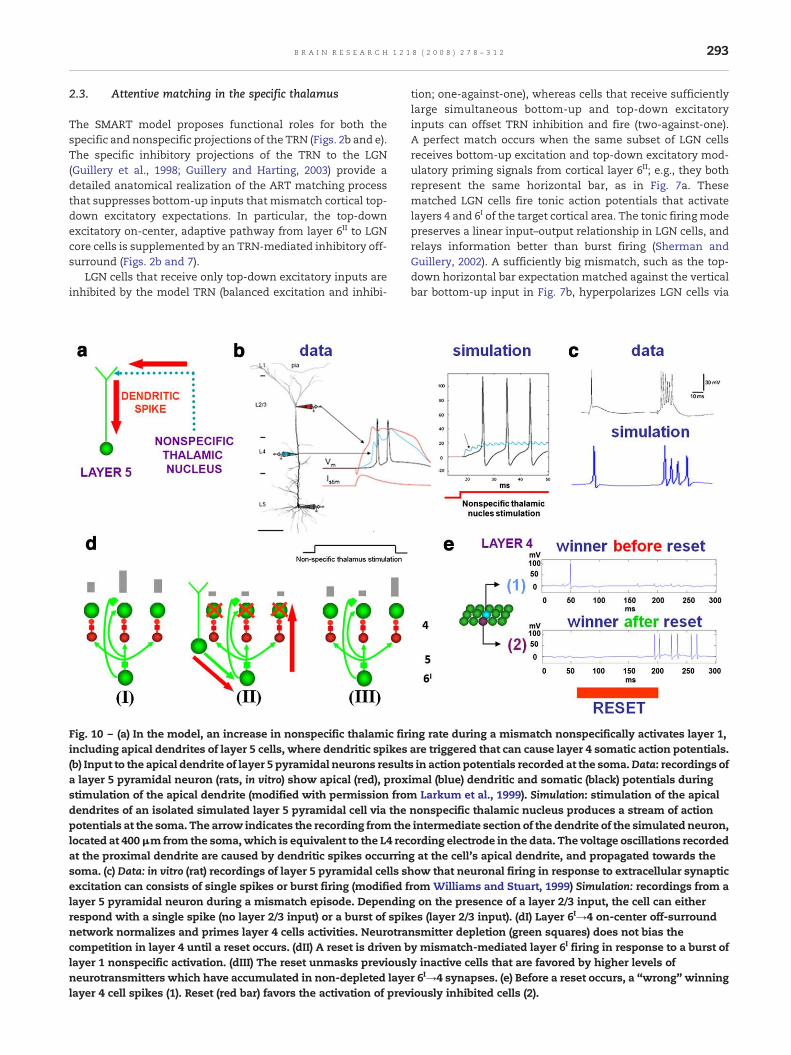
SMART proposes, in accord with known anatomical andphysiological data both in vivo and in vitro (Larkum et al.,1999; Larkum and Zhu, 2002), that layer 5 pyramidal cell firingrate is jointly controlled by nonspecific thalamic inputs andspecific layer 2/3 inputs, thus explaining how layer 5 cellsexhibit two distinct firing modes (Williams and Stuart, 1999):Layer 5 cells that receive layer 2/3 inputs and nonspecificthalamic inputs during a mismatch episode fire in bursts athigh rates (see Section 2.6 for experimental and simulationresults). Active 2/3 cells represent cortical codes that causedthe mismatch. In contrast, single spikes are produced in layer5 cells when only one of these sources is activated, eitherduring a match, or during a mismatch when layer 2/3 cells areinactive.
As noted above, layer 5 pyramidal cells send driving inputsdirectly to higher cortices through the thalamus (e.g., thepulvinar; Sherman and Guillery, 2002; Shipp, 2003; see Fig. 3a),indirectly control corticothalamic feedback at their own cor-tical level through layer 6II (Figs. 2b and 3a), and also controlcorticocortical feedback to layer 4 at their own cortical level vialayer 6I (Figs. 2c and 3c). Layer 5 can hereby generate wide-spread bursts of synchronized activity throughout the neo-cortex mediated by driving layer 5 terminations on higher-order thalamic nuclei (including pathological epileptogenicactivity; Williams and Stuart, 1999), and selectively reset mul-tiple cortical areas by relaying from the nonspecific thalamuslayer 5 bursts to layer 4 via the 6I→4 pathway. In particular,model layer 6I cells are predicted to respond to a thalamicmismatch with selective cortical reset and search for a more
Fig. 8 – Burst and tonic firing in thalamic relay cells. Data: intraceof the low threshold spike for a geniculate relay cell. The same dholding potentials causes either tonic firing (top, cell depolarizedde-inactivated). Modified and reprinted with permission from Sh(horizontal bar) is injected in a simulated LGN cell in the absenceThe hyperpolarization of the cell and the presence of low-threshde-inactivation of the IT current, inducing burst firing.
predictive cortical code in layers 4 and 2/3 (see Section 2.6 forsimulation results).
The nonspecific pathway may also help to regulatemodality-specific attention during reset episodes (Crick,1984; Guillery et al., 1998; Montero, 1997; Weese et al., 1999).SMART predicts how cortical areas that experience strongpredictive mismatches in a given modality may reducepriming of the cortical area of a competing modality byinhibiting the corresponding nonspecific thalamic nucleus. Inparticular, Crabtree and Isaac (2002) have shown that non-specific thalamic nuclei which subserve different modalitiesare linked by mutually inhibitory interactions. SMART simu-lates how TRN-mediated (van Der Werf et al., 2002) inhibitoryinteractions (Crabtree and Isaac, 2002) between nonspecificthalamic nuclei can cause a pause in firing of one nonspecificthalamic nucleus that can transiently down-regulate layer 5pyramidal cells of the competing cortical area (see simulationsin Section 2.9). SMART further predicts that competing specificnuclei, not only nonspecific nuclei as shown by Crabtree andIsaac (2002), might be inhibited by the TRN in cases of strongmismatches, therefore being a possible thalamic substrate forcompetitive allocation of attention.
Learned generalization, vigilance, and acetylcholine. How is thegenerality of recognition categories regulated to representstatistical properties of the environment? As noted above, ARTpredicts that resonance and learning occur when the degree ofmatch between bottom-up and top-down representations isgreater than a gain parameter, called vigilance (see Fig. 4).Vigilance can change due to internal factors, such as fatigue,
llularly in vitro recording illustrating the voltage dependencyepolarizing current pulse administered at two different initial, IT inactivated) or burst firing (bottom, cell hyperpolarized, ITerman and Guillery, 2002. Simulation: a 0.3 nA current(top) or presence (bottom) of a hyperpolarizing voltage clamp.old Ca++ currents (see Eqs. (21)–(27) in Methods) causes the

291B R A I N R E S E A R C H 1 2 1 8 ( 2 0 0 8 ) 2 7 8 – 3 1 2
or external factors, such as predictive mismatch or punish-ment. A baseline vigilance determines how big a mismatch isinitially tolerated before cortical representations are reset.When a predictive error causes a mismatch to occur, thevigilance level is predicted to increase just enough to drive amemory search for a new recognition code. This process iscalled match tracking (Carpenter and Grossberg, 1987; 1991;Carpenter et al., 1992). Match tracking realizes a kind ofminimax learning rule; namely, it enables a learning system tominimize predictive error while maximizing generalization.Choosing a low baseline vigilance leads to the learning ofgeneral categories and thus a minimum use of memoryresources. Match tracking increases this baseline vigilancejust enough to learn the most general categories that areconsistent with predictive success.
The SMART model predicts that one way to controlvigilance may be to modify the excitability of layer 5 cellsduring mismatch episodes (Fig. 5). Anatomical studies inmonkeys, cats and rats have established that the nonspecificthalamus (in particular, the midline and central lateralthalamic nuclei), whose activation is sensitive to the degree
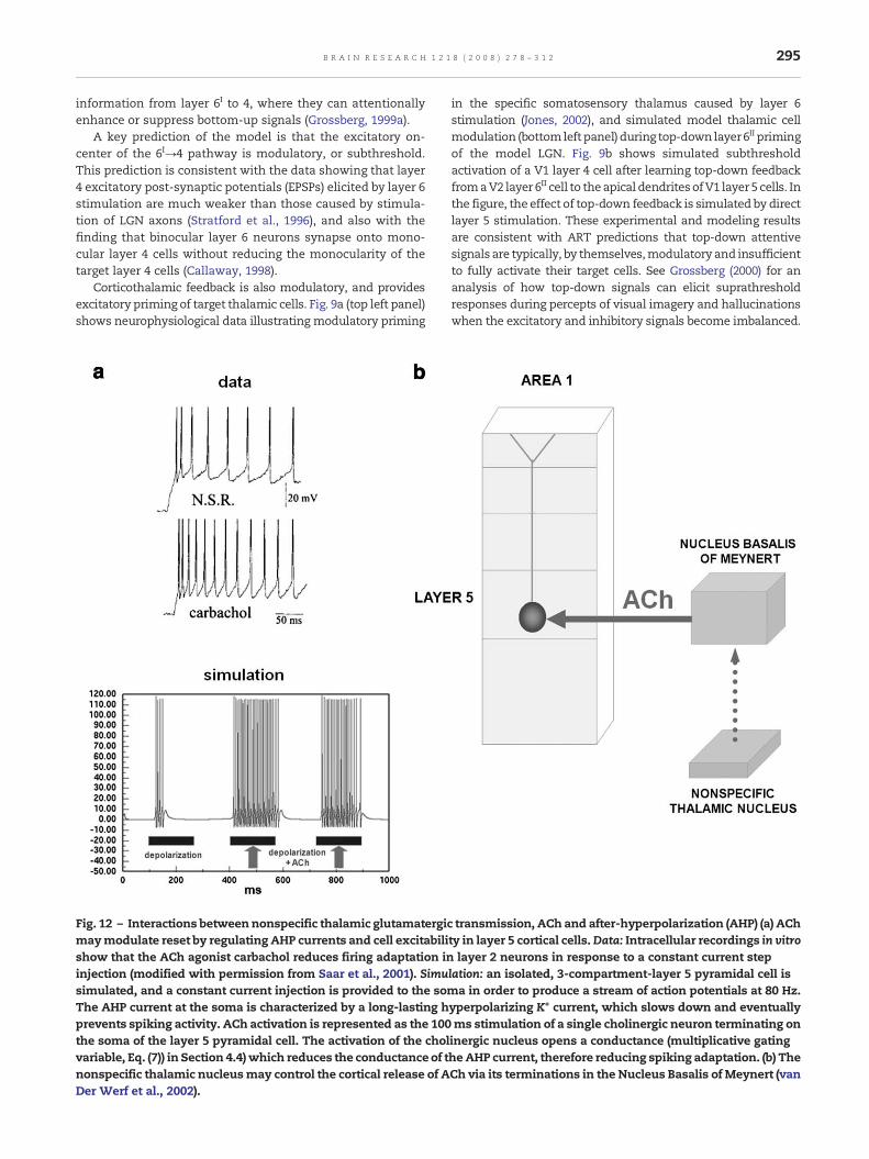
of mismatch, projects to the cholinergic nucleus basalis ofMeynert (van Der Werf et al., 2002), one of the main sources ofcholinergic innervations of the cerebral cortex. The nucleusbasalis of Meynert is also influenced by noxious stimulationand cortical control (Zhang et al., 2004). Saar et al. (2001) haveshown that ACh release reduces the after-hyperpolarization(AHP) current and increases cell excitability in layer 5 corticalcells (see Section 2.8). In SMART, this increased layer 5excitability due to predictive mismatch may cause reset viathe layer 5-to-6I-to-4 circuit, even in cases where top-downfeedbackmay earlier have partiallymatched bottom-up input,which is a key property of vigilance control. The increase ofACh might therefore promote search for finer recognitioncategories in response to environmental feedback, even whenbottom-up and top-down signals have a pretty good match inthe nonspecific thalamus based on similarity alone.
Fig. 3c summarizes all of the simulated SMART circuitry.Table 2 summarizes the main anatomical features simulated,their functional interpretation, and supportive experimentalliterature. TheMethods section provides a detailed descriptionof the model equations and parameters.
292 B R A I N R E S E A R C H 1 2 1 8 ( 2 0 0 8 ) 2 7 8 – 3 1 2
2. Results
2.1. Learning bottom-up oriented filters in the specificpathway
In both the brain and the model, LGN parvalbumin-rich “core”cells receive topographically highly ordered bottom-up sensoryinput andproject to layers 6I and4of cortical areaV1 (Jones 2002,Fig. 1b) in a manner that is sensitive to stimulus orientation(Reid and Alonso, 1995). SMART simulates how adaptivesynapses may become orientationally tuned in the pathwaysfrom LGN core relay cells to V1 layer 4 and layer 6I corticalneurons (Fig. 2a) via postsynaptically gated STDP (Fig. 6a) duringsynchronous match-mediated gamma oscillations.
Fig. 6b illustrates the development of orientation sensi-tivity in a layer 4 cell that wins the competition with itsneighboring cells. It spikes within a few milliseconds after thearrival of the LGN input, while nearby cells are suppressedand their spiking delayed by the on-center off-surround layer6I→4 network. This delay reduces or completely suppresseslearning in cells other than the winning neurons. The gammaoscillations (see Section 2.10) during match episodes allowlayer 4 cells to fire a few ms after LGN cells, and thus withinthe STDP window. The orientation selectivity is expressed interms of LGN→4 synaptic weights (cf., Alonso et al., 2001)before and after a 100 ms exposure to a horizontally-orientedstimulus. Orientationally selective cells in layer 4 of V1 excitelayer 2/3 cells, which in turn project to layer 5 of V1. Layer 5projects to layers 6I and 6II of the same area (Callaway, 1998).Layer 6I closes the layer 2/3→6I→4→2/3 intracortical mod-ulatory excitatory loop that helps select the most activatedcells in layer 4, while strongly suppressing less active cells
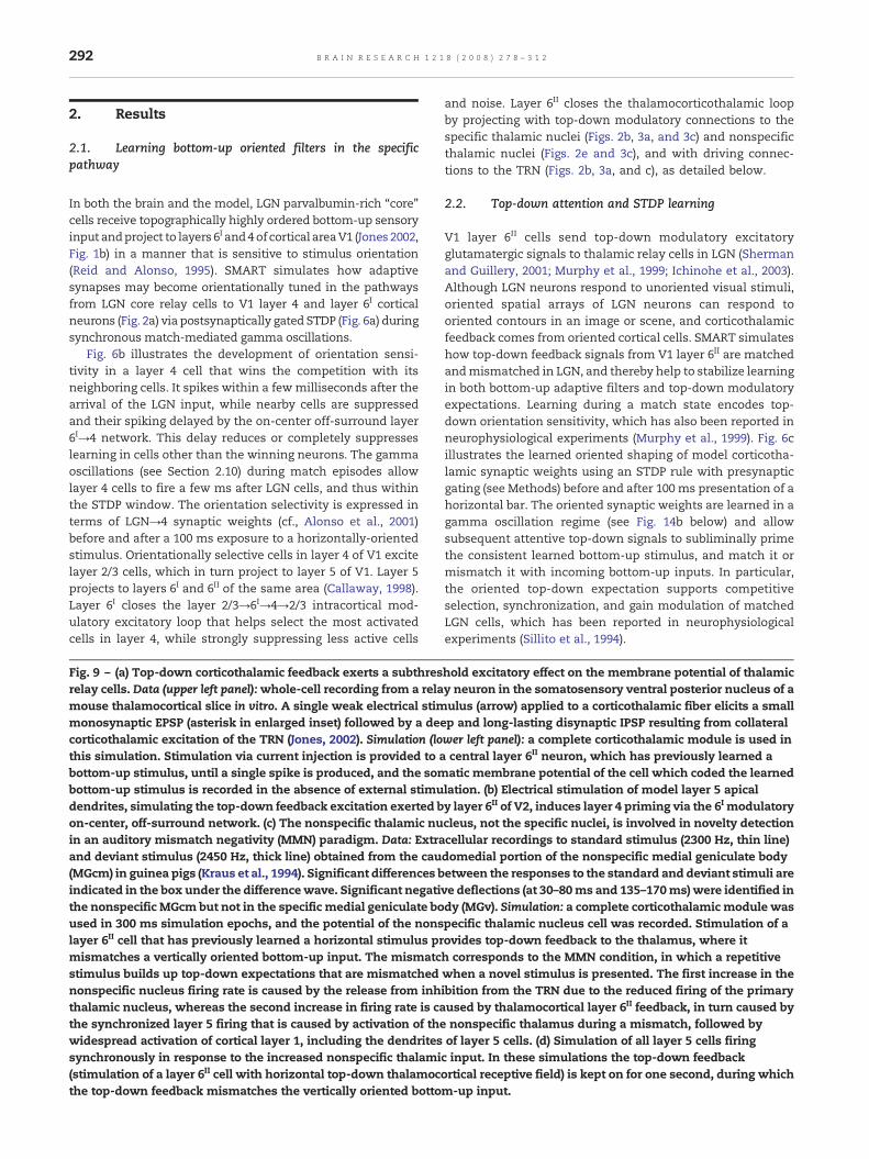
Fig. 9 – (a) Top-down corticothalamic feedback exerts a subthresrelay cells. Data (upper left panel):whole-cell recording from a relamouse thalamocortical slice in vitro. A single weak electrical stimmonosynaptic EPSP (asterisk in enlarged inset) followed by a decorticothalamic excitation of the TRN (Jones, 2002). Simulation (lothis simulation. Stimulation via current injection is provided to abottom-up stimulus, until a single spike is produced, and the sombottom-up stimulus is recorded in the absence of external stimudendrites, simulating the top-down feedback excitation exerted bon-center, off-surround network. (c) The nonspecific thalamic nuin an auditory mismatch negativity (MMN) paradigm. Data: Extraand deviant stimulus (2450 Hz, thick line) obtained from the cau(MGcm) in guinea pigs (Kraus et al., 1994). Significant differences bindicated in the box under the differencewave. Significant negatithe nonspecific MGcm but not in the specific medial geniculate boused in 300 ms simulation epochs, and the potential of the nonslayer 6II cell that has previously learned a horizontal stimulus prmismatches a vertically oriented bottom-up input. The mismatcstimulus builds up top-down expectations that are mismatchednonspecific nucleus firing rate is caused by the release from inhthalamic nucleus, whereas the second increase in firing rate is cathe synchronized layer 5 firing that is caused by activation of thewidespread activation of cortical layer 1, including the dendritessynchronously in response to the increased nonspecific thalami(stimulation of a layer 6II cell with horizontal top-down thalamocthe top-down feedback mismatches the vertically oriented botto
and noise. Layer 6II closes the thalamocorticothalamic loopby projecting with top-down modulatory connections to thespecific thalamic nuclei (Figs. 2b, 3a, and 3c) and nonspecificthalamic nuclei (Figs. 2e and 3c), and with driving connec-tions to the TRN (Figs. 2b, 3a, and c), as detailed below.
2.2. Top-down attention and STDP learning
V1 layer 6II cells send top-down modulatory excitatoryglutamatergic signals to thalamic relay cells in LGN (Shermanand Guillery, 2001; Murphy et al., 1999; Ichinohe et al., 2003).Although LGN neurons respond to unoriented visual stimuli,oriented spatial arrays of LGN neurons can respond tooriented contours in an image or scene, and corticothalamicfeedback comes from oriented cortical cells. SMART simulateshow top-down feedback signals from V1 layer 6II are matchedandmismatched in LGN, and thereby help to stabilize learningin both bottom-up adaptive filters and top-down modulatoryexpectations. Learning during a match state encodes top-down orientation sensitivity, which has also been reported inneurophysiological experiments (Murphy et al., 1999). Fig. 6cillustrates the learned oriented shaping of model corticotha-lamic synaptic weights using an STDP rule with presynapticgating (see Methods) before and after 100 ms presentation of ahorizontal bar. The oriented synaptic weights are learned in agamma oscillation regime (see Fig. 14b below) and allowsubsequent attentive top-down signals to subliminally primethe consistent learned bottom-up stimulus, and match it ormismatch it with incoming bottom-up inputs. In particular,the oriented top-down expectation supports competitiveselection, synchronization, and gain modulation of matchedLGN cells, which has been reported in neurophysiologicalexperiments (Sillito et al., 1994).
hold excitatory effect on the membrane potential of thalamicy neuron in the somatosensory ventral posterior nucleus of aulus (arrow) applied to a corticothalamic fiber elicits a small
ep and long-lasting disynaptic IPSP resulting from collateralwer left panel): a complete corticothalamic module is used incentral layer 6II neuron, which has previously learned aatic membrane potential of the cell which coded the learnedlation. (b) Electrical stimulation of model layer 5 apicaly layer 6II of V2, induces layer 4 priming via the 6I modulatorycleus, not the specific nuclei, is involved in novelty detectioncellular recordings to standard stimulus (2300 Hz, thin line)domedial portion of the nonspecific medial geniculate bodyetween the responses to the standard and deviant stimuli areve deflections (at 30–80ms and 135–170ms) were identified indy (MGv). Simulation: a complete corticothalamic module waspecific thalamic nucleus cell was recorded. Stimulation of aovides top-down feedback to the thalamus, where ith corresponds to the MMN condition, in which a repetitivewhen a novel stimulus is presented. The first increase in theibition from the TRN due to the reduced firing of the primaryused by thalamocortical layer 6II feedback, in turn caused bynonspecific thalamus during a mismatch, followed byof layer 5 cells. (d) Simulation of all layer 5 cells firingc input. In these simulations the top-down feedbackortical receptive field) is kept on for one second, during whichm-up input.
293B R A I N R E S E A R C H 1 2 1 8 ( 2 0 0 8 ) 2 7 8 – 3 1 2
2.3. Attentive matching in the specific thalamus
The SMART model proposes functional roles for both thespecific and nonspecific projections of the TRN (Figs. 2b and e).The specific inhibitory projections of the TRN to the LGN(Guillery et al., 1998; Guillery and Harting, 2003) provide adetailed anatomical realization of the ART matching processthat suppresses bottom-up inputs that mismatch cortical top-down excitatory expectations. In particular, the top-downexcitatory on-center, adaptive pathway from layer 6II to LGNcore cells is supplemented by an TRN-mediated inhibitory off-surround (Figs. 2b and 7).
LGN cells that receive only top-down excitatory inputs areinhibited by the model TRN (balanced excitation and inhibi-
Fig. 10 – (a) In the model, an increase in nonspecific thalamic firincluding apical dendrites of layer 5 cells, where dendritic spikes(b) Input to the apical dendrite of layer 5 pyramidal neurons resulta layer 5 pyramidal neuron (rats, in vitro) show apical (red), proxstimulation of the apical dendrite (modified with permission fromdendrites of an isolated simulated layer 5 pyramidal cell via thepotentials at the soma. The arrow indicates the recording from thelocated at 400μm from the soma,which is equivalent to the L4 recat the proximal dendrite are caused by dendritic spikes occurrinsoma. (c) Data: in vitro (rat) recordings of layer 5 pyramidal cells sexcitation can consists of single spikes or burst firing (modified flayer 5 pyramidal neuron during a mismatch episode. Dependinrespond with a single spike (no layer 2/3 input) or a burst of spiknetwork normalizes and primes layer 4 cells activities. Neurotracompetition in layer 4 until a reset occurs. (dII) A reset is driven blayer 1 nonspecific activation. (dIII) The reset unmasks previousneurotransmitters which have accumulated in non-depleted layelayer 4 cell spikes (1). Reset (red bar) favors the activation of prev
tion; one-against-one), whereas cells that receive sufficientlylarge simultaneous bottom-up and top-down excitatoryinputs can offset TRN inhibition and fire (two-against-one).A perfect match occurs when the same subset of LGN cellsreceives bottom-up excitation and top-down excitatory mod-ulatory priming signals from cortical layer 6II; e.g., they bothrepresent the same horizontal bar, as in Fig. 7a. Thesematched LGN cells fire tonic action potentials that activatelayers 4 and 6I of the target cortical area. The tonic firing modepreserves a linear input–output relationship in LGN cells, andrelays information better than burst firing (Sherman andGuillery, 2002). A sufficiently big mismatch, such as the top-down horizontal bar expectation matched against the verticalbar bottom-up input in Fig. 7b, hyperpolarizes LGN cells via
ing rate during a mismatch nonspecifically activates layer 1,are triggered that can cause layer 4 somatic action potentials.s in action potentials recorded at the soma.Data: recordings ofimal (blue) dendritic and somatic (black) potentials during
Larkum et al., 1999). Simulation: stimulation of the apicalnonspecific thalamic nucleus produces a stream of actionintermediate section of the dendrite of the simulated neuron,ording electrode in the data. The voltage oscillations recordedg at the cell's apical dendrite, and propagated towards thehow that neuronal firing in response to extracellular synapticrom Williams and Stuart, 1999) Simulation: recordings from ag on the presence of a layer 2/3 input, the cell can eitheres (layer 2/3 input). (dI) Layer 6I→4 on-center off-surroundnsmitter depletion (green squares) does not bias they mismatch-mediated layer 6I firing in response to a burst ofly inactive cells that are favored by higher levels ofr 6I→4 synapses. (e) Before a reset occurs, a “wrong” winningiously inhibited cells (2).

294 B R A I N R E S E A R C H 1 2 1 8 ( 2 0 0 8 ) 2 7 8 – 3 1 2
layer 6II→TRN→LGN feedback, and then voltage-dependent T(transient) type Ca+ currents causing burst firing (Shermanand Guillery, 2001). The model exhibits both the tonic and theburst firing modes that are found in the data (Fig. 8).
Consistent with Sherman and Guillery (2002), SMARTclarifies how burst firing might help to switch attention to amodalitywherea suddenbottom-upstimulusoccurs, aswhenasudden visual cue occurs while paying attention to an auditorystimulus. This mechanism complements the nonspecific tha-lamus-mediated mismatch, which can use vigilance control tocause mismatches across multiple modalities. Indeed, usingvigilance control, even if amodality experiences amatch that isgood enough to predict an outcome elsewhere in the brain (e.g.,seeing a visual object predicts its name), a mismatch with thisoutcome can raises vigilance enough to drive a search withinthe originalmodality for a recognition category that can predictthe outcome better in the future (see Section 2.8).
Fig. 11 – Neurotransmitter dynamics with different values of deprespectively, see Eq. (7) in Methods) and different pre-synaptic fiinduced by current injection until firing rates of 23 Hz (panels a, cpre-synaptic cell membrane potential (top) and the level of neurocause larger and quicker neurotransmitter depletion due to the m(c) Increasing the depletion rate ε from 0.5 to 1 results in larger ncounterbalances the effect of depletion.
2.4. Attentive priming via corticocortical and corticothal-amic feedback connections
As noted above, the STDP rule (Fig. 6a; see Methods) is used tolearn the top-down corticocortical attentive connection fromV2 layer 6II cells to layer 1 apical dendrites of V1 layer 5 corticalcells during presentation of a bottom-up input (Fig. 6d). Thislearning correlates V2 layer 6II cell outputs with retrogradedendritic spikes from V1 layer 5 cells to their layer 1 dendrites(Gorchetchnikov and Grossberg, 2007; Grossberg, 1975, 1982;Johnston et al., 1999). Such learning subsequently allows theV2 layer 6II cell to fire the associated V1 layer 5 cell, and fromthere the corresponding V1 layer 6I cell, which in turn primesV1 layer 4 via the modulatory on-center, off-surround layer6I→4 network. This top-down circuit mediates attention in thenetwork. It embodies the concept of “folded feedback” where-by top-down signals are folded into the bottom-up flow of
letion (inactivation, habituation) and recovery rate (ε and τ,ring frequencies in an isolated layer 6I cell. Stimulation was) or 70 Hz (panels b, d) were generated. Each panel shows thetransmitter at the synapse (bottom). (a, b) Higher firing ratesass action (Eq. (7) in Methods), all else being equal.
eurotransmitter depletion. (d) A faster recovery rate τ
295B R A I N R E S E A R C H 1 2 1 8 ( 2 0 0 8 ) 2 7 8 – 3 1 2
information from layer 6I to 4, where they can attentionallyenhance or suppress bottom-up signals (Grossberg, 1999a).
A key prediction of the model is that the excitatory on-center of the 6I→4 pathway is modulatory, or subthreshold.This prediction is consistent with the data showing that layer4 excitatory post-synaptic potentials (EPSPs) elicited by layer 6stimulation are much weaker than those caused by stimula-tion of LGN axons (Stratford et al., 1996), and also with thefinding that binocular layer 6 neurons synapse onto mono-cular layer 4 cells without reducing the monocularity of thetarget layer 4 cells (Callaway, 1998).
Corticothalamic feedback is also modulatory, and providesexcitatory priming of target thalamic cells. Fig. 9a (top left panel)shows neurophysiological data illustrating modulatory priming
Fig. 12 – Interactions betweennonspecific thalamic glutamatergicmaymodulate reset by regulating AHP currents and cell excitabilishow that the ACh agonist carbachol reduces firing adaptation ininjection (modified with permission from Saar et al., 2001). Simusimulated, and a constant current injection is provided to the somThe AHP current at the soma is characterized by a long-lasting hprevents spiking activity. ACh activation is represented as the 100the soma of the layer 5 pyramidal cell. The activation of the cholvariable, Eq. (7)) in Section 4.4) which reduces the conductance of tnonspecific thalamic nucleusmay control the cortical release of ADer Werf et al., 2002).
in the specific somatosensory thalamus caused by layer 6stimulation (Jones, 2002), and simulated model thalamic cellmodulation (bottomleftpanel) during top-down layer6II primingof the model LGN. Fig. 9b shows simulated subthresholdactivation of a V1 layer 4 cell after learning top-down feedbackfromaV2 layer 6II cell to theapical dendritesofV1 layer 5 cells. Inthe figure, the effect of top-down feedback is simulated by directlayer 5 stimulation. These experimental and modeling resultsare consistent with ART predictions that top-down attentivesignals are typically, by themselves,modulatory and insufficientto fully activate their target cells. See Grossberg (2000) for ananalysis of how top-down signals can elicit suprathresholdresponses during percepts of visual imagery and hallucinationswhen the excitatory and inhibitory signals become imbalanced.
transmission, ACh and after-hyperpolarization (AHP) (a) AChty in layer 5 cortical cells.Data: Intracellular recordings in vitrolayer 2 neurons in response to a constant current step
lation: an isolated, 3-compartment-layer 5 pyramidal cell isa in order to produce a stream of action potentials at 80 Hz.
yperpolarizing K+ current, which slows down and eventuallyms stimulation of a single cholinergic neuron terminating oninergic nucleus opens a conductance (multiplicative gatingheAHP current, therefore reducing spiking adaptation. (b) TheCh via its terminations in the Nucleus Basalis of Meynert (van
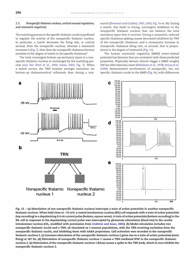
296 B R A I N R E S E A R C H 1 2 1 8 ( 2 0 0 8 ) 2 7 8 – 3 1 2
2.5. Nonspecific thalamicnucleus, cortical arousal regulation,and mismatch negativity
Thematchingprocess in the specific thalamicnuclei is predictedto regulate the activity of the nonspecific thalamic nucleus.In particular, a match decreases the firing rate, or corticalarousal, from the nonspecific nucleus, whereas a mismatchincreases it (Fig. 7). How does the nonspecific thalamus becomesensitive to the degree of match in the specific thalamus?
The total convergent bottom-up excitatory input to a non-specific thalamic nucleus is unchanged by the matching pro-cess (van Der Werf et al., 2002; Jones, 2002; Fig. 7). Whena match occurs, the TRN receives stronger excitation viabottom-up thalamocortical collaterals than during a mis-
Fig. 13 – (a) Stimulation of one nonspecific thalamic nucleus intethalamic nucleus.When held close to −55mV, a rostral intralamin(top recording) to a depolarizing 0.4 nA current pulse (bottom, squRIL cell in response to the depolarizing current pulse was interruintralaminar nucleus (CIL, modified with permission from Crabtrnonspecific thalamic nuclei and a TRN, all simulated as 1-neurononspecific thalamic nuclei, and inhibiting them with GABA prothalamic nucleus 2. (c) Constant stimulation of the nonspecific thafiring) at 187 Hz. (d) Stimulation of nonspecific thalamic nucleusnucleus 2. (e) Stimulation of the nonspecific thalamic nucleus 1 (bnonspecific thalamic nucleus 2.
match (Sherman andGuillery, 2001, 2002; Fig. 7a vs. 5b). Duringa match, this leads to strong, convergent inhibition to thenonspecific thalamic nucleus that can balance the totalexcitatory input that it receives. During a mismatch, reducedspecific thalamus spiking causes decreased inhibition by TRNof the nonspecific thalamus, and a consequent increase innonspecific thalamus firing rate, or arousal, that is propor-tional to the degree of mismatch (Fig. 7c).
The human mismatch negativity (MMN) event-relatedpotential has features that are consistent with these predictedproperties. Physically deviant stimuli trigger a MMN roughly200ms after stimulus onset (Näätänen et al., 1978). Kraus et al.(1994) demonstrated involvement of nonspecific, but notspecific, thalamic nuclei in the MMN (Fig. 9c), with differences
rrupts a train of action potentials in another nonspecificar nucleus (RIL) cell respondswith a train of action potentials
arewave). A train of action potentials (bottom recording) in thepted by glutamate stimulation (black bar) in the caudalee and Isaac, 2002). (b) Model simulation includes twon populations, with the TRN receiving excitation from thejections. Cell activation was recorded at the nonspecificlamic nucleus 2 gives rise to a train of action potentials (tonic1 causes a TRN-mediated IPSP in the nonspecific thalamiclue) causes a spike in the TRN (red), which in turn inhibits the

297B R A I N R E S E A R C H 1 2 1 8 ( 2 0 0 8 ) 2 7 8 – 3 1 2
between novel and standard stimuli at 30–80 ms and 135–170 ms after stimulus onset. The late latency suggests acortical contribution, which involves superficial cortical layers(Karmos et al., 1986).
The SMART model explains and simulates these earlierthalamic and later cortical components using the followingproperties: Mismatch increases nonspecific thalamicnucleus firing at around 50 ms after stimulus onset (Krauset al., 1994; Fig. 9c), reaching cortical layer 1 and causingsynchronized firing in layer 5 (Fig. 9d). The timing of the firstMMN component in SMART is due to synaptic delays andspiking dynamics of the polysynaptic cycle that links LGNand the cortical circuit. During a mismatch, layer 5 apicaldendrites accumulate the incoming EPSPs generated by thenonspecific thalamic nucleus burst until synchronizeddendritic spiking occurs in all layer 5 cells. This layer 5wave then excites layer 6II (Fig. 3c), which in turn reactivatesthe nonspecific thalamic nucleus at around 150 ms afterstimulus onset, thereby causing the second MMN compo-nent. Low threshold Ca+ currents (see Eqs. (21)–(27) in theMethods) in the nonspecific thalamic nucleus generate an
Fig. 14 – Power spectra of cumulative spike histograms of a lamstimulus (horizontal bar, 5 thalamic relay nuclei activated for 100peak in the slowγ frequency band (20–70Hz) in case ofmatch, andanalyzed into three frequency bands (δ and θ, 2–8 Hz; α and β, 8different oscillation frequencies is amatch (c) andmismatch (d). Nfavor of lower-frequency oscillations.
additional burst of spikes. This spiking pattern occurs whena hyperpolarized thalamic cell with low-threshold Ca+ T-current is activated by an excitatory stimulus (Sherman andGuillery, 2001, 2002; Shipp, 2003). These simulation resultsprovide an indirect confirmation of the Karmos et al. (1986)proposal of the cortical origin of the second component inthe MMN.
The SMART model discussion of MMN is related to anearlier prediction about the interpretation of the ART searchcycle that is summarized in Fig. 4; namely, that themismatch, arousal, and STM reset events that are summar-ized in Figs. 4b and c correspond to different human scalp-recorded Event Related Potentials, or ERPs, and that theseevents should tend to co-occur, as they do in the ART searchcycle, if they occur at all. In particular, as ART graduallydeveloped, it predicted that the Processing Negativity, N200(a component of which is MMN), and P300 ERPs correspondto match, arousal, and STM reset events at various levels ofthalamocortical processing (Grossberg, 1978, 1980, 1984).This prediction was supported by ERP experiments using anoddball paradigm with a choice reaction time task, in which
inar primary sensory cortical area during presentation of a0 ms) during match (a) and mismatch (b) conditions show alower frequencies in case ofmismatch. The histogramswere–20 Hz; γ, 20–70 Hz) to highlight the separate contribution ofotably, γ oscillations are drastically reduced in amismatch in

298 B R A I N R E S E A R C H 1 2 1 8 ( 2 0 0 8 ) 2 7 8 – 3 1 2
correlated components of the P120, N200, and P300 behavedlike the predicted mismatch, arousal, and STM reset events(Banquet and Grossberg, 1987).
2.6. Layer 5 regulation of cortical reset
Larkum et al. (1999) found that layer 5 cell dendrites canproduce action potentials that actively propagate to layer 5 cellbodies and cause somatic action potentials (Figs. 10a and b).Until now, there has been no functional explanation of theseregenerative dendritic potentials. Figs. 10b and 10c compare invitro recordings of layer 5 pyramidal apical dendrites andsoma, and model layer 5 cell simulations during match andmismatch episodes.
2.7. Predictive search using mismatch-mediated arousaland habituative synapses
How does a mismatch-mediated layer 5 reset signal choose anew cortical representation that can lead to a better match,and thus a better prediction? How can reset do this without anexternal teacher? How can it be done given that, at themoment of reset, the correct answer is not known, and at thelocation where reset occurs, there is no knowledge of whichactive thalamocortical representations caused the reset? Aproposed solution to this problem (Grossberg, 1980) is hereinrealized using known laminar corticocortical and thalamo-cortical circuits. This solution predicts that the pathwaywhichmediates reset utilizes habituative transmitter gates, alsocalled depressing synapses (Grossberg, 1976, 1980; Carpenter
Fig. 15 – Example of short range (300μm), single unit–single unisimilar orientation preference in V1; top, left: cross-correlation costimulus. top, right: power spectrum of the cross-correlation shofrom Friedman-Hill et al. (2000). Bottom, left: cross-correlation comcells during stimulation of a learned stimulus (lines show the 95cross-correlogram shown on the left.
andGrossberg, 1990; Abbott et al., 1997; Tsodyks andMarkram,1997). In particular, when a bottom-up input froma layer 6I cellactivates its excitatory parthway to layer 4, and its layer 6I-to-4inhibitory interneurons, an activity-dependent fraction ofneurotransmitter in these pathways is released to activatelayer 4 target cells (Fig. 11; see Methods). The transmitterrecovery rate in these pathways is slow relative to its releaserate (see Fig. 11). Thus the net EPSP recorded at a post-synapticsite decreases through time to a habituated level of firing afteran initial burst of activation (Beierlein et al., 2002). Despite thisreduction, synaptic transmission remains unbiased, andstronger inputs produce bigger steady-state EPSPs even asthe corresponding transmitters habituate (Grossberg, 1980;Fig. 10d(I)).
When a layer 5-mediated reset wave later hits layer 6I
(Fig. 10d(II)), this arousal burst changes the balance of totalinput to layer 4 cells. Simulations (Fig. 10e) and mathematicalproofs (Grossberg, 1980; Grossberg and Seidman, 2006) showhow layer 4 cells reset based on their prior activation and thereset wave size, to favor previously inactive or weakly activelayer 4 cells (Fig. 10d(III)).
2.8. Acetylcholine neuromodulation controls vigilance,learning, and generalization
How is the concreteness or abstractness of recognitioncategories controlled in a task-sensitive manner? A clue isprovided by the fact that the nonspecific thalamic nucleuscontrols the excitability of layer 5, and therefore when resetand search for a new recognition category occurs. If the
t correlation of cells with overlapping receptive fields andmputed from the two spike trains during the response to thewn on the left, which shows a peak around 50 Hz. Modifiedputed from the spike trains of two nearby simulated layer 4
% confidence limit). Bottom, right: power spectrum of the
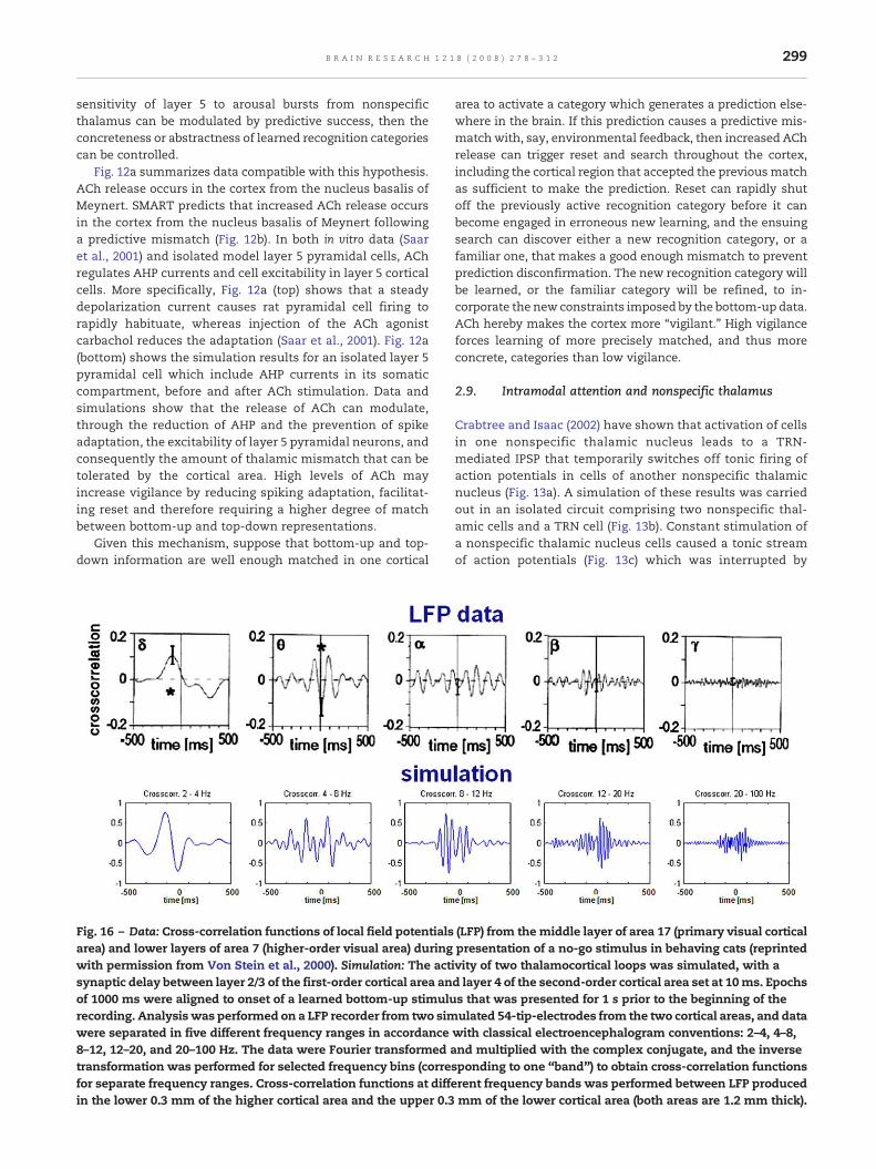
299B R A I N R E S E A R C H 1 2 1 8 ( 2 0 0 8 ) 2 7 8 – 3 1 2
sensitivity of layer 5 to arousal bursts from nonspecificthalamus can be modulated by predictive success, then theconcreteness or abstractness of learned recognition categoriescan be controlled.
Fig. 12a summarizes data compatible with this hypothesis.ACh release occurs in the cortex from the nucleus basalis ofMeynert. SMART predicts that increased ACh release occursin the cortex from the nucleus basalis of Meynert followinga predictive mismatch (Fig. 12b). In both in vitro data (Saaret al., 2001) and isolated model layer 5 pyramidal cells, AChregulates AHP currents and cell excitability in layer 5 corticalcells. More specifically, Fig. 12a (top) shows that a steadydepolarization current causes rat pyramidal cell firing torapidly habituate, whereas injection of the ACh agonistcarbachol reduces the adaptation (Saar et al., 2001). Fig. 12a(bottom) shows the simulation results for an isolated layer 5pyramidal cell which include AHP currents in its somaticcompartment, before and after ACh stimulation. Data andsimulations show that the release of ACh can modulate,through the reduction of AHP and the prevention of spikeadaptation, the excitability of layer 5 pyramidal neurons, andconsequently the amount of thalamic mismatch that can betolerated by the cortical area. High levels of ACh mayincrease vigilance by reducing spiking adaptation, facilitat-ing reset and therefore requiring a higher degree of matchbetween bottom-up and top-down representations.
Given this mechanism, suppose that bottom-up and top-down information are well enough matched in one cortical
Fig. 16 – Data: Cross-correlation functions of local field potentialsarea) and lower layers of area 7 (higher-order visual area) duringwith permission from Von Stein et al., 2000). Simulation: The actsynaptic delay between layer 2/3 of the first-order cortical area anof 1000 ms were aligned to onset of a learned bottom-up stimulurecording. Analysis was performed on a LFP recorder from two simwere separated in five different frequency ranges in accordance8–12, 12–20, and 20–100 Hz. The data were Fourier transformed atransformation was performed for selected frequency bins (correfor separate frequency ranges. Cross-correlation functions at diffein the lower 0.3 mm of the higher cortical area and the upper 0.3
area to activate a category which generates a prediction else-where in the brain. If this prediction causes a predictive mis-matchwith, say, environmental feedback, then increased AChrelease can trigger reset and search throughout the cortex,including the cortical region that accepted the previousmatchas sufficient to make the prediction. Reset can rapidly shutoff the previously active recognition category before it canbecome engaged in erroneous new learning, and the ensuingsearch can discover either a new recognition category, or afamiliar one, that makes a good enough mismatch to preventprediction disconfirmation. The new recognition category willbe learned, or the familiar category will be refined, to in-corporate the new constraints imposed by the bottom-up data.ACh hereby makes the cortex more “vigilant.” High vigilanceforces learning of more precisely matched, and thus moreconcrete, categories than low vigilance.
2.9. Intramodal attention and nonspecific thalamus
Crabtree and Isaac (2002) have shown that activation of cellsin one nonspecific thalamic nucleus leads to a TRN-mediated IPSP that temporarily switches off tonic firing ofaction potentials in cells of another nonspecific thalamicnucleus (Fig. 13a). A simulation of these results was carriedout in an isolated circuit comprising two nonspecific thal-amic cells and a TRN cell (Fig. 13b). Constant stimulation ofa nonspecific thalamic nucleus cells caused a tonic streamof action potentials (Fig. 13c) which was interrupted by
(LFP) from the middle layer of area 17 (primary visual corticalpresentation of a no-go stimulus in behaving cats (reprintedivity of two thalamocortical loops was simulated, with ad layer 4 of the second-order cortical area set at 10ms. Epochss that was presented for 1 s prior to the beginning of theulated 54-tip-electrodes from the two cortical areas, and datawith classical electroencephalogram conventions: 2–4, 4–8,nd multiplied with the complex conjugate, and the inverse
sponding to one “band”) to obtain cross-correlation functionsrent frequency bands was performed between LFP producedmm of the lower cortical area (both areas are 1.2 mm thick).
300 B R A I N R E S E A R C H 1 2 1 8 ( 2 0 0 8 ) 2 7 8 – 3 1 2
the stimulation of a competing nonspecific thalamic cell(Fig. 13d). Crabtree and Isaac (2002) suggested that this effectis due to the TRN, as confirmed by our simulation results inFig. 13e.
2.10. Synchronous oscillation frequency reflects match andmismatch
Gamma (γ, 20–70 Hz) and beta (β, 12–30 Hz) oscillations areobserved in visual cortex during various cognitive, perceptualand attentive states (Singer, 1999; Engel et al., 2001; Hermannet al., 2004; Grossberg, 1976, 2003). Beta oscillations oftencorrelate with long-range synchronous activity of neocorticalregions (Roelfsema et al., 1997), and gamma is restricted tosites within an area (Friedman-Hill et al., 2000) or between twoareas with strong monosynaptic connections (Von Stein et al.,2000). Gamma and beta oscillation frequencies in the modelreflect match and mismatch dynamics, respectively.
Gamma oscillations are amplified between cells of an in-put population, and between cells of an input and a receivingpopulation, when a top-down expectation matches its bot-tom-up input pattern (Figs. 14a and c). During mismatchwithin lower cortical layers, beta oscillations prevail (Figs. 14band d) due to the low frequency, widespread synchronizedfiring of layer 5 cells, as illustrated in Fig. 9d. Layer 1 apicaldendritic potentials slowly depolarize layer 5 cell bodies,causing the low frequency, synchronized spikes of layer 5cells. This frequency component dominates the power spec-trum of the cumulative spike histogram in cases of mis-matches, as illustrated in Figs. 14b and d.
Buffalo, et al. (2004) have reported that gamma oscillationsare more frequent in superficial cortical layers, whereas betaoscillations are found more in deeper cortical layers, consis-tent with the prediction that the beta oscillations may be dueto the initiation of reset in the deeper layers. Their data wouldseem to be due to temporal averaging, since during sustainedperiods of match, one might expect all the cortical layers tosynchronize with the same oscillation frequency due to inter-laminar feedback connections (cf., Yazdanbakhsh and Gross-berg, 2004). This functional interpretation of the gamma/betadifferential may be testable by varying the relative number ofreset events per unit time.
The SMART model also simulates data about short andlong-range synchrony. Friedman-Hill et al. (2000) showedgamma synchronization between two adjacent macaque V1cells with overlapping receptive fields in response to apreferred stimulus (Fig. 15 top). Model V1 layer 4 cells duringmatch of a learned stimulus (Fig. 15 bottom) show a similarcross-correlation power spectrum. These results are consis-tent with the intuition that cells with a shared bottom-upinput would tend to spike in close temporal proximity, andtherefore express high gamma synchronization when nostrong low-frequency synchronization is imposed by mis-match-mediated, synchronized layer 5 discharges.
Von Stein et al. (2000) showed (Fig. 16, top) that synchro-nization between distant cortical areas (middle layers of cor-tical area 17 and lower layers of area 7 in cats) is prevalentin the lower and middle frequency ranges, whereas localinteractions (within areas 17 and 7) show gamma banddominance. The model simulates these properties (Fig. 16,
bottom), showing that synchrony between distant corticalareas is mediated mostly by lower-frequency oscillations.These simulations support the hypothesis that monosynapti-cally connected cells, such as cells within an area or betweennearby areas, can synchronize at gamma frequency bands(Fig. 15), which is compatible with STDP. Top-down interac-tions between lower layers of higher-order and upper levels oflower-order cortical areas are mostly modulatory. Therefore,upper pyramidal layers of lower-order areas should notnecessarily fire in response to a top-down modulatory influ-ence from higher cortical areas, and should not necessarilyexpress gamma frequency synchronization, unless bottom-upand top-down signals match.
3. Discussion
This article describes a model that functionally links single-cell properties, such as spiking dynamics, STDP, and AChmodulation; detailed laminar thalamic and cortical circuitdesigns and their interactions; aggregate cell recordings, suchas current source densities and local field potentials; andsingle-cell and large-scale inter-areal oscillations in thegamma and beta frequency domains, as an expression of thecognitive processing requirements that are needed to regulatefast learning and stablememory of brain representations. As aresult of this wide descriptive range, the model proposesmany testable predictions that link these various levels ofbrain organization as manifestations of how bottom-upadaptive filters and top-down expectations may be learned,matched, and stably remembered during thalamocortical andcorticocortical STDP learning. In particular, the model simu-lates how specific and nonspecific thalamic nuclei regulatelearning via temporal cycles of match/resonance and mis-match/reset, wherein learning is facilitated during match/resonance states and reduced during mismatch/reset states.The predicted involvement of the nonspecific thalamus inlearning is consistent with lesion studies showing a role forthe nonspecific intralaminar/midline thalamic nuclei indeclarative memory (van Der Werf et al., 2003). Moreover,Kraus et al. (1994) have shown that the nonspecific thalamicnuclei, but not the specific thalamic nuclei, show significantdifferential activation in states of match vs. mismatch,consistently with the model prediction that the nonspecificpathway is sensitive to the degree of mismatch betweenbottom-up and top-down cortical signals.
Simulations (Figs. 13c–e) and experimental results (Crab-tree and Isaac, 2002; Fig. 13a) show that the nonspecificthalamic nuclei innervating different cortical areas competevia TRN-mediated inhibitory interactions. The model predictsthat competition in the nonspecific pathway does not inter-fere with bottom-up processing in the specific pathway, butrather transiently decreases the activation of some layer 5cells, therefore preventing the corresponding cortical areafrom further influencing thalamic and cortical areas in thecortical hierarchy. The model suggests that this mechanismmight be a subcortical substrate of cross-modality switchingand competitive deployment of attentional resources. Thisprediction complements the observation (Zeki and Shipp,1988; Sillito et al., 1994) that top-down, layer 6-mediated

301B R A I N R E S E A R C H 1 2 1 8 ( 2 0 0 8 ) 2 7 8 – 3 1 2
attention has similar modulatory effects on target cortical andsubcortical areas even in the absence of bottom-up input(Grossberg, 1999a; Raizada and Grossberg, 2003). In particular,simulation results (Figs. 9a and b) show how layer 6-mediatedtop-down corticothalamic feedback causes both fast primingexcitation and slower inhibitory effects, as has been reportedin experimental data (Jones, 2002).
A novel role for the neurotransmitter acetylcholine ispostulated, linking levels of cortical ACh release to layer 5excitability and layer 4 reset. Experimental results (Singer andRauschecker, 1982; Kilgard andMerzenich, 1998) andmodelingwork (Hasselmo, 1993; Sánchez-Montañés et al., 2000) haveshown that cholinergic modulation is an essential ingredientin cortical plasticity. The model predicts how strong AChrelease, such as during repeated mismatches, or as aconsequence of an environmental stressor, can influence thesharpness of the neural code, by altering the degree of matchrequired for bottom-up and top-down matches to preventreset. Lower levels of ACh favors coarser codes, because higherlevels of mismatch can be tolerated by the system, therebyenabling more variable bottom-up input patterns to beassociated with the same active recognition category withoutcausing recoding.
Themodel for the first timemechanistically links cognitivemechanisms with brain oscillations, notably in the gamma (γ)and beta (β) frequency ranges, recorded from a variety of cor-tical and subcortical structures. Kopell et al. (2000) proposedthat γ and β oscillations might subserve different functionalroles. Their simulations showed that β oscillations are morerobust in synchronizing areas separated by larger transmis-sion delays, whereas γ oscillations tend to be dispersed whensignificant delays are interposed. Olufsen et al. (2003) haveshown that β oscillations allow a different separation between“leading” and “suppressed” cell assemblies than do γ oscil-lations. Gamma oscillations promote a sharp dichotomybetween active/inactive assemblies, a situation similar to a“choice”. The SMART model shows how β oscillations canbecome a signature of modulatory top-down feedback andreset. Top-down processing, in both experimental and modelresults (Fig. 16), shows prevalence of lower-frequency oscilla-tions, consistent with their modulatory nature (Olufsen et al.,2003) and their computational role of priming (Raizada andGrossberg, 2003; Siegel et al., 2000). In accordance with thepresent results, a computational study by Siegel et al. (2000)has shown that top-down feedback is accompanied by anoverall increase of power in the low-frequency domain in thetarget neural population.
The SMART model extends previous modeling work byexplaining how gamma oscillations emerge whenmodulatorytop-down expectations are matched by consistent bottom-upinput patterns. Such a match allows cells to more efficientlycross their spiking threshold to fire action potentials, leadingto an overall increase in local gamma frequency synchroniza-tion among cells sharing common top-down primingmodulation.
The SMARTmodel also links the role of different oscillationfrequencies with STDP. Learning episodes tend to be restrictedto match conditions, when on average presynaptic andpostsynaptic cells spike within 10–20 ms, namely within theSTDP learning window, consistent with experimental results
(Wespatat et al., 2004). The model predicts that STDP furtherreinforces synchronous activation of related cortical andsubcortical areas, and that the effect of spurious synchroniza-tions on long-termmemoryweights in a fast learning regimencan be prevented or rapidly reversed by a synchronousresonance during a match state. Gamma oscillations, ampli-fied in case of a match, may favor propagation of spikesthrough the cortical hierarchy by packing pre-synaptic spikeswithin a narrow temporal window. This prediction is con-sistent with the observation that the efficacy of pairs of pre-synaptic LGN spikes on generating post-synaptic activation inthe visual cortex falls off rapidly in time with the increase ofthe interspike interval (Usrey, 2002).
The different oscillation frequencies associated withmatch/resonance (gamma frequency) and mismatch/reset(beta frequency) link these frequencies not only to selectivelearning, but also to the active search process that candiscover cortical substrates upon which to base new learning.The fact that mismatch is also predicted to be expressed incomponents of the N200 ERP points to new experiments thatcombine ERP and oscillation frequency as indices of thecognitive processes that actively regulate learning.
SMARTpredicts how the nonspecific thalamic nucleusmayplay a crucial role in processing match/mismatch of sensoryinformation and cortical expectations. However, the nonspe-cific thalamic nucleus is not the only structure involved inprocessing novelty. The hippocampus has long been impli-cated as a neural substrate that is sensitive to stimulusnovelty; e.g., Deadwyler, West, and Lynch (1979), Deadwyler,West, and Robinson (1981); O'Keefe and Nadel (1978); Otto andEichenbaum (1992); Vinogradova (1975); see Kumaran andMaguire (2007a) for a review. Previous modeling work (Gross-berg and Schmajuk, 1989; Grossberg and Merrill, 1992, 1996)has proposed how the hippocampus may modulate recogni-tion learning and reinforcement learning by an adaptively-timed inhibition of orienting responses in delayed non-matchto sample paradigms where both temporal delays betweencues and novelty-sensitive recognition processes are involved.This proposed role of the hippocampus as a structure involvedin bridging temporal gaps between temporally-disjoint repre-sentations is consistent with animal (Solomon et al., 1986) andhuman (Clark and Squire, 1998; Büchel et al., 1999) studiesshowing that the hippocampusmaymaintain amemory tracebetween the offset of the conditioned stimulus and thedelayed onset of the unconditioned stimulus to enableassociative learning in trace conditioning. The hippocampushas also been related to sequence novelty, a type of associativenovelty where familiar items appear in a new temporal order(Kumaran and Maguire, 2007b).
SMART explains data consistent with the hypothesis thatfine-grained matches between bottom-up sensory informa-tion and top-down cognitive expectations may occur in thethalamocortical system. The hippocampal role in adaptivetiming, spatial navigation, and declarative (notably, episodic)memory supplements the thalamocortical system with tem-porally and spatially-related expectations (cf. Gorchetchnikovand Grossberg, 2007). When these hippocampal processes aredependent on sensory and cognitive recognition events, thehippocampus might be a recipient, rather than a generator, ofa mismatch response. This hypothesis is consistent with
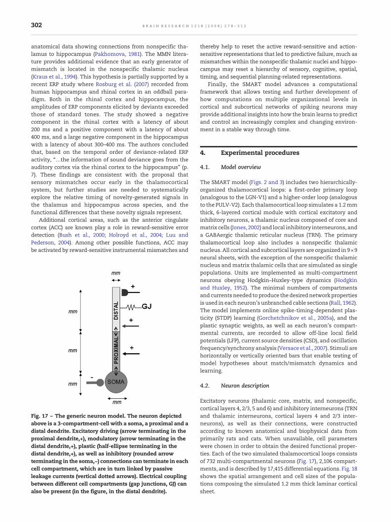
302 B R A I N R E S E A R C H 1 2 1 8 ( 2 0 0 8 ) 2 7 8 – 3 1 2
anatomical data showing connections from nonspecific tha-lamus to hippocampus (Pakhomova, 1981). The MMN litera-ture provides additional evidence that an early generator ofmismatch is located in the nonspecific thalamic nucleus(Kraus et al., 1994). This hypothesis is partially supported by arecent ERP study where Rosburg et al. (2007) recorded fromhuman hippocampus and rhinal cortex in an oddball para-digm. Both in the rhinal cortex and hippocampus, theamplitudes of ERP components elicited by deviants exceededthose of standard tones. The study showed a negativecomponent in the rhinal cortex with a latency of about200 ms and a positive component with a latency of about400 ms, and a large negative component in the hippocampuswith a latency of about 300–400 ms. The authors concludedthat, based on the temporal order of deviance-related ERPactivity, “…the information of sound deviance goes from theauditory cortex via the rhinal cortex to the hippocampus” (p.7). These findings are consistent with the proposal thatsensory mismatches occur early in the thalamocorticalsystem, but further studies are needed to systematicallyexplore the relative timing of novelty-generated signals inthe thalamus and hippocampus across species, and thefunctional differences that these novelty signals represent.
Additional cortical areas, such as the anterior cingulatecortex (ACC) are known play a role in reward-sensitive errordetection (Bush et al., 2000; Holroyd et al., 2004; Luu andPederson, 2004). Among other possible functions, ACC maybe activated by reward-sensitive instrumental mismatches and
Fig. 17 – The generic neuron model. The neuron depictedabove is a 3-compartment-cell with a soma, a proximal and adistal dendrite. Excitatory driving (arrow terminating in theproximal dendrite,+), modulatory (arrow terminating in thedistal dendrite,+), plastic (half-ellipse terminating in thedistal dendrite,+), as well as inhibitory (rounded arrowterminating in the soma,−) connections can terminate in eachcell compartment, which are in turn linked by passiveleakage currents (vertical dotted arrows). Electrical couplingbetween different cell compartments (gap junctions, GJ) canalso be present (in the figure, in the distal dendrite).
thereby help to reset the active reward-sensitive and action-sensitive representations that led to predictive failure, much asmismatches within the nonspecific thalamic nuclei and hippo-campus may reset a hierarchy of sensory, cognitive, spatial,timing, and sequential planning-related representations.
Finally, the SMART model advances a computationalframework that allows testing and further development ofhow computations on multiple organizational levels incortical and subcortical networks of spiking neurons mayprovide additional insights into how the brain learns to predictand control an increasingly complex and changing environ-ment in a stable way through time.
4. Experimental procedures
4.1. Model overview
The SMART model (Figs. 2 and 3) includes two hierarchically-organized thalamocortical loops: a first-order primary loop(analogous to the LGN-V1) and a higher-order loop (analogousto the PULV-V2). Each thalamocortical loop simulates a 1.2mmthick, 6-layered cortical module with cortical excitatory andinhibitory neurons, a thalamic nucleus composed of core andmatrix cells (Jones, 2002) and local inhibitory interneurons, anda GABAergic thalamic reticular nucleus (TRN). The primarythalamocortical loop also includes a nonspecific thalamicnucleus.All cortical and subcortical layers are organized in 9×9neural sheets, with the exception of the nonspecific thalamicnucleus andmatrix thalamic cells that are simulated as singlepopulations. Units are implemented as multi-compartmentneurons obeying Hodgkin–Huxley-type dynamics (Hodgkinand Huxley, 1952). The minimal numbers of compartmentsandcurrentsneeded toproduce thedesirednetworkpropertiesis used in eachneuron's unbranched cable sections (Rall, 1962).The model implements online spike-timing-dependent plas-ticity (STDP) learning (Gorchetchnikov et al., 2005a), and theplastic synaptic weights, as well as each neuron's compart-mental currents, are recorded to allow off-line local fieldpotentials (LFP), current source densities (CSD), and oscillationfrequency/synchrony analysis (Versace et al., 2007). Stimuli arehorizontally or vertically oriented bars that enable testing ofmodel hypotheses about match/mismatch dynamics andlearning.
4.2. Neuron description
Excitatory neurons (thalamic core, matrix, and nonspecific,cortical layers 4, 2/3, 5 and 6) and inhibitory interneurons (TRNand thalamic interneurons, cortical layers 4 and 2/3 inter-neurons), as well as their connections, were constructedaccording to known anatomical and biophysical data fromprimarily rats and cats. When unavailable, cell parameterswere chosen in order to obtain the desired functional proper-ties. Each of the two simulated thalamocortical loops consistsof 732 multi-compartmental neurons (Fig. 17), 2,106 compart-ments, and is described by 17,415 differential equations. Fig. 18shows the spatial arrangement and cell sizes of the popula-tions composing the simulated 1.2 mm thick laminar corticalsheet.

Fig. 18 – Spatial arrangement and cell sizes of the populations composing the 1.2 mm thick laminar cortical sheet. Amulti-compartment model with a spatially explicit structure allows to calculate trans-membrane currents, which can beemployed to derive aggregate cell recordings such as Local Field Potentials (LFP) and Current Source Densities (CSD). Theactivity of dendrites and cell somas of the populations can be recorded by an electrode with variable number of tips (black thickdownward arrow, right). Since each compartment is treated as electrically uniform, the center of the compartment is thephysical point at which the current source/sink is calculated (thick dot at the center of soma/dendrite). Cell bodies and dendritesare vertically aligned parallel to the recording electrode. For each layer, cells are displaced at random distance from therecording electrode. The distance of the electrode to the selected cell in the population is drawn from a uniform distribution onthe interval [10–200] μm, whereas the distance to all other cells in the population is drawn from a uniform distribution on theinterval [10–1000] μm. In the figure, only one cell for each population is shown. Abbreviations: L=layer; S=soma; D=dendrite(D0=proximal dendrite; D1=distal dendrite), INT=interneurons. All measurements are in mm.
303B R A I N R E S E A R C H 1 2 1 8 ( 2 0 0 8 ) 2 7 8 – 3 1 2
Each compartmental membrane potential V[mV] is de-scribed by:
CMdVdt
¼Xi
Ii: ð1Þ
In Eq. (1), CM [μF·cm2] is the membrane capacitance. Ionic orchemically-gated channels and inter-compartmental currentsare described by the current density Ii [μA/cm2] equation:
Ii ¼ gCh VEQ � V� �
; ð2Þ
where the channel conductance gCh and the equilibrium vol-tage VEQ [mV] change according to the nature of the current.Inter-compartmental currents from compartment m to com-partment l are described by Eq. (2), where gCh=gmlDl/4Ll2
[kΩcm], gml is the conductance between compartments m and
l, Dl [mm] and Ll [mm] are the diameter and length of mem-brane compartment l, respectively, and VEQ=Vm and V=Vl
are potentials of the neighboring compartments. Table 3lists dimensions, passive cable properties, and ionic channels(such as sodium (Na++), potassium (K+) and leakage channels)parameters.
In chemically gated channels (AMPA, GABA, etc.) betweenneurons j and k, VEQ=Ei[mV] is the reversal potential of thechannel, and the conductance gCh=wjk gjk g(t), where wjk isthe synaptic weight connecting neurons j and k, and gjk [pS] isthe maximal channel conductance of the synaptic weight wjk.The synaptic weightwjk ¼ Ni
pDL106cm2
h icorresponds to the density of
receptors (millions of channels per membrane cm2). The con-ductance g(t) is defined as a dual exponential factor describingthe time course of the excitatory or inhibitory post-synaptic

Table 3 – Dimensions, passive cable properties and ionic channels parameters of each cellular stage of the thalamocorticalcircuit
Cell Compartmentdiameter
Compartmentlength
Axialresistance
EL gL gNa gK gCa
Units mm mm KΩ cm mV mS/cm2 mS/cm2 mS/cm2 mS/cm2
First-order thalamic relaySoma 0.05 0.06 8 −60 0.01 100 100 –Dendrite 0 0.005 0.008 8 −60 0.01 – – 10Dentric 1 0.005 0.008 8 −60 0.01 – – 10
First-order thalamic (matrix)Soma 0.05 0.06 8 −60 0.01 100 100 –Dendrite 0 0.005 0.008 8 −60 0.01 – – 10Dentric 1 0.005 0.008 8 −60 0.01 – – 10
First-order thalamic interneuronsSoma 0.02 0.02 60 −49 0.01 50 30 –Dendrite 0 0.001 0.1 60 −49 0.01 – – –
TRNSoma 0.05 0.05 10 −69 0.1 100 100 –Dendrite 0 0.01 0.05 10 −69 0.1 – – 10Dentric 1 0.01 0.05 10 −69 0.1 – – 10
Thalamic nonspecificSoma 0.08 0.08 10 −64 0.09 100 100 –Dendrite 0 0.015 0.1 10 −64 0.1 – – 250Dentric 1 0.015 0.1 10 −64 0.1 – – 250
Layer 4 ExcitorySoma 0.05 0.05 40 −65 0.01 50 30 –Dendrite 0 0.01 0.25 40 −65 0.01 – – –
Layer 4 inhibitorySoma 0.02 0.02 100 −50 0.01 50 30 –Dendrite 0 0.01 0.05 100 −50 0.01 – – –
Layer 2/3 excitorySoma 0.05 0.05 100 −65 0.05 50 30 –Dendrite 0 0.02 0.225 100 −65 0.05 – – –
Layer 2/3 inhibitorySoma 0.02 0.02 60 −49 0.01 50 30 –Dendrite 0 0.01 0.05 60 −49 0.01 – – –
Layer 5Soma 0.1 0.15 5 −72 0.1 50 30 –Dendrite 0 0.06 0.4 5 −72 0.03 – – –Dentrite 1 0.06 0.5 5 −72 0.03 50 30 –
Layer 6-I
Soma 0.08 0.1 80 −70 0.15 50 30 –Dendrite 0 0.05 0.1 80 −70 0.9 – – –
Layer 6II
Soma 0.06 0.1 25 −64 0.1 50 30 –Dendrite 0 0.08 0.1 25 −64 0.03 – – –Dentric 1 0.08 0.2 25 −64 0.03 – – –
EL and gL represent the leakage currents equilibrium potential and conductance, respectively. Cell morphologies are the same for cells in thefirst-order and second-order thalamocortical loop.
304 B R A I N R E S E A R C H 1 2 1 8 ( 2 0 0 8 ) 2 7 8 – 3 1 2
potentials (EPSP and IPSP, respectively) triggered by the pre-synaptic spike:
g tð Þ ¼
psf � sr
e� t
sf � e�tsr
� �if sf p sr
tsfe
1� tsf
� �if sf ¼ sr
;
8>>><>>>:
ð3Þ
where t is the time since the onset of a pre-synaptic spike, p is anormalizing coefficient that ensures:
maxp
sf � sre� t
sf � e�tsr
� � !¼ 1 ð4Þ
and τr and τf are the EPSP and IPSP rise and fall time constants,respectively. See Supplementary Table 4 online for parametersof chemically-gated channels.
Unless otherwise specified, simulations were implementedin a full first-order thalamocortical sector including a six-layered cortical structure (V1), a TRN sector, a first-orderthalamic nucleus (LGN), and a nonspecific thalamic nucleus.Eqs. (1)–(27) and (29) below describe the cell properties of thefirst-order thalamocortical sector, whereas Eq. (28) is used onlyin producing the results illustrated in Figs. 12 and 19. Thepassive properties of the cells describing each cellular stage inthe first-order thalamocortical sector are listed in Table 3,
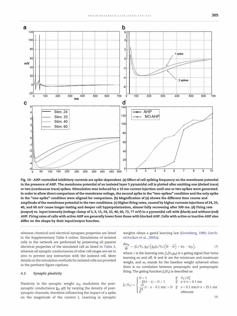
Fig. 19 –AHP-controlled inhibitory currents are spike-dependent. (a) Effect of cell spiking frequency on themembrane potentialin the presence of AHP. Themembrane potential of an isolated layer 5 pyramidal cell is plotted after emitting one (dotted trace)or two (continuous trace) spikes. Stimulation was induced by a 10 ms current injection until one or two spikes were generated.In order to allow direct comparison of themembrane voltage, the second spike in the “two-spikes” condition and the only spikein the “one-spike” condition were aligned for comparison. (b) Magnification of (a) shows the different time course andamplitude of themembrane potential in the two conditions. (c) Higher firing rates, caused by higher currents injections of 24, 33,40, and 60 mV cause longer-lasting and deeper cell hyperpolarization, almost fully recovering after 500 ms. (d) Firing rate(output) vs. input intensity (voltage clamp of 3, 9, 15, 24, 33, 40, 60, 72, 77 mV) in a pyramidal cell with (black) and without (red)AHP. Firing rates of cells with active AHP are generally lower from thosewith blockedAHP. Cells with active or inactive AHP alsodiffer on the shape by their input/output function.
305B R A I N R E S E A R C H 1 2 1 8 ( 2 0 0 8 ) 2 7 8 – 3 1 2
whereas chemical and electrical synapses properties are listedin the Supplementary Table 4 online. Simulations of isolatedcells in the network are performed by preserving all passiveelectrical properties of the simulated cell as listed in Table 3,whereas all synaptic conductances of other cell stages are set tozero to prevent any interaction with the isolated cell. Moredetails on the simulationmethods for isolatedcells areprovidedin the pertinent figure captions.
4.3. Synaptic plasticity
Plasticity in the synaptic weight wjk modulates the post-synaptic conductance g jk g(t) by varying the density of post-synaptic channels, therefore influencing the impact of a spikeon the magnitude of the current Ii. Learning in synaptic
weights obeys a gated learning law (Grossberg, 1980; Gorch-etchnikov et al., 2005a):
dwjk
dt¼ kfG Vk; gjk
� �gjkfN Vkð Þ kw�ww
� �þw0 �wjk
� �; ð5Þ
where λ is the learning rate, fG(Vk,gjk) is a gating signal that turnslearning on and off, w⌣ and w⌢ are the minimum and maximumweight, and w0 stands for the baseline weight achieved whenthere is no correlation between presynaptic and postsynapticfiring. The gating function fG(Vk) is described as:
fG Vkð Þ ¼
Dþ 1 if VkzVhk
�10 t� sð Þ þ Dþ 1 if sbtbsþ 0:1 ms�D25
t� s� 0:1 msð Þ þ D if sþ 0:1 msVtbsþ 25:1 ms0 otherwise
;
8>>><>>>:
ð6Þ

306 B R A I N R E S E A R C H 1 2 1 8 ( 2 0 0 8 ) 2 7 8 – 3 1 2
where Vhk is the spiking threshold potential, t is time, s is the
moment of the postsynaptic spike, and D ¼ kw�w0
� �=
ww�kw� �
.This scaling function is a non-negative function of pre- andpostsynaptic activity and allows synaptic change to occur onlywhen pre- and postsynaptic cell are simultaneously active. SeeGorchetchnikov et al. (2005a) for the derivation and the detaileddiscussion of the rule.
Two forms of gating are used: post-synaptic and dual-ANDgating. Post-synaptic gating, where fG Vk; gjk
� � ¼ Vkð Þ2, is imple-mented in specific thalamic projections terminating in layer 4(bottom-up adaptive weights) and allows the winning layer 4cells to learn the LGN spatio-temporal pattern of activation.Dual-AND gating, where fG Vk; gjk
� � ¼ gjk Vkð Þ2, is implementedin layer 6II projections terminating in the specific thalamus andin layer 1 apical dendrites of layer 5 cells of a previous corticalstage (top-down adaptive weights). This gating allows V2 layer6II cells sending feedbackprojections toV1 layer 5 cells toprime,and then learn the activation pattern of, active layer 5 cells.
4.4. Neurotransmitter release
The neurotransmitter released by the pre-synaptic terminalscales the EPSP or IPSP triggered at the post-synaptic site. Theaccumulation and depletion (inactivation, habituation) ofneurotransmitter zjk at a synapse between neurons j and k isdescribed by (Grossberg, 1980):
dzjkdt
¼ B� zjk� �
s� ed tð Þzjk; ð7Þ
where B=1 is the target level of neurotransmitter at rest,0bεb1 is the depletion coefficient that can scale the amount ofneurotransmitter released at every spike, and 0.1bτb500 is therecovery rate (in ms) regulating the rate of neurotransmitteraccumulation. A spike δ(t) is defined as:
d tð Þ ¼ 1 if V tð Þb0 and V t� Dtð ÞNVh
0 otherwise ;
�ð8Þ
where V (t) is the soma membrane voltage at time t, Vθ is thevoltage threshold that is invariably crossed during spikes(30 mV), V(t−δt) is the soma membrane voltage at time t−δtthat precedes the soma voltage crossing 0 mV. In Eq. (7), theneurotransmitter zjk accumulates towards B at a rate inverselyproportional to the recovery rate τ, and habituates, or isdepleted, by �ed tð Þzjk every time a spike occurs. Neu-rotransmitter depletion allows the EPSP and IPSP to be multi-plicatively gated by the amount of neurotransmitter available,while still ensuring that 0bgjkzjkbB (for all simulations, B=1).Fig. 11 shows how neurotransmitter level varies with differentvalues of ε and τ and different pre-synaptic firing frequencies.
4.5. Ionic currents
Potassium (K+) and Sodium (Na++) currents Ik and INa in Eq. (1)are derived from Traub and Miles (2001), and are described as:
IK ¼ PgKn4 EK � Vð Þ; ð9Þ
where
dndt
¼ a 1� nð Þ � bn; ð10Þ
a ¼ :032 15� Vð Þe15�V
5 � 1; ð11Þ
b ¼ :5e10� V40
; ð12Þ
and
INa ¼ m3hPgNa ENa � Vð Þ; ð13Þ
where
dmdt
¼ a 1�mð Þ � bm; ð14Þ
a ¼ :032 13� Vð Þe13�V
4 � 1; ð15Þ
b ¼ �:28 40� Vð Þe40�V
�5 �1; ð16Þ
dhdt
¼ a 1� hð Þ � bh; ð17Þ
a ¼ :128e27�V18 ; ð18Þ
and
b ¼ 4e 40�V
5 þ 1: ð19Þ
For all neurons, EK=−90 mV and ENa=50 mV. Leakage currentIleak is defined as:
Ileak ¼ � gleakNleak
pDLV; ð20Þ
where gleak is the conductance of the leakage channel, NleakpDL is
the channel density, and Eleak=0 mV.
4.6 Calcium currents in thalamic cells
Low-Threshold T-Type currents (Destexhe, 2000) are implemen-ted in thalamicmatrix, core, TRNandnonspecific thalamiccells,and are described as:
ICa ¼ PgCam3h ECa � Vð Þ; ð21Þ
where
dmdt
¼ 1sm
ml �mð Þ; ð22Þ
sm ¼ 1e�63�V
7:8 þ 1; ð23Þ
ml ¼ 2:44þ 2:506d10�2e�9:84d10�2V ; ð24Þ
dhdt
¼ 1sh
hl � hð Þ; ð25Þ
sh ¼ 1�83�V6:3 þ 1
; ð26Þ
and
hl ¼ 19:5þ 7:171d10�2e�10:54d10�2V : ð27Þ

307B R A I N R E S E A R C H 1 2 1 8 ( 2 0 0 8 ) 2 7 8 – 3 1 2
For all neurons with Ca++ currents, PgCa ¼ 250 mS=cm2, andECa=180 mV.
4.7. Cholinergic modulation and after-hyperpolarizationcurrents (AHP)
Pharmacological and physiological studies have demonstratedthat ACh has facilitatory effects on cortical pyramidal neurons(McCormick and Prince, 1985), and rat cortical layer 5 cells seemto be a preferential target for cholinergic innervation (Turrini etal., 2001). The known electrophysiological excitatory action isthought to be mediated by binding of ACh to muscarinic and/ornicotinic receptors on pyramidal neurons. This causes areduction of membrane K+ conductance in cortical neurons,enhancing depolarization in response to glutamatergic input(McCormick and Prince, 1985) and reducing spike adaptationdue to the after-hyperpolarization current (AHP, Hille, 2001)based on a slow and long-lasting increase in K+ conductance.
AHP and its modulation by acetylcholine are modeled by:
Ii ¼ gCh VEQ � V� �
;
where the AHP current conductance gCh ¼ g⁎PgAHPg tð Þ is modu-lated by the conductance g⁎ controlled by the cholinergicpresynaptic spike, and PgAHP[nS] is the maximal K+ conduc-tance of the AHP channel. The AHP conductance g(t) isdescribed by Eq. (3):
g tð Þ ¼
psf � sr
e� t
sf � e�tsr
� �if sf p sr
tsfe
1� tsfÞ if sf ¼ sr
�8>>><>>>:
where t is time since the action potential of a modulatory cell;p is the scaling coefficient described in Eq. (4). For the AHP usedin these simulations τ r and τ f, namely the rise and fall timeconstants, respectively, are τ r=80 ms and τ f=100 ms. The K+
channels responsible for the AHP are opened by any cell'saxonal output. If there is no spike, t=∞, therefore g=0. If thereis a spike, t=0, causing g to rise. The effect of a cell's spike onthe soma membrane potential in the presence of AHP isillustrated in Fig. 19.
The cholinergic modulation conductance g⁎ is described by
g⁎ ¼1� p
sf � sre� t
sf � e�tsr
� �if sf p sr
1� tsfe
1 � tsf
� �if sf ¼ sr
;
8>>><>>>:
ð28Þ
where τr=5 ms and τf=6 ms, and t is the time since the pre-synaptic cholinergic cell spikes (nucleus basalis of Maynert).These simulations investigate only the fast cholinergic dy-namics, and donot address longer-lasting effect of ACh on targetneural populations (Hasselmo, 1995). The cholinergic input actsby closing the normally open gate g⁎, therefore limiting the totalAHP conductance when ACh modulation is active.
4.8. Network connectivity
Connections between and within cell populations link a pre-synaptic cell with a given postsynaptic cell compartment tar-
get of the axonal projection, and can be categorized as: 1-to-1,1-to-many, or many-to-1 projections. Synaptic weightswij canbe defined between and within layers according to:
wij ¼1ffiffiffiffiffiffiffiffiffiffiffi2pr2
p e� x�Að Þ2=2r2 ; ð29Þ
where μ is the mean and σ is the standard deviation. Eachaxonal pathway can be considered as a delay line, which addsan additional component of delay between pre- and post-synaptic spike, aside from the time required by EPSPs to triggeran action potential. Axonal delays were chosen to be con-sistent with both known transmission delays in cortical andsubcortical areas, and with the spatial conformation of themodel, and are summarized in Supplementary Table 4 online.In general, inhibitory to excitatory connections have smalldelays, intra-cortical feedforward connections have longerdelays (Dinse and Kruger, 1994), and feedback connections(both corticocortial and corticothalamic) have even longerdelays (Schmolesky et al., 1998; Miller, 1996).
The model accounts for the driving vs. modulatory natureof synaptic connections by exploiting both the magnitude ofthe synaptic weight and the passive neuron cable properties.An EPSP occurring at the distal dendrite tends to be attenuatedwith respect to the one occurring at the proximal dendrite orthe soma depending on compartmental length and diameter.Inter-compartmental currents are described by Eq. (2), wheregCh=gml ·Dl/4Ll2 [kΩ⁎cm], and where VEQ and V represent thevoltage of neighboring compartments. The longer and smallerthe dendrite, the more attenuated the post-synaptic currentwill be at the soma. Differential dendritic termination andsynaptic weight magnitudes can be used to simulate theproposed functional differentiation between driving, large,round (R-type) thalamic vesicles occurring at retinothalamicand thalamocortical synapses, and elongated, small vesiclesthat characterize many corticothalamic terminations (Rock-land, 1998; Sherman and Guillery, 2001). Elongated and round-type synapses also widely occur a the level of corticocortialsynapses (Rockland, 2002). Besides the different morphologyof synaptic boutons, inputs to thalamic relay cells are notdistributed evenly on their dendrites (Guillery, 1969; Wilson etal., 1984; Erişir et al., 1997). Retinal and parabrachial inputs arelimited to proximal dendrites, while cortical inputs are locatedmore distally. The SMART model captures these morphologi-cal and functional characteristics by concentrating drivingconnections in proximal dendrites, and modulatory connec-tions with smaller synaptic weights in distal dendrites of thetarget cell. These characteristics are realized between neuronsin the on-center/off-surround architecture implemented byinhibitory interneurons in cortical and thalamic areas.
4.9. Stimuli and initial conditions
Each cell compartment is initialized at the respective restingmembrane potential (see Table 3 online for passive cableproperties and ionic channels conductances). Bottom-upstimuli consist of static vertical or horizontal bars centeredon the 9×9 receptor grid, and implemented via fixeddepolarizing voltage clamp (holding potential=−12 mV; mem-brane resting potential/leakage=−60 mV) to 5 vertically orhorizontally aligned thalamic specific relay neurons until a

308 B R A I N R E S E A R C H 1 2 1 8 ( 2 0 0 8 ) 2 7 8 – 3 1 2
stream of action potential is induced at a firing rate of 40 Hz.When indicated, top-down feedback is induced by injecting astimulating current to the soma of a layer 6II cortical cell. Atypical run of themodel consists of 100ms epoch, or 1000ms inrecording of neural synchrony, with the membrane potentialsof all neuronal compartments recorded along with the plasticsynaptic weights configuration before and after the run.
4.10. Oscillations analysis
Power analysis of single or collective neural signals allows theextraction of information contained in different frequencyranges. Where indicated, analysis of 1000 ms epochs isperformed separately in three different frequency ranges: 2–8(delta and theta, δ and θ), 8–10 (alpha and beta, α and β) and 20–70 Hz (gamma, γ). The mean firing rate is subtracted from thedata, and a Hamming window of 200 ms is applied to smooththe resulting values. The results are then Fourier transformedand multiplied with the complex conjugate (cross- and auto-components), and the inverse transformation is performed forselected, continuous frequency bins (corresponding to one‘‘band'’). In this way, it is possible to reconstruct a time-averaged firing rate for selected frequency ranges. These valuescan then be used to compute cross- and auto-correlations atdifferent frequency ranges (Von Stein et al., 2000).
4.11. Local Field Potential and Current Source Densityanalysis
Cortical LFP and CSD are recorded via a simulated 54-tip-electrode. The distance of the electrode to the selected cell inthe population is drawn from a uniform random distribution inthe interval [10–200] μm, whereas the distance to all other cellsin the layer is drawn from a uniform randomdistribution in theinterval [10–1000] μm. An extracellular inward current flowtowards the interior of the cell creates a current sink, while anoutside flow creates a current source in a particular membranesection. Assuming an extracellular fluid with constant con-ductance, the potential generated by such current dipole is:
Ve ¼ 14pr
Iþ
rþþ I�
r�
� �; ð30Þ
where I-s and r-s are currents and distances between theelectrode and the point where the respective current flowsthrough the membrane (approximated by the center of thecompartment), respectively,+ and − mark the attributes ofsource and sink, respectively, and σ=15 [mS/cm] is theextracellular conductivity. In the case of more complex cellswith many possible sources and sinks, Ve becomes:
Ve ¼ 14pr
Xl
Ilrl: ð31Þ
Compartmental trans-membrane currents Il[μA] are ex-pressed in terms of:
Il ¼ JlSl ¼ � glDl
4L2lVl � VlF1ð Þ 2
pD2l
4þ pDlLl
� �
¼ � glpD2l
4L2lVl � VlF1ð Þ Dl
2þ Ll
� �: ð32Þ
Since CSD and LFP can be measured with multipleelectrodes, for each electrode tip the distance rl to thecompartment center is different. CSD is calculated both inexperimental studies and in the present work by linearapproximation of the second derivative of the voltage:
CSD ¼ Veþ1 þ Ve�1 � 2Ve
Dx; ð33Þ
where Δx is the distance between neighboring electrode tips.
4.12. Simulation environment
The model is implemented in KInNeSS (KDE IntegratedNeuroSimulation Software, www.kinness.net), a softwarepackage that allows the simulation of single neurons withmultiple compartments as well as large networks of suchelements (Versace et al., 2007). All off-line data analysis isimplemented in Matlab (Mathworks Inc.). Simulations are runon 2.80 GHz Intel CPU, 1 GB of RAM, under Linux operatingsystem. The network is described in Neuro Markup Languagecode (NeuroML, http://www.neuroml.org/). NeuroML is avariation of XML designed for modeling different aspects andlevels of neural systems, from intracellular mechanisms andion channel kinetics to the dynamics of networks of recon-structed neurons. The code is downloadable in the Researchsection at http://www.kinness.net/.
Appendix A. Supplementary Table 4
SupplementaryTable4 associatedwith this article canbe found,in the online version, at doi:10.1016/j.brainres.2008.04.024.
R E F E R E N C E S
Abbott, L.F., Sen, K., Varela, J.A., Nelson, S.B., 1997. Synapticdepression and cortical gain control. Science 275, 220–222.
Ahissar, M., Hochstein, S., 2002. View from the top: herarchies andreverse hierarchies in the visual system. Neuron 36, 791–804.
Ahmed, B., Anderson, J.C., Martin, K.A.C., Nelson, J.C., 1997. Map ofthe synapses onto layer 4 basket cells of the primary visualcortex of the cat. J. Comp. Neurol. 380, 230–242.
Alonso, J.M., Usrey, W.M., Redi, R.C., 2001. Rules of connectivitybetween geniculate cells and simple cells in cat primary visualcortex. J. Neurosci. 21, 4002–4015.
Banquet, J.P., Grossberg, S., 1987. Probing cognitive processesthrough the structure of event-related potentials duringlearning: An experimental and theoretical analysis. Appl. Opt.26, 4931–4946.
Bazhenov, M., Timofeev, I., Steriade, M., Sejnowski, T.J., 1998.Computational models of thalamocortical augmentingresponses. J. Neurosci. 18, 6444–6465.
Beierlein, M., Fall, C.P., Rinzel, J., Yuste, R., 2002. Thalamocorticalbursts trigger recurrent activity in neocortical networks: layer 4as a frequency-dependent gate. J. Neurosci. 22, 9885–9894.
Bi, G.Q., Poo, M., 2001. Synaptic modification by correlatedactivity: Hebb's postulate revisited. Annu. Rev. Neurosci. 24,139–166.
Blasdel, G.G., Lund, J.S., 1983. Termination of afferent axons inmacaque striate cortex. J. Neurosci. 3, 1389–1413.
Bosking, W., Zhang, Y., Schofield, B., Fitzpatrick, D., 1997.Orientation selectivity and the arrangement of horizontal
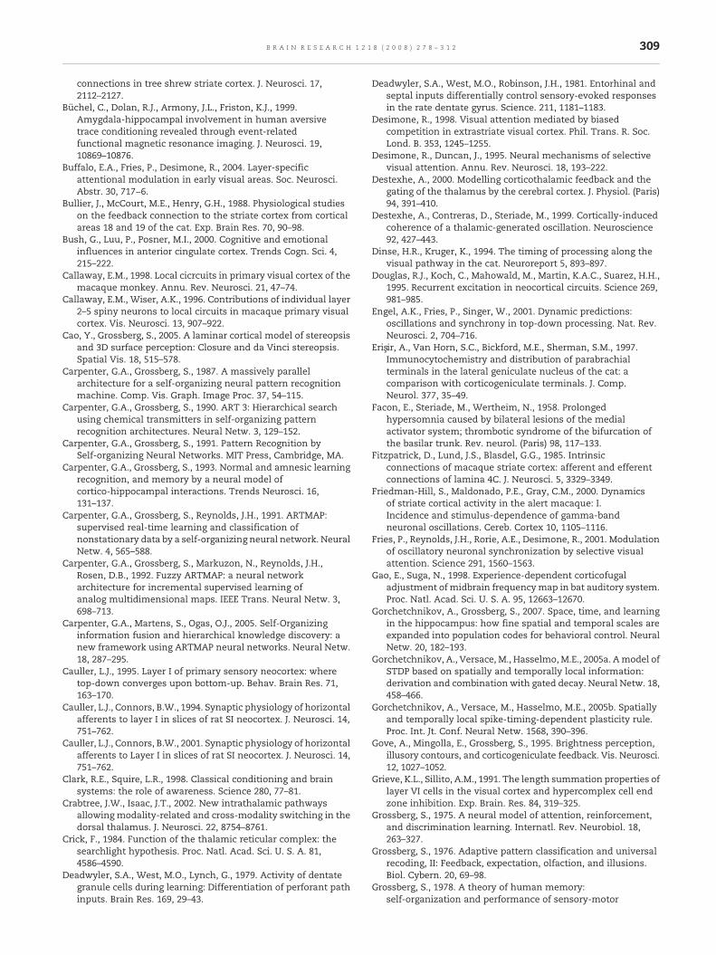
309B R A I N R E S E A R C H 1 2 1 8 ( 2 0 0 8 ) 2 7 8 – 3 1 2
connections in tree shrew striate cortex. J. Neurosci. 17,2112–2127.
Büchel, C., Dolan, R.J., Armony, J.L., Friston, K.J., 1999.Amygdala-hippocampal involvement in human aversivetrace conditioning revealed through event-relatedfunctional magnetic resonance imaging. J. Neurosci. 19,10869–10876.
Buffalo, E.A., Fries, P., Desimone, R., 2004. Layer-specificattentional modulation in early visual areas. Soc. Neurosci.Abstr. 30, 717−6.
Bullier, J., McCourt, M.E., Henry, G.H., 1988. Physiological studieson the feedback connection to the striate cortex from corticalareas 18 and 19 of the cat. Exp. Brain Res. 70, 90–98.
Bush, G., Luu, P., Posner, M.I., 2000. Cognitive and emotionalinfluences in anterior cingulate cortex. Trends Cogn. Sci. 4,215–222.
Callaway, E.M., 1998. Local cicrcuits in primary visual cortex of themacaque monkey. Annu. Rev. Neurosci. 21, 47–74.
Callaway, E.M., Wiser, A.K., 1996. Contributions of individual layer2–5 spiny neurons to local circuits in macaque primary visualcortex. Vis. Neurosci. 13, 907–922.
Cao, Y., Grossberg, S., 2005. A laminar cortical model of stereopsisand 3D surface perception: Closure and da Vinci stereopsis.Spatial Vis. 18, 515–578.
Carpenter, G.A., Grossberg, S., 1987. A massively parallelarchitecture for a self-organizing neural pattern recognitionmachine. Comp. Vis. Graph. Image Proc. 37, 54–115.
Carpenter, G.A., Grossberg, S., 1990. ART 3: Hierarchical searchusing chemical transmitters in self-organizing patternrecognition architectures. Neural Netw. 3, 129–152.
Carpenter, G.A., Grossberg, S., 1991. Pattern Recognition bySelf-organizing Neural Networks. MIT Press, Cambridge, MA.
Carpenter, G.A., Grossberg, S., 1993. Normal and amnesic learningrecognition, and memory by a neural model ofcortico-hippocampal interactions. Trends Neurosci. 16,131–137.
Carpenter, G.A., Grossberg, S., Reynolds, J.H., 1991. ARTMAP:supervised real-time learning and classification ofnonstationary data by a self-organizing neural network. NeuralNetw. 4, 565–588.
Carpenter, G.A., Grossberg, S., Markuzon, N., Reynolds, J.H.,Rosen, D.B., 1992. Fuzzy ARTMAP: a neural networkarchitecture for incremental supervised learning ofanalog multidimensional maps. IEEE Trans. Neural Netw. 3,698–713.
Carpenter, G.A., Martens, S., Ogas, O.J., 2005. Self-Organizinginformation fusion and hierarchical knowledge discovery: anew framework using ARTMAP neural networks. Neural Netw.18, 287–295.
Cauller, L.J., 1995. Layer I of primary sensory neocortex: wheretop-down converges upon bottom-up. Behav. Brain Res. 71,163–170.
Cauller, L.J., Connors, B.W., 1994. Synaptic physiology of horizontalafferents to layer I in slices of rat SI neocortex. J. Neurosci. 14,751–762.
Cauller, L.J., Connors, B.W., 2001. Synaptic physiology of horizontalafferents to Layer I in slices of rat SI neocortex. J. Neurosci. 14,751–762.
Clark, R.E., Squire, L.R., 1998. Classical conditioning and brainsystems: the role of awareness. Science 280, 77–81.
Crabtree, J.W., Isaac, J.T., 2002. New intrathalamic pathwaysallowing modality-related and cross-modality switching in thedorsal thalamus. J. Neurosci. 22, 8754–8761.
Crick, F., 1984. Function of the thalamic reticular complex: thesearchlight hypothesis. Proc. Natl. Acad. Sci. U. S. A. 81,4586–4590.
Deadwyler, S.A., West, M.O., Lynch, G., 1979. Activity of dentategranule cells during learning: Differentiation of perforant pathinputs. Brain Res. 169, 29–43.
Deadwyler, S.A., West, M.O., Robinson, J.H., 1981. Entorhinal andseptal inputs differentially control sensory-evoked responsesin the rate dentate gyrus. Science. 211, 1181–1183.
Desimone, R., 1998. Visual attention mediated by biasedcompetition in extrastriate visual cortex. Phil. Trans. R. Soc.Lond. B. 353, 1245–1255.
Desimone, R., Duncan, J., 1995. Neural mechanisms of selectivevisual attention. Annu. Rev. Neurosci. 18, 193–222.
Destexhe, A., 2000. Modelling corticothalamic feedback and thegating of the thalamus by the cerebral cortex. J. Physiol. (Paris)94, 391–410.
Destexhe, A., Contreras, D., Steriade, M., 1999. Cortically-inducedcoherence of a thalamic-generated oscillation. Neuroscience92, 427–443.
Dinse, H.R., Kruger, K., 1994. The timing of processing along thevisual pathway in the cat. Neuroreport 5, 893–897.
Douglas, R.J., Koch, C., Mahowald, M., Martin, K.A.C., Suarez, H.H.,1995. Recurrent excitation in neocortical circuits. Science 269,981–985.
Engel, A.K., Fries, P., Singer, W., 2001. Dynamic predictions:oscillations and synchrony in top-down processing. Nat. Rev.Neurosci. 2, 704–716.
Erişir, A., Van Horn, S.C., Bickford, M.E., Sherman, S.M., 1997.Immunocytochemistry and distribution of parabrachialterminals in the lateral geniculate nucleus of the cat: acomparison with corticogeniculate terminals. J. Comp.Neurol. 377, 35–49.
Facon, E., Steriade, M., Wertheim, N., 1958. Prolongedhypersomnia caused by bilateral lesions of the medialactivator system; thrombotic syndrome of the bifurcation ofthe basilar trunk. Rev. neurol. (Paris) 98, 117–133.
Fitzpatrick, D., Lund, J.S., Blasdel, G.G., 1985. Intrinsicconnections of macaque striate cortex: afferent and efferentconnections of lamina 4C. J. Neurosci. 5, 3329–3349.
Friedman-Hill, S., Maldonado, P.E., Gray, C.M., 2000. Dynamicsof striate cortical activity in the alert macaque: I.Incidence and stimulus-dependence of gamma-bandneuronal oscillations. Cereb. Cortex 10, 1105–1116.
Fries, P., Reynolds, J.H., Rorie, A.E., Desimone, R., 2001. Modulationof oscillatory neuronal synchronization by selective visualattention. Science 291, 1560–1563.
Gao, E., Suga, N., 1998. Experience-dependent corticofugaladjustment of midbrain frequencymap in bat auditory system.Proc. Natl. Acad. Sci. U. S. A. 95, 12663–12670.
Gorchetchnikov, A., Grossberg, S., 2007. Space, time, and learningin the hippocampus: how fine spatial and temporal scales areexpanded into population codes for behavioral control. NeuralNetw. 20, 182–193.
Gorchetchnikov, A., Versace, M., Hasselmo, M.E., 2005a. Amodel ofSTDP based on spatially and temporally local information:derivation and combination with gated decay. Neural Netw. 18,458–466.
Gorchetchnikov, A., Versace, M., Hasselmo, M.E., 2005b. Spatiallyand temporally local spike-timing-dependent plasticity rule.Proc. Int. Jt. Conf. Neural Netw. 1568, 390–396.
Gove, A., Mingolla, E., Grossberg, S., 1995. Brightness perception,illusory contours, and corticogeniculate feedback. Vis. Neurosci.12, 1027–1052.
Grieve, K.L., Sillito, A.M., 1991. The length summation properties oflayer VI cells in the visual cortex and hypercomplex cell endzone inhibition. Exp. Brain. Res. 84, 319–325.
Grossberg, S., 1975. A neural model of attention, reinforcement,and discrimination learning. Internatl. Rev. Neurobiol. 18,263–327.
Grossberg, S., 1976. Adaptive pattern classification and universalrecoding, II: Feedback, expectation, olfaction, and illusions.Biol. Cybern. 20, 69–98.
Grossberg, S., 1978. A theory of human memory:self-organization and performance of sensory-motor

310 B R A I N R E S E A R C H 1 2 1 8 ( 2 0 0 8 ) 2 7 8 – 3 1 2
codes, maps, and plans. In: Rosen, R., Snell, F. (Eds.),Progress in theoretical biology, Volume 5. Academic Press, NY,pp. 233–374.
Grossberg, S., 1980. How does a brain build a cognitive code? Psych.Rev. 87, 1–51.
Grossberg, S., 1982. A psychophysiological theory ofreinforcement, drive, motivation, and attention. J. Theoret.Neurobiol. 1, 286–369.
Grossberg, S., 1984. Some psychophysiological andpharmacological correlates of a developmental, cognitive,and motivational theory. In: Karrer, R., Cohen, J., Tueting, P.(Eds.), Brain and Information: Event Related Potentials. NYAcad. Sci. pp. 58–142. New York.
Grossberg, S., 1994. 3-D vision and figure-ground separation byvisual cortex. Percept. Psychophys. 55, 48–120.
Grossberg, S., 1995. The attentive brain. Amer. Scientist, 83, 438–449.Grossberg, S., 1999a. How does the cerebral cortex work?
Learning, attention, and grouping by the laminar circuits ofvisual cortex. Spat. Vis. 12, 163–187.
Grossberg, S., 1999b. The link between brain learning, attention,and consciousness. Conscious. Cogn. 8, 1–44.
Grossberg, S., 2000. How hallucinations may arise from brainmechanisms of learning, attention, and volution. J. Int.Neuropsych. Soc. 6, 579–588.
Grossberg, S., 2003. How does the cerebral cortex work?Development, learning, attention, and 3D vision by laminarcircuits of visual cortex. Behav. Cogn. Neur. Rev. 2, 47–76.
Grossberg, S., Stone, G.O., 1986. Neural dynamics of wordrecognition and recall: attentional priming, learning, andresonance. Psych. Rev. 93, 46–74.
Grossberg, S., Schmajuk, N.A., 1989. Neural dynamics ofadaptive timing and temporal discrimination duringassociative learning. Neural Netw. 2, 79–102.
Grossberg, S., Merrill, J.W.L., 1992. A neural network model ofadaptively timed reinforcement learning and hippocampaldynamics. Cog. Brain Res. 1, 3–38.
Grossberg, S., Merrill, J.W.L., 1996. The hippocampus andcerebellum in adaptively timed learning, recognition, andmovement. J. Cogn. Neurosci. 8, 257–277.
Grossberg, S., Yazdanbakhsh, A., 2005. Laminar cortical dynamicsof 3D surface perception: stratification, transparency, and neoncolor spreading. Vision Res. 45, 1725–1743.
Grossberg, S., Seidman, D., 2006. Neural dynamics of autisticbehaviors: Cognitive, emotional, and timing substrates. Psych.Rev. 113, 483–525.
Guillery, R.W., 1969. An abnormal retinogeniculate projection inSiamese cats. Brain Res. 3, 739–741.
Guillery, R.W., Harting, J.K., 2003. Structure and connections of thethalamic reticular nucleus: advancing views over half acentury. J. Comp. Neurol. 463, 360–371.
Guillery, R.W., Feig, S.L., Lozsadi, D.A., 1998. Paying attention to thethalamic reticular nucleus. Trends Neurosci. 21, 28–32.
Hasselmo, M.E., 1993. Acetylcholine and learning in a corticalassociative memory. Neural Comput. 5, 32–44.
Hasselmo, M.E., 1995. Neuromodulation and cortical function:modeling the physiological basis of behavior. Behav. Brain Res.67, 1–27.
Heeger, D.J., 1992. Normalization of cell responses in cat striatecortex. Vis. Neurosci. 9, 181–197.
Heilman, K.M., Bowers, D., Valenstein, E., Watson, R.T., 1993.Disorders of visual attention. Baillière's clin. neurol. 2, 389–413.
Herrmann, C.S., Munk, M.H.J., Engel, A., 2004. Cognitive functionsof gamma-band activity: memory match and utilization.Trends Cognit. Sci. 8, 347–355.
Hille, B., 2001. Ion Channels of Excitable Membranes, ThirdEdition. Sinauer Associates, MA, USA.
Hodgkin, A.L., Huxley, A.F., 1952. A quantitative description ofmembrane current and its application to conduction andexcitation in nerve. J. Neurophysiol. 117, 500–544.
Holroyd, C.B., Nieuwenhuis, S., Mars, R.B., Coles, M.G.H., 2004.Anterior cingulate cortex, selection for action, and errorprocessing. In: Posner, M.I. (Ed.), Cognitive Neuroscience ofAttention. Guilford Publication, Inc, New York, pp. 219–231.
Ichinohe, N., Fujiyama, F., Kaneko, T., Rockland, K.S., 2003.Honeycomb-like mosaic at the border of layers 1 and 2 in thecerebral cortex. J. Neurosci. 23, 1372–1382.
Johnston, D., Hoffman, D.A., Colbert, C.M., Magee, J.C., 1999.Regulation of back-propagating action potentials inhippocampal neurons. Curr. Opin. Neurobiol. 9, 288–292.
Jones, E.G., 2002. Thalamic circuitry and thalamocorticalsynchrony. Phil. Trans. R. Soc. Lond. B. 357, 1659–1673.
Karmos, G., Molnár, M., Csépe, V., Winkler, I., 1986. Evokedpotential components in the layers of the auditory cortex of thecat. Acta neurobiol. exp. 46, 227–236.
Kilgard, M.P., Merzenich, M.M., 1998. Cortical map reorganizationenabled by nucleus basalis activity. Science. 279, 1714–1718.
Kolmac, C.I., Mitrofanis, J., 1997. Organisation of the reticularthalamic projection to the intralaminar and midline nuclei inrats. J. Comp. Neurol. 377, 165–178.
Kopell, N., Ermentrout, G.B., Whittington, M.A., Traub, R.D., 2000.Gamma rhythms and beta rhythms have differentsynchronization properties. Proc. Natl. Acad. Sci. U. S. A. 15,1867–1872.
Kraus, N., McGee, T., Littman, T., Nicol, T., King, C., 1994.Nonprimary auditory thalamic representation of acousticchange. J. Neurophysiol. 72, 1270–1277.
Krupa, D.J., Ghazanfar, A.A., Nicolelis, M.A.L., 1999.Immediate thalamic sensory plasticity depends oncorticothalamic feedback. Proc. Natl. Acad. Sci. U. S. A. 96,8200–8205.
Kumaran, D., Maguire, E.A., 2007a. Which computationalmechanisms operate in the hippocampus during noveltydetection? Hippocampus. 17, 735–748.
Kumaran, D., Maguire, E.A., 2007b. Match–mismatch processesunderlie human hippocampal responses to associativenovelty. J. Neurosci. 27, 8517–8524.
Landisman, C.E., Long, M.E., Beierlein, M., Deans, M.R., Paul, D.L.,Connors, B.W., 2002. Electrical synapses in the thalamicreticular nucleus. J. Neurosci. 22, 1002–1009.
Larkum, M.E., Zhu, J.J., 2002. Signaling of Layer 1 andwhisker-evoked Ca++ and Na++ action potentials in distal andterminal dendrites of rat neocortical pyramidal neurons invitro and in vivo. J. Neurosci. 22, 6991–7005.
Larkum, M.E., Zhu, J.J., Sakmann, B.A., 1999. A new cellularmechanism for coupling inputs arriving at different corticallayers. Nature. 398, 338–341.
Larkum, M.E., Zhu, J.J., Sakmann, B.A., 2002. Signaling ofLayer 1 and whisker-evoked Ca++ and Na++ action potentialsin distal and terminal dendrites of rat neocorticalpyramidal neurons in vitro and in vivo. J. Neurosci. 22,6991–7005.
Larkum, M.E., Senn, W., Lusher, H.R., 2004. Top-down dendriticinput increases the gain of layer 5 pyramidal neurons. Cereb.Cortex 14, 1059–1070.
Levy, W.B., Steward, O., 1983. Temporal contiguity requirementsfor long-term associative potentiation/depression in thehippocampus. Neuroscience 8, 791–797.
Llinas, R.R., Pare, D., 1991. Of dreaming and wakefulness.Neuroscience 44, 521–535.
Llinas, R.R., Leznik, E., Urbano, F.J., 2002. Temporal binding viacortical coincidence detection of specific and nonspecificthalamocortical inputs: a voltage-dependent dye-imagingstudy in mouse brain slices. Proc. Natl. Acad. Sci. U. S. A. 99,449–454.
Lumer, E.D., Edelman, G.M., Tononi, G., 1997. Neural dynamics in amodel of the thalamocortical system I. Layers, loops and theemergence of fast synchronous rhythms. Cereb. Cortex 7,207–227.

311B R A I N R E S E A R C H 1 2 1 8 ( 2 0 0 8 ) 2 7 8 – 3 1 2
Luu, P., Pederson, S.M., 2004. The anterior cingulate cortex:regulating actions in context. In: Posner, M.I. (Ed.),Cognitive neuroscience of attention. Guilford Publication, Inc,New York.
Markram, H., Lubke, J., Frotscher, M., Sakmann, B., 1997.Regulation of synaptic efficacy by coincidence of postsynapticAPs and EPSPs. Science 275, 213–215.
Markram, H., Toledo-Rodriguez, M., Wang, Y., Gupta, A.,Silberberg, G., Wu, C., 2004. Interneurons of the neocorticalinhibitory system. Nat. Rev. Neurosci. 5, 793–807.
Maunsell, J.H.R., Van Essen, D.C., 1983. Anatomical connections ofthe middle temporal visual area in the macaque monkey andtheir relationship to a hierarchy of cortical areas. J. Neurosci. 3,2563–2586.
McCormick, D.A., Prince, D.A., 1985. Two types of muscarinicresponse to acetylcholine in mammalian cortical neurones.Proc. Natl. Acad. Sci. U. S. A. 82, 6344–6349.
McGuire, B.A., Hornung, J.P., Gilbert, C.D., Wiesel, T.N., 1984.Patterns of synaptic input to layer 4 of cat striate cortex. J.Neurosci. 4, 3021–3033.
McGuire, B.A., Gilbert, C.D., Rivlin, P.K., Wiesel, T.N., 1991. Targetsof horizontal connections in macaque primary visual cortex.J. Comp. Neurol. 305, 370–392.
Miller, R., 1996. Corticothalamic interplay and the security ofoperation of neural assemblies and temporal chains in thecerebral cortex. Biol. Cybern. 75, 263–275.
Miller, J.M., Benevento, L.A., 1979. Demonstration of a directprojection from the intralaminar central lateral nucleus to theprimary visual cortex. Neurosci. Lett. 14, 229–234.
Montero, V.M., 1997. C-fos induction in sensory pathways of ratsexploring a novel environment: shifts of active thalamicreticular sectors by predominant sensory cues. Neuroscience76, 1069–1081.
Murphy, P.C., Duckett, S.G., Sillito, A.M., 1999. Feedbackconnections to the lateral geniculate nucleus and corticalresponse properties. Science 286, 1552–1554.
Näätänen, R., Gaillard, A.W.K., Mäntysalo, S., 1978. Early selectiveattention effect on evoked potential reinterpreted. Actapsychol. 42, 313–329.
Nicolelis, M.A., Fanselow, E.E., 2002. Dynamic shifting inthalamocortical processing during different behaviouralstates. Phil. Trans. R. Soc. Lond. B. 357, 1753–1758.
O'Keefe, J., Nadel, L., 1978. The Hippocampus as a Cognitive Map.Oxford University Press, New York.
Olufsen, M.S., Whittington, M.A., Camperi, M., Kopell, N., 2003.New roles for the gamma rhythm: population tuning andpreprocessing for the beta rhythm. J. Comput. Neurosci. 14,33–54.
Otto, T., Eichenbaum, H., 1992. Neuronal activity in thehippocampus during delayed non-match to sampleperformance in rats: evidence for hippocamp processing inrecognition memory. Hippocampus. 2, 323–334.
Pakhomova, A.S., 1981. Neurophysiol. 13, 267–273.Pinault, D., Deschenes, M., 1998. Anatomical evidence for a
mechanism of lateral inhibition in the rat thalamus. E. J.Neurosci., 10, 3462–3469.
Pollen, D.A., 1999. On the neural correlates of visual perception.Cereb. Cortex 9, 4–19.
Raizada, R.D.S., Grossberg, S., 2003. Towards a theory of thelaminar architecture of cerebral cortex: computational cluesfrom the visual system. Cereb. Cortex. 13, 100–113.
Rall, W., 1962. Theory of physiological properties of dendrites.Ann. N. Y. Acad. Sci. 96, 1071–1092.
Reid, R.C., Alonso, J.M., 1995. Specificity of monosynapticconnections from thalamus to visual cortex. Nature 378,281–284.
Rockland, K.S., 1996. Two types of cortico-pulvinar terminations:round, type 2 and elongate, type 1. J. Comp. Neurol. 368, 57–87.
Rockland, K.S., 1998. Convergence and branching patterns ofround, Type 2 corticopulvinar axons. J. Comp. Neurol. 390,515–536.
Rockland, K.S., 2002. Visual cortical organization at the single axonlevel: a beginning. Neurosci. res. 42, 155–166.
Rockland, K.S., Virga, A., 1989. Terminal arbors of individual“feedback” axons projecting from area V2 to V1 in themacaquemonkey: a study using immunohistochemistry ofanterogradelly transported Phaseolus vulgaris leucoagglutinin.J. Comp. Neurol. 285, 54–72.
Rockland, K.S., Andresen, J., Cowie, R.J., Robinson, D.L., 1999. Singleaxon analysis of pulvinocortical connections to several visualareas in the macaque. J. Comp. Neurol. 406, 221–250.
Roelfsema, P.R., Engel, A.K., König, P., Singer, W., 1997. Visuomotorintegration is associated with zero time-lag synchronizationamong cortical areas. Nature 385, 157–161.
Rosburg, T., Trautner, P., Ludowig, E., Schaller, C., Kurthen, M.,Elger, C.E., Boutros, N.N., 2007. Hippocampal event-relatedpotentials to tone duration deviance in a passive oddballparadigm in humans. Neuroimage 37, 274–281.
Rouiller, E.M., Welker, E., 2000. A comparative analysis of themorphology of corticothalamic projections in mammals. BrainRes. Bull. 53, 727–741.
Saar, D., Grossman, Y., Barkai, E., 2001. Long-lasting cholinergicmodulation underlies rule learning in rats. J. Neurosci. 21,1385–1392.
Salin, P.A., Bullier, J., 1995. Corticocortical connections in thevisual system: structure and function. Physiol. Rev. 75, 107–154.
Sánchez-Montañés, M.A., Verschure, P.F.M.J., König, P., 2000. Localand global gating of synaptic plasticity. Neural Comput. 13,543–552.
Schmidt, K.E., Goebel, R., Löwel, S., Singer, W., 1997. Theperceptual grouping criterion of colinearity is reflected byanisotropies of connections in the primary visual cortex. Eur. J.Neurosci. 9, 1083–1089.
Schmolesky, M.T., Wang, Y., Hanes, D.P., Thompson, K.G.,Leutgeb, S., Schall, J.D., Leventhal, A.G., 1998. Signal timingacross the macaque visual system. J. Neurophysiol. 6,3272–3278.
Sherman, S.M., Guillery, R., 2001. Exploring the Thalamus.Academic Press, San Diego.
Sherman, S.M., Guillery, R.W., 2002. The role of the thalamus in theflow of information to the cortex. Phil. Trans. R. Soc. Lond. B.357, 1695–1708.
Shipp, S., 2003. The functional logic of cortico-pulvinarconnections. Phil. Trans. R. Soc. Lond. B. 358, 1605–1624.
Siegel, M., Körding, K.P., König, P., 2000. Integrating top-down andbottom-up sensory processing by somato-dendriticinteractions. J. Comput. Neurosci. 8, 161–173.
Sillito, A.M., Jones, H.E., Gerstein, G.L., West, D.C., 1994.Feature-linked synchronization of thalamic relay cell firinginduced by feedback from the visual cortex. Nature 369,479–482.
Singer, W., 1999. Neuronal synchrony: a versatile code for thedefinition of relations? Neuron 24, 49–65.
Singer, W., Rauschecker, J.P., 1982. Central core control ofdevelopment plasticity in the kitten visual cortex. II: Electricalactivation ofmesencephalic and diencephalic projections. Exp.Brain. Res. 47, 223–233.
Soares, J.G.M., Diogo, A.C.M., Fiorani, M., Souza, A.P.B., Gattass, R.,2004. Effects of inactivation of the lateral pulvinar on responseproperties of second visual area cells in cebus monkeys. Clin.Exp. Pharmacol. Physiol. 31, 580–590.
Sohal, V.S., Huguenard, J.R., 2003. Inhibitory interconnectionscontrol burst pattern and emergent network synchrony inreticular thalamus. J. Neurosci. 23, 8978–8988.
Solomon, P.R., Vander Schaaf, E.R., Thompson, R.F., Weisz, D.J.,1986. Hippocampus and trace conditioning of the rabbit's

312 B R A I N R E S E A R C H 1 2 1 8 ( 2 0 0 8 ) 2 7 8 – 3 1 2
classically conditioned nictitating membrane response. Behav.Neurosci. 100, 729–744.
Steriade, M., Contreras, D., Curro Dossi, R., Nunez, A., 1993. Theslow,b1 Hz oscillation in reticular thalamic andthalamocortical neurons: scenario of sleep rhythm generationin interacting thalamic and neocortical networks. J. Neurosci.13, 3284–3299.
Stratford, K.J., Tarczy-Hornoch, K., Martin, K.A.C., Bannister, N.J.,Jack, J.J.B., 1996. Excitatory synaptic inputs to spiny stellatecells in cat visual cortex. Nature 382, 258–261.
Swadlow, H.A., Gusev, A.G., Bezdudnaya, T., 2002. Activation of acortical column by a thalamocortical impulse. J. Neurosci. 22,7766–7773.
Tamas, G., Somogyi, P., Buhl, E.H., 1998. Differentiallyinterconnected networks of GABAergic interneurons in thevisual cortex of the cat. J. Neurosci. 18, 4255–4270.
Traub, R.D., Miles, R., 2001. Neuronal networks of thehippocampus. Cambridge University Press, Cambridge, UK.
Traub, R.D., Spruston,N., Soltesz, I., Konnerth, A.,Whittington,M.A.,Jefferys, G.R., 1998. Gamma-frequency oscillations: a neuronalpopulation phenomenon, regulated by synaptic and intrinsiccellular processes, and inducing synaptic plasticity. Prog.Neurobiol. 55, 563–575.
Tsodyks, M., Markram, H., 1997. The neural code betweenneocortical pyramidal neurons depends on neurotransmitterrelease probability. Proc. Natl. Acad. Sci. U. S. A. 94, 719–723.
Turrini, P., Casu, M.A.,Wong, T.P., De Koninck, Y., Ribeiro-da-Silva,A., Cuello, A.C., 2001. Cholinergic nerve terminals establishclassical synapses in the rat cerebral cortex: synapticpattern and age-related atrophy. Neuroscience. 105,277–285.
Usrey, W.M., 2002. The role of spike timing for thalamocorticalprocessing. Curr. Opin. Neurobiol. 12, 411–417.
van Der Werf, Y.D., Witter, M.P., Groenewegen, H.J., 2002. Theintralaminar and midline nuclei of the thalamus.Anatomical and functional evidence for participation inprocesses of arousal and awareness. Brain res. Brain Res. Rev.39, 107–140.
van Der Werf, Y.D., Scheltens, P., Lindeboom, J., Witter, M.P.,Uylings, H.B., Jolles, J., 2003. Deficits of memory, executivefunctioning and attention following infarction in the thalamus;a study of 22 cases with localised lesions. Neuropsychologia 41,1330–1344.
Van Essen, D.C., Newsome, W.T., Maunsell, J.H., Bixby, J.L., 1986.The projections from striate cortex, V1 to areas V2 and V3 inthe macaque monkey: asymmetries, areal boundaries, andpatchy connections. J. Comp. Neurol. 244, 451–480.
Versace, M., Ames, A., Leveille, J., Fortenberry, B., Mhatre, H.,Gorchetchnikov, A., 2007. KInNeSS: A modular framework forcomputational neuroscience. CAS/CNSTR-07-007. Submitted toNeuroinformatics.
Vinogradova, O.S., 1975. Functional organization of the limbicsystem in the process of registration of information: Facts andhypotheses. In: Isaacson, R.L., Pribram, K.H. (Eds.), TheHippocampus, vol. 2. Plenum, New York, pp. 3–69.
Vogt, B.A., 1991. The role of layer I in cortical function. In: Peters, A.,Jones, E. (Eds.), Cerebral Cortex. Plenum, New York,pp. 49–79.
Von Stein, A., Chiang, C., König, P., 2000. Top-down processingmediated by interareal synchronization. Proc. Natl. Acad. Sci.U. S. A. 97, 14748–14753.
Wang, S., Eisenback, M.A., Bickford, M.E., 2002. Relativedistribution of synapses in the pulvinar nucleus of the cat:implications regarding the “driver/modulator” theory ofthalamic function. J. Comp. Neurol. 454, 482–494.
Weese, G.D., Phillips, J.M., Brown, V.J., 1999. Attentional orientingis impaired by unilateral lesions of the thalamic reticularnucleus in the rat. J. Neurosci. 19, 1035–10139.
Wespatat, V., Tennigkeit, F., Singer, W., 2004. Phase sensitivity ofsynaptic modifications in oscillating cells of rat visual cortex.J. Neurosci. 24, 9067–9075.
Williams, S.R., Stuart, G.J., 1999. Mechanisms and consequences ofaction potential burst firing in rat neocortical pyramidalneurons. J. Neurophysiol. 521, 467–482.
Wilson, J.R., Friedlander, M.J., Sherman, S.M., 1984. Finestructural morphology of identified X-and Y-cells in the cat'slateral geniculate nucleus. Phil. Trans. R. Soc. Lond. B. 221,411–436.
Yazdanbakhsh, A., Grossberg, S., 2004. Fast synchronization ofperceptual grouping in laminar visual cortical circuits. NeuralNetw. 17, 707–718.
Zeki, S., Shipp, S., 1988. The functional logic of corticalconnections. Nature 335, 311–317.
Zhang, Y.Q., Lu, S.G., Ji, J.P., Zhao, Z.Q., Mei, J., 2004.Electrophysiological and pharmacological properties ofnucleus basalis magnocellularis neurons in rats. Actapharmacologica Sinica 25, 161–170.