sperminemodulatesfungalmorphogenesisandactivatesplasma...
TRANSCRIPT

RESEARCH ARTICLE
Spermine modulates fungal morphogenesis and activates plasmamembrane H+-ATPase during yeast to hyphae transitionAntonio Jesus Dorighetto Cogo1,2, Keilla dos Reis Dutra Ferreira1, Lev A. Okorokov1, Alessandro C. Ramos1,Arnoldo R. Façanha2,* and Anna L. Okorokova-Façanha1,*
ABSTRACTPolyamines play a regulatory role in eukaryotic cell growth andmorphogenesis. Despite many molecular advances, the underlyingmechanism of action remains unclear. Here, we investigate amechanism by which spermine affects the morphogenesis of adimorphic fungal model of emerging relevance in plant interactions,Yarrowia lipolytica, through the recruitment of a phytohormone-likepathway involving activation of the plasma membrane P-typeH+-ATPase. Morphological transition was followed microscopically,and the H+-ATPase activity was analyzed in isolated membranevesicles. Proton flux and acidification were directly probed at living cellsurfaces by a non-invasive selective ion electrode technique.Spermine and indol-3-acetic acid (IAA) induced the yeast-hyphatransition, influencing the colony architecture. Spermine inducedH+-ATPase activity and H+ efflux in living cells correlating with yeast-hypha dynamics. Pharmacological inhibition of spermine and IAApathways prevented the physio-morphological responses, andindicated that spermine could act upstream of the IAA pathway.This study provides the first compelling evidence on the fungalmorphogenesis and colony development as modulated by aspermine-induced acid growth mechanism analogous to thatpreviously postulated for the multicellular growth regulation of plants.
KEY WORDS: Polarized growth, Yarrowia lipolytica, H+ transport,P-typeATPase, Acid growth theory, Scanning ion-selective electrodetechnique, Polyamine
INTRODUCTIONParallels between the morphogenesis of fungi and plants are worthseeking so that we can make use of the conceptual frameworkalready established with the hope of developing models that canshed new light on the conservative evolutionary mechanisms andinfluence a myriad of ecological interactions within and amongthese organisms (Moore, 2013). Fungi are organisms with adaptivemorphological plasticity that enables them to survive under
challenging conditions and colonize new habitats. The hyphaldevelopment and polarized growth contribute to their evolutionarysuccess and are the bases for fungal proliferation and ecologicalinteractions, including virulence and symbiosis with plants(Flor-Parra et al., 2007; Ramos et al., 2008, 2009). Polarizedhyphal growth promotes the substrate invasion, directionaltranslocation between host environments, consolidation of thecolony, nutrient acquisition, and the formation of 3-dimensionalmatrices (Sudbery, 2011; Brand, 2012). Many fungi can grow eitheras unicellular yeast or mycelial forms and can undergo amorphogenesis switch from isotropic to polarized growth (Harris,2011). Cell wall dynamics have been widely explored in plant aswell as fungi cells, and despite their intrinsic differences incomponents and architecture, cell polarity in plants and fungiinvolves a common sequence of events underlying the continuoussynthesis of proteins, lipids and cell wall building blocks, changesin cytoskeletal dynamics, internal hydrostatic pressure, localizedCa2+ gradients, and tip-directed transport of secretory vesicles andaccumulation (Lew, 2011; Riquelme, 2013).
The ascomycete Yarrowia lipolytica has the ability to grow asyeast, pseudohyphae or true hyphae depending on theenvironmental conditions and genetic regulatory mechanism(Dominguez et al., 2000). This dimorphic fungus is one of themore intensively studied ‘non-conventional’ species due to its highbiotechnological potential and wide range of industrial andenvironmental applications (Nicaud, 2012; Barth, 2013; Harzevili,2014; Zinjarde et al., 2014; Ledesma-Amaro and Nicaud, 2016).However, a point so far less explored is that Y. lipolytica has alsobeen considered among the beneficial microorganisms inagriculture, proving to be useful as a biofertilizer, associated ornot with mycorrhizal fungi, modifying soil physico-chemical,biological and fertility properties that enhance plant performance(Vassilev et al., 2001; Medina et al., 2004; Lonhienne et al., 2014).Ecophysiological roles have also been proposed as the halophyteAtriplex halimuswas found to interact with halotolerant Y. lipolyticastrains inhabiting their leaves surfaces (Zvyagilskaya et al., 2001).Moreover, plant-like HAK genes encoding Na+ transporterswere found in Y. lipolytica, suggesting that salt adaptive traits inplants and fungi are more extensive than previously thought (Benitoet al., 2012).
Many environmental factors, including pH, carbon and nitrogensources, and oxygen concentrations, are important modulationfactors involved in hyphal development and growth (Pérez-Campoand Domínguez, 2001; Ruiz-Herrera and Sentandreu, 2002; Bellouet al., 2014). Microarray and proteomic analysis of Y. lipolyticaduring the yeast-to-hypha transition revealed several genes andproteins involved in morphogenetic transition (Morín et al., 2007;Morales-Vargas et al., 2012). The complete genome sequence andefficient genetic tools have also provided important insights onsignaling pathways and transcriptional factors required forReceived 9 September 2017; Accepted 8 January 2018
1Laboratorio de Fisiologia e Bioquımica deMicrorganismos, Universidade Estadualdo Norte Fluminense Darcy Ribeiro, Av. Alberto Lamego, 2000, Pq. California,Campos dos Goytacazes-RJ 28013-602, Brazil. 2Laboratorio de Biologia Celular eTecidual, Centro de Biociências e Biotecnologia, Universidade Estadual do NorteFluminense Darcy Ribeiro, Av. Alberto Lamego, 2000, Pq. California, Campos dosGoytacazes-RJ 28013-602, Brazil.
*Authors for correspondence ([email protected]; [email protected])
A.J.D.C., 0000-0003-1967-6898; K.R.D.F., 0000-0001-5060-2876; A.L.O.-F.,0000-0002-6856-7711
This is an Open Access article distributed under the terms of the Creative Commons AttributionLicense (http://creativecommons.org/licenses/by/3.0), which permits unrestricted use,distribution and reproduction in any medium provided that the original work is properly attributed.
1
© 2018. Published by The Company of Biologists Ltd | Biology Open (2018) 7, bio029660. doi:10.1242/bio.029660
BiologyOpen
by guest on June 19, 2018http://bio.biologists.org/Downloaded from

morphogenesis in Y. lipolytica (Cervantes-Chávez et al., 2009;Martinez-Vazquez et al., 2013). In addition, this non-pathogenicfungus has interesting similarities to the highly virulent pathogenCandida albicans (Herrero et al., 1999). In this way, Y. lipolyticahas emerged as an excellent yeast model to study the mechanismsthat drive the morphogenetic transition in fungi (Dominguez et al.,2000; Herrero et al., 1999).Studies on cell differentiation have demonstrated that polyamines
play a key role in hyphae and colony growth and development ofmany fungal systems (San-Blas et al., 1997; Ueno et al., 2004; 31.Valdés-Santiago et al., 2010; Kummasook et al., 2013). In Y.lipolytica, the intracellular levels of polyamines increase beforethe morphogenetic transition and differentiation process (Guevara-Olvera et al., 1993), but the underlying mechanism is not yetfully understood. Polyamines are low molecular weight positivelycharged aliphatic molecules that facilitate interactions withmacromolecules, stabilizing DNA, RNA, proteins andphospholipids, and modulating gene expression, enzymeactivities, and DNA-protein interactions (Tabor and Tabor, 1985).In addition to the morphogenetic transition, the fungi polyamineshave also been correlated with cell cycle progression(Chattopadhyay et al., 2002), defense against reactive oxygenspecies (Chattopadhyay et al., 2006), and cell lifespan (Eisenberget al., 2009, 2016). The differential polycationic character ofputrescine, spermidine and spermine (Spm), have been related to thedistinct properties and functions of each polyamines (Tabor andTabor, 1985).P-type plasma membrane H+-ATPase plays an essential role in
fungal and plant cells physiology. This proton pump generates theelectrochemical proton-motive force across the membrane thatdrives the energy-dependent uptake of amino acids, sugars,nucleosides, and inorganic ions (Goffeau and Slayman, 1981). Inaddition, H+ transport mediated by this enzyme contributes to theregulation of intracellular pH and surface pH along the hyphae. Inplants, it is widely accepted that the activation of plasma membraneH+-ATPase by indole-3-acetic acid (IAA) underlies the induction ofpolarized growth of roots and pollen tube expansion (Hager, 2003;Zandonadi et al., 2010; Takahashi et al., 2012). This mechanism isthe base of the classical acid growth theory, which postulates that theactivation of proton pump by auxin and subsequent pH decrease inthe apoplast promotes plant cell growth (Hager et al., 1991; Rayleand Cleland, 1992; Frías et al., 1996). Interestingly, although thepresence of IAA in fungi has long been reported (Roberts andRoberts, 1939; Gruen, 1959), it was just recently that a role for auxinhas been related to the morphological transition in Saccharomycescerevisiae, stimulating the morphogenetic switch from yeast cells toa pseudohyphal form (Prusty et al., 2004), and to the hyphal growthin the human pathogen C. albicans (Rao et al., 2010).A transmembrane pH and electrical gradient might be critical in
establishing the cell polarity and regulating the assembly ofcytoskeletal components required for hyphal extension (Harold,1990). A transient increase in the intracellular pH was reportedbefore the morphogenetic transition in C. albicans (Stewart et al.,1988), as well as at the extending hyphal tip in Neurospora crassa(Robson et al., 1996). In fact, P-type H+-ATPase is rate-limiting forgrowth and the decrease of ATPase activity correlates withdecreased intracellular pH in yeast cells (Portillo and Serrano,1989). Moreover, extracellular neutral or alkaline pH induceshyphal development in Y. lipolytica and C. albicans, revealing theimportance of the H+ gradient to hyphal morphogenesis (Ruiz-Herrera and Sentandreu, 2002; Vylkova et al., 2011). Thus, fungaland plant cells share similar features in ion homeostasis and cellular
bioenergetics that might be involved in the modulation of polarizedcell growth.
Although there is a body of evidence suggesting that pH is anessential factor in fungal morphogenesis and that P-type H+-ATPaseregulates the membrane microenvironment pH in these organisms,the actual role of this pump in polarized hyphal growth remainselusive. Moreover, it has been shown that Spm can modulateIAA-dependent P-type H+-ATPase activation and cell polarizedgrowth in plants (Garufi et al., 2007; Pandolfi et al., 2010; Dutraet al., 2013; Pottosin et al., 2014); however, to date, no study hasexplored whether Spm could play a role in the modulation of P-typeH+-ATPase during polarized growth in fungi. Therefore, the presentwork aims to investigate whether Spm modulates the morphogenesisand polarized cell growth of the model fungus Y. lipolytica throughmechanisms similar to those found in plants, underlying an activationof plasma membrane H+-ATPase and the recruitment of auxin-dependent pathways.
RESULTSSpermine induces Y. lipolytica filamentous growthYarrowia lipolytica cells grown in liquid YED medium reached thestationary phase after 22 h. Microscopic analysis of cell morphologyrevealed that the morphogenetic transition took place after 18 h ofgrowth. Consistent with this observation, the number of yeast cellsdeclined after this time point due to the increase of pseudohyphae andhyphae forms (Fig. 1A). Different concentrations of Spm (0.1-2 mM)affected neither the growth nor the morphogenesis start point,although concentrations higher than 1.5 mM Spm reduced cellulargrowth and caused the appearance of abnormal cells (data notshown).
Spermine concentrations between 0.75 and 1.5 mM potentiatedthe dimorphic transition in Y. lipolytica with the highesteffectiveness found at 1 mM Spm. At this concentration of Spm,70-80% of the Y. lipolytica culturewas in the yeast form after 20 h ofgrowth, whereas the corresponding values for the control cultureranged from 85-90%. After 36 h, only 30-35% of cells cultured inthe presence of Spm remained in the yeast form, whereas yeast cellsmade up 50-55% of the control culture (Fig. 1A,D). Furthermore,most of the filamentous forms observed in the cells treated with Spmwere true hyphae (Fig. 1D). Additionally, the presence of 2 mMCHA, a competitive spermidine synthase inhibitor, reducedsignificantly (P≤0.05) the morphogenesis, resulting in 80% of thecells remaining in the yeast form even after 36 h (Fig. 1A,D),although it did not interfere with cell growth. These results indicatethat Spm is required for Y. lipolytica morphogenetic transition.
To investigate a putative signaling pathway involving Spm in themodulation of hyphal growth, we cultivated cells in the presence of1 mM Spm concomitantly or not with an inhibitor of auxinsignaling [α-p-chlorophenoxyisobutyric acid (PCIB), 100 µM] orinhibitor of auxin transport [2,3,5-triiodobenzoic acid (TIBA),100 µM]. At these concentrations, the inhibitors impaired themorphogenetic transition in control culture and did not affect cellgrowth. Treatments involving addition of PCIB and TIBA had asimilar effect to that of CHA, and maintained the yeast form bynearly 80-90% of cells. Addition of low concentrations of IAA(10 pM) induced the morphogenetic transition (Fig. 1B) andreverted the inhibitory effect of CHA (Fig. 1C). On the otherhand, the inhibitory effect of TIBA and PCIB was not antagonizedby Spm (Fig. 1C). The data suggest that modulation ofmorphogenetic transition by Spm might involve IAA signaling.
The morphology of Y. lipolytica colonies was examined on solidmedium (Fig. 2). Colonies of control cells displayed a central
2
RESEARCH ARTICLE Biology Open (2018) 7, bio029660. doi:10.1242/bio.029660
BiologyOpen
by guest on June 19, 2018http://bio.biologists.org/Downloaded from

Fig. 1. Effect of Spm on Y. lipolytica cell morphology. (A-C) Y. lipolytica cells were grown in YED medium supplemented with Spm at the indicatedconcentrations, 10 pM IAA and their inhibitors (2 mM CHA, 100 µM PCIB, 100 µM TIBA). The number of yeast cells at each time point was counted and themaximal value was considered as 100%. Values are representative of at least three independent experiments. (D) Visualization of the Spm-dependent effect onthemorphogenic transition ofY. lipolytica.Cell morphology was examined in cultures grown for 36 h in YEDmedium supplemented or not with Spm or CHA. Scalebar: 20 µm. Ctr, control; Spm, spermine; CHA, spermidine synthase competitive inhibitor cyclohexylamine; IAA, indole-3-acetic acid; PCIB and TIBA, auxininhibitors.
3
RESEARCH ARTICLE Biology Open (2018) 7, bio029660. doi:10.1242/bio.029660
BiologyOpen
by guest on June 19, 2018http://bio.biologists.org/Downloaded from

interlaced patch surrounded by short peripheral extensions orfringes (460±64 µm). In contrast, the presence of 2 mM CHAresulted in ring-like colonies containing much shorter fringes (250±33 µm). Y. lipolytica cells grown on Spm-containing plates formedirregular wrinkled colonies. Fringes developed in the presence of1 mM Spm were distinctly longer, reaching nearly twice the lengthof those observed in control colonies (976±147 µm). The additionof 10 pM IAA also resulted in highly structured wrinkled colonieswith long fringes (749±133 µm), while the auxin inhibitors PCIB orTIBA caused nearly smooth colony phenotype with almost nosurrounding extensions. It is of note that IAA overcame the effect ofCHA and increased the extent of colony wrinkling, as well asrestored the fringe length (Fig. 2A,C).The ability of Spm to induce filamentous growth was further
analyzed. Microscopic examination revealed invasive scars patternsby Y. lipolytica colonies; the extent of agar invasion was enhancedin the presence of 1 mM Spm and abolished by 2 mM CHA(Fig. 3A). Scanning electron microscopy of Y. lipolytica coloniesillustrates in detail that colony morphology is closely associatedwith Y. lipolytica cell morphotypes; less convoluted coloniesformed in the presence of the inhibitor CHAwere composed mainly
of yeast and pseudohyphal cells, whereas wrinkled colonies onSpm-containing plates were composed mainly of invasive hyphalcells (Fig. 3B).
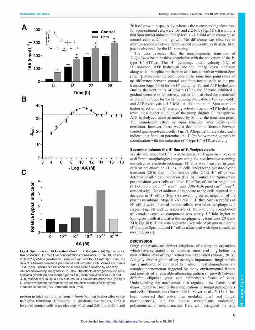
Modulation of morphogenesis by spermine involves theproduction of IAAIn addition to the above described stimulatory effect of Spmon morphogenesis and the activation of P-type H+-ATPase,Y. lipolytica cells grown in the presence of Spm increasedsignificantly the content of IAA in the extracellular media, whencompared with untreated cells (Fig. 4A). Interestingly, the datarevealed a significant difference in IAA content between treated anduntreated cells mainly at the pre-transition stage, suggesting that thesynthesis of this molecule by yeast cells might be also related toyeast-hypha transition. The levels of detected IAA were rangingfrom 15 to 300 nM (Fig. 4A); therefore, we examined the effect ofdifferent exogenous IAA concentrations (from 10−4 to 10−14 M) onY. lipolytica growth and morphogenesis. Additions of lowconcentrations of IAA had slight effect on growth whilesupplementation with 10 or 100 µM IAA promoted inhibition ofgrowth (Fig. 4B). Remarkably, low concentrations such as 10 nM
Fig. 2. Effect of SpmonY. lipolytica colonymorphology. (A) Colonymorphology ofY. lipolytica.Cells were plated onto YEDmedium supplemented with 1 mMSpm and 10 pM IAA and their inhibitors (2 mMCHA, 100 µMPCIB, 100 µM TIBA), incubated for 4 days at 30°C, and photographed using a Zeiss Stereo DiscoverV8 stereomicroscope equipped with an Axiocam MRc5 digital camera. Representative micrographs from three independent experiments are shown. Scale bar:2000 µm. (B) Borders of the colonies grown as described above were documented on a Zeiss Axio Observer A.1 inverted microscope equipped with a digitalcamera. Representative micrographs from three independent experiments are shown. Scale bar: 200 µm. (C) The fringe length after 4 days of growth. Values aremean±s.d. (n=20). Differences between themeans were analyzed by one-way ANOVA followed by Tukey test. Different letters represent significant differences byTukey test (P≤0.05). For abbreviations see Fig. 1.
4
RESEARCH ARTICLE Biology Open (2018) 7, bio029660. doi:10.1242/bio.029660
BiologyOpen
by guest on June 19, 2018http://bio.biologists.org/Downloaded from

and 10 pM induced morphogenesis and hyphal formation (Fig. 4C);the filamentous forms in auxin-containing medium were morepronounced than that of the control and a dominance of hyphae wasobserved in stationary 36-h culture. At high concentrations of IAA(100 µM) the dimorphic switching was delayed (Fig. 4C), and somepseudohyphal/hyphal forms appeared only after 41 h of cultivation(data not shown). These results suggest that IAA can cause bothstimulatory and inhibitory effects on Y. lipolytica depending on theconcentration, and that IAA can induce filamentation atphysiological pM-nM range compatible with amount of IAAproduced by cells.
Spermine stimulates the plasma membrane H+-ATPaseThe P-type plasma membrane H+-ATPase activity was investigatedas a potential target of Spm. First, we demonstrated that 0.2 mMsodium orthovanadate, a specific inhibitor of P-type ATPase,prevented 85-90% of the ACMA fluorescence quenchingcorresponding to the ATP-dependent H+ pumping of totalmembranes vesicles isolated from Y. lipolytica cells cultivated inthe presence or absence of Spm (Fig. 5A). Y. lipolytica cells grownin YED media containing different Spm concentrations for 20 h(morphogenetic transition) exhibited activation of the P-type H+-ATPase, with 1 mM Spm being the most effective. Thisconcentration increased by ∼2.3-fold the amplitude of the H+
pumping (Fmax) and by ∼2-fold the initial velocity of H+ transport(V0), whereas ATP hydrolysis was increased by∼1.5-fold (Fig. 5B).
The effect of 1 mM Spm was also analyzed during theY. lipolytica morphogenesis transition. For this, membranevesicles were isolated at different time points: from yeast cells atpre-transition (14 h), from cells undergoing yeast-to-hyphatransition (20 h), and at late stage of hyphal growth (26 h). Protontransport activity in membranes vesicles of control cells was lowestin the yeast form (14 h), and was enhanced by∼1.5-fold at the yeast-to-hypha transition (20 h) and by ∼4.5-fold in hyphae (26 h).Notably, the H+ pumping activity in Spm-treated cells increased by∼3.3- and 6-fold at 20 and 26 h, respectively, when compared withits 14 h level (Fig. 6A). The initial velocity (V0) of H
+ pumping incontrol cells increased ∼2- and 4-fold after 20 and 26 h of growth,respectively, whereas for Spm-treated cells the induction was ∼3.3-and 4.7-fold (Fig. 6B). The vanadate-sensitive ATP hydrolysisactivity of P-type H+-ATPase also exhibited a continuous increaseaccompanying Y. lipolytica filamentation, and exhibited a similardegree of Spm induction (Fig. 6C).
To verify if the increase in P-type H+-ATPase activity occurredvia regulation of proton pump expression or via enzyme post-translational modification, we performed western blot analysisusing specific anti-Pma1p antibodies against P-type plasmamembrane H+-ATPase. We found that the amount of immuno-reactive
Fig. 3. Spm induces Y. lipolyticafilamentation and invasive growth.(A) Plate-washing assay. Cells were platedonto YEDmedium with or without 1 mMSpmand 2 mM CHA. The plates were incubatedfor 4 days at 30°C to allow the formation ofcolonies. The plates were photographedbefore (upper image) and after (lower image)washing cells off the agar surface. (B)Scanning electron micrographs of borders ofthe colonies after 4 days of growth on YEDplates in the presence or absence of 1 mMSpm and 2 mM CHA as indicated. Scalebars: 20 µm. Representative micrographsare shown.
5
RESEARCH ARTICLE Biology Open (2018) 7, bio029660. doi:10.1242/bio.029660
BiologyOpen
by guest on June 19, 2018http://bio.biologists.org/Downloaded from
protein in total membranes from Y. lipolyticawas higher after yeast-to-hypha transition. Compared to pre-transition values, Pma1plevels in control cells were elevated ∼1.2- and 2.0-fold at 20 h and
26 h of growth, respectively, whereas the corresponding elevationsfor Spm-cultured cells were 1.6- and 2.2-fold (Fig. 6D). It is of notethat Spm further induced Pma1p levels∼1.5-fold when compared tocontrol cells at 20 h of growth. No difference was observed inimmune response between Spm-treated and control cells at the 14 h,just as observed for the H+ pumping.
The data revealed that the morphogenetic transition ofY. lipolytica has a positive correlation with the activation of the P-type H+-ATPase. The H+ pumping, initial velocity (V0) ofH+ transport, ATP hydrolysis and the Pma1p levels increasedalong with dimorphic transition in cells treated with or without Spm(Fig. 7). Moreover, the confluence at the same time point revealedno difference between control and Spm-treated cells at the pre-transition stage (14 h) for the H+ pumping, V0, and ATP hydrolysis.During the next hours of growth (18 h), the enzyme exhibited agradual increase in its activity, and at 20 h reached the maximumactivation by Spm for the H+ pumping (∼2.3-fold), V0 (∼2.0-fold),and ATP hydrolysis (∼1.5-fold). At this time point, Spm exerted ahigher effect on the H+ pumping activity than on ATP hydrolysis,revealing a higher coupling of the pump (higher H+ transported/ATP hydrolyzed ratio) as induced by Spm at the transition point.The stimulatory effect by Spm remained after yeast-hyphatransition; however, there was a decline in difference betweencontrol and Spm-treated cells (Fig. 7). Altogether, these data clearlyindicate that Spm can potentiate the Y. lipolytica morphogenesis incoordination with the induction of P-type H+-ATPase activity.
Spermine induces the H+ flux of Y. lipolytica cellsWe also examined the H+ flux at the surface of Y. lipolytica live cellsat different morphological stages using the non-invasive scanningion-selective electrode technique. H+ flux was measured in yeastcells at pre-transition (16 h), in cells undergoing yeast-to-hyphatransition (20 h) and in filamentous cells (24 h). H+ efflux wasdetected in all these conditions (Fig. 8). Control and Spm-grownpre-transition yeast cells exhibited H+ efflux of similar magnitude(3.18±0.39 pmol cm−2 min−1 and 3.86±0.36 pmol cm−2 min−1,respectively). Direct addition of vanadate to the cells resulted in adecrease in H+ efflux (Fig. 8A), revealing the participation of theplasma membrane P-type H+-ATPase in H+ flux. Similar profiles ofH+ efflux were obtained for the cells at two other morphogeneticstages (Fig. 8B and C, respectively). However, the contributionof vanadate-sensitive component was nearly 1.5-fold higher inSpm-grown cells at and after the morphogenetic transition (20 h and24 h, Fig. 8D). These data highlight a key role of plasma membraneH+ pump in Spm-induced H+ efflux associated with Spm-stimulatedmorphogenesis.
DISCUSSIONFungi and plants are distinct kingdoms of eukaryotic organismswhich have separated in evolution at some level long before themulticellular level of organization was established (Moore, 2013).A highly diverse group of key ecologic importance, fungi remainvastly understudied compared to plants. Fungal dimorphism is acomplex phenomenon triggered by many environmental factorsand consists of a reversible alternating pattern of growth betweendifferent elliptical yeast and filamentous forms of cells.Understanding the mechanisms that regulate these events is ofmajor interest because of their implications in fungal pathogenesisand cell differentiation (Harris, 2011; Mayer et al., 2013). It hasbeen observed that polyamines modulate plant and fungalmorphogenesis, but the precise mechanisms underlyingpolyamine action remain unclear. Here, we investigated this issue
Fig. 4. Spermine and IAA-related effect on Y. lipolytica. (A) Spm inducesIAA production. Extracellular concentrations of IAA after 10, 14, 18, 22 and26 h ofY. lipolytica growth in YEDmediumwith or without 1 mMSpm. Inset: theratio of IAA levels between Spm-treated and untreated cells. Values aremeans±s.d. (n=3). Differences between the means were analyzed by one-wayANOVA followed by Tukey test (*P≤0.05). The effects of exogenous IAA on Y.lipolytica growth (B) and morphogenesis (C) were analyzed after 22 h and36 h, respectively, in liquid YED cultures. In B, values are mean±s.d. (n=3); inC, values represent the relative hyphal induction normalized to hyphalinduction in control (IAA-untreated) cells (n=3).
6
RESEARCH ARTICLE Biology Open (2018) 7, bio029660. doi:10.1242/bio.029660
BiologyOpen
by guest on June 19, 2018http://bio.biologists.org/Downloaded from

by focusing on previously known clues regarding similarities thatthe plants and fungi share in the polarized cell growth. First, fungitogether with plants possess P-type H+-ATPase that generates a H+
electrochemical gradient across the plasma membrane, which isused by ion and metabolite secondary transporters, and is essentialfor pH control (Goffeau and Slayman, 1981; Palmgren and Nissen,
Fig. 5. Spmactivates plasmamembraneP-typeH+-ATPase. (A) ATP-dependent and vanadate-sensitive formation of ΔpH across the total membranes isolatedfrom Y. lipolytica cells grown for 20 h in YED medium with or without 1 mM Spm. H+ transport was initiated by the addition of 1 mM ATP; the proton gradient wasdissipated by 20 mM NH4Cl. H+ transport was sensitive to 0.2 mM vanadate. Data shown are representative of at least three independent membrane isolationexperiments. (B) Concentration-dependent activation of the steady-state (Fmax) and initial velocity (V0) of H+ transport, and ATP hydrolysis by Spm. Y. lipolyticacells were grown for 20 h in liquid YED medium containing the indicated Spm concentrations and used for membrane vesicles isolation as described in theMaterials and Methods. Values are means±s.d. of three independent experiments.
Fig. 6. Modulation of the activity and expression of P-type H+-ATPase by Spm during Y. lipolyticamorphogenesis. Steady-state (A) and initial velocity (B)of H+ pumping, ATP hydrolysis (C) and immuno-response of Pma1p (D) in total membrane vesicles isolated from Y. lipolytica cells grown for 14, 20 or 26 h in YEDmedium supplemented or not with 1 mM Spm. Values are means±s.d. of at least four independent experiments. Differences between the means were analyzedby one-way ANOVA followed by Tukey test. For each point time, means followed by the same uppercase letter are not significantly different by Tukey test(P≤0.05); for each treatment, means followed by the same lowercase letter, at different time point, are not significantly different (P≤0.05) (n=4).
7
RESEARCH ARTICLE Biology Open (2018) 7, bio029660. doi:10.1242/bio.029660
BiologyOpen
by guest on June 19, 2018http://bio.biologists.org/Downloaded from

2011; Falhof et al., 2016). Second, modulation of polarized growthin plants involves an activation of the P-type plasma membrane H+-ATPase through an auxin-dependent pathway (Hager et al., 1991;Rayle and Cleland, 1992). Third, the hyphal growth is characterizedby a transcellular ionic current with attendant electric field and agradient of pH along the hyphae that promote ion uptake and turgorpressure for cellular expansion (Lew, 2011; Harold, 1990). Wehypothesized that since fungi and plants possess P-type H+-ATPaseand a cell wall with complex dynamics, it might be possible thatfungi, like plants, activate this pump to modulate polarized growthby polyamines.In the present study, we have investigated the effect of Spm on
Y. lipolytica morphogenesis. Among polyamines, Spm is the mostcharged polyamine and, consequently, more effective to promoteinteraction with other molecules or proteins, including a potentiatedeffect when compared with putrescine and spermidine on the P-typeH+-ATPase activities and H+ flux in plant cells (Garufi et al., 2007;Pandolfi et al., 2010; Pottosin et al., 2014). We demonstrated a rolefor Spm in the regulation of filamentous growth, the effect of Spmon cell morphology, colony morphology of Y. lipolytica andsubstrate invasiveness (Figs 1–3). Furthermore, Spm caused theenhancement of the H+ pumping and ATP hydrolytic activitymediated by the plasma membrane P-type H+-ATPase (Fig. 5). Theconcentration of 1 mM Spm was more effective, although lowerconcentrations also induced yeast-to-hypha transition and theenzyme activity (Figs 1 and 5). Concentrations higher than1.5 mM Spm caused the appearance of abnormal cells, that is inagreement with previous study reporting the toxicity of high Spmconcentrations for microorganisms (Tabor and Tabor, 1985).The increase of intracellular polyamines content, including Spm,
was detected during filamentous growth of Y. lipolytica (Guevara-Olvera et al., 1993), as well as 2.6-fold upregulation of spermidinesynthase gene expression during yeast-to-hypha transition(Morales-Vargas et al., 2012). The addition of CHA, acompetitive inhibitor of the spermidine synthase enzyme, isknown to block the intracellular synthesis of spermidine and,consequently, of Spm (Kumar et al., 2011). The effect of CHAconfirmed the importance of Spm for Y. lipolytica filamentationsince CHA strongly inhibited yeast-hypha transition and interferedwith colony morphology (Figs 1–3).
Our data suggest that induction of hyphal growth by Spm mightbe related to stimulation of the P-type H+-ATPase. We show thatSpm significantly enhanced the vanadate-sensitive H+-transport andATP hydrolysis mediated by plasma membrane H+-ATPase, as wellas the content of Pma1p during Y. lipolyticamorphogenesis (Fig. 6).These results are consistent with the observation that hyphae areelectrically polarized (Harold, 1990), and the hyphal growth occursafter a transient rise of the intracellular pH (Stewart et al., 1988;Robson et al., 1996). The P-type H+-ATPase activity establishes aputative transmembrane H+ current, an electric field and membranepotentials that could orient the cytoskeleton and cell polarization(Minc and Chang, 2010; Campetelli et al., 2012; Chang and Minc,2014) . The stimulatory effect of Spm was also related to the H+
transport in plants cells (Pandolfi et al., 2010; Dutra et al., 2013;Pottosin et al., 2014), including a dual capacity of Spm onmembrane potential: a hyperpolarization at low concentration anddepolarization at higher concentration, possibly by the P-type H+-ATPase activity (Pottosin et al., 2014). Importantly, Spm was theonly polyamine that increased significantly the hydrolytic activityand the immunoreactivity of P-type H+-ATPase in plants cells(Garufi et al., 2007). The present data on Spm effects on the fungalproton pump revealed a possible influence of Spm on the couplingof this enzyme due to stronger increase of H+ pumping as comparedto the ATPase activity and protein content (Fig. 6). Furthermore, theP-type H+-ATPase stimulation by Spmwas synchronized with the Y.lipolytica morphogenesis (Fig. 7), pointing at the important role ofthe proton pump in the polarized growth of hyphal cells.
A possible mechanism of proton pump modulation by Spm inplants cells was proposed based on an increase of interaction of 14-3-3 proteins with carboxy-terminal phosphorylated domain of P-type H+-ATPase (Garufi et al., 2007). However, the carboxy-terminal domain of fungal proton pump is shorter than in plantscells, its phosphorylation sites are different and thus its regulationmay not involve 14-3-3 proteins (Kühlbrandt, 2004). On the otherhand, 14-3-3 proteins bind to a wide variety of proteins in yeastcells, function as regulators of enzyme activity, and localizationanchors, adapters or scaffolds for many cellular processes (vanHeusden and Steensma, 2006). The Y. lipolytica genome encodestwo 14-3-3 proteins (YlBMH1 and YlBMH2) and the yeast-to-hypha transition is related to the increase in YlBMH1 expression(Hurtado and Rachubinski, 2002). Thus, Spm could promote thestability of proteins and phospholipids membranes, modulatinggene expression, and enzyme activities that support the P-type H+-ATPase activity in fungal as well as in plant cells. The present datawill prompt further investigations regarding H+ pump regulation.
Expansion of plants cells also underlies polarized growthmechanistically described by the acid growth theory, whichpostulates that the growth hormone auxin promotes the cell wallacidification, enhancing the P-type H+-ATPase activities andcontent in the plasma membrane. The acid pH stimulates the cellwall loosening enzymes and initiates the expansion of the cells(Hager, 2003; Rayle and Cleland, 1992; Niczyj et al., 2016).Furthermore, IAA, the most studied auxin, is synthesized bymicroorganisms using pathways quite similar to that described inplants, including a tryptophan-independent pathway (Rao et al.,2010), and is also related with yeast morphogenesis (Prusty et al.,2004; Rao et al., 2010). Indeed, some hydrolytic enzymes involvedin fungal cell wall remodeling exhibit optimum pH at ∼5.0-5.5(Notario, 1982; Hartland et al., 1996; Fontaine et al., 1997).Therefore, we investigated the relationship between Spm and IAAduring Y. lipolytica morphogenesis. We found that addition of Spmto growth medium increased significantly the extracellular content
Fig. 7. Correlation between Spm-induced P-type H+-ATPase activity andY. lipolytica morphogenesis. Ratio of steady-state (Fmax) and initialvelocity (V0) of H+ transport, and ATP hydrolysis between Spm-grown andcontrol cells is shown. Cells were cultivated for 14, 18, 20, 22 or 26 h inYEDmedium with or without 1 mM Spm. Y. lipolyticamorphogenesis is plottedas the percentage of yeast cells (see Fig. 1). The transition took place after18 h. Values are means±s.d. of at least four independent experiments.
8
RESEARCH ARTICLE Biology Open (2018) 7, bio029660. doi:10.1242/bio.029660
BiologyOpen
by guest on June 19, 2018http://bio.biologists.org/Downloaded from

of IAA (up to 300 nM), mainly before the transition point (Fig. 4A),and that nM-pM of IAA induced hyphal formation (Fig. 4C).The interaction of signaling pathways between auxin and
polyamines in the modulation of hyphal growth waspharmacologically investigated using two IAA inhibitors withdifferent mode of action, TIBA and PCIB, and the use of CHA, aninhibitor of spermidine synthase. PCIB impairs the plant auxin-signaling pathway by regulating Aux/IAA protein stability (Oonoet al., 2003; Biswas et al., 2007), while TIBA impairs IAA transportand P-type H+-ATPase vesicle trafficking (Geldner et al., 2001;Soeno et al., 2010). Our data showed that both IAA inhibitorsblocked yeast-to-hypha transition and the formation of colonialperipheral extensions, even in Y. lipolytica cells grown with Spm(Figs 1 and 2), thus indicating that polyamines and IAA act in thesame signaling pathway. In plants cells, there is a cross-regulatoryinteraction between polyamines and auxin during primary andlateral root development (Hausman et al., 1995; Tonon et al., 2001;Saini et al., 2013). Global changes in genes expression resulted fromperturbations of Spm levels were examined by transcript profiling
and showed that a decrease in Spm levels in mutant Arabidopsisplants led to downregulation of the auxin transporters, whereas theincrease of Spm upregulated multiple auxin-responsive proteins(Gonzalez et al., 2011). Higher Spm and spermidine intracellularlevels also enhanced the expression of several auxin-regulated genesin tomato fruit (Kolotilin et al., 2011). Although some experimentaldata point to a cross-regulatory interactions between polyamines andauxin in plant cells, further characterization of the role ofpolyamines in regulating auxin functions is required to elucidatethe role of polyamines in the auxin signaling (Saini et al., 2013;Anwar et al., 2015).
The measurements of H+ flux in live Y. lipolytica cells showed theincrease of the vanadate-sensitive H+ efflux in cells grown in thepresence of Spm (Fig. 8), emphasizing a role of these signalingmolecules in the regulation of H+ transport during the polarizedgrowth of Y. lipolytica. A major advantage of the non-invasivesystem of selective ion electrode technique (SIET) is thatacidification is directly probed very closely to the cell surface(∼10 µm), where local pH is usually quite different from that
Fig. 8. Temporal profile of extracellular H+ efflux inY. lipolytica cells. (A-C) Proton flux over timewasmeasured using non-invasive SIET in live cells after 16 h(A), 20 h (B) and 24 h (C) of growth in YED medium with or without 1 mM Spm. Vanadate (1 mM Na3VO4) was added to cells after stabilization of H+ efflux. Onerepresentative profile of five independent experiments is shown in each case. (D) Comparison of vanadate-sensitive H+ efflux at different morphological stages.Values are means±s.d. (n=5). Differences between the means were analyzed by one-way ANOVA followed by Tukey test (*P≤0.05, between H+ efflux in thecontrol and Spm-grown cells). Ctr, control; Spm, spermine.
9
RESEARCH ARTICLE Biology Open (2018) 7, bio029660. doi:10.1242/bio.029660
BiologyOpen
by guest on June 19, 2018http://bio.biologists.org/Downloaded from

measured in the culture medium. In addition, the SIET data providesa real-time framework for the net H+ flux generated by the H+-ATPases across the plasma membrane, operating in live cells anddetected as a balance between the H+ efflux driven by the pump andthe H+ influx from the co-transport of solutes involved in cellnutrition as well as in building the osmotic pressure necessary forcell expansion.In conclusion, this work provides compelling evidence for a
mechanism by which hyphal growth is modulated by a Spm-dependent stimulation of P-type H+-ATPase, which in turn controlsthe hyphosphere pH, highlighting an unexplored developmentalcharacteristic that fungi share with plants in the induction of cellpolarized growth and morphogenesis towards multicellularity.To the best of our knowledge, this is the first report describing a
phytohormonal-like mechanism of action for fungal morphogenesisinvolving a polyamine-induced plasma membrane H+-ATPaseactivation in close correlation not only with the yeast-to-hyphatransition but also with the colony structural dynamics. Sincemorphogenetic yeast-to-hypha transition and the polarized cellgrowth are key requirements for the ability of many fungi to invade,to adapt and survive under adverse conditions, this phenomenon haswide biological and biotechnological interest. In this context, such amechanism constitutes a key target for interventions aiming tocontrol the fungal production, pathogenesis and symbiosis for manyindustrial, medical and agronomic purposes.
MATERIALS AND METHODSYeast strain, media and cultivation conditionsThe Y. lipolytica strain used in this work was JM12 (MatB leu2-35lys5-12 ura3-18). The strain was kindly provided by DrA. Dominguez (Universidad de Salamanca, Spain) and routinelymaintained on YED medium (1% yeast extract, 1% glucose, 2%agar), supplemented with leucine, lysine, and uracil (each 50 mgl−1)adjusted to pH 4.5 with HCl. Liquid cultures were inoculated to aninitial optical density at 600 nm (OD600) of 0.01 and incubated at30°C at 250 rpm.The polyamine spermine (Spm) stock solution (50 mM) was
adjusted to a pH of 5.2 and filter-sterilized. Yeast cultures weresupplemented or not with the Spm (1 mM) or IAA (10 pM),concomitantly or not with the auxin signaling inhibitor α-p-chlorophenoxy isobutyric acid (PCIB, 100 µM), the auxin effluxinhibitor 2,3,5-triiodobenzoic acid (TIBA, 100 µM), or thecompetitive polyamine biosynthesis inhibitor cyclohexylamine(CHA, 2 mM). All these compounds had no effect on Y. lipolyticacell growth. Cell morphology was analyzed at 2 h intervals using aNeubauer counting chamber in an Axio Imager A.2 microscope(Zeiss, Jena, Germany) and cell growth was monitored usingan LGS53 spectrophotometer (Bel Photonics, Piracicaba, Brazil).
Membrane isolationTotal membranes vesicles from Y. lipolytica cells were isolated asdescribed previously (Okorokov and Lehle, 1998; Lobão et al.,2007) with some modifications. Briefly, the cells grown in YEDmedium pH 4.5 were transformed to the spheroplasts byincubation with lyticase from Arthrobacter luteus (Sigma-Aldrich, ≥200 units mg−1) at 37°C using 1.2 M sorbitol in10 mM Tris-HCl, pH 7.2. Spheroplasts were homogenated inbuffer containing 12.5% sucrose, 20 mM MOPS-Na, pH 7.6,1 mM DTT, 1 mM benzamidine, 1 mM phenylmethanesulphonylfluoride, a cocktail of protease inhibitors and 0.3% BSA, and totalmembranes were precipitated for 45 min at 100,000× g. The totalmembranes were resuspended in buffer containing 12.5% sucrose,
20 mM MOPS-Na, pH 7.6, 1 mM DTT, 1 mM benzamidine,1 mM phenylmethanesulphonyl fluoride and a cocktail ofprotease inhibitors, aliquoted and stored at −70°C.
Plasma membrane H+-ATPase activityTo measure H+ transport, 30 µg of total membrane vesicles wereadded in incubation medium containing 20 mM MOPS-KOH,pH 7.2, 2.5 mM MgCl2, 50 mM KCl, 12.5% sucrose and 1 mM9-amino-6-chloro-2-methoxyacridine (ACMA). H+ transport wasinitiated by addition of 1 mM ATP, pH 7.2, and monitored by thefluorescence quenching of ACMA (Okorokov and Lichko, 1983)using a fluorescence spectrophotometer (RF5301PC, Shimadzu,Kyoto, Japan). Subsequent addition of 20 mM NH4Cl was used toshow recovery of the fluorescence that indicated a collapse ofthe preliminarily formed H+ gradient. Fmax reflects steady-stateamplitude of the ΔpH formation achieved after 10 min of H+
transport; it was calculated as ΔF/F and was expressed as apercentage. For determination of plasma membrane H+ transport,the membranes were pre-incubated with 0.2 mMNa3VO4, a specificinhibitor of P-type ATPase, before addition of ATP.
Initial velocity of H+ transport (V0) was determined by anextrapolation of the fluorescence quenching curve for 1 min. Theplasma membrane ATP-dependent H+ transport was defined as theactivity sensitive to pre-incubation with 0.2 mM sodiumorthovanadate (Na3VO4), a specific inhibitor of P-type ATPase,and resistant to 110 nM concanamycin A, a specific inhibitor of V-H+-ATPase. ATPase activity was determined colorimetrically bymeasuring the release of Pi (Okorokov and Lichko, 1983; Fiske andSubbarow, 1925). The reaction media contained 30 mM MOPS-Tris, pH 6.5, 5 mM ATP, pH 7.2, 9.5 mM MgSO4 and 262 µM(NH4)2MoO4, with or without 0.2 mM Na3VO4. The reaction wasstarted by addition of membrane vesicle protein and stopped withice-cold 5% (w/v) trichloracetic acid after 30 min of incubation at30°C. In all the experiments, the H+-ATPase activity was measuredwith and without vanadate, and the difference between these twoactivities was attributed to the plasma membrane H+-ATPase.
Protein concentrations in membrane preparations weredetermined by the method of Bradford (1976) using bovine serumalbumin as standard.
Western blottingThe immune reactivity of plasma membrane H+-ATPase wasdetected under conditions of low protein content to prevent asaturation of the cross-reactivity. Total membranes vesicles (10-20 µg) isolated from Y. lipolytica cells grown with or without 1 mMSpm for 14, 20, or 26 h were incubated at 65°C for 20 min, wereseparated on 10% (w/v) SDS-PAGE followed by an electrotransferonto a nitrocellulose membrane (Hybond ECL, Amersham/GEHealthcare). H+-ATPase was detected using an anti-PMA1/PMA2polyclonal antibody (1:1000) raised against the S. cerevisiaeH+-ATPase from (Y-300; Santa Cruz Biotechnology).Immunodetection was performed using a peroxidase-conjugatedanti-rabbit IgG secondary antibody (GEHealthcare Bio-Sciences) andwith a chemiluminescence detection system kit (ECLWestern blottingdetection system, GE Healthcare). Intensities of immunoreactivebands on Western blots were quantified using ImageJ densitometricsoftware (https://imagej.nih.gov/ij/).
Plate-washing assayThe ability of Y. lipolytica cells for filamentous growth and agarinvasion was determined as described (Cullen, 2015). Cell aliquotswere spotted onto the surfaces of the YED plates containing or not
10
RESEARCH ARTICLE Biology Open (2018) 7, bio029660. doi:10.1242/bio.029660
BiologyOpen
by guest on June 19, 2018http://bio.biologists.org/Downloaded from

1.0 mMSpm and 2.0 mMCHA. The plates were incubated for 4 daysat 30°C to allow the formation of colonies, and photographed. Platesurface was washed with water and photographed again.
Scanning electron microscopyTo examine cell morphology in colonies, samples were preparedas described previously with modifications (França et al., 2011).Y. lipolytica cells were grown for 4 days at 30°C to allow theformation of colonies. The colonies were cut with agar from the platesand fixated with 2.5% glutaraldehyde, 4% formaldehyde in 0.1 Mphosphate buffer (pH 7.2) for 24 h at a low temperature (5-6°C). Post-fixation was carried out for 2 h at room temperature with 1% osmiumtetroxide. Initial dehydration was accomplished by placing coloniesin the following series of ethanol gradients: 50% and 70% (two timesfor 10 min), 95% (two times for 5 min) and 100% (two times for1 min), respectively. Then, samples were dehydrated with acetone(two times for 30 s). Samples were dried by the critical point methodwith CO2 in CPD-030 (BAL-TEC, Balzers, Liechtenstein).Subsequently, the colonies were coated with gold (20 nm) withsputter coater (SCD 050, BAL-TEC) and examined with a scanningelectron microscope (DSM 962 EVO 40, Zeiss).
Proton flux measurementsProton flux was measured using non-invasive SIET essentially asdescribed (Ramos et al., 2009). The ion-selective cocktails were fromSigma-Aldrich (Hydrogen ionophore I, Cocktail B, Cat. No.25293).Y. lipolytica cells were grown at 30°C for 16, 20 and 24 h in YED pH4.5, with or without 1 mM Spm. The cells were collected bycentrifugation, resuspended and immobilized on YED-agar andplaced in a measuring chamber. Electrodes were positioned near thecell surface and the net H+ flux was measured for 10-15 min over anexcursion distance of 15 µm as a µV difference. To analyze the H+
flux mediated by plasma membrane P-type H+-ATPase, 1 mMsodium orthovanadate was added to the cells and pre-incubated for5 min prior to analysis. Control background measurements wereperformed at 500 to 700 µm distances from the cells, and subtractedfrom measurements performed near the cells surface.
IAA determinationIAA was quantified with a Phytodetec-IAA immunodetection kit(Phytodetek®, Agdia Inc., Elkhart, USA) according to themanufacturer’s instructions. Y. lipolytica cells were grown for 10,14, 18, 22 or 26 h in YED medium at 30°C, pH 4.5, supplementedor not 1 mM Spm. The cells were centrifuged and the supernatantwas collected and immediately methylated with (trimethylsilyl)diazomethane (Sigma-Aldrich, 1:1000).
Statistical analysesThe data were analyzed using GraphPad Prism version 6.0 forWindows (GraphPad Software) and software package Originversion 8.0, and expressed as mean±s.d. All mean data wereobtained from at least three independent experiments. The meanswere analyzed by ANOVA followed by Tukey test (P≤0.05).Statistically significant differences (P≤0.05) are indicated in thefigures by different letters.
AcknowledgementsWe thank V. M. Kokis and L. C. Dutra de Souza for technical support, andM. A. S. C.Dutra for technical assistance with scanning electron microscopy.
Competing interestsThe authors declare no competing or financial interests.
Author contributionsConceptualization: A.R.F., A.L.O.-F.; Methodology: A.J.D.C., K.R.D.F., L.A.O.,A.C.R., A.L.O.-F.; Software: A.C.R.; Validation: A.J.D.C., K.R.D.F.; Formal analysis:A.J.D.C., K.R.D.F.; Investigation: A.J.D.C., K.R.D.F., A.L.O.-F.; Resources: L.A.O.,A.R.F.; Writing - original draft: A.J.D.C., A.L.O.-F.; Writing - review & editing:A.J.D.C., A.R.F., A.L.O.-F.; Visualization: A.J.D.C., A.R.F., A.L.O.-F.; Supervision:L.A.O., A.R.F., A.L.O.-F.; Project administration: A.R.F., A.L.O.-F.; Fundingacquisition: A.R.F.
FundingThis work was supported by Conselho Nacional de Desenvolvimento Cientıfico eTecnologico (CNPq) and Fundaça o Carlos Chagas Filho de Amparo a Pesquisa doEstado do Rio de Janeiro (FAPERJ). A.J.D.C. and K.R.D.F. received a PhDfellowship from Coordenaça o de Aperfeiçoamento de Pessoal de Nıvel Superior(CAPES).
ReferencesAnwar, R., Mattoo, A. K. and Handa, A. K. (2015). Polyamine interactions with
plant hormones: crosstalk at several levels. InPolyamines - A Universal MolecularNexus for Growth, Survival, and Specialized Metabolism (ed. T. Kusano and H.Suzuki), pp. 267-302. New York: Springer.
Barth, G. (2013). Yarrowia lipolytica: Genetics, Genomics and Physiology. Berlin,Heidelberg: Springer-Verlag.
Bellou, S., Makri, A., Triantaphyllidou, I.-E., Papanikolaou, S. and Aggelis, G.(2014). Morphological and metabolic shifts of Yarrowia lipolytica induced byalteration of the dissolved oxygen concentration in the growth environment.Microbiology 160, 807-817.
Benito, B., Garciadeblas, B. and Rodriguez-Navarro, A. (2012). HAKTransporters from Physcomitrella patens and Yarrowia lipolytica mediatesodium uptake. Plant Cell Physiol. 53, 1117-1123.
Biswas, K. K., Ooura, C., Higuchi, K., Miyazaki, Y., Van Nguyen, V., Rahman, A.,Uchimiya, H., Kiyosue, T., Koshiba, T., Tanaka, A. et al. (2007). Geneticcharacterization of mutants resistant to the antiauxin p-chlorophenoxyisobutyricacid reveals that AAR3, a gene encoding a DCN1-like protein, regulatesresponses to the synthetic auxin 2,4-dichlorophenoxyacetic acid in Arabidopsisroots. Plant Physiol. 145, 773-785.
Bradford, M. M. (1976). A rapid and sensitive method for the quantitation ofmicrogram quantities of protein utilizing the principle of protein-dye binding. Anal.Biochem. 72, 248-254.
Brand, A. (2012). Hyphal growth in human fungal pathogens and its role invirulence. Int. J. Microbiol. 2012, 517529.
Campetelli, A., Bonazzi, D. and Minc, N. (2012). Electrochemical regulation of cellpolarity and the cytoskeleton. Cytoskeleton 69, 601-612.
Cervantes-Chavez, J. A., Kronberg, F., Passeron, S. and Ruiz-Herrera, J.(2009). Regulatory role of the PKA pathway in dimorphism andmating in Yarrowialipolytica. Fungal Genet. Biol. 46, 390-399.
Chang, F. and Minc, N. (2014). Electrochemical control of cell and tissue polarity.Annu. Rev. Cell Dev. Biol. 30, 317-336.
Chattopadhyay, M. K., Tabor, C.W. and Tabor, H. (2002). Absolute requirement ofspermidine for growth and cell cycle progression of fission yeast(Schizosaccharomyces pombe). Proc. Natl. Acad. Sci. USA 99, 10330-10334.
Chattopadhyay, M. K., Tabor, C. W. and Tabor, H. (2006). Polyamine deficiencyleads to accumulation of reactive oxygen species in a spe2Δ mutant ofSaccharomyces cerevisiae. Yeast 23, 751-761.
Cullen, P. J. (2015). The plate-washing assay: a simple test for filamentous growthin budding yeast. Cold Spring Harb. Protoc. 2015, 168-171.
Dominguez, A., Ferminan, E. and Gaillardin, C. (2000). Yarrowia lipolytica: anorganism amenable to genetic manipulation as a model for analyzing dimorphismin fungi. Contrib. Microbiol. 5, 151-172.
Dutra, N. T., Silveira, V., de Azevedo, I. G., Gomes-Neto, L. R., Façanha, A. R.,Steiner, N., Guerra, M. P., Floh, E. I. S. and Santa-Catarina, C. (2013).Polyamines affect the cellular growth and structure of pro-embryogenic masses inAraucaria angustifolia embryogenic cultures through the modulation of protonpump activities and endogenous levels of polyamines. Physiol. Plant 148,121-132.
Eisenberg, T., Knauer, H., Schauer, A., Buttner, S., Ruckenstuhl, C., Carmona-Gutierrez, D., Ring, J., Schroeder, S., Magnes, C., Antonacci, L. et al. (2009).Induction of autophagy by spermidine promotes longevity. Nat. Cell Biol. 11,1305-1314.
Eisenberg, T., Abdellatif, M., Schroeder, S., Primessnig, U., Stekovic, S., Pendl,T., Hager, A., Schipke, J., Zimmermann, A., Schmidt, A. et al. (2016).Cardioprotection and lifespan extension by the natural polyamine spermidine.Nat. Med. 22, 1428-1438.
Falhof, J., Pedersen, J. T., Fuglsang, A. T. and Palmgren, M. G. (2016). Plasmamembrane H+-ATPase regulation in the center of plant physiology. Mol. Plant. 9,323-337.
Fiske, C. H. and Subbarow, Y. (1925). The colorimetric determination ofphosphorus. J. Biol. Chem. 66, 375-400.
11
RESEARCH ARTICLE Biology Open (2018) 7, bio029660. doi:10.1242/bio.029660
BiologyOpen
by guest on June 19, 2018http://bio.biologists.org/Downloaded from

Flor-Parra, I., Castillo-Lluva, S. and Perez-Martın, J. (2007). Polar growth in theinfectious hyphae of the phytopathogen Ustilago maydis depends on a virulence-specific cyclin. Plant Cell 19, 3280-3296.
Fontaine, T., Hartland, R. P., Diaquin, M., Simenel, C. and Latge, J. P. (1997).Differential patterns of activity displayed by two exo-β-1,3-glucanases associatedwith the Aspergillus fumigatus cell wall. J. Bacteriol. 179, 3154-3163.
França, E. J. G., Andrade, C. G. T. J., Furlaneto-Maia, L., Serpa, R., Oliveira,M. T., Quesada, R. M. B. and Furlaneto, M. C. (2011). Ultrastructural architectureof colonies of different morphologies produced by phenotypic switching of aclinical strain Candida tropicalis and biofilm formation by variant phenotypes.Micron 42, 726-732.
Frıas, I., Caldeir, M. T., Perez-Castin eira, J. R., Navarro-Avino, J. P., Culian ez-Macia, F. A., Kuppinger, O., Stransky, H., Pages, M., Hager, A. and Serrano,R. (1996). A major isoform of the maize plasma membrane H+-ATPase:characterization and induction by auxin in coleoptiles. Plant Cell 8, 1533-1544.
Garufi, A., Visconti, S., Camoni, L. and Aducci, P. (2007). Polyamines asphysiological regulators of 14-3-3 interaction with the plant plasmamembrane H+-ATPase. Plant Cell Physiol. 48, 434-440.
Geldner, N., Friml, J. K., Stierhof, Y.-D., Jurgens, G. and Palme, K. (2001). Auxintransport inhibitors block PIN1 cycling and vesicle trafficking. Nature 413,425-428.
Goffeau, A. and Slayman, C. W. (1981). The proton-translocating ATPase of thefungal plasma membrane. Biochim. Biophys. Acta 639, 197-223.
Gonzalez, M. E., Marco, F., Minguet, E. G., Carrasco-Sorli, P., Blazquez, M. A.,Carbonell, J., Ruiz, O. A. and Pieckenstain, F. L. (2011). Perturbation ofspermine synthase gene expression and transcript profiling provide new insightson the role of the tetraamine spermine in Arabidopsis defense againstPseudomonas viridiflava. Plant Physiol. 156, 2266-2277.
Gruen, H. E. (1959). Auxins and fungi. Annu. Rev. Plant Biol. 10, 405-440.Guevara-Olvera, L., Calvo-Mendez, C. and Ruiz-Herrera, J. (1993). The role ofpolyamine metabolism in dimorphism of Yarrowia lipolytica. J. Gen. Microbiol.139, 485-493.
Hager, A. (2003). Role of the plasma membrane H+-ATPase in auxin-inducedelongation growth: historical and new aspects. J. Plant Res. 116, 483-505.
Hager, A., Debus, G., Edel, H. G., Stransky, H. and Serrano, R. (1991). Auxininduces exocytosis and the rapid synthesis of a high-turnover pool of plasma-membrane H+-ATPase. Planta 185, 527-537.
Harold, F. M. (1990). To shape a cell: an inquiry into the causes of morphogenesis.Microbiol. Mol. Biol. Rev. 54, 381-431.
Harris, S. D. (2011). Hyphal morphogenesis: an evolutionary perspective. FungalBiol. 115, 475-484.
Hartland, R. P., Fontaine, T., Debeaupuis, J.-P., Simenel, C., Delepierre, M. andLatge, J.-P. (1996). A novel β-(1-3)-glucanosyltransferase from the cell wall ofAspergillus fumigatus. J. Biol. Chem. 271, 26843-26849.
Harzevili, F. D. (2014). Yarrowia lipolytica in biotechnological applications. InBiotechnological Applications of the Yeast Yarrowia lipolytica (ed. F. D. Harzevili),pp. 17-74. Heidelberg: Springer International Publishing.
Hausman, J. F., Kevers, C. and Gaspar, T. (1995). Auxin-polyamine interaction inthe control of the rooting inductive phase of poplar shoots in vitro. Plant Sci. 110,63-71.
Herrero, A. B., Lopez, M. C., Fernandez-Lago, L. and Domınguez, A. (1999).Candida albicans and Yarrowia lipolytica as alternative models for analysingbudding patterns and germ tube formation in dimorphic fungi. Microbiology 145,2727-2737.
Hurtado, C. A. R. and Rachubinski, R. A. (2002). YlBMH1 encodes a 14-3-3protein that promotes filamentous growth in the dimorphic yeast Yarrowialipolytica. Microbiology 148, 3725-3735.
Kolotilin, I., Koltai, H., Bar-Or, C., Chen, L., Nahon, S., Shlomo, H., Levin, I. andReuveni, M. (2011). Expressing yeast SAMdc gene confers broad changes ingene expression and alters fatty acid composition in tomato fruit. Physiol. Plant142, 211-223.
Kuhlbrandt, W. (2004). Biology, structure and mechanism of P-type ATPases. Nat.Rev. Mol. Cell Biol. 5, 282-295.
Kumar, R., Chadha, S., Saraswat, D., Bajwa, J. S., Li, R. A., Conti, H. R. andEdgerton, M. (2011). Histatin 5 uptake by Candida albicans utilizes polyaminetransporters Dur3 and Dur31 proteins. J. Biol. Chem. 286, 43748-43758.
Kummasook, A., Cooper, C. R., Jr, Sakamoto, A., Terui, Y., Kashiwagi, K. andVanittanakom, N. (2013). Spermidine is required for morphogenesis in thehuman pathogenic fungus, Penicillium marneffei. Fungal Genet. Biol. 58-59,25-32.
Ledesma-Amaro, R. and Nicaud, J.-M. (2016). Yarrowia lipolytica as abiotechnological chassis to produce usual and unusual fatty acids. Prog. Lipid.Res. 61, 40-50.
Lew, R. R. (2011). Howdoes a hypha grow? The biophysics of pressurized growth infungi. Nat. Rev. Microbiol. 9, 509-518.
Lobao, F. A., Façanha, A. R., Okorokov, L. A., Dutra, K. R. and Okorokova-Façanha, A. L. (2007). Aluminum impairs morphogenic transition and stimulatesH+ transport mediated by the plasma membrane ATPase of Yarrowia lipolytica.FEMS Microbiol. Lett. 274, 17-23.
Lonhienne, T., Mason, M. G., Ragan, M. A., Hugenholtz, P., Schmidt, S. andPaungfoo-Lonhienne, C. (2014). Yeast as a biofertilizer alters plant growth andmorphology. Crop. Sci. 54, 785-790.
Martinez-Vazquez, A., Gonzalez-Hernandez, A., Domınguez, Á., Rachubinski,R., Riquelme, M., Cuellar-Mata, P. and Guzman, J. C. T. (2013). Identification ofthe transcription factor Znc1p, which regulates the yeast-to-hypha transition in thedimorphic yeast Yarrowia lipolytica. PLoS ONE 8, e66790.
Mayer, F. L., Wilson, D. and Hube, B. (2013). Candida albicans pathogenicitymechanisms. Virulence 4, 119-128.
Medina, A., Vassileva, M., Caravaca, F., Roldan, A. and Azcon, R. (2004).Improvement of soil characteristics and growth of Dorycnium pentaphyllum byamendment with agrowastes and inoculation with AM fungi and/or the yeastYarrowia lipolytica. Chemosphere 56, 449-456.
Minc, N. and Chang, F. (2010). Electrical control of cell polarization in the fissionyeast Schizosaccharomyces pombe. Curr. Biol. 20, 710-716.
Moore, D. (2013). Fungal Biology in the Origin and Emergence of Life. Cambridge:Cambridge University Press.
Morales-Vargas, A. T., Domınguez, A. and Ruiz-Herrera, J. (2012). Identificationof dimorphism-involved genes of Yarrowia lipolytica by means of microarrayanalysis. Res. Microbiol. 163, 378-387.
Morın, M., Monteoliva, L., Insenser, M., Gil, C. and Domınguez, Á. (2007).Proteomic analysis reveals metabolic changes during yeast to hypha transition inYarrowia lipolytica. J. Mass Spectrom. 42, 1453-1462.
Nicaud, J.-M. (2012). Yarrowia lipolytica. Yeast 29, 409-418.Niczyj, M., Champagne, A., Alam, I., Nader, J. and Boutry, M. (2016). Expression
of a constitutively activated plasma membrane H+-ATPase in Nicotiana tabacumBY-2 cells results in cell expansion. Planta 244, 1109-1124.
Notario, V. (1982). β-Glucanases from Candida albicans: purification,characterization and the nature of their attachment to cell wall components.J. Gen. Microbiol. 128, 747-759.
Okorokov, L. A. and Lehle, L. (1998). Ca2+-ATPases of Saccharomycescerevisiae: diversity and possible role in protein sorting. FEMS Microbiol. Lett.162, 83-91.
Okorokov, L. A. and Lichko, L. P. (1983). The identification of a proton pump onvacuoles of the yeast Saccharomyces carlsbergensis ATPase is electrogenic H+
translocase. FEBS Lett. 155, 102-106.Oono, Y., Ooura, C., Rahman, A., Aspuria, E. T., Hayashi, K., Tanaka, A. and
Uchimiya, H. (2003). p-Chlorophenoxyisobutyric acid impairs auxin response inArabidopsis root. Plant Physiol. 134, 1135-1147.
Palmgren, M. G. and Nissen, P. (2011). P-type ATPases. Annu. Rev. Biophys. 40,243-266.
Pandolfi, C., Pottosin, I., Cuin, T., Mancuso, S. and Shabala, S. (2010).Specificity of polyamine effects on NaCl-induced ion flux kinetics and salt stressamelioration in plants. Plant Cell Physiol. 51, 422-434.
Perez-Campo, F. M. and Domınguez, A. (2001). Factors affecting themorphogenetic switch in Yarrowia lipolytica. Curr. Microbiol. 43, 429-433.
Portillo, F. and Serrano, R. (1989). Growth control strength and active site of yeastplasma membrane ATPase studied by site-directed mutagenesis.Eur. J. Biochem. 186, 501-507.
Pottosin, I., Velarde-Buerdıa, A. M., Bose, J., Fuglsang, A. T. and Shabala, S.(2014). Polyamines cause plasma membrane depolarization, activate Ca2+-, andmodulate H+-ATPase pump activity in pea roots. J. Exp. Bot. 65, 2463-2472.
Prusty, R., Grisafi, P. and Fink, G. R. (2004). The plant hormone indoleacetic acidinduces invasive growth inSaccharomyces cerevisiae.Proc. Natl. Acad. Sci. USA101, 4153-4157.
Ramos, A. C., Façanha, A. R. and Feijo, J. A. (2008). Proton (H+) flux signature forthe presymbiotic development of the arbuscular mycorrhizal fungi. New Phytol.178, 177-188.
Ramos, A. C., Martins, M. A., Okorokova-Façanha, A. L., Olivares, F. L.,Okorokov, L. A., Sepulveda, N., Feijo, J. A. and Façanha, A. R. (2009).Arbuscular mycorrhizal fungi induce differential activation of the plasmamembrane and vacuolar H+ pumps in maize roots. Mycorrhiza 19, 69-80.
Rao, R. P., Hunter, A., Kashpur, O. andNormanly, J. (2010). Aberrant synthesis ofindole-3-acetic acid in Saccharomyces cerevisiae triggers morphogenictransition, a virulence trait of pathogenic fungi. Genetics 185, 211-220.
Rayle, D. L. and Cleland, R. E. (1992). The acid growth theory of auxin-induced cellelongation is alive and well. Plant Physiol. 99, 1271-1274.
Riquelme, M. (2013). Tip growth in filamentous fungi: a road trip to the apex. Annu.Rev. Microbiol. 67, 587-609.
Roberts, J. L. and Roberts, E. (1939). Auxin production by soil microorganisms.Soil Sci. 48, 135-140.
Robson, G. D., Prebble, E., Rickers, A., Hosking, S., Denning, D. W., Trinci,A. P. J. and Robertson, W. (1996). Polarized growth of fungal hyphae is definedby an alkaline pH gradient. Fungal Genet. Biol. 20, 289-298.
Ruiz-Herrera, J. E. and Sentandreu, R. (2002). Different effectors of dimorphism inYarrowia lipolytica. Arch. Microbiol. 178, 477-483.
Saini, S., Sharma, I., Kaur, N. and Pati, P. K. (2013). Auxin: a master regulator inplant root development. Plant Cell Rep. 32, 741-757.
12
RESEARCH ARTICLE Biology Open (2018) 7, bio029660. doi:10.1242/bio.029660
BiologyOpen
by guest on June 19, 2018http://bio.biologists.org/Downloaded from

San-Blas, G., San-Blas, F., Sorais, F. E., Moreno, B. and Ruiz-Herrera, J. E.(1997). Polyamines in growth and dimorphism of Paracoccidioides brasiliensis.Arch. Microbiol. 166, 411-413.
Soeno, K., Goda, H., Ishii, T., Ogura, T., Tachikawa, T., Sasaki, E., Yoshida, S.,Fujioka, S., Asami, T. and Shimada, Y. (2010). Auxin biosynthesis inhibitors,identified by a genomics-based approach, provide insights into auxinbiosynthesis. Plant Cell Physiol. 51, 524-536.
Stewart, E., Gow, N. A. R. and Bowen, D. V. (1988). Cytoplasmic alkalinizationduring germ tube formation in Candida albicans. J. Gen. Microbiol. 134,1079-1087.
Sudbery, P. E. (2011). Growth of Candida albicans hyphae. Nat. Rev. Microbiol. 9,737-748.
Tabor, C. W. and Tabor, H. (1985). Polyamines in microorganisms. Microbiol. Mol.Biol. Rev. 49, 81-99.
Takahashi, K., Hayashi, K.-I. and Kinoshita, T. (2012). Auxin activates the plasmamembrane H+-ATPase by phosphorylation during hypocotyl elongation inArabidopsis. Plant Physiol. 159, 632-641.
Tonon, G., Kevers, C. and Gaspar, T. (2001). Changes in polyamines, auxins andperoxidase activity during in vitro rooting of Fraxinus angustifolia shoots: an auxin-independent rooting model. Tree Physiol. 21, 655-663.
Ueno, Y., Fukumatsu, M., Ogasawara, A., Watanabe, T., Mikami, T. andMatsumoto, T. (2004). Hyphae formation of Candida albicans is regulated bypolyamines. Biol. Pharm. Bull. 27, 890-892.
Valdes-Santiago, L., Guzman-de-Pena, D. and Ruiz-Herrera, J. (2010). Lifewithout putrescine: disruption of the gene-encoding polyamine oxidase inUstilagomaydis odc mutants. FEMS Yeast Res. 10, 928-940.
van Heusden, G. P. H. and Steensma, H. Y. (2006). Yeast 14-3-3 proteins. Yeast23, 159-171.
Vassilev, N., Vassileva, M., Azcon, R. and Medina, A. (2001). Application of freeand Ca-alginate-entrapped Glomus deserticola and Yarowia lipolytica in a soil-plant system. J. Biotechnol. 91, 237-242.
Vylkova, S., Carman, A. J., Danhof, H. A., Collette, J. R., Zhou, H. and Lorenz,M. C. (2011). The fungal pathogen Candida albicans autoinduces hyphalmorphogenesis by raising extracellular pH. mBio 2, e00055-11.
Zandonadi, D. B., Santos, M. P., Dobbss, L. B., Olivares, F. L., Canellas, L. P.,Binzel, M. L., Okorokova-Façanha, A. L. and Façanha, A. R. (2010). Nitric oxidemediates humic acids-induced root development and plasma membrane H+-ATPase activation. Planta 231, 1025-1036.
Zinjarde, S., Apte, M., Mohite, P. and Kumar, A. R. (2014). Yarrowia lipolytica andpollutants: interactions and applications. Biotechnol. Adv. 32, 920-933.
Zvyagilskaya, R., Andreishcheva, E., Soares, M. I. M., Khozin, I., Berhe, A. andPersson, B. L. (2001). Isolation and characterization of a novel leaf-inhabitingosmo-, salt-, and alkali-tolerant Yarrowia lipolytica yeast strain. J. Basic Microbiol.41, 289-303.
13
RESEARCH ARTICLE Biology Open (2018) 7, bio029660. doi:10.1242/bio.029660
BiologyOpen
by guest on June 19, 2018http://bio.biologists.org/Downloaded from