special senses: vision chapter 8. agenda anatomy of the eye – internal and external anatomy of the...
Post on 18-Dec-2015
226 views
TRANSCRIPT

Special Senses: Special Senses: VisionVision
Chapter 8Chapter 8
AgendaAgenda
Anatomy of the eye – internal Anatomy of the eye – internal and externaland external
Light and light refractionLight and light refraction
Diseases and disorders of the Diseases and disorders of the eyeeye

The Eye and VisionThe Eye and Vision
Special sense receptors – large, Special sense receptors – large, complex sensory organcomplex sensory organ
70% of sensory receptors70% of sensory receptors
Optic nerve – 1+ million nerve fibersOptic nerve – 1+ million nerve fibers

External and Accessory External and Accessory Structures of EyeStructures of Eye
Extrinsic eye musclesExtrinsic eye muscles
Figure 8.3a

External and Accessory External and Accessory Structures of EyeStructures of Eye
Eyelids – eyelashesEyelids – eyelashes Tarsal glands - Tarsal glands -
lubricationlubrication ConjunctivaConjunctiva Lacrimal Lacrimal
ApparatusApparatus
http://www.virtualmedicalcentre.com/anatomy.asp?sid=28http://www.wisedude.com/health_medicine/eyelids.htm

Conjunctivitis – Pink EyeConjunctivitis – Pink Eye
Conjunctivitis – Conjunctivitis – inflammation of inflammation of conjunctivaconjunctiva
Pink Eye – Pink Eye – Infection of Infection of conjunctiva by conjunctiva by bacteria or virus, bacteria or virus, highly contagioushighly contagious
Leads to red, Leads to red, irritated eyesirritated eyes
https://www.bcbsri.com/BCBSRIWeb/images/mayo_popup/Pinkeye(conjunctivitis).jsp

Internal Anatomy of EyeInternal Anatomy of Eye Humors Humors
Vitreous - Vitreous - posteriorposterior
Aqueous – Aqueous – anterioranterior
LensLens
Wall – 3 layersWall – 3 layers Fibrous layerFibrous layer Vascular layerVascular layer Sensory layerSensory layer
Figure 8.4a
Figure 8.4b

Wall of Eye Wall of Eye
Fibrous Layer – 2 Fibrous Layer – 2 partsparts Sclera – protective, Sclera – protective,
white of eyewhite of eye Cornea – crystal Cornea – crystal
clear, many nerve clear, many nerve endings, vulnerable, endings, vulnerable, extraordinary healingextraordinary healing
Only tissue in body Only tissue in body that can be that can be transplanted w/o fear transplanted w/o fear of rejection – no blood of rejection – no blood vessels immune vessels immune system can’t reachsystem can’t reach http://www.vision-training.com/en/Eye%20Anatomy/Eye%20anatomy.html

Wall of EyeWall of Eye Vascular Layer – 3 Vascular Layer – 3
regionsregions Choroid – blood rich, Choroid – blood rich,
dark pigment, light dark pigment, light scatterscatter
Ciliary body – muscle Ciliary body – muscle structure, lens structure, lens attached via ciliary attached via ciliary zonule (suspensory zonule (suspensory ligament)ligament)
Iris – muscle Iris – muscle structure, pigmented, structure, pigmented, rounded opening rounded opening (pupil), smooth muscle (pupil), smooth muscle
http://canyouspellhubbell.com/wordpress/?p=15

Wall of EyeWall of Eye Sensory Layer – 2 Sensory Layer – 2
layer retinalayer retina Extends to ciliary Extends to ciliary
bodybody Outer layer – Outer layer –
Pigmented LayerPigmented Layer Pigmented cells – Pigmented cells –
absorb and prevent absorb and prevent light scatteringlight scattering
What other structure What other structure also does this?also does this?
Cells act as Cells act as phagocytesphagocytes
Figure 8.5b

Wall of EyeWall of Eye
Sensory Layer – 2 Sensory Layer – 2 layer retinalayer retina Extends to ciliary Extends to ciliary
bodybody Inner Layer – Inner Layer –
Neural LayerNeural Layer TransparentTransparent Contains millions of Contains millions of
photorectptors- photorectptors- cones and rodscones and rods
Figure 8.5

Nerve Impulses from Nerve Impulses from RetinaRetina
Electrical signals Electrical signals from photoreceptors from photoreceptors leave retina as nerve leave retina as nerve impulses via optic impulses via optic nervenerve
Transmitted to optic Transmitted to optic cortex = visioncortex = vision
Photoreceptors on Photoreceptors on entire retina except entire retina except where optic nerve where optic nerve leaves eyeball = optic leaves eyeball = optic disk or blind spotdisk or blind spot
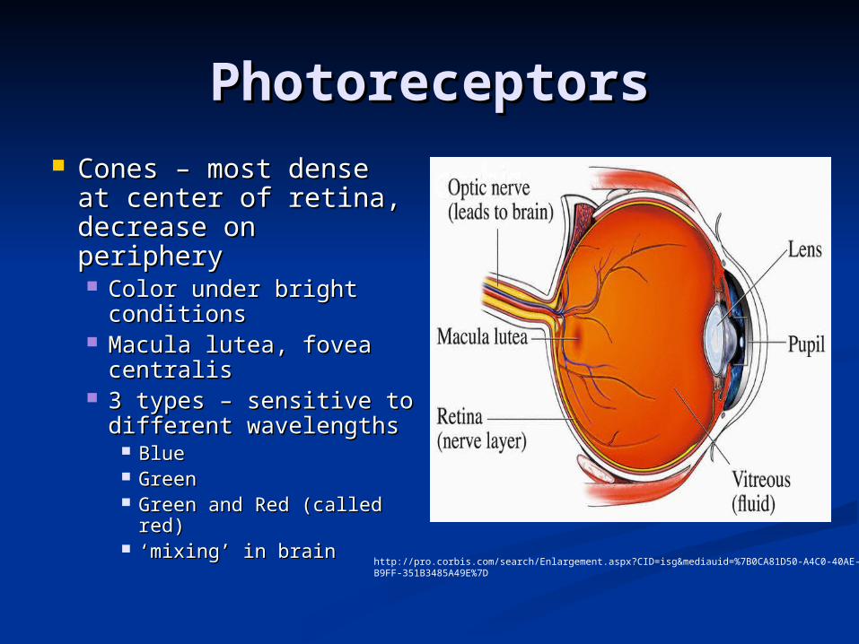
PhotoreceptorsPhotoreceptors Cones – most dense at Cones – most dense at
center of retina, center of retina, decrease on peripherydecrease on periphery Color under bright Color under bright
conditionsconditions Macula lutea, fovea Macula lutea, fovea
centraliscentralis 3 types – sensitive to 3 types – sensitive to
different wavelengthsdifferent wavelengths BlueBlue GreenGreen Green and Red (called Green and Red (called
red)red) ‘‘mixing’ in brainmixing’ in brain http://pro.corbis.com/search/Enlargement.aspx?CID=isg&mediauid=%7B0CA81D50-A4C0-40AE-B9FF-
351B3485A49E%7D
PhotoreceptorsPhotoreceptors
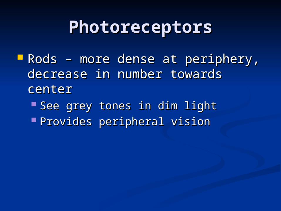
Rods – more dense at periphery, Rods – more dense at periphery, decrease in number towards centerdecrease in number towards center See grey tones in dim lightSee grey tones in dim light Provides peripheral visionProvides peripheral vision

Night or Color BlindnessNight or Color Blindness
Color Blindness – More common in men, Color Blindness – More common in men, most common is lack of red or green most common is lack of red or green receptorsreceptors See red or green as same color, depends on See red or green as same color, depends on
cones presentcones present Can see differences in intensity of colorCan see differences in intensity of color
Night Blindness – most commonly caused Night Blindness – most commonly caused by Vitamin A deficiency, leads to by Vitamin A deficiency, leads to deterioration of neural retinadeterioration of neural retina Vitamin A supplements can restore if Vitamin A supplements can restore if
degenerative changes have not occurreddegenerative changes have not occurred

LensLens
Divides eye into 2 segmentsDivides eye into 2 segments Anterior (Aqueous) SegmentAnterior (Aqueous) Segment
Similar to blood plasmaSimilar to blood plasma Continuously secreted by part of choroidContinuously secreted by part of choroid Provides nutrients for lens and cornea, lack blood Provides nutrients for lens and cornea, lack blood
supplysupply Reabsorbed into venous blood through canal of Reabsorbed into venous blood through canal of
SchlemmSchlemm Posterior (Vitreous) SegmentPosterior (Vitreous) Segment
Gel-like substanceGel-like substance
Both maintain intraocular pressureBoth maintain intraocular pressure

GlaucomaGlaucoma
Drainage of aqueous Drainage of aqueous fluid via canal of fluid via canal of Schlemm blockedSchlemm blocked
pressure increases pressure increases compressing retina compressing retina and optic nerve, and optic nerve, causes pain and causes pain and possibly blindness possibly blindness
Common in elderlyCommon in elderly Slow processSlow process
http://www.steadyhealth.com/articles/Can_Glaucoma_Be_Prevented__a640_f0.html
LensLens
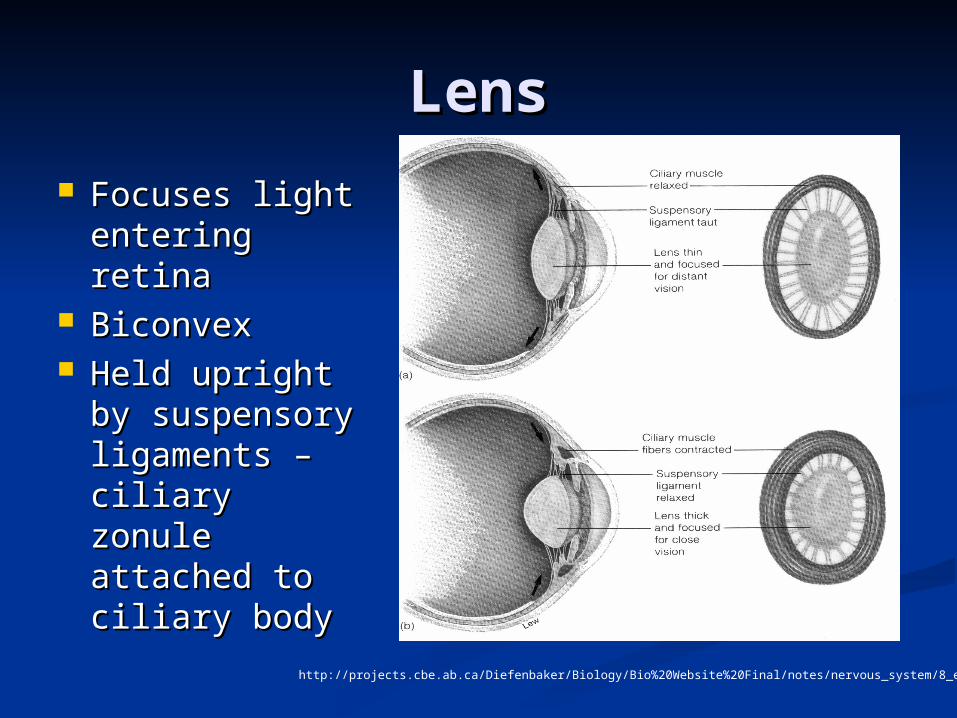
Focuses light Focuses light entering entering retinaretina
BiconvexBiconvex Held upright Held upright
by suspensory by suspensory ligaments – ligaments – ciliary zonule ciliary zonule attached to attached to ciliary bodyciliary body
http://projects.cbe.ab.ca/Diefenbaker/Biology/Bio%20Website%20Final/notes/nervous_system/8_eye_notes.html
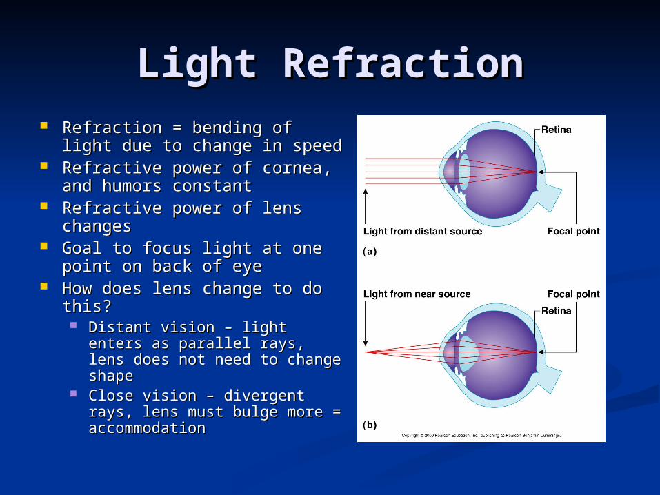
Light RefractionLight Refraction Refraction = bending of light Refraction = bending of light
due to change in speeddue to change in speed Refractive power of cornea, Refractive power of cornea,
and humors constantand humors constant Refractive power of lens Refractive power of lens
changeschanges Goal to focus light at one Goal to focus light at one
point on back of eyepoint on back of eye How does lens change to do How does lens change to do
this?this? Distant vision – light enters Distant vision – light enters
as parallel rays, lens does not as parallel rays, lens does not need to change shapeneed to change shape
Close vision – divergent rays, Close vision – divergent rays, lens must bulge more = lens must bulge more = accommodationaccommodation

Nearsighted or Nearsighted or FarsightedFarsighted

CataractsCataracts
With age, lens With age, lens becomes hard and becomes hard and opaqueopaque
Diabetes mellitusDiabetes mellitus Heavy smokingHeavy smoking Vision becomes hazy Vision becomes hazy
and distorted, will and distorted, will lead to blindnesslead to blindness
Surgical removal or Surgical removal or glassesglasses
http://www.ehponline.org/docs/2005/113-3/niehsnews.html

Binocular visionBinocular vision
““two-eyed” visiontwo-eyed” vision
Provides depth Provides depth perceptionperception
3D vision3D vision
http://www.e-advisor.us/visual_fields.htm

Eye ReflexesEye Reflexes
Convergence = movement of eyes Convergence = movement of eyes medially when viewing close objectsmedially when viewing close objects
Photopupillary reflex = pupils Photopupillary reflex = pupils constrict in bright light to protect constrict in bright light to protect photoreceptorsphotoreceptors
Accommodation pupillary reflex = Accommodation pupillary reflex = pupils constrict when viewing close pupils constrict when viewing close objectsobjects

The EndThe End