special considerations in the management of heel ulcers 2018/presentations/0848 andersen.p… ·...
TRANSCRIPT
Special Considerations in the Management of Heel Ulcers
Charles Andersen MD, FACS, MAPWCAChief Vascular/Endovascular/ Limb Preservation
Surgery Service (Emeritus)Medical Director Wound Care Service
Madigan Army Medical CenterClinical Professor of Surgery UW, USUHS

Bias and Disclosures
• No commercial disclosure
• Vascular Surgeon with a strong interest in Limb Preservation and Wound Care
• Work for US Government; and any opinions or recommendations are my personal recommendations and not the opinions of the US Government
Conclusions
• Heel Ulcers are different that forefoot ulcerations
• Heel ulcers heal slower than forefoot ulcerations, and unfortunately more amputations occur in the presence of heel ulcers.
• The location of the ulcer indicates the etiology and guides treatment
• Posterior, plantar and medial or lateral ulcers require different types of off loading
• Heel ulcers may have an unrecognized ischemic component –standard tests for perfusion may not recognize regional heel ischemia. Fluorescence angiography can detect heel ischemia and document benefits of revascularization
• Early identification and treatment of ischemia may promote healing and prevent amputations

Heel Ulcers - Perspective
• Large amount of data on diabetic foot ulcers and the risk of amputation associated with diabetic foot ulcers
• Many times heel ulcers are grouped with diabetic foot ulcers although the etiology, natural history and treatment are different.
TWO GOALS FOR THE PRESENTATION
• Heel ulcers have different etiologies and different treatments depending on the location of the heel ulcer
• Heel ulcers have special vascular considerations

Heel Ulcers
• Heel ulcers are very common – One million patients in US have heel ulcers
• Heel ulcers - 25% of pressure ulcers in acute care facilities
• 32% following orthopedic hip procedures
• Most common pressure ulcers in chronic care facilities
• Estimated cost per hospital stay ranges from $2000 to $30,000
• Heel ulcers heal slower than forefoot ulcerations, and unfortunately more amputations occur in the presence of heel ulcers.
• Limb salvage rates for patients with heel ulcers have been estimated to be between 65-89%. This is much lower than forefoot ulcerations
• HEEL ULCERS ARE BAD ACTORS
Yosuf MK, Mahadi SI, Mahmoud SM, et al. Diabetic neuropathic forefoot and heel ulcers: management, clinical presentation and outcomes. J Wound Care. 2015;24(9):420-425.
Pickwell KM, Siersma VD, Kars M, et al. Diabetic foot disease: impact of ulcer location on ulcer healing. Diabetes Metab Res Rev. 2013;29(5):377-383.

Heel Ulcers – Ulcer Location
• Ulcer Location – Useful in determining the etiology and treatment• Posterior
• Plantar
• Medial or lateral
Posterior Heel Ulcers
• Very similar to presacral pressure injuries• Prolonged immobility – prolonged hospitalization or chronic care facilities –
Pressure, Shear and Maceration
• Poor nutrition
• Weight loss with underlying significant medical illness
Posterior Heel Ulcers
Treatment – Depends on ambulatory status• Bed rest - Pressure relief critical component of prevention and treatment
• Ambulatory patient - Active off loading – Bulky off loading dressing or TCC• Still need off loading at night
• Standard off loading boots or shoes can make posterior ulcers worse
Goals• Depends on patient – ambulatory or non-ambulatory
• Ambulatory patient - Heal wound
• Non Ambulatory patient• Depends on the patient’s prognosis
• Depending on the prognosis palliative care to prevent infection may be appropriate
• Betadine paint and off loading boot
• It is the secondary infection that leads to sepsis, amputation and/or death

Posterior Heel Ulcers - Case Study
• Elder abuse
• Admitted in Septic Shock
• Bed bound at home – not fed and basic care not provided

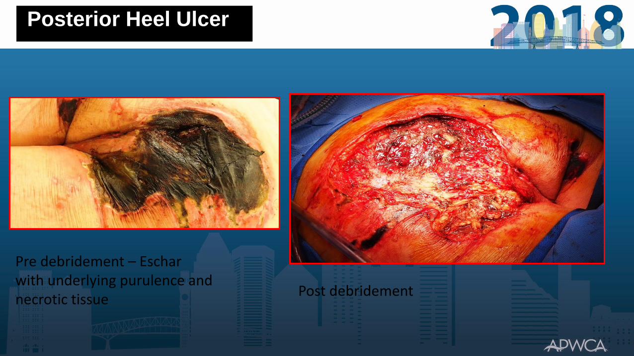
Posterior Heel Ulcer
Posterior Heel Ulcer
Pre debridement – Escharwith underlying purulence and necrotic tissue
Post debridement

Posterior Heel Ulcer
Pressure ulcers elbow, scapular, and head
Posterior Ulcers - Summary
• May be associated with significant systemic issues• Chronic illness
• Poor nutrition
• Altered mental status
• May be in a post op patient going into rehabilitation• Hip Surgery
• Critical is to define goals
• Key to treatment is pressure reduction• If ambulating requires different off loading device or dressing
• In patients with poor prognosis, palliative wound care may be appropriate
Plantar Ulcers
• Very Similar to traditional “diabetic foot ulcers”
• Etiology –• Neuropathy
• Altered biomechanics
• Repetitive unrecognized trauma
• Treatment• Most important is off loading
• More important “what you take off the wound than what you put on the wound”
Plantar Ulcers – Case Study
• 77 y/o male with right heal ulcer• Obese diabetic with neuropathy
• Chronic congestive failure on 40 mgm of Lasix qd
• Chronic post phlebitis syndrome right leg
• Depression with poor compliance

Case Study
• Primary Assessment – Pressure ulcer in neuropathic foot
• Secondary Assessment – Diabetic, congestive failure with edema, venous insufficiency with edema, depression and poor compliance. Good arterial perfusion.
• Initial treatment off loading boot and advanced wound care products
• Ulcer continued to increase in size
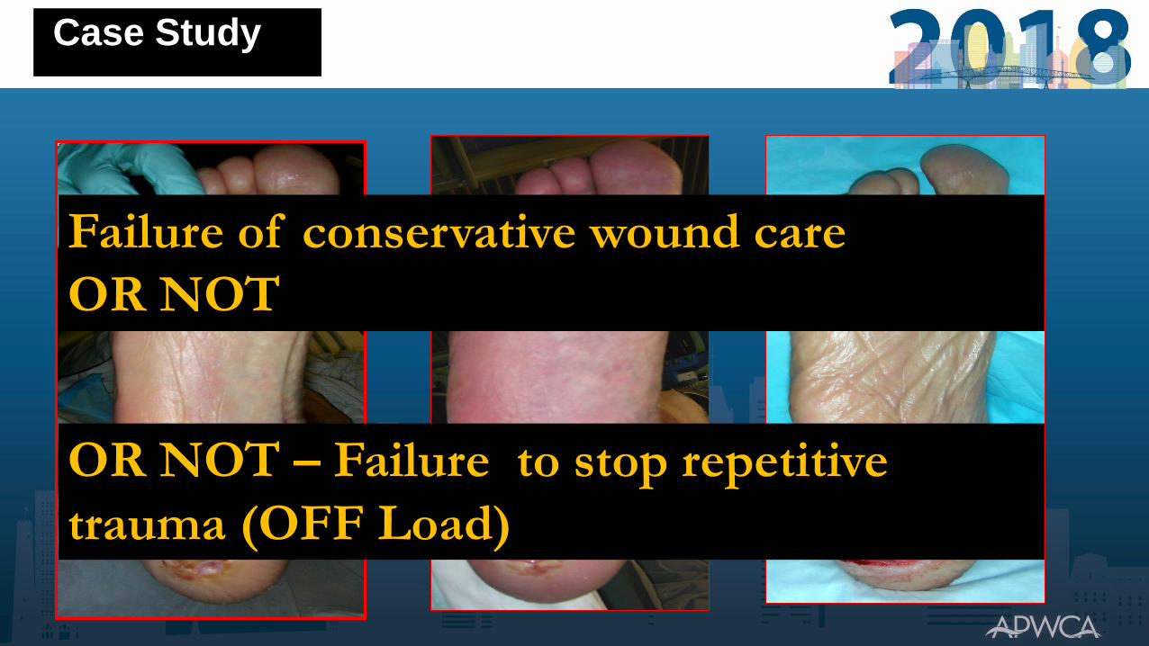
Case Study
Failure of conservative wound care
OR NOT
OR NOT – Failure to stop repetitive
trauma (OFF Load)
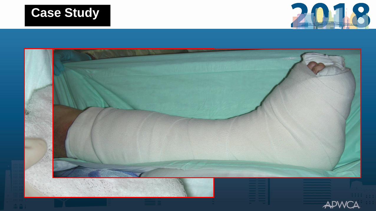
Case Study

Case Study
Following initiation of the Total
Contact Cast,the patient healed
in 6 weeks
Plantar Foot Ulcers - Summary
• Plantar foot ulcers are like forefoot ulcers.
• Key to healing is off loading
• If plantar heel ulcer is not healing don’t think of a new wound care product
• Get better off loading
• For an active plantar ulcer an off loading shoe is often times inadequate
• Gold standard is a TCC

Medial or Lateral Ulcers
• Almost always a traumatic component• Improper shoes
• Wheel chair
• Brace
• Altered position of the leg following hip fracture• Can cause pressure at night
• Treatment• Critical to identify the mechanism of trauma and prevent repetitive trauma

• 65 y/o male status post right BKA for necrotizing infection
• Rehabilitation with poorly fitting off loading shoe
• Developed medial heel ulcer
• Wound continued to increase in size with a standard off loading diabetic shoe
Medial ulcer – Case Study

Medial Heel Ulcer

• Treatment
• Bulky off loading dressing with extra padding over ulcer covered with coban and post op shoe
Medial Heel Ulcer
Key to treatment is identification of the trauma and stop repetitive trauma
Active ulcers require active off loading – high failure with off loading boots
Medial or Lateral Ulcers - Summary
Heel Ulcers Special Vascular Considerations
Heel Ulcers – Special Vascular Considerations
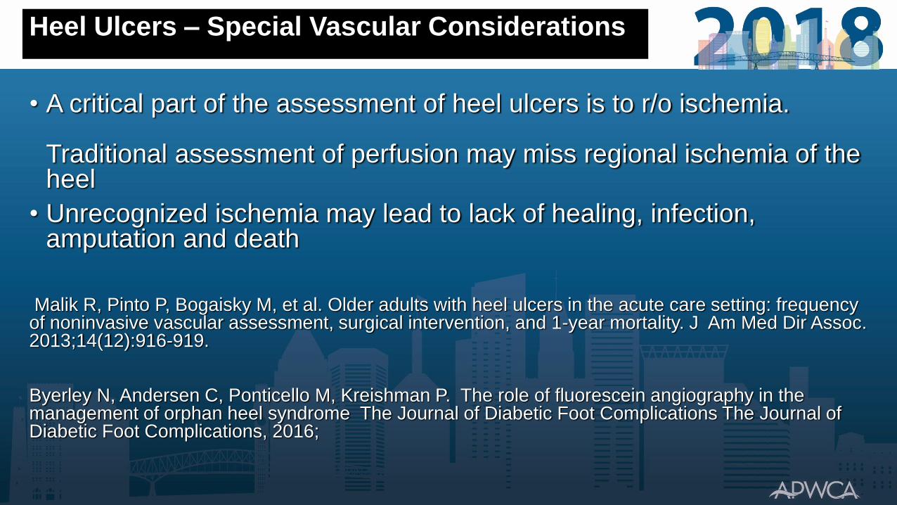
• A critical part of the assessment of heel ulcers is to r/o ischemia.
Traditional assessment of perfusion may miss regional ischemia of the heel
• Unrecognized ischemia may lead to lack of healing, infection, amputation and death
Malik R, Pinto P, Bogaisky M, et al. Older adults with heel ulcers in the acute care setting: frequency of noninvasive vascular assessment, surgical intervention, and 1-year mortality. J Am Med Dir Assoc. 2013;14(12):916-919.
Byerley N, Andersen C, Ponticello M, Kreishman P. The role of fluorescein angiography in the management of orphan heel syndrome The Journal of Diabetic Foot Complications The Journal of Diabetic Foot Complications, 2016;
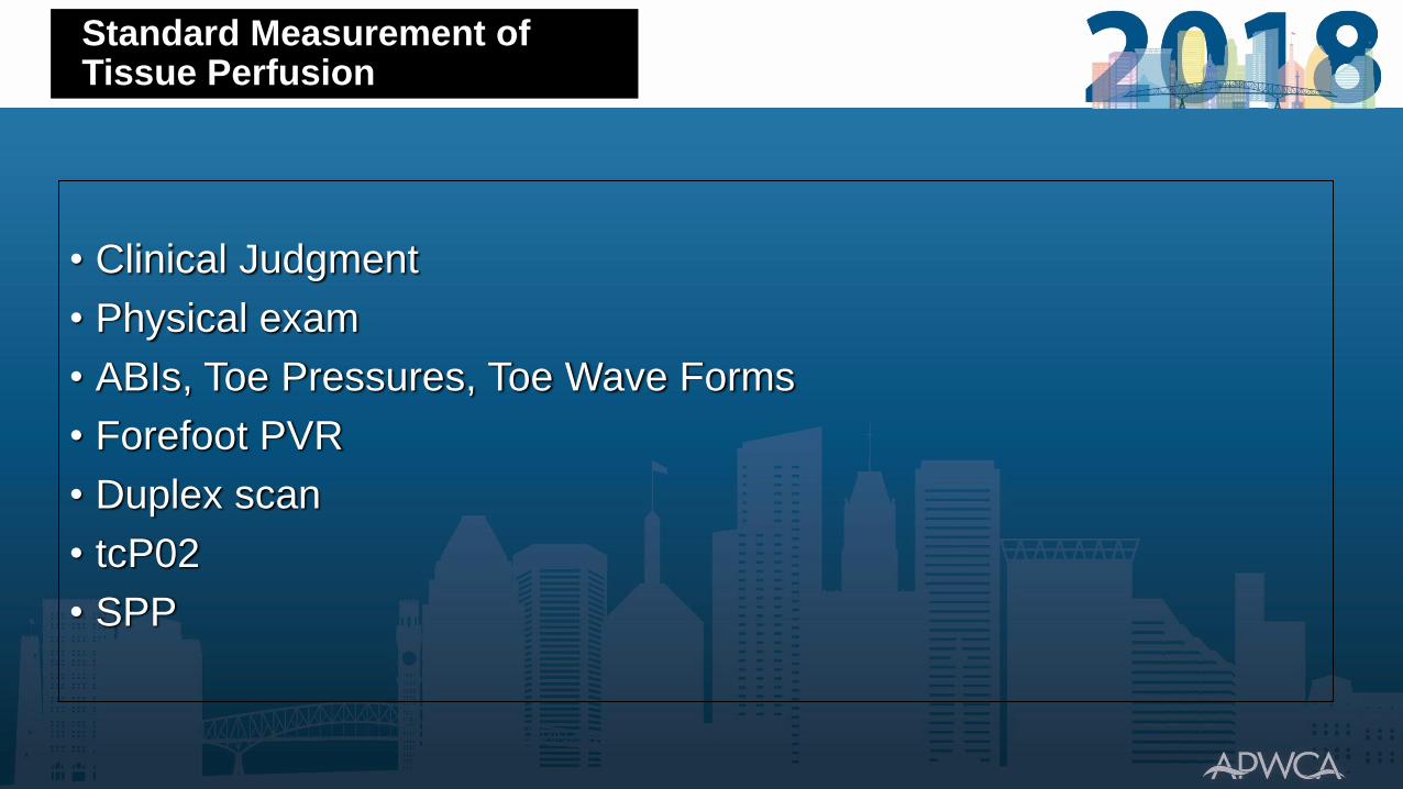
Standard Measurement of Tissue Perfusion
• Clinical Judgment
• Physical exam
• ABIs, Toe Pressures, Toe Wave Forms
• Forefoot PVR
• Duplex scan
• tcP02
• SPP

Assessment of Perfusion
• Traditional methods of measuring tissue perfusion are a poor indicator of heel perfusion.
• These limitations of routine non-invasive vascular studies may not identify focal ischemia giving a false sense of security (Example ABI)
• Fluorescence angiography can measure tissue perfusion in a heel ulcer
• Fluorescence angiography can detect tissue damage prior to ulceration (screening) – Grade DTI
Byerley N, Andersen C, Ponticello M, Kreishman P. The role of fluorescein angiography in the management of orphan heel syndrome The Journal of Diabetic Foot Complications The Journal of Diabetic Foot Complications, 2016;

Heel Ulcers - Ischemia
• Regardless of the etiology, and /or location, heel ulcers may be associated with ischemia
• Because of the unique blood supply• Small collateral branches originating from the posterior tibial (PT) artery and branches of
the perforating peroneal artery.
• The heel is a watershead area that is prone to ischemia (Orphan Heel Syndrome)
• Ischemia may be a contributing factor in development and delayed resolution
• Ischemia most common in patients with diabetes and renal failure
• Taylor Z. The diagnostic triad of orphan heel syndrome: posterior tibial and peroneal artery occlusive disease, poorly controlled diabetes and renal failure. J Vasc Surg 2013;58:565.

• The presence of heel ulcers associated with focal ischemia is referred to as Orphan Heel Syndrome.
• Most common in patients with diabetes and/or renal failure
Orphan Heel Syndrome
The role of fluorescein angiography in the management of orphan heel syndrome Authors: Nicole Byerley, DPM*, Col (Ret) Charles A. Andersen, MD, FACS, FAPWCA1, Mario N. Ponticello, DPM, FACFAS, FAPWCA2, LTC, MC, Peter Kreishman, MD3 The Journal of Diabetic Foot Complications, 2016

Heel Angiosome
• The heel is a unique angiosome due to it having two sources :
posterior tibial artery
peroneal artery
no direct artery to
artery connections
Clemens MW, Attinger CE. Angiosomes and wound care in the diabetic foot. Foot Ankle Clin. 2010;15(3):439-464

Heel Ulcers - Arteriography
• Normal ABIs did not predict heel ischemia
• Patients with heel ulcers having arteriography• 14% - severe malperfusion about the heel
• 33% undergoing endovascular intervention
Taylor Z. The diagnostic triad of orphan heel syndrome: posterior tibial and peroneal artery occlusive disease, poorly controlled diabetes and renal failure. J Vasc Surg 2013;58:565.
Byerley N, Andersen C, Ponticello M, Kreishman P. The role of fluorescein angiography in the management of orphan heel syndrome The Journal of Diabetic Foot Complications The Journal of Diabetic Foot Complications, 2016;

Heel Ulcer - Case Study
• The following case report demonstrates the use of fluorescence angiography to assess and monitor perfusion to a heel ulcer and surrounding soft tissues during treatment.
• In addition, this report documents increased perfusion to the wound and surrounding tissue with an indirect revascularization approach.

Case Study
• 81 year- old female with a past medical history of poorly-controlled insulin-dependent DM type II with neuropathy
• Painful right posterior-lateral heel ulceration that had been present for three weeks

Ischemic Heel Ulcer – Case Study
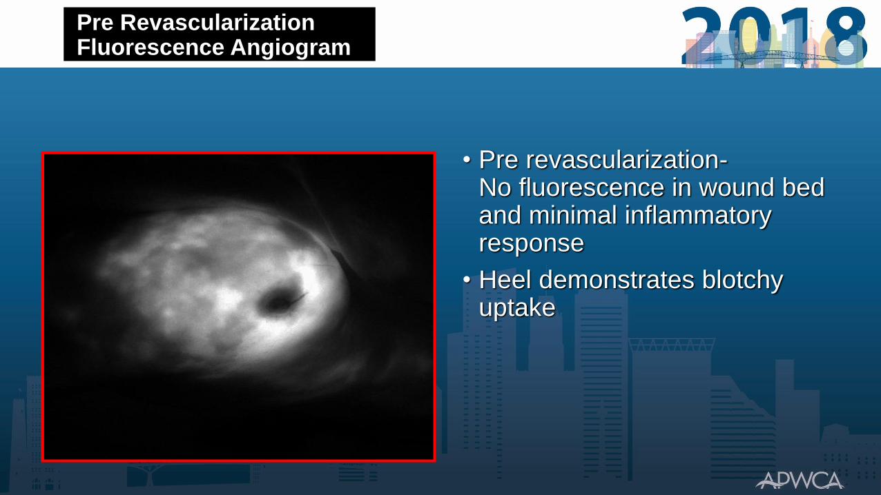
Pre RevascularizationFluorescence Angiogram
• Pre revascularization-No fluorescence in wound bed and minimal inflammatory response
• Heel demonstrates blotchy uptake

Pre RevascularizationFluorescence Angiogram

Revascularization
• An arteriogram demonstrated popliteal occlusion and severe infra-popliteal disease
• Popliteal stent and angioplasty of the tibial peroneal trunk.
• The only runoff vessel was a peroneal artery reconstituting the distal dorsalis pedis artery. The posterior tibial artery was totally occluded
• This is considered indirect revascularization to the heel

Revascularization

Revascularization
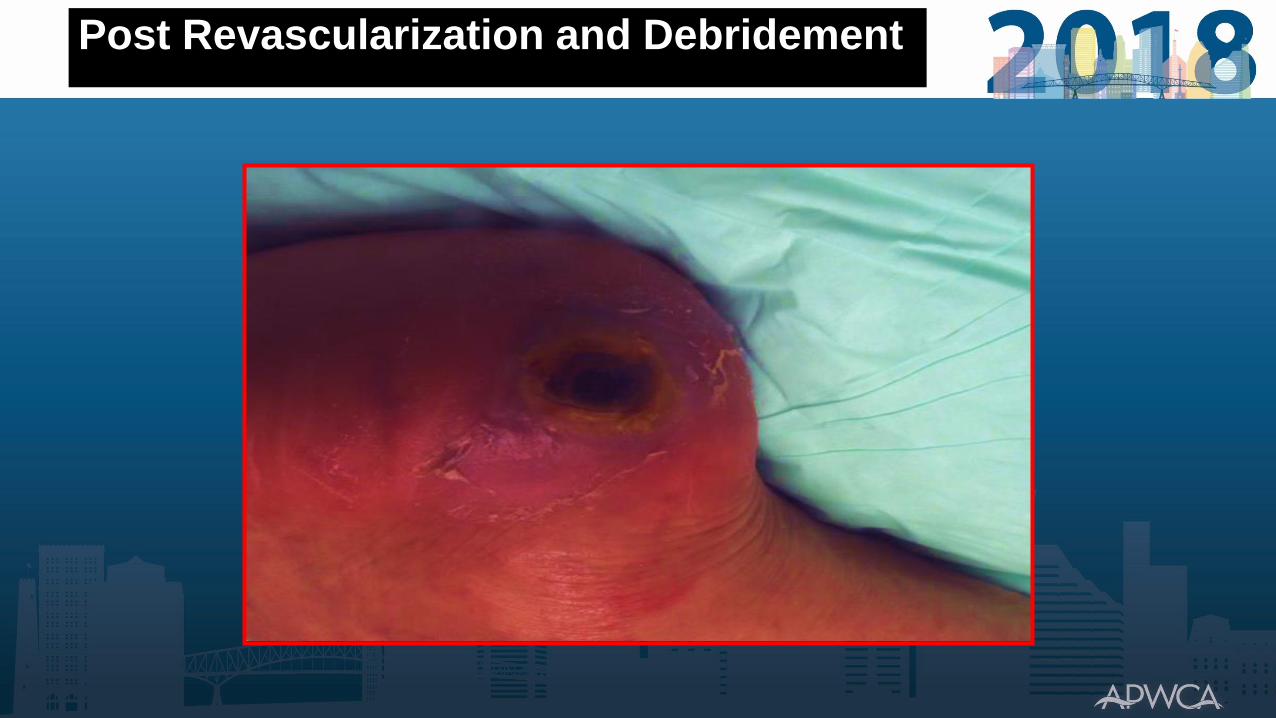
Post Revascularization and Debridement

Post RevascularizationFluorescence Angiogram

One Month Post Revascularization
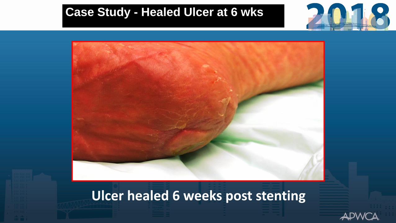
Case Study - Healed Ulcer at 6 wks.
Ulcer healed 6 weeks post stenting

Conclusions
• Heel Ulcers are different that forefoot ulcerations
• Heel ulcers heal slower than forefoot ulcerations, and unfortunately more amputations occur in the presence of heel ulcers.
• The location of the ulcer indicates the etiology and guides treatment
• Posterior, plantar and medial or lateral ulcers require different types of off loading
• Heel ulcers may have an unrecognized ischemic component –standard tests for perfusion may not recognize regional heel ischemia. Fluorescence angiography can detect heel ischemia and document benefits of revascularization
• Early identification and treatment of ischemia may promote healing and prevent amputations


Special Considerations in the Management of Heel Ulcers
Charles Andersen MD, FACS, MAPWCAChief Vascular/Endovascular/ Limb Preservation
Surgery Service (Emeritus)Medical Director Wound Care Service
Madigan Army Medical CenterClinical Professor of Surgery UW, USUHS