soft-tissue hemangiomas of head and neck: mr imaging ... · the diagnosis of hemangioma is normally...
TRANSCRIPT
Page 1 of 14
Soft-tissue hemangiomas of head and neck: MR imagingevaluation
Poster No.: C-3332
Congress: ECR 2010
Type: Educational Exhibit
Topic: Head and Neck
Authors: V. Dimarelos, D. Konstantinou, A. Bintoudi, V. Kyriakou, M.Michaelides, I. Tsitouridis; Thessaloniki/GR
Keywords: hemangioma, head and neck, magnetic resonance imaging
DOI: 10.1594/ecr2010/C-3332
Any information contained in this pdf file is automatically generated from digital materialsubmitted to EPOS by third parties in the form of scientific presentations. Referencesto any names, marks, products, or services of third parties or hypertext links to third-party sites or information are provided solely as a convenience to you and do not inany way constitute or imply ECR's endorsement, sponsorship or recommendation of thethird party, information, product or service. ECR is not responsible for the content ofthese pages and does not make any representations regarding the content or accuracyof material in this file.As per copyright regulations, any unauthorised use of the material or parts thereof aswell as commercial reproduction or multiple distribution by any traditional or electronicallybased reproduction/publication method ist strictly prohibited.You agree to defend, indemnify, and hold ECR harmless from and against any and allclaims, damages, costs, and expenses, including attorneys' fees, arising from or relatedto your use of these pages.Please note: Links to movies, ppt slideshows and any other multimedia files are notavailable in the pdf version of presentations.www.myESR.org
Page 2 of 14
Learning objectives
Hemangiomas are the most frequently encountered vascular soft tissue abnormalitiesand also the most common tumors of infancy, affecting 2%-3% of neonates and 10%-12%of children after 1 year. They are more common in Caucasians, with a female-to-maleratio ranging from 3:1 to 5:1 and the incidence increases with prematurity, especially inthose infants weighing less than 1500g [1]. They can be found in all regions of the body,but there is an anatomic predilection in the head and neck region (60%), followed by thetrunk (25%) and the extremities (15%), that is not yet explained [2]. Almost 80% of allhemangiomas are single lesions, but 20% of affected infants have multiple tumors [3].This increases the risk of having visceral tumors involving the liver, spleen, lung, brain andintestines [4]. Although the majority of hemangiomas occur sporadically, a family historyof hemangioma has been reported to be present in approximately 10% of patients [5].
The diagnosis of hemangioma is normally based on patient history and clinical findings.However, in some cases, there are difficulties in distinguishing hemangiomas from othersoft-tissue tumors of the head and neck and imaging is necessary to characterize thelesion and determine its anatomic extent [6].
Background
Hemangiomas are the result of a derangement in angiogenesis that allows theuncontrolled proliferation of vascular elements [7]. According to the classificationsystem proposed by Mulliken and Glowacki [8], hemangiomas are identified as vasculartumors that exhibit an active growth phase characterized by endothelial proliferation andhyper-cellularity, followed by a period of gradual involution that occurs over several years.In contrast, vascular malformations were characterized as lesions derived from dysplasticcapillaries, veins, lymphatic vessels, arteries or a combination of these.
This classification system was modified in 1997 to include kaposiformhemangioendothelioma, pyogenic granuloma, tufted angioma and hemangiopericytomain the category of vascular tumors [9]. These lesions were previously classified ashemangiomas but were shown to have distinct histopathologic and clinical features (seeTable below).
A. Vascular Tumors

Page 3 of 14
Hemangiomas
Proliferating
Involuting
Kaposiform hemagioma
Tufted angioma (angioblastoma)
Angiosarcoma
Congenital hemangiopericytoma / infantile myofibromatosis
Eccrine angiomatous hamartoma
B. Vascular Malformations
High flow
Arterial malformation-aneurysm, ectasia, stenosis, etc.
Arteriovenous fistula
Arteriovenous malformation
Low flow
Capillary malformation- Port-wine stain
Telangiectasias
Venous malformation
Lymphatic malformation
Macrocystic
Microcystic
Complex combined
Slow flow: Klippel-Trenaunay syndrome (capillary lymphatic venous malformation)
Fast flow: Parkes Weber syndrome (capillary arterial venous malformation)
Hemangiomas usually appear a few weeks after birth and grow more rapidly than theinfant does. The maximum size of the hemangioma is reached at age 6 to 12 months and
Page 4 of 14
the clinical appearance varies with the degree of cutaneous involvement and the depthof the lesions, from an area of simple erythema to a bruise-like macule, a pale patchor a telangiectasia with a pale halo [6]. Involution of the hemangioma generally beginsat about 18 months of age and is characterized by a decrease in cellularity and tumorsize, formation of larger vascular channels, fibro-fatty replacement and development ofa lobular architecture with septa. Involution and proliferation may be happen at the sametime within different parts of a given lesion or among hemangiomas in children withmultiple lesions. An early onset of involution is the only factor associated with improvedoutcome [10].
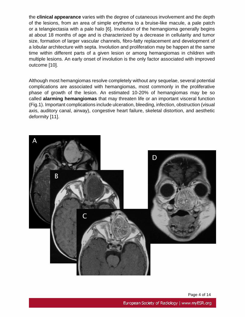
Although most hemangiomas resolve completely without any sequelae, several potentialcomplications are associated with hemangiomas, most commonly in the proliferativephase of growth of the lesion. An estimated 10-20% of hemangiomas may be socalled alarming hemangiomas that may threaten life or an important visceral function(Fig.1). Important complications include ulceration, bleeding, infection, obstruction (visualaxis, auditory canal, airway), congestive heart failure, skeletal distortion, and aestheticdeformity [11].

Page 5 of 14
Fig.: 1. A large hemangioma in the left nasal cavity that obstructs the airway of this11-month-old boy is seen in axial MR images (a, b, c). The coronal plane is useful forunderstanding the extent of the lesion (d).References: V. Dimarelos; Radiology Department, Papageorgiou General Hospital,Thessaloniki, GREECE
The diagnosis of most hemangiomas is based on clinical presentation, history, andphysical examination and do not require any investigation or any treatment for theywill subside spontaneously. However diagnostic imaging is needed in cases of clinicallyatypical soft-tissue masses, questionable superficial lesions, deeper lesions with normaloverlying skin, vascular like lesions presenting in later life, and cases of alarminghemangiomas. Imaging is also required to demonstrate possible visceral involvement orother associated anomalies, guide therapy and assess treatment efficacy [12]. Magneticresonance imaging is considered the modality of choice for the evaluation of soft-tissuemasses and hemangiomas are no exception to this rule [13].
The MR images of 23 patients, (ranging from 1 month to 12 years old) old with soft-tissue hemangiomas in the head and neck were retrospectively studied. The subjectsincluded 13 females and 10 males, ranging from 2 months to 12 years of age (median2.1 years of age). The patients were imaged consecutively at our institution between2005 and 2009. All patients were imaged before biopsy or surgery, when performed, andthe following features were evaluated: detectability of the lesion, location, size, margins,signal intensity on T2- and T1- weighted images, enhancement of contrast medium andthe presence of central low-intensity dots in the lesions.
MRI examinations were performed at 1T with a head, neck or surface coil according tothe size of the patient and the structure to be studied. The scan protocol included axial,coronal or sagittal spin-echo or fast spin-echo T1- and T2- weighted images. T2-weightedimages with fat suppression technique were acquired in some patients. T1-weightedsequences after intravenous administration of paramagnetic contrast agent were alsoobtained in most patients and in at least one orthogonal plane.
Imaging findings OR Procedure details
All lesions in our patients were solitary and unilateral. Hemangiomas were located at-in descending order of frequency- the buccal space, the orbit and periocular area, theparotid and masticator spaces, the soft tissues of the anterior neck, the upper lip andthe nasal cavity. However, in 6 patients, hemangiomas were not localized in a singlecompartment, but involved more than one spaces (Fig.1).

Page 6 of 14
Fig.: 1. A 1-year-old boy with a large hemangioma that infiltrates surrounding tissuesand occupies multiple contiguous spaces of the extracranial head and neck, is seenin axial T2WI (a), axial T1WI (b) and axial (c) and coronal (d) T1WI after contrastadministration.References: V. Dimarelos; Radiology Department, Papageorgiou General Hospital,Thessaloniki, GREECE
MR imaging of hemangiomas in the proliferative phase demonstrated a well-defined,lobulated and heterogeneous mass with a high signal intensity relative to the muscleon T2-weighted images. On T2- weighted MR images with fat suppression technique,hemangiomas tended to show well defined borders, heterogeneity, and high contrastcompared to the surrounding tissues (Fig.2).

Page 7 of 14
Fig.: 2. Hemangioma in the left parotid space of a 2-month-old boy. The mass ishyperintense with lobules and septa on the T2-weighted images (a) and the lesion'scontrast to the surrounding tissues is even higher on the T2-weighted images with theFS technique (b). There is marked enhancement after contrast medium administration(d).References: V. Dimarelos; Radiology Department, Papageorgiou General Hospital,Thessaloniki, GREECE
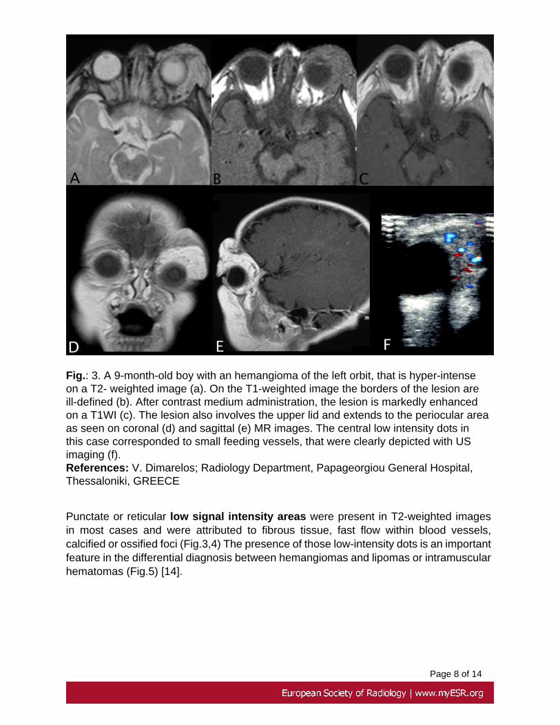
On T1- weighted images, the signal intensity of hemangiomas was intermediate betweenthat of muscle and fat and the lesions had mostly irregular borders and showedheterogeneity (Fig.3).
Page 8 of 14
Fig.: 3. A 9-month-old boy with an hemangioma of the left orbit, that is hyper-intenseon a T2- weighted image (a). On the T1-weighted image the borders of the lesion areill-defined (b). After contrast medium administration, the lesion is markedly enhancedon a T1WI (c). The lesion also involves the upper lid and extends to the periocular areaas seen on coronal (d) and sagittal (e) MR images. The central low intensity dots inthis case corresponded to small feeding vessels, that were clearly depicted with USimaging (f).References: V. Dimarelos; Radiology Department, Papageorgiou General Hospital,Thessaloniki, GREECE
Punctate or reticular low signal intensity areas were present in T2-weighted imagesin most cases and were attributed to fibrous tissue, fast flow within blood vessels,calcified or ossified foci (Fig.3,4) The presence of those low-intensity dots is an importantfeature in the differential diagnosis between hemangiomas and lipomas or intramuscularhematomas (Fig.5) [14].

Page 9 of 14
Fig.: 4. Axial MR images in a 1-year-old girl with an hemangioma above the lefteyebrow, that is hyper-intense on T2-weighted (a,b) and low-to-iso intense on T1-weighted (c) MR imaging. Paramagnetic contrast agent enhanced T1WI MR showsenhancement of the lesion (d). A low signal intensity are seen on unenhanced imagesis still visible after contrast administration.References: V. Dimarelos; Radiology Department, Papageorgiou General Hospital,Thessaloniki, GREECE
T1-weighted images after the intravenous administration of paramagnetic contrastagent showed a moderate to strong centripetal enhancement of all hemangiomas, withprogressive accumulation of contrast material on delayed images (Fig.5,6).

Page 10 of 14
Fig.: 5. An hemangioma is seen in the left masticator space of a 9-year-old girl onaxial T2WI (a) and T1WI (b) MR images. The lesion shows early centripetal (c) anddelayed strong (d) enhancement on T1-weighted images after contrast mediumadministration. There are also prominent vascular signal voids within the lesion.References: V. Dimarelos; Radiology Department, Papageorgiou General Hospital,Thessaloniki, GREECE

Page 11 of 14
Fig.: 6. A 1-year-old boy with an hemangioma in the left upper lip, that is hyper-intenseon T2WI (a) and hypo-intense on T1WI (b) MR. After contrast administration the lesionshows mild centripetal enhancement on T1WI axial (c) and coronal (d) MR imaging.References: V. Dimarelos; Radiology Department, Papageorgiou General Hospital,Thessaloniki, GREECE
Clinically involuting hemangiomas were observed in 3 patients and exhibited areasof high signal intensity on T1-weighted sequences and less contrast enhancement thanproliferating hemangiomas (Fig.7). These findings probably corresponded to fibro-fattyreplacement and the development of a lobular architecture with septa [15].

Page 12 of 14
Fig.: 7. A 7-year-old boy with a right malar hemangioma in clinical regression is seenon axial images (a,b,c). T1WI MR reveals a low-to-intermediate signal of the tumorwith interspersed areas of high signal intensity due to fatty infiltration (b). There is stillconsiderable enhancement of the lesion after contrast agent administration on T1-weighted imaging (c).References: V. Dimarelos; Radiology Department, Papageorgiou General Hospital,Thessaloniki, GREECE
Conclusion
In conclusion, an understanding of the nature and imaging characteristics of hemangiomaof the head and neck soft tissues is important to enable the correct diagnosis, whichis usually based on clinical presentation, history and physical examination. However,in many cases, additional investigation is required and MRI is the preferred modalitybecause of the wealth of information provided, by using a synthesis of morphologic

Page 13 of 14
features, the pattern of enhancement after contrast administration and the signal intensityof the lesion.
Personal Information
V. Dimarelos, D. Konstantinou, A. Bintoudi, V. Kyriakou, M. Michaelides, I. Tsitouridis.Department of Radiology, Papageorgiou General Hospital, Ring Road, Nea Efkarpia,56429, Thessaloniki, Greece.
mail to: [email protected]
References
1. Zheng JW, Zhou GY, Wang YA, Zhang ZY. Management of head and neckhemangiomas in China. Chin Med J (Engl) 2008; 121:1037-1042.
2. Mattassi R, Loose DA, Vaghi M. Hemangiomas and vascular malformations. An atlasof diagnosis and treatment. Springer 2009; pg 17-20.
3. Werner JA, Dunne AA, Folz BJ, et al. Current concepts in the classification, diagnosisand treatment of hemangiomas and vascular malformations of the head and neck. EurArch Otorhinolaryngol 2001; 258:141-149.
4. Enjolras O, Riche MC, Merland JJ, Escande JP. Management of alarminghemangiomas in infancy: a review of 25 cases. Pediatrics 1990; 85:491-498
5. Walter JW, Blei F, Anderson JL, Orlow SJ, Speer MC, Marchuk DA. Genetic mappingof a novel familial form of infantile hemangioma. Am J Med Genet 1999; 82:77-83.
6. Sundine MJ, Wirth GA. Hemangiomas: an overview. Clin Pediatr (Phila) 2007;46:206-221.
7. Folkman J. Is angiogenesis an organizing principle in biology and medicine? J PediatrSurg 2007; 42:1-11.
8. Mulliken JB, Glowacki J. Hemangiomas and vascular malformations in infants andchildren: a classification based on endothelial characteristics. Plast Reconstr Surg 1982;69:412-422.
9. Adams DM, Lucky AW. Cervicofacial vascular anomalies. I. Hemangiomas and otherbenign vascular tumors. Semin Pediatr Surg 2006; 15:124-132.

Page 14 of 14
10. Gampper TJ, Morgan RF. Vascular anomalies: hemangiomas. Plast Reconstr Surg2002; 110:572-585; quiz 586; discussion 587-578.
11. Drolet BA, Esterly NB, Frieden IJ. Hemangiomas in children. N Engl J Med 1999;341:173-181.
12. Dubois J, Garel L. Imaging and therapeutic approach of hemangiomas and vascularmalformations in the pediatric age group. Pediatr Radiol 1999; 29:879-893.
13. Buetow PC, Kransdorf MJ, Moser RP, Jr., Jelinek JS, Berrey BH. Radiologicappearance of intramuscular hemangioma with emphasis on MR imaging. AJR Am JRoentgenol 1990; 154:563-567.
14. Rak KM, Yakes WF, Ray RL, et al. MR imaging of symptomatic peripheral vascularmalformations. AJR Am J Roentgenol 1992; 159:107-112.
15. Vilanova JC, Barcelo J, Villalon M. MR and MR angiography characterization of softtissue vascular malformations. Curr Probl Diagn Radiol 2004; 33:161-170.