ct patterns in histopathologically complex cavernous hemangiomas - ajnr · ajnr :6, may/june 1985...
TRANSCRIPT

Jamshid Ahmadi1 Carol Ann Miller 2
Hervey D. Segall1
Sun-Hyung Park 1.3 Chi-Shing Zee 1
Ronald L. Becker 4
Received August 1, 1984; accepted after revision October 5, 1984.
Presented in part at the annual meeting of the American Society of Neuroradiology, San Francisco, June 1983.
, Department of Radiology, Section of Neuroradiology, University of Southern California School of Medicine, Los Angeles, CA 90033. Address reprint requests to J. Ahmadi , Department of Radiology, LAC/USC Medical Center, Box 2, 1200 N. State St. , Los Angeles, CA 90033.
2 Department of Pathology, Section of Neuropathology, University of Southern California School of Medicine, Los Angeles, CA 90033.
3 Present address: Olympia Radiology, Ltd ., Olympia, WA 98506.
• Department of Radiology, Torrance Memorial Hospital, Torrance, CA 90509 .
AJNR 6:389-393, May/June 1985 0195-6108/85/0603-0389 © American Roentgen Ray Society
389
CT Patterns in Histopathologically Complex Cavernous Hemangiomas
Computed tomographic (CT) studies were correlated with microscopic findings in 10 histologically verified cavernous hemangiomas in nine patients_ In four of the 10 lesions, two or more distinct types of cerebrovascular malformations were identified histopathologically. These included arteriovenous malformations, venous malformations, and telangiectasis. Such coexistence of various types of cerebrovascular malformations has been reported rarely. In each of the four combined lesions, there was evidence of recent or old hemorrhage. The CT findings were nonspecific and were similar to those seen in a variety of intracranial pathologic conditions.
Cavernous hemangiomas are relatively uncommon vascular malformations that may involve any part of the central nervous system. In some cases they are clinically quiescent and are detected at autopsy or, by chance, on a computed tomographic (CT) scan. Others cause seizures, headaches, intracranial hemorrhage, or focal neurologic deficits [1-4). Most cases are diagnosed during the third to sixth decades of life. Identification of these malformations is clinically important because, when accessible, they can be extirpated surgically and are potentially curable [1, 2, 4). Cavernous hemangiomas are more precisely defined by CT than by skull films, radionuclide scans, or cerebral angiograms [5-12] . CT studies of cavernous hemangiomas usually define a well demarcated, hyperdense lesion that frequently contains calcifications. There is no significant mass effect and usually only a mild degree of contrast enhancement. Hypodense areas may sometimes be observed within the lesion because of cystic components [12 , 13). Such a CT feature is nonspecific and may be seen in a variety of intracranial pathologic conditions including various forms of vascular malformations, granulomas, astrocytomas, oligodendrogliomas, craniopharyngiomas, and pineal region neoplasms [14-22]. We describe 10 histologically verified cavernous hemangiomas in which detailed CT studies were correlated with histologic findings.
Subjects and Methods
Five women and four men aged 22-54 years were studied with CT before and after intravenous injection of 300 ml of Reno-M-DIP (42 g I). Selective transfemoral cerebral angiography was performed in seven patients with magnification and subtraction technique. In five cases prolonged contrast injections (15 ml of Conray 60 delivered in 3 sec) were performed. In the other two cases, 10 ml of Conray 60 was injected at a rate of 7 ml/sec. In five patients, direct surgery was performed, while in the other four patients , CT-guided brain biopsies were performed. Tissue was fixed in 10% neutral buffered formalin . Paraffinembedded sections were stained with hematoxylin and eosin . Each specimen was carefully examined in the usual fashion by a neuropathologist who had no knowledge of the CT findings.

390 AHMADI ET AL. AJNR:6, May/June 1985
TABLE 1: Findings in Patients with Histologically Complex Cavernous Hemangiomas
Case No. Site CT
L basal ganglia Moderate enhancement; calcification
R frontal Mild enhancement L occipital'
2 L parietal Moderate enhancement; calcification
3 ... . . .. .. Vermis Mild enhancement 4 L centrum semiovale Moderate enhancement;
calcification 5 .. . . . .... . Medulla oblongata Hematomat
6 . ... . ... . R thalamus Moderate enhancement 7 L middle fossa Marked enhancement 8 .......... R frontal Mild enhancement
9 R frontal Resolving hematoma with ring and tubular enhancementt
Note.-L = left: R = right ; AVM = arteriovenous malformation . • Not biopsied. t Enhancement could not be appreciated within the lesion itself because of hemorrhage.
Histopathologic Diagnosis
Cavernous hemangioma and AVM
Cavernous hemangioma
Cavernous hemangioma
Cavernous hemangioma Cavernous hemangioma
Cavernous hemangioma and AVM
Cavernous hemangioma Cavernous hemangioma Cavernous hemangioma
and AVM Cavernous hemangioma;
venous malformation; telangiectasia
Hemosiderin
+
0
0
0 +
+
0 0 +
+
Specimen
Biopsy
Biopsy
Biopsy
Surgery Biopsy (mild
hemorrhage) Surgery
Biopsy (massive Surgery Surgery
Surgery
.. .
, . 1
Fig. 1.- Postcontrast CT scan. Two cavernous hemangiomas. Lesion adjacent to left lateral ventricle is moderately enhanced and reveals areas of calcification (curved arrow). Other lesion is only minimally enhanced with no apparent calcification (straight arrow). Histologically both lesions contain calcification.
Fig. 2.-A, Precontrast CT scan. Area of slightly increased density (arrow) with average of 51 H. Fourth ventricle is slightly deformed. B, Postcontrast. Minimal enhancement (54 H) (arrow) . C, Histology. Portion of cavernous hemangioma with thick bands of interstitial collagen. Only a few viable endothelial cells remain. Vascular channels are virtually free of blood cells . Dense intraluminar structures (calcifications) (arrows) (x 640).
Results
Eleven lesions were identified by CT in nine patients (table 1). The most common presenting complaints were headaches (seven patients), seizures (four patients), and focal neurologic deficits (four patients). After brain biopsy, one patient developed a mild bleed; another had a massive and fatal hemorrhage.
Although 11 lesions were found in our nine patients, only 10 were confirmed histologically and all contained cavernous
hemangioma. Four of the 10 lesions consisted of two or more types of cerebrovascular malformation. Three manifested a complex vascular pattern that included arteriovenous malformation (AVM) and cavernous hemangioma elements; the fourth involved a distinctly unusual combination of cavernous hemangioma, telangiectasis, and venous malformation. One patient had three discrete lesions, but only two were biopsied and are considered in this report .
Histologically, eight of the cavernous hemangiomas contained calcifications that occurred particularly within throm-
AJNR :6, May/June 1985 CT OF CAVERNOUS HEMANGIOMAS 391
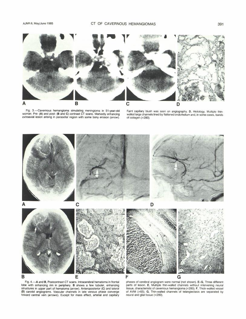
Fig. 3.-Cavernous hemangioma simulating meningioma in 51-year-old woman. Pre- (A) and post- (B and C) contrast CT scans. Markedly enhancing extraaxial lesion arising in parasellar region with some bony erosion (arrow) .
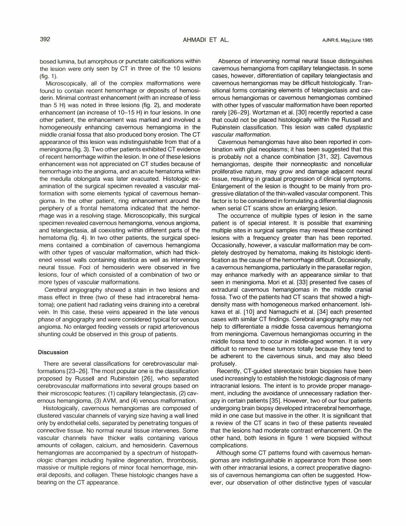
E Fig. 4.-A and B, Postcontrast CT scans. Intracerebral hematoma in frontal
lobe with enhanCing rim in periphery. B shows a few tubular, enhancing structures in upper part of hematoma (arrow). Anteroposterior (C) and lateral (D) carotid angiograms. Vascular channels in late venous phase converge toward central vein (arrows). Except for mass effect, arterial and capillary
D Faint capi llary blush was seen on angiography. 0 , Histology. Multiple thinwalled large channels lined by flattened endothelium and, in some cases, bands of collagen (x 280).
F G phases of cerebral angiogram were normal (not shown). E-G, Three different parts of lesion. E, Multiple thin-walled channels without intervening neural tissue, characteristic of cavernous hemangioma (x280). F, Thick-walled vessel of AVM (x 65). G, Thin-walled channels of telangiectasis are separated by neural and glial tissue (x 280).
392 AHMADI ET AL. AJNR:6. MaylJune 1985
bosed lumina. but amorphous or punctate calcifications within the lesion were only seen by CT in three of the 1 0 lesions (fig . 1).
Microscopically, all of the complex malformations were found to contain recent hemorrhage or deposits of hemosiderin . Minimal contrast enhancement (with an increase of less than 5 H) was noted in three lesions (fig. 2), and moderate enhancement (an increase of 10-15 H) in four lesions. In one other patient, the enhancement was marked and involved a homogeneously enhancing cavernous hemangioma in the middle cranial fossa that also produced bony erosion. The CT appearance of this lesion was indistinguishable from that of a meningioma (fig. 3) . Two other patients exhibited CT evidence of recent hemorrhage within the lesion. In one of these lesions enhancement was not appreciated on CT studies because of hemorrhage into the angioma, and an acute hematoma within the medulla oblongata was later evacuated. Histologic examination of the surgical specimen revealed a vascular malformation with some elements typical of cavernous hemangioma. In the other patient, ring enhancement around the periphery of a frontal hematoma indicated that the hemorrhage was in a resolving stage. Microscopically, this surgical specimen revealed cavernous hemangioma, venous angioma, and telangiectasia, all coexisting within different parts of the hematoma (fig. 4). In two other patients, the surgical specimens contained a combination of cavernous hemangioma with other types of vascular malformation, which had thickened vessel walls containing elastica as well as intervening neural tissue. Foci of hemosiderin were observed in five lesions, four of which consisted of a combination of two or more types of vascular malformations.
Cerebral angiography showed a stain in two lesions and mass effect in three (two of these had intracerebral hematoma); one patient had radiating veins draining into a cerebral vein . In this case, these veins appeared in the late venous phase of angiography and were considered typical for venous angioma. No enlarged feeding vessels or rapid arteriovenous shunting could be observed in this group of patients.
Discussion
There are several classifications for cerebrovascular malformations [23-26]. The most popular one is the classification proposed by Russell and Rubinstein [26] , who separated cerebrovascular malformations into several groups based on their microscopic features: (1) capillary telangiectasis , (2) cavernous hemangioma, (3) AVM, and (4) venous malformation.
Histologically, cavernous hemangiomas are composed of clustered vascular channels of varying size having a wall lined only by endothelial cells, separated by penetrating tongues of connective tissue. No normal neural tissue intervenes. Some vascular channels have thicker walls containing various amounts of collagen , calcium, and hemosiderin . Cavernous hemangiomas are accompanied by a spectrum of histopathologic changes including hyaline degeneration, thrombosis, massive or multiple regions of minor focal hemorrhage, minerai deposits, and collagen . These histologic changes have a bearing on the CT appearance.
Absence of intervening normal neural tissue distinguishes cavernous hemangioma from capillary telangiectasis. In some cases, however, differentiation of capillary telangiectasis and cavernous hemangiomas may be difficult histologically. Transitional forms containing elements of telangiectasis and cavernous hemangiomas or cavernous hemangiomas combined with other types of vascular malformation have been reported rarely [26-29]. Wortzman et al. [30] recently reported a case that could not be placed histologically within the Russell and Rubinstein classification. This lesion was called dysplastic vascular malformation.
Cavernous hemangiomas have also been reported in combination with glial neoplasms; it has been suggested that this is probably not a chance combination [31, 32] . Cavernous hemangiomas, despite their nonneoplastic and noncellular proliferative nature, may grow and damage adjacent neural tissue, resulting in gradual progression of clinical symptoms. Enlargement of the lesion is thought to be mainly from progressive dilatation of the thin-walled vascular component. This factor is to be considered in formulating a differential diagnosis when serial CT scans show an enlarging lesion.
The occurrence of multiple types of lesion in the same patient is of special interest. It is possible that examining multiple sites in surgical samples may reveal these combined lesions with a frequency greater than has been reported. Occasionally, however, a vascular malformation may be completely destroyed by hematoma, making its histologic identification as the cause of the hemorrhage difficult. OccaSionally, a cavernous hemangioma, particularly in the para sellar region, may enhance markedly with an appearance similar to that seen in meningioma. Mori et al. [33] presented five cases of extradural cavernous hemangiomas in the middle cranial fossa. Two of the patients had CT scans that showed a highdensity mass with homogeneous marked enhancement. Ishikawa et al. [10] and Namaguchi et al. [34] each presented cases with similar CT findings. Cerebral angiography may not help to differentiate a middle fossa cavernous hemangioma from meningioma. Cavernous hemangiomas occurring in the middle fossa tend to occur in middle-aged women. It is very difficult to remove these tumors totally because they tend to be adherent to the cavernous sinus, and may also bleed profusely.
Recently, CT-guided stereotaxic brain biopsies have been used increasingly to establish the histologic diagnosis of many intracranial lesions. The intent is to provide proper management, including the avoidance of unnecessary radiation therapy in certain patients [35]. However, two of our four patients undergoing brain biopsy developed intracerebral hemorrhage, mild in one case but massive in the other. It is significant that a review of the CT scans in two of these patients revealed that the lesions had moderate contrast enhancement. On the other hand, both lesions in figure 1 were biopsied without complications.
Although some CT patterns found with cavernous hemangiomas are indistinguishable in appearance from those seen with other intracranial lesions, a correct preoperative diagnosis of cavernous hemangioma can often be suggested . However, our observation of other distinctive types of vascular

AJNR :6, May/June 1985 CT OF CAVERNOUS HEMANGIOMAS 393
malformation in association with cavernous hemangioma in four patients adds a further consideration in CT-pathologic correlation . Such complex malformations may be more common than has been reported heretofore.
REFERENCES
1. Voigt K, Yasargil MG. Cerebral cavernous hemangiomas or cavernomas. Incidence, pathology, localization, diagnosis , clinical features and treatment. Review of the literature and report of an unusual case. Neurochirurgia (Stuttg) 1976;19:59-68
2. Giombini S, Morello G. Cavernous angiomas of the brain . Account of fourteen personal cases and review of the literature. Acta Neurochir (Wien) 1978;40 :61-82
3. McCormick WF, Hardman JM, Boulter TR . Vascular malformations ("angiomas") of the brain , with special reference to those occurring in the posterior fossa. J Neurosurg 1968;28: 241-251
4. Vaquero J, Leunda G, Martinez R, Brav G. Cavernomas of the brain . Neurosurgery 1983;12 : 208-21 0
5. Segall HD, Segal HL, Teal JS, Rumbaugh CL, Bergeron RT. Calcifying cerebral cavernous hemangioma with brain scan and angiographic findings. Neuroradiology 1974;7: 133-138
6. Bartlett JE, Kishore PRS. Intracranial cavernous angioma. AJR 1977;128:653-656
7. Savoiardo M, Passerini C. CT, angiography, and RN scans in intracranial cavernous hemangiomas. Neuroradiology 1978; 16:256-260
8. Becker DH, Townsend JJ , Kramer RA, Newton TH. Occult cerebrovascular malformations. A series of 18 histologically verified cases with negative angiography. Brain 1979 ;102 :249-287
9. Galzio RJ , Zenobii M, Carbonin G, Cristuib-Grizzi L. Hemangioma calcificans. Surg Neuro/1980 ;14 :331-335
10. Ishikawa M, Handa H, Maritake K, Mori K, Nakano Y, Aii H. Computed tomography of cerebral cavernous hemangiomas. J Comput Assist Tomogr 1980;4:587-591
11. Fargueta JS, Iranzo R, Garcia M, Jorda M. Hemangioma calcificans-a benign epi leptic lesion. Surg Neuro/1981;15 :66-70
12. Savoiardo M, Strada L, Passerini A. Intracranial cavernous hemangiomas: neuroradiologic review of 36 operated cases . AJNR 1983;4:945-950.
13. Ramina R, Ingunza W, Vonotakos D. Cystic cerebral cavernous angioma with dense calcification . J Neurosurg 1980;52 :259-262
14. Kramer Ra, Wing SD. Computed tomography of angiographically occult cerebral vascular malformations. Radiology 1977 ; 123 : 649-652
15. Albright AL, Byrd RP, Harrison ML. Angiographically cryptic AVM presenting a pontine tumor. J Neurosurg 1980 ;53 :846-848
16. Yeates A, Enzmann D. Cryptic vascular malformation involving
the brainstem. Radiology 1983 ;146 :71 - 75 17. Foy PF, Lozada L, Shaw MD. Vascular malformation Simulating
a glioma on computed tomography. J Neurosurg 1981 ;54 :125-127
18. Duffner PK, Klein DM , Cohen ME. Calcification of brain stem gliomas. Neurology (N Y) 1978;28 :832-834
19. Price HI , Danziger A. Computed tomography in cranial tuberculosis. AJR 1978;130 :769-771
20. Whelan MA, Stern J. Intracranial tuberculoma. Radiology 1981 ;138:75-81
21 . Vonofakos D, Marcus H, Hacker H. Oligodendrogliomas, CT patterns with emphasis on features indicating malignancy. J Comput Assist Tomogr 1979;3 :783- 788
22. Vaquero J, Carrillo R, Cahezado J, Leunda G, Villoria F, Bravo G. Cavernous angiomas of the pineal region. J Neurosurg 1980;53: 833-835
23. Noran HH . Intracranial vascular tumors and malformations. Arch Pathol Lab Med 1945;39: 393-416
24. Raynor RB, Kingman AF Jr. Hemangioblastoma and vascular malformations as one lesion. Arch Neuro/1965 ;12 :39-48
25. McCormick WF. The pathology of vascular ("arteriovenous") malformations. J Neurosurg 1966;24 :807- 81 6
26. Russell DS, Rubinstein LJ . Pathology of tumors of the nervous system , 3d ed. Baltimore: Williams & Wilkins, 1971:85- 108
27 . Roberson GH, Kase CS, Wolpow ER. Telangiectases and cavernous angiomas of the brainstem: "cryptic" vascular malformations . Report of a case. Neuroradiology 1974;8:83-89
28. Diamond C, Torvik A, Amundsen P. Angiographic diagnosis of telangiectases with cavernous angiomas of the posterior fossa. Acta Radiol [OiagnJ (Stockh) 1976;17 :281-288
29. Hirsh LF. Combined cavernous-arteriovenous malformation. Surg Neuro/1981 ;16:135-139
30. Wortzman G, Sima AF , Morley TP. Cerebral dysplastic vascular malformation: a developmental arrest. Radiology 1983 ;148 :443-446
31 . Falconer MA, Pond DA. Temporal lobe epilepsy with personality and behavior disorders caused by an unusual calcifying lesion. J Neurol Neurosurg Psychiatry 1953;16 :234-244
32. Fischer EG , Sotrel A, Welch K. Cerebral hemangioma with glial neoplasia (angioglioma?). Report of two cases. J Neurosurg 1982;56 : 430-434
33. Mori K, Handa H, Gi H, et al. Cavernomas in the middle fossa. Surg Neuro/1980 ;14 :21-31
34. Namaguchi Y, Kishikawa T, Fukuy M, et al. Prolonged injection angiography for diagnosing intracranial cavernous hemangiomas. Radiology 1979;131 :137-138
35. Apuzzo ML, Sabshin JK. Computed tomographic guidance stereotaxis in the management of intracranial mass lesion. Neurosurgery 1983;12 : 277 - 285