snf1 controls the activity of adr1 through
TRANSCRIPT
Copyright � 2009 by the Genetics Society of AmericaDOI: 10.1534/genetics.109.103432
Snf1 Controls the Activity of Adr1 Through Dephosphorylation of Ser230
Sooraj Ratnakumar, Nataly Kacherovsky, Erin Arms and Elton T. Young1
Department of Biochemistry, University of Washington, Seattle, Washington 98195-7350
Manuscript received March 30, 2009Accepted for publication April 20, 2009
ABSTRACT
The transcription factors Adr1 and Cat8 act in concert to regulate the expression of numerous yeast genesafter the diauxic shift. Their activities are regulated by Snf1, the yeast homolog of the AMP-activated proteinkinase of higher eukaryotes. Cat8 is regulated directly by Snf1, but how Snf1 regulates Adr1 is unknown.Mutations in Adr1 that alleviate glucose repression are clustered between amino acids 227 and 239. Thisregion contains a consensus sequence for protein kinase A, RRAS230F, and Ser230 is phosphorylated in vitroby both protein kinase A and Ca11 calmodulin-dependent protein kinase. Using an antiphosphopeptideantibody, we found that the level of Adr1 phosphorylated on Ser230 was highest in glucose-grown cells anddecreased in a Snf1-dependent manner when glucose was depleted. A nonphosphorylatable Ser230Alamutant was no longer Snf1 dependent for activation of Adr1-dependent genes and could suppress Cat8dependence at genes coregulated by Adr1 and Cat8. Contrary to expectation, neither protein kinase A (PKA)nor Ca11 calmodulin-dependentprotein kinase appeared to havean important role in Ser230phosphorylationin vivo, and a screen of 102 viable kinase deletion strains failed to identify a candidate kinase. We concludethat either Ser230 is phosphorylated by multiple protein kinases or its kinase is encoded by an essential gene.Using the Ser230Ala mutant, we explain a long-standing observation of synergy between Adr1 constitutivemutants and Snf1 activation and conclude that dephosphorylation of Ser230 via a Snf1-dependent pathwayappears to be a major component of Adr1 regulation.
TRANSCRIPTION factors activategeneexpressionbyrecruiting coactivators to a promoter to create a
pre-initiation complex (PIC) containing RNA polII andgeneral factors (Featherstone 2002; Fry and Peterson
2002). Signal transduction pathways often regulatethis process by acetylation or phosphorylation of tran-scription factors to influence their ability to recruitcoactivators (Kwok et al. 1994; Papoutsopoulou andJanknecht 2000; Zhong et al. 2002; Goel andJanknecht 2003). Adr1 is a yeast transcription factorthat acts in concert with Cat8 and Oaf1/Pip2 to activatenumerous genes that are expressed after glucosedepletion, allowing cells to use nonfermentable car-bon sources such as ethanol, glycerol, and fatty acids(Schuller 2003; Young et al. 2003; Santangelo 2006).The signal transduction pathway that activates Adr1 andCat8 is mediated by Snf1 (Rahner et al. 1999; Schuller
2003; Charbon et al. 2004; Santangelo 2006), the yeasthomolog of AMP-activated protein kinase that is widelyregarded as the ‘‘energy sensor of the cell’’ (Hardie et al.1998). Snf1 is activated by phosphorylation on Thr210 bythree upstream kinases (Hong et al. 2003). Snf1-Thr210is dephosphorylated by the PP1-type protein phospha-
tase Glc7, and regulation of this event appears to bethrough the targeting factor Reg1 (Sanz et al. 2000;Rubenstein et al. 2008). As expected, removing thenegative regulatory mechanism by deleting REG1 causesconstitutive activation of Snf1, as measured by expres-sion in glucose of Snf1-dependent genes that arenormally glucose repressed (Dombek et al. 1993; Tu
and Carlson 1995; McCartney and Schmidt 2001;Orlova et al. 2008). Control of Snf1 must includeadditional mechanisms beyond simple phosphorylationand Glc7�Reg1-dependent dephosphorylation, however,because in a reg1D strain, additional phosphorylation ofSnf1 and further activation of some Snf1-dependentgenes is achieved by glucose depletion (Dombek et al.1993; Tachibana et al. 2007; Orlova et al. 2008).Interaction with regulatory proteins or changes inprotein structure have been suggested as additionallevels of Snf1 control (Rubenstein et al. 2008).
Snf1 is necessary for the activity of both Adr1 and Cat8,enhancing transcription of CAT8 through inactivationof the repressor Mig1, as well as activating Cat8 directlyby phosphoryation (Hedges et al. 1995; Rahner et al.1996, 1999; Charbon et al. 2004; reviewed in Schuller
2003). Snf1 is required for Adr1 binding (Young et al.2002) and recruitment of coactivators to Adr1-dependentpromoters (Biddick et al. 2008). Promoter binding byAdr1 is regulated in part by phosphorylation of itsDNA-binding domain (Kacherovsky et al. 2008) andby acetylation of promoter nucleosomes (Verdone et al.
Supporting information is available online at http://www.genetics.org/cgi/content/full/genetics.109.103432/DC1.
1Corresponding author: Department of Biochemistry, University ofWashington, 1705 NE Pacific St., Seattle, WA 98195-7350.E-mail: [email protected]
Genetics 182: 735–745 ( July 2009)

2002; Tachibanaet al. 2007), but it is not known whetherSnf1 activates Adr1 directly or indirectly or has anotherrole at Adr1-dependent promoters, such as nucleosomemodification (Lo et al. 2001).
Selection of mutants that allowed ADH2 expression toescape glucose repression identified rare semidominantADR1c alleles (ADR1-constitutive) (Ciriacy 1979; Denis
et al. 1991). ADR1c alleles allow ADH2 expression tooccur in the presence of glucose and enhance dere-pressed transcription. Surprisingly, the level of Adr1c
protein is much lower than the level of wild-type Adr1(Taylor and Young 1990; Dombek and Young 1997),suggesting that its high transcriptional potency might beassociated with rapid turnover as has been observedfor some other activators (Tansey 2001). ADR1c showsstrong synergism with deletion of the Snf1 regulatoryfactor REG1. In combination with Adr1c, partial activa-tion of Snf1 by deleting REG1 allows much higher levelsof ADH2-repressed expression than are observed witheither mutation acting alone (Dombek et al. 1993). Adr1c
and reg1D are also synergistic in activating a poised butinactive PIC (Tachibana et al. 2007). Although thissuggests that Adr1c and activation of Snf1 by reg1D actthrough different pathways to cause constitutive expres-sion of Snf1-dependent genes, the precise mechanism ofthe synergism is unknown.
Cloning and molecular analysis of ADR1c allelesshowed mutations between amino acids 227 and 239, aregion that contains a consensus sequence for a cyclicAMP-dependent PKA, Arg-Arg-Ala-Ser-Phe (Denis andGallo 1986; Denis et al. 1992). No other mutations inthe Adr1 ORF causing constitutive ADH2 expressionwere isolated, suggesting that the 227–239 region playsa unique role in regulating Adr1 activity. The ADR1c
mutations suggest that Adr1 might be post-translationallyinhibited by phosphorylation of Ser230 in the PKAconsensus sequence (Denis and Gallo 1986; Cherry
et al. 1989). Adr1 appears to be phosphorylated at Ser230in vivo because subtle alterations in its SDS–PAGEelectrophoretic mobility and partial sensitivity to phos-phatase treatment are observed (Vallari et al. 1992;Dombek and Young 1997). Recent studies of the yeastphosphorylome also suggested Adr1 phosphorylation,although the reported site was Ser232, not Ser230 (Chi
et al. 2007).In support of phosphorylation of Adr1 by PKA, both
PKA and calmodulin-dependent protein kinase (CMK)efficiently phosphorylate Ser230 in vitro (Denis et al.1991; Hook et al. 1999). Hyperactivation of PKA bydeletion of its BCY1-encoded regulatory subunit causesloss of ADH2 expression (Cherry et al. 1989), suggest-ing that unregulated PKA activity inhibits Adr1 byconstitutively phosphorylating Ser230. However, replac-ing Ser230 with phosphomimetic Asp gave the sameconstitutively active phenotype as other alterations inthe region, suggesting that either Asp does not mimicphosphorylated Ser or phosphorylation of Ser230 is not
the only determinant of the ADR1c phenotype (Denis
et al. 1992; Dombek and Young 1997). In addition,several of the Adr1c mutations appear to refute PKAas a candidate for the Ser230 kinase in vivo. TheAla229Pro Adr1c mutant shows greatly increased affin-ity for PKA binding in a peptide phosphorylation assay,which would predict greater phosphorylation andlower Adr1 activity. However, the Ala229Pro mutant isone of the most constitutive of the Adr1c mutants(Denis et al. 1992). Therefore, it is not known ifphosphorylation of Ser230 affects the activity of Adr1and what kinase is responsible for the phosphorylationin vivo (Denis et al. 1992; Cook et al. 1994; Dombek andYoung 1997).
How Adr1c achieves its high transcriptional potency isunknown (Denis et al. 1992; Cook et al. 1994; Dombek
and Young 1997). ADR1c alleles map far from the DNA-binding domain and Adr1-Ser230A does not have en-hanced in vitro DNA-binding activity (Taylor andYoung 1990). After characterizing a series of mutantsin amino acids 227–239, as well as internal deletions inthis region of Adr1, Cook et al. (1994) suggested that thisregion either binds a repressive factor that cannotinteract with the mutants or interacts with and blocksthe activation domain. Some evidence has suggestedthat these Adr1c mutants suppress the requirement forSnf1 (Ciriacy 1979; Denis 1984), which would implythat it is not affected by Snf1, but this is contradictory tothe enhanced effect on ADH2 expression seen whenAdr1c is combined with the Snf1-activating reg1D muta-tion. Thus, the questions of how Snf1 affects the activityof Adr1 and whether it interacts with the Adr1c allele areunresolved.
We used an antibody against a Ser230-phosphorylatedpeptide of Adr1 amino acids 217–234 to investigate theconditions under which Ser230 is phosphorylated(Adr1-pSer230) and the effect of Snf1 on this modifica-tion. The antibody was used to screen for a specific Adr1-Ser230 kinase. Phosphorylation was not increased byenhanced PKA activity, nor decreased by loss of PKA orCMK. These and other data suggest that Ser230 may beredundantly phosphorylated in vivo by multiple kinasesor phosphorylated by an essential kinase. Using thenonphosphorylatable Adr1c, we probed the relationshipbetween Adr1-pSer230, Snf1, and the coregulator Cat8,with the results supporting an earlier model for thefunction of the Ser230-containing domain.
MATERIALS AND METHODS
Yeast strains, plasmids, culture conditions, and enzymeassays: The strains used in the study are listed in Table 1.Multiple deletion mutants of TPK and CMK genes wereconstructed by standard genetic techniques. Epitope tag-ging, gene deletion, and marker swapping were according toGuldener et al. (1996), Cross (1997), and Knop et al. (1999),respectively. Deletions and epitope tagging were confirmed byPCR and, for marker swapping, by Western blots. Yeast strains
736 S. Ratnakumar et al.
were grown in complete or synthetic media as described inSherman (1991). Repressing medium contained 5% glucose;derepressing medium contained 0.05% glucose with or with-out 3% glycerol or 2% ethanol. The plasmids used weredescribed previously (Yu et al. 1989; Dombek and Young
1997) and modified in some cases by introducing an epitopetag at the C terminus of Adr1 as described below. For geneexpression studies, CEN-TRP1 plasmids expressing wild-typeADR1 (pKD16) or the S230A (pKD14), R228K (pKD27), or D3(a deletion that removes Adr1 amino acids 226–233; pKD26)alleles of ADR1 from its native promoter were used. To facilitatedetection of Adr1-S230 phosphorylation by Western blotting,2m plasmids expressing wild-type ADR1 (pKD17-HATkanMX(TRP1) and the ADR1-S230A allele (pKD20-HATkanMX(TRP1) from the ADH1 promoter and tagged with an HAepitope were employed. Alternatively, strains with four addi-tional copies of ADR1 integrated at the leu2 locus were used(Sloan et al. 1999). ADR1 was epitope tagged with HATkanMXin this strain. The Adr1-dependent reporter was a UAS1-lacZplasmid, pHDY10, containing 10 Adr1-binding sites (Yu et al.1989). b-Galactosidase assays were performed as described inGuarente (1983). ADH enzyme activity was analyzed byseparating proteins on nondenaturing polyacrylamide gelsand visualized by in-gel chromogenic staining as described(Dombek and Young 1997).
Real-time quantitative PCR: For expression analysis, RNAwas isolated by hot phenol extraction (Collart and Oliveiro
1993) and converted to cDNA with a SuperScript III kit(Invitrogen) according to the manufacturer’s directions.cDNA was quantified by real-time quantitative PCR (RT–qPCR) (Tachibana et al. 2007) with an MJ Research Chromo4system, using ABI or Quantace SYBR Master Mix, according tothe manufacturer’s instructions. Primer sequences are avail-able on request.
Immunoblotting: Whole-cell extracts were analyzed on 3–8% or 6% polyacrylamide gels (NuPage system, Invitrogen)and transferred to PVDF membranes after electrophoresis.Western blots were probed with a-pSer230 followed by a rabbit-specific Licor secondary Ab (l680). A second identical blotwas probed with a monoclonal a-HA antibody and a mouse-specific Licor secondary antibody (l800). Probing a single blotwith both antibodies resulted in bleed-through from the moreabundant HA-epitope-derived signal so either two blots weredone or a single blot was probed sequentially—first, with a-pSer230 and an appropriate Licor secondary antibody and,second, with the a-HA antibody and an appropriate Licorsecondary antibody. Cell extracts were prepared as describedpreviously (Dombek and Young 1997) or by a boiling pro-cedure with the addition of agitation with glass beads for 30 secbefore the boiling step (Invitrogen) (Hahn 2008). An OdysseyInfrared imaging system (Licor Biosciences) was used forquantitation. a-HA (Y-11, Santa Cruz Biochemicals), a-Adr1(Dombek et al. 1993), or antibodies against an Adr1-derivedsynthetic peptide were used at 1:1000 dilution. The peptides,representing Adr1 amino acids 217–234, VKRKYLKKLTRRA(pS)FSAQ, and its nonphosphorylated version were synthe-sized and used to generate and affinity purify the a-pSer230antibody by Bethyl Laboratories (Montgomery, TX). Second-ary IR-dye-conjugated antibodies used at 1:1000–1:3000 weregoat a-mouse Alexa 680 (Molecular Probes) or IRdye800-conjugated a-rabbit IgG (Rockland Immunochemicals).
RESULTS
Adr1-Ser230 phosphorylation in vivo is glucosedependent and dephosphorylation is Snf1 dependent:Antibodies against a phosphorylated peptide represent-
TABLE 1
S. cerevisiae strains
Strain Genotype Source
TYY201(aka W303-1A)
MATa ade2 can1-100 his3-11,15 leu2-13,112 trp1-1 ura3-1 Yeast stock center
VBY20 MATa adh3 ura3 his3 leu2T(pRS315-ADR1)X3 ADH2TYIpADH2/lacZ(TRP1)ADH2T YIpADH2/GFP (URA3)
Voronkova et al. (2006)
NKY85 VBY20 ADR1:HATkanMX This studyNKY87 NK85 reg1DTnatMX This studyNKY91 NK85 snf1DTnatMX This studyTYY204 W303-1A adr1D1TLEU2 ADH2TYIpADH2/lacZTTRP1 Young et al. (2003)TYY497 W303-1A adr1D1TLEU2 ADH2TYIpADH2/lacZTtrp1THIS3 Young et al. (2008)TYY498 W303-1A snf1DTURA3 adr1D1TLEU2 ADH2TYIpADH2/lacZTtrp1THIS3 Young et al. (2008)CKY13 W303-1A adr1DTkanMX This studyCKY26 W303-1A adr1DTnatMX snf1DTkanMX This studyCKY5 W303-1A adr1DTkanMX reg1DTnatMX This studyCKY23 W303-1A adr1DTnatMX cat8DTkanMX This studySRY60 W303-1A reg1DTkanMX cat8DTnatMX adr1DTHYG This studyNKY108 NKY85 cmk1DThphmx cmk2DTnatMX cmk3DTnatMX This studySGP406 MATa his3 leu2-3,112 tpk1TURA3 tpk2THIS3
tpk3TTRP1 trp1 ura3-52 yak1TLEU2Garrett and
Broach (1989)NKY111 NKY85 bcy1DTnatMX This studyCMY323 MATa ade2-101� can1 his3D200 lys2-801 trp1D1 ura3-52 Carl MannCMY328 MATa ade2-101�adr1D2TURA3 can1 his3D200 lys2-801atrp1D1 ura3-52 Dombek and Young (1997)CMY468 MATa ade2-101� his3D leu2D1 lys2-801 tpk1TURA3
tpk2THIS3tpk3ts-1 trp1D1 ura3-52Carl Mann
SGP400 MATa his3 leu2-3,112 trp1 ura3-52 yak1TLEU2 Garrett and Broach (1989)
Adr1c Alleles Suppress Snf1 Deficiency 737
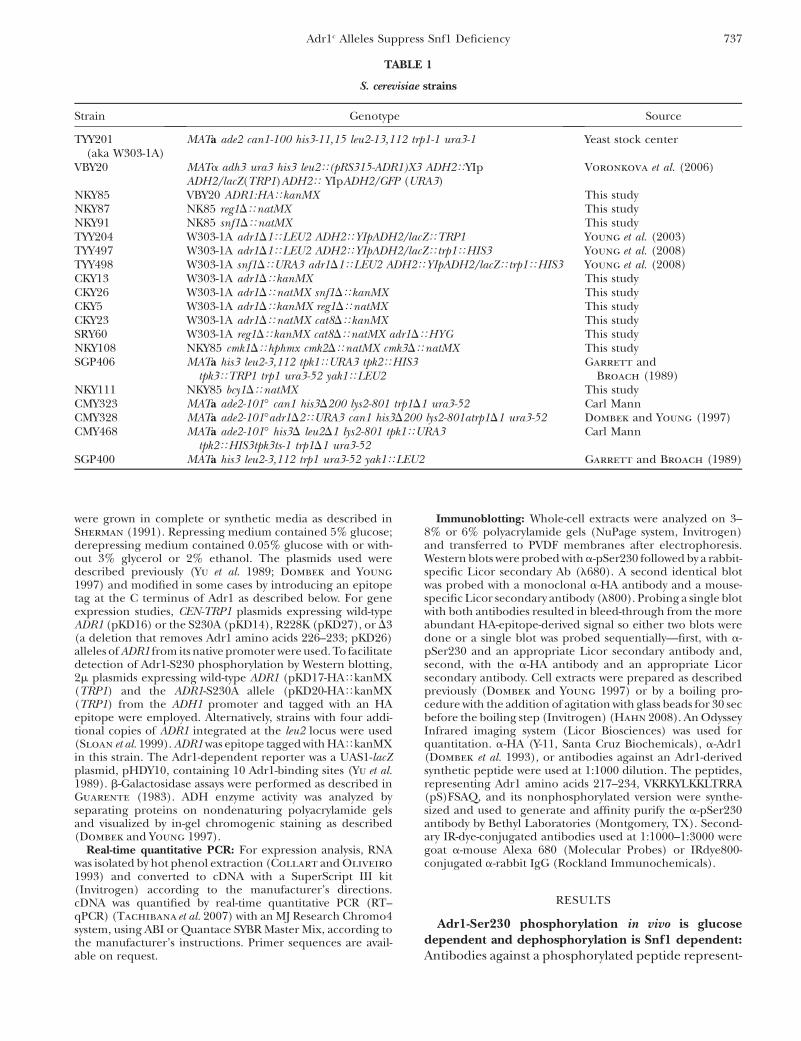
ing Adr1 amino acids 217–234 (VKRKYLKKLTRRA(pS)FSAQ) were generated to assess the level of Adr1phosphorylated on Ser230 in vivo (designatedAdr1�pSer230). ELISA assays indicated that the anti-phosphopeptide antibody (a-pSer230) recognized thephosphorylated peptide 50003 better than the non-phosphorylated peptide (data not shown). Figure 1Ashows that a-pSer230 recognized a protein the size ofAdr1 in a strain expressing Adr1-HA from the strongADH1 promoter on a multicopy plasmid, but not ina strain lacking Adr1. Adr1 containing a Ser230Alamutation (hereafter referred to as Adr1c unless otherADR1c alleles are specified) was recognized poorly by a-pSer230, demonstrating the specificity of the antibodyfor Adr1 with phosphorylated Ser230. Competition withthe phosphorylated and nonphosphorylated peptidesdemonstrated the specificity of a-pSer230 (Figure 1B).These data confirmed that a-pSer230 recognized Adr1-pSer230 and cross-reacted weakly, if at all, with Adr1lacking phosphate on Ser230.
A strain containing multiple integrated copies ofADR1 was employed to facilitate quantitation of Adr1-pSer230 in different growth conditions. At least onecopy of Adr1 in this strain was tagged with an HAepitope to provide a measure of the total amount ofAdr1 in the extract. Adr1 levels in the multicopy straingrown in repressing conditions are similar to Adr1 levelsin wild-type cells grown in derepressing conditions, and
ADH2 regulation appears unperturbed by the modestincrease in Adr1 levels (Sloan et al. 1999; Voronkova
et al. 2006).Western blotting for both HA and Adr1-pSer230
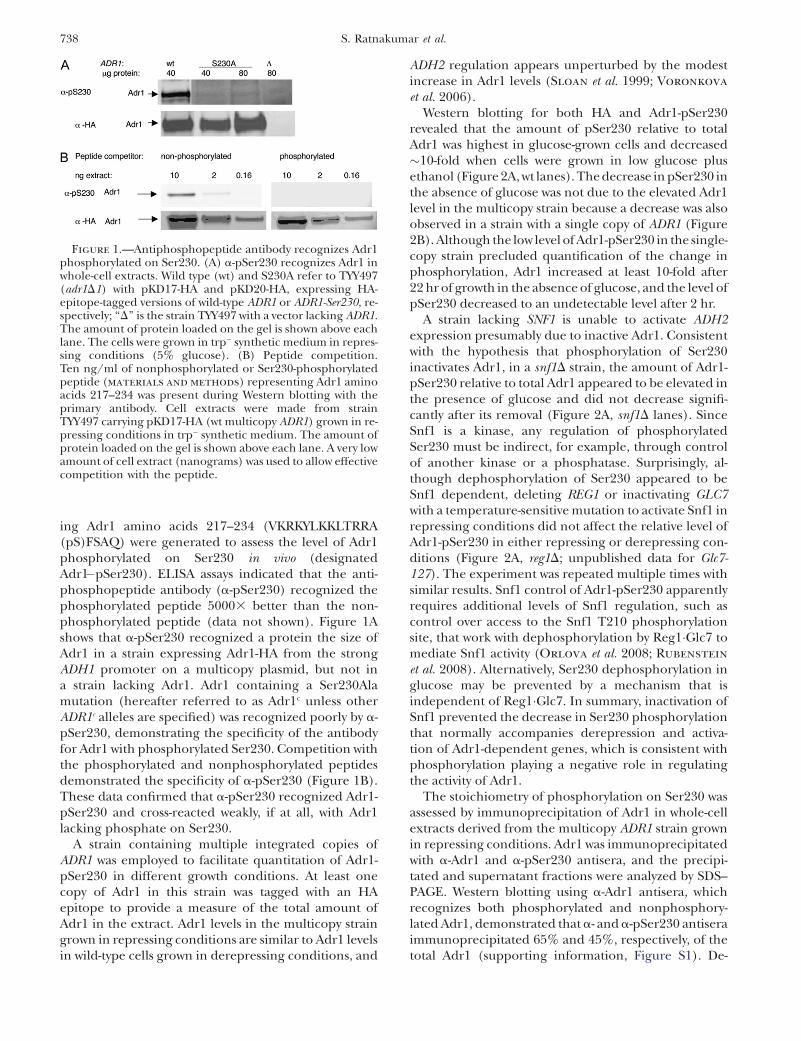
revealed that the amount of pSer230 relative to totalAdr1 was highest in glucose-grown cells and decreased�10-fold when cells were grown in low glucose plusethanol (Figure 2A, wt lanes). The decrease in pSer230 inthe absence of glucose was not due to the elevated Adr1level in the multicopy strain because a decrease was alsoobserved in a strain with a single copy of ADR1 (Figure2B). Although the low level of Adr1-pSer230 in the single-copy strain precluded quantification of the change inphosphorylation, Adr1 increased at least 10-fold after22 hr of growth in the absence of glucose, and the level ofpSer230 decreased to an undetectable level after 2 hr.
A strain lacking SNF1 is unable to activate ADH2expression presumably due to inactive Adr1. Consistentwith the hypothesis that phosphorylation of Ser230inactivates Adr1, in a snf1D strain, the amount of Adr1-pSer230 relative to total Adr1 appeared to be elevated inthe presence of glucose and did not decrease signifi-cantly after its removal (Figure 2A, snf1D lanes). SinceSnf1 is a kinase, any regulation of phosphorylatedSer230 must be indirect, for example, through controlof another kinase or a phosphatase. Surprisingly, al-though dephosphorylation of Ser230 appeared to beSnf1 dependent, deleting REG1 or inactivating GLC7with a temperature-sensitive mutation to activate Snf1 inrepressing conditions did not affect the relative level ofAdr1-pSer230 in either repressing or derepressing con-ditions (Figure 2A, reg1D; unpublished data for Glc7-127). The experiment was repeated multiple times withsimilar results. Snf1 control of Adr1-pSer230 apparentlyrequires additional levels of Snf1 regulation, such ascontrol over access to the Snf1 T210 phosphorylationsite, that work with dephosphorylation by Reg1�Glc7 tomediate Snf1 activity (Orlova et al. 2008; Rubenstein
et al. 2008). Alternatively, Ser230 dephosphorylation inglucose may be prevented by a mechanism that isindependent of Reg1�Glc7. In summary, inactivation ofSnf1 prevented the decrease in Ser230 phosphorylationthat normally accompanies derepression and activa-tion of Adr1-dependent genes, which is consistent withphosphorylation playing a negative role in regulatingthe activity of Adr1.
The stoichiometry of phosphorylation on Ser230 wasassessed by immunoprecipitation of Adr1 in whole-cellextracts derived from the multicopy ADR1 strain grownin repressing conditions. Adr1 was immunoprecipitatedwith a-Adr1 and a-pSer230 antisera, and the precipi-tated and supernatant fractions were analyzed by SDS–PAGE. Western blotting using a-Adr1 antisera, whichrecognizes both phosphorylated and nonphosphory-lated Adr1, demonstrated that a- and a-pSer230 antiseraimmunoprecipitated 65% and 45%, respectively, of thetotal Adr1 (supporting information, Figure S1). De-
Figure 1.—Antiphosphopeptide antibody recognizes Adr1phosphorylated on Ser230. (A) a-pSer230 recognizes Adr1 inwhole-cell extracts. Wild type (wt) and S230A refer to TYY497(adr1D1) with pKD17-HA and pKD20-HA, expressing HA-epitope-tagged versions of wild-type ADR1 or ADR1-Ser230, re-spectively; ‘‘D’’ is the strain TYY497 with a vector lacking ADR1.The amount of protein loaded on the gel is shown above eachlane. The cells were grown in trp� synthetic medium in repres-sing conditions (5% glucose). (B) Peptide competition.Ten ng/ml of nonphosphorylated or Ser230-phosphorylatedpeptide (materials and methods) representing Adr1 aminoacids 217–234 was present during Western blotting with theprimary antibody. Cell extracts were made from strainTYY497 carrying pKD17-HA (wt multicopy ADR1) grown in re-pressing conditions in trp� synthetic medium. The amount ofprotein loaded on the gel is shown above each lane. A very lowamount of cell extract (nanograms) was used to allow effectivecompetition with the peptide.
738 S. Ratnakumar et al.
phosphorylation of pSer230 during immunoprecipita-tion may contribute to the lower level of Adr1 immuno-precipitated by the a-pSer230 antisera compared to thea-Adr1 antisera, or there may be a fraction of Adr1 that isnot phosphorylated on Ser230 even in repressinggrowth conditions. The high level of Adr1 precipitatedby the a-pSer230 antisera indicates that the majority ofAdr1 is phosphorylated in vivo in repressing conditions.
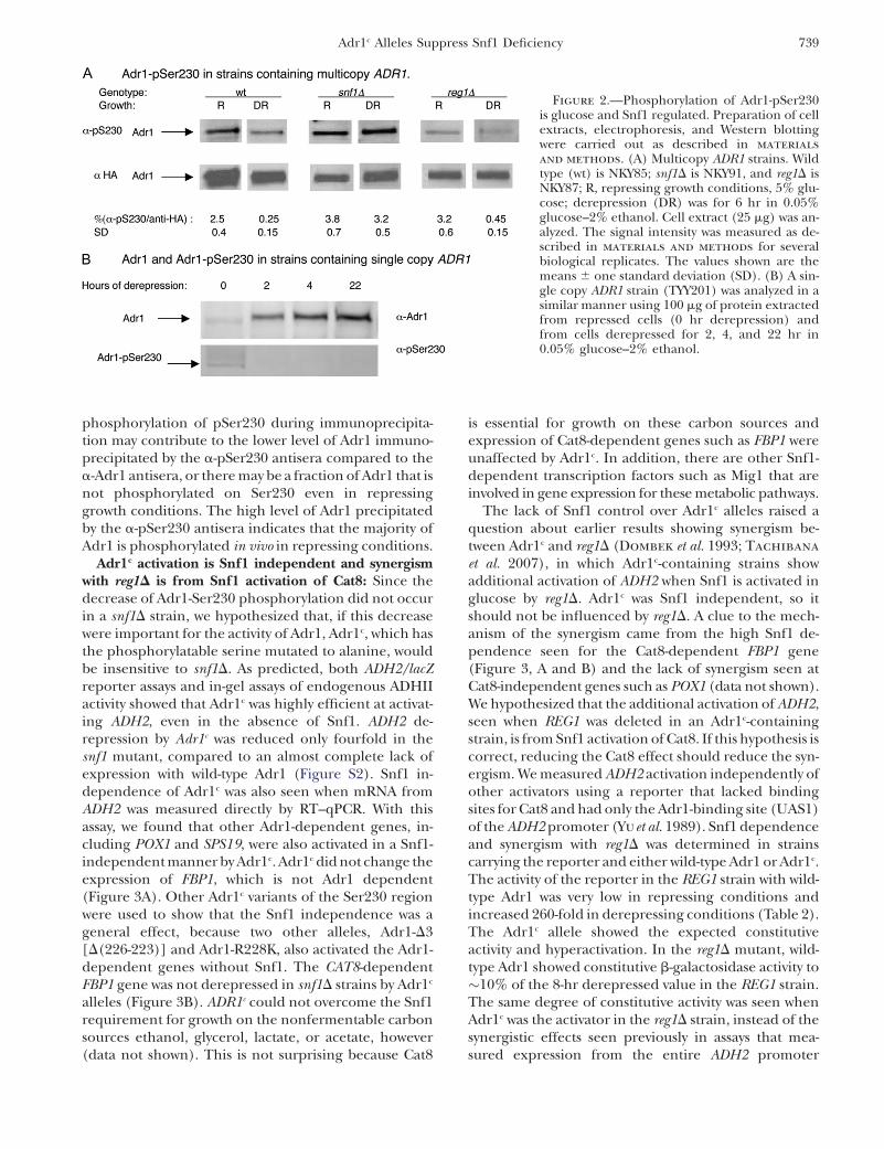
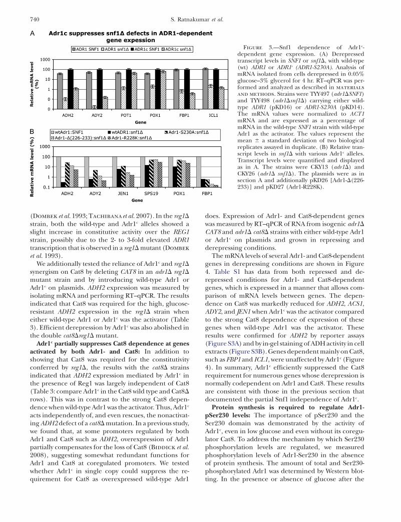
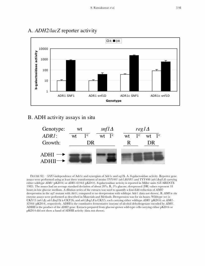
Adr1c activation is Snf1 independent and synergismwith reg1D is from Snf1 activation of Cat8: Since thedecrease of Adr1-Ser230 phosphorylation did not occurin a snf1D strain, we hypothesized that, if this decreasewere important for the activity of Adr1, Adr1c, which hasthe phosphorylatable serine mutated to alanine, wouldbe insensitive to snf1D. As predicted, both ADH2/lacZreporter assays and in-gel assays of endogenous ADHIIactivity showed that Adr1c was highly efficient at activat-ing ADH2, even in the absence of Snf1. ADH2 de-repression by Adr1c was reduced only fourfold in thesnf1 mutant, compared to an almost complete lack ofexpression with wild-type Adr1 (Figure S2). Snf1 in-dependence of Adr1c was also seen when mRNA fromADH2 was measured directly by RT–qPCR. With thisassay, we found that other Adr1-dependent genes, in-cluding POX1 and SPS19, were also activated in a Snf1-independent manner by Adr1c. Adr1c did not change theexpression of FBP1, which is not Adr1 dependent(Figure 3A). Other Adr1c variants of the Ser230 regionwere used to show that the Snf1 independence was ageneral effect, because two other alleles, Adr1-D3[D(226-223)] and Adr1-R228K, also activated the Adr1-dependent genes without Snf1. The CAT8-dependentFBP1 gene was not derepressed in snf1D strains by Adr1c
alleles (Figure 3B). ADR1c could not overcome the Snf1requirement for growth on the nonfermentable carbonsources ethanol, glycerol, lactate, or acetate, however(data not shown). This is not surprising because Cat8
is essential for growth on these carbon sources andexpression of Cat8-dependent genes such as FBP1 wereunaffected by Adr1c. In addition, there are other Snf1-dependent transcription factors such as Mig1 that areinvolved in gene expression for these metabolic pathways.
The lack of Snf1 control over Adr1c alleles raised aquestion about earlier results showing synergism be-tween Adr1c and reg1D (Dombek et al. 1993; Tachibana
et al. 2007), in which Adr1c-containing strains showadditional activation of ADH2 when Snf1 is activated inglucose by reg1D. Adr1c was Snf1 independent, so itshould not be influenced by reg1D. A clue to the mech-anism of the synergism came from the high Snf1 de-pendence seen for the Cat8-dependent FBP1 gene(Figure 3, A and B) and the lack of synergism seen atCat8-independent genes such as POX1 (data not shown).We hypothesized that the additional activation of ADH2,seen when REG1 was deleted in an Adr1c-containingstrain, is from Snf1 activation of Cat8. If this hypothesis iscorrect, reducing the Cat8 effect should reduce the syn-ergism. We measured ADH2 activation independently ofother activators using a reporter that lacked bindingsites for Cat8 and had only the Adr1-binding site (UAS1)of the ADH2 promoter (Yuet al. 1989). Snf1 dependenceand synergism with reg1D was determined in strainscarrying the reporter and either wild-type Adr1 or Adr1c.The activity of the reporter in the REG1 strain with wild-type Adr1 was very low in repressing conditions andincreased 260-fold in derepressing conditions (Table 2).The Adr1c allele showed the expected constitutiveactivity and hyperactivation. In the reg1D mutant, wild-type Adr1 showed constitutive b-galactosidase activity to�10% of the 8-hr derepressed value in the REG1 strain.The same degree of constitutive activity was seen whenAdr1c was the activator in the reg1D strain, instead of thesynergistic effects seen previously in assays that mea-sured expression from the entire ADH2 promoter
Figure 2.—Phosphorylation of Adr1-pSer230is glucose and Snf1 regulated. Preparation of cellextracts, electrophoresis, and Western blottingwere carried out as described in materials
and methods. (A) Multicopy ADR1 strains. Wildtype (wt) is NKY85; snf1D is NKY91, and reg1D isNKY87; R, repressing growth conditions, 5% glu-cose; derepression (DR) was for 6 hr in 0.05%glucose–2% ethanol. Cell extract (25 mg) was an-alyzed. The signal intensity was measured as de-scribed in materials and methods for severalbiological replicates. The values shown are themeans 6 one standard deviation (SD). (B) A sin-gle copy ADR1 strain (TYY201) was analyzed in asimilar manner using 100 mg of protein extractedfrom repressed cells (0 hr derepression) andfrom cells derepressed for 2, 4, and 22 hr in0.05% glucose–2% ethanol.
Adr1c Alleles Suppress Snf1 Deficiency 739
(Dombek et al. 1993; Tachibanaet al. 2007). In the reg1D
strain, both the wild-type and Adr1c alleles showed aslight increase in constitutive activity over the REG1strain, possibly due to the 2- to 3-fold elevated ADR1transcription that is observed in a reg1D mutant (Dombek
et al. 1993).We additionally tested the reliance of Adr1c and reg1D
synergism on Cat8 by deleting CAT8 in an adr1D reg1D
mutant strain and by introducing wild-type Adr1 orAdr1c on plasmids. ADH2 expression was measured byisolating mRNA and performing RT–qPCR. The resultsindicated that Cat8 was required for the high, glucose-resistant ADH2 expression in the reg1D strain wheneither wild-type Adr1 or Adr1c was the activator (Table3). Efficient derepression by Adr1c was also abolished inthe double cat8Dreg1D mutant.
Adr1c partially suppresses Cat8 dependence at genesactivated by both Adr1- and Cat8: In addition toshowing that Cat8 was required for the constitutivityconferred by reg1D, the results with the cat8D strainsindicated that ADH2 expression mediated by Adr1c inthe presence of Reg1 was largely independent of Cat8(Table 3: compare Adr1c in the Cat8 wild type and Cat8D
rows). This was in contrast to the strong Cat8 depen-dence when wild-type Adr1 was the activator. Thus, Adr1c
acts independently of, and even rescues, the nonactivat-ing ADH2 defect of a cat8D mutation. In a previous study,we found that, at some promoters regulated by bothAdr1 and Cat8 such as ADH2, overexpression of Adr1partially compensates for the loss of Cat8 (Biddick et al.2008), suggesting somewhat redundant functions forAdr1 and Cat8 at coregulated promoters. We testedwhether Adr1c in single copy could suppress the re-quirement for Cat8 as overexpressed wild-type Adr1
does. Expression of Adr1- and Cat8-dependent geneswas measured by RT–qPCR of RNA from isogenic adr1D
CAT8 and adr1D cat8D strains with either wild-type Adr1or Adr1c on plasmids and grown in repressing andderepressing conditions.
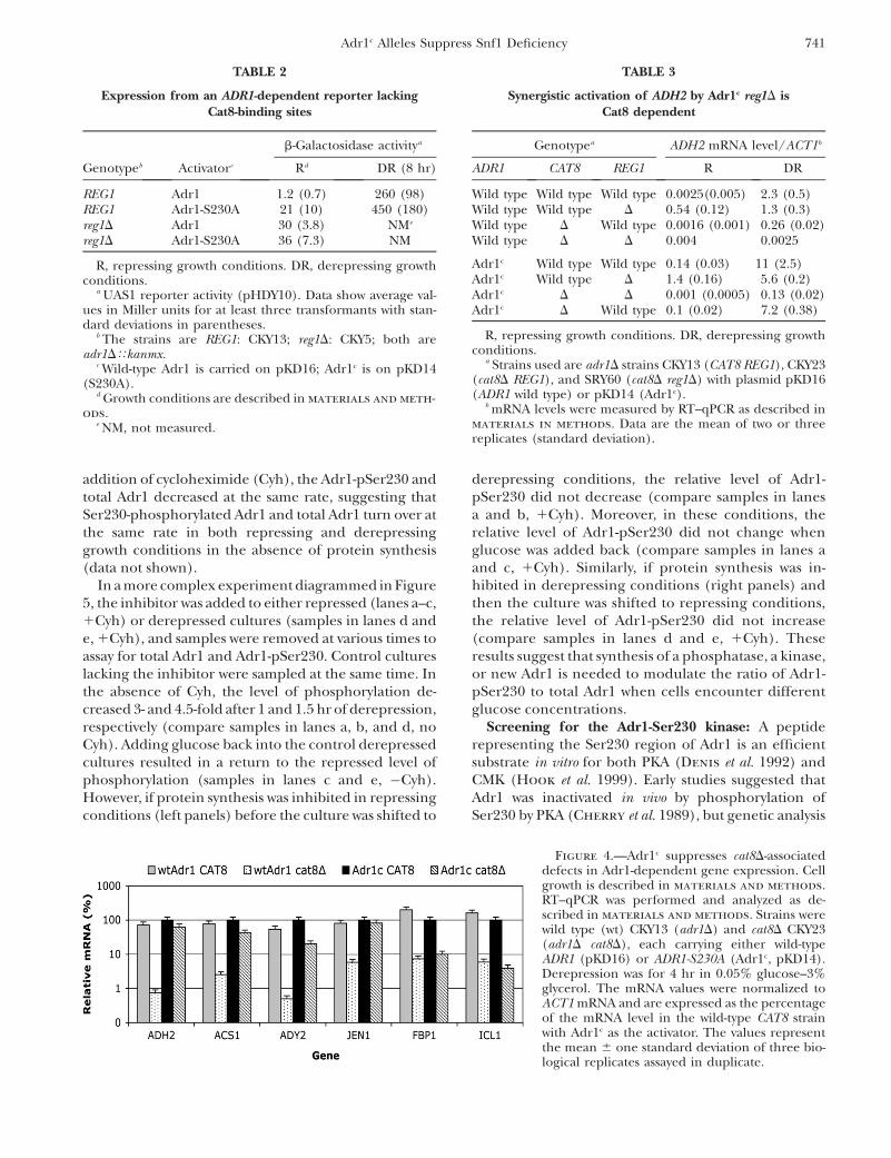
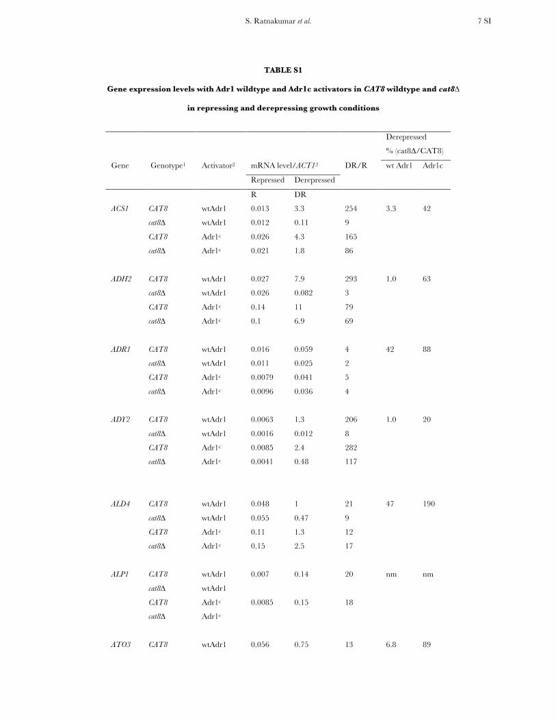
The mRNA levels of several Adr1- and Cat8-dependentgenes in derepressing conditions are shown in Figure4. Table S1 has data from both repressed and de-repressed conditions for Adr1- and Cat8-dependentgenes, which is expressed in a manner that allows com-parison of mRNA levels between genes. The depen-dence on Cat8 was markedly reduced for ADH2, ACS1,ADY2, and JEN1 when Adr1c was the activator comparedto the strong Cat8 dependence of expression of thesegenes when wild-type Adr1 was the activator. Theseresults were confirmed for ADH2 by reporter assays(Figure S3A) and by in-gel staining of ADH activity in cellextracts (Figure S3B). Genes dependent mainly on Cat8,such as FBP1 and ICL1, were unaffected by Adr1c (Figure4). In summary, Adr1c efficiently suppressed the Cat8requirement for numerous genes whose derepression isnormally codependent on Adr1 and Cat8. These resultsare consistent with those in the previous section thatdocumented the partial Snf1 independence of Adr1c.
Protein synthesis is required to regulate Adr1-pSer230 levels: The importance of pSer230 and theSer230 domain was demonstrated by the activity ofAdr1c, even in low glucose and even without its coregu-lator Cat8. To address the mechanism by which Ser230phosphorylation levels are regulated, we measuredphosphorylation levels of Adr1-Ser230 in the absenceof protein synthesis. The amount of total and Ser230-phosphorylated Adr1 was determined by Western blot-ting. In the presence or absence of glucose after the
Figure 3.—Snf1 dependence of Adr1c-dependent gene expression. (A) Derepressedtranscript levels in SNF1 or snf1D, with wild-type(wt) ADR1 or ADR1c (ADR1-S230A). Analysis ofmRNA isolated from cells derepressed in 0.05%glucose–3% glycerol for 4 hr. RT–qPCR was per-formed and analyzed as described in materials
and methods. Strains were TYY497 (adr1DSNF1)and TYY498 (adr1Dsnf1D) carrying either wild-type ADR1 (pKD16) or ADR1-S230A (pKD14).The mRNA values were normalized to ACT1mRNA and are expressed as a percentage ofmRNA in the wild-type SNF1 strain with wild-typeAdr1 as the activator. The values represent themean 6 a standard deviation of two biologicalreplicates assayed in duplicate. (B) Relative tran-script levels in snf1D with various Adr1c alleles.Transcript levels were quantified and displayedas in A. The strains were CKY13 (adr1D) andCKY26 (adr1D snf1D). The plasmids were as insection A and additionally pKD26 [Adr1-D(226-233)] and pKD27 (Adr1-R228K).
740 S. Ratnakumar et al.
addition of cycloheximide (Cyh), the Adr1-pSer230 andtotal Adr1 decreased at the same rate, suggesting thatSer230-phosphorylated Adr1 and total Adr1 turn over atthe same rate in both repressing and derepressinggrowth conditions in the absence of protein synthesis(data not shown).
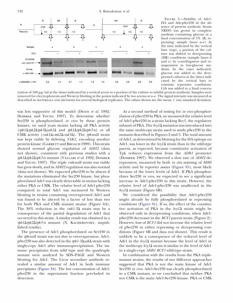
In a more complex experiment diagrammed in Figure5, the inhibitor was added to either repressed (lanes a–c,1Cyh) or derepressed cultures (samples in lanes d ande, 1Cyh), and samples were removed at various times toassay for total Adr1 and Adr1-pSer230. Control cultureslacking the inhibitor were sampled at the same time. Inthe absence of Cyh, the level of phosphorylation de-creased 3- and 4.5-fold after 1 and 1.5 hr of derepression,respectively (compare samples in lanes a, b, and d, noCyh). Adding glucose back into the control derepressedcultures resulted in a return to the repressed level ofphosphorylation (samples in lanes c and e, �Cyh).However, if protein synthesis was inhibited in repressingconditions (left panels) before the culture was shifted to
derepressing conditions, the relative level of Adr1-pSer230 did not decrease (compare samples in lanesa and b, 1Cyh). Moreover, in these conditions, therelative level of Adr1-pSer230 did not change whenglucose was added back (compare samples in lanes aand c, 1Cyh). Similarly, if protein synthesis was in-hibited in derepressing conditions (right panels) andthen the culture was shifted to repressing conditions,the relative level of Adr1-pSer230 did not increase(compare samples in lanes d and e, 1Cyh). Theseresults suggest that synthesis of a phosphatase, a kinase,or new Adr1 is needed to modulate the ratio of Adr1-pSer230 to total Adr1 when cells encounter differentglucose concentrations.
Screening for the Adr1-Ser230 kinase: A peptiderepresenting the Ser230 region of Adr1 is an efficientsubstrate in vitro for both PKA (Denis et al. 1992) andCMK (Hook et al. 1999). Early studies suggested thatAdr1 was inactivated in vivo by phosphorylation ofSer230 by PKA (Cherry et al. 1989), but genetic analysis
TABLE 2
Expression from an ADR1-dependent reporter lackingCat8-binding sites
b-Galactosidase activitya
Genotypeb Activatorc Rd DR (8 hr)
REG1 Adr1 1.2 (0.7) 260 (98)REG1 Adr1-S230A 21 (10) 450 (180)reg1D Adr1 30 (3.8) NMe
reg1D Adr1-S230A 36 (7.3) NM
R, repressing growth conditions. DR, derepressing growthconditions.
a UAS1 reporter activity (pHDY10). Data show average val-ues in Miller units for at least three transformants with stan-dard deviations in parentheses.
b The strains are REG1: CKY13; reg1D: CKY5; both areadr1DTkanmx.
c Wild-type Adr1 is carried on pKD16; Adr1c is on pKD14(S230A).
d Growth conditions are described in materials and meth-
ods.e NM, not measured.
TABLE 3
Synergistic activation of ADH2 by Adr1c reg1D isCat8 dependent
Genotypea ADH2 mRNA level/ACT1b
ADR1 CAT8 REG1 R DR
Wild type Wild type Wild type 0.0025(0.005) 2.3 (0.5)Wild type Wild type D 0.54 (0.12) 1.3 (0.3)Wild type D Wild type 0.0016 (0.001) 0.26 (0.02)Wild type D D 0.004 0.0025
Adr1c Wild type Wild type 0.14 (0.03) 11 (2.5)Adr1c Wild type D 1.4 (0.16) 5.6 (0.2)Adr1c D D 0.001 (0.0005) 0.13 (0.02)Adr1c D Wild type 0.1 (0.02) 7.2 (0.38)
R, repressing growth conditions. DR, derepressing growthconditions.
a Strains used are adr1D strains CKY13 (CAT8 REG1), CKY23(cat8D REG1), and SRY60 (cat8D reg1D) with plasmid pKD16(ADR1 wild type) or pKD14 (Adr1c).
b mRNA levels were measured by RT–qPCR as described inmaterials in methods. Data are the mean of two or threereplicates (standard deviation).
Figure 4.—Adr1c suppresses cat8D-associateddefects in Adr1-dependent gene expression. Cellgrowth is described in materials and methods.RT–qPCR was performed and analyzed as de-scribed in materials and methods. Strains werewild type (wt) CKY13 (adr1D) and cat8D CKY23(adr1D cat8D), each carrying either wild-typeADR1 (pKD16) or ADR1-S230A (Adr1c, pKD14).Derepression was for 4 hr in 0.05% glucose–3%glycerol. The mRNA values were normalized toACT1 mRNA and are expressed as the percentageof the mRNA level in the wild-type CAT8 strainwith Adr1c as the activator. The values representthe mean 6 one standard deviation of three bio-logical replicates assayed in duplicate.
Adr1c Alleles Suppress Snf1 Deficiency 741
was less supportive of this model (Denis et al. 1992;Dombek and Young 1997). To determine whetherSer230 is phosphorylated in vivo by these proteinkinases, we used yeast strains lacking all PKA activity(tpk1Dtpk2Dtpk3Dyak1D and tpk1Dtpk2Dtpk3-ts) or allCMK activity (cmk1Dcmk2Dcmk3D). The tpk-null strainwas kept viable by deleting YAK1, encoding anotherprotein kinase (Garrett and Broach 1989). This strainshowed normal glucose regulation of ADH2 (datanot shown), consistent with previous studies with atpk1Dtpk2Dtpk3-ts mutant (Vallari et al. 1992; Dombek
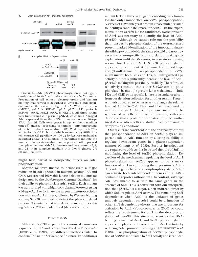
and Young 1997). The triple cmk-null strain was viablebut grew slowly, and its ADH2 regulation was also normal(data not shown). We expected pSer230 to be absent ifthe mutations eliminated the Ser230 kinase, but phos-phorylated Adr1 was clearly detectable in strains lackingeither PKA or CMK. The relative level of Adr1-pSer230compared to total Adr1 was measured by Westernblotting in strains containing overexpressed Adr1 andwas found to be altered by a factor of less than twofor both PKA and CMK mutant strains (Figure 6A).The 30% reduction in the cmk1-3D strain may be aconsequence of the partial degradation of Adr1 thatoccurred in this strain. A similar result was obtained in atpk1Dtpk2Dtpk3-ts mutant (N. Kacherovsky, unpub-lished results).
The presence of Adr1 phosphorylated on Ser230 inthe tpk-null strain was not due to overexpression. Adr1-pSer230 was also detected in the tpk1-3DyakD strain withsingle-copy Adr1 after immunoprecipitation. The im-mune precipitates from wild type and the quadruplemutant were analyzed by SDS–PAGE and Westernblotting for Adr1. The Licor secondary antibody re-vealed a similar amount of Adr1 in both immuneprecipitates (Figure S4). The low concentration of Adr1-pSer230 in the supernatant fraction precluded itsdetection.
As a second method of testing for in vivo phosphor-ylation of pSer230 by PKA, we measured the relative levelof Adr1-pSer230 in a strain lacking Bcy1, the regulatorysubunit of PKA. The bcy1D mutation was introduced intothe same multicopy strain used to study pSer230 in themutants described in Figures 2 and 5. The total amountof Adr1, as determined by blotting for the HA epitope onAdr1, was lower in the bcy1D strain than in the wild-typeparent, as expected, because constitutive activation ofTpk reduces expression from the ADR1 promoter(Dombek 1997). We observed a slow rate of ADH2 de-repression, measured by both in situ staining of ADHactivity and by reporter assays (Figure S5), most likelybecause of the lower levels of Adr1. If PKA phosphor-ylates Ser230 in vivo, we expected to see a significantincrease in Adr1-pSer230 in this strain. However, therelative level of Adr1-pSer230 was unaffected in thebcy1D mutant (Figure 6B).
We considered the possibility that Adr1-pSer230might already be fully phosphorylated in repressingconditions (Figure S1). If so, the effect of the constitu-tive activation of PKA in the bcy1D strain might beobserved only in derepressing conditions, when Adr1-pSer230 decreases in the BCY1 parent strain (Figure 2).However, loss of BCY1 did not increase the relative levelof pSer230 in either repressing or derepressing con-ditions (Figure 6B and data not shown). This result isunlikely to be a consequence of the reduced level ofAdr1 in the bcy1D mutant because the level of Adr1 inthe multicopy bcy1D strain is similar to the level of Adr1in a single-copy ADR1 BCY1 wild-type strain.
In combination with the results from the PKA triple-mutant strains, the results of two different approachessuggested that PKA is not the main kinase of Adr1Ser230 in vivo. Adr1-Ser230 was clearly phosphorylatedin a CMK mutant, so we concluded that neither PKAnor CMK is the main Adr1-Ser230 kinase. PKA or CMK
Figure 5.—Stability of Adr1-HA and Adr-pSer230 in the ab-sence of protein synthesis. StrainNKY85 was grown in completemedium containing glucose at afinal concentration of 5% (R, re-pressing; sample lanes a–c) Atthe time indicated by the verticalbars (top), a portion of the cul-ture was shifted to derepressing(DR) conditions (sample lanes dand e) by centrifugation and re-suspension in low-glucose me-dium. In the cases indicated,glucose was added to the dere-pressed cultures at the times indi-cated by the vertical bars toreinstate repressive conditions.Cyh was added to a final concen-
tration of 100 mg/ml at the times indicated by a vertical arrow to a portion of the culture to inhibit protein synthesis. Samples wereremoved for electrophoresis and Western blotting at the points indicated by two arrows at a–e. The signal intensity was measured asdescribed in materials and methods for several biological replicates. The values shown are the mean 6 one standard deviation.
742 S. Ratnakumar et al.
might have partial or nonspecific effects on Adr1phosphorylation.
Because we were unable to demonstrate a majorreduction in Adr1-pSer230 in mutants lacking PKA andCMK, we screened 102 viable kinase deletion mutants (asdesignated by the Saccharomyces Genome Database) fortheir ability to phosphorylate Adr1-Ser230. Each mutantwas transformed with a high-copy plasmid overexpressingwild-type Adr1 to facilitate the screen. Immunoprecipita-tion with anti-Adr1 antisera, followed by Western blottingwith a-pSer230, was used to detect the phosphorylatedprotein. No mutants that were defective in phosphorylat-ing Adr1-Ser230 were identified (data not shown).
DISCUSSION
Although Ser230 is part of a canonical consensussequence for PKA and is phosphorylated by PKA in vitro(Denis et al. 1992), two different methods failed toconfirm PKA as the Ser230-specific kinase. In addition, a
mutant lacking three yeast genes encoding Cmk homo-logs had only a minor effect on Ser230 phosphorylation.A screen of 102 viable yeast protein kinase mutants failedto identify a candidate kinase for Ser230. In the experi-ments to test Ser230 kinase candidates, overexpressionof Adr1 was necessary to quantify the level of Adr1-pSer230. Although we cannot rule out the possibilitythat nonspecific phosphorylation of the overexpressedprotein masked identification of the important kinase,the wild-type control with the same plasmid did not showexcessive or nonspecific phosphorylation, making thisexplanation unlikely. Moreover, in a strain expressingnormal low levels of Adr1, Ser230 phosphorylationappeared to be present at the same level in wild-typeand tpk-null strains. In vivo phosphorylation of Ser230might involve both Cmk and Tpk, but unregulated Tpkactivity did not significantly increase the level of Adr1-pSer230, making this possibility less likely. Therefore, wetentatively conclude that either Ser230 can be phos-phorylated by multiple protein kinases that may includePKA and CMK or its specific kinase is essential or absentfrom our deletion collection for another reason. Proteinsynthesis appeared to be necessary to change the relativelevel of Adr1-pSer230. This could be interpreted toindicate that an Adr1-specific protein kinase must besynthesized as cells return to repressing growth con-ditions or that a protein phosphatase must be synthe-sized de novo when cells are shifted from repressing toderepressing conditions.
Our results are consistent with the original hypothesisthat phosphorylation of Adr1 on Ser230 plays an im-portant role in Adr1 function by altering its ability toregulate downstream genes in a glucose-dependentmanner (Cherry et al. 1989). Further investigationsare required to address this issue and the role of Snf1 inmodulating the level of Ser230 phosphorylation. Re-gardless of the mechanism, regulating the level of Adr1phosphorylated on Ser230 appears to be a majorfunction of Snf1 in controlling the expression of Adr1-dependent genes because a nonphosphorylatable Adr1c
can activate both Adr1-dependent genes and a UAS1-containing reporter without Snf1. In contrast, wild-typeAdr1 was unable to activate the same genes in theabsence of Snf1. This is consistent with our interpreta-tion that pSer230 is a major, albeit indirect, target bywhich Snf1 regulates Adr1 activity. The remaining Snf1dependence when Adr1c is the activator of genesuniquely dependent on Adr1 could be a function ofother Snf1-dependent pathways that are important foractivation by Adr1 (Voronkova et al. 2006) or couldreflect the requirement for Snf1 in the dephosphor-ylation of pSer98. This site is adjacent to the DNA-binding domain of Adr1, and Ser98 phosphorylationappears to play a repressive role in Adr1 activity byreducing Adr1 promoter binding (Kacherovsky et al.2008). Like phosphorylation of Ser230, phosphoryla-tion of Ser98 is modulated by Snf1 activity and is reduced
Figure 6.—Adr1-pSer230 phosphorylation is not signifi-cantly altered in tpkD and cmkD mutants or a bcy1D mutant.Preparation of cell extracts, electrophoresis, and Westernblotting were carried as described in materials and meth-
ods and in the legend to Figure 1. (A) Wild type (wt) isCMY323, yak1D is SGP400, tpk1D tpk2D tpk3D yak1D isSGP406, cmk1D cmk2D cmk3D is NKY108. All three strainswere transformed with plasmid pNKA1, which has HA-taggedAdr1 expressed from the ADH1 promoter on a multicopyTRP1 plasmid. Cells were grown in trp� synthetic mediumwith 5% glucose (repressing conditions). Fifty microgramsof protein extract was analyzed. (B) Wild type is NKY85and bcy1D is NKY111, both of which are multicopy ADR1. Pro-tein extracts (25 mg wild type, 100 mg bcy1D) were analyzed asdescribed above. The numbers in parentheses indicate thenumber of replicates analyzed and represent both repressed(complete medium with 5% glucose) and derepressed (2, 4,and 22 hr in complete medium with 0.05% glucose–2%ethanol) cultures.
Adr1c Alleles Suppress Snf1 Deficiency 743
in derepressing growth conditions. Whether the twomodifications are causally or functionally related isunder investigation.
We previously suggested that Adr1c and Snf1 act onADH2 expression through separate pathways (Dombek
et al. 1993) on the basis of the strong synergism betweenADR1c and the deletion of REG1. The data presentedhere indicate that the synergism is due to Cat8. Thus theprimary effect of Snf1 on ADH2 expression in a straincarrying ADR1c is due to Cat8 whose expression andactivity are Snf1 dependent. The enhanced activity ofAdr1c may be related to the observation that it activatedgenes normally codependent on Adr1 and Cat8 in theabsence of Cat8. Eukaryotic promoters generally re-spond to different environmental signals through theaction of multiple transcription factors, leading tocombinatorial control of gene expression. Many Adr1-dependent genes are activated by other transcriptionfactors as well. For example, the promoters of genesencoding peroxisomal proteins and the enzymes of b-oxidation bind both Adr1 and the heterodimeric,oleate-responsive transcription factors Oaf1 and Pip2(Simon et al. 1991, 1992; Gurvitz et al. 2000, 2001;Young et al. 2003). We are investigating whetherAdr1c can compensate for the loss of Oaf1/Pip2 in theexpression of peroxisomal genes, as it does for the loss ofCat8 at Adr1- and Cat8-dependent promoters.
The finding that Adr1c can suppress the requirementfor Cat8 suggests that, in spite of its low protein levels,Adr1c has characteristics similar to overexpressed wild-type Adr1, which also escapes Snf1 regulation (Denis
1987) and also compensates for loss of Cat8 at coregu-lated promoters (Biddick et al. 2008). In vitro, by elec-trophoretic mobility shift assays, Adr1c does not haveexceptional DNA-binding activity (Taylor and Young
1990); however, it may be able to bind promotersefficiently in vivo, where factors such as chromatinstructure and stabilization by coactivator interactionare proposed to influence Adr1 binding (Tachibana
et al. 2007; Young et al. 2008). The wide variety ofconstitutive Adr1 mutations, such as a complete deletionof amino acids 220–263 and 262–330 (Cook et al. 1994),suggests that not only the Ser230 phosphorylation site,but also the entire region is important for Adr1 regu-lation. Our results are consistent with a model thatmutations in this area affect protein structure or alterinteraction with other factors (Cook et al. 1994).Although no Adr1-specific repressor has been found inspite of several genetic screens, these findings aboutAdr1c suggest a model that can drive further investiga-tion of the novel interaction between Adr1 and Snf1.
We thank other members of our lab for advice, encouragement, andreagents; Ella Chang for performing some of the quantitative RT–qPCR analyses; Catherine Kehl and Kenneth Dombek for strainconstruction; Carl Mann for strains; and Chris Tachibana for valuablecomments on the manuscript. This work was supported by a grant (no.GM26079) from the National Institutes of Health to E.T.Y.
LITERATURE CITED
Biddick, R. K., G. L. Law and E. T. Young, 2008 Adr1 and Cat8 me-diate coactivator recruitment and chromatin remodeling atglucose-regulated genes. PLoS ONE 3: e1436.
Charbon, G., K. D. Breunig, R. Wattiez, J. Vandenhaute and I.Noel-Georis, 2004 Key role of Ser562/661 in Snf1-dependentregulation of Cat8p in Saccharomyces cerevisiae and Kluyveromy-ces lactis. Mol. Cell. Biol. 24: 4083–4091.
Cherry, J. R., T. R. Johnson, C. Dollard, J. R. Shuster and C. L.Denis, 1989 Cyclic AMP-dependent protein kinase phosphory-lates and inactivates the yeast transcriptional activator ADR1. Cell56: 409–419.
Chi, A., C. Huttenhower, L. Y. Geer, J. J. Coon, J. E. Syka et al.,2007 Analysis of phosphorylation sites on proteins from Saccha-romyces cerevisiae by electron transfer dissociation (ETD) massspectrometry. Proc. Natl. Acad. Sci. USA 104: 2193–2198.
Ciriacy, M., 1979 Isolation and characterization of further cis- andtrans-acting regulatory elements involved in the synthesis of glu-cose-repressible alcohol dehydrogenase (ADHII) in Saccharomy-ces cerevisiae. Mol. Gen. Genet. 176: 427–431.
Collart, M., and S. Oliveiro (Editors), 1993 Current Protocols inMolecular Biology. Greene/Wiley Interscience, New York.
Cook, W. J., D. Chase, D. C. Audino and C. L. Denis,1994 Dissection of the ADR1 protein reveals multiple, function-ally redundant activation domains interspersed with inhibitoryregions: evidence for a repressor binding to the ADR1c region.Mol. Cell. Biol. 14: 629–640.
Cross, F. R., 1997 ‘Marker swap’ plasmids: convenient tools for bud-ding yeast molecular genetics. Yeast 13: 647–653.
Denis, C. L., 1984 Identification of new genes involved in theregulation of yeast alcohol dehydrogenase II. Genetics 108:833–844.
Denis, C. L., 1987 The effects of ADR1 and CCR1 gene dosage onthe regulation of the glucose-repressible alcohol dehydrogenasefrom Saccharomyces cerevisiae. Mol. Gen. Genet. 208: 101–106.
Denis, C. L., and C. Gallo, 1986 Constitutive RNA synthesis for theyeast activator ADR1 and identification of the ADR1–5c muta-tion: implications in posttranslational control of ADR1. Mol.Cell. Biol. 6: 4026–4030.
Denis, C. L., B. E. Kemp and M. J. Zoller, 1991 Substrate specific-ities for yeast and mammalian cAMP-dependent protein kinasesare similar but not identical. J. Biol. Chem. 266: 17932–17935.
Denis, C. L., S. C. Fontaine, D. Chase, B. E. Kemp and L. T. Bemis,1992 ADR1c mutations enhance the ability of ADR1 to activatetranscription by a mechanism that is independent of effects oncyclic AMP-dependent protein kinase phosphorylation of Ser-230. Mol. Cell. Biol. 12: 1507–1514.
Dombek, K. M., and E. T. Young, 1997 Cyclic AMP-dependent pro-tein kinase inhibits ADH2 expression in part by decreasing ex-pression of the transcription factor gene ADR1. Mol. Cell.Biol. 17: 1450–1458.
Dombek, K. M., S. Camier and E. T. Young, 1993 ADH2 expressionis repressed by REG1 independently of mutations that alter thephosphorylation of the yeast transcription factor ADR1. Mol.Cell. Biol. 13: 4391–4399.
Featherstone, M., 2002 Coactivators in transcription initiation:here are your orders. Curr. Opin. Genet. Dev. 12: 149–155.
Fry, C. J., and C. L. Peterson, 2002 Transcription. Unlocking thegates to gene expression. Science 295: 1847–1848.
Garrett, S., and J. Broach, 1989 Loss of Ras activity in Saccharo-myces cerevisiae is suppressed by disruptions of a new kinasegene, YAKI, whose product may act downstream of the cAMP-dependent protein kinase. Genes Dev. 3: 1336–1348.
Goel, A., and R. Janknecht, 2003 Acetylation-mediated transcrip-tional activation of the ETS protein ER81 by p300, P/CAF, andHER2/Neu. Mol. Cell. Biol. 23: 6243–6254.
Guarente, L., 1983 Yeast promoters and lacZ fusions designed tostudy expression of cloned genes in yeast. Methods Enzymol.101: 181–191.
Guldener, U., S. Heck, T. Fiedler, J. Beinhauer and J. H.Hegemann, 1996 A new efficient gene disruption cassette forrepeated use in budding yeast. Nucleic Acids Res. 24: 2519–2524.
Gurvitz, A., L. Wabnegger, H. Rottensteiner, I. W. Dawes, A.Hartig et al., 2000 Adr1p-dependent regulation of the oleicacid-inducible yeast gene SPS19 encoding the peroxisomal
744 S. Ratnakumar et al.
beta-oxidation auxiliary enzyme 2,4-dienoyl-CoA reductase. Mol.Cell Biol. Res. Commun. 4: 81–89.
Gurvitz, A., J. K. Hiltunen, R. Erdmann, B. Hamilton, A. Hartig
et al., 2001 Saccharomyces cerevisiae Adr1p governs fatty acidbeta-oxidation and peroxisome proliferation by regulatingPOX1 and PEX11. J. Biol. Chem. 276: 31825–31830.
Hahn, S., 2008 Rapid yeast protein prep for SDS–PAGE andWestern. http://www.fhcrc.org/science/labs/hahn/methods/biochem_meth/yeast_prot_SDS.html).
Hardie, D. G., D. Carling and M. Carlson, 1998 The AMP-activated/SNF1 protein kinase subfamily: Metabolic sensors of the eukaryoticcell? Annu. Rev. Biochem. 67: 821–855.
Hedges, D., M. Proft and K. D. Entian, 1995 CAT8, a new zinccluster-encoding gene necessary for derepression of gluconeo-genic enzymes in the yeast Saccharomyces cerevisiae. Mol. Cell.Biol. 15: 1915–1922.
Hong, S. P., F. C. Leiper, A. Woods, D. Carling and M. Carlson,2003 Activation of yeast Snf1 and mammalian AMP-activatedprotein kinase by upstream kinases. Proc. Natl. Acad. Sci. USA100: 8839–8843.
Hook, S. S., B. E. Kemp and A. R. Means, 1999 Peptide specificity de-terminants at P-7 and P-6 enhance the catalytic efficiency of Ca21/calmodulin-dependent protein kinase I in the absence of activa-tion loop phosphorylation. J. Biol. Chem. 274: 20215–20222.
Kacherovsky, N., C. Tachibana, E. Amos, D. Fox, III and E. T.Young, 2008 Promoter binding by the Adr1 transcriptional ac-tivator may be regulated by phosphorylation in the DNA-bindingregion. PLoS ONE 3: e3213.
Knop, M., K. Siegers, G. Pereira, W. Zachariae, B. Winsor et al.,1999 Epitope tagging of yeast genes using a PCR-based strategy:more tags and improved practical routines. Yeast 15: 963–972.
Kwok, R. P., J. R. Lundblad, J. C. Chrivia, J. P. Richards, H. P.Bachinger et al., 1994 Nuclear protein CBP is a coactivatorfor the transcription factor CREB. Nature 370: 223–226.
Lo, W. S., L. Duggan, N. C. Emre, R. Belotserkovskya, W. S. Lane
et al., 2001 Snf1—a histone kinase that works in concert withthe histone acetyltransferase Gcn5 to regulate transcription.Science 293: 1142–1146.
McCartney, R. R., and M. C. Schmidt, 2001 Regulation of Snf1 ki-nase. Activation requires phosphorylation of threonine 210 by anupstream kinase as well as a distinct step mediated by the Snf4subunit. J. Biol. Chem. 276: 36460–36466.
Orlova, M., L. Barrett and S. Kuchin, 2008 Detection of endog-enous Snf1 and its activation state: application to Saccharomycesand Candida species. Yeast 25: 745–754.
Papoutsopoulou, S., and R. Janknecht, 2000 Phosphorylation ofETS transcription factor ER81 in a complex with its coactivatorsCREB-binding protein and p300. Mol. Cell. Biol. 20: 7300–7310.
Rahner, A., A. Scholer, E. Martens, B. Gollwitzer and H. J.Schuller, 1996 Dual influence of the yeast Cat1p (Snf1p) pro-tein kinase on carbon source-dependent transcriptional activa-tion of gluconeogenic genes by the regulatory gene CAT8.Nucleic Acids Res. 24: 2331–2337.
Rahner, A., M. Hiesinger and H. J. Schuller, 1999 Deregulationof gluconeogenic structural genes by variants of the transcrip-tional activator Cat8p of the yeast Saccharomyces cerevisiae.Mol. Microbiol. 34: 146–156.
Rubenstein, E. M., R. R. McCartney, C. Zhang, K. M. Shokat, M. K.Shirra et al., 2008 Access denied: Snf1 activation loop phosphor-ylation is controlled by availability of the phosphorylated threo-nine 210 to the PP1 phosphatase. J. Biol. Chem. 283: 222–230.
Santangelo, G. M., 2006 Glucose signaling in Saccharomyces cer-evisiae. Microbiol. Mol. Biol. Rev. 70: 253–282.
Sanz, P., G. R. Alms, T. A. Haystead and M. Carlson, 2000 Reg-ulatory interactions between the Reg1-Glc7 protein phosphataseand the Snf1 protein kinase. Mol. Cell. Biol. 20: 1321–1328.
Schuller, H. J., 2003 Transcriptional control of nonfermentativemetabolism in the yeast Saccharomyces cerevisiae. Curr. Genet.43: 139–160.
Sherman, F., 1991 Getting started with yeast. Methods Enzymol.194: 3–21.
Simon, M., G. Adam, W. Rapatz, W. Spevak and H. Ruis, 1991 TheSaccharomyces cerevisiae ADR1 gene is a positive regulator of tran-scription of genes encoding peroxisomal proteins. Mol. Cell.Biol. 11: 699–704.
Simon, M., M. Binder, G. Adam, A. Hartig and H. Ruis,1992 Control of peroxisome proliferation in Saccharomycescerevisiae by ADR1, SNF1 (CAT1, CCR1) and SNF4 (CAT3). Yeast8: 303–309.
Sloan, J. S., K. M. Dombek and E. T. Young, 1999 Post-translationalregulation of Adr1 activity is mediated by its DNA binding do-main. J. Biol. Chem. 274: 37575–37582.
Tachibana, C., R. Biddick, G. L. Law and E. T. Young, 2007 Apoised initiation complex is activated by SNF1. J. Biol. Chem.282: 37308–37315.
Tansey, W., 2001 Transcriptional activation: risky business. GenesDev. 15: 1045–1050.
Taylor, W. E., and E. T. Young, 1990 cAMP-dependent phosphor-ylation and inactivation of yeast transcription factor ADR1 doesnot affect DNA binding. Proc. Natl. Acad. Sci. USA 87: 4098–4102.
Tu, J., and M. Carlson, 1995 REG1 binds to protein phosphatasetype 1 and regulates glucose repression in Saccharomyces cerevi-siae. EMBO J. 14: 5939–5946.
Vallari, R. C., W. J. Cook, D. C. Audino, M. J. Morgan, D. E. Jensen
et al., 1992 Glucose repression of the yeast ADH2 gene occursthrough multiple mechanisms, including control of the proteinsynthesis of its transcriptional activator, ADR1. Mol. Cell. Biol. 12:1663–1673.
Verdone, L., J. Wu, K. van Riper, N. Kacherovsky, M. Vogelauer
et al., 2002 Hyperacetylation of chromatin at the ADH2 pro-moter allows Adr1 to bind in repressed conditions. EMBO J.21: 1101–1111.
Voronkova, V., N. Kacherovsky, C. Tachibana, D. Yu and E. T.Young, 2006 Snf1-dependent and Snf1-independent pathwaysof constitutive ADH2 expression in Saccharomyces cerevisiae.Genetics 172: 2123–2138.
Young, E. T., N. Kacherovsky and K. van Riper, 2002 Snf1 proteinkinase regulates Adr1 binding to chromatin but not transcrip-tional activation. J. Biol. Chem. 277: 38095–38103.
Young, E. T., K. M. Dombek, C. Tachibana and T. Ideker,2003 Multiple pathways are co-regulated by the protein kinaseSnf1 and the transcription factors Adr1 and Cat8. J. Biol. Chem.278: 26146–26158.
Young, E. T., C. Tachibana, H. W. Chang, K. M. Dombek, E. M. Arms
et al., 2008 Artificial recruitment of mediator by the DNA-bindingdomain of Adr1 overcomes glucose repression of ADH2 expres-sion. Mol. Cell. Biol. 28: 2509–2516.
Yu, J., M. S. Donoviel and E. T. Young, 1989 Adjacent upstreamactivation sequence elements synergistically regulate tran-scription of ADH2 in Saccharomyces cerevisiae. Mol. Cell.Biol. 9: 34–42.
Zhong, H., M. J. May, E. Jimi and S. Ghosh, 2002 The phosphor-ylation status of nuclear NF-kappa B determines its associationwith CBP/p300 or HDAC-1. Mol. Cell 9: 625–636.
Communicating editor: M. Hampsey
Adr1c Alleles Suppress Snf1 Deficiency 745

Supporting Information
http://www.genetics.org/cgi/content/full/genetics.109.103432/DC1
Snf1 Controls the Activity of Adr1 Through Dephosphorylation of
Ser230
Sooraj Ratnakumar, Nataly Kacherovsky, Erin Arms and Elton T. Young
Copyright © 2009 by the Genetics Society of America
DOI:10.1534/genetics.109.103432
S. Ratnakumar et al. 2 SI
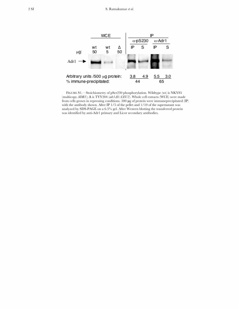
FIGURE S1.—Stoichiometry of pSer230 phosphorylation. Wildtype (wt) is NKY85
(multicopy ADR1); Δ is TYY204 (adr1Δ1::LEU2). Whole cell extracts (WCE) were made from cells grown in repressing conditions. 500 μg of protein were immuneprecipitated (IP) with the antibody shown. After IP 1/5 of the pellet and 1/10 of the supernatant was analyzed by SDS-PAGE on a 6.5% gel. After Western blotting the transferred protein was identified by anti-Adr1 primary and Licor secondary antibodies.
S. Ratnakumar et al. 3 SI
FIGURE S2.—SNF1-independence of Adr1c and synergism of Adr1c and reg1Δ. A. ß-galactosidase activity. Reporter gene assays were performed using at least three transformants of strains TYY497 (adr1ΔSNF1) and TYY498 (adr1Δsnf1Δ) carrying either wildtype ADR1 (pKD16) or ADR1-S230A (pKD14). ß-galactosidase activity is reported in Miller units (GUARENTE 1983). The assays had an average standard deviation of about 20%. R, 5% glucose; derepressed (DR) values represent 18 hours in low glucose medium. A dilution series of the extracts was used to quantify a four-fold reduction of ADH2 derepression in the snf1 mutant with Adr1c, compared to no derepression with wildtype Adr1 (data not shown). B. ADH in situ enzyme assays were performed as described in Materials and Methods. Derepression was for six hours. Wildtype (wt) is CKY13 (adr1Δ); adr1Δsnf1Δ is CKY26, and adr1Δreg1Δ is CKY5, each carrying either wildtype ADR1 (pKD16) or ADR1-S230A (pKD14), respectively. ADHI is the constitutive fermentative isozyme of alcohol dehydrogenase encoded by ADH1. ADHII is the product of the ADH2 gene. Extracts prepared from glucose-grown wild-type cells carrying either pKD16 or pKD14 did not show a band of ADHII activity (data not shown).
S. Ratnakumar et al. 4 SI
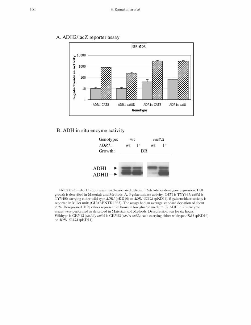
FIGURE S3.—Adr1c suppresses cat8Δ-associated defects in Adr1-dependent gene expression. Cell growth is described in Materials and Methods. A. ß-galactosidase activity. CAT8 is TYY497; cat8Δ is TYY495 carrying either wild-type ADR1 (pKD16) or ADR1-S230A (pKD14). ß-galactosidase activity is reported in Miller units (GUARENTE 1983). The assays had an average standard deviation of about 20%. Derepressed (DR) values represent 20 hours in low glucose medium. B. ADH in situ enzyme assays were performed as described in Materials and Methods. Derepression was for six hours. Wildtype is CKY13 (adr1Δ); cat8Δ is CKY23 (adr1∆ cat8∆) each carrying either wildtype ADR1 (pKD16) or ADR1-S230A (pKD14).
S. Ratnakumar et al. 5 SI
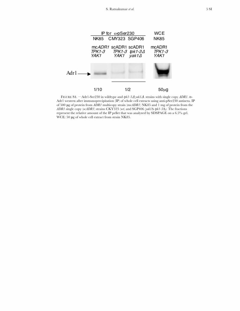
FIGURE S4. —Adr1-Ser230 in wildtype and tpk1-3Δ yak1Δ. strains with single copy ADR1. α-
Adr1 western after immunoprecipitation (IP) of whole cell extracts using anti-pSer230 antisera. IP of 500 μg of protein from ADR1 multicopy strain (mcADR1) NK85 and 1 mg of protein from the ADR1 single copy (scADR1) strains CKY323 (wt) and SGP406 (yak1∆ tpk1-3∆). The fractions represent the relative amount of the IP pellet that was analyzed by SDSPAGE on a 6.5% gel. WCE: 50 μg of whole cell extract from strain NK85.
S. Ratnakumar et al. 6 SI
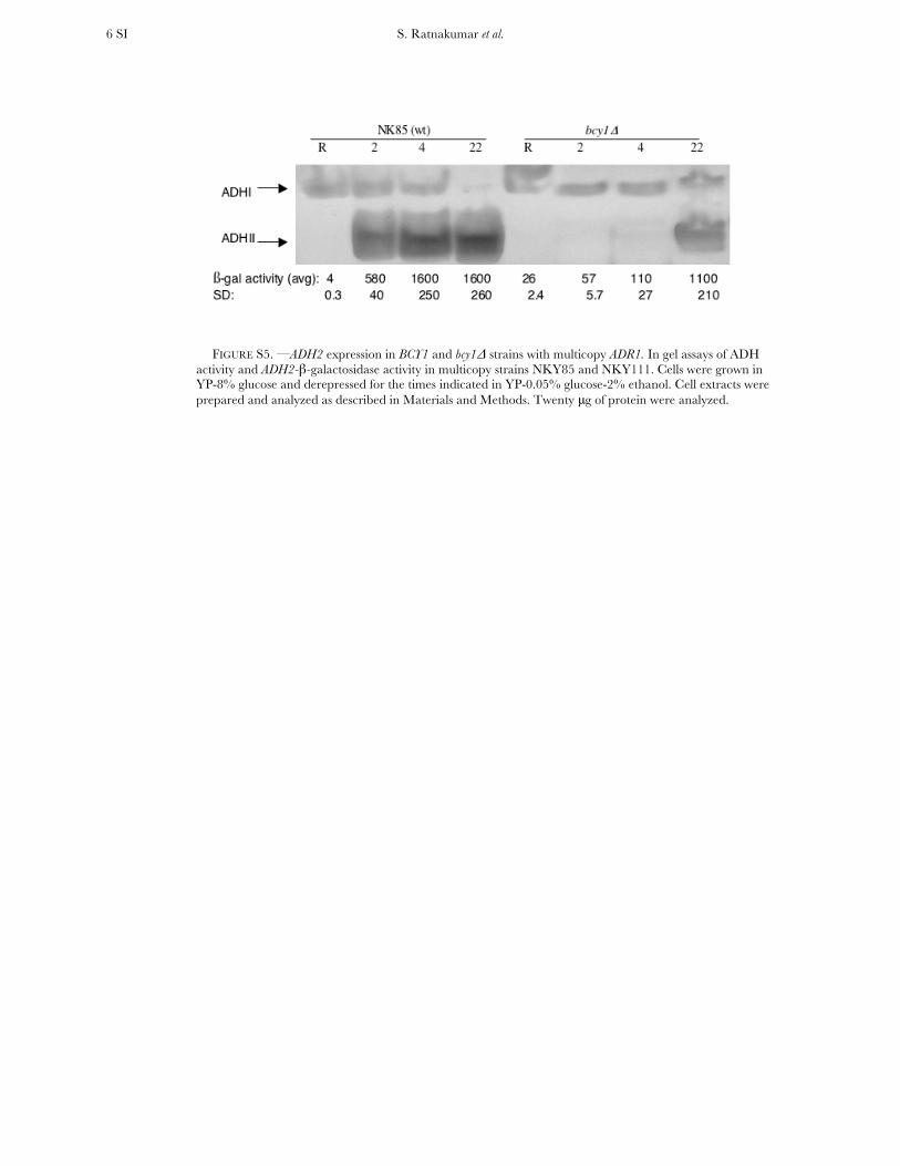
FIGURE S5. —ADH2 expression in BCY1 and bcy1Δ strains with multicopy ADR1. In gel assays of ADH activity and ADH2-β-galactosidase activity in multicopy strains NKY85 and NKY111. Cells were grown in YP-8% glucose and derepressed for the times indicated in YP-0.05% glucose-2% ethanol. Cell extracts were prepared and analyzed as described in Materials and Methods. Twenty μg of protein were analyzed.
S. Ratnakumar et al. 7 SI
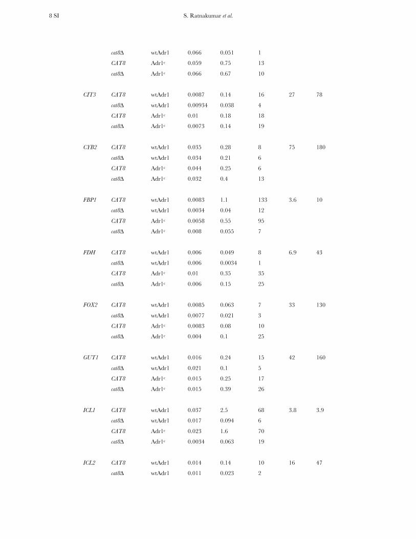
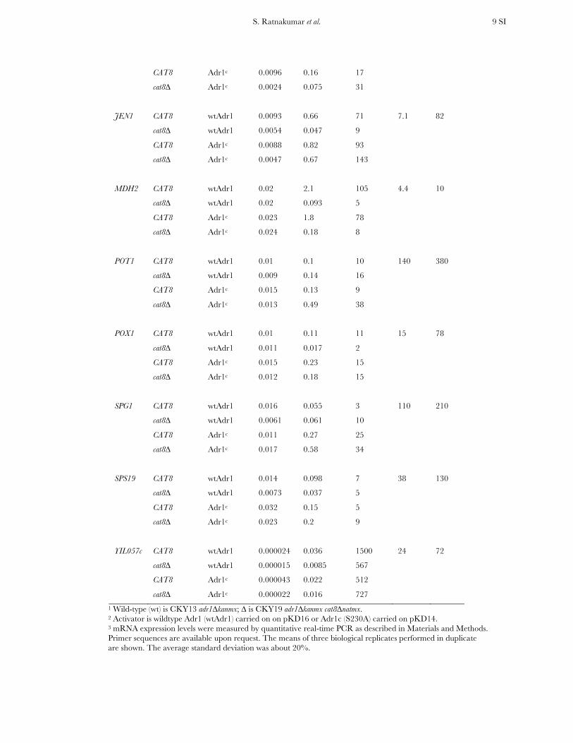
TABLE S1
Gene expression levels with Adr1 wildtype and Adr1c activators in CAT8 wildtype and cat8∆
in repressing and derepressing growth conditions
Derepressed
% (cat8∆/CAT8)
Gene Genotype1 Activator2 mRNA level/ACT13 DR/R wt Adr1 Adr1c
Repressed Derepressed
R DR
ACS1 CAT8 wtAdr1 0.013 3.3 254 3.3 42
cat8∆ wtAdr1 0.012 0.11 9
CAT8 Adr1c 0.026 4.3 165
cat8∆ Adr1c 0.021 1.8 86
ADH2 CAT8 wtAdr1 0.027 7.9 293 1.0 63
cat8∆ wtAdr1 0.026 0.082 3
CAT8 Adr1c 0.14 11 79
cat8∆ Adr1c 0.1 6.9 69
ADR1 CAT8 wtAdr1 0.016 0.059 4 42 88
cat8∆ wtAdr1 0.011 0.025 2
CAT8 Adr1c 0.0079 0.041 5
cat8∆ Adr1c 0.0096 0.036 4
ADY2 CAT8 wtAdr1 0.0063 1.3 206 1.0 20
cat8∆ wtAdr1 0.0016 0.012 8
CAT8 Adr1c 0.0085 2.4 282
cat8∆ Adr1c 0.0041 0.48 117
ALD4 CAT8 wtAdr1 0.048 1 21 47 190
cat8∆ wtAdr1 0.055 0.47 9
CAT8 Adr1c 0.11 1.3 12
cat8∆ Adr1c 0.15 2.5 17
ALP1 CAT8 wtAdr1 0.007 0.14 20 nm nm
cat8∆ wtAdr1
CAT8 Adr1c 0.0085 0.15 18
cat8∆ Adr1c
ATO3 CAT8 wtAdr1 0.056 0.75 13 6.8 89
S. Ratnakumar et al. 8 SI
cat8∆ wtAdr1 0.066 0.051 1
CAT8 Adr1c 0.059 0.75 13
cat8∆ Adr1c 0.066 0.67 10
CIT3 CAT8 wtAdr1 0.0087 0.14 16 27 78
cat8∆ wtAdr1 0.00934 0.038 4
CAT8 Adr1c 0.01 0.18 18
cat8∆ Adr1c 0.0073 0.14 19
CYB2 CAT8 wtAdr1 0.035 0.28 8 75 180
cat8∆ wtAdr1 0.034 0.21 6
CAT8 Adr1c 0.044 0.25 6
cat8∆ Adr1c 0.032 0.4 13
FBP1 CAT8 wtAdr1 0.0083 1.1 133 3.6 10
cat8∆ wtAdr1 0.0034 0.04 12
CAT8 Adr1c 0.0058 0.55 95
cat8∆ Adr1c 0.008 0.055 7
FDH CAT8 wtAdr1 0.006 0.049 8 6.9 43
cat8∆ wtAdr1 0.006 0.0034 1
CAT8 Adr1c 0.01 0.35 35
cat8∆ Adr1c 0.006 0.15 25
FOX2 CAT8 wtAdr1 0.0085 0.063 7 33 130
cat8∆ wtAdr1 0.0077 0.021 3
CAT8 Adr1c 0.0083 0.08 10
cat8∆ Adr1c 0.004 0.1 25
GUT1 CAT8 wtAdr1 0.016 0.24 15 42 160
cat8∆ wtAdr1 0.021 0.1 5
CAT8 Adr1c 0.015 0.25 17
cat8∆ Adr1c 0.015 0.39 26
ICL1 CAT8 wtAdr1 0.037 2.5 68 3.8 3.9
cat8∆ wtAdr1 0.017 0.094 6
CAT8 Adr1c 0.023 1.6 70
cat8∆ Adr1c 0.0034 0.063 19
ICL2 CAT8 wtAdr1 0.014 0.14 10 16 47
cat8∆ wtAdr1 0.011 0.023 2
S. Ratnakumar et al. 9 SI
CAT8 Adr1c 0.0096 0.16 17
cat8∆ Adr1c 0.0024 0.075 31
JEN1 CAT8 wtAdr1 0.0093 0.66 71 7.1 82
cat8∆ wtAdr1 0.0054 0.047 9
CAT8 Adr1c 0.0088 0.82 93
cat8∆ Adr1c 0.0047 0.67 143
MDH2 CAT8 wtAdr1 0.02 2.1 105 4.4 10
cat8∆ wtAdr1 0.02 0.093 5
CAT8 Adr1c 0.023 1.8 78
cat8∆ Adr1c 0.024 0.18 8
POT1 CAT8 wtAdr1 0.01 0.1 10 140 380
cat8∆ wtAdr1 0.009 0.14 16
CAT8 Adr1c 0.015 0.13 9
cat8∆ Adr1c 0.013 0.49 38
POX1 CAT8 wtAdr1 0.01 0.11 11 15 78
cat8∆ wtAdr1 0.011 0.017 2
CAT8 Adr1c 0.015 0.23 15
cat8∆ Adr1c 0.012 0.18 15
SPG1 CAT8 wtAdr1 0.016 0.055 3 110 210
cat8∆ wtAdr1 0.0061 0.061 10
CAT8 Adr1c 0.011 0.27 25
cat8∆ Adr1c 0.017 0.58 34
SPS19 CAT8 wtAdr1 0.014 0.098 7 38 130
cat8∆ wtAdr1 0.0073 0.037 5
CAT8 Adr1c 0.032 0.15 5
cat8∆ Adr1c 0.023 0.2 9
YIL057c CAT8 wtAdr1 0.000024 0.036 1500 24 72
cat8∆ wtAdr1 0.000015 0.0085 567
CAT8 Adr1c 0.000043 0.022 512
cat8∆ Adr1c 0.000022 0.016 727
1 Wild-type (wt) is CKY13 adr1∆kanmx; ∆ is CKY19 adr1∆kanmx cat8∆natmx. 2 Activator is wildtype Adr1 (wtAdr1) carried on on pKD16 or Adr1c (S230A) carried on pKD14. 3 mRNA expression levels were measured by quantitative real-time PCR as described in Materials and Methods. Primer sequences are available upon request. The means of three biological replicates performed in duplicate are shown. The average standard deviation was about 20%.