snare structure drosophila -...
TRANSCRIPT

SEDE AMMINISTRATIVA: UNIVERSITA’ DEGLI STUDI DI PADOVA DIPARTIMENTO DI BIOLOGIA
SCUOLA DI DOTTORATO IN: BIOSCENZE INDIRIZZO: GENETICA E BIOLOGIA MOLECOLARE DELLO SVILUPPO
CICLO XXIII
SNARE complexes associate in a rosette‐like structure at the Drosophila neuromuscular junction
Direttore della Scuola: Ch.mo Prof. GIUSEPPE ZANOTTI
Coordinatore d’indirizzo: Ch.mo Prof PAOLO BONALDO
Supervisore: Prof. MAURO ZORDAN
Dottorando: DAMIANO ZANINI


“Twenty years from now you will be more disappointed by the things that you didn't do than
by the ones you did do. So throw off the bowlines. Sail away from the safe harbor.
Catch the trade winds in your sails.
Explore. Dream. Discover.”
Mark Twain


I
Index
Abstract III
Riassunto V
Abbreviations VII
1. INTRODUCTION 1
1.1. Preface 1
1.2. The fruit fly as model organism 1
1.3. Anatomy of larval body wall 3
1.4. Morphology of the neuromuscular junction 5
1.5. Vesicle fusion cycle 9
1.6. Core of the fusion machinery: the SNARE complex 12
1.7. SNAP‐25: gene and protein 16
1.8. Models of SNAP‐25 mutants 18
1.9. SNAP‐25 and human pathologies 21
1.10 Hypothesis of the SNARE supercomplex 22
2. AIMS of the RESEARCH 27
3. METHODS 29
3.1. Site‐directed mutagenesis: first generation plasmid 29
3.2. Site‐directed mutagenesis: second generation plasmid 31
3.3. Site‐directed mutagenesis: third generation plasmid 32
3.4. Double‐pUAST generation 32
3.5. Fluorescent protein cloning 33
3.6. Transgenes expression 34
3.7. Viability assay 34
3.8. Larva dissection technique 34
3.9. Electrophysiology 35
3.10. Intracellular recording of spontaneous neurotransmitter release 36

II
3.11. Intracellular recording of evoked neurotransmitter release 36
3.12. Data analysis 37
3.13. NMJ Immunohistochemistry 37
3.14. RNA extraction 38
3.15. RT‐PCR 39
3.16. Semi‐quantitative PCR 40
3.17. Analysis of locomotor behavior 41
3.18. Bang test 42
4. RESULTS 45
4.1. Site‐directed mutagenesis 45
4.2. The expression of the SNAP‐25 isoforms does not affect the viability 46
4.3. Expression of the different SNAP‐25 isoforms in the nervous system 48
4.4. Morphology and morphometry of the NMJ 49
4.5. Locomotor behavior 53
4.6. Electrophysiology recordings: spontaneous neurotransmitter release 57
4.7. Electrophysiology recordings: evoked neurotransmitter release 60
4.8. Ca2+ dependency of neuroexocytosis 65
4.9. Bang test 66
4.10. SNARE in vitro assembly 68
5. DISCUSSION 71
5.1. SNAP‐25R206A reduces the probability of vesicle fusion 71
5.2. Evidences of SNARE supercomplex 73
5.3. Fusion pore hypothesis 77
6. CONCLUSION and FUTURE PERSPECTIVES 81
7. REFERENCES 85

III
Abstract
Secretion in all eukaryotic cells involves a family of proteins known as SNAREs (soluble N‐
ethylmaleimide‐sensitive factor attachment protein receptors). In the nervous system, these
proteins are the main molecular constituents of the synaptic vesicle fusion machinery. The
release of molecules contained inside exocytic granules and synaptic vesicles is mediated by
the assembly of a four helix bundle complex: the SNARE complex. It is formed by the coil‐
coiling of three proteins: the v‐SNARE VAMP/Synaptobrevin and the t‐SNAREs, Syntaxin and
SNAP‐25 (synaptosome‐associated protein of 25 kDa). A cooperative interaction between
SNARE complexes has been suggested to be necessary for the fusion of a synaptic vesicle
with the presynaptic membrane to occur. Previous experiments, made with botulinum
neurotoxins type A and E on mouse neuromuscular junction, led to predict a central role of
the SNAP‐25 C‐terminus in protein‐protein contacts between SNARE complexes. Previously
conducted sequence comparisons and molecular dynamics simulations, led to the finding
that this central role in such interactions could be played by a key Arginine residue at the
SNAP‐25 C‐terminus. In particular, the SNAP‐25 Arg206 residue possibly forms an ionic bond
with a negative charge amino acid of Syntaxin from an adjacent SNARE complex. A
computational model, based on such interactions, proposes that the formation of the
vesicle/presynaptic membrane fusion pore is catalyzed by a SNARE supercomplex consisting
in a "ten petal" rosette. This prediction was tested in Drosophila melanogaster by altering
the putative key amino acids, thought to be involved in rosette formation, by site‐directed
mutagenesis. Two transgenic lines (harboring the mutation bearing constructs in a SNAP‐25
wild‐type background) were thus generated: UAS‐SNAP‐25WT (control) and UAS‐SNAP‐
25R206A. Using the UAS/GAL4 system the expression of these constructs was directed
specifically in the nervous system. The mRNA expression profiles, the locomotor behaviour
and the morphology of the neuromuscular junction of these mutants were characterized in
third instar larvae. Electrophysiological recordings performed at the larval neuromuscular
junction showed that the ElavALl4/UAS‐SNAP‐25R206A mutants present a decrease in the
probability of vesicle fusion as compared to ElavGAL4/UAS‐SNAP‐25WT, our control, in both
spontaneous and evoked exocytosis. These findings highlight the key role of Arg206 and
support the model entailing the involvement of a rosette of SNARE complexes in the process
of neuroexocytosis.

IV
V
Riassunto
L’esocitosi regolata è un processo essenziale in tutte le cellule eucariotiche. A livello della
sinapsi, il rilascio di molecule contenute all’interno delle vescicole è mediato dalla
formazione del complesso SNARE, costituito dall’interazione di tre proteine: sintassina,
SNAP‐25 e VAMP/sinaptobrevina. Numerose evidenze sperimentali suggeriscono che la
cooperazione tra complessi SNARE adiacenti sia necessaria affinché una vescicola sinaptica si
fonda con la membrane plasmatica. Da un’analisi della sequenza amminoacidica
dell’isoforma neuronale di questa proteina è emerso che, nella porzione C‐terminale, un
residuo aminoacidico è altamente conservato anche se non è situate nella regione di
interazione tra SNAP‐25 e le altre componenti molecolari del complesso. In mammifero, tale
residuo è l’arginina in posizione 198. Esperimenti condotti incubando la giunzione
neuromuscolare di topo in presenza della tossina botulinica di tipo A hanno evidenziato il
ruolo chiave della porzione C‐terminale della proteina SNAP‐25 nel processo di fusion
vescicolare e, in particolare, dell’Arg198, poiché la tossina taglia esattamente la proteina a
monte di questo residuo, determinando il blocco dell’esocitosi. Inoltre, simulazione di
modeling effettuate al computer hanno suggerito che tale arginine interagisce con un
amminoacido carico negativamente posizionato sulla sintassina del complesso SNARE
adiacente, formando di un legame ionico. Questa ipotesi è stata verificata in vivo usando
Drosophila melanogaster come organismo modello. Poiché nel moscerino della frutta la
sequenza di SNAP‐25 è più lunga di quella in mammifero, l’arginina conservata risulta essere
in posizione 206. Tale Arg206 è stata sostituita con un’alanina mediante mutagenesi sito‐
specifica. Quindi, sono state generate due linee transgeniche in cui i costrutti pUAST‐SNAP‐
25WT e pUAST‐SNAP‐25R206A sono stati inseriti in background SNAP‐25 wild‐type. Sfruttando il
sistema GAL4/UAS, i transgeni sono stati espressi nel sistema nervoso utilizzando il driver
pan‐neuronale ElavGAL4. In entrambe le linee ElavGAL4/UAS‐SNAP‐25WT (controllo)
(controllo) e ELAV‐GAL4/UAS‐SNAP‐25R206A (mutante) sono stati quantificati i profili di
espressione dell’mRNA dei trascritti mutante e wild‐type; inoltre, è stata caratterizzata la
giunzione neuromuscolare a livello larvale, musurandone la morfologia e la morfometria.
L’analisi elettrofisiologica del rilascio spontaneo ed evocato ha mostrato una significativa
riduzione della probabilità di fusione tra vescicola e membrana plasmatica. Questo risultato
conferma il ruolo chiave ricoperto dall’Arg206 durante il processo di esocitosi a livello del
terminale sinaptico e supporta l’ipotesi secondo cui i complessi SNARE cooperano tra loro,
formando un supercomplesso necessario alla formazione del poro di fusione.

VI

VII
Abbreviations
ADHD: attention‐deficit/hyperactivity disorder
BoNT: botulinum neurotoxin
CNS: central nervous system
DA: dopamine
ECP: exo/endo cycling pool
EJP: evoked junction potential
ELAV: embryonic lethal abnormal vision
HRP: horseradish peroxidase
IRP: immediately releasable vesicles
ISN: intersegmental nerve branch
MEPC: miniature end plate current
MEPP: miniature end plate potential
NMJ: neuromuscular junction
NSF: N‐ethylmaleimide sensitive fusion ATPase
RP: reserve pool
RRP: readily releasable pool
SDT: short‐term synaptic depression
SN: segmental nerve
SNAP‐25: synaptosome‐associated protein 25 kDa
SNARE: soluble N‐ethyl maleimide sensitive factor attachment protein receptors
SYX: syntaxin
SV: synaptic vesicle
SYT: synaptotagmin
VAMP: vesicle‐associated membrane protein
TN: transverse nerve

VIII

1
1. INTRODUCTION
1.1. Preface
Everyone's favorite research fruit fly is Drosophila melanogaster (Greek for dark‐bellied dew
lover), a species of Diptera, of the Drosophilidae family.
Within a few years of the rediscovery of Mendel's rules in 1900, Drosophila became a
favorite model organism for genetics research. Starting from Charles William Woodworth
(credited with first breeding Drosophila in quantity, while he was at Harvard University) and
Thomas Hunt Morgan (awarded the Nobel Prize in Physiology or Medicine in 1933), the fruit
fly has become one of the most valuable of organisms in biological research, particularly in
genetics and developmental biology.
Its importance for human health was recognized by the award of the Nobel Prize in
medicine/physiology to Edward Bok Lewis, Christiane Nusslein‐Volhard and Eric Wieschaus
in 1995.
1.2. The fruit fly as model organism
Drosophila was among the first organisms used for genetic analysis, and today it is one of
the most widely used and genetically best‐known of all eukaryotic organisms. The reasons
for its popularity are several: they go from the ease of maintenance due to its high fecundity
and short life cycle, to the large availability of genetic transformation techniques that allow
the manipulation of the completely sequenced genome (Adams et al., 2000), passing from
the easy identification of different phenotypes and the possibility of keeping stocks carrying
lethal by alleles using recessive lethal and multiple inversion‐bearing "balancer
chromosomes" which act as suppressors of recombination with their non‐inverted
homologs.
Furthermore the Drosophila larval body wall muscles, and in particular the neuromuscular
junction (NMJ), constitutes a versatile model system to address most of the basic questions
concerning synaptic function such as ion channels function, mechanisms of fast
2
neurotransmitter release, synapse physiology, functional and structural synaptic plasticity,
and synapse development and formation. Due to its relative simplicity, easy accessibility,
and the presence of large muscles with a well‐defined synapse, the larval body wall
preparation has contributed importantly to our understanding of the genetic mechanisms of
synaptic vesicle release and vesicle recycling (Budnik and Ruiz‐Cañada, 2006).
In the mid to late 1970s Yuh Nung Jan and Lily Yeh Jan were the pioneer investigators the
third instar larva body wall and NMJ (Jan and Jan, 1976a, b). They described the
pharmacological and physiological properties of this model system and they also performed
the first genetic analysis of synaptic transmission and short‐term synaptic plasticity (Jan and
Jan, 1978).
In the 1980s Chun‐Fang Wu and Barry Ganetzky used the body wall preparation to study
properties of ion channels involved in synaptic transmission (Ganetzky and Wu, 1982a,
1983). Few years later studies by the Keshishian lab highlighted the accessibility of the larval
synapse, making developmental analysis simple (Anderson et al., 1988).
In the 1990s Vivian Budnik demonstrated that the larval NMJ is involved in regulatory
mechanisms which allow synaptic plasticity in neuronal networks (Budnik et al., 1990). Soon
after the NMJ became the subject of studies by the Goodman lab when they focused on the
identification of genes involved in the formation of a functional synapse, to be more precise
on the mechanism of axonal pathfinding (Nose et al., 1992).
Thus, for all the above reasons, as well as the well‐characterized genetics, nowadays the fly
NMJ constitute an excellent model system for investigating features of synaptic transmission
such as synapse physiology, development and plasticity, since many of the molecules
involved in vesicle fusion at the synapse terminal are highly conserved between humans and
Drosophila (Zhang, 2010), making the studies on larval body wall muscles and the NMJ highly
relevant for the scientific community.
Even if the Drosophila genome size is less than 1/20 of the human one, about 50% of fly
protein sequences have mammalian homologs and almost 75% of known human disease
genes have a recognizable match in the genetic code of fruit flies (Reiter et al., 2001). The
fruit fly is also being used as a genetic model for several human diseases including
neurodegenerative disorders such as Down syndrome as well as Parkinson, Huntington, and
Alzheimer diseases. Furthermore, Drosophila has been used in neuropharmacological
research, including studies on the effects of cocaine (Andretic et al., 1999) and alcohol
(Heberlein, 2000) on the nervous system and behavior.

3
1.3. Anatomy of larval body wall
As already anticipated, the short life cycle is one reason for the use of Drosophila as a model
system. In lab conditions, at 25°C, generation time is about 10 days: the egg hatches after
12‐15 hours. The resulting larva grows for about 4 days while molting into second instar at
about 24 h and third instar approximately 48 h after hatching. Then the larva encapsulates in
the puparium and undergoes a four‐day‐long metamorphosis after which the adult emerges.
The Drosophila larval neuromuscular system exhibits bilaterally symmetric and segmentally
reiterated organization (Fig. 1.1). Each segment contains a repeated pattern of 30 skeletal
muscle fibers which share common physiological and contractile properties (Crossley, 1978).
These supercontractile muscles are very large and the identification is unique, based on their
size, such as shape, orientation within the muscle fields, relative attachment sizes within the
cuticle, as well as the innervations by the central nervous system (CNS). Muscles anchor to
specialized cells in the epidermis called tendon cells and may also attach to each other. Due
to the dimensions (a longitudinal muscle can reach the size of 40µm wide x 400µm long), a
muscle can be impaled with microelectrodes relatively easily, providing for relatively
straightforward electrophysiological intracellular records. Each mature muscle consists of a
single myofiber, a single multinucleated cell which is anchored to the cuticle by its internal
projections; in contrast each muscle in the Drosophila adult or vertebrate system consists of
several bundles of myofibers (Budnik and Ruiz‐Cañada, 2006).
During the development of the embryo, reiterated sets of approximated 80 motorneurons
are generated in each segment of the ventral nerve chord. Each motorneuron has its unique
cellular identity and is characterized by its axonal trajectory and innervations of a particular
target muscle, as well as by its operation and its dendritic harborization within the CNS
(Landgraf et al., 2003). From the ventral ganglion of the CNS, 32 motorneurons (Landgraf et
al., 1997) arise to extend their axons to innervate each hemisegment of the larval body wall
muscles. Those motorneuron axons are grouped into six major nerve branches: ISN
(intersegmental nerve branch), SNa (segmental nerve branch a), SNb (segmental nerve
branch b), SNc (segmental nerve branch c), SNd (segmental nerve branch d), and TN
(transverse nerve) (Hoang and Chiba, 2001).

4
Fig. 1.1. Drosophila larval body wall muscles. (A) Third instar larva. Anterior is on the left. Body wall preparation of a dissected flattened, and fixed third instar larva observed (B) at the optical microscope and (C) at the fluorescent microscope after a phalloidin‐conjugated rhodamine staining (phalloidin binds filamentous actin); the abdominal segments and the repeated pattern of musculature rearrangements are illustrated (the brain and the ventral nerve chord are removed in these images). (D) Highlight of a larval abdominal hemisegment with (E) the correspondent schematic representation of the main muscle fibers, with the fibers illustrated given their identifying numbers (Johansen et al., 1989b).
The dorsal (distal) region of the neuromuscular system includes 10 muscles (1, 2, 3, 4, 9, 10,
11, 18, 19, and 20) and a single ISN nerve branch that supplies all motorneuron axons that
reach these muscles. The lateral neuromuscular region contains six muscles (5, 8, 21, 22, 23,
and 24). A single SNa nerve branch supplies all motorneuron axons innervating these
muscles. The motorneurons in the SNb and SNd nerve branches innervate the 10 ventral
(proximal) muscles (6, 7, 12, 13, 14, 15, 16, 17, 28, and 30). The SNc and TN motorneurons

5
innervate four ventral (proximal) muscles (25, 26, 27, and 29) in the superficial (close to the
cuticle) layers of the musculature (Hoang and Chiba, 2001).
Fig. 1.2. Neuromuscular system of the Drosophila larva. Body wall preparation of an ElavGAL4/UAS‐EGFP larva seen (A) at the optical microscope and (B) at the fluorescent microscope; the whole nervous system is labeled in green. The bilaterally symmetrical and segmentally reiterated organization of the CNS (C) and the body wall muscles (D) are illustrated. The schematic shows the dorsal view of a fillet‐dissected preparation after a dorsal midline incision. Anterior is at the top. The second abdominal right half‐segment (A2) is highlighted. Each CNS segment has a dorsal midline nerve exit, from which the bilaterally symmetric TN nerve branches extend, and a pair of lateral nerves exit on each side, from which the ISN, SNa, SNb, SNc, and SNd nerve branches arise. Thirty muscles (1–30) in each half segment are innervated by the motorneurons through these six nerve branches (Hoang and Chiba, 2001).
1.4. Morphology of the neuromuscular junction
Chemical synaptic transmission is an important means of neuronal communication in the
nervous system. When the Ca2+ concentration increases due to a depolarization of the axon
terminal, synaptic vesicles (SVs) fuse with the plasma membrane and the neurotransmitter is
released in the synaptic cleft. Transmitters elicit synaptic response in the postsynaptic cell by

6
binding to specific receptors and activating them. The exocytosis is followed by the recycling
of SVs at the presynaptic terminals.
As already anticipated, the Drosophila larval NMJ shares several important features with
central excitatory synapses in the vertebrate brain, also in humans: it is glutamatergic (Jan
and Jan, 1976a), with homologous ionotropic glutamate receptors, and it is organized into a
series of boutons that can be added or eliminated during development. Both the Drosophila
NMJ and vertebrate central synapses exhibit dynamic functional plasticity (Petersen et al.,
1997). In addition, the basic cellular and molecular mechanisms which regulates the cycle of
synaptic vesicles are the same in both fly and humans (Zhang, 2010). Because of these
properties, the fly NMJ constitutes an excellent model system for dissecting the cellular and
the molecular mechanism of synaptic transmission.
In the larva body wall, the abdominal segments from A2 to A7 are almost identically
organized (Bate, 1993). All larval motorneurons in these segments have been
morphologically described and, during the development of the neuroectoderm, the
respective precursor cells have been identified. They have been also linked to their muscle
targets (Jia et al., 1993). Almost all the morphological features of these motorneurons have
been defined (Hoang and Chiba, 2001).
Except for muscle 4 which is monosynaptic, all the body wall muscles are innervated by two
specific motorneuron axons. At the larval stage, motorneurons can be divided into three
classes on the base of the dimension of their boutons at the axon terminal (Atwood et al.,
1993).
The most abundant class of motorneurons are of type‐I. Type‐I boutons are restricted in
distribution and possess relatively prominent terminal enlargements. Within this class, the
axons may be divided into 2 subclasses on the base of the bouton dimensions, big (Ib) and
small (Is) (Johansen et al., 1989a): type‐Ib boutons are large (about 3‐5 mm), the terminals
bearing them tend to be short and minimally branching and they contain mainly small and
clear vesicles of 30/40nm diameter. Type‐Is boutons are, on the average, slightly smaller
(about 1‐1.5 mm) than type‐Ib boutons, the terminals bearing them can often be longer and
more elaborate than those with type‐Ib boutons and they contain a cocktail of different
clear and dense‐core vesicles (Jia et al., 1993). Both these two types of motorneurons
primarily release glutamate, the main excitatory neurotransmitter at the NMJ (Atwood et al.,
1993).
The other two classes (type‐II and type‐III) have a neuromodulatory function and they
release octopamine (Monastirioti et al., 1995) or a variety of peptides (Cantera and Nassel,

7
1992). Type‐II boutons are small (about 1‐2 um), more widely distributed and the terminals
bearing them are very long and the most elaborate of all axon terminal types. Moreover,
type‐II boutons are thought to be on most muscles (Johansen et al., 1989). Type‐III boutons
are of medium size (about 2‐3 µm) and the terminals bearing them are of medium length but
are thought to occur only on muscle 12. In addition to glutamate, type‐III boutons contain
insulin, a putative neural cotransmitter (Gorczyca et al., 1993).
The characteristic peristaltic locomotor behavior of a larva is the result of four principal
components: the cuticular exoskeleton, which provides frictional contact with the substrate
through hooklike protrusions (denticles), the arrays of body wall muscles, the motorneurons
located in the ventral chord which innervate these muscles and the input provided by
interneurons (predominantly cholinergic). This makes Drosophila larva an attractive
experimental model to study how the neuronal network that underlies such a simple
behavior can regulates the motor system of the animal.
Muscles labeled as 6, 7, 12 and 13 constitute the ventral longitudinal abdominal muscles.
Three classes of nerve innervate these muscles: type‐Ib, type‐Is, and type‐II. Whereas
muscles 12 and 13 receive both type of innervations, muscles 6 and 7 receive only type‐I
endings (Fig. 1.3) (Atwood et al., 1993). This difference in innervation pattern allows the two
axons to be reliably identified. Consistent with vertebrate studies, there is a correlation
between muscle fiber size and terminal size (Lnenicka and Keshishian, 2000).
Neurons use electrical (electrical synapses) and chemical (chemical synapses) signals to
receive, process, and communicate informations. The basic currency of signaling by excitable
cells (neurons and muscles) is electrical current. Accordingly, electrophysiology is the study
of how neurons generate, integrate and transmit electrical signals in neuronal networks,
causing all nervous system responses and behaviors. The fly larval body wall muscles do not
express voltage gated Na+ channels (Hong and Ganetzky, 1994; Wu and Haugland, 1985) and
thus do not produce rapid action potentials. Neither do they exhibit Ca2+‐dependent
regenerating potentials in the absence of the tracheal system, which is the open respiratory
system composed of spiracles, tracheae, and tracheoles that terrestrial arthropods have to
transport metabolic gases to and from tissues (Yamaoka and Ikeda, 1988). Hence, all
recordings from the muscle fibres are passive membrane properties or pure synaptic
responses (Wu and Haugland, 1985). Moreover, the muscle is isopotential (Jan and Jan,
1976b). Hence the electrode can be inserted anywhere in the muscle to record the synaptic
potential. For electrophysiological recordings on the larval NMJ the scientific community has
chosen to focus most of their investigations on the muscle 6 and 7 NMJs because of their

8
large size and their central position in the body wall, that preserve them from any damage
during the process of larval dissection.
Fig. 1.3. Synapse structure at muscle fiber 6 and 7. Single‐cell Lucifer yellow labeling of individual motorneuron axons and pan‐neuronal immunological counter‐labeling with anti‐HRP antibodies (green). The axon terminals are in the right half‐segments. The dashed line represents the midline of the CNS. The cell bodies of the three motorneurons examined are located contralaterally (across the midline) to the target muscles. The larval motorneurons MN6/7‐Ib form a type Ib bouton in the cleft between muscles 6/7 (B) and whose cell body is found proximal to, but across from, the CNS midline (A) (Hoang and Chiba, 2001).
Two axons with distinctive endings contribute to the innervation of muscles 6 and 7: axon 1
and axon 2. The differences between axon 1 and axon 2 are several. The axon 1 terminal
creates numerous synapses containing presynaptic dense bodies, which are the putative
active zones for transmitter release; this axon also has more numerous mitochondria, and a
profuse subsynaptic reticulum around or under the synaptic boutons (Atwood et al., 1993).
The dimension of the boutons at the NMJ is bigger, 30% wider than the axon 2 boutons
(Lnenicka and Keshishian, 2000). Axon 2 presents fewer synapses and dense bodies per
bouton, fewer intraterminal mitochondria, and a less‐developed subsynaptic reticulum. The
dimension of axon 2 boutons at the NMJ is smaller. Approximately 800 synapses are
provided by axon 1, and approximately 250 synapses are provided by axon 2 (Atwood et al.,
1993). Also the electrophysiological profile of these two axons is different. The excitatory
postsynaptic potential produced by axon 1 is generally smaller than that produced by axon 2
(Lnenicka and Keshishian, 2000).
The morphological structure of the synapse is also different from that of vertebrate
neuromuscular junctions. The Drosophila nerve terminal is embedded in the muscle and its
perimeter is completely surrounded by subsynaptic muscle membrane, which is extensively
folded at synaptic terminals. In contrast to vertebrate NMJs that present a 50‐60 nm wide
cleft between the nerve and the muscle membranes and contain a basal lamina (Peters et
al., 1991), the fly NMJ show a 15‐20 nm wide cleft and has no basal lamina; this structure in
9
Drosophila is comparable to central synapses in humans (Prokop and Meinertzhagen, 2006).
Furthermore, the main neurotransmitter at the larval NMJ is glutamate; the same amino
acid is used at the great majority (over 90%) of fast excitatory synapses in the brain and
spinal cord of vertebrates (Usherwood, 1981).
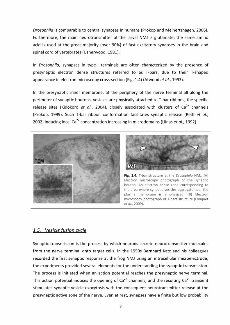
In Drosophila, synapses in type‐I terminals are often characterized by the presence of
presynaptic electron dense structures referred to as T‐bars, due to their T‐shaped
appearance in electron microscopy cross‐section (Fig. 1.4) (Atwood et al., 1993).
In the presynaptic inner membrane, at the periphery of the nerve terminal all along the
perimeter of synaptic boutons, vesicles are physically attached to T‐bar ribbons, the specific
release sites (Kidokoro et al., 2004), closely associated with clusters of Ca2+ channels
(Prokop, 1999). Such T‐bar ribbon conformation facilitates synaptic release (Reiff et al.,
2002) inducing local Ca2+ concentration increasing in microdomains (Llinas et al., 1992).
Fig. 1.4. T‐bar structure at the Drosophila NMJ. (A) Electron microscopy photograph of the synaptic bouton. An electron dense zone corresponding to the area where synaptic vesicles aggregate near the plasma membrane is emphasized. (B) Electron microscopy photograph of T‐bars structure (Fouquet et al., 2009).
1.5. Vesicle fusion cycle
Synaptic transmission is the process by which neurons secrete neurotransmitter molecules
from the nerve terminal onto target cells. In the 1950s Bernhard Katz and his colleagues
recorded the first synaptic response at the frog NMJ using an intracellular microelectrode;
the experiments provided several elements for the understanding the synaptic transmission.
The process is initiated when an action potential reaches the presynaptic nerve terminal.
This action potential induces the opening of Ca2+ channels, and the resulting Ca2+ transient
stimulates synaptic vesicle exocytosis with the consequent neurotransmitter release at the
presynaptic active zone of the nerve. Even at rest, synapses have a finite but low probability
10
of release, causing spontaneous events of exocytosis. In both cases the neurotransmitter is
released in defined units, called quantal units. The units appeared to be the same in both
spontaneous and evoked release processes (Fatt and Katz, 1951, 1952).
Effective neurotransmission requires the precise spatial regulation of protein‐lipid
interactions. More than 1000 proteins function in the presynaptic nerve terminal, and
hundreds are thought to participate in exocytosis (Sudhof, 2004). Also plasma membrane
and vesicle lipids regulate functional aspects of neurotransmission (Davletov and
Montecucco, 2010; Rohrbough and Broadie, 2005). In fact neuronal lipid rafts, sphingolipid‐
and cholesterol‐enriched micro domains that are distributed in both surface and organelle
membranes, are involved in the targeting, localization and functional modulation of many
neuronal ion channels and receptors (Tsui‐Pierchala et al., 2002) as well as for synaptic
vesicle targeting, docking, priming and fusion at the active zone (Rohrbough and Broadie,
2005). Lipid rafts play a key role in the regulation of membrane fluidity and curvature,
parameters directly relevant to membrane restructuring during synaptic vesicle fusion and
budding (Chernomordik and Kozlov, 2008; Megighian et al., 2007; Rohrbough and Broadie,
2005).
Synaptic vesicles undergo a trafficking cycle in the nerve terminal to support rapid and
repeated rounds of release (Fig. 1.5). That cycle can be divided into several sequential steps:
the synaptic vesicles are charged with the neurotransmitter that is actively transported into
the lumen (Sudhof, 2004); cytoskeletal and tethering proteins interact with vesicles to drive
them in proximity of the active zone (Rohrbough and Broadie, 2005). In preparation for
neurotransmitter release, synaptic vesicles are converted into a state of competence for
Ca2+‐triggered fusion‐pore opening (Sudhof, 2004). Three soluble N‐ethylmaleimide‐sensitive
factor (NSF)‐attachment protein receptors (SNAREs) are involved in these preliminary steps:
synaptobrevin, syntaxin and synaptosome‐associated protein‐25 kDa (SNAP‐25). These
proteins drive the assembly of the SNARE complex which constitutes the minimal fusion
machinery. This complex regulates the neuronal exocytosis process allowing: the
establishment of tight membrane contact between vesicle and plasma membrane (docking),
the formation of a scaffolding on which to build the fusion machine, and the binding of lipid
surfaces (priming) (Sollner et al., 1993; Weber et al., 1998).
When an action potential invades a nerve terminal, voltage‐gated Ca2+ channels open, and
the resulting wave of intracellular Ca2+ triggers fusion‐pore opening and neurotransmitter
release. The calcium sensor of exocytosis is constitute by synaptotagmin, a protein with two
C‐terminal C2 domains to which Ca2+ bind driving the fusion between vesicle and plasma
membranes (Sudhof and Rizo, 1996).

11
Fig. 1.5. The synaptic vesicle cycle. (A) Electron micrograph of the presynaptic terminal bouton at the Drosophila NMJ, containing vesicles (red arrow), presynaptic organelles and an electron‐dense active zone, which defines the site of vesicular fusion and neurotransmitter release. (B) Scheme of the presynaptic terminal, highlighting the main stages of the synaptic vesicle cycle. Ten steps can be defined: (1) synaptic vesicle docking to the plasma membrane, (2) vesicle priming for fusion, (3) Ca2+‐triggered vesicle fusion, (4) clathrin‐mediated budding and synaptic vesicle formation, (5) fission of a new vesicle, (6) clathrin uncoating, (7) neurotransmitter loading, (8) vesicle trafficking, (9) tethering in a reserve pool and (10) mobilization and targeting to the plasma membrane release site. The plasma membrane (turquoise zone) shows where exocytosis occurs in lipid raft domains that localize SNARE proteins. Endocytosis occurs in adjacent specialized regions which might have distinct raft‐like features. Endophilin regulates membrane reshaping, facilitating vesicle budding. Cytoskeletal and tethering proteins interact with vesicle lipids to traffic and sequester synaptic vesicles (Rohrbough and Broadie, 2005).
Synaptic vesicles recycling may follow three alternative pathways: (1) vesicles are reacidified
and refilled with neurotransmitters without undocking, thus remaining in the readily
releasable pool (step 6, called “kiss‐and‐stay”) (Barker et al., 1972); (2) vesicles undock and
recycle locally (called “kiss‐and‐run”) (Ceccarelli et al., 1973) to reacidify and refill with
neurotransmitters; or (3) vesicles endocytose via clathrin‐coated pits and reacidify and refill
with neurotransmitters either directly or after passing through an endosomal intermediate
(Heuser and Reese, 1973).
In Drosophila NMJ, two types of endocytosis have been observed: active zone endocytosis
that occurs at the presynaptic active zone facing the specialized postsynaptic membrane and
non‐active zone endocytosis that operates at the area away from the active zone. Once SVs
are formed by endocytosis, they are delivered to two SV pools: the exo/endo cycling pool
(ECP) the reserve pool (RP) (Kidokoro, 2006). Different pools of synaptic vesicles were
defined on the basis of the rates of release under various stimulation conditions. In a
temperature‐sensitive paralytic mutant of Drosophila, shibire, two distinct pools have been

12
identified and characterized (Poodry and Edgar, 1979). The ECP may be
electrophysiologically divided into the readily releasable pool (RRP) and the immediately
releasable pool (IRP). While the RRP and the IRP are recruited in synaptic transmission
during low frequency nerve‐stimulation, while the RP participates only during high frequency
stimulation (Kidokoro, 2006). Moreover, in the case of high sustained stimulation,
endosomes were revealed to be an obligatory component of the vesicle recycling pathway in
all synapses (Wucherpfennig et al., 2003). The IRP and the RRP locates in the vicinity of
release sites, exactly in the periphery of presynaptic boutons, while the RP spreads toward
the center of boutons. These two pools are separately replenished by endocytosis but the
vesicles can move from a RP to RRP, and vice versa, leading to changes in synaptic efficacy.
The size of the RP is approximately 62,000 vesicles, five to seven times larger than that of
the RRP. The IRP is smaller (estimated to be 230 vesicles) and can be exhausted with a few
stimuli and probably represents docked vesicles (Kidokoro et al., 2004). Vesicles in the IRP
are in dynamic equilibrium with those in the RRP (Li and Schwarz, 1999).
1.6. Core of the fusion machinery: the SNARE complex
Intracellular membrane or vesicle fusion involves a highly conserved family of proteins
termed SNAREs. One particular example of vesicular fusion is exocytosis, defined as fusion of
an intracellular trafficking vesicle with the plasma membrane. Constitutive exocytosis
comprises all fusion processes where vesicles are generated, transported, and undergo
exocytosis continuously. In contrast, regulated exocytosis is triggered by second messengers
such as Ca2+ in response to activation or membrane depolarization (Burgess and Kelly, 1987).
The same molecular mechanism is required in all eukaryotic cells, from the yeast secretion,
example of metazoan constitutive exocytosis, to the mammalian synaptic neurotransmitter
release, example of regulated exocytosis (Bennett and Scheller, 1994; Sollner and Rothman,
1994).
The release of molecules contained inside exocytic granules and synaptic vesicles is
mediated by the assembly of a SNARE complex which constitutes the main molecular
constituents of the synaptic vesicle fusion machinery. As in humans and other vertebrates,
also the synaptic vesicle fusion at the Drosophila NMJ requires the same synaptic proteins.
The ‘minimal fusion machinery’ is composed by two target‐SNAREs (t‐SNAREs), syntaxin and
SNAP‐25 on the plasma membrane, and one vesicle‐associated SNARE (v‐SNARE), neuronal
synaptobrevin (also known as VAMP) on synaptic vesicles (Weber and Cyran, 1998). Those

13
SNAREs create a stable ternary core complex, with 1:1:1 stoichiometry, from helices in their
cytoplasmic domains with the formation of a 4‐helix bundle with 1 helix donated by syntaxin
and by synaptobrevin and 2 helices donated by SNAP‐25 (Montecucco et al., 2005; Sollner et
al., 1993).
Neuronal synaptobrevin, also known as VAMP (vesicle‐associated membrane protein), is a
13 kDa integral membrane proteins of the vesicle. The protein structure consists of four
parts: a N‐terminal proline‐rich segment; a segment of around 60 aminoacids, which is well
conserved among isoforms and contains heptad repeats typical for coiled‐coils and a short a‐
helix that inserts into the interfacial region of the lipid bilayer (de Haro et al., 2004); a single
transmembrane segment; and a poorly‐conserved intra‐lumenal tail (Montecucco et al.,
2005).
Drosophila has two characterized members of synaptobrevin gene family. Mutant
phenotypes and gene‐expression patterns indicate that one isoform is exclusively neuronal
(n‐syb) and required for only synaptic vesicle secretion, whereas the other synaptobrevin
isoform (syb) is ubiquitous and essential for cell viability (Sudhof et al., 1989). The nerve‐
evoked synaptic currents were absent from the neuromuscular junctions of n‐syb mutant
embryos, while minis were readily detected (Kidokoro, 2003).
Neuronal syntaxin (Cerezo et al., 1995) is a t‐SNARE expressed ubiquitously in the plasma
membrane, but it acts as the central member of the core complex, mediating synaptic
vesicle fusion only at presynaptic active zones (Jahn and Hanson, 1998). The protein is bound
to the presynaptic membrane of neurons by a transmembrane segment linked to a short C‐
terminal domain exposed to the cell surface and a large cytosolic portion that includes two
domains with distinct structural features. The N‐terminal domain consists of three long α‐
helices that are likely to be involved in protein‐protein interactions, whereas the central
portion contains coiled‐coil forming heptad repeats required for the assembly and stability
of the SNARE complex mediating vesicular fusion at the synapse (Bennett et al., 1993).
Several syntaxin isoforms coexist within the nervous tissue. Targeted vesicle fusion is
regulated by a large number of syntaxin‐binding interactions that control its functional
conformation (Bajjalieh and Scheller, 1995).
In experiments performed at the Drosophila neuromuscular synapse, nether nerve‐evoked
synaptic currents nor minis were observed in syxnull mutant embryos (Kidokoro, 2003).
Furthermore the fundamental role in neurotransmission, syntaxin is absolutely required in
non‐neuronal processes. In Drosophila, proper cellularization of the embryo requires this
protein that is known to be important for membrane trafficking and biogenesis (Burgess et

14
al., 1997). Syntaxin is also essential for cell viability in the developing eye, it is required for
the completion of oogenesis, and it may play a role in membrane stabilization in the nervous
system (Schulze and Bellen, 1996).
SNAP‐25 (Oyler et al., 1989) is localized on the cytosolic face of the nerve membrane via
palmitoylated cysteines located in the middle of the polypeptide chain. Both the N‐ and C‐
terminal halves of SNAP‐25 contain heptad repeats. SNAP‐25 is highly conserved among
species with little variation in length. Upon interaction with syntaxin, SNAP‐25 forms a three‐
helix bundle complex, which might function as a Synaptobrevin receptor on the plasma
membrane (An and Almers, 2004). Moreover, SNAP‐25 stoichiometrically binds to the
putative calcium sensor synaptotagmin and this interaction is believed to be important for
the calcium‐dependent phase of neurotransmitter release (Bai et al., 2004).
Prior to exocytosis, the t‐ and v‐SNAREs are thought to form a trans complex composed of
one helix each from syntaxin and synaptobrevin and two helices contributed by SNAP‐25
(Fig. 1.6) (Sutton et al., 1998). Starting from the N‐termini of the SNAREs, the coil‐coiling
interactions proceed until the formation of a four‐helices bundle with a parallel orientation,
which brings the transmembrane segment of VAMP and syntaxin close to each other and
close to the C‐terminus and to the palmitoylated central loop of SNAP‐25 (Lin and Scheller,
1997).
This SNARE complex has a trans configuration with respect to the membrane location of the
two TM domains, one of which resides in the synaptic vesicle membrane and the other is
embedded in the synaptic plasma membrane. This conformation of the pre‐fusion SNARE
complex is stabilized by a core complex of 16 hydrophobic interactions layers (Lagow et al.,
2007). The surface of the SNARE complex has major grooves and charges enabling the
possibility of additional specific protein‐protein interactions (Lin and Scheller, 1997).
At this state the vesicle is docked, linked to the plasma membrane. The vesicles then
undergo a priming process in which they become fusion competent; at this stage the SNARE
complex, which is forming in the N‐ to C‐terminal direction, is in a partly assembled
intermediate (Sorensen et al., 2006) and the vesicle is in a hemifused state.
Vesicle fusion can be constitutive or triggered by calcium. In the latter case when the Ca2+
concentration increases due to the depolarization of the axon terminal, the fusion pore
forms and the neurotransmitter is released (Katz and Miledi, 1967). Exocytosis is triggered
within about 0.2 ms of the Ca2+ influx (Li et al., 1995) through calcium channels located near
the ‘active zones’, which are the preferred sites of neuroexocytosis (Jahn et al., 2003).

15
Fig. 1.6. Structure of the synaptic SNARE complex. (A) Cartoon representing the primary domain structure of neuronal SNAREs. Synaptobrevin (blue) and syntaxin (red) present one transmembrane domain (TM) and one conserved domain (corresponding to the α‐helix core domains) each. On the other hand, SNAP‐25 (green) presents two conserved domains, no TM but four palmitoylation sites (green tails) which anchor the protein to the plasma membrane. (B) Ribbon model of the synaptic trans‐SNARE complex helical bundle formed by the coil coiling interactions of synaptobrevin (blue) syntaxin (red) and SNAP‐25 (grey). The helical bundle is based upon high resolution crystal data. (C) Model of the trans state of two SNARE complexes that dock a liposome to a supported bilayer in vitro. This model was obtained by modifying the membrane‐proximal end of the crystal structure of the neuronal SNARE complex to allow the transmembrane domains to enter into the juxtaposed membranes. The transmembrane domains were assumed to be helical. The connecting regions between the transmembrane domains and the core complex are likely flexible. Two SNARE complexes are shown (Brunger et al., 2009).
SNAREs are linked directly to Ca2+ triggering of exocytosis, most likely in conjunction with a
Ca2+ sensor (Sorensen et al., 2002). Numerous auxiliary proteins have been found to be
essential for Ca2+‐dependent neurotransmitter release, such as synaptotagmin, the Ca2+

16
sensor, (Perin et al., 1990), complexin (McMahon et al., 1995), Munc18 (Bracher et al.,
2000), and Munc13 (Varoqueaux et al., 2002).
As vesicles undergo fusion, the SNARE complex rearranges its configuration from trans to cis
(Fig. 1.7). After this remodeling, all the SNARE proteins are localized to one membrane (Lin
and Scheller, 1997).
After fusion, the SNARE complex assumes a cis configuration (i.e. with the two TM regions
within the same membrane). The cis complex is then thought to be rapidly disrupted by the
N‐ethylmaleimide sensitive fusion ATPase (NSF) (Lin and Scheller, 1997) and additional
proteins; the coiled four‐helix bundle is unwound, the SNAREs are allowed to be recycled
into synaptic vesicles (Sankaranarayanan and Ryan, 2000) and the synaptic vesicles are
retrieved by membrane fission (Jahn et al., 2003).
Fig. 1.7. Model of SNARE complex assembly and disassembly in a synaptic vesicle cycle. (1) Synaptobrevin forms a partial trans SNARE complex with syntaxin and SNAP‐25. (2) By zippering in an N‐ to C‐termini direction, the SNARE proteins form a trans complex and bring the synaptic vesicle close to the plasma membrane. SNARE‐mediated synaptic vesicle exocytosis occurs either spontaneously (3) or evoked by Ca2+ (4). (5) cis SNARE complexes are thought to be disassembled by NSF ATPase prior to vesicle recycling. PM, plasma membrane; SV, synaptic vesicle (Lagow et al., 2007).
1.7. SNAP‐25: gene and protein
SNAP‐25 is a component of the core complex involved in the docking and fusion of synaptic
vesicles in nerve terminals. It is located to the plasma membrane (Oyler et al., 1989),
anchored by palmitoylated cysteine residues in the central part of the protein (Hess et al.,
1992a). SNAP‐25 contributes with two a‐helices to the formation of the SNARE complex
where it assembles with syntaxin and synaptobrevin, which contribute with a single a‐helix

17
each (Sollner et al., 1993). Also the loopy region of the protein, linking the two helices,
seems to be intimately involved in fast Ca2+ triggering (Nagy et al., 2008).
In mammals, the protein is critical for evoked glutamatergic and cholinergic transmission in
central neurons and at neuromuscular junctions (Washbourne et al., 2002). Beyond this
specialized functions in regulated secretion pathways in neuronal and neuroendocrine cells,
SNAP‐25 expression has been detected in granulosa cells (Jo et al., 2004), pancreatic cells
(Marshall et al., 2005), chromaffin cells (Lopez et al., 2007; Sorensen et al., 2003), sperm
(Hutt et al., 2005), and cumulus cells of ovulating follicles (Shimada et al., 2007). Also in
these non neuronal cells, SNAP‐25 is required for regulating the release of specific vesicle‐
contained factors by forming a complex with syntaxin and synaptobrevin.
Like other nerve terminal proteins, SNAP‐25 shows strong evolutionary conservation
(Catsicas et al., 1991) and sequence homologies to yeast secretion proteins (Bennett and
Scheller, 1993). The human SNAP‐25 gene is located on chromosome 20; it spans more than
88 kb of genomic DNA and consists of eight different exons (Bark and Wilson, 1994). The
protein is located within the presynaptic terminals of hippocampal mossy fibers and the
inner molecular layer of the dentate gyrus. The mRNA was found to be enriched within
neurons of the neocortex, hippocampus, piriform cortex, anterior thalamic nuclei, pontine
nuclei, and granule cells of the cerebellum (Oyler et al., 1989).Furthermore, the human Snap
locus, like the chicken (Bark, 1993), rat (Jacobsson and Meister, 1996), goldfish and zebrafish
(Risinger and Larhammar, 1993), has two alternatively spliced copies of exon 5 (5A and 5B).
SNAP‐25 is alternatively spliced into two forms, SNAP‐25A and SNAP‐25B, which results in a
difference of nine amino acid residues in the composition of the palmitoylated cysteine‐rich
domain thought to be responsible for association of the protein with membranes (Bark and
Wilson, 1994). This implies that the two isoforms differ in their ability to stabilize vesicles in
the primed state (Sorensen et al., 2003) and their capacity or efficiency to be modified by
fatty acylation, suggesting divergent abilities to interact with neuronal membranes (Bark,
1993). The B form predominating in the adult nervous tissue. In addition, zebrafish and
goldfish have two loci encoding SNAP‐25 due to a gene duplication that seems to be unique
for Actinopterygian fish (Risinger and Larhammar, 1993) .
In the fly the gene is located on chromosome 3, in an heterochromatic region (Fig. 1.8).
Drosophila Snap‐25 contains eight exons distributed over a distance larger than 120 kb and it
presents a complex gene organization similar to the human one. In contrast to mammals,
chicken, and Actinopterygian fish which have two alternatively used forms of exon 5, only
one version of exon 5 has been found in Drosophila (Risinger et al., 1997).

18
Nevertheless the fly Snap‐25 has 3 annotated transcripts and 3 annotated polypeptides:
SNAP‐25A, SNAP‐25C and SNAP‐25D. The A isoform corresponds to the neuronal SNAP‐25
protein, while the D isoform (shorter due to the alternative spicing of exon 7) has an
unknown function. Also the C isoform, which differs from the A one due to an alternative
spicing of exon 4, has an unnoted function. Neuronal SNAP‐25A mRNA expression and
protein are primarily if not exclusively located in the central nervous system (Risinger et al.,
1997), as observed in human (Oyler et al., 1989).
While the human protein consists in 206 amino acid residues, in Drosophila this sequence is
a bit longer, precisely 212 bp (base pairs). This difference affects the N‐terminal portion of
the protein, where the fly isoform presents one more α‐helix turn.
Fig. 1.8. Gene model and products of Drosophila Snap‐25 (http://flybase.org).
1.8. Models of SNAP‐25 mutants
SNAP‐25 mutants have been generated not only in cell cultures but also in few animal
models, such as the fly and the mouse.
Flies carrying null alleles of Snap‐25, generated by X‐ray‐induced mutagenesis (Vilinsky et al.,
2002), die at the pharate adult stage, and electroretinogram recordings of these animals
reveal that synaptic transmission is blocked. Surprisingly, the synaptic physiology at the
larval NMJ is normal. This has been explained by the presence of SNAP‐24, a closely related
Drosophila SNAP‐25 homologue that can substitute for SNAP‐25 in null larvae. Nevertheless,
the endogenous levels of SNAP‐24 are low in the adult nervous system, so the vicariant role
of SNAP‐24 is not sufficient to restore a normal phenotype of SNAP‐25 null animals (normal
synaptic transmission in the optic lobes) (Vilinsky et al., 2002). However, if a wild‐type or

19
mutant form of SNAP‐25 is present, then SNAP‐24 does not appear to take part in
neurotransmitter release at the larval NMJ. The apparent redundancy between SNAP‐25 and
SNAP‐24 is due to inappropriate genetic substitution and highly dependent on the genetic
and temporal context (Rao et al., 2001).
Looking at the amino acid sequences, SNAP‐24 differs from SNAP‐25 in the cysteine‐rich
domain, where it contains three instead of four cysteine residues, which could potentially
affect its membrane association dynamics. In vitro studies, however, show that SNAP‐24 can
form core complexes with syntaxin and both synaptic and non‐synaptic v‐SNAREs (Niemeyer
and Schwarz, 2000). In vivo, SNAP‐24 is expressed throughout development and this protein
is found at high levels relative to SNAP‐25 at the larval stage (Rao et al., 2001). SNAP‐24 is
involved in the massive glue secretion in salivary glands (Niemeyer 2000). During
metamorphosis into adulthood SNAP‐25 expression rises significantly relative to SNAP‐24
(Rao et al., 2001). In the adult brain, the protein is found concentrated in the mushroom
body neuropil, a region where SNAP‐25 levels are very low (Vilinsky et al., 2002). Within
neurons, SNAP‐25 is concentrated at synapses, while SNAP‐24 is predominantly in the cell
body (Niemeyer and Schwarz, 2000), suggesting that it is not likely to play a major role in
neurotransmitter release in normal larval NMJs. These findings underline that the presence
of SNAP‐25 is fundamental for viability at the pharate adult stage, underscoring the fact that
intracellular vesicular trafficking and membrane fusion are important processes for nervous
system development and for the function of neural circuits (Vilinsky et al., 2002). Therefore,
the roles of SNAP‐24 and SNAP‐25 in Drosophila may be in some ways analogous to the roles
of SNAP‐25A and SNAP‐25B in mammals (Rao et al., 2001).
SNAP‐25ts is a temperature‐sensitive paralytic mutant which affects synaptic transmission in
3rd instar larvae (Rao et al., 2001). The point mutation involves an amino acid near the N‐
terminus, the glycine at the position 50, which is substituted with a glutamine. The SNAP‐25ts
mutant has two effects on neurotransmitter release at the larval NMJ, depending upon
temperature: at 22°C, the mutation causes the SNARE complex to be more fusion competent
and so evoked release of neurotransmitter is greatly increased; at 37°C, the same mutation
leads to fusion incompetence, the single SNARE complex becomes unstable causing the
dissociation of the multimer and, as a consequence, the release of neurotransmitter is
reduced.
At the non‐permissive temperature, the SNAP‐25ts isoform acts as a “poison” subunit by
impairing the normal activity of the SNARE protein complex and causes loss of function. In
SNAP‐25ts heterozygotes, the amount of wild‐type SNAP‐25 is sufficient to outcompete the
SNAP‐25ts poison subunit: hence, the mutation has a recessive phenotype. However, in

20
SNAP‐25ts homozygotes, SNAP‐24 levels are insufficient to outcompete SNAP‐25ts, resulting
in an altered synaptic physiology (Rao et al., 2001). The SNAP‐25ts larval phenotype appears
stronger than the null allele one (Vilinsky et al., 2002). This evidence suggested that
missense mutants may therefore have a stronger phenotype and may be ultimately more
informative as to the function of the gene than complete null mutants (Rao et al., 2001).
In the mouse a semi‐dominant negative allele of Snap‐25 was generated using the chemical
mutagen ethylnitrosourea (Nolan et al., 2000). The name of the mutation explains the
related phenotype consisting in an altered sensory processing of the animals, which reflects
in anxiety behavior exhibited in the light/dark test (blind) and apathetic behavior observed in
the playground paradigm test (drunk) (Jeans et al., 2007). The electrophysiological
phenotype of the mutants results in a reduction in the frequency of spontaneous mEPSCs,
although these were of normal amplitude, and a marked short‐term depression of EPSCs at
low frequency stimulation. The blind‐drunk mutation describes an activity‐dependent
decrease in neurotransmitter release. This effect is the results of a single amino acid
substitution (I67T) in a highly conserved domain of the first SNAP‐25 α‐helix which is
orientated toward the core of the complex. As a consequence, the binding affinity within the
core SNARE complex results increased and the synaptic vesicle mobilization and
replenishment of the RRP are strongly reduced.
As observed in Drosophila null mutants, also in the mouse one the absence of SNAP‐25 does
not lead to a severe phenotype. In fact vesicle docking persisted, but primed vesicle pools
were empty and fast calcium‐triggered release abolished. Single vesicular fusion events
showed normal characteristics, except for a shorter duration of the fusion pore. Snap‐25null
mice rescued by the ubiquitous over expression of SNAP‐23. Over expression of SNAP‐23 did
not support a standing pool of primed vesicles. This shorter (SNAP‐23) isoform can partially
vicariate for SNAP‐25 (Sorensen et al., 2003).
In vitro experiments have been performed on PC12 cells, a widely employed cell line derived
from pheochromocytoma, a neuroendocrine tumor of the medulla of the adrenal glands
(Bertherat and Gimenez‐Roqueplo, 2005). In mammals, chromaffin cells are neuroendocrine
cells found just in the medulla of the adrenal gland (a gland located above the kidneys) and
in other ganglia of the sympathetic nervous system. These cells, innervated by the
splanchnic nerve, secrete numerous bioactive substances including adrenaline (epinephrine),
noradrenaline (norepinephrine), and enkephalin endogenous ligands (endorphins) (Young
and Abboud, 2006). Transmitters, hormones neuropeptides, and chromogranins are stored
in chromaffin granules, large dense core vesicles (Winkler and Westhead, 1980) from which
they are released by regulated exocytosis in a calcium ion‐dependent and hormonally
21
controlled manner (Aunis and Langley, 1999). Due to the similarity between neuroexocytosis
and neurosecretion, the chromaffin granule has been a model for membrane fusion. The
analysis of the molecular mechanism controlling neuroexocytosis has been greatly helped by
studies on chromaffin cells. The priming of docked granules followed by their Ca2+‐driven
fusion with the plasma membrane and the release of the granule content has been studied
in these cells, the role of key factors involved has been highlighted, and the cytosolic and
membrane proteins have been linked to priming and fusion processes (Jahn and Sudhof,
1999).
With an elegant work using adrenal chromaffin cells, Sorensen and colleagues (Sorensen et
al., 2006) demonstrated that SNAP‐25 mutations within the N‐terminal portion of the SNARE
four helix bundle selectively disrupt vesicle priming by interfering with the stability of the
hydrophobic interaction layers holding the complex together. Mutations in the C‐terminal
end affected fusion triggering. In contrast, similar N‐terminal mutations were without effects
on exocytosis. Mutations in the middle of the complex selectively interfered only with
vesicle priming.
In summary, all the existing mutant SNAP‐25 isoforms interfere with the stability of the
hydrophobic interaction layers holding the complex together; as a result the mutations
provoke the non‐assembly or the disassembly of the SNARE complex impairing the vesicle
docking or priming/fusion processes.
1.9. SNAP‐25 and human pathologies
Recently, several evidences suggest that SNAP‐25, in concert with other genes and
environmental factors, may be involved in different neuropsychiatric and neurological
disorders. Alterations in the Snap‐25 gene structure, expression and/or function may
contribute directly to abnormal behavioral phenotypes, including schizophrenia, attention‐
deficit/hyperactivity disorder (ADHD) and epilepsy (Corradini et al., 2009).
Schizophrenia (MIM ‐ Mendelian Inheritance in Man ‐ 181500) is a common mental disorder
with a prevalence of approximately 0.5‐1% and significant heritability estimated to be about
85% (Cardno and Gottesman, 2000). The clinical symptoms can be classified into two main
categories: psychotic or “positive” symptoms, including hallucinations, altered emotional
activity, disorganized behavior; and “negative” symptoms, such as delusions, reduced
interest and motivation, and cognitive impairment. Evidence from several
immunohistochemical and Western blot studies in postmortem brains showed discrepancies

22
in altered expression levels of SNAP‐25 between different brain regions. These evidences
implicate SNAP‐25 in the etiology of schizophrenia, explaining the depressed functionality of
certain neural circuits, but also the hyperactivity of other pathways that may possibly result
from compensatory mechanisms (Thompson et al., 1998).
ADHD (MIM 143465) is one of the most prominent childhood neuropsychiatric disorders.
The gene encoding SNAP‐25 has been identified as responsible for hyperkinetic behavior
based on analysis of the coloboma mutant mouse. This mutant mouse is heterozygous for a
semi‐dominant deletion mutation that encompasses 10‐12 genes on chromosome 2,
including Snap‐25 (Hess et al., 1996). In coloboma mutant mice (Cm/+), deletion of the Snap‐
25 gene results in 50% lower amounts of the Snap‐25 mRNA and protein expression
compared to wild‐type mice. Coloboma mice exhibit normal circadian rhythms and, as in
case of children with ADHD, they are hyperactive during their active (nocturnal) phase, with
locomotor activity averaging three fold the activity of control littermates (Hess et al., 1992b).
Epilepsy (MIM 604233) is a group of heterogeneous neurologic disorders affecting almost
1% of the population. It is characterized by recurrent, unprovoked seizure episodes, due to
abnormal synchronous firing of groups of neurons, arising from periodic neuronal hyper
excitability. The coloboma mutant mouse, in addition to its hyperkinetic activity, displays
frequent spontaneous epileptic seizures, which are thought to arise from abnormalities in
calcium transients, caused by a SNAP‐25 deficiency, in modulating presynaptic voltage‐gated
calcium channels (Pozzi et al., 2008).
Besides its well characterized role in regulating exocytosis, there is increasing evidence that
SNAP‐25 also modulates calcium dynamics in response to depolarization. Effects on SNAP‐25
expression could result from altered neural circuits or network function, or from more
general perturbations to brain development (Corradini et al., 2009).
1.10. Hypothesis of the SNARE supercomplex
The functional importance of SNARE proteins was demonstrated by observations that it is a
target of the eight neurotoxins produced by the anaerobic bacteria of the genus Clostridium:
one tetanus neurotoxin, TeNT, and seven botulinum neurotoxins, BoNT/A‐G (Fig. 1.9).
BoNTs (around 150 kDa) are comprised of three independent domains that mediate neuron
intoxication: the heavy chain domain mediates the entry into the neuron by binding to
neuronal membrane receptor(s), and the consequent uptake into an endosome‐like

23
compartment; the amino‐terminal segment of the heavy chain domain allows the
penetration of the light chain into the endosome membrane via a pH‐dependent
translocation process; the light chain, once within the synaptic cytoplasm, mediates the
proteolytic cleavage of specific sites on the neuronal SNAREs (Keller et al., 2004) causing the
neuromuscular blockade.
Fig. 1.9. Location of neurotoxin‐mediated cleavage sites on the hypothetical model of the synaptic fusion complex as it joins two membranes. The synaptobrevin (blue) neurotoxin‐mediated cleavage site for tetanus toxin (TeNT) and botulinum toxin (BoNT) type B (BoNT/B) is between Gln76 and Phe77; for BoNT/F, between Gln58 and Lys59; for BoNT/G, between Ala81 and Ala82; and for BoNT/D, between Lys59 and Leu60. The syntaxin (red) BoNT/C cleavage site is between Lys253 and Ala254. Cleavage sites in SNAP‐25 (green) are between Asp193 and Glu194 for BoNT/E, and between Arg176 and Gln177 for BoNT/A (Sutton et al., 1998).
In the case of SNAP‐25, three neurotransmission‐blocking botulinum toxins can cleave the
protein: botulinum neurotoxin type A (BoNT/A) (Blasi et al., 1993a), type C (BoNT/C) (Foran
et al., 1996), and type E (BoNT/E) (Schiavo et al., 1993). Considering the mouse sequence,
BoNT/A cleaves SNAP‐25 at position 197, precisely nine residues from the C‐terminus;
BoNT/E cleaves at position 180, in the middle of the second a‐helix which interacts with the
other three a‐helixes of the SANREs; BoNT/C cleaves at position 198 (Vaidyanathan et al.,
1999). While the substrate of BoNT/A and E is SNAP‐25, BoNT/C cleaves also syntaxin (Blasi
et al., 1993b).
A striking feature of the action of different BoNTs within nerve terminals is the large
difference in the duration of their inhibitory action both in vitro, in neuronal cultures (Keller
et al., 2004), and also in vivo, on the mouse NMJ (Montecucco et al., 2005). BoNT/A and
BoNT/C have a longer inhibitory effect on the nerve terminal, while BoNT/E causes a shorter
paralysis. Moreover, in the mouse NMJ, cleavage of only 10% of SNAP‐25 by BoNT/A is
sufficient to block glycine release; in the case of BoNT/E almost a complete cleavage is
needed to have the same inhibition on neurotransmitter release (Fig. 1.10).
BoNT/A cleaved SNAP‐25 seems to act as a negative dominant isoform, suggesting that the
C‐terminal region of SNAP‐25 may play a central role in the function of the proper

24
neuroexocytosis machinery. These findings lead to the suggestion that several SNARE
complexes may bind together and cooperate to allow the formation of a functional fusion
pore (Montecucco et al., 2005).
Fig. 1.10. On the top, cartoon of SNAP‐25 and the position of BoNT/A and BoNT/E cleavages. On the bottom, dose response curves for the effects of the two botulinum neurotoxins on SNAP‐25 proteolysis (purple squares, continuous line) and glycine release (green circles, broken line). The vertical bars indicate the values (C/KSD) for SNAP‐25 proteolysis (purple) and glycine release (green).
A sequence conservation analysis was performed on neuronal SNAP‐25 amino acid
sequences among different species to investigate which amino acids might be involved in
contacts between the SNARE complex and other proteins or lipids. This approach highlighted
an arginine residue which is highly conserved even if it is not involved in the contacts
between the α‐helices of the molecular components of the complex (Fig. 1.11). This arginine
(at position 189 in mammals) points outside the complex and, due to its positive charge, it is
thought to interact with an amino acid, with a negative charge, on the adjacent SNARE
complex in order to stabilize the formation of a SNARE supercomplex.
Currently, there are only few scattered pieces of evidence in favor of the hypothesis of a
multimeric SNARE supercomplex as a necessary structure involved in the exocytosis of
synaptic vesicles. Therefore the kinetics of the vesicle fusion process has been characterized
from the energetic and temporal points of view.
SNAP‐25 proteolysis (%)Glycine release (%)

25
Fig. 1.11. Consensus diagram of SNAP‐25 amino acid sequence. The conserved residues have a negative score; the unconserved ones have a positive score (i.e. in the loopy region). The N‐terminus sequence of the protein is highlighted. Dashed lines indicate the α‐helix steps and the amino acids overlapping those positions are highly conserved due to their interaction with syntaxin and synaptobrevin α‐helices. Arg189 (human numeration), which corresponds to Arg206 (Drosophila numeration), has a negative score even if it is not involved in contacts with the other SNARE proteins within the single complex (Pantano 2007, data not published).
On the basis of the energetic requirement for membrane fusion, it has been estimated that
the formation of three or more SNARE complexes provides sufficient free energy to drive the
process (Cohen and Melikyan, 2004). Considering that the time delay between the cytosolic
Ca2+ trigger and the fusion of docked and ready‐to‐fuse synaptic vesicles is around 100
microseconds (Kasai, 1999), even if it varies for different types of synapses (Sudhof and
Malenka, 2008), it has been proposed that the docked vesicles may be in a state of
hemifusion with the presynaptic membrane (Chernomordik and Kozlov, 2008). This would
require that the ready‐to‐fuse vesicles should be actually stably juxtaposed on the cytosolic
face of the plasma membrane and this appears to entail the involvement of several SNARE
complexes at the same site (Montecucco et al., 2005).
In the last decade, the SNARE supercomplex model has been supported by many
biochemical and in vitro experiments. Moreover, the precise stoichiometry of the
supercomplex has not been established yet. However, the necessity for a multimeric SNARE
supercomplex has not been demonstrated in vivo yet.

26

27
2. AIMS of the RESEARCH
Recently, Montecucco and collaborators (Montecucco et al., 2005) highlighted the key role
of the C‐terminus of SNAP‐25 by performing experiments on the mouse NMJ using different
types of botulinum neurotoxins. They proposed that several SNARE complexes should be
present at the same site to allow the fusion of a synaptic vesicle with the presynaptic
membrane: the assembly of a rosette‐shaped oligomer of SNARE complexes, which binds
together and cooperate to origin a functional fusion pore, may constitute a crucial event in
neuroexocytosis.
Performing sequence conservation analysis on neuronal SNAP‐25 protein sequences among
different species, a single amino acid which is highly conserved in the protein’s C‐terminal
region was identified, even if this residue does not face the α‐helices of syntaxin and
synaptobrevin, but instead appears to point outside the complex. In the murine sequence
this amino acid is an arginine in position 198. This amino acid is at the first position of the
peptide bond cleaved by BoNT/A and it is predicted to not to be involved in the contacts
necessary for the formation of the SNARE complex.
To prove the experimental hypothesis and to demonstrate that Arg198 is fundamental for
the proper function of the neuroexocytosis apparatus, we decided to introduce non
functional SNARE complexes within the rosette in order to interrupt the interactions within
the adjacent SNARE complexes.
Drosophila was used as an animal model to test the key role of Arg198 in the process of
vesicle fusion. In Drosophila the homolog of murine Arg198 is the arginine at the position
206. We performed site‐directed substitution in order to replace Arg206 with alanine, a
neutral amino acid that should not alter the α‐helix structure of SNAP‐25 C‐terminus or the
assembly of single SNARE complexes. We hypothesized that the site‐directed substitution
could interfere, in vivo, preventing the cooperation between the SNARE complexes. As a
result we expected an impairment of vesicle fusion with the consequent decrease of
neurotransmitter release at the NMJ.
28
We generated two constructs carrying different isoforms of SNAP‐25 under the UAS
promoter: UAS‐SNAP‐25WT, a vector carrying the wild‐type isoform, our control; and UAS‐
SNAP‐25R206A, a vector carrying the point mutated isoform of the protein, our mutant.
The plasmids were microinjected to generate several transgenic lines in a SNAP‐25 wild‐type
background. The Elav‐GAL4 diver was used to direct the expression of the transgenes within
the nervous system. The expression levels of the transgenes were analyzed, the
electrophysiological profiles were recorded from the larval NMJ, the morphology and the
morphometry of the synapse was measured, and the behavior of the larvae and the adults
was characterized. Moreover, we verified, in vitro, the assembly of the SNARE complex in the
presence of mutated SNAP‐25.

29
3. METHODS
3.1. Site‐directed mutagenesis: first generation plasmids
Four isoforms of SNAP‐25 (three mutants and one wild‐type) were produced by polymerase
chain reaction (PCR) using primers carrying the required point mutations. The ORF coding for
SNAP‐25 was amplified from the full‐length cDNA derived from w1118 flies using the following
oligonucleotides (5’‐3’ orientation):
NAME SEQUENCE
sense SNAP‐25 CGGAATTCATGCCAGCGGATCCATCTGAAG
antisense SNAP‐25WT CTCTCGAGTTACTTTAATAGTTGATGTGCC
antisense SNAP‐25R206A CTCTCGAGTTACTTTAATAGTTGATGTGCCGCTTGATTAGC
antisense SNAP‐25R199A CTCTCGAGTTACTTTAATAGTTGATGTGCCCTTTGATTAGCAACTGCTA
TCGCCGCTTCATTAG
antisense SNAP‐25R199A,R206A CTCTCGAGTTACTTTAATAGTTGATGTGCCGCTTGATTAGCAACTGCTA
TCGCCGCTTCATTAG
Sense and antisense primers have at their 5’ ends the restriction sites for EcoRI (GAATTC)
and XhoI (CTCGAG), respectively (underlined nucleotides). Mutated sites are highlighted.
PCRs were performed in a 50‐μl reaction mixture containing:
1X Expand High Fidelity Buffer
1.5 mM MgCl2
10 pmol each pair of primers
1 mM dNTPs
50 ng cDNA
2.6 U Expand High Fidelity enzyme mix (Roche, USA)
Amplification program:
first denaturation step: 4’ at 94°C
35 rounds of amplification: 94°C for 30’’(denaturation), 68°C for 30’’ (annealing), 72°C for 1’ (elongation)
final incubation step: 72°C for 10’
A further incubation step at 72°C for 15’ was necessary with GoTaq DNA polymerase
(Promega, USA) to add an adenine (A) at the 3’‐term of each PCR product.
30
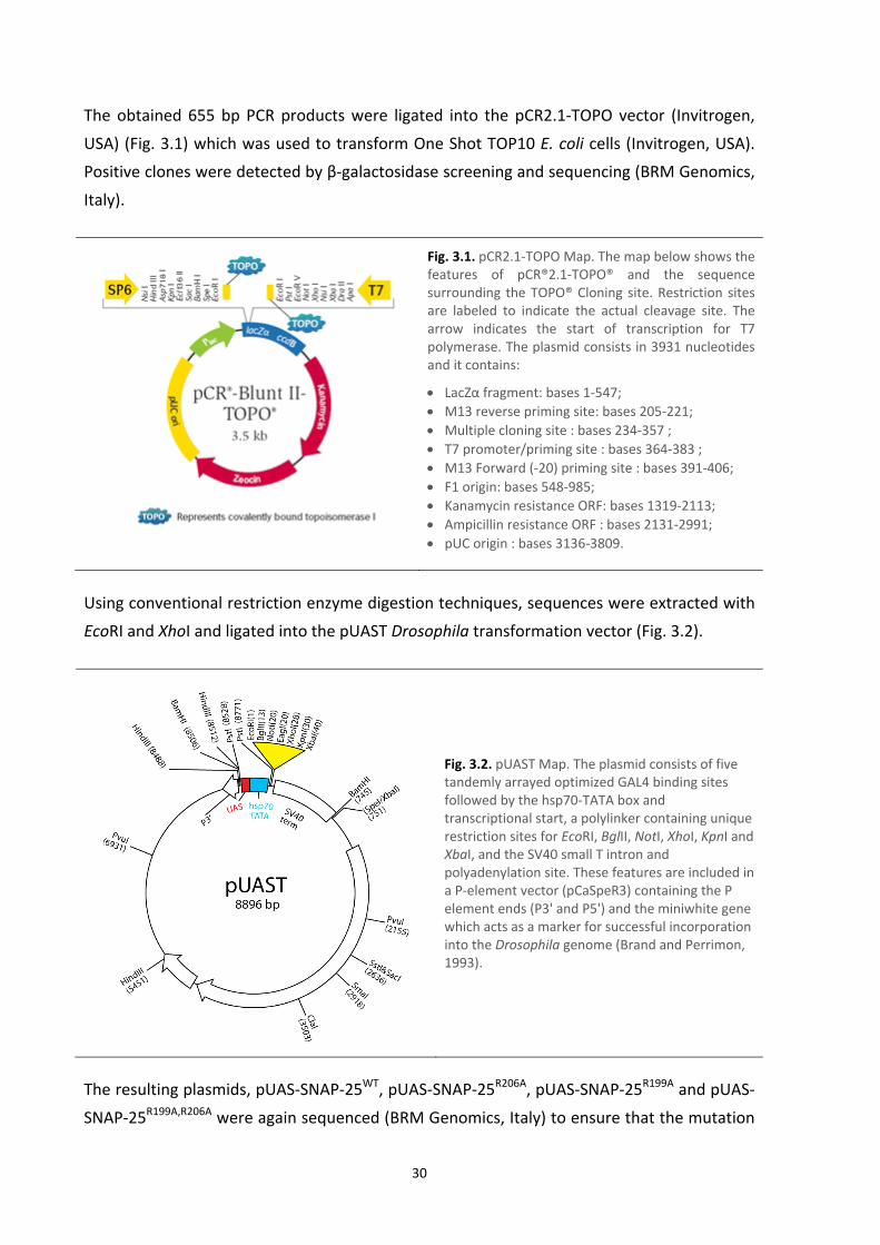
The obtained 655 bp PCR products were ligated into the pCR2.1‐TOPO vector (Invitrogen,
USA) (Fig. 3.1) which was used to transform One Shot TOP10 E. coli cells (Invitrogen, USA).
Positive clones were detected by β‐galactosidase screening and sequencing (BRM Genomics,
Italy).
Fig. 3.1. pCR2.1‐TOPO Map. The map below shows the features of pCR®2.1‐TOPO® and the sequence surrounding the TOPO® Cloning site. Restriction sites are labeled to indicate the actual cleavage site. The arrow indicates the start of transcription for T7 polymerase. The plasmid consists in 3931 nucleotides and it contains:
LacZα fragment: bases 1‐547;
M13 reverse priming site: bases 205‐221;
Multiple cloning site : bases 234‐357 ;
T7 promoter/priming site : bases 364‐383 ;
M13 Forward (‐20) priming site : bases 391‐406;
F1 origin: bases 548‐985; Kanamycin resistance ORF: bases 1319‐2113;
Ampicillin resistance ORF : bases 2131‐2991;
pUC origin : bases 3136‐3809.
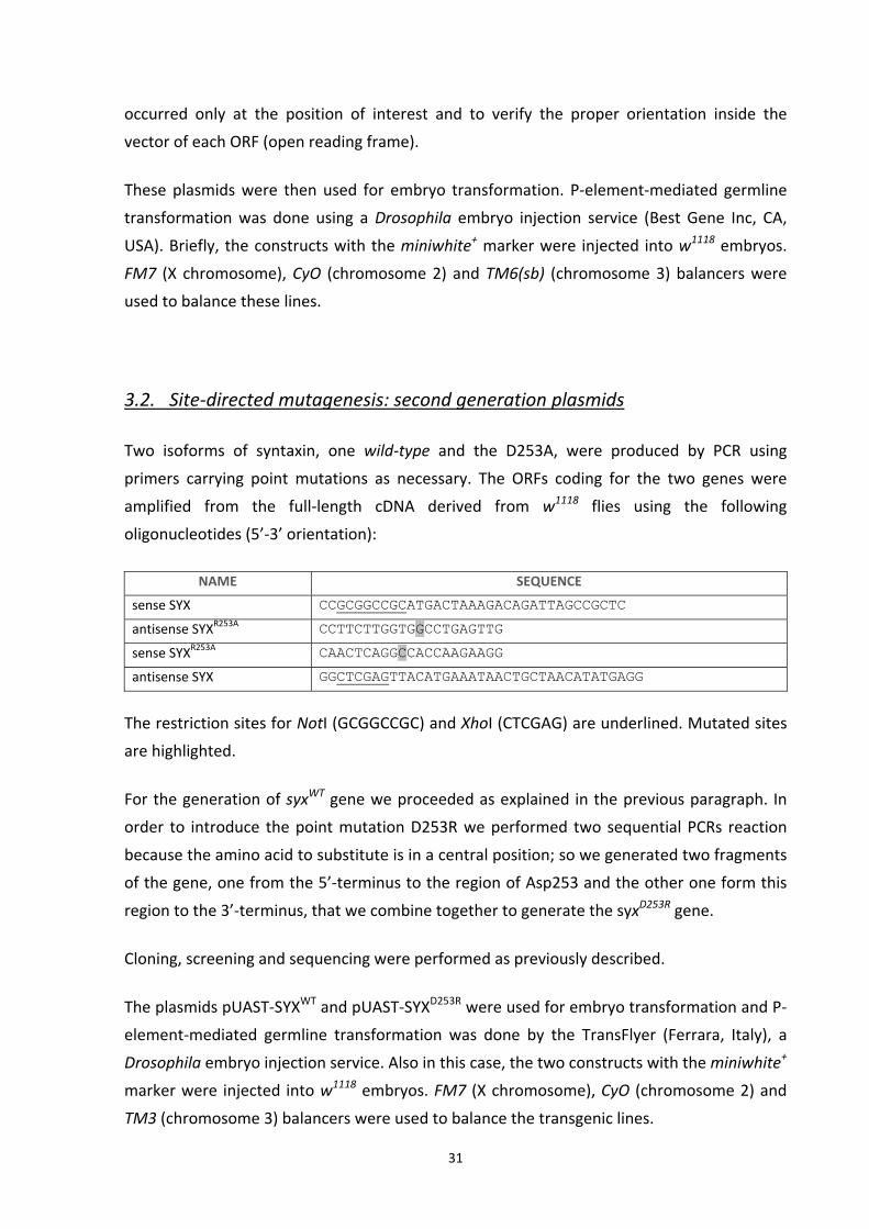
Using conventional restriction enzyme digestion techniques, sequences were extracted with
EcoRI and XhoI and ligated into the pUAST Drosophila transformation vector (Fig. 3.2).
Fig. 3.2. pUAST Map. The plasmid consists of five tandemly arrayed optimized GAL4 binding sites followed by the hsp70‐TATA box and transcriptional start, a polylinker containing unique restriction sites for EcoRI, BglII, NotI, XhoI, KpnI and XbaI, and the SV40 small T intron and polyadenylation site. These features are included in a P‐element vector (pCaSpeR3) containing the P element ends (P3' and P5') and the miniwhite gene which acts as a marker for successful incorporation into the Drosophila genome (Brand and Perrimon, 1993).
The resulting plasmids, pUAS‐SNAP‐25WT, pUAS‐SNAP‐25R206A, pUAS‐SNAP‐25R199A and pUAS‐
SNAP‐25R199A,R206A were again sequenced (BRM Genomics, Italy) to ensure that the mutation
31
occurred only at the position of interest and to verify the proper orientation inside the
vector of each ORF (open reading frame).
These plasmids were then used for embryo transformation. P‐element‐mediated germline
transformation was done using a Drosophila embryo injection service (Best Gene Inc, CA,
USA). Briefly, the constructs with the miniwhite+ marker were injected into w1118 embryos.
FM7 (X chromosome), CyO (chromosome 2) and TM6(sb) (chromosome 3) balancers were
used to balance these lines.
3.2. Site‐directed mutagenesis: second generation plasmids
Two isoforms of syntaxin, one wild‐type and the D253A, were produced by PCR using
primers carrying point mutations as necessary. The ORFs coding for the two genes were
amplified from the full‐length cDNA derived from w1118 flies using the following
oligonucleotides (5’‐3’ orientation):
NAME SEQUENCE
sense SYX CCGCGGCCGCATGACTAAAGACAGATTAGCCGCTC
antisense SYXR253A CCTTCTTGGTGGCCTGAGTTG
sense SYXR253A CAACTCAGGCCACCAAGAAGG
antisense SYX GGCTCGAGTTACATGAAATAACTGCTAACATATGAGG
The restriction sites for NotI (GCGGCCGC) and XhoI (CTCGAG) are underlined. Mutated sites
are highlighted.
For the generation of syxWT gene we proceeded as explained in the previous paragraph. In
order to introduce the point mutation D253R we performed two sequential PCRs reaction
because the amino acid to substitute is in a central position; so we generated two fragments
of the gene, one from the 5’‐terminus to the region of Asp253 and the other one form this
region to the 3’‐terminus, that we combine together to generate the syxD253R gene.
Cloning, screening and sequencing were performed as previously described.
The plasmids pUAST‐SYXWT and pUAST‐SYXD253R were used for embryo transformation and P‐
element‐mediated germline transformation was done by the TransFlyer (Ferrara, Italy), a
Drosophila embryo injection service. Also in this case, the two constructs with the miniwhite+
marker were injected into w1118 embryos. FM7 (X chromosome), CyO (chromosome 2) and
TM3 (chromosome 3) balancers were used to balance the transgenic lines.

32
3.3. Site‐directed mutagenesis: third generation plasmid
Finally, one more mutant isoforms of SNAP‐25 (R206D) and syntaxin (D253R) were
produced. The ORFs coding for the two genes were amplified from the full‐length cDNA
derived from w1118 flies using the following oligonucleotides (5’‐3’ orientation):
NAME SEQUENCE
sense SNAP‐25 CGGAATTCATGCCAGCGGATCCATCTGAAG
antisense SNAP‐25R206D GGACGCGTTTACTTTAATAGTTGATGTGCGTCTTGATTAGCAACTG
sense SYX CCGCGGCCGCATGACTAAAGACAGATTAGCCGCTC
antisense SYXD253R CCTTCTTGGTGCGCTGAGTTG
sense SYXD253R CAACTCAGCGCACCAAGAAGG
antisense SYX GGCTCGAGTTACATGAAATAACTGCTAACATATGAGG
Restriction sites for EcoRI (GAATTC), MluI (ACGCGT) and XhoI (CTCGAG), are underlined.
Mutated sites are highlighted.
PCRs, cloning, screening, sequencing and microinjection followed as described in the
previous paragraphs.
3.4. Double‐pUAST generation
A new expression plasmid was generated by the duplication of a region of the pUAST vector
(Brand and Perrimon, 1993) containing an hsp70‐TATA box, transcriptional start, a
polylinker, the SV40 small T intron and polyadenylation site. The polylinker was substituted
to introduce new unique restriction sites for enzymes different from those already present in
the original polylinker.
By PCRs were generated two regions: one containing hsp70 TATA box, transcriptional start
and a new polylinker (introduced by the primer), the other containing the SV40 small T
intron and polyadenylation site. The following oligonucleotides (5’‐3’ orientation) were used:
NAME SEQUENCE
sense UAS+hsp70 CCAGATCTCTCTCCGGATCCAAGCTTGC
antisense UAS+hsp70 GGGCGGCCGCCCAATTCCCTATTCAGAGTTCTCTTC
sense polylinker+SV40 GGGAATTCACGCGTGGCCCGCGGGGCCACTAGTATTTAAATGGATCTTTGT
GAAGGAACCTTAC
antisense SV40 GGAGATCTGAATTAGGCCTTCTAGTGGATCC

33
Restriction sites for BglII (AGATCT), NotI (GCGGCCGC) and EcoRI (GAATTC) are underlined.
The new polylinker is highlighted.
As showed in Fig. 3.3, first, the fragment polylinker+SV40 was cloned in pUAST using EcoRI
and BglII sites; then, the fragment UAS+hsp70 was cloned using BglII and NotI sites.
Fig. 3.3. Scheme describing the generation of double‐pUAST.
3.5. Fluorescent proteins cloning
The two fluorescent proteins, EGFP and dsRED.T4.NLS, were amplified by PC using the
following oligonucleotides (5’‐3’ orientation):
NAME SEQUENCE
sense EGFP GGCCGCGGCCGCGAATTCATGGTGAGCAAGGGCGAGGAGCT
antisense EGFP CCGGCTCGAGACGCGTTTACTTGTACAGCTCGTCCATGC
sense dsRED CGCCGCGGCCGCGAATTCATGGCCTCCTCCGAGGACGTCAT
antisense dsRED CCGGCTCGAGACGCGTCTAGAGTCGCGGCCGGCCGCTGT
Sense primers have the restriction sites for both NotI and EcoRI. Antisense primers have the
restriction sites for both MluI XhoI. This will allow the cloning of the two genes in both the
polylinkers of the double‐pUAST using EcoRI‐MluI and NotI‐XhoI enzyme pairs.

34
3.6. Transgenes expression
These new alleles of SNAP‐25 were expressed in flies with a SNAP‐25 wild‐type background
using the GAL4/UAS system (Brand and Perrimon 1993). This system consists of two parts:
the GAL4 gene, encoding the yeast transcription activator protein Gal4, and the UAS
(Upstream Activation Sequence), a short part of the promoter region, to which Gal4
specifically binds to activate gene transcription.
ElavGAL4 (pan‐neuronal promoter) virgin females were crossed to homozygous UAS‐SNAP‐
25WT, UAS‐SNAP‐25R206A and UAS‐SNAP‐25R199A,R206A males. Experiments were carried on the
F1 progeny.
Flies were raised on a standard yeast‐glucose‐agar medium and were maintained at 22°C,
70% relative humidity, in a 12 hours light:12 hours dark cycle.
3.7. Viability assay
100 male transgenic flies, carrying the different isoforms of SNAP‐25, were crossed with 200
virgin female ElavGAL4/ElavGAL4 and placed in an egg‐laying chamber that consists in a
small agar plate with a dab of live yeast covered by a plastic beaker with tiny holes to allow
fresh air exchange. Flies were incubated at 22°C to allow egg laying on the agar plate.
After 1 hour the flies were removed from the chambers and the agar plates were
photographed; the number of embryos were counted. The plates were reattached to the
beakers and the embryos were allowed to develop.
After 5 days the number of pupae on the walls of the tubes were counted. Finally, after 5
more days, the number of adults was counted.
3.8. Larva dissection technique
Third instar larvae were dissected in Ca2+‐free haemolymph‐like saline‐3 (HL3) (Stewart et
al., 1994) and pinned on the silicone coated surface (Sylgard 184; Dow Corning, USA) of a 35
mm Petri dish using fine insect forceps, scissors and pins (FST, Germany) (Jan and Jan,
1976b).

35
HL3 solution: (in mM) 70 NaCl, 5 KCl, 0.2 CaCl2, 20 MgCl2, 10 NaHCO3, 5 trehalose, 115
sucrose, and 5 HEPES, pH 7.3.
Dissection procedure:
1. 500 µl of cold HL3 was placed on the dissection plate;
2. the selected third instar larva was placed in the drop of HL3;
3. first, a pin was placed squarely in the head of the larva near the mouth hooks, then
another pin between the posterior spiracles. The animal was stretched out
lengthwise;
4. using the scissors, a vertical cut along the dorsal side of the larva was made, just from
the anterior pin to the posterior one;
5. the flaps were pinned, stretching the body wall both horizontally and vertically;
6. the organs were removed taking extra care not to damage the muscles. First, the
tracheal system was removed. Then, the rest of the organs were grabbed using the
forceps and removed using the scissors. Finally, if needed, the nerves were cut at the
base of the brain and removed;
7. the body wall preparation was washed with fresh HL3.
3.9. Electrophysiology
Following dissection, third instar larval body walls were washed two times with 1 mM Ca2+
HL3 and left to incubate in fresh 1 mM Ca2+ HL3 solution for at least 15’ before experiment
onset.
Electrophysiological recordings were performed on fibres 6 or 7 of abdominal segment 3 or 4
using intracellular glass microelectrodes (1.2 mm o.d.; 0.69 mm i.d.; 10‐12 MW resistance;
Science Products, Germany) filled with a 1:2 solution of 3 M KCl and 3 M CH3CO2K. Fibres
with a membrane resting potential lower than ‐60 mV were discarded. In each fibre both
spontaneous and evoked neurotransmitter release were recorded. No more than one fibre
for each larval body wall was utilized for electrophysiological recording.
In all experiments, particular care was taken to avoid the microelectrode exiting from the
fibre or fibre membrane damage. Because evoked release can influence spontaneous
neurotransmitter release, each activity was recorded in a separate set of experiments. The
experiments lasted maximum 60’ to prevent shift of resting membrane potential or fibre
alterations due to tissues degeneration.

36
For quantifying Ca2+ dependence of both spontaneous and evoked neurotransmitter release,
after dissection, body walls were first incubated in 1 mM Ca2+ HL3 for at least 15’. After the
spontaneous or evoked release was recorded, the bathing solution was replaced with one
containing 3 mM Ca2+ HL3, taking particular care to maintain in place the intracellular
microelectrode and that resting membrane potential remained substantially unchanged
(apart from the physiological shift due to the different extracellular Ca2+ concentrations).
After an additional 15’ incubation, spontaneous or evoked release was recorded again, and
the bathing solution was changed to one containing 5 mM Ca2+ HL3; following a further 15’
incubation, a new recording of spontaneous or evoked release from the same fibre was
performed.
3.10. Intracellular recording of spontaneous neurotransmitter release
Spontaneous neurotransmitter release was analyzed by intracellularly recording miniature
end‐plate potentials (MEPPs) under current‐clamp conditions. Resting membrane potential
was clamped at ‐70 mV.
Spontaneous neurotransmitter release was also analyzed by intracellularly recording
miniature end‐plate currents (MEPCs) under current‐clamp conditions. In this case,
microelectrode series resistances were corrected to 95%.
3.11. Intracellular recording of evoked neurotransmitter release
Evoked junction potentials (EJPs) were intracellularly recorded under current‐clamp
conditions. Resting membrane potential was clamped at ‐70 mV. The single segmental nerve
innervating abdominal segment A3 or A4 was stimulated (stimulus duration 0.1 milliseconds,
1.5 threshold voltage (Wu and Haugland, 1985)) using a suction microelectrode, filled with
extracellular bathing solution and connected to a stimulator (S88, Grass, USA) via a stimulus
isolation unit (SIU5, Grass, USA).
A protocol was used to perform intracellular recordings of neurotransmitter release at
several frequencies of stimulation:
4‘: no stimulation;
2’: stimulation at 0.5 Hz;

37
40’’: stimulation at 10 Hz;
2’: stimulation at 0.5 Hz;
5 series of paired pulse stimulation, which consists on the application of 2 stimuli in
close succession; the spacing intervals used were: 100 ms, 75 ms, 50 ms and 25 ms;
10’: stimulation at 30 Hz.
Signals were amplified in current‐clamp mode by a patch‐clamp amplifier (Axopatch 200,
Axon, USA).
3.12. Data analysis
Amplified signals were digitized using a digital A/C interface (National Instruments, USA) and
then fed to a PC for both on‐line visualization and off‐line analysis using an appropriate
software (WinEDR, Starthclyde University; pClamp, Axon, USA).
Stored data, were analyzed off‐line using appropriate software (pClamp, Axon, USA).
Statistical analysis and graph construction were carried out using Prism software (GraphPad,
USA).
3.13. NMJ immunohistochemistry
NMJ axon terminals and synaptic boutons of A3‐A4 fibres 6 and 7 were stained in body‐wall
preparations using the neuronal membrane marker anti‐HRP antibody. During the dissection
the central nervous system was not removed to maintain intact the nerve organization of
the body wall.
PBS solution: 137 mM NaCl, 2.7 mM KCl, 10 mM sodium phosphate dibasic, 2 mM potassium
phosphate monobasic, pH of 7.4.
The coloration of the nervous system followed a procedure performed at RT. The body wall
preparation was:
1. washed with phosphate buffered saline (PBS);
2. incubated with 4% Paraformaldehyde (Sigma, USA) in PBS;
3. washed with 0.2% Triton X‐100 (Sigma, USA) in PBS;
4. washed 3 times for 5’ with 0.1% Tween 20 (Sigma, USA) in PBS;

38
5. incubated 30’ with 0.1% Bovine Serum Albumin (BSA) (Sigma, USA) in PBS;
6. incubated 2 hours with the primary anti‐HRP and anti‐NC82 antibodies (dilution
1:300 in PBS) (Cappel MP Biomedicals, USA);
7. washed with 0.2% Triton X‐100 in PBS;
8. Washed 3 times for 5’ with 0.1% Tween 20 in PBS;
9. incubated 2 hours with the secondary FITC‐conjugated rabbit anti‐goat IgG antibodies
and TRITC‐conjugated rabbit anti‐mouse IgG (dilution 1:300 in PBS) (Sigma‐Aldrich,
USA);
10. washed with 0.2% Triton X‐100 in PBS;
11. washed 3 times for 5’ with 0.1% Tween 20 in PBS;
12. briefly washed in fresh H2O milliQ.
Lastly, the pins were remove from the body wall preparation and the sample was mounted
using Mowiol 4‐88 medium (Polysciences Inc., USA).
Images were acquired with a video‐confocal microscope (VCM; Mangoni Biomedica, Pisa,
Italy). Briefly, in VC microscopy an arc lamp is used as a multi‐point excitation source and a
CCD camera as an image detector. Confocal performance is achieved using an original
processing method (US patent 6.016.367), which allows high spatial resolution and spectral
flexibility.
Optical sections of 0.5 μm were acquired using a 20X/0.75 NA dry objective (Nikon
Instruments, Japan). Acquired images were then collapsed into a single image with a
maximum intensity projection algorithm. Bouton diameter was measured with NIS AR
software (Nikon Instruments, Japan). Bouton area was measured on digital images, using
ImageJ software. Statistical analysis of bouton areas and diameters was performed with
Prism software.
3.14. RNA extraction
Total RNA was extracted from approximately 10 larvae and 30 larval brains with Trizol
Reagent (Invitrogen) following the manufacturer’s instruction:
Homogenization:
animal tissue samples (~100mg) were grinded in 1 ml of Trizol Reagent using a power homogenizer;
samples were incubate at RT for 5‘.

39
Phase separation:
0.2 ml of chloroform were added; samples were vigorously mixed, using the vortex for 15’’ , and incubated at RT for 3’;
samples were centrifuged at 12,000 x g for 15’ at 4°C;
following centrifugation, the mixture separated into lower red, phenol‐chloroform phase, an interphase, and a colorless upper aqueous phase which contains the RNA.
RNA precipitation:
without disturbing the interphase, the aqueous phase was carefully transferred into fresh tube containing 0.5 ml of isopropyl alcohol;
samples were incubated at RT for 10’;
samples were centrifuged at 12,000 x g for 10’. The RNA precipitate formed a pellet on the side and bottom of the tube.
RNA wash:
the supernatant was completely removed;
the RNA pellet was washed once with 1 ml of 75% ethanol;
samples were mixed by vortexing and centrifuged at 7,500 x g for 5’ at RT;
all leftover ethanol was removed.
Redissolving RNA:
the RNA pellet was air‐dry for 10’ at RT;
the RNA was dissolved in DEPC (DiEthyl PyroCarbonate)‐treated water by passing solution a few times through a pipette tip. DEPC is an efficient, nonspecific inhibitor of RNases;
the RNA was resuspended in 10 μl of DEPC‐treated water.
Spectophotometric analysis:
dilute 1 μl of RNA with 999 μl of DEPC‐treated water (1:1000 dilution);
using the spectrophotometer (Beckman‐Coulter DU530) take OD at 260 nm and 280 nm to determine sample concentration and purity. The A260/ A280ratio should be between 1.6 and 2. Apply the convention that 1 OD at 260 equals 40 μg/ml RNA to calculate the RNA concentration using the formula: [RNA] μl/ml = 40*A260
3.15. RT‐PCR
The first‐strand of cDNA was synthesized by reverse transcription (RT)‐PCR on mRNA,
employing SuperScript II (Invitrogen, USA) according to the manufacturer’s instructions.
Reaction mix placed in 12 μl volumetric flask with DEPC‐treated water:

40
1 μg RNA
1 mM dNTPs
1 μM oligo‐dT (17 polyT)
Samples were incubated 5’ at 65°C to denature RNA hairpins and other secondary
structures, then the following reagents were add in a total volume of 20 μl of DEPC‐treated
water:
1X First‐Strand Buffer
0.1 M DiThioThreitol (DTT) (Invitrogen, USA)
40 U RNaseOUTTM (Invitrogen, USA)
100 U Superscript II
Samples were incubated 1 hour at 42°C. Afterwards the enzyme was inactivated by a
following incubation of 15’ at 75°C.
3.16. Semi‐quantitative PCR
In each PCR reaction, a fragment of the constitutively expressed rp49 (ribosomal protein
49KDa) mRNA and the 3’‐term portion of one SNAP‐25 isoform were amplified, using the
following oligonucleotides (5’‐3’ oriented):
NAME SEQUENCE
sense SNAP‐25 GGATAACGAACGACGCTAGAGA
antisense SNAP‐25WT TACTTTAATAGTTGATGTGCCCT
antisense SNAP‐25R206A TACTTTAATAGTTGATGTGCCGC
sense rp49 ATCGGTTACGGATCGAACAA
antisense rp49 GACAATCTCCTTGCGCTTCT
Antisense SNAP‐25WT and antisense SNAP‐25R206A are different in the last 2 nucleotides at
the 3’‐term (underlined), which determine the specificity of each primer for the
corresponding isoform.
PCRs were performed in a 50 μl reaction mixture containing:
1X Expand High Fidelity Buffer
1.5 mM MgCl2
10 pmol each pair of primers
1 mM dNTPs
50 ng cDNA
2.6 U Expand High Fidelity enzyme mix (Roche, USA)

41
Amplification program:
first denaturation step: 4’ at 94°C
35 rounds of amplification: 94°C for 30’’(denaturation), 65°C for 30’’ (annealing), 72°C for 45’ (elongation)
final incubation step: 72°C for 7’
The sizes of the PCR products were 210 bp in the case of the SNAP‐25 isoforms and 164 bp in
the case of rp49. The PCR products were analyzed by 1% agarose gel electrophoresis and the
resultant digital image of the gel was captured using a UV transilluminator.
The intensity of the bands was quantified using Image Pro Plus 6.0 (Media Cybernetics, USA).
WT and mutant SNAP‐25 isoforms expression was calculated relative to the median values of
the rp49 housekeeping mRNA.
3.17. Analysis of locomotor behavior
Larvae
The locomotor activity of a single third instar larva inside an arena was recorded for a 10’’
period using a video tracking system.
Third instar wandering larvae emerging from the food of a vial were selected, briefly washed
with water and put in the middle of the arena. The arena consisted in a Petri dish (5 cm in
diameter) covered by a thin layer of agar gel 1%. If the larva did not start to move, it was
stimulated using a drop of water on the dorsal surface.
The Petri dish was placed inside a box, the internal walls of which were painted black,
containing a ring of ultrabright white leads to generate a uniform illumination. After having
closed the box, the movement of the larva inside the arena was video recorded using a
Canon digital video camera (10 frames per second). Video signals were fed online to a PC
through a video‐interface. A specific software (AnyMaze, Stoelting, USA) was utilized to track
the path covered by the moving animal during the recording time period of 10’’. The
software calculated the total length of the path, the average speed and the maximum speed
of the larvae. A total number of 50 larvae were analyzed for each genotype. The tests were
performed at the same time interval of the day for all strains.

42
Adults
The locomotor activity of adult flies was analyzed by recording with a video tracking system
as in the case of the larvae. In this case movements of single adult flies inside an arena were
recorded for a 10’ period.
Adult flies collected 3‐5 days after pupal eclosion, were anaesthetized under CO2 and single
adult flies were collected and placed into small transparent vials (4.5 cm in length and 1.5 cm
in diameter) with no food and water ad libitum (provided by a piece of wet paper inserted
under the cotton plug). Each vial contained a single fly. Flies were then left to rest overnight
(at least 10 hours) in order to have a complete recovery from CO2 anesthesia. Five minutes
before the experiment, the wet paper was removed from each vial. The vial was then placed
inside a box with the uniform and ultrabright white illumination. After having closed the box,
the movement of the fly inside the vial (the arena) was video recorded. Every time the
animal stopped moving for more than 2’’, the pause was considered an immobility episode.
During the immobility episode, the animal body did not change its coordinates inside the
arena, even if it was moving its legs or wings. The tracking software automatically analyzed
the number of immobility episodes and their total time duration (sum of the time duration
of each immobile episode). The software calculated also the time spent during active
movement together with the average speed during active movement and the total distance
covered. It was impossible to analyze the maximum speed because the adults can jump
inside the vial, skewing the data. A total number of 24 flies were analyzed for each genotype.
Again, the tests were performed at the same time interval of the day for all strains.
3.19. Bang test
Flies were collected under CO2 at 3‐5 days after eclosion and kept at 6 animals/vial for 1 day
before behavioral analysis in order to have a complete recovery from CO2 anesthesia. Vials
were transparent, 15 cm in length and 1.2 cm in diameter, with no food inside and water ad
libitum through a piece of wet paper that was removed 5 minutes before the experiment.
Each vial was mechanically stimulated by placing on a bench‐top vortex for 10’’ at the
maximum setting. After the stimulation, the vial was allowed to drop into a graduated
cylinder (with marks at 28, 56, 84 and 112 mm) so that the flies would all fall on the bottom.
Subsequently a webcam recorded the movements of the flies for 30’’ at 10 frames per
second. Video signals were fed online to a PC through a video‐interface. Video recordings
were analyzed using Image Pro Plus 6.0 (Media Cybernetics, USA). Different phenotypes

43
were defined as follow: flies that were lying on the bottom of the vial unable to right
themselves; flies that remained on the bottom of the vial but were able to stand on their
legs; and flies able to climb the wall of the vial, reaching the different marks. The number of
flies in each group was counted. Moreover, within the group of the flies able to climb the
wall of the vial, the time needed to reach the different marks was also measured.

44

45
4. RESULTS
4.1. Site‐directed mutagenesis
In order to destabilize the multimeric SNARE complex, we mutated the key arginine residue.
As already anticipated, the Drosophila protein is longer than the mouse one, respectively
212 and 206 amino acids, and it presents one more α‐helix turn at the N‐terminus. From a
predicted three‐dimensional structure of the fly protein we identified two homologs of the
murine Arg198: the Arg199 and the Arg206; these two aminoacids faces the same side of the
α‐helix and are close together in the predicted three‐dimensional structure (the
crystallographic analysis of the Drosophila SNAP‐25 is not reported in any database).
We decided to substitute the arginine, which has a positive charge, with alanine, which is a
neutral amino acid often present in α‐helix structures so it should not produce alteration in
the structure of the protein. Moreover, alanine has a small apolar side chain which should
not introduce steric hindrance in SNARE complex formation or change the surface charge of
the complex. This is important, because several other proteins (e.g. synaptotagmin,
complexin) bind to the surface of the SNARE complex through polar interactions.
We decided also to not attach a tag to the transgenic protein in order not to risk altering the
interactions between SNAP‐25 and other regulatory elements, protein or lipids.
Several independent transformant lines were established for each construct except pUAS‐
SNAP‐25R199A which was not injected due to technical problems. We thus obtained:
5 lines carrying UAS‐SNAP‐25WT
7 lines carrying UAS‐SNAP‐25R206A
6 lines carrying UAS‐SNAP‐25R199A,R206A
Lines carrying the UAS‐SNAP‐25WT transgene, the UAS‐SNAP‐25R206A transgene and the UAS‐
SNAP‐25R199A,R206A transgene on the 2nd or the 3rd chromosome were chosen on the basis of
their homozygous viability.
We focused our investigation only on the R206A mutation because preliminary results
carried out the UAS‐SNAP‐25R199A,R206A were almost the same as the ones obtained with the

46
UAS‐SNAP‐2R206A and also because a further comparison between the human and the fly
SNAP‐25 sequences indicated Arg206 as the most probable candidate as shown in Fig. 4.1.
human MPADPSEEVAPQVPKTELEELQINAQGVADESLESTRRMLALCEESKEAGIRTLVALDDQ 60 fly MAEDAD-------MRNELEEMQRRADQLADESLESTRRMLQLVEESKDAGIRTLVMLDEQ 53 *. *.. :.****:* .*: :************ * ****:******* **:* Human GEQLDRIEEGMDQINADMREAEKNLSGMEKCCGICVLPCNKSQSFKEDDGTWKGNDDGKV 120 fly GEQLERIEEGMDQINKDMKEAEKNLTDLGKFCGLCVCPCNKLKSSDAYKKAWGNNQDG-V 112 ****:********** **:******:.: * **:** **** :* . . :* .*:** * human VNNQPQRVMDDRNGMMAQAGYIGRITNDAREDEMEENMGQVNTMIGNLRNMALDMGSELE 180 fly VASQPARVVDEREQMAISGGFIRRVTNDARENEMDENLEQVSGIIGNLRHMALDMGNEID 172 * .** **:*:*: * ..*:* *:******:**:**: **. :*****:******.*:: human NQNRQIDRINRKGESNEARIAVANQRAHQLLK-- 212 fly TQNRQIDRIMEKADSNKTRIDEANQRATKMLGSG 206 .******** .*.:**::** ***** ::*
Fig. 4.1. Clustal v.2.0.12 alignment of human (Swiss‐Prot: P60880.1) and fly (Swiss‐Prot: P36975.1) SNAP‐25 sequences.
4.2 ‐ The expression of the SNAP‐25 isoforms does not affect the viability
First of all we analyzed if the presence of the mutant protein and the overexpression of the
wild‐type one in the nervous system had any effect on the life cycle of the animals.
Homozygous ElavGAL4 virgin females were crossed to males homozygous for either the UAS‐
SNAP‐25WT or the UAS‐SNAP‐25R206A constructs. The F1 animals produced the SNAP‐25 wild‐
type protein, encoded by the corresponding endogenous gene, as well as either of also the
transgenic SNAP‐25 proteins (wild‐type or mutant), encoded by the transgenes.
The elav (embryonic lethal abnormal vision) gene of Drosophila is necessary for the proper
development of the embryonic and adult nervous systems. in situ RNA localization data
showed that elav is transcribed exclusively within the central nervous system as well as the
peripheral nervous system during the whole life cycle of the fly, from the embryonic stage (6
hours post fertilization) till the adult stage (Robinow and White, 1988).
To verify if the expression of the transgenes affected the vitality of the animals, we counted
the number of adults which developed from the embryos. The results are shown in Fig. 4.2,
while the absolute numbers obtained were as follows:

47
GENOTYPE N° EMBRYOS N° ADULTS
w1118 111 109
UAS‐SNAP‐25WT 182 159
UAS‐SNAP‐25R206A 182 159
ElavGAL4/ UAS‐SNAP‐25WT 165 154
ElavGAL4/ UAS‐SNAP‐25R206A 347 324
We found that almost the totality of the F1 embryos ElavGAL4/UAS‐SNAP‐25WT and
ElavGAL4/UAS‐SNAP‐25R206A reached adulthood. No lethality was observed in the transgene
expressing animals if compared with the parental lines UAS‐SNAP‐25WT and UAS‐SNAP‐
25R206A (controls). We observed limited mortality in the parental lines during the pupal stage,
probably because they are homozygous for the transgene insertion while the transgene
expressing lines are heterozygous for the insertions. It could thus be possible that the
transgene has been inserted in a genome region interrupting a gene necessary for the
proper development of the animals, resulting in a slightly deleterious recessive mutation.
Only a delay (about 24 hours) in the pupal eclosion was observed for the ElavGAL4;UAS‐
SNAP‐25R206A/+ line but we did not investigate the possible mechanisms for this
developmental delay.
Fig. 4.2. Percentage of embryos which developed to adulthood.
The presence of a mutant isoform of SNAP‐25 does not produce lethality at any of the life
stages of the fly. Moreover, also the overexpression of the wild‐type protein has no effect on
the animals. This is in contrast of the data previously presented by Babcock and
collaborators (Babcock et al., 2004) showing that, of the two lines, overexpressing SNAP‐25
in the nervous system, analyzed by them, one resulted in embryonic lethality, while the
other presented no defects except wing inflation defects. They explained this severe
phenotype as a consequence of the secretion inhibition produced by SNAP‐25
overexpression. In our hands SNAP‐25 overexpression was not toxic and did not lead to any
phenotypical alterations neither in larvae nor adults.
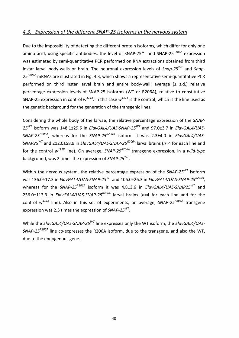
48
4.3. Expression of the different SNAP‐25 isoforms in the nervous system
Due to the impossibility of detecting the different protein isoforms, which differ for only one
amino acid, using specific antibodies, the level of SNAP‐25WT and SNAP‐25R206A expression
was estimated by semi‐quantitative PCR performed on RNA extractions obtained from third
instar larval body‐walls or brain. The neuronal expression levels of Snap‐25WT and Snap‐
25R206A mRNAs are illustrated in Fig. 4.3, which shows a representative semi‐quantitative PCR
performed on third instar larval brain and entire body‐wall: average (± s.d.) relative
percentage expression levels of SNAP‐25 isoforms (WT or R206A), relative to constitutive
SNAP‐25 expression in control w1118. In this case w1118 is the control, which is the line used as
the genetic background for the generation of the transgenic lines.
Considering the whole body of the larvae, the relative percentage expression of the SNAP‐
25WT isoform was 148.1±29.6 in ElavGAL4/UAS‐SNAP‐25WT and 97.0±3.7 in ElavGAL4/UAS‐
SNAP‐25R206A, whereas for the SNAP‐25R206A isoform it was 2.3±4.0 in ElavGAL4/UAS‐
SNAP25WT and 212.0±58.9 in ElavGAL4/UAS‐SNAP‐25R206A larval brains (n=4 for each line and
for the control w1118 line). On average, SNAP‐25R206A transgene expression, in a wild‐type
background, was 2 times the expression of SNAP‐25WT.
Within the nervous system, the relative percentage expression of the SNAP‐25WT isoform
was 136.0±17.3 in ElavGAL4/UAS‐SNAP‐25WT and 106.0±26.3 in ElavGAL4/UAS‐SNAP‐25R206A,
whereas for the SNAP‐25R206A isoform it was 4.8±3.6 in ElavGAL4/UAS‐SNAP25WT and
256.0±113.3 in ElavGAL4/UAS‐SNAP‐25R206A larval brains (n=4 for each line and for the
control w1118 line). Also in this set of experiments, on average, SNAP‐25R206A transgene
expression was 2.5 times the expression of SNAP‐25WT.
While the ElavGAL4/UAS‐SNAP‐25WT line expresses only the WT isoform, the ElavGAL4/UAS‐
SNAP‐25R206A line co‐expresses the R206A isoform, due to the transgene, and also the WT,
due to the endogenous gene.

49
Fig. 4.3. Semi‐quantitative PCR analysis of wild‐type (WT) and mutant (R206A) SNAP‐25 isoforms in third instar larvae brains. Agarose gel electrophoresis of PCR‐amplified fragments produced on cDNA from (A) brain total RNA and (B) body wall total RNA. Lane M is the molecular mass marker (FX174/HaeIII). The bottommost band in lanes 1‐6 is the housekeeping gene rp49 (164 bp); the other bands are SNAP‐25 fragments (WT in lanes 1, 3 and 5; R206A in lanes 2, 4 and 6; 210 bp). Average (± s.d.) relative percentage expression levels of SNAP‐25 isoforms (WT or R206A) in ElavGAL4/UAS‐SNAP‐25WT and ElavGAL4/UAS‐SNAP‐25R206A (C) larval brains (n=4 for each line) and (D) whole larval body (n=4 for each line), relative to constitutive SNAP‐25 (=100%; dotted line) expression in control w1118 larval brains (n=4).
4.4. Morphology and morphometry of the NMJ
As other Drosophila synaptic mutants with alteration in synaptic function were reported to
show a morphological change at the level of the NMJ, we analyzed the morphology and
morphometry of the NMJ after staining with the neuronal‐membrane‐specific anti‐HRP
antibody. The enzyme horseradish peroxidase (HRP) recognizes a carbohydrate epitope that
is selectively expressed in the insect nervous system; a large number of neuronal
glycoproteins and cell adhesion molecules that may be involved in axon outgrowth and
guidance process bear the HRP carbohydrate epitope (Desai et al., 1994).
To analyze synaptic changes in the ElavGAL4/UAS‐SNAP‐25R206A mutant we investigated the
projections of motorneurons (axon 1 and axon 2) onto of abdominal segment 3 muscle 6 and
7 in third instar larvae. These neurons form type‐I synaptic terminals and use glutamate as
their major neurotransmitter (Hoang and Chiba, 2001; Johansen et al., 1989a).

Fig. 4.4 sh
abdominal
stained wit
Fig. 4.4. Imm(right) larva.(green) were
The innerv
morpholog
which the
maximum
axon was
branches.
ElavGAL4/
morphome
indeed the
case of Ela
10
hows repre
l segment 3
th anti‐HRP
ElavGAL4/U
munohistochem The neuronae used for the
vation of m
gy of SNAP
synapses
surface of t
expanded
In ElavGAL4
/UAS‐SNAP‐2
etrical data
e average ex
avGAL4/UAS
0 µm
esentative
3 of ElavGA
P to visualize
UAS‐SNAP‐25W
mical stainingal membrane staining.
muscles 6/7
P‐25WT cont
appeared
the muscles
in SNAP‐2
4/UAS‐SNAP
25WT. Th
(Fig. 4.5B)
xtension of
S‐SNAP‐25W
confocal im
AL4/UAS‐SN
e neural pro
WT
g of abdominamarker anti‐
7 in SNAP‐
trol neuron
correctly h
s with their
25R206A mut
P‐25R206A la
e morph
showing n
ElavGAL4/UWT it was 63
50
mages of t
NAP‐25WT a
ojections, a
al segment 3 fHRP antibody
25R206A mu
s; mutants
harborized
r secondary
tants, we
rvae, the N
hological
o differenc
UAS‐SNAP‐2
39.9±121.4
he NMJ of
nd ElavGAL
nd anti‐NC8
ElavGAL4
fibres 6 and 7 y (red) and th
tants did n
presented
in order to
branches.
measured
NMJ length w
observation
ces between
25R206A is 65
µm2. These
f muscles
L4/UAS‐SNA
82 to visual
4/UAS‐SNAP‐2
NMJ of contrhe active zone
not differ f
d a well de
o cover an
To determi
the synap
was extend
n was
n control an
56.3±126.4
e results sug
6 and 7 in
AP‐25R206A la
ize active zo
25R206A
rol (left) and me marker anti
from the ty
veloped NM
d stimulate
ne how lon
tic spans o
ded as the o
confirmed
nd mutant
µm2 while i
ggest that S
n the
arvae
ones.
mutant i‐NC82
ypical
MJ in
e the
ng the
of all
one in
by
NMJ;
in the
SNAP‐

51
25R206A is not involved in alteration of bouton number or length of synaptic innervation at
the NMJ.
To quantify the SNAP‐25R206A phenotype, we counted the number of type‐I boutons. When
compared to controls, ElavGAL4/UAS‐SNAP‐25R206A larvae had approximately the same
number of boutons.
It has been reported that muscle size can be correlated with the size of the NMJ (Schuster et
al., 1996). But, because the expression of the transgenes is localized within the nervous
system, and, as consequence, the muscle surface area should not differ between mutants
and controls.
If compared with a other synaptic mutant previously characterized in the lab, the Splynull
mutant, the structure of the ElavGAL4/UAS‐SNAP‐25R206A NMJ seemed to be not altered.
Sphingosine‐1‐phosphate lyase (Sply) gene codes for an enzyme involved in sphingolipid
metabolism. The mutant lacks Sphingosine‐1‐phosphate, a sphingolipid metabolite which
regulates cell proliferation, migration and apoptosis, and which is important for adult muscle
development and integrity, reproduction and larval viability (Herr et al., 2003). Splynull larvae
display a severe alteration in the architecture of the NMJ, affecting both the axon terminal
arborization, synaptic boutons size, and the muscle apparatus (Magnabosco, PhD thesis and
personal communication). Otherwise, at the ElavGAL4/UAS‐SNAP‐25R206A NMJ the boutons
were easily recognizable, well bound and determined with the characteristic round shape,
not aggregated or fused together
To test whether type‐Is and Ib boutons might be altered in the mutant in general, the
average number (Fig. 4.5A) and diameter (Fig. 4.5D) of each group of boutons was
measured. The average number of type‐Is and Ib boutons counted in mutants and controls
were, respectively, 39.2±4.9 and 37.1±4.5. The bouton diameter distribution of
ElavGAL4/UAS‐SNAP‐25R206A overlaps the ElavGAL4/UAS‐SNAP‐25WT one; the first peak, at
around 1.5‐2 µm, corresponds to type‐Is boutons, while the second peak, at around 3‐4 µm,
corresponds to type‐Ib boutons. Those values are comparable to the ones reported in
literature (Hoang and Chiba, 2001).
Morphometrical analysis indicates that neither the total number of boutons per NMJ nor the
diameter of type‐Ib and Is boutons were changed in ElavGAL4/UAS‐SNAP‐25R206A compared
with ElavGAL4/UAS‐SNAP‐25WT. Mutants displayed a normal architecture of the NMJ at this
resolution level.

52
To analyze more in detail the boutons architecture, we determined the number of putative
active zones in control and mutant larvae. The NMJ was labeled with an antibody against the
active zone specific marker Bruchpilot, a presynaptic active zone protein which promotes
active zone assembly, Ca2+ channel clustering, and vesicle release in the larval nervous
system (Kittel et al., 2006).
It has been reported that the number of active zones increases in proportion to bouton size
(Atwood et al., 1993). Therefore, the number of active zones in the type‐Is and Ib boutons
were counted for the fibres 6 and 7 synapse of a abdominal segment 3 (Fig. 4.5C). We found
that this parameter was slightly increased in mutants; the ElavGAL4/UAS‐SNAP‐25R206A
larvae had, on average, 452.6±86.6 active zones per NMJ, while controls presented
348.9±56.0 anti‐NC82‐positive spots.
Analysis of several Drosophila mutants suggests that bouton number and size might be
regulated by separate signaling processes. The synaptic growth of neurons during the
development and adult life of an animal is a very dynamic and highly regulated process,
especially during larval development. In this period new boutons and branches are added at
the glutamatergic NMJ until a balance between neuronal activity and morphological
structures is reached (Budnik, 1996). It can be possible that the increase active zone number
of about 22% in mutants respect to controls can be a sort of compensation for the
impairment of vesicle fusing process determined by the presence of SNAP‐25R206A. So, the
increase in active zone number may correlate with a change in synaptic transmission
determining an increase in the vesicle fusion events in order to compensate the impairment
caused by the presence of mutant SNAP‐25 isoform at the synaptic terminal.
Thus the presence of SNAP‐25R206A did not alter the morphometry of the synapse, at least
not at the level of analysis performed, but instead it produced a slight structure
rearrangement at the bouton terminals, i.e. the increase in active zone number.
Experiments performed with murine spinal cord cultures have shown that the removal on
the part of BoNT/A of the SNAP‐25 C‐terminal segment beginning with Arg198 did not
change either the appearance of the synaptic terminal or the number and distribution of
synaptic terminals (Neale et al., 1999). Also in the Drosophila SNAP‐25ts mutant, the
synapses in the larvae appeared to be grossly normal at both the permissive and non‐
permissive temperatures even if, in this case, the mutation produced more severe effects
than the ones studied by us (i.e. altering the structure of the protein and its interaction with
the other SNAREs, synaptobrevin and syntaxin (Rao et al., 2001)). On the basis of these

53
considerations, we did not extend our morphological analysis to the electron microscopic
level.
Fig. 4.5. Third instar larvae NMJ morphological and morphometrical analyses. (A) Mean (± s.e.m.) number of NMJ type‐Ib and Is boutons from abdominal segment 3 fibre 6/7 of controls and mutants (n=8). (B) Mean (± s.e.m.) of the surface covered by the synapse (n=8). (C) Mean (± s.e.m.) number of active zones (n=8). (D) Relative percentage frequency distribution of type‐Ib and Is bouton diameters. Data were fitted with a double Gaussian function (R2=0.95 and 0.94 for ElavGAL4/UAS‐SNAP‐25WT and ElavGAL4/UAS‐SNAP‐25R206A data, respectively) showing the presence of a double peak corresponding to type‐Ib and type‐Is bouton diameters.
4.5. Locomotor behavior
Locomotion is an integral component of most animal behaviors. This behavior presents
extreme variation in nature which is attributable to multiple interacting quantitative trait
loci whose expression is influenced by the environment (Jordan et al., 2006). Insects have
long proven to be popular subject for the study of both innate and adaptative behavior.
Insects have elaborated a sensory systems, are affected by internal state, and exhibit a wide
array of stereotyped and nonstereotyped behaviors; they display several phenomena that,
from our point of view, can be described as thirst, hunger, lust, aggression, attention, sleep,
C
A B
D

54
and learning and memory. Even if the genetic basis of such complex behavior is largely
uncharacterized (Jordan et al., 2007), studying Drosophila can indeed inform us of the
driving forces involved (Zhang et al., 2010).
At the larval stage the animal are photophobic and escape light, so locomotion is required
for localization of food and dark environments. The Drosophila larva’s forward and backward
movements are produced by peristaltic waves of contraction of the body wall musculature.
The rhythmic contractions of the body segments are coordinated by a central pattern
generator (Jordan et al., 2007). Larvae have a simpler nervous system than adults, they have
a less elaborate repertoire of behaviors and their behavior is unavoidably linked to ongoing
development. Both the body and the nervous system continuously grow as the animal
transitions through the three instars and prepares for pupariation (Zhang et al., 2010). At the
adult stage the flies are no longer photophobic but they are attracted by light sources.
Locomotion is required for localization of food and mates, escape from predators, and
response to stress (Jordan et al., 2006).
The locomotor behavior of ElavGAL4/UAS‐SNAP‐25R206A larvae was characterized to verify if
the impairment of the vesicle fusion at the NMJ resulted in an altered locomotor phenotype.
To evaluate the condition of the animals we measured the total distance covered, the
average speed and the maximum speed. In all the three parameter considered the mutants
appeared to perform worse than the controls (Fig. 4.6).
In 10 seconds ElavGAL4/UAS‐SNAP‐25R206A larvae covered, on average, 4 mm more than the
controls (which corresponds to the length of a third instar larvae). This is probably due to the
fact that the mutant larvae move slowly; in fact their average and maximum speed are
respectively 15% and 17% lower than ElavGAL4/UAS‐SNAP‐25WT larvae. Moreover the
mutant larvae appeared to be less active and they needed to be stimulated to move (i.e. by
using a drop of water placed on the dorsal abdomen) more often than controls.
To complete the characterization, adult flies were also tested to verify if the presence of the
mutated SNAP‐25 produced any effects on adult locomotion. For this analysis other
parameters were considered; in addition to the total distance covered and the average
speed, the time spent resting was considered.
Also in adulthood (Fig. 4.7) the mutants exhibited the same sluggish behavior already seen at
the larval stage; the phenotype persists in both sexes. Indeed, in ten minutes, control males
and females covered, on average, 338.9 and 348.1 cm respectively, while the mutants
walked for 238.7 and 287.5 cm; there is a difference of about one meter between the
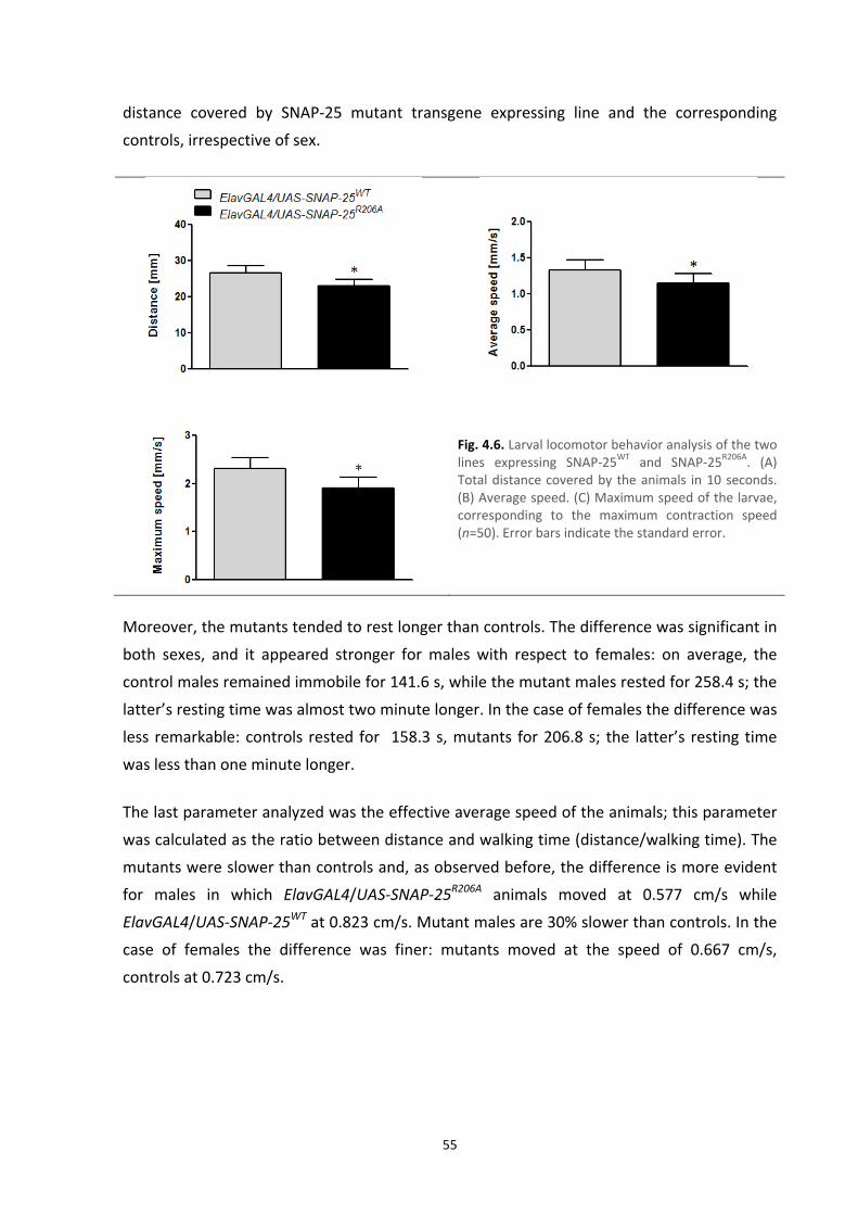
55
distance covered by SNAP‐25 mutant transgene expressing line and the corresponding
controls, irrespective of sex.
Fig. 4.6. Larval locomotor behavior analysis of the two lines expressing SNAP‐25WT and SNAP‐25R206A. (A) Total distance covered by the animals in 10 seconds. (B) Average speed. (C) Maximum speed of the larvae, corresponding to the maximum contraction speed (n=50). Error bars indicate the standard error.
Moreover, the mutants tended to rest longer than controls. The difference was significant in
both sexes, and it appeared stronger for males with respect to females: on average, the
control males remained immobile for 141.6 s, while the mutant males rested for 258.4 s; the
latter’s resting time was almost two minute longer. In the case of females the difference was
less remarkable: controls rested for 158.3 s, mutants for 206.8 s; the latter’s resting time
was less than one minute longer.
The last parameter analyzed was the effective average speed of the animals; this parameter
was calculated as the ratio between distance and walking time (distance/walking time). The
mutants were slower than controls and, as observed before, the difference is more evident
for males in which ElavGAL4/UAS‐SNAP‐25R206A animals moved at 0.577 cm/s while
ElavGAL4/UAS‐SNAP‐25WT at 0.823 cm/s. Mutant males are 30% slower than controls. In the
case of females the difference was finer: mutants moved at the speed of 0.667 cm/s,
controls at 0.723 cm/s.

56
Fig. 4.7. Adult locomotor behavior analysis of the two lines expressing SNAP‐25WT and SNAP‐25R206A isoforms. Male and female animals are represented. (A) Total distance covered by the animals in 10 minutes. (B) Total time immobile. (C) Average speed (n=25). Error bars indicate the standard error.
The neuromodulatory function of dopamine (DA) is an inherent feature of nervous systems
of all animals. Despite the evolutionary distance between mammals and the fly, functional
and molecular parallels exist between their dopaminergic systems. In the fly, DA neurons in
the fly brain are involved in the regulation of specific behaviors (e.g. locomotor activity,
sexual function, and response to drugs of abuse). These neurons show cluster‐specific,
stereotypical projection patterns with terminal arborization in target areas which include the
mushroom bodies, involved in memory formation and motivation, and the central complex,
involved in the control of motor behavior. The circuit from the dopamine system to
mushroom bodies is crucial for choice behavior in Drosophila; mutants lacking DA
biosynthesis showed reduced activity, extended sleep time, negative geotaxis, abolished
olfactory learning and they are hypophagic (Riemensperger et al., 2011; White et al., 2010).
It can be possible that ElavGAL4/UAS‐SNAP‐25R206A animals had an altered modulation of the
dopaminergic system which resulted in defects in motor behavior, deficit with characterize
many human health problems (e.g. Parkinson's disease, Huntington's disease, activity
disorders and depression). The ElavGAL4/UAS‐SNAP‐25R206A mutant may represent a model
to investigate neuropsychiatric and neurological disorders related to alterations in SNAP‐25
function.
A
C
B

57
4.6. Electrophysiology recordings: spontaneous neurotransmitter release
Two modes of vesicular release of transmitter occur at a synapse: spontaneous release, in
the absence of a stimulus, and evoked release, that is triggered by Ca2+ influx. Evoked and
spontaneous release occur in a quantal manner. These modes often have been presumed to
represent the same exocytotic apparatus functioning at different rates in different Ca2+
concentrations. Spontaneous discharges consisted of events which were randomly
distributed in time and of bursts of temporally ordered events (Budnik and Ruiz‐Cañada,
2006). To investigate the mechanism of transmitter release in ElavGAL4/UAS‐SNAP‐25R206A
and ElavGAL4/UAS‐SNAP‐25WT third instar larvae, the neurotransmitter release was
monitored at the NMJ of ventrolateral muscles 6 and 7.
Fig. 4.8A and Fig. 4.9A show that the co‐expression of SNAP‐25R206A with SNAP‐25WT led to a
significant (P<0.05) reduction in spontaneous neurotransmitter release. The mean (± s.e.m.)
frequency values of spontaneous release were: 1.85±0.35 Hz in ElavGAL4/UAS‐SNAP‐25WT
and 1.01±0.11 Hz in ElavGAL4/UAS‐SNAP‐25R206A under current‐clamp conditions.
To be more precise, the spontaneous neurotransmitter release was analyzed also under
voltage‐clamp conditions. This technique was used for the accuracy of minis detection (low
signal to noise ratio) and the possibility, due to 3rd instar larval muscle fiber biophysical
properties, to utilize only one microelectrode for voltage‐clamping muscle fibre. This
particular advantage is lost when larger currents (for instance evoked excitatory junctional
currents) must be clamped, making necessary to use the more complex two‐electrode
voltage clamp (Wu and Haugland, 1985). Using this configuration the average miniature end‐
plate current (MEPC) amplitude values were: 1.61±0.05 Hz in ElavGAL4/UAS‐SNAP‐25WT and
1.07±0.04 Hz in ElavGAL4/UAS‐SNAP‐25R206A.
The average miniature end‐plate potential (MEPP) amplitudes (in current‐clamp conditions)
were: 0.97±0.06 mV and 0.94±0.02 mV in ElavGAL4/UAS‐SNAP‐25WT and ElavGAL4/UAS‐
SNAP‐25R206A, respectively (Fig. 4.8B). Data were not significantly different (Student’s t‐test
for unpaired samples). Fig. 4.8C shows that the cumulative MEPP amplitudes distributions
were not significantly different (Kolmogorov‐Smirnov D=0.069; P=NS).

58
Fig. 4.8. Spontaneous neurotransmitter release in SNAP‐25WT and SNAP‐25R206A transgenic third instar larvae, recorded in current‐clamp conditions. (A) Mean MEPP (± s.e.m.) frequency in ElavGAL4/UAS‐SNAP‐25WT (n=5) and ElavGAL4/UAS‐SNAP‐25R206A (n=5) larvae. One NMJ for each animal was analyzed. *P<0.05; Student’s t‐test for unpaired data. (B) Mean (±s.e.m.) MEPP amplitude in ElavGAL4/UAS‐SNAP‐25WT (556 events analyzed from five different larvae) and ElavGAL4/UAS‐SNAP‐25R206A (314 events analyzed in five different larvae). Data are not statistically significantly different (Student’s t‐test for unpaired data). (C) Relative cumulative frequency distribution of MEPP amplitudes from ElavGAL4/UAS‐SNAP‐25WT and ElavGAL4/UAS‐SNAP‐25R206A. Data are not statistically different (Kolmogorov‐Smirnov test).
By contrast, the average miniature end plate current (MEPC) amplitude (under voltage‐
clamp conditions) was significantly reduced in ElavGAL4/UAS‐SNAP‐25R206A (132.6±2.2 pA;
mean ± s.e.m.) with respect to ElavGAL4/UAS‐SNAP‐25WT (167.8±2.5 pA; mean ± s.e.m.; Fig.
4.9B). And this is reflected in the cumulative MEPC amplitudes distributions, showing a shift
towards smaller amplitudes in the case of ElavGAL4/UAS‐SNAP‐25R206A (Kolmogorov‐Smirnov
D=0.186; P=0.001; Fig. 4.9C).
Because concentration of neurotransmitter is the same in all synaptic vesicles, the volume of
the vesicle determines how much transmitter is released. Thus, variation in vesicular volume
may contribute to observed variance of synaptic quantal unit size. Spontaneous fusions
occur in a random manner, each event being independent from the previous and next one
(Kim et al., 2000). Accordingly, the frequency distribution of MEPCs inter‐event intervals is
well described by a Poisson distribution in both controls and SNAP‐25 mutants (Fig. 4.9D),
indicating that the random nature of spontaneous fusion was not altered by the presence of
SNAP‐25R206A.
A B
C
*

59
ElavGAL4/UAS‐SNAP‐25WT ElavGAL4/UAS‐SNAP‐25R206A
Fig. 4.9. Spontaneous neurotransmitter release in SNAP‐25WT and SNAP‐25R206A transgenic third instar larvae, recorded in voltage‐clamp conditions. (A) Representative traces of MEPCs recorded in control and mutant (black line) larvae. (B) Mean MEPC (± s.e.m.) frequency in ElavGAL4/UAS‐SNAP‐25WT (n=7) and ElavGAL4/UAS‐SNAP‐25R206A (n=6) larvae; n indicates the number of NMJ analyzed. One neuromuscular junction for each animal was analyzed. *Data are statistically different (P<0.05; Student’s t‐test for unpaired data). (C) Mean (± s.e.m.) MEPC amplitude in ElavGAL4/UAS‐SNAP‐25WT (2616 events analyzed in seven different NMJs) and ElavGAL4/UAS‐SNAP‐25R206A (1539 events analyzed in six different NMJs). *Data are statistically different (P<0.05; Student’s t‐test for unpaired data). (D) Relative cumulative frequency distribution of MEPC amplitudes from ElavGAL4/UAS‐SNAP‐25WT and ElavGAL4/UAS‐SNAP‐25R206A. Data are statistically different (Kolmogorov‐Smirnov test). (E) Percentage relative frequency distribution of inter‐event intervals (gray dots: ElavGAL4/UAS‐SNAP‐25WT; black dots: ElavGAL4/UAS‐SNAP‐25R206A). Black and grey lines are the monoexponential curve fitting of ElavGAL4/UAS‐SNAP‐25WT and ElavGAL4/UAS‐SNAP‐25R206A data, respectively. The good R2 of each curve fit indicates the random behavior of spontaneous release.
B C
D E
A

60
4.7. Electrophysiology recordings: evoked neurotransmitter release
To complete the characterization of the electrophysiological profile of the ElavGAL4/UAS‐
SNAP‐25R206A and ElavGAL4/UAS‐SNAP‐25WT NMJ, the evoked release of quanta form the
synaptic terminals has been examined. To fully investigate the mechanism of transmitter
release, the records were performed stimulating the nerve at different frequencies.
Low frequency stimulation
Initially, to not stress the NMJ, the nerve was stimulated at low frequency; synapses do not
display fatigue at this frequency, the RRP pool results not depleted, and, as consequence,
the vesicle cycle is not accelerated. During the low frequency stimulation only the vesicles of
the RRP, generally thought to be docked to the presynaptic active zone and primed, fuse
with the target membrane and release the neurotransmitter into the synaptic cleft (Rizzoli
and Betz, 2005).
Fig. 4.10B shows that the mean amplitude of EJPs recorded at third instar larva NMJ
following segmental nerve stimulation at 0.5 Hz, was clearly (P<0.05) reduced in
ElavGAL4/UAS‐SNAP‐25R206A mutants, with respect to the controls (33.7±0.6 mV in
ElavGAL4/UAS‐SNAP‐25WT and 26.0±0.3 mV in ElavGAL4/UAS‐SNAP‐25R206A; mean ± s.e.m.;
n=6 and 11, respectively).
On the same fibres we recorded the EJPs after a period higher frequency (10 Hz) of
stimulation of the nerve; Fig. 4.10C shows that the mean amplitude of EJPs recorded. Values
were 35.0±0.5 mV in ElavGAL4/UAS‐SNAP‐25WT and 28.6±0.3 mV in ElavGAL4/UAS‐SNAP‐
25R206A (mean ± s.e.m.; n=6 and 11, respectively). The 10 Hz stimulation of the nerve
determined the rise of postsynaptic membrane potential of a value of 2 mV in both mutants
and controls. This event is described as short term plasticity, a phenomenon, lasting a few
seconds, which synaptic enhancement results from an increase in the probability that
synaptic terminals will release transmitters in response to presynaptic action potentials.
Synapses strengthen for a short time due to either an increase in size of the readily
releasable pool of packaged transmitter or an increase in the amount of packaged
neurotransmitter released in response to each action potential (Stevens and Wesseling,
1999).
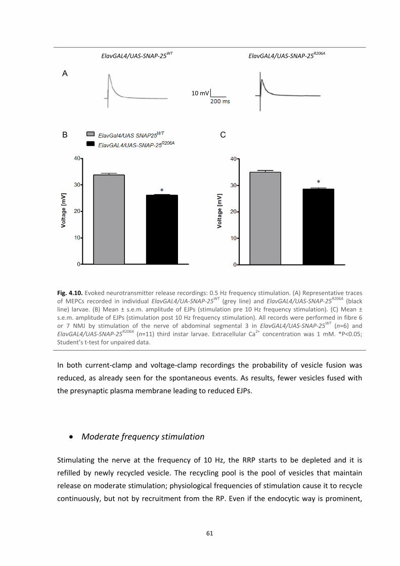
61
ElavGAL4/UAS‐SNAP‐25WT ElavGAL4/UAS‐SNAP‐25R206A
Fig. 4.10. Evoked neurotransmitter release recordings: 0.5 Hz frequency stimulation. (A) Representative traces of MEPCs recorded in individual ElavGAL4/UA‐SNAP‐25WT (grey line) and ElavGAL4/UAS‐SNAP‐25R206A (black line) larvae. (B) Mean ± s.e.m. amplitude of EJPs (stimulation pre 10 Hz frequency stimulation). (C) Mean ± s.e.m. amplitude of EJPs (stimulation post 10 Hz frequency stimulation). All records were performed in fibre 6 or 7 NMJ by stimulation of the nerve of abdominal segmental 3 in ElavGAL4/UAS‐SNAP‐25WT (n=6) and ElavGAL4/UAS‐SNAP‐25R206A (n=11) third instar larvae. Extracellular Ca2+ concentration was 1 mM. *P<0.05; Student’s t‐test for unpaired data.
In both current‐clamp and voltage‐clamp recordings the probability of vesicle fusion was
reduced, as already seen for the spontaneous events. As results, fewer vesicles fused with
the presynaptic plasma membrane leading to reduced EJPs.
Moderate frequency stimulation
Stimulating the nerve at the frequency of 10 Hz, the RRP starts to be depleted and it is
refilled by newly recycled vesicle. The recycling pool is the pool of vesicles that maintain
release on moderate stimulation; physiological frequencies of stimulation cause it to recycle
continuously, but not by recruitment from the RP. Even if the endocytic way is prominent,
C
A
B
10 mV

62
the synapse can respond to this stimulation without displaying fatigue (Rizzoli and Betz,
2005).
The only remarkable effect is the short‐term synaptic depression (STD), one of several
phenomena underlying synaptic plasticity, a propriety which allows the synaptic connection
functional strength to be modified by neuronal activity . When stimulated repetitively under
physiological conditions, many synapses exhibit rapid depression that progresses to a
steady‐state level and recovers in seconds after stimulation. Many forms of STD reflect
activity‐dependent changes in neurotransmitter release (Zucker and Regehr, 2002).
Stimulating the nerve at 10 Hz frequency, the first few spikes presented an increased EJP
value if compared to the others recorded subsequently in a row. The STD effect occurred in
both ElavGAL4/UAS‐SNAP‐25WT and ElavGAL4/UAS‐SNAP‐25R206A NMJs.
In Fig. 4.11 the mean amplitude of EJPs, recorded during 10 Hz nerve stimulation, are shown.
The neurotransmitter release quantal size was clearly (P<0.05) reduced in ElavGAL4/UAS‐
SNAP‐25R206A mutants, with respect to the controls as previously observed in the case of low
frequency stimulation. EJPs mean amplitude values were 36.2±0.3 mV in ElavGAL4/UAS‐
SNAP‐25WT and 25.2±0.2 mV in ElavGAL4/UAS‐SNAP‐25R206A; mean ± s.e.m.; n=6 and 11,
respectively. The difference between mutants and controls in case of moderate stimulation
(∆10Hz=11 mV) is increased respect to the difference observed in case of low frequency
stimulation (∆0.5HzPRE =7.7 mV, ∆0.5HzPOST =6.4 mV).
Fig. 4.11. Evoked neurotransmitter release recordings: 10 Hz frequency stimulation. Mean amplitude of EJPs. Records were performed in fibre 6 or 7 NMJ by stimulation of the nerve of abdominal segmental 3 in ElavGAL4/UAS‐SNAP‐25WT (n=6) and ElavGAL4/UAS‐SNAP‐25R206A (n=11) third instar larvae. Extracellular Ca2+ concentration was 1 mM.
Paired pulse stimulation
Synaptic plasticity is the ability of the synapse to change in strength in response to either use
or disuse of transmission over synaptic pathways (Hughes, 1958). There are several

unde
the q
Facil
their
seco
trans
the C
facili
Wym
excit
syna
the a
stimu
NMJs
75 an
Fig. 4larval 20, 30
The d
while
displ
Paired
RespoElavG
RespoElavG
ElaUA
ElaUA
erlying mec
quantity of n
itation of m
r strength; t
nd impulse
sient outwa
Ca2+‐depend
tated state
man, 1980).
tability for a
pse and we
amplitude
ulus were
s. The nerv
nd 100 ms i
4.12. Paired pNMJ. Repres0, 50, 75 and 1
differences
e the contr
ay a respon
d pulse Interv
onse to the seGAL4/UAS‐SNA
onse to the seGAL4/UAS‐SNA
avGAL4/ AS‐SNAP‐25R20
avGAL4/ AS‐SNAP‐25WT
chanisms th
neurotrans
membrane e
this effects
e. The mec
ard current
dent spike‐
persists fo
. Because a
a short time
e recorded
of the eve
characteriz
ve was stim
ntervals we
pulses recordentative reco100 ms.
in paired p
rol NMJ wa
nse to them
val [ms]
econd pulse inAP‐25WT [fails
econd pulse inAP‐25R206A [fa
06A
T
hat coopera
mitters rele
electrical ex
s consists in
hanism of
t by prior m
like respon
r around 1
a single ne
e, with the
the EJPs of
ents but als
ed in ElavG
ulated by p
ere chosen f
dings performrdings of pair
pulse respo
as able to r
m all. Values
n s/total events
n ails/total even
63
ate to achie
eased into a
xcitability is
n the increa
facilitation
membrane
se has a de
second afte
erve‐driven
paired puls
f the secon
so the abil
GAL4/UAS‐S
paired pulse
for the ana
med on ElavGAred pulses in c
onses betwe
respond to
were as fol
s]
nts]
eve synapti
a synapse (G
s the ability
asing in pos
involves th
depolarizat
ecreased cu
er a membr
spike is su
se stimulati
nd response
ity of the
SNAP‐25R206
es differing
lysis (Fig. 4.
AL4/UA‐SNAPcontrol and m
een mutant
all stimula
llows:
20
0/33
69/144
c plasticity,
Gaiarsa et a
y of chemic
stsynaptic p
he inactivat
tion. During
urrent and v
rane active
ufficient to
ion we indu
e of the mu
NMJ to re6A and Elav
for the inte
.12A).
P‐25WT and Elmutant NMJ; p
ts and cont
tion series,
30
0/34 0
52/139 25
, including
al., 2002).
cal synapses
potential ev
tion of an
g the facilit
voltage thre
response (
o facilitate
uced facilita
uscular fibre
espond to t
vGAL4/UAS‐
erval time;
lavGAL4/UAS‐paired pulse in
rols were r
, the mutan
50 75
0/33 0/35
/134 17/14
10
10 mV
10 mV
10
changes in
s to change
voked by a
early, fast,
tated state,
eshold. The
Salkoff and
membrane
ation in the
e. Not only
the second
‐SNAP‐25WT
20, 30, 50,
‐SNAP‐25R206A
ntervals were:
emarkable;
nts did not
100
5 0/37
42 3/172
0 ms
00 ms
n
e
a
,
,
e
d
e
e
y
d T
,
A :
;
t

64
The number of EJP failures is maximum when the interval is 20 ms. Increasing the distance
between the two stimuli, the ElavGAL4/UAS‐SNAP‐25R206A reacquired the ability to respond
to the second pulse. At 100 ms the mutant phenotype is comparable to the control one, and
the failure events occurred only few times.
4.7.4. Tetanic stimulation
To complete our electrophysiological characterization of the mutant we analyzed the EJPs at
high frequency stimulation (30 Hz), called tetanic stimulation (Fig. 4.13). It consisted in a
high‐frequency sequence of individual stimulations of a neuron which results in a depletion
of both RRP and recycling vesicle pool. This high‐frequency stimulation causes the
mobilization of the RP, which is called into action only upon strong, often unphysiological
stimulation (Rizzoli and Betz, 2005).
Only a qualitative analysis of the EJPs was performed. The results showed that the mutants
failed to respond more frequently than controls during the stimulation. ElavGAL4/UAS‐
SNAP‐25WT NMJs were able to maintain the stimulation for few minutes while
ElavGAL4/UAS‐SNAP‐25R206A NMJs became unable to respond earlier, in some cases within
the first minute of stimulation. Reducing the frequency of stimulation the mutants became
able to respond to the stimuli, showing that the phenotype was not caused by a depletion of
the ready releasable pool of synaptic vesicles.
Fig. 4.12. Representative records 30 Hz stimulation recordings performed on ElavGAL4/UA‐SNAP‐25WT and ElavGAL4/UAS‐SNAP‐25R206A larval NMJ.
ElavGAL4/UAS‐SNAP‐25WT
ElavGAL4/UAS‐SNAP‐25R206A
200 ms10 mV

65
4.8. Ca2+ dependency of neuroexocytosis
At the synapse, neurotransmitter release is triggered physiologically by Ca2+ influx through
voltage‐gated Ca2+ channels which origins high calcium concentration microprofiles at the
axon terminal. The control of transmitter release at the neuromuscular synapse is achieved
also by intracellular calcium; while membrane‐bound organelles, such as mitochondria and
the endoplasmic reticulum, regulate Ca2+ homeostasis intracellularly, performing
electrophysiological experiments on the larval NMJ it is possible to alter the extracellular
Ca2+ concentration of the bath solution.
It is known that calcium ions trigger the final step of vesicle fusion (Katz and Miledi, 1967)
but also regulate other steps in the synaptic vesicle cycle, such as vesicle priming, vesicle‐
pool refilling, and endocytosis (Burgoyne and Morgan, 1995; Sudhof, 2004). It has been
demonstrated that SNAP‐25 is involved in the control of Ca2+ dynamics (close to) at the
active zone (Pozzi et al., 2008) and interacts with the voltage gated Ca2+ channel via its C‐
terminus (Catterall, 1999; Zhong et al., 1999). Moreover, it has been well documented that
high external Ca2+ concentrations can partially rescue the neuroexocytosis blocking effect of
BoNT/A which cleaves SNAP‐25 at the Gln198‐Arg199 peptide bond; the paralysis caused by
BoNT/A, but not that caused by BoNT/E, can be partially reversed by raising intracellular
calcium (Binz et al., 1994; Capogna et al., 1997; Schiavo et al., 1993). To test for the
possibility that the Arg‐Ala replacement affects the Ca2+ sensitivity of neuroexocytosis, the
Ca2+ dependence of evoked neurotransmitter release was measured.
EJPs were recorded on the same fibre increasing the external Ca2+ concentration. We
observed a reduced Ca2+ sensitivity in ElavGAL4/UAS‐SNAP‐25R206A compared with
ElavGAL4/UAS‐SNAP‐25WT (Fig. 4.12A) with no change in the slope of the linear fit to the
data of voltage change recorded at different extracellular Ca2+ concentrations (Fig. 4.12B).
This result suggests that the Ca2+ cooperativity of the process is unchanged between
ElavGAL4/UAS‐SNAP‐25R206A and ElavGAL4/UAS‐SNAP‐25WT transgenic lines.

66
Fig. 4.12. Calcium sensitivity of evoked release. (A) Mean ± s.e.m. amplitude of EJPs evoked in fibre 6 or 7 NMJ by stimulation of the nerve of abdominal segment 3 or 4 fibre at different extracellular Ca2+ concentrations in ElavGAL4/UAS‐SNAP‐25WT (n=8; grey bars) and ElavGAL4/UAS‐SNAP‐25R206A (n=9; black bars) third instar larvae. Only fibres in which it was possible to maintain the intracellular recording throughout the whole experiments (i.e. at different external Ca2+ concentrations) were considered for analysis. *Statistically significant (P<0.05) difference at each Ca2+ concentration tested (Student’s t‐test for unpaired data). (B) Linear regression of the same data as in A. R2 for each linear regression is indicated for the wild‐type SNAP‐25 (grey) and mutated SNAP‐25 (black) samples.
4.9. Bang test
Due to the potential implications of SNAP‐25, together with other genes, in the pathologies
of neuropsychiatric and neurological disorders (e.g. schizophrenia, ADHD and epilepsy), we
also tested the functionality of central neuronal circuits in the ElavGAL4/UAS‐SNAP‐25R206A
flies by performing the bang sensitivity assay on adult flies.
The bang test (Ganetzky and Wu, 1982b) consists in a hyper stimulation of the animals that
promote the fusion of a large number of SVs of the RRP pool in the neurons. As results the
animals can show different phenotypes: they can show a sustained paralysis after a brief
seizure, such as occurs in the case of the easily shocked (eas) mutant do; the can temporally
lose the climbing ability (wild‐type flies normally show negative geotaxis), as happens in the
case of the shibirets mutant; they can show a positive geotaxis phenotype or they can briefly
recover and show a normal phenotype, as wild‐type individuals normally do.
Males and females were distinguished in this test to pick out any possible sexual
dimorphism. In general, males are more active than females, and the bang sensitivity assay
reflected this difference (Fig. 4.13). Within the same sexual class, ElavGAL4/UAS‐SNAP‐
A B

67
25R206A flies needed more time to recover after the hyperstimulation. As a consequence,
they reached the different marks later that corresponding controls (i.e. the ElavGAL4/UAS‐
SNAP‐25WT animals). The worse behavior of the mutant was significant for the first two
marks, 28 and 56 mm respectively, where the animals showed, on average, a delay of about
4 seconds with respect to the concurrent controls.
Furthermore, few animals showed short seizures, defined as transient signs and/or
symptoms of abnormal, excessive or synchronous neuronal activity in the brain (Fisher et al.,
2005). During the epileptic transient they performed uncontrolled bursts of wing flappings.
As soon as the seizure ended, few animals were not able to stand stably on their legs (Fig.
4.13B and 4.13D, columns KO), and some others tended to remain in the same area of the
test arena (Fig. 4.13B and 4.13D, columns 0) showing a phenotype that can be related to the
defective behavioral activity observed in the coloboma mutant mouse (Hess et al., 1992b), in
which, SNAP‐25 is partially lacking.
After the recovery, the mutant flies usually displayed negative geotaxis as also observed in
controls; these animals climbed the walls of the vial passing the four climbing marks to reach
the top. The proportion of flies reaching the different marks was lower in the ElavGAL4/UAS‐
SNAP‐25R206A line than the ElavGAL4/UAS‐SNAP‐25WT line, because not only the mutants
moved more slowly, as previously observed in the locomotor assay, but also the delay in the
post‐stimulation recovery time was longer (5 seconds more on average, with respect to the
concurrent controls).
It has been proposed that, in the case of human neuropsychiatric disorders associated with
SNAP‐25 defects, the effects on SNAP‐25 expression could be downstream and secondary to
maintaining synaptic homeostasis, resulting from altered neural circuits or network function.
In fact it is probable that each of the candidate genes thought to be involved in
neuropsychiatric disease, among which Snap‐25, appear to contribute only a small effect
towards the overall phenotype of disability (Corradini et al., 2009). This is in keeping with the
lack of a severe phenotype in the case of the overexpression of the mutant isoform
described in this work.

68
Fig. 4.13. Bang sensitivity of adults. Female data are reported in red, male data in blue. Light color refers to ElavGAL4/UAS‐SNAP‐25WT, dark refers to ElavGAL4/UAS‐SNAP‐25R206A. (A and B) Average time needed by the animals to reach the marks positioned on the vial at 28, 56, 84 and 112 mm from the bottom. (C and D) Percentage of animals which reached the different marks. KO refers to animals unable to recover after the epileptic seizure. 0 refers to animals which did not present the typical climbing ability, by remaining instead at the bottom of the testing vial.
4.10. SNARE in vitro assembly
To verify if the presence of the mutated SNAP‐25 could prevent the assembly of single
SNARE complexes, in vitro experiments were performed in our laboratory in order to assay
the extent and time‐course of the formation of SNARE complexes. Mouse GST‐SNAP‐25WT or
the mutant mouse GST‐SNAP‐25R198A were in turn incubated together with the cytoplasmic
domains of VAMP and syntaxin and the capacity to form complexes in vitro determined
(Fasshauer et al., 1998).
Fig. 4.14A shows that no difference in the formation of SNARE complexes in vitro was
detected between the wild‐type and mutated proteins.
A
C
B
D

69
Furthermore, a more sensitive and relevant end point was also determined by assaying the
time‐course of SNARE complexes formation.
Fig. 4.14B shows that both wild‐type and mutated SNAP‐25 very rapidly formed a SNARE
complex with VAMP and syntaxin as found previously (Fasshauer et al., 2002) with no
difference in the time course.
Fig. 4.14. The point mutation R198A of GST−SNAP‐25 does not prevent SNARE complex assembly. SNARE complex formation was analyzed using recombinant VAMP2 1‐96, syntaxin1A 1‐261 and GST‐SNAP‐25WT (lanes 1, 2) or the mutant R198A (lanes 3, 4). Reactions were stopped at 30 minutes (A) or at the indicated time points (B) by adding SDS‐containing sample buffer. Samples were then subjected to SDS‐PAGE with or without boiling. Protein were visualized by Coomassie staining (A). Panel B shows the time course of the formation of the SNARE complexes quantified by densitometry of the SDS‐PAGE gels (n=3) (Megighian et al., 2010).
It has been show that mutations affecting the properties of core complex formation and
vesicular fusion, such BoNT/A cleaved SNAP‐25, do not affect the capability of forming the in
vitro complex but rather the thermostability of this complex (Zhang et al., 2002). The results
obtained in these important control experiments confirmed our expectation since, as
pointed out, the mutated residue points outward in the crystallographic structure of the
SNARE complex (Brunger et al., 2009; Sutton et al., 1998) and C‐terminal mutations of SNAP‐
25 have been shown not to affect SNARE assembly (Sorensen et al., 2006).
A B

70

71
5. DISCUSSION
5.1. SNAP‐25R206A reduced the probability of vesicle fusion
The probability of fusion of a synaptic vesicle is strictly dependent on the correct formation
of SNARE complexes (Sorensen, 2009; Sudhof and Rothman, 2009). The fact that BoNT/A is
known to cleave nine amino acid residues from the COOH‐terminus of SNAP‐25 indicates
that this part of the molecule must play an essential role in the assembly of the exocytotic
machinery. Performing sequence conservation analysis on neuronal SNAP‐25 protein
sequences among different species, a single amino acid which is highly conserved in the
protein’s C‐terminal region was identified: the arginine in position 206. This amino acid is at
the first position of the peptide bond cleaved by BoNT/A. Using site‐directed mutagenesis,
the conserved Arg206 was replaced with alanine. The results obtained by the in vivo
expression of SNAP‐25R206A within the Drosophila nervous system reflect defective SNARE
complex assembly. As consequence, the probability of neurotransmitter release at the larval
NMJ was reduced, leading to the decrease of the frequency of the spontaneous fusion
events and the amplitude of evoked events.
Nowadays it is debated if the two forms of neurotransmitter liberation involve the same
molecular machinery. Regarding to the spontaneous vesicle fusion, the phenomenon occurs
both in the absence of action potentials and without any apparent stimulus and is hence
thought to be Ca2+‐independent. However, increasing evidence shows that this form of
neurotransmitter release can be modulated by changes in intracellular Ca2+ concentration,
suggesting that it is not fully spontaneous (Glitsch, 2008). For example, synaptotagmin‐1
(SYT‐1) (Perin et al., 1990), a protein of a family of transmembrane proteins containing
tandem Ca2+‐binding C2 domains, plays the role as sensor which trigger Ca2+‐induced
exocytosis. The protein, localized to synaptic vesicles, is involved in the vesicle fusion as well
as recycling. When the Ca2+ concentration increases within the axon terminal, the two C2
domains, after binding Ca2+ ions, induce membrane curvature bringing the two membranes
destined to fuse into close proximity, increasing the probability of vesicle fusion. Genetic
studies in Drosophila and mice have demonstrated that loss of syt‐1 specifically eliminates
the fast synchronous component of release, without removing the slow asynchronous
component (Geppert et al., 1994; Loewen et al., 2001; Yoshihara and Littleton, 2002).
72
Also the presence of SNAP‐25R206A led to an alteration in the frequency of spontaneous
vesicle fusion, supporting the idea that spontaneous release is not an independent process
but can be modulated, for example, by interfering with a protein involved in the exocytotic
process.
As seen for the syt‐1null mutants, modulation of spontaneous discharge at the level of the
release machinery is not always accompanied by corresponding modulation of action
potential‐evoked release, suggesting that two independent processes may exist for
spontaneous and evoked exocytosis. For this reason, also the electrophysiological profile of
the evoked neurotransmitter release has been characterized in the transgene expressing
SNAP‐25R206 isoform.
Action potential‐evoked potentials recorded at ElavGAL4/UAS‐SNAP‐25R206A third instar larva
NMJ were reduced if compared to controls both at low and high frequency stimulation,
respectively 0.5 and 10 Hz. Assuming that the concentration of neurotransmitter is the same
in all synaptic vesicles, the volume of the vesicle determines how much transmitter is
released. Thus, the presence of SNAP‐25R206A determined a decrease in fusion event number
in response to a stimulus. As a consequence of the depletion of vesicle fusion with the
plasma membrane, the amplitude of the peaks recorded in current‐clamp condition is
reduced, if compared with controls.
From voltage‐clamp recordings it was possible to appreciate that the vesicles pool which
fused with the plasma membrane was enriched in small diameter vesicles; a possible
explanation for this can be correlated to the different energetic requirement to fuse big and
small vesicles. If a big vesicle require more energy to fuse, probably more SNARE complexes
have to mediate the process; on the other hand, a small vesicle may require less SNARE
complexes. For this reason the presence of the SNAP‐25R206A isoform may be more probable
at the big vesicles, and less probable at the small vesicles.
Moreover, the tetanic stimulation (30 Hz) showed a more severe electrophysiological
phenotype: the expression of SNAP‐25R206A had repercussion on the origin of action
potentials. The ElavGAL4/UAS‐SNAP‐25R206A NMJ did not respond to the stimulation already
after a few minutes of stimulation, while ElavGAL4/UAS‐SNAP‐25WT synapses were able to
sustain the strong stimulation for several minutes before displaying a failure in the response.
Also this results reflects an impairment of vesicle fusion, as observed for the lower frequency
stimulation experiments.
Lastly, from the paired pulse recordings it appeared clear that the presence of SNAP‐25R206A
affected the probability of fast vesicle fusion. In fact, when the ElavGAL4/UAS‐SNAP‐25R206A

73
synapse has been stimulated with temporally closed spaced pulses, the synapse was not able
to respond to the second stimulus; while, by increasing the temporal spacing of the stimuli,
the ElavGAL4/UAS‐SNAP‐25R206A synapse could respond to both pulses, reacquiring a
phenotype similar to the controls. This result can be explained assuming that when the first
action potential invaded the axon terminal, all the fusion‐competent vesicles fused with the
plasma membrane, while the fusion of vesicles presenting a non‐functional SNARE complex
is prevented. When the second action potential arrives, the active zone may be still enriched
in vesicles which have a SNAP‐25R206A isoform in their SNARE complex, vesicles which are not
able to fuse; thus, in these conditions, the number of fusion events is strongly reduced.
Nothing is known about the turnover rate of SNARE proteins but it is plausible that barely
any vesicles were able to fuse and collapse; as consequence, the neurotransmitter released
in the synaptic cleft may not be sufficient to activate all the channels on the muscle
membrane, resulting in no post synaptic potential arising.
These results prove the experimental hypothesis demonstrating that Arg198 is fundamental
for the proper functioning of the neuroexocytosis apparatus. Because the amino acid does
not face the α‐helices of syntaxin and synaptobrevin, but instead points outside the
complex, it appears plausible that this residue might be involved in contacts with lipids or
other proteins which regulate and/or modulate the synaptic vesicle fusion process.
5.2. Evidences of the SNARE supercomplex
Currently it is still debated whether assembly of one SNARE complex generates sufficient
energy to initiate vesicle fusion. Several biochemical studies have been performed to identify
the minimum number of complexes necessary to promote vesicle fusion (Lu et al., 2008).
While the energy profile of SNARE complex assembly reaction and the thermodynamic
properties of the SNARE complex are almost clarified, just how the energy is transferred
from the complex across lipid bilayers is still obscure (Wiederhold and Fasshauer, 2009).
Models of specific kinetics, interaction and assembly/dissociation forces of the SNAREs
harnessed for membrane fusion remain speculative (Yersin et al., 2003).
It was recently reported that in vitro liposomes can fuse with the help of a single SNARE
complex, albeit with low speed (van den Bogaart et al., 2010). It was hypothesized that
vesicles resident at the plasma membrane have time to form several SNARE complexes in
the absence of stimulation, achieving faster speeds of fusion when triggered by calcium.

74
However, vesicles arriving during conditions of sustained high calcium concentrations might
fuse using fewer (or possibly only a single SNARE complex) (Mohrmann et al., 2010).
In vitro experiments, using liposomes or isolated synaptic vesicles, gave only ambiguous
proof that one SNARE is sufficient for the fusion process. Even in the fastest reconstituted
biochemical systems, the fusion process is still at least 20 times slower than fusion of
synaptic vesicles in neurotransmission; in these in vitro systems, also the docking occurs
stochastically over many tens of minutes (Domanska et al., 2009). Moreover, all these
models do not consider that neuronal docking and calcium‐dependent secretion are tightly
regulated, both spatially and temporally, by a multitude of proteins. Thus the in vivo
complexity, where many regulatory proteins, such as Munc18, complexin and
synaptotagmin, interact with the SNAREs, is vastly increased compared to in vitro systems. It
is conceivable that binding of these proteins siphons away some of the energy of SNARE
assembly and that under these conditions one SNARE complex may no longer suffice to bring
about fusion (van den Bogaart et al., 2010).
The membrane‐bridging assembly of the neuronal SNARE complex is central in exocytosis of
secretory cells (Jahn and Scheller, 2006). Even if the SNARE proteins are responsible for the
mechanism of fusion in the both chromaffin cells and nerve terminals, the differences
between neuroexocytosis and neurosecretion are several: the two processes differ in many
parameters, such as the vesicle dimension and density in the cytosol, the fusion timeline,
and the recycling process.
To become available for exocytosis, granules must move from the cytosol to the plasma
membrane and dock there. In chromaffin cells, a layer of filamentous actin covers the
internal side of the plasma membrane (Steyer and Almers, 1999). This “cortical” actin layer
appears to be a barrier to granules on their way to the plasma membrane (Nakata and
Hirokawa, 1992), reducing the mobility of isolated chromaffin granules in vitro (Miyamoto et
al., 1993), although whether cortical actin helps or hinders the motion of granules to the
plasma membrane remains unresolved. At the axonal terminal this structure is not present.
Furthermore, neuroendocrine cells that release hormones and peptides typically contain
vesicles of larger diameters than those at classical synapses: at the Drosophila NMJ, vesicles
have a mean diameter of about 40 µm (Zhang et al., 1998) while chromaffin granules have a
diameter between 330 and 360 µm (Plattner et al., 1997), almost 10 times larger than
synaptic vesicles.

75
Although dense‐cored vesicles which contain peptides are found within nerve terminals of
non‐neuroendocrine cells, they occur with lower frequency than the clear‐cored vesicles
which contain neurotransmitter (Kupfermann, 1991).
Lastly, different types of cells present different types of presynaptic Ca2+ signals.
‘Nanodomains’ arise from local diffusion from single open Ca2+ channels, ‘microdomains’
from multiple open Ca2+ channels, and ‘radial gradients’ from long‐distance movements of
Ca2+ away from the channels. On the basis of this classification, very local, nanodomain Ca2+
signals are used when optimizing for speed, microdomains are used at slightly slower
synapses, and radial shells mediate the delayed secretion of peptides (Augustine, 2001). Not
only the Ca2+ signals are different in neuroexocytosis and neurosecretion but also the fusion
processes follow different time courses. Probably granules do not attain exocytic
competence immediately after docking, and the docking complex must undergo subsequent
maturation. Moreover, once at the plasmalemma, a granule evidently reaches fusion
competence within minutes (Steyer et al., 1997). The chromaffin cells present two distinct
populations of granules, a readily releasable pool and a slowly releasable pool, which follow
two distinct secretory pathways (Liu et al., 2010): at high intracellular Ca2+ concentration
(>10 µM) a fast secretion occurs; at low intracellular Ca2+ concentration (between 5 and 10
µM) a slow one occurs (Kasai et al., 1996). On the other hand, in the presynaptic cell, the
time course of the local, rapidly decaying Ca2+ concentration at the release site or intrinsic
kinetics of the fusion apparatus does not control the timing of action potential‐evoked
release. Moreover, in the chromaffin cells, the maximal rate constant of the fast exocytosis
is larger than 30 ms (Kasai et al., 1996), while in the axon terminal a synaptic vesicle takes
only 2‐3 ms to fully fuse with the plasma membrane (Heidelberger et al., 1994).
After a high frequency stimulation to deplete the RRP, the plasmalemmal granule population
is partially replenished with a time constant of 6 min in the chromaffin cells (Steyer et al.,
1997). This is >10‐fold slower than at neuronal synapses, where endocytic vesicles become
release‐ready synaptic vesicles in only 15‐30 s (frog neuromuscular junction) (Betz and Wu,
1995).
All these evidences suggest that the two processes may differ for the number of SNARE
complexes and/or for the interactions between the molecular partners involved. These
elements can distinguish between the fast vesicle fusion of the nerve and the slow vesicle
fusion of the chromaffin cells. So, although the chromaffin granule has been a good model
to study the basic mechanism of membrane fusion, currently it cannot be considered a
precise model reflecting the tonic vesicle fusion process which occurs at the neuronal
synapse.

76
In spite of all above limitations, multimers containing a variable number of SNARE complexes
have been observed under various circumstances in vitro (Antonin et al., 2000; Fasshauer et
al., 1998). SNARE complexes were also isolated from detergent‐treated squid synaptosomes
(Tokumaru et al., 2001). The Ca2+‐cooperativity of neurotransmitter release was found to be
linked to the number of SNARE proteins (Stewart et al., 2000). Remarkably, star‐shaped
oligomers, comprising three to four SNARE complexes, were isolated from detergent‐treated
homogenized bovine brain with a monoclonal antibody directed toward the acetylated N‐
terminus of SNAP‐25 (Rickman et al., 2005). In vitro experiments performed with different
biophysical approaches have led to the suggestion that 5 to 11 SNARE complexes might be
involved in faster modes of fusion (Karatekin et al., 2010; Lu et al., 2008; Yersin et al., 2003).
Another study, performed using a peptide that inhibits granule fusion in a chromaffin cell
line, estimated that a minimum of three SNARE complexes is sufficient to support
exocytosis; in this case it was proposed that SNARE complexes form an ‘heterogeneous’
fusion pore together with the lipids (Hua and Scheller, 2001). In the same PC12 cells,
mutagenesis of the transmembrane domain of the SNARE presynaptic membrane protein
syntaxin led to an estimate of 5 to 8 syntaxin molecules being involved in catecholamine
release (Han et al., 2004). However, careful experiments performed with mouse spinal cord
motorneurons and with the frog NMJ intoxicated with botulinum neurotoxins suggested a
higher number (Keller et al., 2004; Keller and Neale, 2001; Montecucco et al., 2005;
Raciborska et al., 1998).
The involvement of multi SNARE complexes in neuroexocytosis also accounts for the
strikingly long duration of BoNT/A poisoning (>3 months in human skeletal muscles) (Eleopra
et al., 1998; Meunier et al., 2003) and for the fact that transfection of BoNT/A‐truncated
SNAP‐25 inhibits exocytosis (Fang et al., 2008; Huang et al., 1998; Wei et al., 2000).
Our data suggests that the SNAP‐25R206A isoform formed a stable and inhibitory SNARE
complex; several SNARE complexes assembled together and formed a multimeric SNARE
ring, which is ultimately responsible of the exocytic event; and the truncated SNAP‐25R206A
prevented the assembly of the multimer SNARE ring or might gave rise to a non‐functional
SNARE ring. A similar mechanism of function was hypothesized for BoNT/A‐truncated SNAP‐
25 protein, which acts as a dominant‐negative factor of exocytosis reducing the quantal
release as observed at the NMJ in the presence of SNAP‐25R206A (Montecucco et al., 2005).

77
5.3. Model of SNARE supercomplex
On the basis of this rich set of data indicating the role of an oligomeric SNARE supercomplex
in exocytosis, we have modeled here a ten‐SNARE complex rosette and have run molecular
dynamics simulations (Fig 5.1). Such modeling is justified by the fact that the three SNARE
proteins are sufficient to promote membrane fusion in vitro and can, therefore provide
relevant information on protein‐protein contacts among SNARE complexes in membranes
(Karatekin et al., 2010; Sudhof and Rothman, 2009). The number of SNARE complexes used
here (ten) derives from analysis of different experiments performed with botulinum
neurotoxins type A and E on the frog NMJ (Raciborska et al., 1998) and on mouse spinal cord
motorneurons (Keller et al., 2004). Moreover, the steric effects of the proteins involved has
been considered and, in particular, the ten‐SNARE complex model calculates one
synaptotagmin protein every two SNARE complexes. Clearly, the number ten is an
approximate value, but it should be noted that using 8 to 13 complexes does not significantly
change the position of the SNAP‐25 Arg with respect to the neighboring partner.
Fig. 5.1. Model of the SNARE complex rosette apparatus of neuroexocytosis. In the highlighted frame, the dark α‐helix turns at the end of the orange protein (SNAP‐25 C‐terminus) corresponds to the peptide cleaved by BoNT/A. SNAP‐25 Arg206 is represented with its specific R chain (Megighian et al., 2010).
Given the necessity of forming a ring of ‘petals’ to define a central area where membrane
fusion may take place, such an approach shows that few protein‐protein contacts between
SNARE complexes are involved, and Arg206 is at the centre of the contact areas between the
petals of the rosette. Changing the number of petals of the rosette between 8 and 13 results

78
in little alteration in the amount of protein‐protein contacts between each SNARE complex,
while Arg206 remains in a central position within the area of contact.
The simple replacement of this charged residue with a helix‐promoting, but uncharged,
alanine residue does not appear to change the secondary structure, and indeed we did not
find any effect of the alanine replacement in the rate and extent of assembly of the SNARE
complex in vitro. However, this single mutated SNAP‐25 is sufficient to decrease the number
of MEPPs events as well as EJPs. It is even more remarkable that it does so in a wild‐type
SNAP‐25 background at the Drosophila larva NMJ.
Although further analysis with expression of the mutant SNAP‐25 in a null background is
required to substantiate the present results, the consistency of the data reported here
suggests that the presence of a single mutant SNARE complex in the very critical region of
protein‐protein contact between the petals of the rosette is sufficient to block the activity of
the rosette. This does explain the dominant negative nature of the mutation introduced here
with respect to neuroexocytosis, and fits well with the specific action of BoNT/A and BoNT/C
which cleave SNAP‐25 just before and after Arg206, respectively, causing a dominant‐
negative effect with the characteristic long duration of action of these two neurotoxins
(Eleopra et al., 1998; Eleopra et al., 1997; Meunier et al., 2003).
The present model also explains the remarkable findings that the replacements of Lys201
and Leu203 (human numeration) with a negatively charged glutamate residue do not affect
SNARE assembly but inhibits exocytosis in chromaffin cells (Criado et al., 1999; Gil et al.,
2002) as these changes are disturbing the protein‐protein contacts between adjacent SNARE
complexes, causing their repulsion.
5.3. Fusion pore hypothesis
Within the scientific community, there are two schools of thought, with different
explanations concerning the main determinants involved in the origin of the fusion pore: the
lipidic school and the protein school. In one view, the fusion pore is purely lipidic, and the
pathway to fusion passes through the stalk intermediate (hemifusion); the SNAREs, perhaps
in concert with additional proteins, serve as catalysts that lower the intrinsic barrier to
bilayer fusion. In the other view, the fusion pore is formed by the cylindrical stacking of a
ring of several SNARE complexes which anchor the vesicle to the plasma membrane.

79
The involvement of a SNARE supercomplex highlights not only the functional but also the
structural role of the SNARE proteins during the fusion process. The ionic interactions
between adjacent SNARE complexes may coordinate the single complexes pushing them
close together. This model supports the protein fusion pore hypothesis, not excluding the
participation of lipids in the stalk intermediate.

80

81
6. CONCLUSIONS and FUTURE PERSPECTIVES
The replacement of the conserved Arg206 residue of Drosophila SNAP‐25 with alanine
affects the in vivo probability of neurotransmitter release, reducing the frequency of the
spontaneous fusion events and the amplitude of evoked release.
The present finding is highly relevant because it identifies a key residue in the process of
vesicle exocytosis which is of paramount importance for the function of the nervous system.
However, as the process is a complex one, the end result is open to different interpretations.
We provided evidence against the possibilities that:
this change might have affected the assembly of the SNARE complex;
the calcium concentration dependence of the phenomenon could have been altered;
the morphology and/or morphometry of the NMJ were changed (i.e. developmental
processes involved in synaptic formation were not affected).
Molecular modeling suggests that the Arg206 residue is not involved in protein‐protein
interactions between the three SNARE proteins, and points outward from the complex,
suggesting that it may be involved in the interaction with other proteins of the
neuroexocytosis machinery.
The presence of the mutant SNAP‐25 isoform affects also the behavior of the animals at
both larval and adult stages. Locomotor activity is reduced, due to a decrease in the speed of
the animals, most likely as a consequence of impairment in the NMJ. The animals also
appears less active and less reactive, suggesting a more generalized malfunctioning of
neuronal circuits involved in motivating (i.e. decision making) and eliciting locomotion. So
the presence of the point‐mutated isoform seems to produce a widespread effect on the
nervous system.
Although, this is not a direct evidence of the assembly of the SNARE supercomplex, there
are, however, several pieces of evidence in favor of the hypothesis of a rosette of SNARE
complexes as a necessary structure involved in the exocytosis of synaptic vesicles in vivo.
In Drosophila, we believe we have identified the amino acid with which SNAP‐25 Arg206
takes contact: this should be syntaxin Asp253. The two amino acids present opposite charges

82
and are probably involved in an ionic interaction, specifically of the salt bridge type, which
could be involved in the binding together of adjacent SNARE complexes.
To prove that Asp253 plays a key role in the assembly of the SNARE complex, we have
already generated two constructs expressing different isoforms of neuronal syntaxin:
pUAST‐syntaxinWT, our control;
pUAST‐syntaxinD253A, a mutant isoform with an alanine instead of the aspartate.
Transgenic Drosophila lines were generated carrying the two transgenes in a syntaxin wild‐
type background. We are planning to express the transgenes pan‐neuronally as in the case of
the SNAP‐25 transgenes. Also in this case, we expect that the co‐expression of the
endogenous wild‐type isoform together with the mutant form should result in a reduction of
the probability of neurotransmitter release. More precisely, in this situation the specific
SNAP‐25 arginine in position 206 should not be able to interact with the syntaxin alanine in
position 253, so we expect to see similar effects to the ones observed in the presence of
SNAP‐25R206A.
But to prove that the two residues have the leading role and bind together, we also
generated another set of constructs carrying other mutant isoforms of the two SNAREs:
pUAST‐SNAP‐25R206D
pUAST‐syntaxinD253R
These plasmids will be respectively injected in a SNAP‐25 and syntaxin wild‐type background
in order to generate a repulsion of adjacent SNARE complexes. In this case, two amino acids
with the same charge will face each other, so we expect to have a more severe phenotype
than the one observed in the case of the isoforms having a neutral amino acid in the
respective positions.
In the light of the above experimental approaches, we are also planning definitive proof of
principle experiments in which SNAP‐25R206D and syntaxinD253R will be co‐expressed in a
SNAP‐25 and syntaxin double null background. This experiment entails the swapping of the
charges involved in the ionic interaction between adjacent SNARE complexes: the inversion
of the charges should maintain the formation of the hypothesized salt bridge. For this
purpose we have designed a new tool allowing the cloning of two different genes in the
same plasmid, a double‐pUAST vector, in which SNAP‐25R206D and syntaxinD253R will be
inserted. Before cloning the cDNA of the two SNARE genes, the functionality of the cloning
sites and the expression levels of the two UAS units contained in the plasmid will be

83
characterized. For this purpose a green fluorescent protein with a cytoplasmic localization
(EGFP) and a red one with a nuclear localization signal (dsRED.T4‐ NLS) will be cloned into
the double‐pUAST vector. The resulting plasmid will then be used to transform a Drosophila
S2 cell line together with a pGAL4 plasmid, and the fluorescent intensity level of the two
fluorophores will be measured using fluorescent and/or confocal microscopy. Once the in
vitro tests have been successfully concluded, the characterization of the plasmid will be
carried out also in vivo.
In conclusion, all the previously described SNAP‐25 mutants present mutations which alter
the association with syntaxin and/or synaptobrevin, thus preventing the proper formation of
a functional SNARE complex. The mutant generated and characterized in this work is the
only one which carries a mutation which does not affect the formation of the SNARE
complex itself, but, nonetheless, acts as a dominant negative. In this case the phenotype
observed is similar to the one observed in the presence of BoNT/A cleaved SNAP‐25. Our
data support the hypothesis that several SNARE complexes are involved in the formation of a
supercomplex necessary for neuroexocytosis. In this supercomplex SNAP‐25 Arg206 plays a
key role as suggested by computer molecular dynamic simulations. Finally, in relation to the
issue concerning the potential involvement of SNAP‐25 in neuropsychiatric and neurological
disorders, other experiments should be performed in the future to more specifically address
central nervous system function in our animal model. This issue could be tackled by
implementing assays specifically designed to test for learning and memory abilities (i.e.
courtship conditioning test (Mehren and Griffith, 2004)) together with approaches such as
the proboscis extension reflex assay, designed to test conditioning in a central nervous
system reflex circuit (DeJianne et al., 1985).

84

85
7. REFERENCES
Adams, M.D., Celniker, S.E., Holt, R.A., Evans, C.A., Gocayne, J.D., Amanatides, P.G., Scherer, S.E., Li, P.W., Hoskins, R.A., Galle, R.F., et al. (2000). The genome sequence of Drosophila melanogaster. Science 287, 2185‐2195.
An, S.J., and Almers, W. (2004). Tracking SNARE complex formation in live endocrine cells. Science 306, 1042‐1046.
Anderson, M.S., Halpern, M.E., and Keshishian, H. (1988). Identification of the neuropeptide transmitter proctolin in Drosophila larvae: characterization of muscle fiber‐specific neuromuscular endings. J Neurosci 8, 242‐255.
Andretic, R., Chaney, S., and Hirsh, J. (1999). Requirement of circadian genes for cocaine sensitization in Drosophila. Science 285, 1066‐1068.
Antonin, W., Holroyd, C., Fasshauer, D., Pabst, S., Von Mollard, G.F., and Jahn, R. (2000). A SNARE complex mediating fusion of late endosomes defines conserved properties of SNARE structure and function. EMBO J 19, 6453‐6464.
Atwood, H.L., Govind, C.K., and Wu, C.F. (1993). Differential ultrastructure of synaptic terminals on ventral longitudinal abdominal muscles in Drosophila larvae. J Neurobiol 24, 1008‐1024.
Augustine, G.J. (2001). How does calcium trigger neurotransmitter release? Curr Opin Neurobiol 11, 320‐326.
Aunis, D., and Langley, K. (1999). Physiological aspects of exocytosis in chromaffin cells of the adrenal medulla. Acta Physiol Scand 167, 89‐97.
Babcock, M., Macleod, G.T., Leither, J., and Pallanck, L. (2004). Genetic analysis of soluble N‐ethylmaleimide‐sensitive factor attachment protein function in Drosophila reveals positive and negative secretory roles. J Neurosci 24, 3964‐3973.
Bai, J., Wang, C.T., Richards, D.A., Jackson, M.B., and Chapman, E.R. (2004). Fusion pore dynamics are regulated by synaptotagmin*t‐SNARE interactions. Neuron 41, 929‐942.
Bajjalieh, S.M., and Scheller, R.H. (1995). The biochemistry of neurotransmitter secretion. J Biol Chem 270, 1971‐1974.
Bark, I.C. (1993). Structure of the chicken gene for SNAP‐25 reveals duplicated exon encoding distinct isoforms of the protein. J Mol Biol 233, 67‐76.
Bark, I.C., and Wilson, M.C. (1994). Human cDNA clones encoding two different isoforms of the nerve terminal protein SNAP‐25. Gene 139, 291‐292.
Barker, L.A., Dowdall, M.J., and Whittaker, V.P. (1972). Choline metabolism in the cerebral cortex of guinea pigs. Stable‐bound acetylcholine. Biochem J 130, 1063‐1075.
Bate, M. (1993). The mesoderm and its derivatives. In “The Development of Drosophila melanogaster” (ed. Bate, M. and Martinez‐Arias, A.), pp 1013‐1090. Cold Spring Harbor Laboratory Press, Plainview, NY.

86
Bennett, M.K., Garcia‐Arraras, J.E., Elferink, L.A., Peterson, K., Fleming, A.M., Hazuka, C.D., and Scheller, R.H. (1993). The syntaxin family of vesicular transport receptors. Cell 74, 863‐873.
Bennett, M.K., and Scheller, R.H. (1993). The molecular machinery for secretion is conserved from yeast to neurons. Proc Natl Acad Sci U S A 90, 2559‐2563.
Bennett, M.K., and Scheller, R.H. (1994). Molecular correlates of synaptic vesicle docking and fusion. Curr Opin Neurobiol 4, 324‐329.
Bertherat, J., and Gimenez‐Roqueplo, A.P. (2005). New insights in the genetics of adrenocortical tumors, pheochromocytomas and paragangliomas. Horm Metab Res 37, 384‐390.
Betz, W.J., and Wu, L.G. (1995). Synaptic transmission. Kinetics of synaptic‐vesicle recycling. Curr Biol 5, 1098‐1101.
Binz, T., Blasi, J., Yamasaki, S., Baumeister, A., Link, E., Sudhof, T.C., Jahn, R., and Niemann, H. (1994). Proteolysis of SNAP‐25 by types E and A botulinal neurotoxins. J Biol Chem 269, 1617‐1620.
Blasi, J., Chapman, E.R., Link, E., Binz, T., Yamasaki, S., De Camilli, P., Sudhof, T.C., Niemann, H., and Jahn, R. (1993a). Botulinum neurotoxin A selectively cleaves the synaptic protein SNAP‐25. Nature 365, 160‐163.
Blasi, J., Chapman, E.R., Yamasaki, S., Binz, T., Niemann, H., and Jahn, R. (1993b). Botulinum neurotoxin C1 blocks neurotransmitter release by means of cleaving HPC‐1/syntaxin. EMBO J 12, 4821‐4828.
Bracher, A., Perrakis, A., Dresbach, T., Betz, H., and Weissenhorn, W. (2000). The X‐ray crystal structure of neuronal Sec1 from squid sheds new light on the role of this protein in exocytosis. Structure 8, 685‐694.
Brand, A.H., and Perrimon, N. (1993). Targeted gene expression as a means of altering cell fates and generating dominant phenotypes. Development 118, 401‐415.
Brunger, A.T., Weninger, K., Bowen, M., and Chu, S. (2009). Single‐molecule studies of the neuronal SNARE fusion machinery. Annu Rev Biochem 78, 903‐928.
Budnik, V. (1996). Synapse maturation and structural plasticity at Drosophila neuromuscular junctions. Curr Opin Neurobiol 6, 858‐867.
Budnik, V., and Ruiz‐Cañada, C. (2006). The Fly Neuromuscular Junction: Structure and Function, Volume 75: Second Edition (International Review of Neurobiology). Academic Press.
Budnik, V., Zhong, Y., and Wu, C.F. (1990). Morphological plasticity of motor axons in Drosophila mutants with altered excitability. J Neurosci 10, 3754‐3768.
Burgess, R.W., Deitcher, D.L., and Schwarz, T.L. (1997). The synaptic protein syntaxin1 is required for cellularization of Drosophila embryos. J Cell Biol 138, 861‐875.
Burgess, T.L., and Kelly, R.B. (1987). Constitutive and regulated secretion of proteins. Annu Rev Cell Biol 3, 243‐293.
Burgoyne, R.D., and Morgan, A. (1995). Ca2+ and secretory‐vesicle dynamics. Trends Neurosci 18, 191‐196.
Cantera, R., and Nassel, D.R. (1992). Segmental peptidergic innervation of abdominal targets in larval and adult dipteran insects revealed with an antiserum against leucokinin I. Cell Tissue Res 269, 459‐471.
87
Capogna, M., McKinney, R.A., O'Connor, V., Gahwiler, B.H., and Thompson, S.M. (1997). Ca2+ or Sr2+ partially rescues synaptic transmission in hippocampal cultures treated with botulinum toxin A and C, but not tetanus toxin. J Neurosci 17, 7190‐7202.
Cardno, A.G., and Gottesman, II (2000). Twin studies of schizophrenia: from bow‐and‐arrow concordances to star wars Mx and functional genomics. Am J Med Genet 97, 12‐17.
Catsicas, S., Larhammar, D., Blomqvist, A., Sanna, P.P., Milner, R.J., and Wilson, M.C. (1991). Expression of a conserved cell‐type‐specific protein in nerve terminals coincides with synaptogenesis. Proc Natl Acad Sci U S A 88, 785‐789.
Catterall, W.A. (1999). Interactions of presynaptic Ca2+ channels and snare proteins in neurotransmitter release. Ann N Y Acad Sci 868, 144‐159.
Ceccarelli, B., Hurlbut, W.P., and Mauro, A. (1973). Turnover of transmitter and synaptic vesicles at the frog neuromuscular junction. J Cell Biol 57, 499‐524.
Cerezo, J.R., Jimenez, F., and Moya, F. (1995). Characterization and gene cloning of Drosophila syntaxin 1 (Dsynt1): the fruit fly homologue of rat syntaxin 1. Brain Res Mol Brain Res 29, 245‐252.
Chernomordik, L.V., and Kozlov, M.M. (2008). Mechanics of membrane fusion. Nat Struct Mol Biol 15, 675‐683.
Cohen, F.S., and Melikyan, G.B. (2004). The energetics of membrane fusion from binding, through hemifusion, pore formation, and pore enlargement. J Membr Biol 199, 1‐14.
Corradini, I., Verderio, C., Sala, M., Wilson, M.C., and Matteoli, M. (2009). SNAP‐25 in neuropsychiatric disorders. Ann N Y Acad Sci 1152, 93‐99.
Criado, M., Gil, A., Viniegra, S., and Gutierrez, L.M. (1999). A single amino acid near the C terminus of the synaptosomeassociated protein of 25 kDa (SNAP‐25) is essential for exocytosis in chromaffin cells. Proc Natl Acad Sci U S A 96, 7256‐7261.
Crossley, A.C. (1978). The morphology and development of the Drosophila muscular system. In “The Genetics and Biology of Drosophila” (ed. Ashburner, M., and Wright, T. R. F.), vol. 2b, pp. 499‐560. Academic Press, NY.
Davletov, B., and Montecucco, C. (2010). Lipid function at synapses. Curr Opin Neurobiol 20, 543‐549.
de Haro, L., Ferracci, G., Opi, S., Iborra, C., Quetglas, S., Miquelis, R., Leveque, C., and Seagar, M. (2004). Ca2+/calmodulin transfers the membrane‐proximal lipid‐binding domain of the v‐SNARE synaptobrevin from cis to trans bilayers. Proc Natl Acad Sci U S A 101, 1578‐1583.
DeJianne, D., McGuire, T.R., and Pruzan‐Hotchkiss, A. (1985). Conditioned suppression of proboscis extension in Drosophila melanogaster. J Comp Psychol 99, 74‐80.
Desai, C.J., Popova, E., and Zinn, K. (1994). A Drosophila receptor tyrosine phosphatase expressed in the embryonic CNS and larval optic lobes is a member of the set of proteins bearing the "HRP" carbohydrate epitope. J Neurosci 14, 7272‐7283.
Domanska, M.K., Kiessling, V., Stein, A., Fasshauer, D., and Tamm, L.K. (2009). Single vesicle millisecond fusion kinetics reveals number of SNARE complexes optimal for fast SNARE‐mediated membrane fusion. J Biol Chem 284, 32158‐32166.
Eleopra, R., Tugnoli, V., Rossetto, O., De Grandis, D., and Montecucco, C. (1998). Different time courses of recovery after poisoning with botulinum neurotoxin serotypes A and E in humans. Neurosci Lett 256, 135‐138.

88
Eleopra, R., Tugnoli, V., Rossetto, O., Montecucco, C., and De Grandis, D. (1997). Botulinum neurotoxin serotype C: a novel effective botulinum toxin therapy in human. Neurosci Lett 224, 91‐94.
Fang, Q., Berberian, K., Gong, L.W., Hafez, I., Sorensen, J.B., and Lindau, M. (2008). The role of the C terminus of the SNARE protein SNAP‐25 in fusion pore opening and a model for fusion pore mechanics. Proc Natl Acad Sci U S A 105, 15388‐15392.
Fasshauer, D., Antonin, W., Subramaniam, V., and Jahn, R. (2002). SNARE assembly and disassembly exhibit a pronounced hysteresis. Nat Struct Biol 9, 144‐151.
Fasshauer, D., Eliason, W.K., Brunger, A.T., and Jahn, R. (1998). Identification of a minimal core of the synaptic SNARE complex sufficient for reversible assembly and disassembly. Biochemistry 37, 10354‐10362.
Fatt, P., and Katz, B. (1951). An analysis of the end‐plate potential recorded with an intracellular electrode. J Physiol 115, 320‐370.
Fatt, P., and Katz, B. (1952). Spontaneous subthreshold activity at motor nerve endings. J Physiol 117, 109‐128.
Fisher, R.S., van Emde Boas, W., Blume, W., Elger, C., Genton, P., Lee, P., and Engel, J., Jr. (2005). Epileptic seizures and epilepsy: definitions proposed by the International League Against Epilepsy (ILAE) and the International Bureau for Epilepsy (IBE). Epilepsia 46, 470‐472.
Foran, P., Lawrence, G.W., Shone, C.C., Foster, K.A., and Dolly, J.O. (1996). Botulinum neurotoxin C1 cleaves both syntaxin and SNAP‐25 in intact and permeabilized chromaffin cells: correlation with its blockade of catecholamine release. Biochemistry 35, 2630‐2636.
Fouquet, W., Owald, D., Wichmann, C., Mertel, S., Depner, H., Dyba, M., Hallermann, S., Kittel, R.J., Eimer, S., and Sigrist, S.J. (2009). Maturation of active zone assembly by Drosophila Bruchpilot. J Cell Biol 186, 129‐145.
Gaiarsa, J.L., Caillard, O., and Ben‐Ari, Y. (2002). Long‐term plasticity at GABAergic and glycinergic synapses: mechanisms and functional significance. Trends Neurosci 25, 564‐570.
Ganetzky, B., and Wu, C.F. (1982a). Drosophila mutants with opposing effects on nerve excitability: genetic and spatial interactions in repetitive firing. J Neurophysiol 47, 501‐514.
Ganetzky, B., and Wu, C.F. (1982b). Indirect Suppression Involving Behavioral Mutants with Altered Nerve Excitability in DROSOPHILA MELANOGASTER. Genetics 100, 597‐614.
Ganetzky, B., and Wu, C.F. (1983). Neurogenetic analysis of potassium currents in Drosophila: synergistic effects on neuromuscular transmission in double mutants. J Neurogenet 1, 17‐28.
Geppert, M., Goda, Y., Hammer, R.E., Li, C., Rosahl, T.W., Stevens, C.F., and Sudhof, T.C. (1994). Synaptotagmin I: a major Ca2+ sensor for transmitter release at a central synapse. Cell 79, 717‐727.
Gil, A., Gutierrez, L.M., Carrasco‐Serrano, C., Alonso, M.T., Viniegra, S., and Criado, M. (2002). Modifications in the C terminus of the synaptosome‐associated protein of 25 kDa (SNAP‐25) and in the complementary region of synaptobrevin affect the final steps of exocytosis. J Biol Chem 277, 9904‐9910.
Glitsch, M.D. (2008). Spontaneous neurotransmitter release and Ca2+‐‐how spontaneous is spontaneous neurotransmitter release? Cell Calcium 43, 9‐15.
Gorczyca, M., Augart, C., and Budnik, V. (1993). Insulin‐like receptor and insulin‐like peptide are localized at neuromuscular junctions in Drosophila. J Neurosci 13, 3692‐3704.
Han, X., Wang, C.T., Bai, J., Chapman, E.R., and Jackson, M.B. (2004). Transmembrane segments of syntaxin line the fusion pore of Ca2+‐triggered exocytosis. Science 304, 289‐292.
89
Heberlein, U. (2000). Genetics of alcohol‐induced behaviors in Drosophila. Alcohol Res Health 24, 185‐188.
Heidelberger, R., Heinemann, C., Neher, E., and Matthews, G. (1994). Calcium dependence of the rate of exocytosis in a synaptic terminal. Nature 371, 513‐515.
Herr, D.R., Fyrst, H., Phan, V., Heinecke, K., Georges, R., Harris, G.L., and Saba, J.D. (2003). Sply regulation of sphingolipid signaling molecules is essential for Drosophila development. Development 130, 2443‐2453.
Hess, D.T., Slater, T.M., Wilson, M.C., and Skene, J.H. (1992a). The 25 kDa synaptosomal‐associated protein SNAP‐25 is the major methionine‐rich polypeptide in rapid axonal transport and a major substrate for palmitoylation in adult CNS. J Neurosci 12, 4634‐4641.
Hess, E.J., Collins, K.A., and Wilson, M.C. (1996). Mouse model of hyperkinesis implicates SNAP‐25 in behavioral regulation. J Neurosci 16, 3104‐3111.
Hess, E.J., Jinnah, H.A., Kozak, C.A., and Wilson, M.C. (1992b). Spontaneous locomotor hyperactivity in a mouse mutant with a deletion including the Snap gene on chromosome 2. J Neurosci 12, 2865‐2874.
Heuser, J.E., and Reese, T.S. (1973). Evidence for recycling of synaptic vesicle membrane during transmitter release at the frog neuromuscular junction. J Cell Biol 57, 315‐344.
Hoang, B., and Chiba, A. (2001). Single‐cell analysis of Drosophila larval neuromuscular synapses. Dev Biol 229, 55‐70.
Hong, C.S., and Ganetzky, B. (1994). Spatial and temporal expression patterns of two sodium channel genes in Drosophila. J Neurosci 14, 5160‐5169.
Hua, Y., and Scheller, R.H. (2001). Three SNARE complexes cooperate to mediate membrane fusion. Proc Natl Acad Sci U S A 98, 8065‐8070.
Huang, X., Wheeler, M.B., Kang, Y.H., Sheu, L., Lukacs, G.L., Trimble, W.S., and Gaisano, H.Y. (1998). Truncated SNAP‐25 (1‐197), like botulinum neurotoxin A, can inhibit insulin secretion from HIT‐T15 insulinoma cells. Mol Endocrinol 12, 1060‐1070.
Hughes, J.R. (1958). Post‐tetanic potentiation. Physiol Rev 38, 91‐113.
Hutt, D.M., Baltz, J.M., and Ngsee, J.K. (2005). Synaptotagmin VI and VIII and syntaxin 2 are essential for the mouse sperm acrosome reaction. J Biol Chem 280, 20197‐20203.
Jacobsson, G., and Meister, B. (1996). Molecular components of the exocytotic machinery in the rat pituitary gland. Endocrinology 137, 5344‐5356.
Jahn, R., and Hanson, P.I. (1998). Membrane fusion. SNAREs line up in new environment. Nature 393, 14‐15.
Jahn, R., Lang, T., and Sudhof, T.C. (2003). Membrane fusion. Cell 112, 519‐533.
Jahn, R., and Scheller, R.H. (2006). SNAREs‐‐engines for membrane fusion. Nat Rev Mol Cell Biol 7, 631‐643.
Jahn, R., and Sudhof, T.C. (1999). Membrane fusion and exocytosis. Annu Rev Biochem 68, 863‐911.
Jan, L.Y., and Jan, Y.N. (1976a). L‐glutamate as an excitatory transmitter at the Drosophila larval neuromuscular junction. J Physiol 262, 215‐236.
Jan, L.Y., and Jan, Y.N. (1976b). Properties of the larval neuromuscular junction in Drosophila melanogaster. J Physiol 262, 189‐214.

90
Jan, Y.N., and Jan, L.Y. (1978). Genetic dissection of short‐term and long‐term facilitation at the Drosophila neuromuscular junction. Proc Natl Acad Sci U S A 75, 515‐519.
Jeans, A.F., Oliver, P.L., Johnson, R., Capogna, M., Vikman, J., Molnar, Z., Babbs, A., Partridge, C.J., Salehi, A., Bengtsson, M., et al. (2007). A dominant mutation in Snap25 causes impaired vesicle trafficking, sensorimotor gating, and ataxia in the blind‐drunk mouse. Proc Natl Acad Sci U S A 104, 2431‐2436.
Jia, X.X., Gorczyca, M., and Budnik, V. (1993). Ultrastructure of neuromuscular junctions in Drosophila: comparison of wild type and mutants with increased excitability. J Neurobiol 24, 1025‐1044.
Jo, M., Gieske, M.C., Payne, C.E., Wheeler‐Price, S.E., Gieske, J.B., Ignatius, I.V., Curry, T.E., Jr., and Ko, C. (2004). Development and application of a rat ovarian gene expression database. Endocrinology 145, 5384‐5396.
Johansen, J., Halpern, M.E., Johansen, K.M., and Keshishian, H. (1989a). Stereotypic morphology of glutamatergic synapses on identified muscle cells of Drosophila larvae. J Neurosci 9, 710‐725.
Johansen, J., Halpern, M.E., and Keshishian, H. (1989b). Axonal guidance and the development of muscle fiber‐specific innervation in Drosophila embryos. J Neurosci 9, 4318‐4332.
Jordan, K.W., Carbone, M.A., Yamamoto, A., Morgan, T.J., and Mackay, T.F. (2007). Quantitative genomics of locomotor behavior in Drosophila melanogaster. Genome Biol 8, R172.
Jordan, K.W., Morgan, T.J., and Mackay, T.F. (2006). Quantitative trait loci for locomotor behavior in Drosophila melanogaster. Genetics 174, 271‐284.
Karatekin, E., Di Giovanni, J., Iborra, C., Coleman, J., O'Shaughnessy, B., Seagar, M., and Rothman, J.E. (2010). A fast, single‐vesicle fusion assay mimics physiological SNARE requirements. Proc Natl Acad Sci U S A 107, 3517‐3521.
Kasai, H. (1999). Comparative biology of Ca2+‐dependent exocytosis: implications of kinetic diversity for secretory function. Trends Neurosci 22, 88‐93.
Kasai, H., Takagi, H., Ninomiya, Y., Kishimoto, T., Ito, K., Yoshida, A., Yoshioka, T., and Miyashita, Y. (1996). Two components of exocytosis and endocytosis in phaeochromocytoma cells studied using caged Ca2+ compounds. J Physiol 494 ( Pt 1), 53‐65.
Katz, B., and Miledi, R. (1967). The timing of calcium action during neuromuscular transmission. J Physiol 189, 535‐544.
Keller, J.E., Cai, F., and Neale, E.A. (2004). Uptake of botulinum neurotoxin into cultured neurons. Biochemistry 43, 526‐532.
Keller, J.E., and Neale, E.A. (2001). The role of the synaptic protein snap‐25 in the potency of botulinum neurotoxin type A. J Biol Chem 276, 13476‐13482.
Kidokoro, Y. (2003). Roles of SNARE proteins and synaptotagmin I in synaptic transmission: studies at the Drosophila neuromuscular synapse. Neurosignals 12, 13‐30.
Kidokoro, Y. (2006). Vesicle trafficking and recycling at the neuromuscular junction: two pathways for endocytosis. Int Rev Neurobiol 75, 145‐164.
Kidokoro, Y., Kuromi, H., Delgado, R., Maureira, C., Oliva, C., and Labarca, P. (2004). Synaptic vesicle pools and plasticity of synaptic transmission at the Drosophila synapse. Brain Res Brain Res Rev 47, 18‐32.
Kim, S., Atwood, H.L., and Cooper, R.L. (2000). Assessing accurate sizes of synaptic vesicles in nerve terminals. Brain Res 877, 209‐217.

91
Kittel, R.J., Wichmann, C., Rasse, T.M., Fouquet, W., Schmidt, M., Schmid, A., Wagh, D.A., Pawlu, C., Kellner, R.R., Willig, K.I., et al. (2006). Bruchpilot promotes active zone assembly, Ca2+ channel clustering, and vesicle release. Science 312, 1051‐1054.
Kupfermann, I. (1991). Functional studies of cotransmission. Physiol Rev 71, 683‐732.
Lagow, R.D., Bao, H., Cohen, E.N., Daniels, R.W., Zuzek, A., Williams, W.H., Macleod, G.T., Sutton, R.B., and Zhang, B. (2007). Modification of a hydrophobic layer by a point mutation in syntaxin 1A regulates the rate of synaptic vesicle fusion. PLoS Biol 5, e72.
Landgraf, M., Bossing, T., Technau, G.M., and Bate, M. (1997). The origin, location, and projections of the embryonic abdominal motorneurons of Drosophila. J Neurosci 17, 9642‐9655.
Landgraf, M., Jeffrey, V., Fujioka, M., Jaynes, J.B., and Bate, M. (2003). Embryonic origins of a motor system: motor dendrites form a myotopic map in Drosophila. PLoS Biol 1, E41.
Li, C., Davletov, B.A., and Sudhof, T.C. (1995). Distinct Ca2+ and Sr2+ binding properties of synaptotagmins. Definition of candidate Ca2+ sensors for the fast and slow components of neurotransmitter release. J Biol Chem 270, 24898‐24902.
Li, J., and Schwarz, T.L. (1999). Genetic evidence for an equilibrium between docked and undocked vesicles. Philos Trans R Soc Lond B Biol Sci 354, 299‐306.
Lin, R.C., and Scheller, R.H. (1997). Structural organization of the synaptic exocytosis core complex. Neuron 19, 1087‐1094.
Liu, Y., Schirra, C., Edelmann, L., Matti, U., Rhee, J., Hof, D., Bruns, D., Brose, N., Rieger, H., Stevens, D.R., et al. (2010). Two distinct secretory vesicle‐priming steps in adrenal chromaffin cells. J Cell Biol 190, 1067‐1077.
Llinas, R., Sugimori, M., and Silver, R.B. (1992). Microdomains of high calcium concentration in a presynaptic terminal. Science 256, 677‐679.
Lnenicka, G.A., and Keshishian, H. (2000). Identified motor terminals in Drosophila larvae show distinct differences in morphology and physiology. J Neurobiol 43, 186‐197.
Loewen, C.A., Mackler, J.M., and Reist, N.E. (2001). Drosophila synaptotagmin I null mutants survive to early adulthood. Genesis 31, 30‐36.
Lopez, I., Giner, D., Ruiz‐Nuno, A., Fuentealba, J., Viniegra, S., Garcia, A.G., Davletov, B., and Gutierrez, L.M. (2007). Tight coupling of the t‐SNARE and calcium channel microdomains in adrenomedullary slices and not in cultured chromaffin cells. Cell Calcium 41, 547‐558.
Lu, X., Zhang, Y., and Shin, Y.K. (2008). Supramolecular SNARE assembly precedes hemifusion in SNARE‐mediated membrane fusion. Nat Struct Mol Biol 15, 700‐706.
Marshall, C., Hitman, G.A., Partridge, C.J., Clark, A., Ma, H., Shearer, T.R., and Turner, M.D. (2005). Evidence that an isoform of calpain‐10 is a regulator of exocytosis in pancreatic beta‐cells. Mol Endocrinol 19, 213‐224.
McMahon, H.T., Missler, M., Li, C., and Sudhof, T.C. (1995). Complexins: cytosolic proteins that regulate SNAP receptor function. Cell 83, 111‐119.
Megighian, A., Rigoni, M., Caccin, P., Zordan, M.A., and Montecucco, C. (2007). A lysolecithin/fatty acid mixture promotes and then blocks neurotransmitter release at the Drosophila melanogaster larval neuromuscular junction. Neurosci Lett 416, 6‐11.

92
Megighian, A., Scorzeto, M., Zanini, D., Pantano, S., Rigoni, M., Benna, C., Rossetto, O., Montecucco, C., and Zordan, M. (2010). Arg206 of SNAP‐25 is essential for neuroexocytosis at the Drosophila melanogaster neuromuscular junction. J Cell Sci 123, 3276‐3283.
Mehren, J.E., and Griffith, L.C. (2004). Calcium‐independent calcium/calmodulin‐dependent protein kinase II in the adult Drosophila CNS enhances the training of pheromonal cues. J Neurosci 24, 10584‐10593.
Meunier, F.A., Lisk, G., Sesardic, D., and Dolly, J.O. (2003). Dynamics of motor nerve terminal remodeling unveiled using SNARE‐cleaving botulinum toxins: the extent and duration are dictated by the sites of SNAP‐25 truncation. Mol Cell Neurosci 22, 454‐466.
Miyamoto, S., Funatsu, T., Ishiwata, S., and Fujime, S. (1993). Changes in mobility of chromaffin granules in actin network with its assembly and Ca(2+)‐dependent disassembly by gelsolin. Biophys J 64, 1139‐1149.
Mohrmann, R., de Wit, H., Verhage, M., Neher, E., and Sorensen, J.B. (2010). Fast vesicle fusion in living cells requires at least three SNARE complexes. Science 330, 502‐505.
Monastirioti, M., Gorczyca, M., Rapus, J., Eckert, M., White, K., and Budnik, V. (1995). Octopamine immunoreactivity in the fruit fly Drosophila melanogaster. J Comp Neurol 356, 275‐287.
Montecucco, C., Schiavo, G., and Pantano, S. (2005). SNARE complexes and neuroexocytosis: how many, how close? Trends Biochem Sci 30, 367‐372.
Nagy, G., Milosevic, I., Mohrmann, R., Wiederhold, K., Walter, A.M., and Sorensen, J.B. (2008). The SNAP‐25 linker as an adaptation toward fast exocytosis. Mol Biol Cell 19, 3769‐3781.
Nakata, T., and Hirokawa, N. (1992). Organization of cortical cytoskeleton of cultured chromaffin cells and involvement in secretion as revealed by quick‐freeze, deep‐etching, and double‐label immunoelectron microscopy. J Neurosci 12, 2186‐2197.
Neale, E.A., Bowers, L.M., Jia, M., Bateman, K.E., and Williamson, L.C. (1999). Botulinum neurotoxin A blocks synaptic vesicle exocytosis but not endocytosis at the nerve terminal. J Cell Biol 147, 1249‐1260.
Niemeyer, B.A., and Schwarz, T.L. (2000). SNAP‐24, a Drosophila SNAP‐25 homologue on granule membranes, is a putative mediator of secretion and granule‐granule fusion in salivary glands. J Cell Sci 113 ( Pt 22), 4055‐4064.
Nolan, P.M., Peters, J., Strivens, M., Rogers, D., Hagan, J., Spurr, N., Gray, I.C., Vizor, L., Brooker, D., Whitehill, E., et al. (2000). A systematic, genome‐wide, phenotype‐driven mutagenesis programme for gene function studies in the mouse. Nat Genet 25, 440‐443.
Nose, A., Mahajan, V.B., and Goodman, C.S. (1992). Connectin: a homophilic cell adhesion molecule expressed on a subset of muscles and the motoneurons that innervate them in Drosophila. Cell 70, 553‐567.
Oyler, G.A., Higgins, G.A., Hart, R.A., Battenberg, E., Billingsley, M., Bloom, F.E., and Wilson, M.C. (1989). The identification of a novel synaptosomal‐associated protein, SNAP‐25, differentially expressed by neuronal subpopulations. J Cell Biol 109, 3039‐3052.
Perin, M.S., Fried, V.A., Mignery, G.A., Jahn, R., and Sudhof, T.C. (1990). Phospholipid binding by a synaptic vesicle protein homologous to the regulatory region of protein kinase C. Nature 345, 260‐263.
Peters, A., Palay, S.L., and Webster, H.d. (1991). The fine structure of the nervous system : neurons and their supporting cells, 3rd edn (New York, Oxford University Press).
Petersen, S.A., Fetter, R.D., Noordermeer, J.N., Goodman, C.S., and DiAntonio, A. (1997). Genetic analysis of glutamate receptors in Drosophila reveals a retrograde signal regulating presynaptic transmitter release. Neuron 19, 1237‐1248.

93
Plattner, H., Artalejo, A.R., and Neher, E. (1997). Ultrastructural organization of bovine chromaffin cell cortex‐analysis by cryofixation and morphometry of aspects pertinent to exocytosis. J Cell Biol 139, 1709‐1717.
Poodry, C.A., and Edgar, L. (1979). Reversible alteration in the neuromuscular junctions of Drosophila melanogaster bearing a temperature‐sensitive mutation, shibire. J Cell Biol 81, 520‐527.
Pozzi, D., Condliffe, S., Bozzi, Y., Chikhladze, M., Grumelli, C., Proux‐Gillardeaux, V., Takahashi, M., Franceschetti, S., Verderio, C., and Matteoli, M. (2008). Activity‐dependent phosphorylation of Ser187 is required for SNAP‐25‐negative modulation of neuronal voltage‐gated calcium channels. Proc Natl Acad Sci U S A 105, 323‐328.
Prokop, A. (1999). Integrating bits and pieces: synapse structure and formation in Drosophila embryos. Cell Tissue Res 297, 169‐186.
Prokop, A., and Meinertzhagen, I.A. (2006). Development and structure of synaptic contacts in Drosophila. Semin Cell Dev Biol 17, 20‐30.
Raciborska, D.A., Trimble, W.S., and Charlton, M.P. (1998). Presynaptic protein interactions in vivo: evidence from botulinum A, C, D and E action at frog neuromuscular junction. Eur J Neurosci 10, 2617‐2628.
Rao, S.S., Stewart, B.A., Rivlin, P.K., Vilinsky, I., Watson, B.O., Lang, C., Boulianne, G., Salpeter, M.M., and Deitcher, D.L. (2001). Two distinct effects on neurotransmission in a temperature‐sensitive SNAP‐25 mutant. EMBO J 20, 6761‐6771.
Reiff, D.F., Thiel, P.R., and Schuster, C.M. (2002). Differential regulation of active zone density during long‐term strengthening of Drosophila neuromuscular junctions. J Neurosci 22, 9399‐9409.
Reiter, L.T., Potocki, L., Chien, S., Gribskov, M., and Bier, E. (2001). A systematic analysis of human disease‐associated gene sequences in Drosophila melanogaster. Genome Res 11, 1114‐1125.
Rickman, C., Hu, K., Carroll, J., and Davletov, B. (2005). Self‐assembly of SNARE fusion proteins into star‐shaped oligomers. Biochem J 388, 75‐79.
Riemensperger, T., Isabel, G., Coulom, H., Neuser, K., Seugnet, L., Kume, K., Iche‐Torres, M., Cassar, M., Strauss, R., Preat, T., et al. (2011). Behavioral consequences of dopamine deficiency in the Drosophila central nervous system. Proc Natl Acad Sci U S A 108, 834‐839.
Risinger, C., Deitcher, D.L., Lundell, I., Schwarz, T.L., and Larhammar, D. (1997). Complex gene organization of synaptic protein SNAP‐25 in Drosophila melanogaster. Gene 194, 169‐177.
Risinger, C., and Larhammar, D. (1993). Multiple loci for synapse protein SNAP‐25 in the tetraploid goldfish. Proc Natl Acad Sci U S A 90, 10598‐10602.
Rizzoli, S.O., and Betz, W.J. (2005). Synaptic vesicle pools. Nat Rev Neurosci 6, 57‐69.
Robinow, S., and White, K. (1988). The locus elav of Drosophila melanogaster is expressed in neurons at all developmental stages. Dev Biol 126, 294‐303.
Rohrbough, J., and Broadie, K. (2005). Lipid regulation of the synaptic vesicle cycle. Nat Rev Neurosci 6, 139‐150.
Salkoff, L., and Wyman, R. (1980). Facilitation of membrane electrical excitability in Drosophila. Proc Natl Acad Sci U S A 77, 6216‐6220.
Sankaranarayanan, S., and Ryan, T.A. (2000). Real‐time measurements of vesicle‐SNARE recycling in synapses of the central nervous system. Nat Cell Biol 2, 197‐204.

94
Schiavo, G., Santucci, A., Dasgupta, B.R., Mehta, P.P., Jontes, J., Benfenati, F., Wilson, M.C., and Montecucco, C. (1993). Botulinum neurotoxins serotypes A and E cleave SNAP‐25 at distinct COOH‐terminal peptide bonds. FEBS Lett 335, 99‐103.
Schulze, K.L., and Bellen, H.J. (1996). Drosophila syntaxin is required for cell viability and may function in membrane formation and stabilization. Genetics 144, 1713‐1724.
Schuster, C.M., Davis, G.W., Fetter, R.D., and Goodman, C.S. (1996). Genetic dissection of structural and functional components of synaptic plasticity. II. Fasciclin II controls presynaptic structural plasticity. Neuron 17, 655‐667.
Shimada, M., Yanai, Y., Okazaki, T., Yamashita, Y., Sriraman, V., Wilson, M.C., and Richards, J.S. (2007). Synaptosomal‐associated protein 25 gene expression is hormonally regulated during ovulation and is involved in cytokine/chemokine exocytosis from granulosa cells. Mol Endocrinol 21, 2487‐2502.
Sollner, T., and Rothman, J.E. (1994). Neurotransmission: harnessing fusion machinery at the synapse. Trends Neurosci 17, 344‐348.
Sollner, T., Whiteheart, S.W., Brunner, M., Erdjument‐Bromage, H., Geromanos, S., Tempst, P., and Rothman, J.E. (1993). SNAP receptors implicated in vesicle targeting and fusion. Nature 362, 318‐324.
Sorensen, J.B. (2009). Conflicting views on the membrane fusion machinery and the fusion pore. Annu Rev Cell Dev Biol 25, 513‐537.
Sorensen, J.B., Matti, U., Wei, S.H., Nehring, R.B., Voets, T., Ashery, U., Binz, T., Neher, E., and Rettig, J. (2002). The SNARE protein SNAP‐25 is linked to fast calcium triggering of exocytosis. Proc Natl Acad Sci U S A 99, 1627‐1632.
Sorensen, J.B., Nagy, G., Varoqueaux, F., Nehring, R.B., Brose, N., Wilson, M.C., and Neher, E. (2003). Differential control of the releasable vesicle pools by SNAP‐25 splice variants and SNAP‐23. Cell 114, 75‐86.
Sorensen, J.B., Wiederhold, K., Muller, E.M., Milosevic, I., Nagy, G., de Groot, B.L., Grubmuller, H., and Fasshauer, D. (2006). Sequential N‐ to C‐terminal SNARE complex assembly drives priming and fusion of secretory vesicles. EMBO J 25, 955‐966.
Stevens, C.F., and Wesseling, J.F. (1999). Augmentation is a potentiation of the exocytotic process. Neuron 22, 139‐146.
Stewart, B.A., Atwood, H.L., Renger, J.J., Wang, J., and Wu, C.F. (1994). Improved stability of Drosophila larval neuromuscular preparations in haemolymph‐like physiological solutions. J Comp Physiol A 175, 179‐191.
Stewart, B.A., Mohtashami, M., Trimble, W.S., and Boulianne, G.L. (2000). SNARE proteins contribute to calcium cooperativity of synaptic transmission. Proc Natl Acad Sci U S A 97, 13955‐13960.
Steyer, J.A., and Almers, W. (1999). Tracking single secretory granules in live chromaffin cells by evanescent‐field fluorescence microscopy. Biophys J 76, 2262‐2271.
Steyer, J.A., Horstmann, H., and Almers, W. (1997). Transport, docking and exocytosis of single secretory granules in live chromaffin cells. Nature 388, 474‐478.
Sudhof, T.C. (2004). The synaptic vesicle cycle. Annu Rev Neurosci 27, 509‐547.
Sudhof, T.C., Czernik, A.J., Kao, H.T., Takei, K., Johnston, P.A., Horiuchi, A., Kanazir, S.D., Wagner, M.A., Perin, M.S., De Camilli, P., et al. (1989). Synapsins: mosaics of shared and individual domains in a family of synaptic vesicle phosphoproteins. Science 245, 1474‐1480.
95
Sudhof, T.C., and Malenka, R.C. (2008). Understanding synapses: past, present, and future. Neuron 60, 469‐476.
Sudhof, T.C., and Rizo, J. (1996). Synaptotagmins: C2‐domain proteins that regulate membrane traffic. Neuron 17, 379‐388.
Sudhof, T.C., and Rothman, J.E. (2009). Membrane fusion: grappling with SNARE and SM proteins. Science 323, 474‐477.
Sutton, R.B., Fasshauer, D., Jahn, R., and Brunger, A.T. (1998). Crystal structure of a SNARE complex involved in synaptic exocytosis at 2.4 A resolution. Nature 395, 347‐353.
Thompson, P.M., Sower, A.C., and Perrone‐Bizzozero, N.I. (1998). Altered levels of the synaptosomal associated protein SNAP‐25 in schizophrenia. Biol Psychiatry 43, 239‐243.
Tokumaru, H., Umayahara, K., Pellegrini, L.L., Ishizuka, T., Saisu, H., Betz, H., Augustine, G.J., and Abe, T. (2001). SNARE complex oligomerization by synaphin/complexin is essential for synaptic vesicle exocytosis. Cell 104, 421‐432.
Tsui‐Pierchala, B.A., Encinas, M., Milbrandt, J., and Johnson, E.M., Jr. (2002). Lipid rafts in neuronal signaling and function. Trends Neurosci 25, 412‐417.
Usherwood, P.N. (1981). Glutamate synapses and receptors on insect muscle. Adv Biochem Psychopharmacol 27, 183‐193.
Vaidyanathan, V.V., Yoshino, K., Jahnz, M., Dorries, C., Bade, S., Nauenburg, S., Niemann, H., and Binz, T. (1999). Proteolysis of SNAP‐25 isoforms by botulinum neurotoxin types A, C, and E: domains and amino acid residues controlling the formation of enzyme‐substrate complexes and cleavage. J Neurochem 72, 327‐337.
van den Bogaart, G., Holt, M.G., Bunt, G., Riedel, D., Wouters, F.S., and Jahn, R. (2010). One SNARE complex is sufficient for membrane fusion. Nat Struct Mol Biol 17, 358‐364.
Varoqueaux, F., Sigler, A., Rhee, J.S., Brose, N., Enk, C., Reim, K., and Rosenmund, C. (2002). Total arrest of spontaneous and evoked synaptic transmission but normal synaptogenesis in the absence of Munc13‐mediated vesicle priming. Proc Natl Acad Sci U S A 99, 9037‐9042.
Vilinsky, I., Stewart, B.A., Drummond, J., Robinson, I., and Deitcher, D.L. (2002). A Drosophila SNAP‐25 null mutant reveals context‐dependent redundancy with SNAP‐24 in neurotransmission. Genetics 162, 259‐271.
Washbourne, P., Thompson, P.M., Carta, M., Costa, E.T., Mathews, J.R., Lopez‐Bendito, G., Molnar, Z., Becher, M.W., Valenzuela, C.F., Partridge, L.D., et al. (2002). Genetic ablation of the t‐SNARE SNAP‐25 distinguishes mechanisms of neuroexocytosis. Nat Neurosci 5, 19‐26.
Weber, H.S., and Cyran, S.E. (1998). Transvenous "snare‐assisted" coil occlusion of patent ductus arteriosus. Am J Cardiol 82, 248‐251.
Weber, T., Zemelman, B.V., McNew, J.A., Westermann, B., Gmachl, M., Parlati, F., Sollner, T.H., and Rothman, J.E. (1998). SNAREpins: minimal machinery for membrane fusion. Cell 92, 759‐772.
Wei, S., Xu, T., Ashery, U., Kollewe, A., Matti, U., Antonin, W., Rettig, J., and Neher, E. (2000). Exocytotic mechanism studied by truncated and zero layer mutants of the C‐terminus of SNAP‐25. EMBO J 19, 1279‐1289.
White, K.E., Humphrey, D.M., and Hirth, F. (2010). The dopaminergic system in the aging brain of Drosophila. Front Neurosci 4, 205.

96
Wiederhold, K., and Fasshauer, D. (2009). Is assembly of the SNARE complex enough to fuel membrane fusion? J Biol Chem 284, 13143‐13152.
Winkler, H., and Westhead, E. (1980). The molecular organization of adrenal chromaffin granules. Neuroscience 5, 1803‐1823.
Wu, C.F., and Haugland, F.N. (1985). Voltage clamp analysis of membrane currents in larval muscle fibers of Drosophila: alteration of potassium currents in Shaker mutants. J Neurosci 5, 2626‐2640.
Wucherpfennig, T., Wilsch‐Brauninger, M., and Gonzalez‐Gaitan, M. (2003). Role of Drosophila Rab5 during endosomal trafficking at the synapse and evoked neurotransmitter release. J Cell Biol 161, 609‐624.
Yamaoka, K., and Ikeda, K. (1988). Electrogenic responses elicited by transmembrane depolarizing current in aerated body wall muscles of Drosophila melanogaster larvae. J Comp Physiol A 163, 705‐714.
Yersin, A., Hirling, H., Steiner, P., Magnin, S., Regazzi, R., Huni, B., Huguenot, P., De los Rios, P., Dietler, G., Catsicas, S., et al. (2003). Interactions between synaptic vesicle fusion proteins explored by atomic force microscopy. Proc Natl Acad Sci U S A 100, 8736‐8741.
Yoshihara, M., and Littleton, J.T. (2002). Synaptotagmin I functions as a calcium sensor to synchronize neurotransmitter release. Neuron 36, 897‐908.
Young, B., Lowe, J.S., Stevens, A., and Heath J.W. (2006). Wheater's Functional Histology: A Text and Colour Atalas, 5th Edition. Churchill Livingstone Elsevier.
Young, W.F., Jr., and Abboud, A.L. (2006). Editorial: paraganglioma‐‐all in the family. J Clin Endocrinol Metab 91, 790‐792.
Zhang, B., Freeman, M.R., and Wadde, S. (2010). Drosophila Neurobiology: A Laboratory Manual. Cold Spring Harbor Laboratory Press, Plainview, NY.
Zhang, B., Koh, Y.H., Beckstead, R.B., Budnik, V., Ganetzky, B., and Bellen, H.J. (1998). Synaptic vesicle size and number are regulated by a clathrin adaptor protein required for endocytosis. Neuron 21, 1465‐1475.
Zhang, X., Kim‐Miller, M.J., Fukuda, M., Kowalchyk, J.A., and Martin, T.F. (2002). Ca2+‐dependent synaptotagmin binding to SNAP‐25 is essential for Ca2+‐triggered exocytosis. Neuron 34, 599‐611.
Zhong, H., Yokoyama, C.T., Scheuer, T., and Catterall, W.A. (1999). Reciprocal regulation of P/Q‐type Ca2+ channels by SNAP‐25, syntaxin and synaptotagmin. Nat Neurosci 2, 939‐941.
Zucker, R.S., and Regehr, W.G. (2002). Short‐term synaptic plasticity. Annu Rev Physiol 64, 355‐405.

Acknowledgments
In the first place, I want to thanks Prof. Zordan, my supervisor, for giving me the possibility
to work in his lab and for the critical reading of my thesis.
Thanks to Prof. Megighian for introducing me in the field of electrophysiology and for the
teaching in the field and to Prof. Costa, Prof. Montecucco and Prof. Rossetto and also their
collaborators for their suggestions and technical help.
Special thanks to Mike, not only a labmate but also a friend.
My sincere thanks to everyone who have either contributed time and resources, or been
kind enough to support my studies: Caterina, Clara, Diego, Elena, Francesca, Jessica, Luca,
Pamela, Paola C, Paola dP, Paola Q, Stefano M and Stefano V.
Finally I would like to arknowledge Fondazione CaRiPaRo (Cassa di Risparmio di Padova e
Rovigo) for providing me with founding to carry out my research.
