smart implants for mucoperiosteal tissue expansion in
TRANSCRIPT

Smart implants for mucoperiosteal tissue expansion in cleft palate defects
Inaugural dissertation
to
be awarded the degree of Dr. sc. med. presented at
the Faculty of Medicine
of the University of Basel
by
Kasturi Koteswara Prasad Nalabothu
From Chennai, India
Original document stored on the publication server of the University of Basel edoc.unibas.ch
Basel, 2021

2
Approved by the Faculty of Medicine On application of First supervisor Prof. Dr. med. dent. Carlalberta Verna Second supervisor Prof. Dr. med. Dr. med. dent. Hans- Florian Zeilhofer External expert Prof. Dr. Shinji Kobayashi Further advisors Prof. PhD. Michel Dalstra PD Dr. med. Dr. med. dent. Andreas Müller Basel, 12.02.2021
(Date of acceptance of the Faculty)
………………………………………..
Dean
Prof. Primo Leo Schar

3
Contents Page numbers
1 Acknowledgements 5
2 Abstract 7
3 List of Abbreviations 9
4. Introduction 10
4.1 Incidence 10
4.1.1 Distribution of cleft 10
4.2 Embryology of CLP 11
4.3 Aetiology of CLP 11
4.3.1 Genetic aetiology 11
4.3.2 Environmental risk factors 12
4.3.3 Associated anomalies 13
4.4 Cleft anatomy 14
4.4.1 Anatomy of UCLP 14
4.4.2 Anatomy of BCLP 14
4.4.3 Anatomy of CP 14
4.5 Cleft classification 15
4.6 Cleft size and intrinsic tissue deficiency 15
4.6.1 Evidence of tissue deficiency 15
4.6.2 Current methods to solve tissue deficiency 16

4
4.6.2.1 Surgical methods to mitigate tissue deficiency 16
4.6.2.2 Non-surgical methods to mitigate tissue deficiency 17
4.6.2.3 Distraction osteogenesis 18
4.6.2.4 Mucoperiosteal tissue expansion (MTE) 19
4.6.2.5 Smart implants for guiding tissue expansion 20
4.6 Finite element analysis 20
4.7 Outcome measurement: cleft palate morphology 21
5. Aim of the study 22
6. First study: Three-Dimensional Changes of the True Cleft under Passive
Presurgical Orthopaedics in Unilateral Cleft Lip and Palate: A Retrospective
Cohort Study 23
7. Second study: The biomechanical evaluation of magnetic forces to Drive
osteogenesis in newborns with cleft lip and palate 37
8. Third study: Load transfer during magnetic mucoperiosteal distraction in
newborns with complete unilateral and bilateral orofacial clefts: a three-dimensional
finite element analysis 47
9. Discussion 61
10. Conclusion and future perspectives 65
11. References 68
12. List of publications on the PhD topic 83
5
Acknowledgements
First and foremost, I would like to express my profound and genuine gratitude to my main
supervisor Professor Carlalberta Verna for giving me the opportunity to join the Orthodontic
Research group and work in a multidisciplinary project. I am sincerely grateful to her constant
guidance, sharing knowledge and being an exceptional source of inspiration throughout my
course. I shall remain indebted to her forever for making me a better individual both
professionally and personally.
I would like to sincerely thank my co-supervisor Professor Hans- Florian Zeilhofer for his constant
support, supervision and suggestions. He has been a driving force with strong motivation for
innovation and offering invaluable advice through the entire process.
It is with great respect and candour that I express my thanks to Dr. Andreas Müller for his
relentless encouragement, support and magnificent guidance in helping me better myself and
scaffolding my ideas with this constant encouragement, support and timely help throughout the
process. Without his innovative ideas, surgical expertise, motivation and encouragement, this
study would not have been possible. It was an enriching experience to have spent four years of
my life under his guidance. I am very excited about our future research together!
With deep sense of gratitude, I would like to sincerely thank my co- supervisor Michel Dalstra for
sharing his knowledge and ideas about 3-D imaging and guiding me through the hidden world
of finite element modelling. I am forever indebted for his constant support and dedication and
always having time for discussion at any hour of the day. I am also extremely grateful to Mr.
Markus Steineck for his help with the magnetic experiments and technical know-how.

6
I would like to express my sincere thanks and gratitude to Dr. Ravikant Singh for providing the
access to use his patients which made my research possible. I extend my sincere thanks to Dr.
Benito Benitez for his constant encouragement, support and motivation towards completion of
my PhD
I express a note of special thanks to my friends and colleagues who went through hard times
along with me, cheered me on and celebrated my accomplishments. I want to take a moment to
thank them Dario Arnold, Kim Mueller, Vesna Vidovic, Oliver Stadler, Simone Horn, Olivia
Engeler, Gianluca Cassina and Kevin Sieber. I extend my special thanks to Ignacio Filippon,
Remi Ammann and Tobias Horn for their moral support and taking good care of me.
I am deeply indebted to Jasna and Hans Nussberger for taking me into their family and taking
good care of me like their own son from the time I arrived to Switzerland till now. I would also
like to thank them for introducing me to the incredible swiss culture, cuisine and lovely friends.
I would like to thank my parents Rama Rao and Vimala for the sacrifices they have made to see
me succeed for which I am deeply indebted.
Last but not least I wish to thank my wife Michelle who has stood by me throughout this entire
process and has made countless sacrifices to help me get to this point. I am forever indebted to
her for her endless love, encouragement and support to me and my children Devansh and Aaria
Faye during my studies here.

7
Abstract
Cleft lip and palate are the most common craniofacial malformations, affecting one in every 500
to 700 live births, thus accounting for about 220,000 new cases each year worldwide with
tremendous variations across geographic areas, ethnic groups and socioeconomic status.
Affected children have a range of both functional and aesthetic problems comprising of feeding
difficulties due to incomplete oral seal, swallowing, nasal regurgitation, respiratory problems,
hearing difficulties due to abnormalities in the palatal musculature, and speech impairments due
to air escape and articulations problems. The surgery can solve the problems, but the two major
factors which determine a good surgical outcome and its assessment are the interpretation of
the actual size of the cleft and generation of periosteal tissue to close the defect. The surgeons
faced a challenge to measure the cleft size due to wide diversity in methodologies employed
which resulted in improper estimation of the deficient palatal tissue and thus resulted
contradictory results in measuring outcomes such as occlusion or midface skeletal development.
We have introduced the vomer edge for establishing a validated 3D measuring method for the
width, area and height of the true cleft with reproducible landmarks for easy and accurate
measurement of the outcomes in unilateral cleft lip and palate patients. The passive plate
therapy provided to UCLP patients gave favourable anatomical conditions for subsequent
surgical palatal repair in patients by alleviating the problems of tissue deficiency to some extent.
We therefore adopted periosteal tissue distraction osteogenesis as potential treatment strategy
to target the tissue deficiency while using the magnetic forces to exert necessary strain. In our
study, we have assessed whether the dental magnets have the potential to act as a device to
generate mucoperiosteal tissue in UCLP. We have used in-silica approach in the form of 3D FE-
model and found that strain levels in the palatal segments of the cleft for the load cases do reach
8
1500 µstrain limit, a requirement for bone formation, according to the mild overload window of
the Mechanostat theory proposed by Harold Frost. We further examined the forces, which reach
threshold for regeneration of the hard and soft tissue volumes along the cleft edges in both UCLP
and BCLP by means of periosteal distraction. We found that a 5N attraction force could initiate
generation of soft and hard tissues along the cleft edges in in-silico model within the optimal
biological limits.

9
List of abbreviations
BCLP Bilateral cleft lip and palate
CFA Craniofacial anomalies
CL Cleft lip
CL/P Cleft lip- with or without cleft palate
CP Cleft palate
CLP Cleft lip and palate
OFC Orofacial clefts
RCT Randomized controlled trial
UCLP Unilateral cleft of the lip and palate
BCLP Bilateral cleft of the lip and palate
VPI Velo-pharyngeal incompetence
WHO World Health Organization
GWAS Genome wide association studies
ICD International classification of diseases
DO Distraction osteogenesis
PDO Periosteal distraction osteogenesis
MTE Mucoperiosteal tissue expansion
FE Finite Element
3D Three-dimensional

10
4. Introduction
Orofacial clefting represents the most occurring congenital deformity after heart deformities,
spina bifida and limb deformities [1]. Cleft lip and palate (CLP) can affect maxilla, palate, vomer,
and soft tissue structures such as the lips and nose resulting on orofacial and craniofacial
deformity. Affected children acquire a spectrum of functional as well as aesthetic complications.
These comprise feeding complications at birth due to challenges associated with oral seal,
swallowing, nasal regurgitation, respiratory problems, hearing difficulties due to deformities in
the palatal musculature and speech disorders due to nasal escape and articulation disturbances
[1] . Therefore, early and complete surgical repair of lip and palate is encouraged to normalize
food intake, speech development and aesthetics [2][3][4].
4.1 Incidence of orofacial cleft lip and or palate
Cleft lip and palate affects about 1.7 per 1000 live births worldwide with large variations across
geographic areas, ethnic groups and socioeconomic status [4][5]. The highest birth prevalence
is seen in Asian or Amerindian populations with 1/500 live births, intermediate range was noted
in European-derived populations at about 1/1000 and the lowest was noted in African derived
populations at 1/2500 [1][4][5][6]. In South America, the incidence of CL/P is 1/1150 [7] and the
recurrence rate among siblings is about 4% similar to sibling’s risk in other populations [8].
In Switzerland, the birth prevalence of cleft lip with or without cleft palate (CL/P) is between 0.83
and 0.96 and for isolated cleft palate from 0.59 to 0.69 per 1000 live births [4][9].
4.1.1 Distribution of cleft
Most often the predominance of CL/P is seen in males and predominance of CP is seen in
females [10][11]. Unilateral clefts form 76% of all CLP with higher predominance on left side

11
(52%) compared to the right side (24%) [11]. Bilateral clefts form around 24% of all the CLP
cases in Europe and world-wide [11].
4.2 Embryology of CLP
The development of the face begins during the 4th week of intrauterine life when neural crest
cells migrate and combine with mesoderm to form the facial primordia [12]. The central segment
of the face comprises the forehead, supraorbital ridges, nose, philtrum and primary palate are
derived from the frontonasal process. The fusion of the frontonasal process, right and left
nasomedial process of the maxilla results in formation of the upper lip and premaxilla [4]. The
upper lip formation completes during the 5th or 6th week of embryonic development and failure
of fusion results in cleft lip. Prior to the 6th week of development, the primary palate includes that
portion of the upper airway that will develop into the lip, alveolar ridge and the hard palate
segment extending back to the incisive foramen. The separation between nasal and oral cavities
occurs during the 6th week and contains the premaxilla and four maxillary incisor teeth. The
fusion of the palatal shelves begins at the incisive foramen and advances towards the posterior
palate and the fusion is completed at the end of the 12 weeks of intrauterine life. Failure of the
fusion process results in a cleft palate [4].
4.3 Aetiology of CLP
The aetiology of orofacial clefts is complex and heterogeneous. It can be characterized as part
of syndrome where it is termed syndromic and non-syndromic or isolated when it occurs beyond
other malformations or syndromes. The majority of orofacial clefts are categorized as non-
syndromic clefts that is 70% of all CL/P cases [13] and are as aforementioned to be multifactorial
in origin. The causes for CLP are linked to genetics with involvement of wide variety of genes,
environmental and gene-environment interaction.
4.3.1 Genetic aetiology
12
The understanding of genetic factors involved in clefting is crucial to improve the clinical care of
individuals with cleft lip and palate malformations. Fogh-Andersen [14] was among the early
ones who proposed that genetic factors contribute to the non-syndromic CL/P after observing
elevated prevalence of clefting in relatives of a patient with cleft. The evidence has shown that
in siblings of children affected with cleft lip with or without cleft palate the frequency of occurrence
is 4%, whereas the frequency increases to 16.7% if one of the parents has been affected [15].
The risk of having a cleft palate in siblings of a family with unaffected relatives is 1.7%, when
there is a history of clefting in family members other than the parents the occurrence risk
increases to 7.2% [15]. When one of the parents of a patient with a cleft palate has clefting the
frequency of a sibling having the same malformation increases to 15.4% [15].
Various approaches like linkage analysis and associations studies of candidate genes have been
conducted to seek for genetic non-syndromic CL/P. The first gene associated with non-
syndromic CL/P transforming growth factor alpha (TGF- alpha) locus has been identified and
has shown variation in different population groups [16]. Genome-wide association studies
(GWAS) have led to the detection of at least 43 genes/loci associated with non-syndromic CL/P
[17][18][19]. The genetic variants in the region of the interferon regulatory factor 6 (IRF6) gene
have shown the strongest association with non-syndromic CL/P among different
populations[17][18][20]. The mutations in the gene for IRF6 gene causes Van der Woude’s
syndrome [21], which account for 2% of all the CL/P cases with a prevalence of 1/34000 live
births worldwide [22]. MSX1 gene was also identified having a major role in the development of
cleft palate (CP) and tooth agenesis and any alterations in MSX1 led to CP more frequently than
alterations in other genes [23].
4.3.2 Environmental risk factors
Several environmental factors have been found to increase the risk of CL/P and are classified
into four extensive groups: womb environment, external environment, nutrition and drugs. There

13
are several known teratogens that increase the risk for CL/P and these include antiepileptic
drugs (phenytoin, valproic acid) thalidomide, dioxin (pesticide), retinoic acid [24], and maternal
alcohol use and maternal cigarette smoking [25]. It was found that embryonic exposure to
tobacco during the first trimester resulted in increased risk for CL/P [25]. Twenty or more
cigarettes per day result in a twofold increase whereas less than 20 cigarettes per day resulted
in a 1.5-fold increase and this was due to recurrent deficiency in the amount of oxygen reaching
the tissues induced by nicotine affects facial development [25].
Nutrition during pregnancy has been suggested as another contributing factor for CL/P. Daily
intake of folic acid during the first three months of pregnancy seem to partially prevent the
occurrence of orofacial clefts and is widely used in different national health protocols [26].
4.3.3 Associated anomalies
There are over 487 syndromes identified to be associated with CL/P in the 2001 version of the
London Dysmorphology Database [27]. The overall incidence of cleft associated anomalies was
around 29.2% and the most common ones affected are the musculoskeletal, cardiac and central
nervous system were the predominant ones according to the EUROCAT workgroup in the
European Union [28]. The most frequent chromosomal anomaly was trisomy with higher
prevalence in CL/P than CL only [28]. The musculoskeletal anomalies, polydactyly and limb
reductions were also reported along with malformation in the cardiovascular system. In 28.9%
malformations of the cardiovascular system, the ventricular and atrial septal defect were the
most common followed by tetralogy of Fallot [28].
Middle ear (otitis media) is a very common condition in children with CL/P and CP and with a
prevalence rate of a least 90% [29]. Feeding, speech and language difficulties, hearing
impairment and low self-esteem are other problems associated with CL/P patients [30].

14
4.4 Cleft anatomy
The extent of involvement of hard and soft tissue can vary considerably among the patients. It
might range from a small notch of the vermillion border of the lip to a complete defect affecting
the alveolus up to the floor of the nose unilaterally or bilaterally.
4.4.1 Anatomy of UCLP
The anterior part of the primary palate segment on the cleft side often tilts superiorly and medially
into the cleft area in complete UCLP. The deficiency of mucosa and the underlying bone are
characteristic features of clefts of the secondary hard palate. The amount of distance between
the palatal shelves varies depending on the severity of the cleft. Shortening of the velar
musculature and its abnormal insertion contribute to the deficiency of the palatal mucosa [31].
4.4.2 Anatomy of BCLP
The alveolar ridge is divided into three segments in patients with BCLP. The premaxillary
segment is often protruded and might be deviated to one side while the lateral segments are
generally collapsed. The anatomy in BCLP patients may vary considerably and can be
summarized based on the extent and severity of the cleft. Usually the columella is remarkably
short and the nasal tip is broad. The nasal alae are flat adopting an S-shaped curvature due to
latero-inferior and posterior displacement. The deformation of the lateral cartilages with short
extensively separated medial crura causes the nostril to be horizontally conformed. The nasal
floor is missing and there is a displacement of the septum and the anterior nasal spine [32].
4.4.3 Anatomy of CP
In cleft palate (CP) patients where the secondary palate is affected, both the hard and soft tissues
may be involved. Palatal clefts might extend from a bifid uvula to a V-shaped cleft reaching the
incisive foramen. The degree of separation of the palatal shelves varies substantially [31].
15
4.5 Cleft classification
Different systems have been described to classify orofacial clefts based on the anatomical,
embryological and epidemiological conditions [14][33][34][35]. Tessier described the most
extensive and comprehensive classification of craniofacial clefting using 14 meridians [36]. The
pictographic Y-stripe representation of cleft phenotype developed by Kernahan and Stark was
blamed for being too complicated and burdensome to use [34]. The LAHSHAL classification
describe by Kriens [37] represents the cleft involvement for lip, A for alveolus for hard palate and
S for soft palate. LASH points out a right sided cleft while SHAL left sided cleft involvement.
LAHSHAL represents a bilateral cleft. Lower case refers to submucosal clefts and bifid uvula.
The classification of Van Der Meulen delineates orofacial clefts based on where the development
arrest occurs in the embryogenesis [38]. The international classification of diseases (ICD) [39]
has simplified the classification of clefts and is globally used for epidemiological, health
management and diagnostic purposes. ICD-10 includes congenital malformations such as
different cleft types. Group Q35 includes cleft palate, Q36 refers to cleft lip and Q37 for cleft lip
and palate. The code Q30.2 is used to identify cleft associated malformations of the nose [39].
4.6 Cleft size and intrinsic tissue deficiency
The cleft size/width at birth is highly variable and its assessed-on appearance at birth. The two
main major factors which influence a good surgical outcome and its assessment are the amount
of palatal tissue present and interpretation of cleft size. Cleft severity is interpreted as either an
intrinsic amount of palatal and alveolar tissue deficiency.
4.6.1 Evidence of tissue deficiency
A comparison with non-treated cleft lip and palate patients the mid-facial growth is similar when
compared with non-cleft patients without apparent restriction of the growth [40]. However,
significant correlation between the cleft size and the growth of maxilla in patients with large cleft

16
had a more retrusive maxilla [41]. This impairment of growth is due to extensive deficient tissue
in critical areas rather than damaging the bone itself [42]. Further investigations confirmed that
4.1° reductions of SNA between 8 and 16 years and further reduction of the anteroposterior
intermaxillary relationship (ANB) was due to the tissue deficiency and not influenced by
mandibular growth. Deficient tissue adjacent to the pterygopalatine tuberosity sutures can inhibit
the forward growth of the maxilla leading to reduction in maxillary length [42]. The vertical growth
of the maxilla will also be negatively affected by deficient tissue in the palate that anchors the
periodontal fibers attached to the teeth [42].This reduction in anteroposterior and vertical growth
of the maxilla will result in a malocclusion in patients with cleft in their adolescence. Hence it is
important to have enough tissue to mitigate all the associated problems.
4.6.2 Current methods to solve tissue deficiency
Intrinsic palatal tissue deficiency finds its basis in the genetic and anatomical etiological factors.
The genetically determined developmental malformations would lead to more severe defects
with a larger palatal tissue deficiency [14] which results in being the main determinant for the
development of upper jaw [43].
The approaches to mitigate the tissue deficiency in modern day use have their own advocates
and numerous methods were employed, but is still not known which the best is for a given
individual. The majority of the approaches for tissue deficiency mitigations are primarily based
on non-surgical and surgical methods.
4.6.2.1 Surgical methods to mitigate tissue deficiency
Cleft palate closure by surgery depends upon the adequacy of available palatal mucosal tissue.
Insufficiency of such soft tissue is a cause for concern in the closure of clefts of the hard palate.
Several different surgical techniques evolved over the years to mitigate the problem of tissue
deficiency [33][44][45][46][47][48][49][50][51][52][53]. There is no consensus on which is the
best protocol until date. In a survey of over 210 European cleft centres it was found that

17
seventeen different surgical sequences for repairing unilateral complete clefts were identified
[54]. In 2005 a scientific basis was put forward for determining the optimal conditions for palatal
cleft closure in UCLP and BCLP patients [55]. It was recommended that a surgery of the palate
should be performed when the surface area of the cleft space does not exceed 10% of the
surrounding palatal surface bounded by alveolar ridges independent of patients age for
achieving good facial balance [55]. However, this holds true for palatal clefts which are lesser in
size, whereas in larger clefts this recommendation seems to be a challenge as generation of
new tissue is an adversity [56].
The designed surgical techniques were based on the three basic surgical principles with minor
variations [57] based on the extent of tissue deficit. The first principle is based on single-layer
closure as this procedure involves turning over the mucosa from either the oral of the nasal cavity
like a page in book while leaving an open wound on the back side of the turned over mucosa
[44][45][46][47]. This procedure is also limited to closure only to either the oral or the nasal
mucosal side. The second principle involves detaching the oral tissue and shifting it over the
cleft area while creating an open wound that heals outside the cleft area from where the tissue
has been detached [33][48][49] . The third principle involves tissue from outside the palatal area
is used to cover the wound areas that result from the first or second principles
[50][51][52][53].The above described principles when applied to close the gap are effective but
none were able to generate new mucoperiosteal tissue.
4.6.2.2 Non-surgical methods to mitigate tissue deficiency
The most common non-surgical methods employed to mitigate the tissue deficiency is by
applying the principles of presurgical maxillary orthopaedics through palatal appliances
[58][59][60]. The principle behind these appliances was gradual approximation of the palatal
shelves and thus reduces the alveolar and cleft width prior to surgery. Numerous techniques and
appliances existed but they are classified based on the intensity of force levels employed for

18
infant orthopaedics. The early proponents of this technique were McNeil in 1950 [59] and Hotz
and Gnoinski [61] employed medium forces to approximate the segments. The higher forces
were applied in the early 1976 by using Latham appliance [62] and later modified by Latham and
Millard [63]. The introduction of nasoalveolar molding technique by Grayson with moderate
forces to approximate the cleft width and increasing the columella length had bought some
respite for cleft surgeons [64]. The vast majority of the proponents of this technique claimed that
gradual adaptation of the appliance can stimulate palatal tissue growth [59][60][62][63]. It was
later shown that these approaches resulted in retardation of growth of palatal tissue [65]. A
randomized controlled trial concluded that using a palatal appliance did not result in any
permanent growth change or tissue gain [66].
4.6.2.3 Distraction osteogenesis
The vast majority of the methods placed emphasis on presurgical manipulation of tissue to create
dynamics that are more favourable at the time of the definitive repair. However, the surgical and
non-surgical methods do not correct the deficiency of the periosteal bone and soft tissue; rather
they move the existing tissue into a better location. The introduction of distraction osteogenesis
(DO) for the correction of the craniofacial skeleton in the early 1990 [67] has brought some new
hope as the process relies on the mechanical induction of new bone formation between bony
surfaces that are gradually separated. Transport osteogenesis is a specific type of DO in which
a transport disk is created and moved to fill in a defect. First, osteotomies are made and
distraction device is applied. A short latency period is allowed to elapse before the distraction
phase begins. The bone segments are separated by 0.5 to 1mm/d and osteogenesis is induced
between the segments. The tension placed on the bone as the segments are gradually
separated also stimulates soft tissue expansion and accommodation of lengthened bone. Not
only is new bone created but any related muscles, blood vessels, nerves and mucosa also
elongate [67][68][69][70].There is no evidence of application of DO in new born human hard
19
palate except for its application in treating palatal fistula in adults [70]. The reasons could be
because of its surgical procedure, device size and potential damage of dental follicles embedded
in the palatal shelf ridge.
Periosteal distraction osteogenesis seems to be a viable option. It is the combination of the
guided bone regeneration and tissue expansion. It builds an artificial space between bone
surface and periosteum by expanding the periosteum, muscle and skin at the same time. This
procedure has the potential to eliminate the need for having osteotomy like in DO [71]. The vast
majority of the researches explored the feasibility and superiority through many animal
experiments [72][73][74][75][76]. In an experiment in the New Zealand rabbits the periosteal
distractor were placed sub-periosteally and on mechanical traction of the periosteum, formation
of new bone was observed [73]. Similarly in an another animal experiment in Japanese white
rabbits, the area of new bone was formed after 8 weeks of periosteal distraction [75].There is
also increasing evidence that magnetic fields can promote bone regeneration directly by
attracting growth factors, hormones and polypeptides to the implantation site [77]. The
investigations in animal experiments provided evidence suggesting that periosteal distraction
osteogenesis (PDO) is an appealing option for utilizing the principles of DO [78][79][80] by
evading the challenges posed by DO. PDO results in combined bone and soft tissue generation
while avoiding detrimental effects on the bone.
4.6.2.4 Mucoperiosteal tissue expansion (MTE)
The tissue expansion was first reported in the context of palatal fistula repair in 1990 [81].
Following this report, the other authors explored various regiments of MTE for the repair of
primary cleft palate [82][83][84]. Mucoperiosteal tissue expansion is a mechanical process of
tissue expansion which can be achieved by conventional, prolonged expansion for one to three
months and intraoperative tissue expansion called as “intraoperative sustained limited
expansion” [85][86]. The general principle employed is that of an “expander” placed under the

20
mucosa on either ends of the palatal defect in an effort to generate additional tissue that can be
used to close the defect. The challenge of this MTE includes expander extrusion or
displacement, the possible need for second surgery to remove the expander and repair the
palate if the expansion is staged.
4.6.2.5 Smart implants for guiding tissue expansion
Smart implantation of biomaterials began to play a central role in modern strategies in
regenerative medicine [87][88][89] and the vast majority of the studies focussed on application
of biomaterials for purely bone regeneration [90].The new-born cleft palate comprises of bone
and two mucosal layers which makes it difficult target for biomaterial or tissue engineering
techniques. Static magnetic fields have been used for bone formation in medical research under
a weak magnetic force for a long period [91]. Magnetic forces have also been used in
orthodontics to generate tooth movement and to promote tissue reactions [92]. The application
of mechanical loading by implantation of magnetic smart implants at the borders of
mucoperiosteum of cleft borders seems to provide tissue formation by gradual expansion. The
amount of mechanical loading necessary to stimulate tissue formation is not known and use of
computational finite element analysis would aid in finding the optimal forces necessary for tissue
formation.
4.7 Finite element analysis
The finite element (FE) analysis is a numerical tool used to provide a quantified estimate of the
stresses and strains generated in the bone structure under external loading. It brings to light the
distribution of the internal loads and deformations. Computational FE analysis has provided
crucial basic data for understanding mechanical interactions between the magnets and
mucoperiosteal tissue for tissue expansion [93]. The loading distribution depends on the shape,
size, architecture of individual anatomical structures and malformations. The exact morphology

21
of these alterations in turn determines the extent of necessary tissue shift at the time of surgical
cleft closure.
4.8 Outcome measurements: cleft palate morphology
A distinctive feature of the cleft palate deformity is the curved vomer. The curved vomer ‘”portion
du vomer incurvè” [33] together with true cleft is referred to as “palatal cleft” . Despite the clinical
importance of the true cleft region in all cleft palate surgery techniques, to our knowledge this
region has not previously been investigated previously in three dimensions. Apart from that there
is no consensus on how to measure the cleft size and categorize its severity three dimensionally.
This might be because the separation of the maxillary segments [94] is easier to measure while
the extent of tissue deficiency is difficult to quantify. Further, defining the landmarks and
measuring the cleft palate are commonly performed in two dimensions [94][95][96][97] which
represents the simplification of the three dimensional complexity of cleft palate. Hence, there is
a need for development of new analysis method based on three dimensional standardized
reproducible landmarks using vomer.

22
5. Aims of the study
The aim of this PhD project was to quantify the tissue deficit in cleft palate using 3D reproducible
methods and to find mechanical approaches to minimize the tissue deficit. The aim also involves
to understand the principles involved in mitigating tissue deficiency in the cleft palate by
quantifying the stresses and strains in the palate of newborns with UCLP and BCLP, simulating
the mucoperiosteal loading through magnetic forces and to verify whether loading could reach
the necessary threshold for tissue expansion.
To fulfil the aim the following objectives were set in studies (I-III)
I. The aim of this cohort study was to use a new analysis method based on 3D
standardized, reproducible landmarks to quantify the morphological changes of the
palatal cleft and true cleft areas under passive plate therapy.
II. To quantify the stresses and strains in the palate of newborns with a unilateral cleft lip
and palate simulating the periosteal loading through magnetic forces and to verify
whether loading could reach the threshold necessary for bone formation.
III. To compare the load transfer of magnetic forces used for periosteal distraction
osteogenesis in both UCLP and BCLP in silico models and examine whether forces
reach the threshold necessary for regeneration of the hard and soft tissue volumes
along the cleft edges in both UCLP and BCLP.

23
6 First study:
Three-Dimensional Morphological Changes of the True Cleft under Passive Presurgical
Orthopaedics in Unilateral Cleft Lip and Palate: A Retrospective Cohort Study
Study design: Dr. Prasad Nalabothu, Prof. Michel Dalstra
Financing: FAG (Voluntary Academic Association): CHF 13,406
Project leaders: PD Dr. med. Dr. med. dent. Dr. phil. Andreas A. Mueller
Publication: First authorship, Journal of Clinical Medicine, Impact factor 5.68 (2020), Ranking
15/160 (Q1) in “Medicine, General and Internal”

24
Article
Three-Dimensional Morphological Changes of the True Cleft
under Passive Presurgical Orthopaedics in Unilateral Cleft
Lip and Palate: A Retrospective Cohort Study
Prasad Nalabothu 1,2, †, Benito K. Benitez 2, †, Michel Dalstra 1, Carlalberta Verna 1 and
Andreas A. Mueller 2, *
1 Department of Orthodontics and Pediatric Dentistry, University Center for Dentistry, Basel 4031, Switzerland;
[email protected] (P.N.); [email protected] (M.D.); [email protected] (C.V.) 2 Department of Oral and Craniomaxillofacial Surgery, University Hospital Basel, Basel 4031, Switzerland;
[email protected] (B.K.B); [email protected] (A.A.M.) † They are joint first authors and contributed equally to this work.
* Correspondence: [email protected]; Tel.: +41-61-328-60-95
Received: 10 March 2020; Accepted: 28 March 2020; Published: date
Abstract: The aim of this cohort study was to quantify the morphological changes in the palatal cleft and true cleft
areas with passive plate therapy using a new analysis method based on three-dimensional standardized
reproducible landmarks. Forty-five casts of 15 consecutive patients with complete unilateral cleft lip and palate
were laser scanned and investigated retrospectively. The landmarks and the coordinate system were defined, and
the interrater and intrarater measurement errors were within 1.0 mm. The morphological changes of the cleft
palate area after a period of 8 months of passive plate therapy without prior lip surgery are presented graphically.
The median decrease in cleft width was 38.0% for the palatal cleft, whereas it was 44.5% for the true cleft. The
width of the true and palatal cleft decreased significantly over a period of 8 months. The true cleft area decreased
by 34.7% from a median of 185.4 mm2 (interquartile range, IQR = 151.5–220.1) to 121.1 mm2 (IQR = 100.2–144.6).
The palatal cleft area decreased by 31.5% from a median of 334 mm2 (IQR = 294.9–349.8) to 228.8 mm2. The most
important clinical considerations are the reproducibility and reliability of the anatomical points, as well as the
associated morphological changes. We propose using the vomer edge to establish a validated measuring method
for the width, area, and height of the true cleft.
Keywords: cleft lip and palate; cleft palate; three-dimensional; presurgical orthopaedics; true cleft; passive plate;
vomer
1. Introduction
The area that surrounds the entrance from the oral into the nasal cavity in cleft lip and palate was described
by Victor Veau as “fente vraie” [1], which we translate as “true cleft”. Whereas the curved vomer “portion du vomer
incurvé” [1] together with the true cleft is referred to as “palatal cleft” and denotes the gap in palatal mucosa. All
types of hard palate surgeries aim for tissue to cover the true cleft region in order to produce a functional seal
between the oral and nasal cavities. Despite the fundamental clinical importance of the true cleft region in all cleft
palate surgery techniques, to our knowledge this region has not previously been investigated in three dimensions.
The anatomical and functional alterations of the cleft lip and palate result in dimensional alterations in the
palate. The exact morphology of these alterations in turn determines the extent of the necessary tissue shift at the
time of surgical cleft closure and has consequences for healing and growth. The presurgical cleft palate morphology
is therefore of great significance for the perioperative and long-term rehabilitation of patients.
However, there is no consensus on how to measure the cleft size and categorize its severity. This might be
because the separation of the maxillary segments [2] is easier to measure, while the extent of tissue deficiency is
difficult to quantify. Further, defining the landmarks and measuring the cleft palate are commonly performed in

25
two dimensions [2–5], which represents a simplification of the three-dimensional (3D) complexity of the cleft palate.
Moreover, there is a wide diversity of methodologies applied to describe the cleft palate morphology in children
with complete unilateral cleft lip and palate. Some researchers measure only the separation between two segments
anteriorly [4,5], whereas others measure the cleft width or area between the palatal shelves, which is commonly
defined as the cleft area [3,6,7]. The cleft area is quantified as a percentage of the total palatal area rather than as an
absolute number. The use of these methods to establish the correlation between the initial cleft size and outcome
measurements, such as occlusion or midface skeletal development, has produced contradictory results due to ill-
defined landmarks, low-quality dental casts, and lack of general reproducibility [7–9].
The previously used two-dimensional (2D) measurement techniques comprise direct measurements of real
plaster casts and measurements of its photographs or photogrammetric models, and measurements of occlusal
radiographs [2–7,10–12]. The projection of 3D points onto a 2D plane is affected by the orientation of the cast and
the plaster-cast surface area. Most measurements in cleft lip and palate studies, such as of the inclination of the
palatal shelves or the surface area of the palatal segments, are 3D in nature and therefore only strictly valid when
evaluated in three dimensions [13,14].
The aim of this cohort study was to use a new analysis method based on 3D standardized, reproducible
landmarks to quantify the morphological changes of the palatal cleft and true cleft areas under passive plate
therapy.
2. Materials and Methods
2.1. Patients and Plaster Casts
This study retrospectively analysed 15 consecutive patients with complete unilateral clefts of the lip, alveolus,
and palate who were treated at the last author’s (A.A.M.) institute. The subjects comprised 3 females and 12 males,
and none of them had Simonart’s band. Each infant received passive plate therapy, that lead to 3 plaster casts that
had been taken at the following different intervals (total of 45 casts): during the first week after birth (before passive
plate therapy) (T0), 3–4 months after birth (ongoing passive plate therapy) (T1), and prior to primary surgery at
around 8 months (at the end of passive plate therapy) (T2). Passive plate therapy was applied by the same surgeon
(A.A.M.) to all patients. Informed consent was obtained from the children’s parents or guardians. The study was
performed in accordance with the Declaration of Helsinki, and it was approved by the Ethics Commission of
Northwest and Central Switzerland (EKNZ) (project-ID 2018-01561).
2.2. Passive Plate Therapy
After birth, an impression of the palate was taken in the awake infant using an individual impression tray and
silicone (Epiform-flex, Dreve-Dentamid, Unna, Germany). The cleft depressions on the plaster cast were blocked
out using soft putty (President, Coltène/Whaledent, Altstätten, Switzerland) in order to simulate a normal palatal
vault shape and create a free space to the vomer mucosa, to the palatal shelves, and between the alveolar segments.
The alternating application of monomer spray (Orthocryl liquid monomer, Dentaurum, Ispringen, Germany) and
sprinkling of acrylic powder (Orthocryl polymethylmethacrylate, Dentaurum, Ispringen, Germany) formed a
passive plate with a target thickness of 1.5–2.0 mm. A caregiver removed the plate once daily to clean and disinfect
it (Octenisept, Schuelke, Norderstedt, Germany). A thin film of tasteless denture adhesive (Kukident Neutral Extra
Strong, Kukident, Weinheim, Germany) kept the plate in place. The plate typically became unstable after 3–4
months, when it was renewed. The third impression was taken prior to the lip and palate repair in one cleft surgery
at around 8 months.
2.3. Three-Dimensional Analysis
Plaster cast of the infants were scanned and digitized using a high-precision laser scanner (Iscan L2, Imetric
Swiss 3D Scanning Systems, Switzerland, precision of <15 μm) and were exported in the STL (stereolithographic)
file format. The exported models were then imported into dedicated 3D analysis software (Mimics version 20.0,
Materialise, Leuven, Belgium).

26
The 45 casts were analysed by marking 14 landmarks on each digitized model based on the principles of Stöckli
[2] and Mazaheri et al. [10]. The 3D definitions of the measurement landmarks are presented in Tables 1 and 2 and
illustrated in Figures 1 and 2.
Table 1. Definition of landmarks for the 3D analysis.
Abbreviation Name Definition
Q Lateral sulcus vertex Point where the lateral sulcus intersects the crest of the
ridge of the greater segment [4]
T/T' Tuberosity vertex Points where the tuberosity border intersects the crest of the
ridge of the greater (T) and lesser (T') segments [10]
The base plane runs through T and T' and is perpendicular
to the plane defined by (Q, T, T')
The base line connects T–T' within the base plane
g Greater ridge Path of the greater segment’s palatal shelf ridge, that is at
the junction with the vomer [1]
v Vomer edge Path along the maximal curvature of the vomer
l Lesser ridge Path of the lesser segment’s palatal shelf ridge
GA (=VA) Greater anterior
(=Vomer anterior)
Most-anterior point on the ridge of the greater segment
where it intersects with the vomer edge
GT Greater posterior Point where the ridge of the greater segment intersects the
base plane
GM Greater midpoint Point halfway between GA and GT following the path on
the ridge of the greater segment
LA Lesser anterior Most-anterior point on the ridge of the lesser segment ridge
LT Lesser posterior Point where the ridge of the lesser segment intersects the
base plane
LM Lesser midpoint Point halfway between LA and LT following the path on
the ridge of the lesser segment
VT Vomer posterior Point where the vomer edge intersects the base plane
VM Vomer midpoint Point halfway between VA (=GA) and VT following the
path on the vomer edge
The coordinate system was established as described by Botticelli et al. [15], by a horizontal plane passing
through Q–T/T' and a posterior vertical plane perpendicular to the previous one passing through T/T'. The most-
anterior point on the palatal ridge of the greater segment (GA) and the most-anterior point of the vomer edge (VA)
were at the same point (Figure 1C, D). This point is referred to anatomically as the innominate sulcus (or “unnamed
furrow”) [16].
27
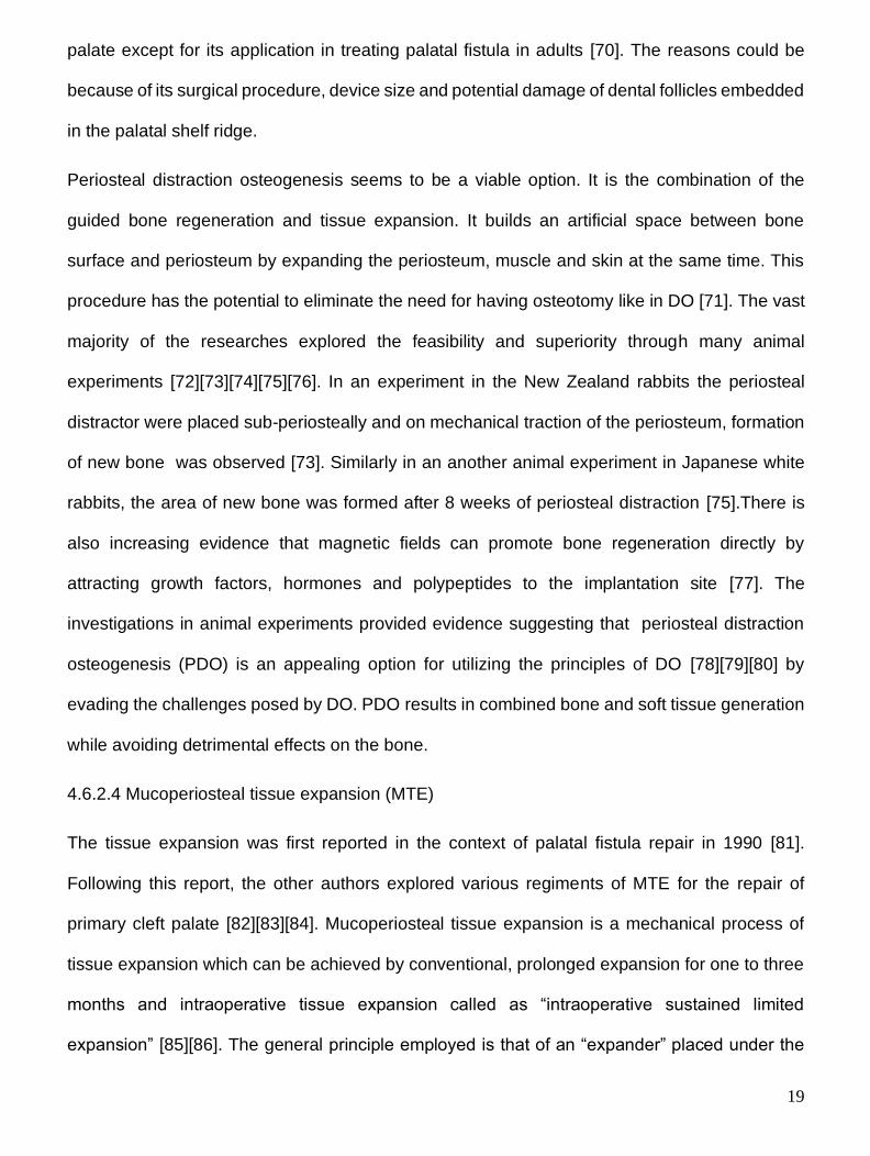
Figure 1. Establishment of a three-dimensional coordinate system. (A) and (B) A horizontal plane (green) is defined
by (Q, T, T'), and a vertical plane (red) is defined by (T, T'). (C) and (D) The line between VT–VA denotes the vomer
edge (v), the line between GT–GA denotes the palatal shelf ridge of the greater segment (g), and the line between LT–
LA represents the palatal shelf ridge of the lesser segment (l).
The palatal cleft was delimited by the greater segment’s palatal shelf ridge (g) at the junction to the vomer and
the lesser segment’s shelf ridge (l). The true cleft was delimited by the vomer edge (v) and the lesser segment’s
palatal shelf ridge (l). The height measurements were performed along the three paths g, l, and v at nine equidistant
points each, generated from the ascending order of 0 to 100% (0%, 12.5%, 25%, 37.5%, 50%, 62.5%, 75%, 87.5%,
100%). The height in each point was measured perpendicular to the horizontal plane (Figure 2C, D).
28
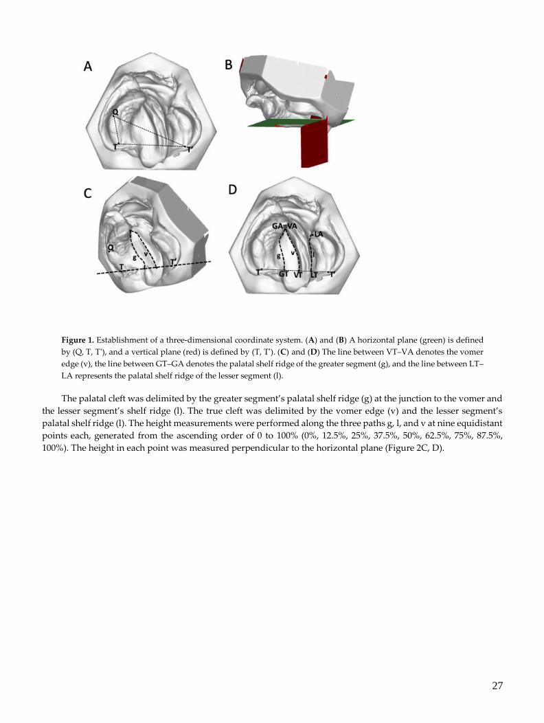
Table 2. Definitions of 3D landmark measurements.
Abbreviation Description
Cleft area dimensions
GA/GM/GT–LA/LM/LT Total palatal cleft area (PCA)
VA/VM/VT–LA/LM/LT Total true cleft area (TCA)
Transverse dimensions
GA–LA Anterior palatal cleft width
GM–LM Middle palatal cleft width
GT–LT Posterior palatal cleft width
VA–LA Anterior true cleft width
VM–LM Middle true cleft width
VT–LT Posterior true cleft width
Vertical dimensions1
g-height Height of the palatal shelf ridge of the greater segment
perpendicular to the horizontal plane
l-height Height of the palatal shelf ridge of the lesser segment
perpendicular to the horizontal plane
v-height Height of the vomer edge perpendicular to the horizontal
plane 1 Vertical dimension measured at nine equidistant points along the paths g, l, and v.
Figure 2. (A) Palatal cleft: palatal cleft width (dashed lines) and palatal cleft area (shaded area). (B) True cleft: true
cleft width (dashed lines) and true cleft area (shaded area). (C) and (D) The height of the palate to the horizontal plane
at the vomer edge (v) and at the greater (g) and lesser (l) palatal shelf ridges.
Connecting corresponding equidistant points from g to l and v to l (0% and 0%, 12.5% and 12.5%, and so forth)
led to 8 equidistant quadrangles, which were split into two triangles each, leading to a total of 16 triangles (Figure
3). Surface measurements of defined areas were then approximated as the sum of its comprising triangles.

29
Figure 3. True cleft area measurement connecting equidistant points from the vomer edge to the lesser palatal shelf
ridge (0% and 0%, 12.5% and 12.5%, and so forth) led to 8 equidistant quadrangles, which were split into two triangles
each, leading to a total of 16 triangles. Surface measurements of defined areas were then approximated as the sum of
its comprising triangles.
2.4. Statistical Analysis
The measurements made at time points T0 and T2 were compared using a Wilcoxon signed-ranks test.
Statistical significance was assumed at p < 0.05. The abovementioned procedures for calculating the cleft width, cleft
area, and height of the cleft edges were repeated for 15 of the 45 casts both by the same rater and by a second rater.
The differences were investigated to quantify the measurement error of the method according to Dahlberg’s
formula [17]. The statistical analysis was performed using Stata (version 15.1, StataCorp LLC, Texas, USA).
3. Results
The analysis of landmark positioning in the 3D cast analysis showed that the intrarater measurement errors
ranged from 0.7 to 0.9 mm and those for interrater measurements ranged from 0.5 to 1.0 mm.
3.1. Cleft Width
The median palatal cleft width at T0 was 11.4 mm (interquartile range, IQR = 9.8–14.4 mm) in the anterior
region (GA–LA), 14.8 mm (IQR = 14.0–15.9 mm) in the midpalatal region (GM–LM), and 13.7 mm (IQR = 12.3–16.7
mm) in the posterior region (GT–LT). The median true cleft width was 13.3 mm (IQR = 10.6–14.4 mm) in the anterior
region (VA–LA), 9.9 mm (IQR = 8.1–11.0 mm) in the midpalatal region (VM–LM), and 7.4 mm (IQR = 5.8–10.4 mm)
in the posterior region (VT–LT). The narrowing of the palatal and true cleft from T0 to T2 resulted in its width
becoming more even from anterior to posterior locations along the cleft (Figure 4). The median palatal cleft width
decreased significantly at T2, from 11.4 to 6.5 mm (z = 3.237, p = 0.0012) in the anterior region (GA–LA), from 14.8
to 9.3 mm (z = 3.41, p = 0.0007) in the midpalatal region (GM–LM), and from 13.7 to 10.5 mm (z = 3.18, p = 0.0015) in
the posterior region (GT–LT). Similar changes were seen in the true cleft width in the anterior region (VA–LA) (from
13.3 to 6.8 mm, z = 3.41, p = 0.0007), in the midpalatal region (VM–LM) (from 9.9 to 5.0 mm, z = 3.41, p = 0.0007), and
in the posterior region (VT–LT) (from 7.4 to 4.9 mm, z = 3.24, p = 0.0012) (Figure 4). The median decrease (from T0
to T2) in cleft width was 38.0% for the palatal cleft, whereas it was 44.5% for the true cleft.
30
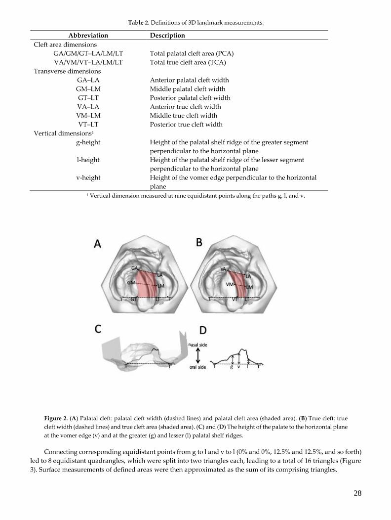
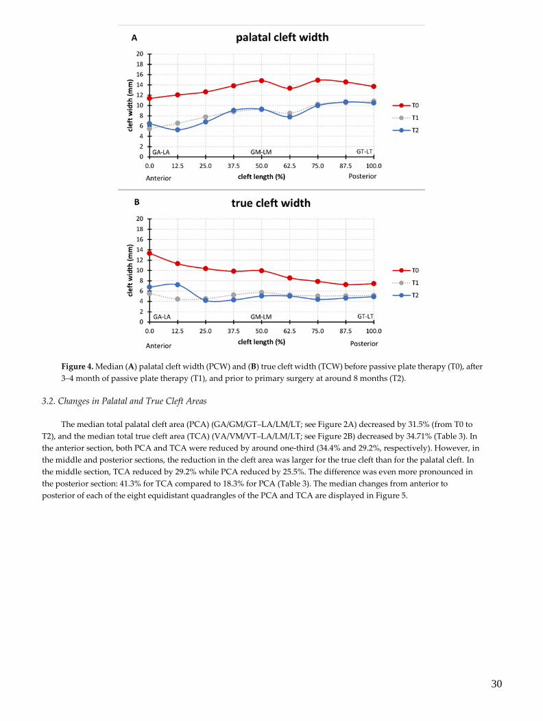
Figure 4. Median (A) palatal cleft width (PCW) and (B) true cleft width (TCW) before passive plate therapy (T0), after
3–4 month of passive plate therapy (T1), and prior to primary surgery at around 8 months (T2).
3.2. Changes in Palatal and True Cleft Areas
The median total palatal cleft area (PCA) (GA/GM/GT–LA/LM/LT; see Figure 2A) decreased by 31.5% (from T0 to
T2), and the median total true cleft area (TCA) (VA/VM/VT–LA/LM/LT; see Figure 2B) decreased by 34.71% (Table 3). In
the anterior section, both PCA and TCA were reduced by around one-third (34.4% and 29.2%, respectively). However, in
the middle and posterior sections, the reduction in the cleft area was larger for the true cleft than for the palatal cleft. In
the middle section, TCA reduced by 29.2% while PCA reduced by 25.5%. The difference was even more pronounced in
the posterior section: 41.3% for TCA compared to 18.3% for PCA (Table 3). The median changes from anterior to
posterior of each of the eight equidistant quadrangles of the PCA and TCA are displayed in Figure 5.

31
Table 3. Measured changes in palatal cleft area (PCA) and true cleft area (TCA).
Cleft Area
(mm2) Section
T0
Median (IQR)
T2
Median (IQR) p-Value
PCA Total 0%–100% 334 (294.9–349.8) 228.8 (205–287.9) 0.0015
Anterior 0%–25% 75.3 (67.2–93.3) 49.4 (32.0–70.0) 0.0090
Middle 25%–75% 157.0 (141.5–173.8) 116.9 (99.7–135.0) 0.0076
Posterior 75%–100% 91.8 (77.5–102.6) 75.0 (61.5–84.2) 0.0090
TCA Total 0%–100% 185.4 (151.5–220.1) 121.1 (100.2–144.6) 0.0015
Anterior 0%–25% 56.9 (40.9–66.6) 41.7 (18.1–51.3) 0.0409
Middle 25%–75% 84.7 (69.6–102.8) 60.0 (42.3–62.2) 0.0007
Posterior 75%–100% 41.2 (31.5–48.5) 24.2 (20.3–32.35) 0.0012
PCA, palatal cleft area; TCA, true cleft area; IQR, interquartile range.
Figure 5. Median (A) palatal cleft area (PCA) and (B) true cleft area (TCA) at T0, T1, and T2 in eight equidistant
quadrangles (0%–12.5%, 12.5%–25%, 25%–37.5%, and so forth) from anterior to posterior.
3.3. Changes in the Height of the Palatal Surface
The height of the palatal surface was measured to the horizontal plane (Q–T/T') along the longitudinal course
of the cleft along three different paths: the greater palatal ridge (g), the lesser palatal ridge (l), and the vomer edge
(v) (Figure 2C, D). At birth, the greater and lesser palatal shelf ridges followed a horizontal course at a height of
around 8 mm, becoming skewed towards the posterior end. Both shelf ridges run at the same level, while the vomer
edge paralleled their course at about 2 mm higher (Figure 6). The heights of the greater and lesser palatal shelf
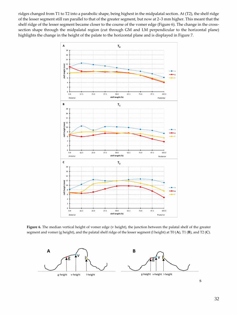
32
ridges changed from T1 to T2 into a parabolic shape, being highest in the midpalatal section. At (T2), the shelf ridge
of the lesser segment still ran parallel to that of the greater segment, but now at 2–3 mm higher. This meant that the
shelf ridge of the lesser segment became closer to the course of the vomer edge (Figure 6). The change in the cross-
section shape through the midpalatal region (cut through GM and LM perpendicular to the horizontal plane)
highlights the change in the height of the palate to the horizontal plane and is displayed in Figure 7.
Figure 6. The median vertical height of vomer edge (v height), the junction between the palatal shelf of the greater
segment and vomer (g height), and the palatal shelf ridge of the lesser segment (l height) at T0 (A), T1 (B), and T2 (C).
s

33
Figure 7. Cross-section through GM and LM perpendicular to the horizontal plane, displaying the height of g, v, and
l above the horizontal plane at T0 (A) and T2 (B).
4. Discussion
4.1. Three-Dimensional Analysis
The dimensions and shape of the cleft alveolus and palate play an important role in the outcome of any primary
surgery [18]. Different methods are used to measure the cleft dimensions, with some investigators only measuring
the separation between two segments anteriorly, whereas others measure the cleft width at several palatal levels or
measure the cleft area in relation to the total palatal area [2–7,10–13,18–20]. Aberrant anatomical structures on dental
casts pose a major challenge to the clinician attempting to identify and determine the correct anatomical landmarks
[20–22]. Vague landmarks are the most important factor contributing to inaccuracy of these measurement, and there
is no standard protocol for identifying the points since demarcating these landmarks is extremely difficult
[8,12,20,21].
4.2. Cleft Width
The palatal cleft width and true cleft width decreased in all cases, which is consistent with previous reports
[3,9,18] (Figure 4). The true and palatal cleft width decreased evenly along its longitudinal course, but slightly less
in the posterior region, whereas the main effect was already observed at T1 (Figure 4).
A presurgical reduction in the width of the cleft is considered a positive predictor of the surgical results,
because this reduces the undermining mobilization of the tissues [5,23]. We reason this is achieved simply by
blocking out the depressions on the plaster cast prior to plate fabrication to keep the palatal shelves and vomer
surfaces free and not applying any pressure from the tongue. No gradual trimming of the plate was performed,
which contrasts with other techniques such as nasoalveolar moulding (NAM) [5] and the Hotz-type plate [24].
4.3. Changes in the Palatal and True Cleft Areas
Measuring the cleft area indirectly quantifies the shortage of palatal tissue when performing surgical repairs,
and this shortage seems to be related to the amount of subsequent maxillary growth disturbances later in life [5].
Assuming that no tissue is freely transplanted into the cleft area in a primary repair, greater tissue shortage during
cleft repair leads inevitably to (a) an increased area of secondary wound healing over soft tissue or bone or (b) an
increased volume of dead space between tissues. Both effects are typically present but to variable extents depending
on the precise surgical technique used, whether it is tissue turnover, tissue shift in the horizontal or vertical
direction, and single- or double-layer tissue closure. However, these two effects are considered to equally increase
the invasiveness of surgical repair due to more scarring and a greater risk of wound healing disturbances with, for
example fistula formation.
It has been proposed that a given ratio between the cleft area and the entire palatal vault surface within the
alveolar ridges can help to define the time point when surgical repair leads to minimal side effects on growth [13].
When considering the effect of surgery on future growth, it is therefore important not only to discriminate between
the surgical time point and technique, but also whether the surgical repair addresses the closure of the palatal or
(in contrast) the true cleft region only—which constitutes only about half the surface area (Table 3)—and thus
markedly reduces the degree of tissue shift.
However, in addition to the total amount of tissue shift differing between PCA and TCA, there are also
differences in geometry. This becomes clear when the cleft area is divided into eight sections from anterior to
posterior (Figure 5). The palatal cleft area was minimal in the central section while being largest in the posterior
section (Figure 5A). Clinical translation of this finding means maximized need for tissue shift in the posterior section
of the palate. However, in the posterior section the palatal artery might impede free tissue movement, thus resulting
in increased tissue tension, which is also a negative factor for wound healing and thus a risk factor for a residual
fistula. Indeed, a meta-analysis [25] identified a fistula as being most frequently located in the posterior hard palate
section, at the junction with the soft palate.
In the true cleft region, the area of the posterior section was about the same as that in the central section (Figure
5B). If the surgical cleft closure was limited to the true cleft region, it would have to be investigated whether this

34
could lead to a more homogeneous tissue displacement and tissue tension due to the more even distribution of the
area in all sections.
4.4. Changes in the Height of the Palatal Surface
3D changes of the palatal curved surface at different stages have been studied by taking the bilateral tuberosity
points, incisal point or canine points, which lacks objectivity and reproducibility [26]. The measurement technique
must instead be based on a larger number of data points along a curve denoting the actual height of the palatal
segments. Our method offers objectivity, reproducibility, and reliability at different stages (Figure 2C,D). However,
the changes measured in the present study cannot be compared to those in previous studies due to differences in
the parameters measured and the protocols used when treating cleft lip and palate [27]. With respect to a potential
palatal repair after 8 months of pure passive plate therapy, two important discrepancies between TCA and PCA
were identified. First, the tissue borders in the true cleft region (the vomer edge and the lesser segment) are
vertically closer together than the tissue borders in the palatal cleft region (the greater segment and the lesser
segment), thus from a surgical point of view requiring less vertical tissue displacement to come into the same
vertical plane for cleft repair, which also minimizes the contiguous amount of dead space (Figure 6C). Second, the
tissue borders are higher in the true cleft region than in the palatal cleft region (Figure 7), with this difference being
more pronounced in the anterior region (Figure 6C). Thus, a repair following the tissue levels in the true cleft region
might allow more space for the tongue in the anterior palatal region to have an undisturbed posture and
articulation.
4.5. Clinical Translation of the Findings
The long history of presurgical orthopaedic treatment [28] has led to various technical variations depending
on the effect aimed for. Some techniques [29] aim to guide the alveolar positions into an optimal position, not mainly
for surgery but rather with the intent to optimize the long-term arch form. The plate is periodically trimmed every
couple of weeks to guide the alveolar segments, and the orthopaedic therapy continues after lip surgery and soft
palate surgery until when hard palate surgery is performed. However, a large randomized controlled trial failed to
demonstrate—aside from the narrowing effect before lip surgery [30]—any persistent effect on arch form [31] or
occlusion [32] from this specific type of presurgical orthopaedics (Hotz-type plate). Those authors concluded that
lip surgery and subsequent palatal surgery overrode the forming effect of presurgical orthopaedics. We therefore
refrained from using grinding to actively guide the alveolar segments, instead using a purely passive type of plate
therapy similar to the passive appliance of Huddart [28] but without extraoral wires.
In line with the findings of the aforementioned randomized trial, two main ways of clinical reasoning were
observed: either presurgical orthopaedics are abandoned and one relies on the palatal shape changes that take place
after lip surgery, or the presurgical orthopaedics focus mainly on the narrowing of the anterior cleft region to
facilitate the primary repair in the lip, alveolus, and nose region (in NAM), with the palatal shape changes not being
taken into account in the primary surgery.
However, the present finding of narrowing of the true cleft region would also allow a third clinical reasoning—
using the effect of presurgical orthopaedics in exchange for performing a separate lip repair before palatal closure.
As with most one-stage cleft repair techniques, the effect of preoperative orthopaedics is used in exchange for
performing a separate lip repair prior to palatal closure. If the results can be confirmed in further studies, the
common belief that early isolated lip surgery is necessary to provide optimal conditions for later palatal repair
could be questioned.
4.6. Strengths and Limitations
We used the vomer edge (also called “Poutriquet’s ridge”) [1, 16] and innominate sulcus [16] as new
anatomically reproducible landmarks that can be easily used to measure the true cleft width, area, and curvature
in all dimensions (Figure 2). To the best of our knowledge, no previous 3D study has differentiated between TCA
and PCA. Our measurement errors were found to be well within the ranges found in previous similar studies [8].
The landmarks used in the present study have biological correlates or distinct morphologies that facilitate their
identification with high precision and reproducibility [22], which probably explains why the errors in the interrater
transverse measurements were less than 0.5 mm, which is lower than that in all previous studies [8,21]. Moreover,

35
no previous study has measured the height of the vomer edge to the horizontal plane. The errors of our interrater
measurements were within the acceptable limit of 1.0 mm, making it a practical cleft landmark with a defined cleft
anatomical correlate—it is the zenith of the palatal vault prior to surgery (Figure 2D).
The limitation of our study were the small sample size and the lack of a control group without passive plate
therapy. We cannot draw any conclusion about whether the passive plate is also purely passive towards the
intrinsic growth of each palatal half. Further, no data on a potential long-term effect of the plate therapy on the
growth or shape of segments can be provided. This will be further complicated by the additional bias of the wide
range of surgical techniques. Future studies involving larger numbers of patients and longer observation times are
necessary, as well as 3D evaluations of a control group that does not receive passive plate therapy, in order to fully
appreciate the effect of on the growth, remodelling and relocation of the palatal segments and vomer.
5. Conclusions
In conclusion, few studies have investigated the morphological changes of the cleft palate width by using 3D
standardized, reproducible landmarks in unilateral cleft lip and palate patients. We have introduced the vomer
edge for establishing a validated measuring method for the width, area, and height of the true cleft. To the best of
our knowledge, this is the first 3D study to show the 3D morphological changes of a pure passive plate therapy
over a long period of 8 months in the absence of previous lip surgery. In the investigated cohort without prior lip
repair, passive plate therapy provided favourable anatomical conditions for subsequent surgical palatal repair.
Author Contributions: Conceptualization, A.A.M.; data curation, P.N.; formal analysis, B.K.B. and M.D.; funding acquisition,
P.N., C.V., and A.A.M.; investigation, P.N. and B.K.B.; methodology, P.N., B.K.B., M.D., and A.A.M.; project administration,
A.A.M.; resources, C.V. and A.A.M.; software, P.N. and M.D.; supervision, M.D., C.V., and A.A.M.; validation, P.N. and B.K.B.;
visualization, P.N., M.D., and C.V.; writing—original draft, P.N. and B.K.B.; writing—review and editing, P.N., B.K.B., M.D.,
C.V., and A.A.M. All authors have read and agreed to the published version of the manuscript.
Funding: This research was partially funded from a research grant of the Freiwillige Akademische Gesellschaft (FAG), Basel,
Switzerland.
Conflicts of Interest: The authors declare no conflict of interest. The funders had no role in the design of the study; the collection,
analysis, or interpretation of data; in the writing of the manuscript; or the decision to publish the results.
References
1. Veau, V. Division Palatine. Anatomie. Chirurgie. Phonétique. Avec la Collaboration de Mlle S. Borel.; Impr. Darantière Masson et
Cie éditeurs: Dijon/Paris, France, 1931.
2. Stöckli, P.W. Application of a quantitative method for arch form evaluation in complete unilateral cleft lip and palate. Cleft
Palate J. 1971, 8, 322–341.
3. Grabowski, R.; Kopp, H.; Stahl, F.; Gundlach, K.K.H. Presurgical orthopaedic treatment of newborns with clefts - functional
treatment with long-term effects. J. Cranio-Maxillofacial Surg. 2006, 34, 34–44.
4. Wada, T.; Miyazaki, T. Treatment principles for the changing arch form in children with complete unilateral cleft lips and
palates. Cleft Palate J 1976, 13, 273–283.
5. Peltomäki, T.; Vendittelli, B.L.; Grayson, B.H.; Cutting, C.B.; Brecht, L.E. Associations between Severity of Clefting and
Maxillary Growth in Patients with Unilateral Cleft Lip and Palate Treated with Infant Orthopedics. Cleft Palate-Craniofacial
J. 2001, 38, 582–586.
6. Jorge, P.K.; Gnoinski, W.; Vaz Laskos, K.; Felício Carvalho Carrara, C.; Gamba Garib, D.; Okada Ozawa, T.; Andrade
Moreira Machado, M.A.; Pinelli Valarelli, F.; Oliveira, T.M. Comparison of two treatment protocols in children with
unilateral complete cleft lip and palate: Tridimensional evaluation of the maxillary dental arch. J. Craniomaxillofac. Surg.
2016, 44, 1117–1122.
7. Yamanishi, T.; Nishio, J.; Kohara, H.; Hirano, Y.; Sako, M.; Yamanishi, Y.; Adachi, T.; Miya, S.; Mukai, T. Effect on Maxillary
Arch Development of Early 2-Stage Palatoplasty by Modified Furlow Technique and Conventional 1-Stage Palatoplasty in
Children With Complete Unilateral Cleft Lip and Palate. J. Oral Maxillofac. Surg. 2009, 67, 2210–2216.
8. Seckel, N.G.; Van der Tweel, I.; Elema, G.A.; Specken, T.F.J.M.C. Landmark positioning on maxilla of cleft lip and palate
infant - A reality? Cleft Palate-Craniofacial J. 1995, 32, 434–441.
9. Shetty, V.; Agrawal, R.K.; Sailer, H.F. Long-term effect of presurgical nasoalveolar molding on growth of maxillary arch in
unilateral cleft lip and palate: Randomized controlled trial. Int. J. Oral Maxillofac. Surg. 2017, 46, 977–987.
10. Mazaheri, M.; Harding, R.L.; Cooper, J.A.; Meier, J.A.; Jones, T.S. Changes in arch form and dimensions of cleft patients.
36
Am. J. Orthod. 1971, 60, 19–32.
11. Schmidt-Flath, I.; Fränkel, R.; Grabowski, R.; Opitz, C.; Wiemann, C. Methoden zur Ausmessung des Säuglingskiefers, des
Milch- und bleibenden Gebisses beim Spaltträger. Fortschr. Kieferorthop. 1972, 33, 457–476.
12. Berkowitz, S.; Pruzansky, S. Stereophotogrammerty of serial casts of cleft palate. Angle Orthod. 1968, 38, 136–149.
13. Berkowitz, S.; Duncan, R.; Evans, C.; Friede, H.; Kuijpers-Jagtman, A.M.; Prahl-Anderson, B.; Rosenstein, S. Timing of cleft
palate closure should be based on the ratio of the area of the cleft to that of the palatal segments and not on age alone. Plast.
Reconstr. Surg. 2005, 115, 1483–1499.
14. Leighton, B.C. Morphologische Variationender Alveolarbögen beim Neugeborenen. Fortschr. Kieferorthop. 1976, 37, 8–14.
15. Botticelli, S.; Pedersen, T.K.; Küseler, A.; Nørholt, S.E.; Cattaneo, P.M. Novel 3-D Analysis for the Assessment of Cleft
Dimensions on Digital Models of Infants With Unilateral Cleft Lip and Palate. Cleft Palate. Craniofac. J. 2019, 56, 127–133.
16. Malek, R. Cleft Lip and Palate: Lesions, Pathophysiology and Primary Treatment, 1st ed.; Martin, D., Ed.; CRC Press: London,
UK, 27 November 2000; ISBN 978-1853174919.
17. Dahlberg, G. Statistical Methods for Medical and Biological Students. Br. Med. J. 1940, 2, 358.
18. Robertson, N.R.; Fish, J. Early dimensional changes in the arches of cleft palate children. Am. J. Orthod. 1975, 67, 290–303.
19. Long, R.E.; Daskalogiannakis, J.; Mercado, A.M.; Hathaway, R.R.; Fessler, J.; Russell, K.A. The americleft project: Plaster
dental casts versus digital images for GOSLON yardstick ratings when used in intercenter comparisons. J. Craniofac. Surg.
2017, 28, 1269–1273.
20. Ye, B.; Ruan, C.; Hu, J.; Yang, Y.; Ghosh, A.; Jana, S.; Zhang, G. A Comparative Study on Dental-arch Morphology in Adult
Unoperated and Operated Cleft Palate Patients. J. Craniofac. Surg. 2010, 21, 811–815.
21. Brief, J.; Behle, J.H.; Stellzig-Eisenhauer, A.; Hassfeld, S. Precision of landmark positioning on digitized models from
patients with cleft lip and palate. Cleft Palate. Craniofac. J. 2006, 43, 168–173.
22. Oxnard, C.E. The measurement of form: Beyond biometrics. Sausages and stars, dumbbells and doughnuts: Peculiar views
of anatomical structures. Cleft Palate J. 1986, 23, 110–128.
23. Santiago, P.E.; Grayson, B.H.; Cutting, C.B.; Gianoutsos, M.P.; Brecht, L.E.; Kwon, S.M. Reduced Need for Alveolar Bone
Grafting by Presurgical Orthopedics and Primary Gingivoperiosteoplasty. Cleft Palate-Craniofacial J. 1998, 35, 77–80.
24. Hotz, M.M.; Gnoinski, W.M.; Nussbaumer, H.; Kistler, E. Early maxillary orthopedics in CLP cases: Guidelines for surgery.
Cleft Palate J. 1978, 15, 405–411.
25. Bykowski, M.R.; Naran, S.; Winger, D.G.; Losee, J.E. The rate of oronasal fistula following primary cleft palate surgery: A
meta-analysis. Cleft Palate-Craniofacial J. 2015, 52, e81–e87.
26. Isogawa, N.; Ochiai, S.; Mito, T.; Kindaichi, J.; Ishibashi, N.; Takagi, Y.; Ishikawa, M. Three-Dimensional Comparison in
Palatal Forms Between Modified Presurgical Nasoalveolar Molding Plate and Hotz’s Plate Applied to the Infants with
Unilateral Cleft Lip and Palate. Singapore Dent. J. 2010, 31, 36–42.
27. Mishima, K.; Sugahara, T.; Mori, Y.; Sakuda, M. Three-Dimensional Comparison between the Palatal Forms in Infants with
Complete Unilateral Cleft Lip, Alveolus, and Palate (UCLP) with and without Hotz’s Plate. Cleft Palate-Craniofacial J. 1996,
33, 245–251.
28. Huddart, AG. Presurgical changes in unilateral cleft palate subjects. Cleft Palate J. 1979, 16, 147–157.
29. Gnoinski, W. Infant Orthopedics and Later Orthodontic Monitoring for Unilateral Cleft Lip and Palate Patients in Zurich; Bardach,
J., Morris, H., Eds.; WB Saunders Co: Philadelphia, PA, USA, 1990.
30. Prahl, C.; Kuijpers-Jagtman, A.M.; van’t Hof, M.A.; Prahl-Andersen, B. A randomised prospective clinical trial into the
effect of infant orthopaedics on maxillary arch dimensions in unilateral cleft lip and palate (Dutchcleft). Eur. J. Oral Sci.
2001, 109, 297–305.
31. Bongaarts, C.A.M.; van ’t Hof, M.A.; Prahl-Andersen, B.; Dirks, I.V.; Kuijpers-Jagtman, A.M. Infant orthopedics has no
effect on maxillary arch dimensions in the deciduous dentition of children with complete unilateral cleft lip and palate
(Dutchcleft). Cleft Palate. Craniofac. J. 2006, 43, 665–672.
32. Noverraz, R.L.M.; Disse, M.A.; Ongkosuwito, E.M.; Kuijpers-Jagtman, A.M.; Prahl, C. Transverse dental arch relationship
at 9 and 12 years in children with unilateral cleft lip and palate treated with infant orthopedics: A randomized clinical trial
(DUTCHCLEFT). Clin. Oral Investig. 2015, 19, 2255–2265.
© 2020 by the authors. Licensee MDPI, Basel, Switzerland. This article is an open access
article distributed under the terms and conditions of the Creative Commons Attribution
(CC BY) license (http://creativecommons.org/licenses/by/4.0/).

37
7. Second study:
The biomechanical evaluation of magnetic forces to drive osteogenesis in newborn’s
with cleft lip and palate
Study design: Dr. Prasad Nalabothu, Prof. Michel Dalstra
Financing: FAG (Voluntary Academic Association): CHF 13,406
Project leaders: Professor Dr. med. dent. Carlalberta Verna
Publication: First authorship, Journal of Materials Science: Materials in Medicine, Impact factor
2.46 (2020), Ranking 7876 (Q2) in “Biomedical Engineering” and “Bioengineering”.

38
Journal of Materials Science: Materials in Medicine (2020) 31:79
https://doi.org/10.1007/s10856-020-06421-6
Original Research
The biomechanical evaluation of magnetic forces to drive osteogenesis in newborn’s with cleft lip and palate
Prasad Nalabothu 1,2 ● Carlalberta Verna1 ● Markus Steineck1 ● Andreas Albert Mueller2 ● Michel Dalstra1
Received: 26 February 2020 / Accepted: 20 July 2020 / Published online: 20 August 2020
© The Author(s) 2020
Abstract
This study examined the potential for dental magnets to act as a driving force for osteogenesis in the palate of newborns with
a unilateral cleft lip and palate. In the first part of the study dental magnets were arranged in a set up mimicking a distraction
device and the curves of the magnetic attraction force versus gap distance curves generated, with and without the presence of
palatal rugae tissue in between both sides of the distraction device. The attraction forces ranged from 1 to 12 N depending on
the gap distance and the presence of soft tissue in the gap. In the second part of the study these forces were used as input for a
3D finite element model of the palate of a newborn affected by unilateral cleft lip and palate. In the analysis of load transfer,
it was found that the strains generated by a magnetically induced distraction exceed 1,500 µstrain suggesting that bone
locally is submitted to mild overload leading to bone apposition.
Graphical Abstract
1 Introduction
These authors contributed equally: Andreas Albert Mueller, Michel
Dalstra
* Prasad Nalabothu
1 Department of Paediatric Oral Health and Orthodontics, University
Center for Dental Medicine UZB, Basel, Switzerland
2 Department of Oral and Craniomaxillofacial Surgery, University
Hospital Basel, Basel, Switzerland
Cleft lip and palate (CL/P) is the most common presenting
congenital condition of the face and cranial bones [1]. It is
the most frequent craniofacial malformation affecting 1:500
to 1:700 live births worldwide with tremendous variations
across geographic areas, ethnic groups and socioeconomic
status [2]. Cleft palate closure by surgery depends upon the
adequacy of available palatal mucosal tissue. Insufficiency
of such soft tissue is a cause for concern in the closure of
severe wide clefts of the hard palate [3]. The tissue shortage
makes it necessary to extensively mobilize the tissue
adjacent to the cleft, leading to a wider tissue wound with
consequent scarification due to the wound healing process.
During wound repair, various
ENGINEERING AND NANO-ENGINEERING APPROACHES FOR MEDICAL DEVICES
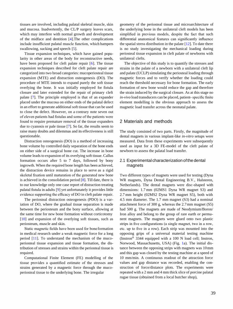
39
tissues are involved, including palatal skeletal muscle, skin
and mucosa. Inadvertently, the CL/P surgery leaves scars,
which may interfere with normal growth and development
of the midface and dentition [4].The other complications
include insufficient palatal muscle function, which hampers
swallowing, sucking and speech [5].
Tissue expansion techniques, which have gained popu-
larity in other areas of the body for reconstructive needs,
have been proposed for cleft palate repair [6]. The tissue
expansion techniques described for cleft palate repair are
categorized into two broad categories: mucoperiosteal tissue
expansion (MTE) and distraction osteogenesis (DO). The
procedure of MTE intends to expand purely the soft tissue
overlying the bone. It was initially employed for fistula
closure and later extended for the repair of primary cleft
palate [7]. The principle employed is that of an expander
placed under the mucosa on either ends of the palatal defect
in an effort to generate additional soft tissue that can be used
to close the defect. However, on a contrary note seven out
of eleven patients had fistulas and some of the patients were
found to require premature removal of the tissue expanders
due to cyanosis or pale tissue [7]. So far, the results seem to
raise many doubts and dilemmas and its effectiveness is still
questionable.
Distraction osteogenesis (DO) is a method of increasing
bone volume by controlled daily separation of the bone ends
on either side of a surgical bone cut. The increase in bone
volume leads to expansion of its overlying soft tissue. Callus
formation occurs after 5 to 7 days, followed by bony
ingrowth. When the required bone length has been achieved,
the distraction device remains in place to serve as a rigid
skeletal fixation until maturation of the generated new bone
is achieved in the consolidation period [8]. Till date, there is
to our knowledge only one case report of distraction treating
palatal fistula in adults [9] yet unfortunately it provides little
evidence supporting the efficacy of DO in cleft palate repair.
The periosteal distraction osteogenesis (PDO) is a var-
iation of DO, where the gradual tissue separation is made
between the periosteum and the bony surface, allowing at
the same time for new bone formation without corticotomy
[10] and expansion of the overlying soft tissues, such as
periosteum, muscle and skin.
Static magnetic fields have been used for bone formation
in medical research under a weak magnetic force for a long
period [11]. To understand the mechanism of the muco-
periosteal tissue expansion and tissue formation, the dis-
tribution of stresses and strains within the periosteal tissue is
required.
Computational Finite Element (FE) modelling of the
tissue provides a quantified estimate of the stresses and
strains generated by a magnetic force through the muco-
periosteal tissue to the underlying bone. The irregular
geometry of the periosteal tissue and microarchitecture of
the underlying bone in the unilateral cleft models has been
simplified in previous models, despite the fact that such
differential anatomical features can significantly influence
the spatial stress distribution in the palate [12]. To date there
is no study investigating the mechanical loading during
periosteal tissue expansion in cleft palate of newborns with
unilateral clefts.
The objective of this study is to quantify the stresses and
strains in the palate of a newborn with a unilateral cleft lip
and palate (UCLP) simulating the periosteal loading through
magnetic forces and to verify whether the loading could
reach the threshold necessary for bone formation. The early
formation of new bone would reduce the gap and therefore
the strain induced by the surgical closure. As at this stage no
in-vivo load transducers can be placed, patient- specific finite
element modelling is the obvious approach to assess the
magnetic load transfer across the neonatal palate.
2 Materials and methods The study consisted of two parts. Firstly, the magnitude of
dental magnets in various implant-like in-vitro setups were
measured. Data from these experiments were subsequently
used as input for a 3D FE-model of the cleft palate of
newborn to assess the palatal load transfer.
2.1 Experimental characterization of the dental magnets
Two different types of magnets were used for testing (Dyna
WR magnets, Dyna Dental Engineering B.V., Halsteren,
Netherlands). The dental magnets were disc-shaped with
dimensions: 1.7 mm (02MS1 Dyna WR magnet S3) and
2.7 mm height (02MS2 Dyna WR magnet S5), both with
4.5 mm diameter. The 1.7 mm magnet (S3) had a nominal
attachment force of 300 g, whereas the 2.7 mm magnet (S5)
had 500 g. The magnets are made of Neodymium/Boron/
Iron alloy and belong to the group of rare earth or perma-
nent magnets. The magnets were glued onto two plastic
strips in five configurations (a single magnet, two in a row,
etc. up to five in a row). Each strip was mounted into the
opposing grips of a universal material testing machine
(Instron® 3344 equipped with a 100 N load cell; Instron,
Norwood, Massachusetts, USA) (Fig. 1a). The initial dis-
tance between the opposing strips with magnets was 10 mm
and this gap was closed by the testing machine at a speed of
10 mm/min. A continuous readout of the attraction force
values and gap distance was recorded, enabling the con-
struction of force/distance plots. The experiments were
repeated with a 2 mm and 4 mm thick slice of porcine palatal
rugae tissue (obtained from a local butcher shop),
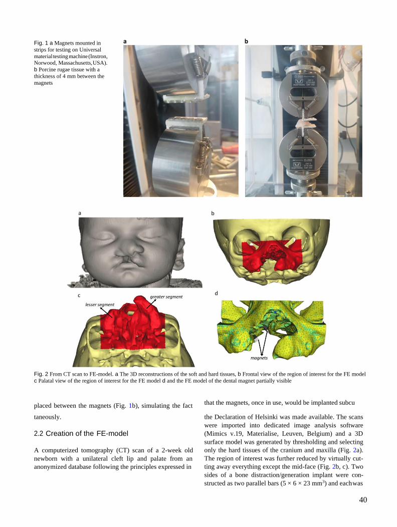
40
Fig. 1 a Magnets mounted in
strips for testing on Universal
material testing machine (Instron,
Norwood, Massachusetts, USA).
b Porcine rugae tissue with a
thickness of 4 mm between the
magnets
Fig. 2 From CT scan to FE-model. a The 3D reconstructions of the soft and hard tissues, b Frontal view of the region of interest for the FE model
c Palatal view of the region of interest for the FE model d and the FE model of the dental magnet partially visible
placed between the magnets (Fig. 1b), simulating the fact that the magnets, once in use, would be implanted subcu
taneously.
2.2 Creation of the FE-model
A computerized tomography (CT) scan of a 2-week old
newborn with a unilateral cleft lip and palate from an
anonymized database following the principles expressed in
the Declaration of Helsinki was made available. The scans
were imported into dedicated image analysis software
(Mimics v.19, Materialise, Leuven, Belgium) and a 3D
surface model was generated by thresholding and selecting
only the hard tissues of the cranium and maxilla (Fig. 2a).
The region of interest was further reduced by virtually cut-
ting away everything except the mid-face (Fig. 2b, c). Two
sides of a bone distraction/generation implant were con-
structed as two parallel bars (5 × 6 × 23 mm3) and each was

41
Table 1 Mechanical properties of the materials used in the present study. For the different bone tissues (1 until 10) the percentage signifies the
cumulative amount of bone elements belonging to this specific tissue
Material Apparent Density (gr/cm3) Young’s Modulus (MPa) Poisson’s Ratio (-) Yield Strength
Compressive (MPa) Tensile (MPa)
Bone 1 (0.1%) 0.04 5 0.4 −0.06 0.04
Bone 2 (0.5%) 0.10 51 0.4 −0.61 0.40
Bone 3 (30.9%) 0.18 157 0.4 −1.89 1.26
Bone 4 (89.6%) 0.26 351 0.4 −4.22 2.81
Bone 5 (97.8%) 0.36 677 0.4 −8.12 5.41
Bone 6 (98.8%) 0.50 1212 0.4 −14.54 9.70
Bone 7 (99.4%) 0.66 2115 0.4 −25.37 16.92
Bone 8 (99.8%) 0.83 3752 0.4 −45.02 30.02
Bone 9 (99.9%) 1.20 7226 0.4 −86.72 57.81
Bone 10 (100%) 1.90 18048 0.4 −216.58 144.38
Implant – 100 0.4 – –
placed at either side of the cleft (Fig. 2d). Using the pro-
gram’s FE module, the model, including the two implant
bars, was transformed into a 3D FE-mesh consisting of
238,243 4-node tetrahedral elements with 43,822 nodes.
The position of the centroid of each element was calculated
and a grey value was associated to it, based on spatial
interpolation of the grey values of the nearest voxels. Using
arithmetic relationships between grey values, apparent
densities and Young’s moduli, relevant material properties
were assigned to the elements [12–15] (Table 1). Subse-
quently, the Young’s moduli were stratified into ten mate-
rial property groups, ranging from 5.2 MPa, representing
very low-density cancellous bone (ρapp = 0.04 gr/cm3) to
18,050 MPa, representing high density cortical bone (ρapp =
1.90 gr/cm3). The FE-model was then exported from the
image analysis program in NASTRAN format and imported
into an FE-analysis program (Strand7, Sydney, Australia).
The material properties of the bone were assumed non-
linear, as the stress-strain curves for the bone in the ten
material property groups were adapted such that the bone
would behave as a perfectly plastic material with a 150 per
cent higher compressive strength than a tensile strength
[16].
2.3 Preprocessing and FE-analysis
Prior to the analysis the boundary conditions were defined
by restraining the degrees of freedom of the nodes on the cut
border surfaces perpendicular to these surfaces of the model.
The nodes were allowed to move in the plane of these border
surfaces to simulate residual stiffness of the (non-modeled)
rest of the skull.
External forces were assigned to the two facing sides of
either bar implant. The force magnitude derived from the
abovementioned experiments with the dental magnets, while
the direction followed from the direction of mutual attraction.
The FE-analysis was conducted with the above- mentioned
material considerations, boundary conditions and external
loading, using a non-linear solver with logarithmically
increasing increments from 0.01 to 1 (100% load). The
various components and invariants of the deformations,
stresses and strains were calculated and used
for subsequent post-processing.
3 Results
3.1 Experimental characterization of dental magnets
The experimental force/distance curves of each magnet
with different combinations were obtained. The combi-
nation of two oppositely placed single 500 g magnets
yielded a maximum attraction force of 2.8 N, right before
the two magnets came in contact with each other and
compressive forces started to develop. Adding up to 5
magnets on either side of the testing device increased the
attraction force to 11.7 N (Fig. 3). The distance between
the magnets, however, had a significant influence on the
magnitude of the attraction force. When the separation
between the magnets is 1 mm the attraction forces are
merely 1.0 and 4.6 N for a set of one and five opposing
magnets, respectively. Adding a layer of porcine palatal
rugae tissue in itself did not influence the test curves, as
long as the two sides were sufficiently apart. However, the
presence of rugae tissue prevented the magnets to come
closer together, which in turn prevented the attraction
forces to reach levels seen for the curves without rugae
tissue present (Fig. 4).
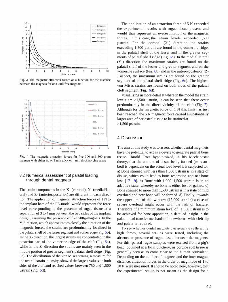
42
Fig. 3 The magnetic attraction forces as a function for the distance
between the magnets for one until five magnets
Fig. 4 The magnetic attraction forces for five 300 and 500 gram
magnets with either no or 2 mm thick or 4 mm thick porcine rugae
3.2 Numerical assessment of palatal loading through dental magnets
The strain components in the X- (coronal), Y- (medial/lat-
eral) and Z- (anterior/posterior) are different in each direc-
tion. The application of magnetic attraction forces of 1 N to
the implant bars of the FE-model would represent the force
level corresponding to the presence of rugae tissue at a
separation of 3 to 4 mm between the two sides of the implant
design, assuming the presence of five 500g-magnets. In the
Y-direction, which approximates closely the direction of the
magnetic forces, the strains are predominantly localized in
the palatal shelf of the lesser segment and vomer edge (Fig. 5b).
In the X- direction, the largest strains are concentrated in the
posterior part of the vomerine edge of the cleft (Fig. 5a),
while in the Z- direction the strains are mainly seen in the
middle portion of greater segment’s palatal shelf ridge (Fig.
5c). The distribution of the von Mises strains, a measure for
the overall strain intensity, showed the largest values on both
sides of the cleft and reached values between 750 and 1,500
µstrain (Fig. 5d).
The application of an attraction force of 5 N exceeded
the experimental results with rugae tissue present and
would thus represent an overestimation of the magnetic
forces. In this case, the strain levels exceeded 1,500
µstrain. For the coronal (X-) direction the strains
exceeding 1,500 µstrain are found in the vomerine ridge,
in the palatal shelf of the lesser and in the greater seg-
ments of palatal shelf ridge (Fig. 6a). In the medial/lateral
(Y-) direction the maximum strains are found on the
palatal shelf of the lesser and greater segment and on the
vomerine surface (Fig. 6b) and in the antero-posterior (Z-
) aspect, the maximum strains are found on the greater
segment of the palatal shelf ridge (Fig. 6c). The highest
von Mises strains are found on both sides of the palatal
cleft segment (Fig. 6d).
Visualizing in more detail at where in the model the strain
levels are >1,500 µstrain, it can be seen that these occur
predominantly in the direct vicinity of the cleft (Fig. 7).
Although for the magnetic force of 1 N this limit has just
been reached, the 5 N magnetic force caused a substantially
larger area of periosteal tissue to be strained at
>1,500 µstrain.
4 Discussion The aim of this study was to assess whether dental mag- nets
have the potential to act as a device to generate palatal bone
tissue. Harold Frost hypothesized, in his Mechanostat
theory, that the amount of tissue being formed (or resor-
bed) is dependent on the actual load level it is subjected to:
a) Bone strained with less than 1,000 µstrain is in a state of
disuse, which could lead to bone resorption and net bone
loss [17–19]. b) Bone with 1,000–1,500 µstrain is in an
adaptive state, whereby no bone is either lost or gained. c)
Bone strained to more than 1,500 µstrain is in a state of mild
overload and new bone will be formed. d) Finally, towards
the upper limit of this window (15,000 µstrain) a case of
severe overload might occur with the risk of fracture.
Therefore, if a minimum strain level of 1,500 µstrain is to
be achieved for bone apposition, a detailed insight in the
palatal load transfer mechanism in newborns with cleft lip
and palate is required.
To see whether dental magnets can generate sufficiently
high forces, several set-ups were tested, including the
absence or presence of rugae tissue between the magnets.
For this, palatal rugae samples were excised from a pig’s
head, obtained at a local butchery, as porcine soft tissue is
generally seen as to come close to the human equivalent.
Depending on the number of magnets and the inter-magnet
distance, attraction forces in the order of magnitude of 1 to
10 N were measured. It should be noted here, however, that
the experimental set-up is not meant as the design for a

43
Fig. 5 The distribution of the three orthogonal strain components von Mises strains for magnetic attraction forces of 1 N a palatal view with the
X-axis pointing coronally, b Y-axis pointing left, c Z-axis pointing posteriorly and d the strain intensity (von Mises strain)
Fig. 6 The distribution of the three orthogonal strain components von Mises strains for magnetic attraction forces of 5 N a palatal view with the
X-axis pointing coronally, b Y-axis pointing left, c Z-axis pointing posteriorly and d the strain intensity (von Mises strain)
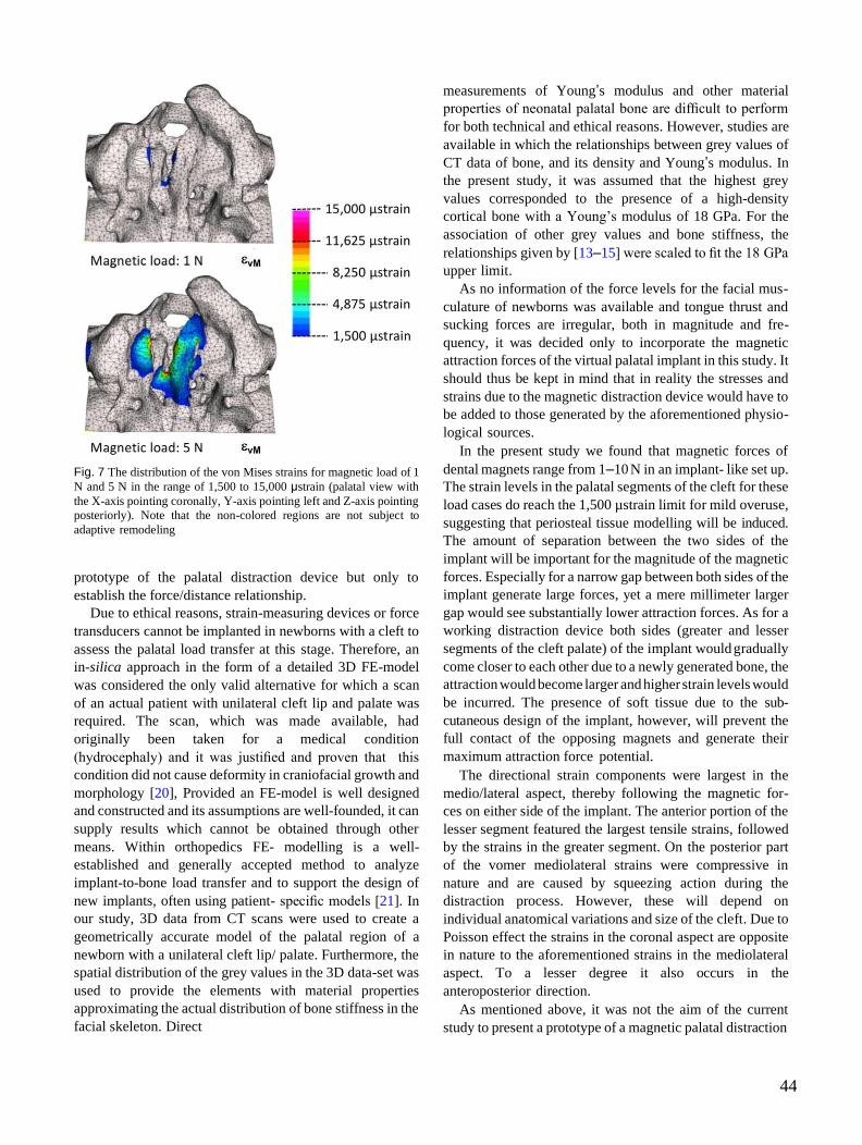
44
Fig. 7 The distribution of the von Mises strains for magnetic load of 1
N and 5 N in the range of 1,500 to 15,000 μstrain (palatal view with
the X-axis pointing coronally, Y-axis pointing left and Z-axis pointing
posteriorly). Note that the non-colored regions are not subject to
adaptive remodeling
prototype of the palatal distraction device but only to
establish the force/distance relationship.
Due to ethical reasons, strain-measuring devices or force
transducers cannot be implanted in newborns with a cleft to
assess the palatal load transfer at this stage. Therefore, an
in-silica approach in the form of a detailed 3D FE-model
was considered the only valid alternative for which a scan
of an actual patient with unilateral cleft lip and palate was
required. The scan, which was made available, had
originally been taken for a medical condition
(hydrocephaly) and it was justified and proven that this
condition did not cause deformity in craniofacial growth and
morphology [20], Provided an FE-model is well designed
and constructed and its assumptions are well-founded, it can
supply results which cannot be obtained through other
means. Within orthopedics FE- modelling is a well-
established and generally accepted method to analyze
implant-to-bone load transfer and to support the design of
new implants, often using patient- specific models [21]. In
our study, 3D data from CT scans were used to create a
geometrically accurate model of the palatal region of a
newborn with a unilateral cleft lip/ palate. Furthermore, the
spatial distribution of the grey values in the 3D data-set was
used to provide the elements with material properties
approximating the actual distribution of bone stiffness in the
facial skeleton. Direct
measurements of Young’s modulus and other material
properties of neonatal palatal bone are difficult to perform
for both technical and ethical reasons. However, studies are
available in which the relationships between grey values of
CT data of bone, and its density and Young’s modulus. In
the present study, it was assumed that the highest grey
values corresponded to the presence of a high-density
cortical bone with a Young’s modulus of 18 GPa. For the
association of other grey values and bone stiffness, the
relationships given by [13–15] were scaled to fit the 18 GPa
upper limit.
As no information of the force levels for the facial mus-
culature of newborns was available and tongue thrust and
sucking forces are irregular, both in magnitude and fre-
quency, it was decided only to incorporate the magnetic
attraction forces of the virtual palatal implant in this study. It
should thus be kept in mind that in reality the stresses and
strains due to the magnetic distraction device would have to
be added to those generated by the aforementioned physio-
logical sources.
In the present study we found that magnetic forces of
dental magnets range from 1–10 N in an implant- like set up.
The strain levels in the palatal segments of the cleft for these
load cases do reach the 1,500 µstrain limit for mild overuse,
suggesting that periosteal tissue modelling will be induced.
The amount of separation between the two sides of the
implant will be important for the magnitude of the magnetic
forces. Especially for a narrow gap between both sides of the
implant generate large forces, yet a mere millimeter larger
gap would see substantially lower attraction forces. As for a
working distraction device both sides (greater and lesser
segments of the cleft palate) of the implant would gradually
come closer to each other due to a newly generated bone, the
attraction would become larger and higher strain levels would
be incurred. The presence of soft tissue due to the sub-
cutaneous design of the implant, however, will prevent the
full contact of the opposing magnets and generate their
maximum attraction force potential.
The directional strain components were largest in the
medio/lateral aspect, thereby following the magnetic for-
ces on either side of the implant. The anterior portion of the
lesser segment featured the largest tensile strains, followed
by the strains in the greater segment. On the posterior part
of the vomer mediolateral strains were compressive in
nature and are caused by squeezing action during the
distraction process. However, these will depend on
individual anatomical variations and size of the cleft. Due to
Poisson effect the strains in the coronal aspect are opposite
in nature to the aforementioned strains in the mediolateral
aspect. To a lesser degree it also occurs in the
anteroposterior direction.
As mentioned above, it was not the aim of the current
study to present a prototype of a magnetic palatal distraction
45
device for the use in newborns. It has merely confirmed the hypothesis that dental magnets do have the potential
to generate levels of tissue strains, associated with mild overload, resulting in bone apposition. As the feasibility
of dental magnets for the purpose of osteogenesis has now been demonstrated, the next step will be to produce a
prototype design of the device, and test it in an ex-vivo and in- vivo animal model to validate the findings of the
presented FE-analyses.
5 Conclusions
(1) Dental magnets can produce attraction forces large enough to be used for adaptive remodelling or guided bone
formation on infant cranio-facial complexes.
(2) The strains in the periosteum and the underlying palatal bone generated by magnetic attraction forces are
compatible with adaptive bone formation in the unilateral cleft palate model.
(3) With magnitudes exceeding 1,500 µstrains, the tensile strains in the lesser and greater segments will act as
stimulus for guided bone formation in the medio-lateral aspect.
Acknowledgements The authors would like to thank Dyna Dental Engineering B.V., Halsteren, Netherlands for kindly supplying
the magnets for this study. This study was partially supported from a grant of the Freiwillige Akademische Gesellschaft (FAG), Basel,
Switzerland. Open access funding provided by University of Basel.
Compliance with ethical standards
Conflict of interest: The authors declare that they have no conflict of interest.
Publisher’s note Springer Nature remains neutral with regard to jurisdictional claims in published maps and institutional affiliations.
Open Access This article is licensed under a Creative Commons Attribution 4.0 International License, which permits use, sharing,
adaptation, distribution and reproduction in any medium or format, as long as you give appropriate credit to the original author(s)
and the source, provide a link to the Creative Commons license, and indicate if changes were made. The images or other third party
material in this article are included in the article’s Creative Commons license, unless indicated otherwise in a credit line to the
material. If material is not included in the article’s Creative Commons license and your intended use is not permitted by statutory
regulation or exceeds the permitted use, you will need to obtain permission directly from the copyright holder. To view a copy of
this license, visit http://creativecommons. org/licenses/by/4.0/.
References
1. Mossey PA, Little J, Munger RG, Dixon MJ, Shaw WC. Cleft lip and palate. Lancet 2009; 374:1773–85.
2. Mossey P. Global strategies to reduce the healthcare burden of craniofacial anomalies. British Dental Journal. 2003; 195:613.
3. Latham RA, Smiley GR, Gregg JM. The problem of tissue deficiency in cleft palate: An experiment in mobilising the palatine
bones of cleft dogs. Br J Plast Surg. 1973; 26:252–60.
4. Shetye PR, Evans CA. Midfacial morphology in adult unoperated complete unilateral cleft lip and palate patients. Angle Orthod
2006; 76:810–6.
5. Monroy PLC, Grefte S, Kuijpers-Jagtman AM, Wagener FADTG, Von Den Hoff JW. Strategies to improve regeneration of the
soft palate muscles after cleft palate repair. Tissue Eng - Part B: Rev. 2012; 18:468–77.
6. Shash H, Al-Halabi B, Jozaghi Y, Aldekhayel S, Gilardino MS. A review of tissue expansion-assisted techniques of cleft palate
repair. J Craniofac Surg. 2016; 27:760–6.
7. Kobus KF. Cleft palate repair with the use of osmotic expanders: a preliminary report. J Plast Reconstr Aesthetic Surg. 2007;60:414–21.
8. McCarthy JG, Schreiber J, Karp N, Thorne CH, Grayson BH. Lengthening the human mandible by gradual distraction. Plast
Reconstr Surg. 1992; 89:1–8.
9. Alkan A, Baş B, Özer M, Bayram M. Closure of a large palatal fistula with maxillary segmental distraction osteogenesis in a
cleft palate patient. Cleft Palate-Craniofacial J 2007; 44:112–5.
10. Schmidt BL, Kung L, Jones C, Casap N. Induced osteogenesis by periosteal distraction. J Oral Maxillofac Surg. 2002; 60:1170–
5.
11. Yan QC, Tomita N, Ikada Y. Effects of static magnetic field on bone formation of rat femurs. Med Eng Phys. 1998; 20:397–
402.
12. Wang D, Cheng L, Wang C, Qian Y, Pan X. Biomechanical analysis of rapid maxillary expansion in the UCLP patient. Med
Eng Phys. 2009; 31:409–17.
13. McPherson GK, Kriewall TJ. The elastic modulus of fetal cranial bone: A first step towards an understanding of the
biomechanics of fetal head molding. J Biomech 1980; 13:9–16.
14. Bauer FX, Heinrich V, Grill FD, Wölfle F, Hedderich DM, Rau A, et al. Establishment of a finite element model of a neonate’s
skull to evaluate the stress pattern distribution resulting during nasoalveolar molding therapy of cleft lip and palate patients. J
Cranio- Maxillofac Surg. 2018; 46:660–7.
15. Pan X, Qian Y, Yu J, Wang D. Biomechanical Effects of Rapid Palatal Expansion on the Craniofacial Skeleton. Cleft Palate-
Craniofacial J 2007; 44:149–54.
16. Carter DR, Schwab GH, Spengler DM. Tensile fracture of cancellous bone. Acta Orthop 1980; 51:733–41.
17. Frost HM. The Utah paradigm of skeletal physiology: An over- view of its insights for bone, cartilage and collagenous tissue

46
organs. J Bone Min Metab. 2000; 18:305–16.
18. Frost HM. A 2003 update of bone physiology and Wolff s law for clinicians. Angle Orthod 2004; 74:3–15.
19. Turner CH. Toward a mathematical description of bone biology: The principle of cellular accommodation. Calcif Tissue Int.
1999; 65:466–71.
20. Yusof A Craniofacial growth changes in Malaysian Malay children and young adults: a cross-sectional 3-dimensional CT study.
[Internet]. University of Adelaide; 2007. Available from: https:// digital.library.adelaide.edu.au/dspace/handle/2440/39388.
21. Poelert S, Valstar E, Weinans H, Zadpoor AA. Patient-specific finite element modeling of bones. Proc Inst Mech Eng Part H J
Eng Med. 2013; 227:464–78.

47
8. Third study:
Load transfer during magnetic mucoperiosteal distraction in newborns with
complete unilateral and bilateral orofacial clefts: a three-dimensional finite
element analysis
Study design: Dr. Prasad Nalabothu, Prof. Michel Dalstra and PD Dr. med. Dr. med.
dent. Dr. phil. Andreas A. Mueller
Financing: FAG (Voluntary Academic Association): CHF 13,406
Project leaders: Professor Dr. med. dent. Carlalberta Verna
Publication: First authorship, Journal of applied Sciences, Impact factor 2.47 (2020),
Ranking 85/299 (Q2) in ‘General engineering and 233/460 in ‘General Materials
Science’.

48
applied sciences
Article
Load Transfer during Magnetic Mucoperiosteal
Distraction in Newborns with Complete Unilateral
and Bilateral Orofacial Clefts: A Three-Dimensional
Finite Element Analysis
Prasad Nalabothu 1,2, *, Carlalberta Verna 1, Benito K. Benitez 2, Michel Dalstra 1, † and Andreas
A. Mueller 2, †
1 Department of Paediatric Oral Health and Orthodontics, University Center for Dental Medicine UZB,
4058 Basel, Switzerland; [email protected] (C.V.); [email protected] (M.D.) 2 Department of Oral and Craniomaxillofacial Surgery, University Hospital Basel, 4031 Basel, Switzerland;
[email protected] (B.K.B.); [email protected] (A.A.M.)
* Correspondence: [email protected]; Tel.: +41-61-328-60-95
† Joint last authors.
Received: 17 September 2020; Accepted: 23 October 2020; Published: 31 October 2020 check for updates
Abstract: The primary correction of congenital complete unilateral cleft lip and palate (UCLP) and
bilateral cleft lip and palate (BCLP) is challenging due to inherent lack of palatal tissue and small
extent of the palatal shelves at birth. The tissue deficiency affects the nasal mucosa, maxillary bone
and palatal mucosa. This condition has driven the evolution of several surgical and non-surgical
techniques for mitigating the inherent problem of anatomical deficits. These techniques share the
common principle of altering the neighboring tissues around the defect area in order to form a
functional seal between the oral and nasal cavity. However, there is currently no option for rectifying
the tissue deficiency itself. Investigations have repeatedly shown that despite the structural tissue
deficiency of the cleft, craniofacial growth proceeds normal if the clefts remain untreated, but the
cleft remains wide. Conversely, craniofacial growth is reduced after surgical repair and the related
alteration of the tissues. Therefore, numerous attempts have been made to change the surgical
technique and timing so as to reduce the effects of surgical repairs on craniofacial growth, but they
have been only minimally effective so far. We have determined whether the intrinsic structural soft
and hard tissue deficiency can be ameliorated before surgical repair using the principles of periosteal
distraction by means of magnetic traction. Two three-dimensional maxillary finite element models,
with cleft patterns of UCLP and BCLP, respectively, were created from computed tomography slice
data using dedicated image analysis software. A virtual dental magnet was positioned on either side
of the cleft at the mucoperiosteal borders, and an incremental magnetic attraction force of up to 5 N
was applied to simulate periosteal distraction. The stresses and strains in the periosteal tissue induced
by the magnet were calculated using finite element analysis. For a 1 N attraction force the maximum
strains did not exceed 1500 µstrain suggesting that adaptive remodeling will not take place for
attraction forces lower than 1 N. At 5 N the regions subject to remodeling differed between the UCLP
and BCLP models. Stresses and strains at the periosteum of the palatal shelf ridges in the absence of
compressive forces at the alveolar borders were greater in the UCLP model than the BCLP model. The
findings suggest that in newborns with UCLP and BCLP, periosteal distraction by means of a magnetic
5 N attraction force can promote the generation of soft and hard tissues along the cleft edges and
rectify the tissue deficiency associated with the malformation.
Keywords: cleft lip and palate; finite element model; periosteal bone remodeling; unilateral cleft
palate; bilateral cleft palate; distraction
Appl. Sci. 2020, 10, 7728; doi:10.3390/app10217728
Appl. Sci. 2020, 10, 7728
49
1. Introduction
Cleft lip and palate (CLP) is the most common craniofacial malformation, affecting one in every
500 to 700 live births, thus accounting for about 220000 new cases each year worldwide with disparity
across geographic areas, racial groups, and socioeconomic status [1]. No effective preventive measures
exist. The ongoing manifestation of CLP as well as its broad implications on psychological well-being,
speech, orofacial development, and therapeutic burden underline the need to identify and targeting the
intrinsic causes [2–4].
The etiology of CLP is complex, which could occur as a result of multiple genetic and environmental
factors or a combination of both [5,6]. The clefting of the lip and/or palate may occur in a unilateral
or bilateral fashion, and the embryological pattern of clefting results from a fusion failure during the
development of maxilla and palate. The normal fusion of the medial frontonasal process with the
maxillary process of the first pharyngeal arch takes place at 4–6 weeks of gestation forming a primary
palate [7]. A complete fusion failure results in a cleft of the lip through the vermillion border that
extends to the nasal sill and in a cleft of the alveolar process [7]. The cleft of the secondary palate occurs
at 8–12 weeks of gestation due to fusion failure of the palatal shelves of the maxillary processes [8,9]
that results in a cleft in the palatal roof and soft palate that extends to the uvula.
Affected children have a range of both functional and esthetic problems. They comprise feeding
difficulties at birth due to incomplete oral seal, swallowing, nasal regurgitation, respiratory problems,
hearing difficulties due to abnormalities in the palatal musculature, and speech impairments due to air
escape and articulation problems [10]. Therefore, an ideal early and complete cleft treatment would
allow the normalizing of food intake, speech development, growth and esthetics - while imposing
minimal treatment burden to child and family.
Several different surgical protocols have been advocated for managing children with CLP.
The amount of palatal mucosa present and timing of the surgery play important roles in healing success
after surgery. Clefting of the palate at birth structurally results in 16% less palatal mucosa compared
with a normal child [11] and anatomically smaller palatal shelves on the cleft and noncleft sides [12,13].
The intrinsic tissue deficiency in the palate has no spontaneous improvement trend. It persists until
adulthood in both unilateral cleft lip and palate (UCLP) and bilateral cleft lip and palate (BCLP) [14].
A scientific basis was proposed in 2005 for determining the optimal conditions for palatal cleft
closure in complete UCLP and BCLP [15]. This should be performed when the surface area of the
cleft space does not exceed 10% of the surrounding palatal surface bounded by the alveolar ridges,
because under these circumstances the facial growth is less affected, independent of the patient’s age at
surgery [15]. This recommendation may be applicable for clefts which are smaller in size, whereas
in cases of wider clefts it is imperative for surgeons to mobilize the tissue adjoining the cleft more
extensively which may lead to open wound areas that undergo secondary healing. This may lead to
structural tissue deficiencies and pronounced interference with the normal growth and development
of the midface [16].
The use of a palatal appliance for presurgical maxillary orthopedics is a widely accepted non-
surgical therapeutic adjunct for addressing the problem of tissue deficiency in wider clefts. Such an
appliance approximates the palatal shelves and thus reduces the alveolar and palatal cleft width prior
to surgery [17]. The effect of those appliances on the growth is controversial. Some proponents of
this technique have claimed that gradual adaptations of the appliance can stimulate palatal tissue
growth [18,19]. Others have shown that this approach resulted in retarded growth of the palatal tissue
[20]. Finally, a randomized controlled trial concluded that using a palatal appliance does not result in
any permanent growth change or tissue gain [21].
Various surgical techniques have also been applied in an attempt to mitigate the problems of
tissue deficiency. These techniques varied between surgeons, and there is still no consensus on which
is the best protocol for treating CLP patients. Seventeen different surgical sequences for repairing
unilateral complete clefts were identified in a survey of over 201 European cleft centers [22].

Appl. Sci. 2020, 10, 7728
50
Three main surgical principles to mitigate the tissue deficiency in CLP have evolved [23]. The first
principle limits the closure only to either the oral or the nasal mucosa layer by applying a so called
single-layer closure [24–27]. This procedure involves turning over the mucosa from either the oral or the
nasal cavity like a book page while leaving an open wound on the back side of the turned-over mucosa.
The second principle involves detaching the oral tissue and shifting it over the cleft area while creating
an open wound that heals outside the cleft area from where the tissue has been detached [28–30]. In the
third principle, tissue from outside the palatal area is used to cover the open wound areas that result
from the first or second technique [31–34]. While all of these surgical techniques are able to close the
cleft gap using the available tissue, none of them result in the generation of new mucoperiosteal tissue.
Adjuvant therapies or strategies such as tissue expansion [35] and distraction osteogenesis
(DO) [36] have been attempted to increase the volume of tissue in the areas of tissue deficiency in
clefts. The principles of DO require a bone cut through the palatal shelf ridges. However, in infants
this would damage the dental follicles that are embedded in the palatal shelf ridges. Most tissue
engineering studies have focused on the application of biomaterials purely for bone generation [37].
However, the palatal cleft comprises bone and two mucosal layers, which makes it a difficult target for
biomaterial or tissue-engineering techniques.
Animal experiments suggest that periosteal distraction osteogenesis (PDO) is an appealing option
for utilizing the principles of DO [38–40] for combined bone and soft tissue generation without the need
for a bone cut. PDO is performed by gradual lift-up on the mucoperiosteum from the underlying bone
by applying mechanical loading. The physical tissue strain and biological tissue stimulus to bone and
periosteum lead to new tissue formation. We therefore address PDO as a potential treatment strategy
to target the tissue deficiency in orofacial clefts while using magnetic forces to exert the necessary
tissue strain. Magnetic forces have been used in orthodontics to generate tooth movement and to
promote tissue reactions [41].
Finite element (FE) analysis is a numerical tool to compute the stress and strain fields in the
bone structure under external loading. It thereby reveals the distribution of the internal loads and
deformations. Computational FE analysis has provided crucial basic data for understanding mechanical
interactions between the magnets and mucoperiosteal tissue for tissue expansion [42]. The loading
distribution depends on the shape and size of individual anatomical structures and malformation.
Previously, this method has been used to assess the maxillary load transfer during conventional DO in
patients with cleft lip and palate [43].
In our previous study we have examined the feasibility of dental magnets as distraction devices
in newborns with UCLP [44]. Unilateral clefts form 76% of all CLP cases worldwide which still leaves
the BCLP, accounting for one fourth (24%) of all CLP unaddressed [45]. As the anatomy of BCLP is
different from UCLP, it is the aim of this study to compare the load transfer of magnetic forces used for
PDO in both UCLP and BCLP in silico models. It examines whether the forces reach the threshold
necessary for regeneration of the hard and soft tissue volumes along the cleft edges in both UCLP
and BCLP.
2. Materials and Methods
2.1. Ethical Statement and Patient Data
The reported investigations and procedures were conducted according to the principles expressed
in the Declaration of Helsinki. Two computed tomography (CT) scans of anonymized 2- and 3-week-old
newborns with complete UCLP and BCLP, respectively, were obtained from an existing database and
analyzed. The DICOM data sets of 2 CT scans were obtained from a commercial CT scanner using a
voxel size of 0.3 mm (Philips CT scanner, Eindhoven, The Netherlands).

Appl. Sci. 2020, 10, 7728
51
2.2. Model Construction
The CT scans of the UCLP and BCLP newborns were imported into dedicated image analysis
software (Mimics version 19, Materialise, Leuven, Belgium) and three-dimensional (3D) surface models
were generated by thresholding and selecting only the hard tissues of the cranium and maxilla. The
region of interest was further defined by virtually removing all structures except for the midface,
including the maxilla and the adjoining bony segments of the face (first, second and third rows in
Figure 1).
Figure 1. From computed tomography scans to finite element (FE) models with magnets in
the unilateral cleft lip and palate (UCLP, left column) and bilateral cleft lip and palate (BCLP,
right column). Frontal views of three-dimensional reconstructions of the soft tissue (first row)
and hard tissue (second row). Regions of interest of the FE models (third row) and the models
with magnetic strips which are blue in color (fourth row).

Appl. Sci. 2020, 10, 7728
52
2.3. Magnet Implantation
The rare-earth permanent magnets used for in vitro testing were constructed from a neodymium-
boron-iron alloy (Dyna WR magnets, Dyna Dental Engineering, Halsteren, The Netherlands). The magnets
were disc shaped with a height of 1.7 mm and a diameter of 4.5 mm (02MS1 Dyna WR magnet S3). The
mucoperiosteum borders on the cleft and non-cleft sides were covered with two 23 mm long parallel bars
of five magnets each placed in a row in the UCLP and BCLP models (Figure 1, fourth row). In the
following analyses, each row of the magnets was assumed to exert attracting forces relative to the
oppositely placed row of magnets. Experimental force-distance curves for these magnets were measured
[44] with the results used to define incremental forces with a magnitude of up to 5 N to either side of the
cleft in the UCLP and BCLP models.
2.4. FE Model Design
The FE mesh was constructed based on the CT scans of the 3D UCLP and BCLP models. The UCLP
FE mesh consisted of 238243 4-node tetrahedral elements with 43822 nodes, whereas the BCLP FE
mesh consisted of 409027 4-node tetrahedral elements with 74551 nodes. The position of the centroid
of each element was calculated and a gray scale was associated with it based on spatial interpolation of
the gray scale values of the nearest voxels. The gray scale values from the CT scans were correlated
with the material density and the material stiffness. Relevant material properties were assigned for the
elements representing bone, arithmetic relationships between gray scale values, apparent densities and
Young’s moduli [46–49]. Depending on its apparent density and the associated gray scale values, the
bone tissue was stratified into ten different material property groups, ranging from very low-density
cancellous bone (bone 1) to dense cortical bone (bone 10). The material properties of these ten types of
bone (apparent density, Young’s modulus, Poisson’s ratio and yield strength) were same as used by
Nalabothu et al. [44].To highlight the compositional difference between the UCLP and BCLP model
each type of bone density (bone 1 to bone 10) has been cumulatively compared between both models
(Figure 2).
Figure 2. The cumulative percentage of the ten material property groups for the unilateral
cleft lip and palate (UCLP) and bilateral cleft lip and palate (BCLP) models, respectively. The
material properties of the ten different bone types are retrieved from Nalabothu et al. [44].

Appl. Sci. 2020, 10, 7728
53
2.5. FE Analysis
The UCLP and BCLP FE models were exported from the image analysis program in NASTRAN
format and imported into an FE analysis program (Strand7, Sydney, Australia). The material properties
of the bone were assumed to be non-linear, with its stress-strain curves in the ten groups adapted so
that the bone would behave as a perfectly plastic material whose compressive strength was 150%
higher than its tensile strength [50]. The FE analyses were conducted using a nonlinear solver with
logarithmically increasing increments from 0.01 to 1 (100% load). The calculated stresses and the
associated strains were used for post-processing.
3. Results
The application of a 1 N attraction force did not produce strain levels in the 1500 µstrain range
in both the UCLP and BCLP model. As this was considered a minimum requirement for adaptive
remodeling, in the following only the results for a 5 N attraction force are presented.
3.1. Overall Deformation of the Maxilla
The maximum deformation in the UCLP and BCLP models occurred around the sites adjacent to
the magnetic strips at the vomer edge of the greater and lesser segments of the palatal shelf ridge. The
overall deformation was greater in the UCLP model (left column of Figure 3) than in the BCLP model.
In the BCLP model, the deformations occurred more in the posterior area of the palatal shelf ridges
(right column of Figure 3) than in the anterior one.
Figure 3. Distribution of the overall periosteal deformation in mm for 5 N loading in
the UCLP (left column) and BCLP (right column) models. Frontal (top row) and palatal
(bottom row) views are shown.

Appl. Sci. 2020, 10, 7728
54
3.2. Stresses and Strains in the Maxilla
The obtained stress/strain components of the UCLP and BCLP FE models in the X-direction
(coronal), Y-direction (medial/lateral) and Z-direction (anterior/posterior) were different in each aspect.
In the BCLP model no strains higher than 1500 µstrain were seen for a magnetic load of 1 N (Figure 4).
However, a load of 5 N regions with strains beyond 1500 µstrain were observed though less than the
UCLP model [44].
Figure 4. Areas showing the overall strain levels in BCLP model during loading of 1 N
(left) and 5 N (right) from the palatal view.
The orthogonal stresses in the coronal (X) direction occurred in the UCLP model mainly at the
anterior part of the ridge of the lesser segment and the posterior part of the vomer edge (Figure 5:
left column, top row), while in the BCLP model it occurred bilaterally in the posterior border of
palatal shelf ridge (Figure 5: right column, top row). The orthogonal stresses in the medial/lateral
(Y)-direction occurred in the UCLP model mainly at the middle portion of the lesser segment’s ridge
and in some sparse areas at the middle portion of the vomer edge and the greater segment’s ridge
(Figure 5: left column, second row), while in the BCLP model it occurred bilaterally on the palatal shelf
ridges at the anterior tip and posterior border (Figure 5: right column, second row).
The orthogonal stresses in the antero/posterior (Z) direction occurred in the UCLP model mainly
on the anterior portion of the lesser segment’s ridge and on the middle portion of the vomer edge and
on the anterior portion of the greater segment’s ridge (Figure 5: left column, third row), while there
were no stresses in this direction in the BCLP model (Figure 5: right column, third row).
The von Mises stresses (stress intensity) in the UCLP model were highest on the entire greater
and lesser palatal shelf ridges and on the vomer edge (Figure 5: left column, bottom row), while in
the BCLP model von Mises stresses were highest bilaterally on the anterior and posterior parts of the
palatal shelf ridges. (Figure 5: right column, bottom row).
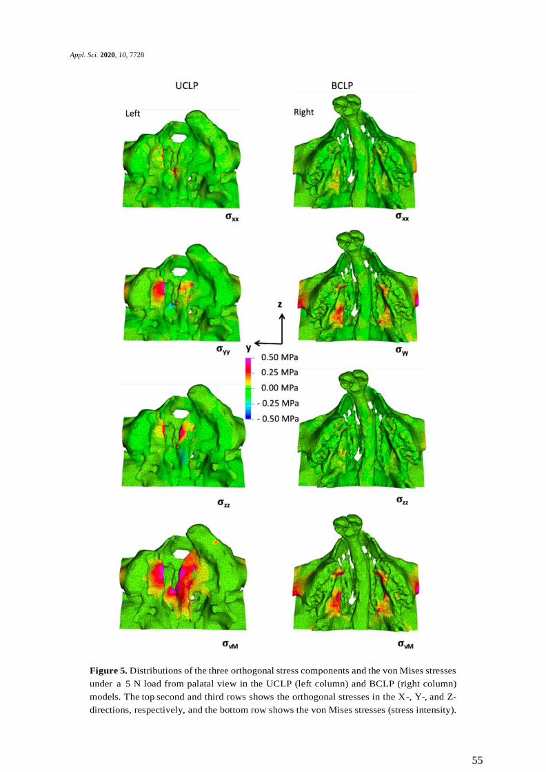
Appl. Sci. 2020, 10, 7728
55
Figure 5. Distributions of the three orthogonal stress components and the von Mises stresses
under a 5 N load from palatal view in the UCLP (left column) and BCLP (right column)
models. The top second and third rows shows the orthogonal stresses in the X-, Y-, and Z-
directions, respectively, and the bottom row shows the von Mises stresses (stress intensity).

Appl. Sci. 2020, 10, 7728
56
4. Discussion
The success of surgical hard palate closure in UCLP and BCLP is dependent on the availability of
tissue in relation to the cleft area that needs to be closed. In wider clefts it is imperative to mobilize the
tissue adjacent to the cleft more extensively, which leads to a wider tissue wound and tissue reparation
rather than regeneration [51]. This situation may interfere with the normal growth and development
of the midface and dentition [12,52]. The growth in patients with clefts remains a challenge regardless
of which surgical protocol is applied. Applying adjuvant therapies before surgery can alleviate the
problems of tissue deficiency to some extent by approximation of the palatal shelves using active
presurgical orthopedics [53] or passive plate therapy [17], which facilitate tissue closure across the cleft
area. However, the stocks of bone and soft tissue still remain deficient since such therapies do not aid
in the regeneration of local tissue.
Therefore, in this study we tested a contrasting strategy based on increasing the bone and soft tissue
prior to cleft surgical repair by means of magnetic periosteal distraction. It aims for true generation of
the deficient osteo-mucoperiosteal tissue−thus, tackling the inherent palatal tissue deficiency which is
a basic feature of the cleft malformation that does not disappear over time. With this intention, we
analyzed the in silico stress distribution induced by magnetic strips placed on the cleft palatal shelves
in newborns with UCLP and BCLP. We tested the hypothesis that magnets can generate bone inducing
strains in UCLP and BCLP newborns.
Analyses of load transfer, by studying the distribution of stresses and strains, are a prerequisite
before in vivo testing since incorrect loading or overloading may lead to disorganized tissue structures,
eventually leading to tissue necrosis. Determining these stresses and strains in vivo is not feasible in
newborns for ethical reasons, and so 3D in silico models were deemed a valid alternative approach.
Although our experiments were confined to tissue generation by means of stresses induced by magnetic
periosteal distraction, there is increasing evidence that magnetic fields can further promote bone
regeneration directly by attracting growth factors, hormones and polypeptides to the implantation
site [54]. The length of the magnetic strips was defined as 23 mm since the average length of hard
palate at birth is 25.6 mm ±1.6 mm [55]. Previous tests on the magnetic attraction forces across porcine
palatal mucosa (rugae) tissue allowed the simulation of the thickness of mucosa in the newborns [44].
In the present study we found that applying 5 N attraction forces using magnetic strips induced
stresses in the UCLP and BCLP models mainly at the vomer edge and the palatal shelf ridges of
the greater and lesser segments (left and right columns, Figure 3). Areas of tissue generation were
delineated by applying the principles of Frost’s Mechanostat theory, which states that mechanical
loading influences the bone structure by changing the mass (amount of bone) and architecture
(arrangement of surrounding tissues) if the induced strain exceeds 1500 µstrain. This implies that
periosteal tissue regeneration will be induced at sites where the strain in the underlying bone exceeds
1500 µstrain, as the periosteum is mechanically subservient to the bone [56] (Figure 4).
The area of periosteal tissue that was subjected to at least 1500 µstrain was larger in the UCLP
model than the BCLP model, which could be due to differences in material properties of the bone and
anatomical variations. In the two FE models there is more low-density bone type 3 present in BCLP
than in the UCLP model (Figure 2). Moreover, the direction of the magnetic forces is also more
horizontally oriented in BCLP model compared to the UCLP model. The stresses and strains, able to
induce adaptive remodeling, as exhibited in both UCLP and BCLP models, were localized to the area of
the intrinsic tissue deficit of the cleft malformation while at the same time not affecting the neighboring
areas, thus providing evidence that no compressive forces were transmitted to the alveolar ridges by
the magnets.
The abovementioned geometrical and structural differences between the bone anatomy of the
UCLP and BCLP mean that each will require a different design of implant. The larger cleft width
in case of BCLP would make a more powerful magnet necessary to generate equivalent forces as in
case of UCLP. Alternatively, the use of spacers to partially bridge the distance, or the placement of an
auxiliary implant at the vomer could be considered as an option. The present findings obtained in

Appl. Sci. 2020, 10, 7728
57
the UCLP and BCLP FE models might not be generalizable to all clefts of similar distances between
the affected and non-affected segments, since the 3D bone morphology might vary even when the
morphology in the cleft area is identical. However, the results of this study give guidance about the
distribution of palatal load transfer around the cleft segments by inducing PDO within the optimal
biological limits [56]. Future studies could test this hypothesis of mucoperiosteal distraction in an
in vivo cleft animal model [57] in order to validate the findings of the present FE analyses.
5. Conclusions
The following conclusions can be drawn from this study
1. The present FE models have shown that the use of magnets as approved for dental applications
can exert attraction with the potential to induce PDO around the greater and lesser segments of
the palatal shelf ridge in cases of UCLP and BCLP.
2. A magnetic attraction force of 5 N was found to be sufficient to reach strain values in excess of
1500 µstrain in the palatal bone in both UCLP and BCLP models, and thereby have the potential
to initiate adaptive remodeling.
3. The effects of magnetic load transfer were localized to their area of application, and did not spread
out to distant structures.
4. The anatomical variations both in external bone geometry and internal bone composition affect
the load transfer across the palate.
Author Contributions: Conceptualization, A.A.M. and C.V.; data curation, P.N. and M.D.; formal analysis, P.N.
and M.D.; funding acquisition, C.V., A.A.M. and H.F.Z.; investigation, P.N. and M.D.; methodology, P.N., M.D. and A.A.M.; supervision, M.D., C.V. and A.A.M.; validation, P.N., A.A.M. and M.D.; visualization, P.N., A.A.M.,
B.K.B., M.D. and C.V.; writing—original draft, P.N.; writing—review and editing, P.N., M.D., B.K.B., C.V. and
A.A.M. All authors have read and agreed with submitting this version of the manuscript.
Funding: This study was partially funded by a research grant from the Freiwillige Akademische Gesellschaft (FAG), Basel, Switzerland.
Acknowledgments: The authors thank Dyna Dental Engineering B.V., Halsteren, Netherlands for kindly supplying
the magnets used in this study. Ravi Kant Singh (chief consultant at Peace Pant Hospital, Varanasi, India) for
providing the CT scans, Markus Steineck (University Center for Dental Medicine UZB, Basel, Switzerland) for supporting the magnet-based experiments and Hans Florian Zeilhofer for funding acquisition and support of the
project.
Conflicts of Interest: The authors declare that they have no conflict of interest. The funders had no role in the
design of the study; the collection, analysis, or interpretation of data; the writing of the manuscript; or the decision to publish the results.
References
1. Mossey, P. Global strategies to reduce the healthcare burden of craniofacial anomalies. Br. Dent. J. 2003, 195,
613. [CrossRef] [PubMed]
2. Mossey, P.A.; Little, J.; Munger, R.G.; Dixon, M.J.; Shaw, W.C. Cleft lip and palate. Lancet 2009, 374, 1773–1785.
[CrossRef]
3. Schutte, B.C.; Murray, J.C. The many faces and factors of orofacial clefts. Hum. Mol. Genet. 1999, 8, 1853–1859.
[CrossRef] [PubMed]
4. Mossey, P.A. Epidemiology of oral clefts: An international perspective. In Cleft Lip and Palate: From Origin to
Treatment; Oxford University Press: Oxford, UK, 2002; ISBN 0195139062.
5. Mossey, P. Epidemiology underpinning research in the aetiology of orofacial clefts. Orthod. Craniofac. Res.
2007, 10, 114–120. [CrossRef]
6. Wong, F.K.; Hägg, U. An update on the aetiology of orofacial clefts. Hong Kong Med. J. 2004, 10, 331–336.
[PubMed]
7. Nanci, A.; Ten, C.A.R. Ten Cate’s Oral Histology Development, Structure, and Function; Elsevier: Amsterdam,
The Netherlands, 2017.
Appl. Sci. 2020, 10, 7728
58
8. Bush, J.O.; Jiang, R. Palatogenesis: Morphogenetic and molecular mechanisms of secondary palate
development. Development 2012, 139, 231–243. [CrossRef]
9. Tarr, J.T.; Lambi, A.G.; Bradley, J.P.; Barbe, M.F.; Popoff, S.N. Development of normal and Cleft Palate: A
central role for connective tissue growth factor (CTGF)/CCN2. J. Dev. Biol. 2018, 6, 18. [CrossRef]
10. Bührer, C.; Zimmermann, A. Cleft palate. In Neonatal Emergencies: A Practical Guide for Resuscitation, Transport
and Critical Care of Newborn Infants; Cambridge University Press: Cambridge, UK, 2009; Volume 1, pp. 460–463.
[CrossRef]
11. Huddart, A.G. Arch Alignment and Presurgical Treatment—The West Midlands Approach. In Early Treatment
of Cleft Lip and Palate; Hans Huber: Berne, Switzerland, 1986; ISBN 3-456-81406-2.
12. Latief, B.S.; Lekkas, K.C.; Schols, J.G.J.H.; Fudalej, P.S.; Kuijpers, M.A.R. Width and elevation of the palatal
shelves in unoperated unilateral and bilateral cleft lip and palate patients in the permanent dentition. J. Anat.
2012, 220, 263–270. [CrossRef]
13. Atherton, J.D. A Descriptive Anatomy of the Face in Human Fetuses with Unilateral Cleft Lip and Palate.
Cleft Palate J. 1967, 4, 104–114. [CrossRef]
14. Diah, E.; Lo, L.J.; Huang, C.S.; Sudjatmiko, G.; Susanto, I.; Chen, Y.R. Maxillary growth of adult patients with
unoperated cleft: Answers to the debates. J. Plast. Reconstr. Aesthetic Surg. 2007, 60, 407–413. [CrossRef]
15. Berkowitz, S.; Duncan, R.; Evans, C.; Friede, H.; Kuijpers-Jagtman, A.M.; Prahl-Anderson, B.; Rosenstein, S.
Timing of Cleft Palate Closure Should Be Based on the Ratio of the Area of the Cleft to That of the Palatal
Segments and Not on Age Alone. Plast. Reconstr. Surg. 2005, 115, 1483–1499. [CrossRef] [PubMed]
16. Kuijpers-Jagtman, A.M.; Long, R.E., Jr. The influence of surgery and orthopedic treatment on maxillofacial
growth and maxillary arch development in patients treated for orofacial clefts. Cleft Palate-Craniofac. J. 2000,
37, 527. [CrossRef]
17. Nalabothu, P.; Benitez, B.K.; Dalstra, M.; Verna, C.; Mueller, A.A. Three-Dimensional Morphological Changes
of the True Cleft under Passive Presurgical Orthopaedics in Unilateral Cleft Lip and Palate: A Retrospective
Cohort Study. J. Clin. Med. 2020, 9, 962. [CrossRef] [PubMed]
18. McNeil, C.K. Oral and Facial Deformity; Pitman: London, UK, 1954.
19. Fish, J. Growth of the palatal shelves of post-alveolar cleft palate infants. Effects of stimulation appliances.
Br. Dent. J. 1972, 132, 492–501. [CrossRef] [PubMed]
20. Huddart, A.G. Presurgical changes in unilateral cleft palate subjects. Cleft Palate J. 1979, 16, 147–157.
21. Prahl, C.; Kuijpers-Jagtmann, A.M.; Van’t Hof, M.A.; Prahl-Andersen, B. A randomised prospective clinical
trial into the effect of infant orthopaedics on maxillary arch dimensions in unilateral cleft lip and palate
(Dutchcleft). Eur. J. Oral Sci. 2001, 109, 297–305. [CrossRef]
22. Shaw, W.C.; Semb, G.; Nelson, P.; Brattström, V.; Mølsted, K.; Prahl-Andersen, B.; Gundlach, K.K.H. The
Eurocleft project 1996-2000: Overview. J. Cranio-Maxillofac. Surg. 2001, 29, 131–140. [CrossRef]
23. Dorrance, G.M.; Shirazy, E. The Operative Story of Cleft Palate; W.B. Saunders Company: Philadelphia, PA,
USA, 1933.
24. Campbell, B.Y.A. The closure of congenital clefts of the hard palate. Br. J. Surg. 1926, 13, 715–719. [CrossRef]
25. Pichler, H. Zur Operation der doppelten Lippen-Gaumenspalten. Dtsch. Z. Cir. 1926, 195, 104–107. [CrossRef]
26. Bütow, K.W. Caudally-based single-layer septum-vomer flap for cleft palate closure. J. Cranio-Maxillofac. Surg.
1987, 15, 10–13. [CrossRef]
27. Abyholm, F.E. Primary Closure of Cleft Lip and Palate. In Facial Clefts and Craniosynostosis: Principles and
Management; Turvey, T.A., Vig, K.W.L., Fonseca, R.J., Eds.; W.B. Saunders Company: Philadelphia, PA, USA,
1996.
28. Langenbeck, B. Die Uranoplastik mittelst Ablösung des mucös-periostalen Gaumenüberzuges. Uranoplasty
by means of raising mucoperiosteal flaps. Hirschwald: Berlin, 1861. Plast. Reconstr. Surg. 1972, 49, 326–330.
[CrossRef]
29. Veau, V.; Borel, S. Division palatine: Anatomie, Chirurgie Phonétique/Victor Veau, Avec la Collaboration de S. Borel;
Masson: Paris, France, 1931.
30. Bardach, J. Two-Flap palatoplasty: Bardach’s technique. Oper. Tech. Plast. Reconstr. Surg. 1995, 2, 211–214.
[CrossRef]
31. Millard, D.R., Jr. The island flap in cleft palate surgery. Surg. Gynecol. Obstet. 1963, 116, 297–300. [CrossRef]
32. Mukherji, M.M. Cheek flap for short palates. Cleft Palate J. 1969, 6, 415–420. [PubMed]

Appl. Sci. 2020, 10, 7728
59
33. Stricker, M.; Chancholle, A.R.; Flot, F.; Malka, G.; Montoya, A. La greffe periostee dans la reparation de la
fente totale du palais primaire [Periosteal graft in the repair of complete primary cleft palate]. Ann. Chir.
Plast. 1977, 22, 117–125.
34. Neiva, C.; Dakpe, S.; Gbaguidi, C.; Testelin, S.; Devauchelle, B. Calvarial periosteal graft for second-stage cleft
palate surgery: A preliminary report. J. Cranio-Maxillofac. Surg. 2014, 42, e117–e124. [CrossRef]
35. Shash, H.; Al-Halabi, B.; Jozaghi, Y.; Aldekhayel, S.; Gilardino, M.S. A review of tissue expansion-assisted
techniques of cleft palate repair. J. Craniofac. Surg. 2016, 27, 760–766. [CrossRef] [PubMed]
36. Alkan, A.; Bas, B.; Özer, M.; Bayram, M. Closure of a large palatal fistula with maxillary segmental distraction
osteogenesis in a cleft palate patient. Cleft Palate-Craniofac. J. 2007, 44, 112–115. [CrossRef]
37. Martín-Del-Campo, M.; Rosales-Ibañez, R.; Rojo, L. Biomaterials for cleft lip and palate regeneration. Int. J.
Mol. Sci. 2019, 20, 2176. [CrossRef]
38. Schmidt, B.L.; Kung, L.; Jones, C.; Casap, N. Induced osteogenesis by periosteal distraction. J. Oral Maxillofac.
Surg. 2002, 60, 1170–1175. [CrossRef]
39. Sencimen, M.; Aydintug, Y.S.; Ortakoglu, K.; Karslioglu, Y.; Gunhan, O.; Gunaydin, Y. Histomorphometrical
analysis of new bone obtained by distraction osteogenesis and osteogenesis by periosteal distraction in rabbits.
Int. J. Oral Maxillofac. Surg. 2007, 36, 235–242. [CrossRef] [PubMed]
40. Sato, K.; Haruyama, N.; Shimizu, Y.; Hara, J.; Kawamura, H. Osteogenesis by gradually expanding the interface
between bone surface and periosteum enhanced by bone marrow stem cell administration in rabbits. Oral Surg.
Oral Med. Oral Pathol. Oral Radiol. Endodontology 2010, 110, 32–40. [CrossRef] [PubMed]
41. Darendeliler, M.A.; Darendeliler, A.; Mandurino, M. Clinical application of magnets in orthodontics and
biological implications: A review. Eur. J. Orthod. 1997, 19, 431–442. [CrossRef]
42. Brand, R.A.; Stanford, C.M.; Swan, C.C. How do tissues respond and adapt to stresses around a prosthesis?
A primer on finite element stress analysis for orthopaedic surgeons. Iowa Orthop. J. 2003, 23, 13–22.
43. Ghasemianpour, M.; Ehsani, S.; Tahmasbi, S.; Bayat, M.; Ghorbanpour, M.; Safavi, S.M.; Mirhashemi, F.S.
Distraction osteogenesis for cleft palate closure: A finite element analysis. Dent. Res. J. 2014, 11, 92–99.
44. Nalabothu, P.; Verna, C.; Steineck, M.; Mueller, A.A.; Dalstra, M. The biomechanical evaluation of magnetic
forces to drive osteogenesis in newborn’s with cleft lip and palate. J. Mater. Sci. Mater. Med. 2020, 31, 1–8.
[CrossRef]
45. Gundlach, K.K.H.; Maus, C. Epidemiological studies on the frequency of clefts in Europe and world-wide.
J. Cranio-Maxillofac. Surg. 2006, 34, 1–2. [CrossRef]
46. Wang, D.; Cheng, L.; Wang, C.; Qian, Y.; Pan, X. Biomechanical analysis of rapid maxillary expansion in the
UCLP patient. Med. Eng. Phys. 2009, 31, 409–417. [CrossRef]
47. McPherson, G.K.; Kriewall, T.J. The elastic modulus of fetal cranial bone: A first step towards an understanding
of the biomechanics of fetal head molding. J. Biomech. 1980, 13, 9–16. [CrossRef]
48. Bauer, F.X.; Heinrich, V.; Grill, F.D.; Wölfle, F.; Hedderich, D.M.; Rau, A.; Wolff, K.D.; Ritschl, L.M.;
Loeffelbein, D.J. Establishment of a finite element model of a neonate’s skull to evaluate the stress pattern
distribution resulting during nasoalveolar molding therapy of cleft lip and palate patients.
J. Cranio-Maxillofac. Surg. 2018, 46, 660–667. [CrossRef]
49. Pan, X.; Qian, Y.; Yu, J.; Wang, D. Biomechanical Effects of Rapid Palatal Expansion on the Craniofacial
Skeleton with cleft palate: A three-dimensional finite element analysis. Cleft Palate-Craniofac. J. 2007, 44,
149–154. [CrossRef] [PubMed]
50. Carter, D.R.; Schwab, G.H.; Spengler, D.M. Tensile fracture of cancellous bone. Acta Orthop. 1980, 51, 733–741.
[CrossRef] [PubMed]
51. Kim, T.; Ishikawa, H.; Chu, S.; Handa, A.; Iida, J.; Yoshida, S. Constriction of the maxillary dental arch by
mucoperiosteal denudation of the palate. Cleft Palate-Craniofac. J. 2002, 39, 425–431. [CrossRef] [PubMed]
52. Shetye, P.R.; Evans, C.A. Midfacial morphology in adult unoperated complete unilateral cleft lip and palate
patients. Angle Orthod. 2006, 76, 810–816. [CrossRef]
53. Grayson, B.H.; Cutting, C.B. Presurgical nasoalveolar orthopedic molding in primary correction of the nose, lip,
and alveolus of infants born with unilateral and bilateral clefts. Cleft Palate-Craniofac. J. 2001, 38, 193–198.
[CrossRef]
54. Cutting, C.B.; Ortolani, A.; Bianchi, M.; Mosca, M.; Caravelli, S.; Fuiano, M.; Marcacci, M.; Russo, A.
The prospective opportunities offered by magnetic scaffolds for bone tissue engineering: A review. Joints
2017, 4, 228–235. [CrossRef]

60
55. Ashley-Montagu, M.F. The form and dimensions of the palate in the newborn. Int. J. Orthod. Dent. Child.
1934, 20, 810–827. [CrossRef]
56. Frost, H.M. The Utah paradigm of skeletal physiology: An overview of its insights for bone, cartilage and
collagenous tissue organs. J. Bone Miner. Metab. 2000, 18, 305–316. [CrossRef]
57. Stern, M.; Dodson, T.B.; Longaker, M.T.; Lorenz, H.P.; Harrison, M.R.; Kaban, L.B. Fetal cleft lip repair in
lambs: Histologic characteristics of the healing wound. Int. J. Oral Maxillofac. Surg. 1993, 22, 371–374.
[CrossRef]
Publisher’s Note: MDPI stays neutral with regard to jurisdictional claims in published maps and
institutional affiliations.
© 2020 by the authors. Licensee MDPI, Basel, Switzerland. This article is an open access
article distributed under the terms and conditions of the Creative Commons Attribution
(CC BY) license (http://creativecommons.org/licenses/by/4.0/).

61
9. Discussion
The primary correction of congenital clefts of the lip and palate in unilateral clefts
(UCLP) and bilateral clefts (BCLP) is challenging due to inherent lack of palatal
mucosa and petite extent of the palatal shelves at birth. The two main major factors,
which influence a good surgical outcome and its assessment, are the interpretation of
cleft size and generation of new tissue to close the defect.
Several different surgical and non-surgical techniques evolved to mitigate the inherent
problem of anatomical deficits to an extent by altering the neighbouring tissues around
the defect to form a functional seal between the oral and nasal cavity leaving but
without option to rectify tissue deficiency itself. The vast majority of clinicians faced a
challenge to measure the cleft size due to wide diversity in methodologies employed
which resulted in not finding proper correlation in estimating the amount of deficient
tissue. This produced contradictory result’s in measuring the outcomes, such as
occlusion or midface skeletal development [98][99][100].
The outcome measurement and assessments of the cleft width and dimensions of the
maxillary dental arches is key to a successful treatment outcome of any primary cleft
surgery [101][102][103]. However, the aberrant anatomical structures on the dental
casts pose the biggest challenge to clinicians for identification and determination of
correct landmarks [104]. Our study overcame this challenge with the use of high
resolution scanners which have the advantage of rotating and manipulating the
produced 3D images and were also able to give more details to an extent that
positioning the landmarks is possible with accuracy and precision [105]. The vast
majority of hard palate surgeries aim for tissue to cover the palatal and true cleft [33]
in order to produce a functional seal between the nasal and oral cavities. Despite the

62
clinical importance of the true cleft region in all palate surgeries, this region has not
been investigated in three dimensions. Our first study focussed on developing a new
3D standardized landmark analysis by identifying the true cleft width and observe the
morphological changes of true cleft width passive plate therapy [58].
In our first study we used the vomers edge (also called “Poutriquet’s ridge) [33][106]
and innominate sulcus [106] as new anatomically reproducible landmarks that can be
easily used to measure the true cleft ,width, area and curvature in all dimensions [58].
Our measurement errors were found to be well within the ranges found in previous
similar studies [99].The landmarks used in our first study have biological correlates or
distinct morphologies that facilitate their identification with high precision and
reproducibility [107], which probably explains why the errors in the interrater
transverse measurements were less than 0.5mm, which is lower than in all previous
studies [99][108]. Ours was the first study using vomer edge to compare the effects
of passive plate therapy in three dimensions.
Presurgical orthopaedics has been widely used as a non-surgical method to
approximate the segments to mitigate the problem of tissue deficiency [65], but it led
to various technical variations depending on the effect. Adjuvant therapies are used
for approximation of the palatal shelves prior to surgery by active [64] or passive plate
therapy [58]. However, the stocks of bone and soft tissue remain deficient since such
therapies do not aid in the regeneration of local tissue.
Various surgical techniques have also been attempted to mitigate the problem of
tissue deficiency. There is still no consensus about the best protocol for treating CLP
patients. Seventeen different surgical sequences for repairing unilateral complete
clefts were identified in a survey of over 201 European cleft centres [54]. While all of
the surgical techniques were able to close the cleft gap using the available tissue,

63
none of them result in the generation of new mucoperiosteal tissue [33] [44][45][46][47]
[49][50][51][52][53][57]. The use of adjuvant therapies like tissue expansion [109] and
distraction osteogenesis [70] have been attempted to increase the volume of tissue ,
but the principles of distraction osteogenesis requires cut which would damage the
dental follicles that are embedded in the palatal shelf ridges. However the animal
experiments suggested that periosteal distraction osteogenesis [78][79][80] is an
appealing option that can be performed without cutting the bone by gradual lift-up on
the mucoperiosteum from the underlying bone by applying mechanical loading. The
physical tissue strain and biological tissue stimulus to bone and periosteum lead to
new tissue formation. We therefore adopted the concept of PDO as a potential
treatment strategy to target the tissue deficiency in orofacial clefts while using
magnetic forces to exert the necessary strain.
In our second study we have assessed whether the dental magnets have the potential
to act as a device to generate mucoperiosteal tissue in UCLP. The magnets have been
widely used in the field of orthodontics for variety of tooth movement for a long period
of time [92][110][111][112]. A clinical study on maxillary expansion with repulsive
magnets reported that magnetic force can produce dental and skeletal movements in
a light force expansion concept [113]. The magnets have been used for lengthening
the long bones [114] but there is no evidence in the literature on the use of magnets
to generate mucoperiosteal tissue in cleft palate defects. Hence, in our study we have
tested the set-ups to see whether the magnets can generate sufficiently high forces
by the absence or presence of rugae tissue between magnets. The porcine rugae
obtained from the a local butcher shop was used for mimicking the normal palatal
mucoperiosteum and thickness of 2-4mm was used based on the thickness levels in
the literature [115]. This testing was done to establish the force/distance relationship

64
[116]. Since strain measuring devices or force transducers cannot be implanted in new
born with a cleft to assess the palatal load transfer, we have used an in silico approach
in the form of a detailed 3D FE-model. We found that the strain levels in the palatal
segments of the cleft for these load cases do reach 1500 µstrain limit for mild overuse
which was in accordance with the hypothesis of Harold Frost in his Mechanostat theory
[117].
Unilateral clefts form 76% of all CLP cases worldwide which still leaves the BCLP
accounting for one fourth (24%) of all CLP unaddressed [11].The third study examined
whether forces reach the threshold necessary for regeneration of the hard and soft
tissue volumes along the cleft edges in both UCLP and BCLP. Although our third study
was confined to tissue generation by means of stresses induced by magnetic
periosteal distraction, there is increasing evidence that magnetic fields can further
promote bone regeneration directly by attracting growth factors, hormones and
polypeptides to the implantation site [77]. The length of the magnetic strips was
defined as 23mm since the average length of hard palate at birth is 25.6 mm ± 1.6mm
[118]. The stresses and strains induced by the magnet were calculated using finite
element analysis, for a 1 N attraction force the maximum strains did not exceed 1500
µstrain suggesting that adaptive remodelling will not take place for attraction forces
lower than 1 N. At 5 N the regions subject to remodelling differed between the UCLP
and BCLP models [119]. Stresses and strains at the periosteum of the palatal shelf
ridges in the absence of compressive forces at the alveolar borders were greater in
the UCLP model than the BCLP model. Thus, the findings suggest that in new-borns
with UCLP and BCLP, periosteal distraction by means of a magnetic 5N attraction
force can promote the generation of soft and hard tissues along the cleft edges. The
results of this study give guidance about the distribution of palatal load transfer around

65
the cleft segments by inducing PDO within the optimal biological limits [120]. Future
studies could test this hypothesis of mucoperiosteal distraction in an in vivo cleft
animal model [121] in order to validate the findings of the present FE analyses.
10. Conclusion and future perspective
The conclusions from study I-III were as follows
Study I
The proposed three-dimensional (3D) analysis using vomer edge to define cleft size
at palatal cleft and true cleft were validated with optimal intra-examiner and inter-
examiner reliability scores. The results were further validated by quantifying the
morphological changes of palatal cleft and true cleft width, area and height after
passive plate therapy over a long period of 8 months in the absence of previous lip
surgery.
Study II
The potential for dental magnets to act as a driving force for tissue expansion in the
palate of new-borns was achievable in the computational finite element modelling.
The strains produced on application of magnetic attraction forces are compatible with
adaptive bone formation in the unilateral cleft palate model.
Study III
The magnets used for dental applications have shown the potential to induce PDO
around the greater and lesser segments of palatal shelf ridge in cases of UCLP and
BCLP. The potential to initiate adaptive remodelling by magnetic attraction force of 5
N was found to be sufficient to reach strain values in excess of 1500 µstrain in the
palatal bone in both UCLP and BCLP models.

66
The area that surrounds the entrance from the oral into nasal cavity in cleft lip and
palate which was described by Victor Veau as "fente vraie” [33] which when translated
refers to as “true cleft”. The curved vomer together with the true cleft is referred to as
“palatal cleft” and have until now received little attention despite the clinical importance
to know the extent of tissue deficiency of the region in all cleft palate surgery
techniques. This might be due to defining the landmarks and measuring the cleft palate
are commonly performed in two dimensions [94][95][96][97] which represents a
simplification of the three-dimensional (3D) complexity of the cleft palate. The vast
majority of the measurement’s in cleft lip and palate studies are 3D in nature and are
valid when evaluated in three dimensions [55]. The landmarks used in our study have
biological correlates or distinct morphologies that facilitate their identification with high
precision and reproducibility [108]. We have investigated our new landmark analysis
on measuring the outcome of palatal cleft and true cleft areas with passive plate
therapy. The limitations of our study were the small sample size and lack of control
group without passive plate therapy. Moreover, most of the measurements were
localized to the regions of the palatal cleft and true cleft without taking into
consideration to the adjacent structures like alveolar ridges for total surface area
evaluation. We intend to continue our research by extending our measurements to
determine the optimal conditions for complete cleft closure in complete UCLP and
BCLP based on the vomer edge landmark. A scientific basis was proposed in 2005
that the surface area of the cleft space should not exceed 10% of the surrounding
palatal surface bounded by alveolar ridges, because under these conditions the facial
growth is less affected [55]. This holds true when the measurements are made using
anatomically reproducible vomer edge landmark. Hence, we continue our future
research in measuring the total surface area of the palate in the control and UCLP

67
patients who received the passive plate to appreciate the effect of the growth,
remodelling and relocation of the palatal segments and vomer.
Study II examined the potential for dental magnets to act as a driving force for tissue
expansion in the palate of new-borns was achievable in the computational finite
element modelling. It was found that the quantified stresses and strains in the palate
of newborns with UCLP by simulating the periosteal loading through magnetic forces
are compatible with adaptive bone formation. The limitations of our study are strain
measuring devices or force transducers cannot be implanted to measure the palatal
load transfer as this was computational study. The other limitation was the force levels
for the facial musculature, tongue thrust and sucking forces could not be considered
due to the in vitro model. We intend to continue these investigations in an in vivo
animal model to validate the findings of the presented FE analysis.
Study III found that the load transfer of magnetic forces used for periosteal distraction
around the greater and lesser segments of the palatal shelf ridge in UCLP and BCLP
could promote generation of soft and hard tissues along the cleft edges to rectify the
tissue deficiency. The results also gave the guidance for palatal load transfer around
the cleft segments within the optimal biological limits [120].The limitations of this study
are that the obtained findings [121] are from an in silico model. These can only be
validated further in an in vivo transgenic mouse models where knockout in gene
expression for Msx1 [122] [123] or Tgfb3 [124][125] in results in 100% cleft palate. The
obtained results from the animal experiments will be extrapolated for clinical usage.
The choice of materials and strategies are all variables in preclinical studies for future
preclinical work. On a whole estimating the cleft size and decreasing the tissue deficit
by implantation of the magnets with the potential to induce PDO around the greater
68
and lesser segments might have great potential to achieve functional and aesthetic
treatment outcomes.
11. References
1. Mossey PA, L.J. Epidemiology of oral clefts: an international perspective. In
Wyszynski DF (ed): Cleft lip and palate, from origin to treatment. Oxford: Oxford
University press, 2002: ISBN 0195139062.
2. Losee, Joseph E., Richard E.Kirschner. Comprehensive Cleft Care; Volume 2,
The McGraw Hill Company: New York, 2009: ISBN:9783132067516
3. Strong EB, Buckmiller LM. Management of the Cleft palate. Facial plast Surg
Clin North Am. 2001, 9, 15–25, vii. PMID:11465002
4. Mossey, P.A.; Little, J.; Munger, R.G.; Dixon, M.J.; Shaw, W.C. Seminar, Cleft
lip and palate. Lancet 2009, 374, 1773–1785.doi:1016/S01406736(09)60695-4
5. Vanderas, A.P. Incidence of cleft Lip, Cleft Palate, and Cleft Lip and Palate
Among Races: A Review. Cleft Palate J 1987, 24, 216–225.
6. Butali, A.; Mossey, P. Epidemiology of Orofacial clefts in Africa: Methodological
challenges in ascertainment. Pan Afr. Med. J. 2010, 2, 1–9,
doi:10.4314/pamj.v2i1.51705.
7. Menegotto, B.G.; Salzano, F.M. Epidemiology of Oral Clefts in a Large South
American Sample. Cleft Palate-Craniofacial J. 1991, 28, 373–377.
8. Mitchell, L.E.; Christensen, K. Analysis of the recurrence patterns for
nonsyndromic cleft lip with or without cleft palate in the families of 3,073 Danish
probands. Am. J. Med. Genet. 1996, 61, 371–376, doi:10.1002/(SICI)1096-
8628(19960202)61:4<371::AID-AJMG12>3.0.CO;2-P.
9. Kapitanova, M.; Knebel, J.F.; El Ezzi, O.; Artaz, M.; de Buys Roessingh, A.S.;
Richard, C. Influence of infancy care strategy on hearing in children and

69
adolescents: A longitudinal study of children with unilateral lip and /or cleft
palate. Int. J. Pediatr. Otorhinolaryngol. 2018, 114, 80–86,
doi:10.1016/j.ijporl.2018.08.031.
10. Mossey, P. Epidemiology underpinning research in the aetiology of orofacial
clefts. Orthod. Craniofacial Res. 2007, 10, 114–120.
11. Gundlach, K.K.H.; Maus, C. Epidemiological studies on the frequency of clefts
in Europe and world-wide. J. Cranio-Maxillofacial Surg. 2006, 34, 1–2,
doi:10.1016/S1010-5182(06)60001-2.
12. Stanier, P.; Moore, G.E. Genetics of cleft lip and palate: Syndromic genes
contribute to the incidence of non-syndromic clefts. Hum. Mol. Genet. 2004, 13,
73–81, doi:10.1093/hmg/ddh052.
13. Jugessur Astanand, Min Shi , Hakon Kristian Gjessing , Rolv Terje Lie, A.J.;
Wilcox , Clarice Ring Weinberg , Kaare Christensen , Abee Lowman Boyles, S.;
Daack-Hirsch , Truc Trung Nguyen , Lene Christiansen , Andrew Carl Lidral,
and J.; Murray, C. Fetal genetic risk of isolated cleft lip only versus isolated cleft
lip and palate: a subphenotype analysis using two population-based studies of
orofacial clefts in Scandinavia. Birth Defects Res A Clin Mol Teratol 2011, 91,
85–92, doi:10.1002/bdra.20747.
14. Poul, Fogh-Andersen. Inheritance of Harelip and Cleft Palate; Copenhagen:
Munksgaard, 1942;
15. Elizabeth J. Curtis; F. Clarke Fraser; Dorothy Warburton. Risk Figures for
Counseling. Am J Dis Child. 1961, 102, 853–857,
doi:10.1001/archpedi.1961.02080010855007.
16. Ardinger, H.H.; Buetow, K.H.; Bell, G.I.; Bardach, J.; VanDemark, D.R.; Murray,
J.C. Association of genetic variation of the transforming growth factor-alpha
gene with cleft lip and palate. Am. J. Hum. Genet. 1989, 45, 348–353.
17. Beaty, T.H.; Ruczinski, I.; Murray, J.C.; Marazita, M.L.; Munger, R.G.;
Hetmanski, J.B.; Murray, T.; Redett, R.J.; Fallin, M.D.; Yee, K. Evidence for
gene-environment interaction in a genome wide study of isolated, non-
70
syndromic cleft palate. Genet. Epidemiol. 2011, 35, 469–478,
doi:10.1002/gepi.20595.Evidence.
18. Ludwig, K.U.; Mangold, E.; Herms, S.; Nowak, S.; Paul, A.; Becker, J.; Herberz,
R.; Alchawa, T.; Böhmer, A.C.; Mattheisen, M.; et al. Genome-wide meta-
analyses of nonsyndromic cleft lip with or without cleft palate identify six new
risk loci. Nat Genet. 2012, 44, 968–971, doi:10.1038/ng.2360.Genome-wide.
19. Elizabeth J. Leslie, Jenna C. Carlson, John R. Shaffer, Azeez Butali, Carmen J.
Bux, Eduardo E. Castilla, Kaare Christensen, Fred W.-B. Deleyiannis, L. Leigh
Field, Jacqueline T. Hecht, Lina Moreno, Ieda M. Orioli, Carmencita Padilla,
Alexandre R. Vieira, and M.L.M. Genome-wide meta-analyses of nonsyndromic
orofacial clefts identify novel associations between FOXE1 and all orofacial
clefts, and TP63 and cleft lip with or without cleft palate. Hum Genet. 2017, 136,
275–286, doi:0.1007/s00439-016-1754-7.
20. Theresa M. Zucchero, Margaret E. Cooper, Brion S. Maher, Sandra Daack-
Hirsch, Buena Nepomuceno, Lucilene Ribeiro, Diana Caprau, Kaare
Christensen, Yasushi Suzuki, Junichiro Machida, Nagato Natsume, Koh-Ichiro
Yoshiura, Alexandre R. Vieira, Ieda M. Orioli, Eduardo E. Castilla, Lina Moreno,
Mauricio Arcos-Burgos, Andrew C. Lidral, L. Leigh Field, You-e Liu, Ajit Ray,
Toby H. Goldstein, Rebecca E. Schultz, Min Shi, Marla K. Johnson, Shinji
Kondo, Brian C. Schutte, Mary L. Marazita and Jeffrey C. Murray. Interferon
Regulatory Factor 6 (IRF6) Gene Variants and the Risk of Isolated Cleft Lip or
Palate. N Engl J Med 2004, 351, 769–780, doi:10.1056/NEJMoa032909.
21. Kondo, S.; Schutte, B.C.; Richardson, R.J.; Bjork, B.C.; Knight, A.S.; Watanabe,
Y.; Howard, E.; de Lima, R.L.L.F.; Daack-Hirsch, S.; Sander, A.; et al. Mutations
in IRF6 cause Van der Woude and popliteal pterygium syndromes. Nat. Genet.
2002, 32, 285–289, doi:10.1038/ng985.
22. Rizos, M.; Spyropoulos, M.N. Van der Woude syndrome: A review. Cardinal
signs, epidemiology, associated features, differential diagnosis, expressivity,
genetic counselling and treatment. Eur. J. Orthod. 2004, 26, 17–24,
doi:10.1093/ejo/26.1.17.

71
23. Marie-José H. van den Boogaard, Marinus Dorland, F.A.B.& H.K.P. van A.
MSX1 mutation is associated with orofacial clefting and tooth agenesis in
humans. Nat. Genet. 2000, 24, 342–343, doi:https://doi.org/10.1038/74155.
24. Abbott, B.D. The etiology of cleft palate: A 50-year search for mechanistic and
molecular understanding. Birth Defects Res. Part B - Dev. Reprod. Toxicol.
2010, 89, 266–274, doi:10.1002/bdrb.20252.
25. Shaw, G.M.; Wasserman, C.R.; Lammer, E.J.; O’Malley, C.D.; Murray, J.C.;
Basart, A.M.; Tolarova, M.M. Orofacial clefts, parental cigarette smoking, and
transforming growth factor-alpha gene variants. Am. J. Hum. Genet. 1996, 58,
551–561.
26. Kelly, D.; O’Dowd, T.; Reulbach, U. Use of folic acid supplements and risk of
cleft lip and palate in infants: A population-based cohort study. Br. J. Gen. Pract.
2012, 62, 466–472, doi:10.3399/bjgp12X652328.
27. Winter RM, B.M. London dysmorpholgy Database, London neurogenetics
database and Dysmorphology photo library on CD-ROM (version 3) Oxford;
Oxford university Press: Oxford, 2001;
28. Calzolari, E.; Pierini, A.; Astolfi, G.; Bianchi, F.; Neville, A.J.; And, F.R.; Group,
E.W. Associated Anomalies in Multi-MalformedInfants With Cleft Lip and
Palate:An Epidemiologic Study of Nearly 6 MillionBirths in 23 EUROCAT
Registries. Am. J. Med Genet. 2007, 143A, 528–537, doi:10.1002/ajmg.a.
29. Alper, C.M.; Losee, J.E.; Seroky, J.T.; Mandel, E.M.; Richert, B.C.; Doyle, W.J.
Resolution of otitis media with effusion in children with cleft palate followed
through five years of age. Cleft Palate-Craniofacial J. 2016, 53, 607–613,
doi:10.1597/15-130.
30. Giselle Firmino Torres de Sousa, Angelo Giuseppe Roncalli. Orofacial clefts in
Brazil and surgical rehabilitation under the Brazilian National Health System.
Braz. oral. res. 2017,31:e23. https://doi.org/10.1590/1807-3107bor-
2017.vol.31.0023
31. Sommerlad, B. Anatomy and Function. In Management of cleft lip and palate;
72
Watson, A.C.H., Sell, D., Grunwell, P., Eds.; Whurr Publishers: London, 2001;
pp. 25–43.
32. Janusz Bardach; Morris, H.L. Multidisciplinary management of cleft lip and
palate; W.B. Saunders Ltd: Philadelphia,Pennsylvania, 1990;
33. Veau, V.; Borel, S. Division palatine: anatomie,chirurgie phonétique/Victor
Veau, avec la collaboration de S. Borel.; 1931st ed.; Paris: Masson: Dijon, Paris,
1931;
34. Kernahan, D.A.; Stark, R.B. A New Classification for cleft lip and cleft palate.
Plast. Reconstr. Surg. 1958, 22, 435–441.
35. Allori, A.C.; Mulliken, J.B.; Meara, J.G.; Shusterman, S.; Marcus, J.R.
Classification of cleft lip/palate: Then and now. Cleft Palate-Craniofacial J. 2017,
54, 175–188, doi:10.1597/14-080.
36. P, Tessier. Anatomical classification facial, cranio-facial and latero-facial clefts.
J Maxillofac Surg. 1976, 4, 69–92, doi:10.1016/s0301-0503(76)80013-6.
37. Otto Kriens What is a cleft lip and palate? In Proceedings of the A
multidisciplinary update : proceedings of an advanced workshop, Bremen 1987;
Thieme: Stuttgart, 1989.
38. Shkoukani, Mahdi A.Lawrence A Lauren,Liebertz J Daniel, S.F.P. Cleft palate:
a clinical review. Birth Defects Res C Embryo Today 2014, 102, 333–42,
doi:10.1002/bdrc.21083.
39. WHO International Statistical Classification of Diseases and Related Health
Problems 10th Revision (ICD-10)-2015-WHO Version for ;2015 Available online:
https://icd.who.int/browse10/2015/en#/XVII (accessed on Aug 6, 2020).
40. Shetye, P.R.; Evans, C.A. Midfacial morphology in adult unoperated complete
unilateral cleft lip and palate patients. Angle Orthod. 2006, 76, 810–816,
doi:10.1043/0003-3219(2006)076[0810:MMIAUC]2.0.CO;2.
41. Liao, Y.F.; Prasad, N.K.K.; Chiu, Y.T.; Yun, C.; Chen, P.K.T. Cleft size at the
time of palate repair in complete unilateral cleft lip and palate as an indicator of

73
maxillary growth. Int. J. Oral Maxillofac. Surg. 2010, 39, 956–961,
doi:10.1016/j.ijom.2010.01.024.
42. Ross, R. Treatment Variables Affecting Facial Growth in complete Unilateral
Cleft lip and Palate. Part 7: An overview of treatment and facial growth. Cleft
Palate-Craniofacial J. 1987, 24, 5–77.
43. Mølsted, K. Treatment outcome in cleft lip and palate: Issues and perspectives.
Crit. Rev. Oral Biol. Med. 1999, 10, 225–239,
doi:10.1177/10454411990100020801.
44. Campbell, B.Y.A. The closure of congenital clefts of the hard palate. Br. J. Surg.
1926, 13, 715–719.
45. Pichler,H. Zur operation der doppelten lippen-gaumenspalten. Dtsch Z Cir 1926,
195, 104.
46. Bütow, K.W. Caudally-based single-layer septum-vomer flap for cleft palate
closure. J. Cranio-Maxillofacial Surg. 1987, 15, 10–13, doi:10.1016/S1010-
5182(87)80006-9.
47. Abyholm,F.E., Primary closure of cleft lip and palate; Turvey TA, Vig KWL, F.R.,
Ed.; WB Saunders Co: Philadelphia, 1996;
48. Langenbeck B. Die Uranoplastik mittelst Ablösung des Mucös-periostalen
Gaumenüberzuges.Uranoplasty by means of raising mucoperiosteal
flaps.Hirschwald: Berlin,1861 Plast. Reconstr. Surg. 1972, 49, 326–330,
doi:10.1097/00006534-197203000-00016.
49. Bardach, J. Two-Flap palatoplasty: Bardach’s technique. Oper. Tech. Plast.
Reconstr. Surg. 1995, 2, 211–214, doi:10.1016/S1071-0949(06)80034-X.
50. Millard, D.R. The island flap in cleft palate surgery. Surg Gynecol Obs. 1963,
116, 297–300.
51. Mukherji, M.M. Cheek flap for short palates. Cleft Palate J. 1969, 6, 415–420.
52. Stricker M, Chancholle AR, Flot F, Malka G, Montoya A.; La greffe periostee
74
dans la reparation de la fente totale du palais primaire [The periosteal graft in
the repair of primary cleft palate]. Ann.Chir.Plast. 1976, 22, 117-125.
53. Neiva, C.; Dakpe, S.; Gbaguidi, C.; Testelin, S.; Devauchelle, B. Calvarial
periosteal graft for second-stage cleft palate surgery: A preliminary report. J.
Cranio-Maxillofacial Surg. 2014, 42, e117–e124,
doi:10.1016/j.jcms.2013.07.007.
54. Shaw, W.C.; Semb, G.; Nelson, P.; Brattström, V.; Mølsted, K.; Prahl-Andersen,
B.; Gundlach, K.K.H. The Eurocleft project 1996-2000: Overview. J. Cranio-
Maxillofacial Surg. 2001, doi:10.1054/jcms.2001.0217.
55. Berkowitz, S.; Duncan, R.; Evans, C.; Friede, H.; Kuijpers-Jagtman, A.M.; Prahl-
Anderson, B.; Rosenstein, S. Timing of Cleft Palate Closure Should Be Based
on the Ratio of the Area of the Cleft to That of the Palatal Segments and Not on
Age Alone. Plast. Reconstr. Surg. 2005, 115, 1483–1499,
doi:10.1097/01.PRS.0000161673.31770.23.
56. Kuijpers-Jagtman, A.M.; Long, J. The influence of surgery and orthopedic
treatment on maxillofacial growth and maxillary arch development in patients
treated for orofacial clefts. Cleft Palate-Craniofacial J. 2000, 37, 527,
doi:10.1597/1545-1569(2000)037<0527:TIOSAO>2.0.CO;2.
57. Dorrance,G.M.; The operative story of cleft palate; W.B.Saunders Company:
Philadelphia,PA,USA 1933.
58. Nalabothu, P.; Benitez, B.K.; Dalstra, M.; Verna, C.; Mueller, A.A. Three-
Dimensional Morphological Changes of the True Cleft under Passive
Presurgical Orthopaedics in Unilateral Cleft Lip and Palate: A Retrospective
Cohort Study. J. Clin. Med. 2020, 9, 962, doi:10.3390/jcm9040962.
59. McNeil CK Oral and Facial Deformity; Pitman: London,UK, 1954;
60. Fish, J. Growth of the palatal shelves of post-alveolar cleft palate infants. Effects
of stimulation appliances. Br. Dent. J. 1972, 132, 492–501,
doi:10.1038/sj.bdj.4802863.

75
61. Gnoinski, W.M.; Nussbaumer, H.; Speech-pathologist, C.; Kistler, E.; Speech-
pathologist, C. Early Maxillary Orthopedics in CLP Cases: Guidelines for
Surgery. Cleft Palate J. 1978, 15, 405–411.
62. Latham, R.A.; Kusy, R.P.; Georgiade, N.G. An extraorally activated expansion
appliance for cleft palate infants. Cleft Palate J. 1976, 13, 253–261.
63. Millard, D.R.; Latham, R.A. Improved primary surgical and dental treatment of
clefts. Plast. Reconstr. Surg. 1990, 86, 856–871, doi:10.1097/00006534-
199011000-00006.
64. Grayson, B.H.; Cutting, C.B. Presurgical nasoalveolar orthopedic molding in
primary correction of the nose, lip, and alveolus of infants born with unilateral
and bilateral clefts. Cleft Palate-Craniofacial J. 2001, 38, 193–198,
doi:10.1597/1545-1569(2001)038<0193:PNOMIP>2.0.CO;2.
65. Huddart, A.G. Presurgical changes in unilateral cleft palate subjects. Cleft
Palate J. 1979, 16, 147–157.
66. Prahl, C., Kuilpers-Jagtman,A.M.,, Van’t Hof M.A., Prahl -Andersen, B. A
randomised prospective clinical trial into the effect of infant orthopaedics on
maxillary arch dimensions in unilateral cleft lip and palate (Dutchcleft). Eur. J.
Oral Sci. 2001, 109, 297–305.
67. McCarthy, J.G.; Schreiber, J.; Karp, N.; Thorne, C.H.; Grayson, B.H.
Lengthening the human mandible by gradual distraction. Plast. Reconstr. Surg.
1992, 89, 1–8, doi:10.1097/00006534-199289010-00001.
68. Figueroa, A.; Polley, J.; Ko, E. Maxillary distraction for the management of cleft
maxillary hypoplasia with a rigid external distraction system. Semin. Orthod.
1999, 5, 46–51, doi:10.1016/s1073-8746(99)80042-5.
69. Liou, E.; Chen, P.; C, H.; Y, C. Interdental distraction osteogenesis and rapid
orthodontic tooth movement: a novel approach to approximate a wide alveolar
cleft or bony defect. Plast. Reconstr. Surg. 2000, 105, 1262–1272,
doi:10.1097/00006534-200004040-00002.

76
70. Alkan, A.; Baş, B.; Özer, M.; Bayram, M. Closure of a large palatal fistula with
maxillary segmental distraction osteogenesis in a cleft palate patient. Cleft
Palate-Craniofacial J. 2007, 44, 112–115, doi:10.1597/05-195.
71. Zhao, D.; Wang, Y.; Han, D. Periosteal Distraction Osteogenesis: An Effective
Method for Bone Regeneration. Biomed Res. Int. 2016, 2016,
doi:10.1155/2016/2075317.
72. Kessler Peter, Bumiller Lars, Schlegel Andreas, Birkholz Torsten, N.W.F.
Dynamic periosteal elevation. Br. J. Oral Maxillofac. Surg. 2006, 45, 284–287,
doi:https://doi.org/10.1016/j.bjoms.2006.09.010.
73. Casap, N.; Venezia, N.B.; Wilensky, A.; Samuni, Y. Induction of osteogenesis
by periosteal distraction in rabbits. Int. J. Oral Maxillofac. Surg. 2007, 36, 1005,
doi:10.1016/j.ijom.2007.08.163.
74. Abrahamsson, P.; Isaksson, S.; Gordh, M.; Andersson, G. Periosteal expansion
of rabbit mandible with an osmotic self-inflatable expander. Scand. J. Plast.
Reconstr. Surg. Hand Surg. 2009, 43, 121–125,
doi:10.1080/02844310902771798.
75. Oda, T.; Kinoshita, K.; Ueda, M. Effects of Cortical Bone Perforation on
Periosteal Distraction: An Experimental Study in the Rabbit Mandible. J. Oral
Maxillofac. Surg. 2009, 67, 1478–1485, doi:10.1016/j.joms.2008.06.085.
76. Lethaus B, Tudor C, Bumiller L, Birkholz T, Wiltfang J, K.P. Guided bone
regeneration: dynamic procedures versus static shielding in an animal model. J
Biomed Mater Res B Appl Biomater. 2010, 95, 126–130,
doi:doi:10.1002/jbm.b.31691.
77. Bianchi, M.; Mosca, M.; Caravelli, S.; Fuiano, M.; Marcacci, M. The prospective
opportunities offered by magnetic scaffolds for bone tissue engineering: a
review. 2016, 4, 228–235.
78. Schmidt, B.L.; Kung, L.; Jones, C.; Casap, N. Induced osteogenesis by
periosteal distraction. J. Oral Maxillofac. Surg. 2002, 60, 1170–1175,
doi:10.1053/joms.2002.34993.

77
79. Sencimen, M.; Aydintug, Y.S.; Ortakoglu, K.; Karslioglu, Y.; Gunhan, O.;
Gunaydin, Y. Histomorphometrical analysis of new bone obtained by distraction
osteogenesis and osteogenesis by periosteal distraction in rabbits. Int. J. Oral
Maxillofac. Surg. 2007, 36, 235–242, doi:10.1016/j.ijom.2006.08.016.
80. Sato, K.; Haruyama, N.; Shimizu, Y.; Hara, J.; Kawamura, H. Osteogenesis by
gradually expanding the interface between bone surface and periosteum
enhanced by bone marrow stem cell administration in rabbits. Oral Surgery, Oral
Med. Oral Pathol. Oral Radiol. Endodontology 2010, 110, 32–40,
doi:10.1016/j.tripleo.2009.11.005.
81. De Mey, A.; Malevez, C.; Lejour, M. Treatment of palatal fistula by expansion.
Br. J. Plast. Surg. 1990, 43, 362–364, doi:10.1016/0007-1226(90)90090-M.
82. Abramo, A.; Viola, J.; Angelo, A. Intraoperative rapid expansion in cleft palate
repair. Plast. Reconstr. Surg. 1993, 91, 441–445, doi:10.1097/00006534-
199303000-00008.
83. Peled, I.J.; Barak, A.; Taran, A.; Ullman, Y. Intraoperative Expansion of the
Palate by the Tumescent Technique. Plast. Reconstr. Surg. 1997, 100, 100–
103.
84. Kobus, K.F. Cleft palate repair with the use of osmotic expanders: a preliminary
report. J. Plast. Reconstr. Aesthetic Surg. 2007, 60, 414–421,
doi:10.1016/j.bjps.2006.01.053.
85. Sasaki, G.H. Intraoperative Sustained Limited Expansion (ISLE) as an
Immediate Reconstructive Technique. Clin. Plast. Surg. 1987, 14, 563–573,
doi:https://doi.org/10.1016/S0094-1298(20)31526-1.
86. Machida, B.K.; Liu-Shindo, M.; Sasaki, G.H.; Rice, D.H.; Chandrasoma, P.
Immediate versus chronic tissue expansion. Ann. Plast. Surg. 1991, 26, 227—
31; discussion 232, doi:10.1097/00000637-199103000-00004.
87. Facca, S.; Cortez, C.; Mendoza-Palomares, C.; Messadeq, N.; Dierich, A.;
Johnston, A.P.R.; Mainard, D.; Voegel, J.C.; Caruso, F.; Benkirane-Jessel, N.
Active multilayered capsules for in vivo bone formation. Proc. Natl. Acad. Sci.
78
U. S. A. 2010, 107, 3406–3411, doi:10.1073/pnas.0908531107.
88. Mendoza-Palomares, C.; Ferrand, A.; Facca, S.; Fioretti, F.; Ladam, G.;
Kuchler-Bopp, S.; Regnier, T.; Mainard, D.; Benkirane-Jessel, N. Smart hybrid
materials equipped by nanoreservoirs of therapeutics. ACS Nano 2012, 6, 483–
490, doi:10.1021/nn203817t.
89. Schiavi, J.; Keller, L.; Morand, D.-N.; Isla, N. De; Huck, O.; Lutz, J.C.; Mainard,
D.; Schwinté, P.; Benkirane-Jessel, N. Active implant combining human stem
cell microtissues and growth factors for bone-regenerative nanomedicine.
Nanomedicine 2015, 10, 753–763, doi:10.2217/nnm.14.228.
90. Martín-Del-Campo, M.; Rosales-Ibañez, R.; Rojo, L. Biomaterials for cleft lip and
palate regeneration. Int. J. Mol. Sci. 2019, 20, doi:10.3390/ijms20092176.
91. Yan, Q.C.; Tomita, N.; Ikada, Y. Effects of static magnetic field on bone
formation of rat femurs. Med. Eng. Phys. 1998, 20, 397–402,
doi:10.1016/S1350-4533(98)00051-4.
92. Ali Darendeliler, M.; Darendeliler, A.; Mandurino, M. Clinical application of
magnets in orthodontics and biological implications: A review. Eur. J. Orthod.
1997, 19, 431–442, doi:10.1093/ejo/19.4.431.
93. Brand, R.A.; Stanford, C.M.; Swan, C.C. How do tissues respond and adapt to
stresses around a prosthesis? A primer on finite element stress analysis for
orthopaedic surgeons. Iowa Orthop. J. 2003, 23, 13–22.
94. Stöckli, P.W. Application of a quantitative method for arch form evaluation in
complete unilateral cleft lip and palate. Cleft Palate J 1971, 8, 322–341.
95. Grabowski. R. ; Kopp H.; Stahl, F.; Gundlacj, K.K.H. Presurgical orthopaedic
treatment of newborns with clefts - functional treatment with long-term effects.
J. Cranio-Maxillofacial Surg. 2006, 34, 34–44, doi:10.1016/S1010-
5182(06)60009-7.
96. Wada, T.; Miyazaki, T. Treatment principles for the changing arch form in
children with complete unilateral cleft lips and palates. Cleft Palate J 1975, 13,

79
273–283.
97. Grayson, B.H.; Cutting, C.B.; Brecht, L.E.; Peltomäki, T.; Vendittelli, B.L.
Associations between Severity of Clefting and Maxillary Growth in Patients with
Unilateral Cleft Lip and Palate Treated with Infant Orthopedics. Cleft Palate-
Craniofacial J. 2001, 38, 582–586, doi:10.1597/1545-
1569_2001_038_0582_absoca_2.0.co_2.
98. Yamanishi, T.; Nishio, J.; Kohara, H.; Hirano, Y.; Sako, M.; Yamanishi, Y.;
Adachi, T.; Miya, S.; Mukai, T. Effect on Maxillary Arch Development of Early 2-
Stage Palatoplasty by Modified Furlow Technique and Conventional 1-Stage
Palatoplasty in Children With Complete Unilateral Cleft Lip and Palate. J. Oral
Maxillofac. Surg. 2009, 67, 2210–2216, doi:10.1016/j.joms.2009.04.038.
99. Seckel, N.G.; Van der Tweel, I.; Elema, G.A.; Specken, T.F.J.M.C. Landmark
positioning on maxilla of cleft lip and palate infant - A reality? Cleft Palate-
Craniofacial J. 1995, doi:10.1597/1545-
1569(1995)032<0434:LPOMOC>2.3.CO;2.
100. Shetty, V.; Agrawal, R.K.; Sailer, H.F. Long-term effect of presurgical
nasoalveolar molding on growth of maxillary arch in unilateral cleft lip and palate:
randomized controlled trial. Int. J. Oral Maxillofac. Surg. 2017, 46, 977–987,
doi:10.1016/j.ijom.2017.03.006.
101. Robertson, N.R.E.; Fish, J. Early dimensional changes in the arches of cleft
palate children. Am. J. Orthod. 1975, 67, 290–303, doi:10.1016/0002-
9416(75)90051-2.
102. Botticelli, S.; Pedersen, T.K.; Küseler, A.; Nørholt, S.E.; Cattaneo, P.M. Novel
3-D Analysis for the Assessment of Cleft Dimensions on Digital Models of Infants
With Unilateral Cleft Lip and Palate. Cleft Palate. Craniofac. J. 2019, 56, 127–
133, doi:10.1177/1055665618770795.
103. Botticelli, S.; Küseler, A.; Marcusson, A.; Mølsted, K.; Nørholt, S.E.; Cattaneo,
P.M.; Pedersen, T.K. Do Infant Cleft Dimensions Have an Influence on Occlusal
Relations? A Subgroup Analysis Within an RCT of Primary Surgery in Patients

80
With Unilateral Cleft Lip and Palate. Cleft Palate-Craniofacial J. 2020, 57, 378–
388, doi:10.1177/1055665619875320.
104. Botticelli, S.; Küseler, A.; Mølsted, K.; Ovsenik, M.; Nørholt, S.E.; Dalstra, M.;
Cattaneo, P.M.; Pedersen, T.K. Palatal morphology in unilateral cleft lip and
palate patients: Association with infant cleft dimensions and timing of hard
palate repair. Orthod. Craniofacial Res. 2019, 22, 270–280,
doi:10.1111/ocr.12318.
105. Long, R.E.; Daskalogiannakis, J.; Mercado, A.M.; Hathaway, R.R.; Fessler, J.;
Russell, K.A. The americleft project: Plaster dental casts versus digital images
for GOSLON yardstick ratings when used in intercenter comparisons. J.
Craniofac. Surg. 2017, 28, 1269–1273, doi:10.1097/SCS.0000000000003728.
106. Malek, R. Cleft Lip and Palate: Lesions, Pathophysiology and Primary
Treatment; 1ST ed.:Martin, D., ED.; CRC Press: London,UK., 27November 2000:
ISBN 978-1853174919.
107. Oxnard, C.E. The measurement of form: beyond biometrics. Sausages and
stars, dumbbells and doughnuts: peculiar views of anatomical structures. Cleft
Palate-Craniofacial J. 1986, 23 Suppl 1, 110–128.
108. Jakob Brief., Jan H Behle, Angelika Stellzig-Eisenhauer, Stefan Hassfeld.
Precision of landmark positioning on digitized models from patients with cleft lip
and palate. Cleft Palate-Craniofacial J. 2006, 43, 168–173, doi:10.1597/04-
106.1.
109. Shash, H.; Al-Halabi, B.; Jozaghi, Y.; Aldekhayel, S.; Gilardino, M.S. A review
of tissue expansion-assisted techniques of cleft palate repair. J. Craniofac. Surg.
2016, 27, 760–766, doi:10.1097/SCS.0000000000002468.
110. Muller, M. The use of magnets in orthodontics: An alternative means to produce
tooth movement. Eur. J. Orthod. 1984, 6, 247–253, doi:10.1093/ejo/6.1.247.
111. Gianelly, A.A.; Vaitaa, A.S.; Thomas, W.M. The use of magnets to move molars
distally. Am. J. Orthod. Dentofac. Orthop. 1989, 96, 161–167, doi:10.1016/0889-
5406(89)90257-6.

81
112. Darendeliler, M.A.; Joho, J.P. Magnetic activator device II (MAD II) for correction
of Class II, division 1 malocclusions. Am. J. Orthod. Dentofac. Orthop. 1993,
103, 223–239, doi:10.1016/0889-5406(93)70003-7.
113. Darendeliler, M.A.; Strahm, C.; Joho, J.P. Light maxillary expansion forces with
the magnetic expansion device. A preliminary investigation. Eur. J. Orthod.
1994, 16, 479–490, doi:10.1093/ejo/16.6.479.
114. Cheung, J.P.Y.; Cahill, P.; Yaszay, B.; Akbarnia, B.A.; Cheung, K.M.C. Special
article: Update on the magnetically controlled growing rod: Tips and pitfalls. J.
Orthop. Surg. 2015, 23, 383–390, doi:10.1177/230949901502300327.
115. Lee, S.M.; Park, J.H.; Bayome, M.; Kim, H.S.; Mo, S.S.; Kook, Y.A. Palatal Soft
Tissue Thickness at Different Ages Using an Ultrasonic Device. J. Clin. Pediatr.
Dent. 2012, 36, 405–409, doi:10.17796/jcpd.36.4.58tm38928v522283.
116. Nalabothu, P.; Verna, C.; Steineck, M.; Mueller, A.A.; Dalstra, M. The
biomechanical evaluation of magnetic forces to drive osteogenesis in newborn’s
with cleft lip and palate. J. Mater. Sci. Mater. Med. 2020, 31, 1–8,
doi:10.1007/s10856-020-06421-6.
117. Frost, H.M. A 2003 update of bone physiology and Wolff s law for clinicians.
Angle Orthod. 2004, 74, 3–15, doi:10.1043/0003-
3219(2004)074<0003:AUOBPA>2.0.CO;2.
118. Ashley-Montagu M.F. The form and dimensions of the palate in the newborn. Int
J orthod Dent Child 1934, 20, 694–704. https://doi.org/10.1016/S0097-
0522(34)90177-5.
119. Nalabothu, P.; Verna, C.; Benitez, B.K.; Dalstra, M.; Mueller, A.A. Load transfer
during magnetic mucoperiosteal distraction in newborns with complete unilateral
and bilateral orofacial clefts: A three-dimensional finite element analysis. Appl.
Sci. 2020, 10, 1–13, doi:10.3390/app10217728.
120. Frost, H.M. The Utah paradigm of skeletal physiology: An overview of its insights
for bone, cartilage and collagenous tissue organs. J. Bone Miner. Metab. 2000,
18, 305–316, doi:10.1007/s007740070001.

82
121. Stern M, Dodson TB, Longaker MT, Lorenz HP, Harrison MR, K.L. Fetal cleft lip
repair in lambs: histologic characteristics of the healing wound. Int J Oral
Maxillofac Surg. 1993, 22, 371–374, doi:10.1016/s0901-5027(05)80672-1.
122. Satokata, I.; Maas, R. Msx1 deficient mice exhibit cleft palate and abnormalities
of craniofacial and tooth development. Nat. Genet. 1994, 6, 348–356,
doi:https://doi.org/10.1038/ng0494-348.
123. Houzelstein, D.; Cohen, A.; Buckingham, M.E.; Robert, B. Insertional mutation
of the mouse Msx1 homeobox gene by an nlacZ reporter gene. Mech. Dev.
1997, 65, 123–133, doi:10.1016/S0925-4773(97)00065-8.
124. Proetzel, G.; Pawlowski, S.A.; Wiles, M. V; Yin, M.; Gregory, P.; Howles, P.N.;
Ding, J.; Ferguson, M.W.J.; Doetschman, T. Transforming growth factor-β3 is
required for secondary palate fusion. Nat. Genet. 1995, 11, 409–414,
doi:10.1038/ng1295-409.Transforming.
125. Kaartinen, V.; Voncken, J.W.; Shuler, C.; Warburton, D.; Bu, D.; Heisterkamp,
N.; Groffen, J. Abnormal lung development and cleft palate in mice lacking TGF–
β3 indicates defects of epithelial–mesenchymal interaction. Nat. Genet. 1995,
11, 415–421, doi:https://doi.org/10.1038/ng1295-415.

83
12. List of publications on PhD topic
Journal publications
Nalabothu P, Benitez BK, Dalstra M, Verna C, Mueller AA.
Three-Dimensional Morphological Changes of the True Cleft under Passive
Presurgical Orthopaedics in Unilateral Cleft Lip and Palate: A Retrospective Cohort
Study. J Clin Med. 2020 Mar 31;9(4):962. doi: 10.3390/jcm9040962. PMID: 32244361;
PMCID: PMC7230798.
Nalabothu P, Verna C, Steineck M, Mueller AA, Dalstra M.
The biomechanical evaluation of magnetic forces to drive osteogenesis in newborn's
with cleft lip and palate. J Mater Sci Mater Med. 2020 Aug 20;31(9):79. doi:
10.1007/s10856-020-06421-6. PMID: 32816120; PMCID: PMC7441089.
Nalabothu, P.; Verna, C.; Benitez, B.K.; Dalstra, M.; Mueller, A.A.
Load Transfer during Magnetic Mucoperiosteal Distraction in Newborns with Complete
Unilateral and Bilateral Orofacial Clefts: A Three-Dimensional Finite Element
Analysis. Appl. Sci. 2020, 10, 7728.
Proceedings
Prasad Nalabothu, Michel Dalstra, Hans-Florian Zeilhofer, Andreas Mueller,
Carlalberta Verna. Periosteal distraction by magnets for cleft palate defects – a finite
element study. available online page 148, abstract no.350 Abstracts of Lectures and
Scientific Posters, European Journal of Orthodontics, Volume 40, Issue 5, October
2018, Pages e1–e223, https://doi.org/10.1093/ejo/cjy060
Prasad Nalabothu, Michel Dalstra, Hans-Florian Zeilhofer, Andreas Mueller,
Carlalberta Verna. Biomechanical analysis of mucoperiosteal tissue strains during

84
treatment of cleft palate defects. Available online page 130, abstract no.288 Abstracts
of Lectures and Scientific Posters, European Journal of Orthodontics, Volume 41,
Issue 5, October 2019, Pages e1–e189, https://doi.org/10.1093/ejo/cjz073
Prasad Nalabothu, Benito Benitez, Andreas Mueller, Carlalberta Verna, Michel
Dalstra. Three dimensional morphological changes of true cleft width passive plate
therapy in patients with a cleft lip and palate. Available online Page 29, abstract
no.4320. Abstracts of short oral and scientific poster presentations, European Journal
of Orthodontics, Volume42, Issue5, October 2020, Pages e1–
e44, https://doi.org/10.1093/ejo/cjaa059
Oral presentations
10/10/ 2020 Comprehensive cleft care workshop. Anterior Maxillary distraction in
growing children. (Virtual mode)
14/06/2019 European cleft palate craniofacial anomalies (ECPCA), Utrecht,
Netherlands. Periosteal distraction by magnets for cleft palate defects,
30/11/2018 SGLKG Swiss society for clefts and Craniofacial anomalies. Inselspital,
Bern, Switzerland. Periosteal distraction by magnets for cleft palate
defects
25/08/2017 VIDEC 10, Hanoi, Vietnam. Maxillary hypoplasia in growing and non-
growing patients with cleft lip and palate.
28/07/2017 Faculty of medicine University of Indonesia. Distraction of severe
maxillary hypoplasia in patients with cleft
28/06/2017 Summer school (Nanomedicine) Biomaterial science center, University
of Basel, organized at Saas fe, Switzerland Smart implants for
mucoperiosteal tissue expansion in cleft palate defects.
16/06/2017 13th International Bern Spiessl symposium, Basel, Switzerland. Stability
of anterior maxillary advancement using tooth supported distraction
osteogenesis.
10/02/2017 International Cleft congress, Chennai, India. Tooth supported anterior
maxillary distraction in cleft lip and palate patients.