sleep bruxism and the role of peripheral sensory influences
TRANSCRIPT

Topical Review: Sleep Bruxism and the Role ofPeripheral Sensory Influences
Introduction and Overview of Sleep Bruxism
“Bruxism” has been defined as an oral parafunctional activityregardless of the time of its occurrence.1 This broad definitionincludes not only tooth grinding and clenching, but also other oralhabits such as nail biting, tongue pushing, and jaw bracing. Morerecently, however, bruxism has been classified into primary (idio-pathic) and secondary (iatrogenic) forms (Fig 1).2–5 According tothis latter proposal, the primary form includes daytime clenchingand sleep bruxism (SB) in the absence of a medical cause, whilethe secondary form is associated with either neurologic, psychi-atric, sleep disorders, use of medication, and/or possibly any com-bination of these.
Bruxism occurring during wakefulness needs to be differentiatedfrom bruxism occurring during sleep, since wakefulness and sleepare different physiologic states with different influences on oromo-tor excitability.6–8 The American Academy of Sleep Medicine (for-merly the American Sleep Disorders Association) has classified SB
Takafumi Kato, DDS, PhDResearch AssistantFaculty of Medicine and DentistryUniversity of MontréalSleep and Biological Rhythm Research
CenterHospital du Sacré-Cœur of MontréalQuebec, Canada
Matsumoto Dental UniversityMatsumoto, Nagano, Japan
Norman M.R. Thie, DDS, MScClinical Associate ProfessorFaculty of Medicine and DentistryUniversity of AlbertaEdmonton, Alberta, Canada
Nelly Huynh, BScGraduate StudentFaculty of Medicine and DentistryUniversity of MontréalSleep and Biological Rhythm Research
CenterHospital du Sacré-Cœur of MontréalQuebec, Canada
Shouichi Miyawaki, DDS, PhDAssistant ProfessorDepartment of Orthodontics and
Dentofacial OrthopedicsGraduate School of Medicine and
DentistryOkayama UniversityOkayama, Japan
Gilles J. Lavigne, DMD, MSc, FRCD(c)Professor Faculty of Medicine and DentistryUniversity of MontréalSleep and Biological Rhythm Research
CenterHospital du Sacré-Cœur of MontréalQuebec, Canada
Correspondence to:Drs Takafumi Kato and Gilles LavigneUniversité of MontréalFaculties of Dentistry and MedicineCP 6128, succursale Centre-villeMontréal (Québec) Canada, H3C 3J7Fax: (514) 343-2233E-mail: [email protected]
Journal of Orofacial Pain 191
Sleep bruxism (SB) is an unusual orofacial movement described asa parafunction in dentistry and as a parasomnia in sleep medicine.Since several peripheral influences could be involved in sleep-wakeregulation and the genesis of rhythmic jaw movements, theauthors have reviewed the relevant literature to facilitate under-standing of mechanisms possibly involved in SB genesis. Variousanimal and human studies indicate that during either wakefulnessor anesthesia, orofacial sensory inputs (eg, from periodontium,mucosa, and muscle) could influence jaw muscle activity.However, the role of these sensory inputs in jaw motor activityduring sleep is unclear. Interestingly, during sleep, the jaw is usu-ally open due to motor suppression; tooth contact most likelyoccurs in association with sleep arousal. Recent physiologic evi-dence supports an association between sleep arousal and SB; asequential change from autonomic (cardiac) and brain corticalactivities precede SB-related jaw motor activity. This suggests thatthe central and/or autonomic nervous systems, rather than periph-eral sensory factors, have a dominant role in SB genesis. However,some peripheral sensory factors may exert an influence on SBthrough their interaction with sleep-wake mechanisms. The intentof this review is to integrate various physiologic concepts in orderto better understand the mechanisms underlying the genesis of SB.J OROFAC PAIN 2003;17:191–213.
Key words: bruxism, micro-arousal, orofacial sensory system,occlusion, rhythmic masticatory muscle activity,sleep, trigeminal reflexes

Kato et al
192 Volume 17, Number 3, 2003
as a sleep parasomnia: “a sleep disorder which isnot an abnormality of the processes responsible forsleep and awake states per se but an undesirablephysical phenomenon that occurs during sleep.”9
Accordingly, we propose the following definitionof SB: a parasomnia and an oral parafunctionalactivity that is characterized during sleep by eitherjaw clenching (tonic activity) and/or repetitive,phasic jaw muscle activity that produce toothgrinding.
Sleep bruxism episodes are further subclassifiedinto either phasic, tonic, or both (mixed) typesaccording to the duration of jaw-closing musclebursts.10 It has been found that approximately 90%of SB episodes are of the phasic and mixed typesinvolving repetitive phasic muscle bursts occasion-ally associated with tooth grinding.10–13 Recently,our group introduced the term “rhythmic mastica-tory muscle activity (RMMA),”14,15 since repetitivephasic muscle activity, in the absence of toothgrinding, is observed during sleep in several sleepdisorders (eg, somnambulism, rapid eye movementsleep disorders) and is observed in approximately60% of normal control subjects.11,16–18
The mechanisms involved in the genesis of SBare not yet fully understood. It has been suggestedthat several neurochemicals are involved, as hasalso been reported in other sleep movement disor-ders.2,4,19–21 When L-dopa (a precursor ofdopamine, adrenaline, and noradrenaline) wastested in young healthy SB patients in a controlledtrial, it produced a modest (≈ 30%) but significantreduction in SB activity.22 The specificity of
dopamine in the genesis of SB remains to be deter-mined because a recent controlled study using amodest dopamine agonist (eg, bromocriptine) didnot reveal any effect in SB patients.23 Furthermore,the finding of a subtle side-to-side asymmetry butnormal striatal dopamine binding in young SBpatients suggests no early nigrostriatal degenera-tion as seen in patients with Parkinson’s disease.24
Propranolol, a catecholamine beta-adrenergicreceptor blocker, has been reported to reduce SB.However, this finding was based on a comparisonof treatment effects made several months after thebaseline measurement in only 1 SB patient.25 Inaddition, the administration of selective serotoninreuptake inhibitors (eg, fluoxetine, sertraline, flu-voxamine, and paroxetine) has been associatedwith reports of tooth clenching or tooth grindingduring sleep in the absence of quantitative record-ings.26 In the absence of controlled double-blindstudies, it is difficult to draw conclusions about thespecific roles of propranolol or serotonin inhibitorsin SB pathophysiology. It should be noted that theabove neurochemical substances have a broadspectrum of influence on mechanisms related tosleep-wake regulation, orofacial sensory/motoractivity, and autonomic functions.19,20,27,28
The role of the autonomic nervous system in SBpathophysiology is not clearly understood. In anopen study, 64% of SB patients were reported tohave abnormal responses to autonomic functiontests during wakefulness but other researchershave not been able to reproduce this observa-tion.5,29–31 However, in response to a simple reac-tion task during wakefulness, polygraphically diag-nosed SB patients showed similar changes in heartrate compared to normal subjects.31 Although SB isassociated with an arousal-related heart rateincrease, several controlled polygraphic studiesfailed to show an abnormality in heart rate mea-sures during sleep.7,32,33 Overall, SB is not likely tobe a primary disorder of the autonomic nervousfunction, and more detailed analyses (eg, heart ratespectral analysis) are needed to clarify the influ-ence of the balance between the sympathetic andparasympathetic components of the autonomicnervous system in relation to SB.33.34
A psychologic response to stress has beenthought to facilitate SB genesis but little physio-logic information is available. Several case studieshave suggested a positive association betweenstress and masseter electromyographic (EMG)activity during sleep35; however, when massetermuscle activities were recorded during sleep for 15nights, self-reported daily stress was positively correlated with these activities in only 8 of 100
Sleep Wakefulness
Primary bruxism
Secondary bruxism
Fig 1 Proposed bruxism classification. Bruxism can beclassified based on the basis of: (1) occurrence duringsleep or wakefulness and (2) primary or secondarycauses. Some subjects may report both sleep and awakebruxism, hence the overlap of the circles indicated in thefigure. Secondary bruxism is associated with medicalconditions (eg, disease, medication) that may exaggerateprimary bruxism or may cause bruxism.

Kato et al
Journal of Orofacial Pain 193
subjects.36 In a psychophysiologic reaction taskstudy performed during wakefulness, SB patientsreported higher levels of anxiety concerning testperformance than normal subjects.31 Clearly, fur-ther studies are needed to demonstrate the roles ofpsychologic aspects in SB pathophysiology.
Several studies have reported SB in patients withchronic pain, sleep apnea, and insomnia related toanxiety-related stress. It is well known that thesemedical conditions alter sleep microstructure andmacrostructure and sensory perception. In studieswhere EMG activity was recorded during sleep inSB patients with orofacial pain complaints, incomparison to those without pain, the number ofSB episodes and/or the amplitude of masseterEMG activity were significantly lower in the for-mer group.37,38 Sleep apnea is another conditioncharacterized by numerous sleep arousals (eg,micro-arousals, awakenings).39 Concomitant SB isreported in some patients with obstructive sleepapnea (OSA),40,41 but time sequence analysisbetween RMMA-tooth grinding episodes andapnea events fails to reveal a clear associationbetween the two.41,42 This suggests that in sleepapnea patients, sleep arousal related to SB motorevents is not time-related to either the instability ofrespiratory control that occurs in central sleepapnea or with the decrease in upper airway muscletone that occurs in OSA.43–46 Sleep bruxism hasalso been reported in patients with insomnia,47 butthe specificity of the association is questionablesince insomnia is a condition often associated withanxiety, depression, and stress.48–50
In view of the emphasis in the SB literature onocclusal factors, psychologic influences as well asfactors related to motor and sleep neurophysiol-ogy, and oropharyngeal obstruction, the focus ofthis review is on the sensory influences that mayoccur during sleep and that could be involved inthe genesis of SB.
Sleep Processes
Sleep is a very different physiologic condition com-pared to wakefulness. In the dental community,however, this is often overlooked in discussions ofthe pathophysiology of SB. The authors thereforeprovide a summary of most relevant informationon sleep physiology.
Sleep Macrostructure
Sleep usually lasts 7 to 9 hours in adults and about30 minutes less in the elderly.51.52 We usually sleepto recover energy loss from waking activity; it isprobable that sleep also contributes to our mentalhealth. It is widely recognized that lack of sleep, orfrequently interrupted sleep (eg, fragmentation),triggers low daytime performance, poor vigilance,loss of memory, etc.51–53
Sleep is divided into non-rapid eye movement(NREM) and rapid eye movement (REM) cycles(Fig 2).51,54 NREM sleep is further classified into4 stages, from light (stages 1 and 2) to deep(stages 3 and 4) sleep. A normal subject usuallyfalls asleep within 10 to 20 minutes to progressinto NREM sleep stage 1, then to stage 2, andnext to deeper stages 3 and 4, and returns into abrief period of light sleep before reaching REMsleep. NREM and REM sleep alternate cyclicallywith a period of about 90 minutes across thenight (sleep cycles; see Fig 2). These cycles arerepeated 3 to 5 times across a typical night. Inthe first third of total sleep, the duration ofstages 3 and 4 is the longest and decreases or dis-appears in the last third. REM sleep duration iscompletely opposite: it increases toward the lastthird of sleep (Fig 2).51,54
Stage 1 NREM sleep is usually observed in theminutes following sleep onset and can reappearthrough the night in relation to transitional stageperiods or body movements (eg, SB); this stage is
Fig 2 Sleep histogram (hypnogram) of a23-year-old female normal subject. Thishypnogram shows the time course ofsleep (eg, sleep stages/sleep cycles) acrossthe night. Sleep started (arrowhead) sev-eral minutes after the light was turned off.In this subject, 4 sleep cycles wererepeated across nights. The duration ofnon-rapid eye movement (NREM) sleepstages 3 and 4 (deep sleep) decreases fromfirst cycle and that of rapid eye movement(REM) sleep (thick bars) increases towardthe fourth one.

Kato et al
194 Volume 17, Number 3, 2003
characterized by low-voltage cortical electroen-cephalographic (EEG) activity. Stage 2 occupiesmore than 50% of the total sleep time; several pha-sic EEG patterns (eg, K-complex, sleep spindles)characterize this stage.52,55 Sleep stages 3 and 4occur for 15% to 20% of the total sleep time andare associated with the sleep “recovery” effect;stages 3 and 4 are characterized by the presence ofhigh-voltage slow wave activity (75 µV, � 4 Hz).REM sleep occupies 20% to 25% of total sleep timeand is the sleep period with the most vivid dreams;it is characterized by low-voltage cortical EEG activ-ity with bursts of rapid eye movement, an increasein heart rate, sexual organ tumescence, and musclehypotonia (muscle tone is reduced to a nearly para-lyzed state). Interestingly, autonomic nervous sys-tem activity (eg, cardiac, respiratory activation) isalso altered during sleep. As sleep becomes deeperfrom stages 1 to 4 of NREM sleep, sympatheticactivity decreases and parasympathetic activityincreases (heart rate becomes less variable).34 DuringREM sleep, sympathetic activity is sometimes evenhigher than during wakefulness. Since REM sleep isdominant during the early morning hours (in thelast third of the total sleep period), it has been pro-
posed that there is a higher risk of cardiac arrestoccurring during sleep in this period.34
Sleep Microstructure and Arousal
The microstructure of sleep consists of a series ofphysiologic events that are present within a givensleep stage (Table 1).52,55 For example, in sleepstage 2, EEG events (eg, K-alphas, K-complexes, orsingle K) are observed 1 to 3 times per minute andhelp to preserve sleep continuity under exogenous(eg, noise) or endogenous (eg, fluctuation in bloodpressure) influences.
Sleep arousals are classified as micro-arousals orawakenings. Sleep micro-arousal is defined as atransient and brief cortical, autonomic (cardiac),and motor activation in the absence of conscious-ness.52,56,57 Micro-arousals are thought to be aphysiologic adjustment to environmental andendogenous influences; they are more frequent atthe end of a sleep cycle when sleep becomeslighter.46,52,58 Awakening is an arousal activity last-ing more than half of a scoring epoch (10 or 15seconds) and could be associated with a sleep stageshift (eg, from deeper to a lighter stage).46,59
Table 1 Sleep Microstructure Variables in NREM and REM Sleep
Phasic events Sleep stages Characteristics
K-complex NREM stage 2 (sometimes Bi-triphasic EEG complex, consisting of an initial negative in stages 3 and 4) and successive slow waves (duration: � 0.5 s; amplitude:
� 75 µV; density: 1–5/min)Sleep spindle NREM stage 2 (sometimes Sequences of sinusoidal and fusiform waves at 12 to 14 Hz
in stages 3 and 4) (duration: 0.5 to 2.0 s; amplitude: � 5 µV; density: 3–10/min)Delta burst Prevalent in NREM stages Sequence of 2 or more slow waves (0.5 to 4 Hz) with a
3 and 4 (but clearly observed voltage � 100 µV or of at least 1/3 greater than that of in stage 2) background activity
Saw-tooth waves A few seconds before or Trains of 3 or more angular EEG waves (2 to 5 Hz); during REM sleep amplitude: 20 to 100 µV
Sleep arousals NREM and REM An abrupt EEG desynchronization• with alpha (8 to 12 Hz)/beta (12 to 14 Hz) activity and/or
frequencies more than 16 Hz in NREM• with duration of � 3s; can be accompanied by increase
in muscle tone and/or body movementMicro-arousal An arousal lasting more than 3 to 10 or 15 s (the half of the
scoring epoch)Awakenings An arousal lasting more 10 or 15 s (half the scoring epoch);
scored as wakeSleep stage shift NREM and REM Rapid changes from deeper to lighter sleep stageCyclic alternating NREM stages 1 to 4 Sequences of EEG activity that repeat cyclically; prevalent inpattern (CAP) NREM sleep; CAP cycle may last from 2 to 60 sPhase A (A1-A3) Associated with micro-arousals, either isolated or associated
with slow waves:• A1: synchronized EEG pattern• A2: desynchronized EEG pattern preceded by high voltage
slow waves• A3: desynchronized EEG pattern exceeding by at least 2/3 of the length of the entire phase A
Phase B Quiet background EEG activity
Kato et al
Journal of Orofacial Pain 195
The cyclic alternating pattern (CAP) represents acondition of sleep instability that protects and reg-ulates the macrostructure of sleep. It is dominantin non-REM sleep in which clusters of cortical,autonomic, and motor activities are repeated every20 to 60 seconds.46,60
Conclusion
The macrostructure and microstructure of sleeprepresent the substrate for sleep, and the sensoryinputs that can influence sleep arousal operatewithin the context of the homeostatic balancebetween wakefulness and sleep. The following sec-tions consider those sensory influences that origi-nate from orofacial structures.
The Orofacial Sensory System, Oromotor Activity, and Sleep
Although oral health professionals have fordecades associated peripheral morphologic factors
(eg, occlusal interferences) with the genesis of SB(eg, initiation of SB), very little attention has beengiven to the physiologic aspects of sensory inputson SB or RMMA during sleep. The role of orofa-cial sensory inputs in jaw motor activity has beenextensively investigated in numerous animal andhuman studies. However, there are little datashowing how these afferent inputs influence theoromotor system in sleep. The focus of this sectionis to review orofacial sensory factors and theirpotential physiologic influence on SB genesis.
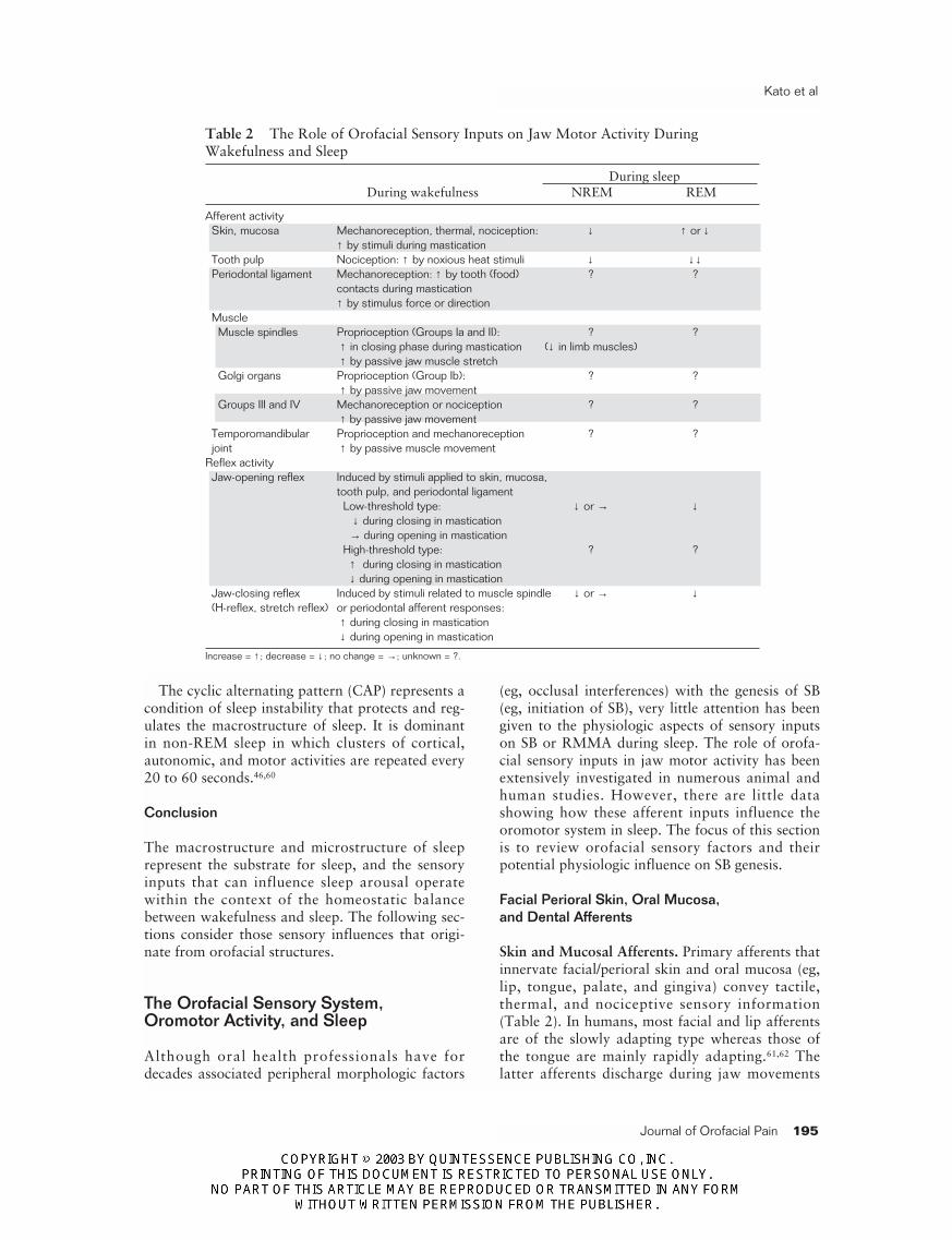
Facial Perioral Skin, Oral Mucosa, and Dental Afferents
Skin and Mucosal Afferents. Primary afferents thatinnervate facial/perioral skin and oral mucosa (eg,lip, tongue, palate, and gingiva) convey tactile,thermal, and nociceptive sensory information(Table 2). In humans, most facial and lip afferentsare of the slowly adapting type whereas those ofthe tongue are mainly rapidly adapting.61,62 Thelatter afferents discharge during jaw movements
Table 2 The Role of Orofacial Sensory Inputs on Jaw Motor Activity DuringWakefulness and Sleep
During sleepDuring wakefulness NREM REM
Afferent activitySkin, mucosa Mechanoreception, thermal, nociception: ↓ ↑ or ↓
↑ by stimuli during masticationTooth pulp Nociception: ↑ by noxious heat stimuli ↓ ↓↓Periodontal ligament Mechanoreception: ↑ by tooth (food) ? ?
contacts during mastication↑ by stimulus force or direction
MuscleMuscle spindles Proprioception (Groups Ia and II): ? ?
↑ in closing phase during mastication (↓ in limb muscles)↑ by passive jaw muscle stretch
Golgi organs Proprioception (Group Ib): ? ?↑ by passive jaw movement
Groups III and IV Mechanoreception or nociception ? ?↑ by passive jaw movement
Temporomandibular Proprioception and mechanoreception ? ?joint ↑ by passive muscle movement
Reflex activityJaw-opening reflex Induced by stimuli applied to skin, mucosa,
tooth pulp, and periodontal ligamentLow-threshold type: ↓ or → ↓
↓ during closing in mastication→ during opening in mastication
High-threshold type: ? ?↑ during closing in mastication↓ during opening in mastication
Jaw-closing reflex Induced by stimuli related to muscle spindle ↓ or → ↓(H-reflex, stretch reflex) or periodontal afferent responses:
↑ during closing in mastication↓ during opening in mastication
Increase = ↑ ; decrease = ↓ ; no change = →; unknown = ?.

Kato et al
196 Volume 17, Number 3, 2003
(usually jaw closure), in response to either pressurefrom a food bolus and/or deformation of theirreceptive fields, and to contacts between 2 oralparts (eg, tongue and lip, upper and lowerlips).61–64
The influence of sensory inputs from facial andperioral skin and from oral mucosa on the jawmotor system has been studied in humans and ani-mals. The excitatory and inhibitory influences onthe jaw motor system are thought to depend onthe stimulus modality, stimulus intensity, back-ground motor excitability, and location of theapplied stimuli.65–73 In general, low-intensity (non-noxious) stimulation that activates low-thresholdafferents produces excitatory effects on jaw-closingmuscles whereas activation of high-threshold affer-ents by high-intensity (noxious) stimulationinhibits jaw-closing muscles (also see sections onjaw-opening reflex).64 Stimulating skin andmucosal afferents in humans and animals alsomodulates rhythmic jaw movements.74–77 Thus,cutaneous and mucosal afferents from the orofa-cial region provide sensory information for coordi-nating movements of facial and oral structuresduring mastication, swallowing, and speaking.
When conscious tactile stimulus detection andspatial resolution of the lower lip was recently com-pared between patients, with or without an aware-ness of SB, no difference was found between groups.This result suggests that perioral sensory acuity (eg,2-point discrimination) is normal during wakeful-ness in subjects with an awareness of tooth grindingduring sleep.78 During wakefulness, patients withOSA showed an impaired sensory function (eg,vibratory detection) of the mucous membrane in theupper airway and a reduction in genioglossus muscletone following anesthesia of nasopharynx topicalreceptors, indicating the importance of the suprahy-oid musculature in maintaining airway patency.79,80
This emphasizes the importance of receptors in theupper airway which can respond to chemical, ther-mal, and mechanical stimuli and influence jaw andtongue position and thereby airway patency.81–83 Toour knowledge, intraoral and upper airway mucosalsensory influences on jaw and upper airway muscleshave not been tested in SB patients.
In the case of the influences of these variousafferents during sleep, cutaneous (ie, skin) sensoryinputs from the infraorbital region (division II ofthe trigeminal nerve) to the trigeminal sensorynucleus complex (TSNC) of the brainstem in ani-mals are facilitated during quiet sleep (equivalentto NREM sleep in humans) but are suppressedduring active sleep (equivalent to REM sleep inhumans) (Table 2).84 When compared to wakeful-
ness, the response of TSNC neurons to electricalstimulation of the inferior alveolar nerve (trigemi-nal nerve division III containing periodontal, toothpulp, and lip cutaneous afferents) is also sup-pressed during active REM sleep but suppression isnot obvious during quiet NREM sleep.85,86 Theseobservations support the idea that synaptic trans-mission is likely suppressed during REM sleep.87
Interestingly, sensory neurons projecting to thethalamus from the TSNC discharge differentlyduring REM sleep: neurons innervating hairmechanoreceptors around the face show facilitatedresponses to air-puff stimulation but those thatrespond to tooth pulp stimuli are suppressed.88
Since in animals this type of stimulus and responsemay be more relevant for bodily protection andsurvival, different types of sensory informationmay be processed differently based on their bio-logic relevance during sleep.89,90 This was alsoobserved in human experiments using non-trigemi-nal sensory stimulation during sleep. Non-noxiousvibrotactile stimulation applied to the arm wasfound to induce more awakening in light NREMsleep only. Following experimental muscular pain,however, the awakening response was preserved inall sleep stages.91 In another study, cutaneous heatpain induced more frequent sleep arousals thannon-noxious warm and cold stimulation.92
Periodontal Afferents. In the periodontal liga-ment, afferents convey nociceptive and tactile sen-sory information. The latter is especially subservedby rapidly and slowly adapting mechanoreceptorsthat respond to the mechanical stimulation (eg,displacement, pressure) of the teeth (Table 2). Therapidly adapting receptors fire abruptly after toothcontact associated with food chewing, while thefiring frequency of the slowly adapting receptorsincreases as the forces applied to the teethincrease.64,93,94 In addition, the afferent fibers fromthe rapidly adapting receptors have higher thresh-olds compared to those of slowly adapting recep-tors.93
The excitatory and inhibitory influences of periodontal afferents on jaw muscle activity have been studied in both humans and ani-mals.64,95–98 When activated by the insertion of chew-ing objects (eg, steel ball, plastic strips)99,100 or anunexpected mechanical load,101 low-threshold–typeafferents are thought to facilitate jaw-closing muscleactivity through a positive feedback loop. The acti-vation of high-threshold afferents, on the otherhand, suppresses jaw-closing muscles and activatesjaw-opening muscles in humans and animals; thishas been considered as a protective mechanism toguarantee the integrity of the oral tissues.99,102–105

Kato et al
Journal of Orofacial Pain 197
There has been only the 1 sleep study notedabove that investigated the response of TSNC neu-rons to electrical stimulation of the inferior alveo-lar nerve, which contains periodontal nerve affer-ents.85 Here the response of TSNC neurons wasfound to be lower in REM sleep when comparedto NREM sleep and wakefulness. From this indi-rect information, however, it is difficult to extractdefinitive information on the influence of perio-dontal afferents on jaw muscle activity duringsleep in relation to SB in humans.
Afferents from Muscles
Muscle sensory organs inform the brain about mus-cle length, displacement velocity, and nociception(Table 2). Their associated afferents are classifiedinto several groups (Ia, Ib, II, III, IV) according tothe afferent fiber diameter.64,97,106 Within the jawmusculature, muscle spindles are distributed mainlyin the jaw-closing muscles (eg, masseter, temporalis,and medial pterygoid muscles) while none or veryfew are distributed in the jaw-opening (eg, digastric)muscles.107
The larger afferents, groups Ia and II, innervatethe muscle spindles. Group Ia afferents have pri-mary endings that are mainly responsive to thevelocity and acceleration of muscle stretch whereasgroup II afferents mainly respond to the length ofmuscle stretch.108,109 The sensitivity of these musclespindle afferents to muscle stretch is controlled bythe activity of the fusimotor (gamma motor)system.106 The muscle spindle afferents are mostactive during jaw movement and voluntary jaw-closing muscle contraction.110–113 They have alsobeen reported to facilitate jaw-closing muscle activ-ity during experimental chewing in animals.100,114–116
The group Ib afferents are also muscle afferentfibers but their role in the masticatory muscles hasnot been clearly documented.64,117,118 These typesof afferents convey information about muscle ten-sion that is sensed by Golgi tendon organs. Theyare reported to be excited by muscle stretch and byjaw muscle contraction during mastication. Thelast groups, III and IV muscle afferents, have beenreported to carry sensory information frommechanosensitive and nociceptive receptors. Thegroup III muscle afferents are reported to show aresponse to jaw muscle stretch similar to that ofmuscle spindle afferents. Interestingly, some recentstudies have revealed that activation of musclenociceptive afferents suppresses agonistic muscleactivity but facilitates antagonistic muscle activitywhen muscle pain is induced although others haveindicated that both muscle groups (jaw opener and
closer) may be activated simultaneously in certainconditions.119–122 A decrease in movement speedand amplitude is observed both in humans andanimals. Experimental muscle pain also reducesthe jaw-closing muscle activity during chewing inhumans and the frequency of rhythmic musclebursts in anesthetized animals.123–125
There have been no studies of jaw muscle affer-ents during sleep. However, in the limb muscles ofcats, the discharge of muscle spindle afferents istonically suppressed during NREM sleep and sub-stantially decreased in REM sleep.126 A transientfacilitation of discharge during REM sleep canoccur spontaneously, however, with or withoutsimultaneous muscle twitch, probably due to tran-sient activation of gamma motoneurons.126,127
When transient sleep arousals were induced byexperimental pinch stimulation, some of thegamma motor fibers exhibited a phasic increase intheir firing frequency.127
Afferents of the Temporomandibular Joint (TMJ)
The temporomandibular joint afferents that inner-vate the joint capsule are sensitive to small dis-placements and movements (eg, opening, laterotru-sive, and protrusive jaw movements)117,129 andmechanical or chemical stimuli (Table 2).122,130,131
However, these afferents do not discharge atextreme jaw positions.117 This suggests that thesereceptors are important for proprioceptive jawmovement control. It has also been reported thatmasseter motor nerve activity is suppressed whenthe TMJ condyle is pressed against the joint cap-sule in cats.129 Only a few studies have investigatedthe role of TMJ afferents on jaw motor con-trol64,97,121,122,132 and, to our knowledge, no studyhas been made during sleep.
Trigeminal Reflexes
Jaw-opening Reflex (JOR). The JOR is a veryrapid di-synaptic motor response associated withthe protection of oral tissues (Table 2). In animals,it can be evoked by stimulation of orofacial low-threshold or high-threshold mechanoreceptors (eg,lip and oral mucosa), electrical stimulation of thetrigeminal nerve (eg, inferior alveolar nerve), andmechanical impact on teeth.64,93,97,99,104,133 The inhi-bition of jaw-closing motoneurons and the facilita-tion of jaw-opening motoneurons has been exten-sively studied in animals but its role in humans isless clear.64,76,93,134,135 In animals, when the JOR istriggered during chewing by the stimulation oflow-threshold mechanoreceptors (eg, non-painful

Kato et al
198 Volume 17, Number 3, 2003
stimuli), the reflex is inhibited during the closingphase but is preserved during the openingphase.64,133,136 In contrast, when the JOR is inducedby stimulation of high-threshold mechanoreceptors(eg, using high intensity of electrical stimulation),it is facilitated during the closing phase and inhib-ited during the opening phase.64,105 The latterreflex system may also play an important role dur-ing chewing by protecting oral structures fromdamage (eg, tooth fracture, oral mucosa bit-ing).64,105 Its role in reducing jaw-closing muscleactivity during jaw closure is more evident if a per-son bites either their tongue or an unexpected hardobject in food.
Although in cats the amplitude of the JOR hasbeen reported to be facilitated during NREM sleepwhen compared to wakefulness,137 the oppositefinding was reported recently in rabbits (Table2).138 However, in REM sleep, the JOR wasstrongly reduced in both studies.137,138 Althoughobservations in human experiments using electricalstimulation to trigger a leg flexion reflex haveshown that the reflex threshold was increased inNREM sleep compared to wakefulness and wasmaximal in REM sleep, no direct information isavailable for the JOR in humans.137
Lateral Jaw Reflex. In anesthetized animals, thelateral jaw reflex occurs in response to tonicmechanical stimuli of periodontal mechanorecep-tors, which enhances lateral pterygoid muscleactivity, leading to the large lateral jaw excursionsobserved during the slow closing phases of chew-ing.99,114,139,140 Although its relevance in humans isnot as clearly defined, it has been observed thatlarger lateral jaw movements, related to anincreased duration of the jaw-closing muscle burstsand chewing cycle, are present when both humansand animals chew hard foods.100,114,141–144
Jaw-closing Reflex. In awake humans, amonosynaptic reflex of the jaw-closing muscles iselicited by a mechanical chin tap that stretchesmuscle receptors (stretch reflex) or by direct elec-trical stimulation of muscle spindle afferents in thetrigeminal nerve (H-reflex), or by mechanical orelectrical activation of periodontal recep-tors.64,81,97,128,145–147 The amplitude of these reflexesdepends on the excitability of the trigeminal alphaand gamma motoneurons and the amount of mus-cle spindle afferent input to the alpha motoneu-rons. These monosynaptic reflexes are increasedduring the jaw-closing phase of rhythmic jawmovements and during tooth-clenching tasks(Table 2).148–151 In animals, the monosynaptic mas-seteric reflex (MMR) is evoked by electrical stimu-lation of neurons in the trigeminal mesencephalic
nucleus where the primary afferent cell bodies ofjaw muscle spindle afferents are located. Thisreflex activity is increased during the jaw-closingphase and inhibited during the jaw-opening phaseof mastication.136
To our knowledge, there have been no studieson trigeminal monosynaptic reflex modulationduring sleep in humans, perhaps due to the inva-sive technique required for nerve stimulation andproblems associated with stability of the stimula-tion set-up (eg, electrode displacement in responseto body movements during sleep). In animals, theamplitude of the MMR did not significantlydecrease from wakefulness to NREM sleep, andwas markedly suppressed during REM sleep.129,138
However, there are controversial findings that theamplitude of the MMR is facilitated or suppressedin relation to rapid eye movement episodes duringREM sleep.129,138
Orofacial Sensory Inputs Triggering RhythmicJaw Muscle Activity
Several animal studies have reported that rhythmicjaw muscle activity is observed following periph-eral sensory stimulation applied to oral mucosalmechanoreceptors.64,152–154 It was also reported thatunder light anesthesia, both tonic mechanical stim-ulation of the palate and specific directional pres-sure applied to the incisor teeth elicited rhythmicjaw muscle activity in rats.155,156 To the authors’knowledge, these types of experimental responsesdo not occur in humans, and so their relevance toSB is difficult to determine.
Conclusion
Several animal studies have shown either facilita-tion or inhibition of the trigeminal peripheral oro-facial sensory factors and/or related reflexes inrelation to sleep but their relevance to SB genesishas not yet been elucidated in humans. Clearly,further studies on orofacial sensory feedbackmechanisms in relation to sleep are needed.
Jaw Muscle Tone, Mandibular Posture,and Tooth Contact During Sleep
When discussing the relevance of peripheral sensoryfactors in SB genesis (including tooth contact), theclassic question arises: “Which occurs first, toothcontact or jaw muscle activation?” The followingsection is focused on the putative role of muscletone and mandibular posture during sleep in SB.

Kato et al
Journal of Orofacial Pain 199
Muscle Tone and Mandibular Posture
Generally, during sleep, skeletal muscles arerelaxed and muscle tone is lower when comparedto wakefulness.6 Similarly, masticatory muscletone decreases from wakefulness to NREM sleepto a similar level between sleep stages 1 to 4.7,8
During REM sleep, muscle tone is normally mini-mal due to the powerful motor suppression, alsotermed “muscle atonia” to reflect the completeloss of muscle tone.6,7,20,157 However, masticatorymuscle activity does not disappear completely dur-ing REM sleep; very low residual muscle tone maypersist, suggesting that the term “hypotonia”would more accurately describe the state of masti-catory muscle tone during REM sleep.7,157
With the aid of intraoral devices that measurethe vertical distance between the maxillary andmandibular incisors, it has been demonstrated thatduring sleep the jaw is usually open 1 to 5 mm forapproximately 90% of the total sleep time.158 Thisdistance can be significantly greater during REMsleep when compared to the lighter non-REMsleep stages (eg, stage 1). Interestingly, a sleeparousal reaction precedes an increase in jaw mus-cle tone (likely associated with jaw closure) duringthe sleep of normal subjects, sleep apnea, and SBpatients.7,31,158,159 These observations suggest thattooth contact is most likely to be a final outcomein jaw closure secondary to the jaw muscle activa-tion during sleep.
The use of ambulatory systems to record mas-seter EMG activity has revealed that edentuloussubjects also show RMMA during sleep even inthe absence of dentures in the mouth.160 This find-ing further supports the idea that jaw motor acti-vation during sleep can occur in the absence oftooth contact and feedback from periodontalreceptors.
Jaw Motor Events and Tooth Contacts
There are several studies reporting data on theduration and frequency of jaw motor events in nor-mal and SB patients (Table 3).11,12,161–170 The jawmotor events, scored as SB, were observed more fre-quently in SB patients (3.6 to 6.8 episodes per hourof sleep) than in normal subjects (0.5 to 5.6episodes), and the total duration of these events perhour of sleep was longer in SB patients (0.6 to 1.65minutes) compared to normal subjects (0.12 to 0.89minutes). On the other hand, only a few studieshave directly measured tooth contact during sleep(Table 3).171–176 These studies used intraoral sensorsto assess tooth contact but no recordings were
made of quantitative sleep variables. One studyreported that SB patients with a temporomandibu-lar disorder (TMD) showed significantly higher fre-quency and duration of tooth contacts compared tonormal subjects and patients with TMD.176 Severaltechniques allow indirect assessment of the numberof tooth contacts in relation to SB-related toothgrinding (Table 3).10,11,170,177 The results from thepolygraphic studies using an audio monitor revealthat the incidence of tooth grinding-related noise ismore frequently observed in SB patients. From theseresults, we can summarize that SB patients haveapproximately a 3 to 4 times higher number andduration of SB episodes and tooth contacts com-pared to normal subjects. In addition, the presenceof a tooth grinding noise seems to be characteristicof SB patients.
When interpreting these data, however, thereader should consider a number of limitationsthat may account for discrepancies between studyresults: (1) The different methods used to recordSB and tooth contact (eg, polygraphic versusambulatory recording systems, EMG of mastica-tory muscles versus mechanical impact on sensorsto score motor events). (2) Standardized cut-offcriteria for normal subjects and SB patients werenot used; this creates heterogeneity in the studysample. (3) Different scoring criteria for SBepisodes (eg, EMG threshold, muscle burst pat-terns) were used by the research groups, with noclear consensus on the definition of an SB episode.As an aside, our group has recently carried out apost-hoc analysis for EMG threshold, which wepreviously proposed as more than 20% of maxi-mum voluntary contraction (MVC) of massetermuscle activity during wakefulness. A retrospectiveanalysis of the 20% criteria used has shown, inreality, that some SB events were also scored in therange between 10% and 20% MVC. Thus, in ourrecent studies, we used 10% to 20% of MVC as aminimum EMG threshold, similar to anotherstudy.11,162 (4) Finally, several studies havereported that the patterns of jaw muscle contrac-tions (eg, in closer and opener muscles) and jawmovements related to SB are not the same as thosefound in awake chewing in humans.7,14,20,32,166,178,179
This suggests that tooth contact related to SBtooth grinding may take a different form from thatoccurring during wakefulness.180–184
Conclusion
During sleep, the lower jaw tends to be open dueto jaw-closer muscle relaxation. Following a sleeparousal, it is frequently observed that an increase

Kato et al
200 Volume 17, Number 3, 2003
in jaw muscle activity may lead to the jaw closure.Thus, tooth contact seems to be the consequenceof jaw motor activation during sleep rather than acause. It has also been clearly shown that jaw mus-cle EMG events and tooth contact occur more fre-quently in SB patients than normal subjects. Thisobservation is supported by polysomnographicstudies that demonstrate more frequent toothgrinding episodes in SB patients in comparison tonormal subjects.
Sleep Processes and SB
This section aims to clarify the association of SBwith sleep processes.
Sleep Macrostructure and SB
Interestingly, most young and healthy SB patientsexhibit a normal sleep structure and usually do notcomplain of sleep disturbance unless they haveeither concomitant chronic pain and/or other sleepinfluencing disorders (eg, sleep breathing disor-ders).2–5,185 In contrast to an original report that
Table 3 SB and Related Variables: EMG Events, Tooth Contacts, Tooth Grinding Sound
Thresholds CriteriaSample Recording for SB for SB/
Author (age/range or mean) Frequency† Duration† system episode normals‡
SB-related jaw motor eventsGallo et al164* 21 normals (31;22–37) 10.2 0.8 min Ambulatory EMG recording Algorithms 1,2,3Okeson et al161* 20 normals (42.5;21–58) 5.6 0.6 min PSG � 40% MVC —Okeson et al165* 30 normals (70.2;60–87) 3.0 0.3 min PSG “ —Clarke et al168* 10 adults 4.9 (0.3–11.6) 0.7 min (0.1–2.3) Ambulatory EMG recording � 2-second duration —Amemori et al166* 2 SB patients and 8.2 (4.5–10.9) 1.8 min (0.8–2.9) PSG + jaw movement � 5% MVC 1,2
1 normal (26–32) recordingIkeda et al162* 9 SB patients (26.2) 3.6 0.6 min Ambulatory EMG recording � 10% MVC 2,3Pierce and Gale169 100 SB patients (18–72) 16.7 (1.4–67.6) 0.03–2.6 min Ambulatory EMG recording � 20 µV 1,2,5Nishigawa et al167* 5 normals (29.6;26–36) 3.5 (0.3–11.5) 0.4 min (0.02–1.4) Ambulatory bite force recording � 5 kgf 1,2,3
5 SB patients (27.6;23–33) 3.5 (0.5–7.5) 0.4 min (0.03–0.9) with full arch oral applianceKydd and Daly163* 10 normals (21–43) — 0.4 min (0.4–0.6) Ambulatory EMG recording Unknown 1,2
10 SB patients (21–43) — 1.4 min (0.4–2.0)Sjöholm et al12 6 normals (26.3;17–36) 1.9 0.9 min (0.3–1.4) PSG � 20% MVC 1,2,3Lavigne et al11 31 normals (27.6;15–40) 1.8 (0.1–12.6) — PSG with audio-video � 10/20% MVC‡ 1,2,4
33 SB patients (26.8;20–50) 5.8 (1.2–15.2) —Miyawaki et al170 7 normals (25.3) 0.5 0.12 min (0.05–0.25) PSG with audio-video � 10/20% MVC‡ 1,2,4
9 SB patients (23.2) 6.8 1.15 min (0.68–1.9)Tooth contactPowell and Zander171 7 normals (34–56) 29.8 (6–140) — A sensor in fixed bridge � 1,2,3Graf172 1 normal — 0.16 min A strain gauge on the molar � —Brewer173 Dental patients — (0.38–18.8 min) Radio transmitter in denture � —Yamashita et al174* 3 normals (26–31) 40.7 (25.7–65.1) 0.8 min (0.3–1.5) Micro-photo sensor in metal � —
attachment on dentitionsAkamatsu et al175* 2 SB patients (26 and 27) 52.4 (10.3–94.5) 0.9 min (0.5–1.3) Magnetic sensor in metal attach- � 1
ment on upper/lower dentitionsTrenouth176* 10 normals (21.4;20–28) 45 0.68 min An electric circuit consist with � 1,2,3
9 SB patients with TMD 165 4.8 min wires placed on the incisors(21.7;16–36)6 TMD patients (21.5;15–35) 125 1.4 min
Tooth grinding noise incidentsReding et al177* 18 normals (24;17–56) 0 0 PSG with microphone � 40 µV 1
40 SB patients (20.6;7–50) 5.8 0.34 minSB episodes with grinding noiseLavigne et al10* 18 normals 0 — PSG with audio-video � 10/20% MVC§ 1,2,4
18 SB patients 0.97 —Lavigne et al11* 31 normals (27.6;15–40) 0 — PSG with audio-video � 10/20% MVC§ 1,2,4
33 SB patients (26.8;20–50) 1.9 —Miyawaki et al170 7 normals (25.3) 0.1 (0–0.3) 0.03 min (0–0.07) PSG with audio-video � 10/20% MVC§ 1,2,4
9 SB patients (23.2) 3.4 (0.1–10) 0.62 min (0.01–1.54)
*The numbers were estimated from the data presented in the article.†Frequency and duration [mean (range)] were presented as times per hour of sleep and minutes per hour of sleep, respectively.‡Selection criteria: 1 = a history/awareness of tooth grinding; 2 = clinical signs of SB including tooth wear and morning jaw muscle discomfort and masseter muscle hypertrophy; 3 = clinicalsigns of temporomandibular disorders; 4 = polysomnographic criteria; 5 = ambulatory criteria.§Ten percent of SB episodes were between 10% and 20% of MVC range (see text).PSG = polysomnography; MVC = maximum voluntary clenching during wakefulness; TMD = temporomandibular disorders. / = unavailable or unknown.

Kato et al
Journal of Orofacial Pain 201
was later refuted,186,187 and to studies describingcases of secondary SB,46,188–190 most of the oromo-tor activity related to SB occurs in stages 1 and 2of NREM sleep (60% to 85%). Very few SBepisodes occur in sleep stages 3 and 4 of NREMsleep (� 5%), while on occasion SB is observed inREM sleep (� 10%).10,13,177,191–194 The occurrenceof SB episodes in REM sleep is an interesting para-dox since this sleep stage is usually characterizedby muscle hypotonia. So far, the only explanationgiven for the occurrence of SB in REM sleep inpatients without sleep disorders is the presence ofbrief and transient sleep arousal activity that canoccur during this sleep stage.6,46,56
Sleep Microstructure, Micro-arousal, and SB
Previously, SB tooth-grinding episodes werereported to be associated with K-complexes in SBpatients.176,193 However, in a recent controlledstudy, K-complexes were found to be associatedwith only 12% of RMMA episodes in SB patientsand with 21% in normal subjects.185 Moreover, SBpatients were found to present 42.7% fewer K-complexes during sleep compared to normal sub-jects.184 Although the number of sleep spindles didnot differ between SB patients and normal sub-jects, SB episodes were not associated with sleepspindles.175,185
Since an episode of SB lasts anywhere from 5 to 15 seconds, it can be considered a transientmotor event occurring during sleep (Table4).11,12,32,161–163,177,190,195 As previously described, SBpatients show approximately 3 times moreRMMA episodes, twice as many muscle bursts,and 40% higher EMG burst amplitudes duringsleep than normal control subjects.11,12 These find-ings suggest that SB is an exaggerated transientmotor (muscle) activity occurring during sleep inan otherwise “normal” sleeper.
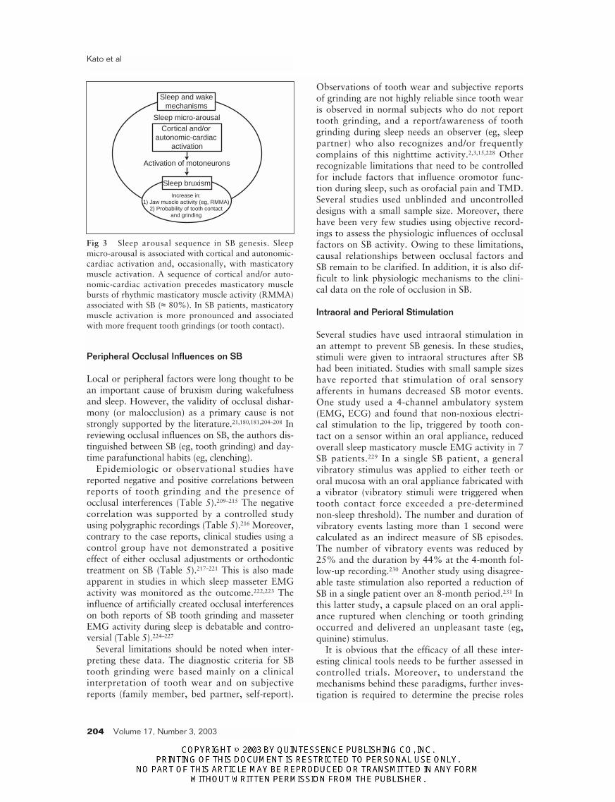
In most studies, young healthy SB patients displaya normal incidence of micro-arousals, but the inci-dence lies in the upper range of the normallimit.11,13,32,185 However, polysomnographic studiesfrom as early as the 1960s have reported that SBepisodes are associated with physiologic changes (eg,micro-arousals) that include transient EEG activity,increases in heart rate, and frequent sleep stage shifts(Table 5).177,190,193,196,197 Furthermore, recent studiesnow support the association between SB and a tran-sient EEG/autonomic activation. The first few ofthese studies have shown that nearly 80% of SBepisodes are scored in association with a phase A3of the cyclic alternating pattern (CAP).13,60,198,199
Additional recent evidence supporting an association
between SB and sleep arousals is derived from theobservation that in both SB patients and normalsubjects most of the RMMA episodes are precededby suprahyoid muscle activation, an EMG sign ofsleep micro-arousal.11,32 Interestingly, severalpolysomnographic studies have also reported otherspecific physiologic changes (eg, cortical EEGchange and heart rate acceleration) before the occur-rence of an SB episode (Table 4). For example,changes in EEG frequency are observed before SBepisodes and alpha EEG intrusion precedes 50% ofSB episodes.177,190,195–197 In addition, an increase inheart rate is followed by jaw-closer muscle activ-ity.177,195–197 When these changes in EEG activity andheart rate are measured in relation to the onset ofRMMA episodes in SB and normal subjects, a tran-sient sequence is found: (1) Sleep bruxism patientshave a significant increase in EEG activity (alphaand delta EEG amplitude) approximately 4 secondsbefore the onset of an RMMA episode. (2) A signifi-cant increase in heart rate occurs 1 cardiac cyclebefore an RMMA episode in SB patients, but theoverall time course from EEG activity and increasesin heart rate related to RMMA onset are statisticallysimilar in both SB patients and normal control sub-jects.32 Thus, a sequence that starts with corticalEEG activation followed by heart rate accelerationprecedes jaw-closer muscle activation, with theRMMA episode in SB being the final event in thissleep micro-arousal reaction (Fig 3). This finding hasbeen supported by our experimental study in whichRMMA occurred following experimentally inducedarousals.7 Furthermore, several studies have revealedthat small and subtle changes in autonomic-cardiacheart rate variability can precede sleep arousals (�10 seconds).34,200 After applying frequency analysison heart rate variability, our preliminary observa-tions suggested that in the 3 minutes before RMMAepisodes, SB patients showed a higher sympatheticand a lower parasympathetic activity compared tonormal subjects.33 The changes in EEG and EMGactivity are probably under the influence of the auto-nomic nervous system fluctuation during sleep.
Several issues must be considered when inter-preting the literature mentioned above:
1. In most studies, patients were monitored in asleep laboratory setting, which is not a naturalsleep environment, even though environmentalhabituation is done prior to recording.9
2. The analysis of autonomic (cardiac) activity wascarried out in a time domain (beats per minute),which does not allow for the detection of subtlechanges in sympathetic and parasympatheticcomponents that could have preceded the EEG,

Kato et al
202 Volume 17, Number 3, 2003
electrocardiogram (EKG), and EMG activa-tion.34,201,202 This issue is currently being investi-gated in our laboratory.33
3. Tonic clenching activity (� 10% of all SBepisodes) was not analyzed.
4. Sleep bruxism occurring in REM (� 10% of allSB episodes) is not well understood.
5. Most recent studies used only severe SB patients.It is not known whether the association betweenEEG, EKG, and jaw EMG activity is related tothe severity (light to severe levels) of SB patients.
6. Studies were not population-based (ie, SBpatients were mostly in their twenties and theinfluence of age on sleep architecture is alreadyknown, especially after the fourth decade203).
Conclusion
Although SB episodes occur frequently in younghealthy SB patients, gross sleep architecture isundisturbed. Based on polysomnographic studies,SB seems to occur in relation to subtle changes in
both sleep macrostructure (eg, in light non-REMsleep stages 1 and 2) and microstructure (eg, EEG-cardiac activation). These studies suggest that SBmay be a masticatory muscle activation related tothe exaggerated responses often found associatedwith sleep arousals. The observation that withsleep arousals a rise in EEG and cardiac activitiesprecedes RMMA in SB patients further suggests aprimary role for autonomic and central nervoussystems in the genesis of SB.
Interaction Between Sensory Inputs,Sleep, and Oromotor Activity
Sleep architecture (eg, macrostructure and micro-structure including arousal) can be altered by vari-ous sensory influences.52,53,89 Therefore, interactionsbetween sensory input, autonomic, and central ner-vous systems are important in sleep and wake regu-lation. In this section, the roles of various sensoryinfluences in SB genesis will be reviewed.
Table 4 Summary of Evidence that Micro-Arousal is Related to SB
SB Heart rateSample episode acceleration Motor
Author (age/range or mean) duration % increase EEG change activation
Takahama196 7 SB patients / Increase during episode Change before episode Wrist/masticatory musclesactivity before episode
Tani et al197 3 SB patients / Increase before episode Theta, alpha, beta before /(13–25) episode
Reding et al177 40 SB patients 9.0 s 4 beats before episode Low amplitude and high 24% with body movement(20.6; 7–50) (2.7–66.5) (26.8%; 2.0–68) frequency EEG before episode18 normals: not described (24.0;17–56)
Satoh and Harada193 15 SB patients / Increased Associated with alpha activity /(19–48) and K complex
Kydd and Daly163 10 SB patients / Increased 18 beats/min / Never(21–43) (2%–30%)10 normals / Increased 18 beats/min / Never(21–43) (2%–30%)
Okeson et al161 20 normals 6.7 s (16.6%; 6.1–40.2) 57.7% with leg jerk(42.5; 21–58)
Ikeda et al162 9 SB patients 10 s (10%–14.9%) / /(26.2)
Okura et al195 5 SB patients 9.1 s 4 to 8 s before episode Beta and delta activity /(33.8; 25–48) before episode
Bader et al190 24 SB patients 6.3–11.1 s At the onset of masseter EEG alpha activity before /(35; 27–67) burst (17.6%) episode
Macaluso et al13 6 SB patients (34) / (19.3%) 87.3% during CAP 79.6% with leg jerk6 normals (34):not described
Lavigne et al11 8 SB patients 9.4 s / / 78.6% preceded by(27.6) jaw-opener activation8 normals 7.8 s / / 69.1% preceded by(26.8) jaw-opener activation
Kato et al32 10 SB patients 14.9 s 1 heart beat before 4 s before episode 87.5% with neck and leg(26.5; 20–36) episode (26.7%) jerk10 normals 11.9 s Statistically similar to Statistically similar to 85% with neck and leg(24.2; 18–41) SB patients (28.1%) SB patients jerk
CAP = cyclic alternating pattern./ = unavailable or unknown.

Kato et al
Journal of Orofacial Pain 203
Table 5 Studies of SB-Tooth Grinding and Occlusal Factors
Intervention a: Selection criteria† Outcomes andAuthor Sample* or design b: Assessment of SB† results Critical comments
Epidemiologic or observational studiesNilner209 440 children Oral examination, interview a,b: 1 Correlation with OI Cross-sectional analysis,
(7–14; 222M, 218 F) subjective assessmentNilner210 309 adolescents Oral examination, interview a,b: 1 No correlation with OI Cross-sectional analysis,
(15–18; 147 M, 162 F) subjective assessmentBrandt211 1,342 children Oral examination, interview a,b: 1 Association with overjet, Cross-sectional analysis,
(6–17; 667 M, 675 F) overbite, and mesiodistal subjective assessmentmolar relationships
Gunn et al212 151 children Oral examination, interview a,b: 1 No correlation with Subjective assessment,(6–18; 67 M, 84 F) occlusal measures small sample
Lindqvist213 79 teenagers (14) A: SB (n = 34); B: controls (n 45) a,b: 1 No. of subjects with OI: Subjective assessment,oral examination A (55%) � B (29%) small sample
Henrikson et al214 183 girls (11–15) A: Class II malocclusion a,b: 1 No. of grinding reports: Subjective assessment(n = 123); B: normals (n= 60) 23/123 (A) � 9/60 (B)oral examination
Kampe et al215 225 children (13–15) A: with restorations (n = 129) a,b: 1 No. of grinding reports: Subjective assessmentB: without restorations (n = 96) 9/129 (A) � 4/96 (B)
Polygraphic studiesLobbezoo et al216 10 SB patients and A: 10 SB patients; B: 10 normals; a: 1,2,4 No group differences for any Objective assessment but
10 normals cephalometry, cast model, b: 4 occlusal and morphological cross-sectional analysisoral examination variables
Clinical studiesForssell et al217 91 TMD patients with Double-blind placebo controlled a,b: 1 Before treatment No statistical analysis;
headache A: Occlusal adjustments (n = 48) A: 16/48; B: 14/43 subjective assessment;(16–52; 11 M, 80 F) 5- to 20-month follow-up After treatment orofacial pain
B: Placebo adjustments (n = 43) A: 9/48; B: 8/432- to 6-month follow-up
Kirveskari et al218 65 healthy university Double-blind placebo controlled a,b: 1 3 drop-outs in A No statistical analysis,students (19 M, 46 F) A: Occlusal treatment (n = 33) A: improved in 1 and subjective assessment
B: Placebo treatment (n = 32) impaired in 5 subjects no description of SB2-year follow-up B: impaired in 7 subjects population
Henrikson and Nilner219 183 girls (11–15) A: Class II-treatment (n = 65) a,b: 1 Before treatment Subjective assessmentB: Class II-no treatment (n = 58) A: 23%; B: 22%; C: 9%C: Normals (n = 60) 2 years after treatment2-year follow-up A: 11%; B: 15%; C: 7%
Kerstein and Farrell220 32 SB patients with Descriptive open trial a: 1 Marked decrease or complete Poor description for jaw muscle pain occlusal equilibration b: 6 absence for 7 days after orofacial pain(15–51; n = 32) treatment
Yustin et al221 86 TMD patients The use of anterior bite splint a: 6 Most patients relieved from Poor description for (36.4; 18 M, 68 F) during sleep (15.5 months) b: 6 bruxism orofacial pain
followed by the continuation ofsplint, occlusal adjustment, ortho-dontic/prosthetic treatment ororthognathic surgery
Bailey and Rugh222 9 SB patients Occlusal adjustment a: 1 No change in 6, increase Abstract, unclear diagnosisbaseline-intervention-postbaseline b: 5 in 2 and decrease in 1
Kardachi et al223 16 SB patients Randomized trials a: 6 No difference between groups Unclear diagnostic criteria4 normals A: Occlusal adjustment (n = 4) b: 5 for SB, randomized but (18–39; 8 M, 12 F) B: Placebo adjustment (n = 4) small sample, unclear for
C: Biofeedback (n = 4) specific SB activityD: Control feedback (n = 4)E: Normals (no intervention; n = 4)Comparison: baseline-intervention
Experimental studiesShiau and Syu224 13 SB patients Open trial: occlusal interferences a,b: 1 A: 4 dropouts; no increase Subjective assessment
14 normals created by metal overlay (1.5 mm in 5; decrease or absence(18–30; 15 M, 12 F) thickness) 1-month follow-up in 4
B: 2 dropouts, no changeRugh et al225 8 SB patients Cross-over design a: 1,2,5 No difference between Objective assessment,
(23–46; 2 M, 6 F) splint: canine guidance (A) and b: 3,5 the 2 splints influence of splint insertion,molar guidance (B) unclear for specific SB 1-week recording activity
Rugh et al226 10 normal subjects Open design, occlusal inter- a: 1,2 Increase in 1, no changes Objective assessment,(26–41; 5 M, 5 F) ference created by crown b: 3,5 in 4, decrease in 5 unclear for specific SB
(0.5–1.0 mm); 1-week recording activityTakeda et al227 7 normal subjects Open design, occlusal interference a: 6 Increased masseter EMG Abstract, objective assess-
created by class I inlay (0.1 mm); b: 6 activity after intervention ment, unclear for specific 1-week recording SB activity
*Age (range or average); sex (F = female; M = male).†Criteria to select SB patients and assess SB activity: 1 = a history/awareness of tooth grinding; 2 = clinical signs of SB including tooth wear and morning jaw muscle discomfort and massetermuscle hypertrophy; 3 = clinical signs of temporomandibular disorders (TMD); 4 = polysomnographic criteria; 5 = sleep masseter muscle activity with ambulatory system; and 6 = unclear.OI = occlusal interferences.
Kato et al
204 Volume 17, Number 3, 2003
Peripheral Occlusal Influences on SB
Local or peripheral factors were long thought to bean important cause of bruxism during wakefulnessand sleep. However, the validity of occlusal dishar-mony (or malocclusion) as a primary cause is notstrongly supported by the literature.21,180,181,204–208 Inreviewing occlusal influences on SB, the authors dis-tinguished between SB (eg, tooth grinding) and day-time parafunctional habits (eg, clenching).
Epidemiologic or observational studies havereported negative and positive correlations betweenreports of tooth grinding and the presence ofocclusal interferences (Table 5).209–215 The negativecorrelation was supported by a controlled studyusing polygraphic recordings (Table 5).216 Moreover,contrary to the case reports, clinical studies using acontrol group have not demonstrated a positiveeffect of either occlusal adjustments or orthodontictreatment on SB (Table 5).217–221 This is also madeapparent in studies in which sleep masseter EMGactivity was monitored as the outcome.222,223 Theinfluence of artificially created occlusal interferenceson both reports of SB tooth grinding and masseterEMG activity during sleep is debatable and contro-versial (Table 5).224–227
Several limitations should be noted when inter-preting these data. The diagnostic criteria for SBtooth grinding were based mainly on a clinicalinterpretation of tooth wear and on subjectivereports (family member, bed partner, self-report).
Observations of tooth wear and subjective reportsof grinding are not highly reliable since tooth wearis observed in normal subjects who do not reporttooth grinding, and a report/awareness of toothgrinding during sleep needs an observer (eg, sleeppartner) who also recognizes and/or frequentlycomplains of this nighttime activity.2,3,15,228 Otherrecognizable limitations that need to be controlledfor include factors that influence oromotor func-tion during sleep, such as orofacial pain and TMD.Several studies used unblinded and uncontrolleddesigns with a small sample size. Moreover, therehave been very few studies using objective record-ings to assess the physiologic influences of occlusalfactors on SB activity. Owing to these limitations,causal relationships between occlusal factors andSB remain to be clarified. In addition, it is also dif-ficult to link physiologic mechanisms to the clini-cal data on the role of occlusion in SB.
Intraoral and Perioral Stimulation
Several studies have used intraoral stimulation inan attempt to prevent SB genesis. In these studies,stimuli were given to intraoral structures after SBhad been initiated. Studies with small sample sizeshave reported that stimulation of oral sensoryafferents in humans decreased SB motor events.One study used a 4-channel ambulatory system(EMG, ECG) and found that non-noxious electri-cal stimulation to the lip, triggered by tooth con-tact on a sensor within an oral appliance, reducedoverall sleep masticatory muscle EMG activity in 7SB patients.229 In a single SB patient, a generalvibratory stimulus was applied to either teeth ororal mucosa with an oral appliance fabricated witha vibrator (vibratory stimuli were triggered whentooth contact force exceeded a pre-determinednon-sleep threshold). The number and duration ofvibratory events lasting more than 1 second werecalculated as an indirect measure of SB episodes.The number of vibratory events was reduced by25% and the duration by 44% at the 4-month fol-low-up recording.230 Another study using disagree-able taste stimulation also reported a reduction ofSB in a single patient over an 8-month period.231 Inthis latter study, a capsule placed on an oral appli-ance ruptured when clenching or tooth grindingoccurred and delivered an unpleasant taste (eg,quinine) stimulus.
It is obvious that the efficacy of all these inter-esting clinical tools needs to be further assessed incontrolled trials. Moreover, to understand themechanisms behind these paradigms, further inves-tigation is required to determine the precise roles
Sleep and wakemechanisms
Sleep micro-arousal
Cortical and/or autonomic-cardiac
activation
Activation of motoneurons
Sleep bruxism
Increase in:1) Jaw muscle activity (eg, RMMA)
2) Probability of tooth contactand grinding
Fig 3 Sleep arousal sequence in SB genesis. Sleepmicro-arousal is associated with cortical and autonomic-cardiac activation and, occasionally, with masticatorymuscle activation. A sequence of cortical and/or auto-nomic-cardiac activation precedes masticatory musclebursts of rhythmic masticatory muscle activity (RMMA)associated with SB (≈ 80%). In SB patients, masticatorymuscle activation is more pronounced and associatedwith more frequent tooth grindings (or tooth contact).

Kato et al
Journal of Orofacial Pain 205
of sensory stimulations on sleep architectures (eg,sleep stages, micro-arousal) and behavioral condi-tioning (eg, biofeedback paradigms) in relation totrigeminal nerve excitability.20,89,90,98,232–234
Visceral Sensory Stimuli, Salivation, andSwallowing
During wakefulness, chemical or mechanical stim-uli applied to the pharynx and esophagus (eg, byeither experimental stimuli or refluxed stomachcontent) induces swallowing, which prevents aspi-ration of undesirable substances and damage toesophageal structures.98,235,236 The stimulation ofvisceral vagal afferents (tenth cranial nerve) inthese structures activates the swallowing centerslocated in the brainstem.237,238
Interestingly, visceral vagal afferents also projectto central nervous system structures (eg, the nucleustractus solitarius) involved in both arousal andswallowing.238–242 During sleep, human swallowingcan be induced by stimulation of the pharyngealand esophageal tissues243–247 and sleep arousals havebeen found to concomitantly increase the number ofswallowing episodes.244,247 This suggests that sen-sory input from vagal visceral afferents could alsotrigger sleep arousals, which may in turn facilitateorofacial activity during sleep. In fact, gastroe-sophageal reflux (GR) events resulting in decreasingesophageal pH were reported to be associated withboth increased sleep arousals and swallowing dur-ing sleep.235,248,249 Although an increase of swallow-ing in relation to GR events is characteristic inpatients with GR, healthy subjects showed swallow-ing during sleep in the absence of reflux events.250,251
Salivary flow is known to decrease significantlyduring sleep.252,253 However, even during sleep,saliva still plays important physiologic roles suchas the lubrication and protection of oro-esophageal structures, teeth, and mucosa.236
During sleep, dryness, and/or acidification of oro-esophageal tissues may generate sensory signals,possibly as a “warning” signal, which inducessleep micro-arousal with subsequent salivation sec-ondary to sleep RMMA.235,236,254,255 Interestingly,salivary flow is associated with the stimulation ofintraoral mechanoreceptors and gustatory recep-tors.93,256–259 Salivation is high on the chewing sideof the mouth and depends on chewing frequencyand bite force.256,260 Thus, it has been hypothesizedthat RMMA may be associated with salivary secre-tion and lubrication of the oral cavity and theupper alimentary tract during sleep.236
SB and swallowing occur more frequently dur-ing light NREM sleep and they are associated with
sleep arousal. Recently, we found that close to60% of RMMA episodes in SB patients and nor-mal subjects occurred concomitantly with swal-lowing-related laryngeal movements.170 Moreover,swallowing occurred more frequently in the lastthird of SB episodes. It remains possible thatRMMA associated with SB is also triggered byoral dryness, which in turn can cause more dentaldamage due to the absence of lubricant.236 Therelationships between sleep micro-arousals, sleepRMMA/swallowing, and salivation during sleepneed further investigation before the abovehypotheses are confirmed.
Other Sensory Stimuli that Influence SleepBruxism Activity
In a biofeedback experiment, high-intensity audi-tory stimuli (� 65dB) were applied at the initiationof an SB episode in order to arouse patients fromsleep. Each auditory stimulus was triggered whenmasticatory EMG activity and/or biting forceexceeded a predefined non-sleep threshold.234 Whenauditory arousing stimuli were used, the duration(but not frequency) of SB episodes was found todecrease,234,261,262 but the effect did not persist overtime.3,234 On the other hand, when SB patients wereawakened from sleep by auditory stimuli and thenforced to do behavioral tasks, both the frequencyand duration of SB episodes were reduced and theeffects persisted over long periods of time.234,263
In contrast to these previous findings, a fewstudies reported that SB might be induced byapplying sensory stimulation during sleep. In nor-mal subjects with no orofacial pain problems, asudden increase of masseter EMG activity wasscored in sleep following loud auditory noise (�60dB).264 In an open study, tooth-grinding episodesduring sleep were found to follow 7.8% of experi-mental arousals (tactile, auditory, and photic stim-uli) in patients who reported a history of toothgrinding.193 In a recent controlled study, experi-mental micro-arousals were induced by auditory(� 45dB) and arm vibrotactile stimuli (without fullawakening) in 8 SB patients diagnosed bypolysomnography and in 8 normal control sub-jects.7 The probability of experimentally inducedtooth-grinding episodes (7.9% of experimentalarousal) was found to be similar to the previousstudy.7,193 Moreover, RMMA and/or tooth-grind-ing episodes occurred in all 8 SB patients but only1 control subject showed RMMA (without toothgrinding). Therefore, in patients diagnosed withSB, 7 times more RMMA episodes were inducedsecondarily by experimental micro-arousal.7

Kato et al
206 Volume 17, Number 3, 2003
Conclusion
Several sensory stimuli can be associated witharousal and oromotor systems during sleep.Orofacial stimulations, given after SB initiation,seem to interrupt any ongoing SB episode.Similarly, SB can be interrupted by high-intensityauditory stimuli if they are strong enough to arousesleeping subjects. The mechanisms involving suchinfluences on SB require further study. The influ-ence of visceral sensory stimuli on sleep arousaland oromotor activity has been documented in theliterature. The concomitant occurrence of SB andswallowing in relation to visceral sensory stimuliand salivation still needs to be investigated.Interestingly, SB episodes can also result from sen-sory-induced micro-arousal in SB patients, support-ing a strong association between sleep arousal andSB genesis. These findings suggest that there is apossible interaction between sensory input andsleep-wake mechanisms in SB genesis.
Concluding Remarks
To date, polysomnographic studies have revealed arelationship between sleep micro-arousal (ie, corticaland autonomic activation preceding jaw-closer mus-cle motor activation) and SB episodes, suggestingthat autonomic/central nervous system activationare primary factors responsible for initiating SB.Further studies are needed to confirm the role of theautonomic nervous system as well as the central ner-vous system in the pathophysiology of SB. From theabove evidence, it appears that peripheral sensoryinfluences are more likely to be secondary ratherthan primary contributing factors in the genesis ofSB. Since at present there is insufficient informationto adequately define the role of the periodontal-occlusal influences on orofacial sensory- motor sys-tem(s) during sleep, it is probably wise to beextremely cautious in interpreting data. Moreover, itshould be reiterated that periodontal factors are oneof the numerous sensory inputs that can influencetrigeminal sensorimotor function, homeostasis, andthe sleep-wake system. The age of describing a dis-ease or disorder with only 1 neurotransmitter or 1gene or 1 factor is past. An integrated approach ismandatory when the mechanism of a condition,such as SB, is under investigation.
With these concepts in mind, and in terms of themanagement of SB in clinical practice, occlusalsplints, for example, could be used in order to pre-vent the undesirable consequences of SB (eg, toothwear, tooth-grinding sounds, pain) rather than
being advocated as a means of preventing the initi-ation of SB episodes.181 Other interventions thatmay in fact influence either the central oromotorsystem and/or the sleep process (eg, medication,behavioral-cognitive strategies) could then betested to see whether they reduce the probability ofSB genesis or decrease oromotor responsiveness tomicro-arousal. The efficacy of SB managementinterventions also needs to be assessed in patientswith concomitant clinical problems frequentlyassociated with increased sleep micro-arousals,such as chronic pain, sleep apnea, and insomnia.
Acknowledgments
The research of authors is supported by the Canadian Institutesof Health Research (formerly Canadian MRC) and Fonds de laRecherche en santé du Québec. The authors thank M. Saber forher technical support. S. Miyawaki was a Visiting ResearchFellow at the Faculté de médecine dentaire, Université deMontréal.
References
1. American Academy of Orofacial Pain. Okeson JP (ed).Orofacial Pain: Guidelines for Assessment, Diagnosis, andManagement. Chicago: Quintessence, 1996.
2. Lavigne GJ, Manzini C. Sleep bruxism and concomitantmotor activity. In: Kryger MH, Roth T, Dement WC(eds). Principles and Practice of Sleep Medicine.Philadelphia: Saunders, 2000:773–785.
3. Kato T, Thie N, Montplaisir J, Lavigne GJ. Bruxism andorofacial movements during sleep. In: Attanasio R, BaileyDR (eds). Dental Clinics of North America. Vol 45.Philadelphia: Saunders, 2001:657–684.
4. Kato T, Blanchet PJ, Montplaisir JY, Lavigne GJ. Sleepbruxism and other disorders with orofacial activity duringsleep. In: Chokroverty S, Hening WA, Walters AS (eds).Sleep and Movement Disorders. Philadelphia: ButterworthHeinemann, 2003:273–285.
5. Bader G, Lavigne GJ. Sleep bruxism: Overview of an oro-mandibular sleep movement disorder. Sleep Med Rev2000;4:27–43.
6. Chase MH, Morales FR. Control of motoneurons duringsleep. In: Kryger MH, Roth T, Dement WC (eds).Principles and Practice of Sleep Medicine. Philadelphia:Saunders, 2000:155–168.
7. Kato T, Montplaisir JY, Guitard F, Sessle BJ, Lund JP,Lavigne GJ. Evidence that experimentally-induced sleepbruxism is a consequence of transient arousal. J Dent Res2003;82:284–288.
8. Tangel DJ, Mezzanotte WS, Sandberg EJ, White DP.Influences of NREM sleep on the activity of tonic vs.inspiratory phasic muscles in normal men. J Appl Physiol1992;73:1058–1066.
9. Thorpy MJ. International classification of sleep disorders:Diagnostic and coding manual. Rev ed. Rochester,Minnesota: Allen Press, 1997.

Kato et al
Journal of Orofacial Pain 207
10. Lavigne GJ, Rompré PH, Montplaisir J. Sleep bruxism:Validity of clinical research diagnostic criteria in a con-trolled polysomnographic study. J Dent Res 1996;75:546–552.
11. Lavigne GJ, Rompré PH, Poirier G, Huard H, Kato T,Montplaisir JY. Rhythmic masticatory muscle activityduring sleep in humans. J Dent Res 2001;80:443–448.
12. Sjöholm T, Lehtinen I, Helenius H. Masseter muscle activ-ity in diagnosed sleep bruxists compared with non-symp-tomatic controls. J Sleep Res 1995;4:48–55.
13. Macaluso GM, Guerra P, Di Giovanni G, Boselli M,Parrino L, Terzano MG. Sleep bruxism is a disorderrelated to periodic arousals during sleep. J Dent Res1998;77:565–573.
14. Lavigne GJ, Lobbezoo F, Montplaisir J. The genesis ofrhythmic masticatory muscle activity and bruxism duringsleep. In: Morimoto T, Matsuya T, Takada T (eds). Brainand Oral Functions. Netherlands: Elsevier, 1995:249–255.
15. Lobbezoo F, Lavigne GJ. Do bruxism and temporo-mandibular disorders have a cause-and-effect relationship?J Orofac Pain 1997;11:15–23.
16. Halász P, Ujszaszi J, Gadoros J. Are microarousals pre-ceded by electroencephalographic slow wave synchroniza-tion precursors of confusional awakenings? Sleep1985;8:231–238.
17. Halász P, Ujszaszi J. Chewing automatism in sleep con-nected with micro-arousals: An indicator of propensity toconfusional awakenings? In: Koella WP, Schulz OF, VisserP (eds). Sleep 1986. New York: Gustav Fisher Verlag.Stuttgart, 1988:235–239.
18. Sforza E, Zucconi M, Petronelli R, Lugaresi E, CirignottaF. REM sleep behavioral disorders. Eur Neurol 1988;28:295–300.
19. Mignot E, Taheri S, Nishino S. Sleeping with the hypotha-lamus: Emerging therapeutic targets for sleep disorders.Nature Neurosci 2002;5:1071–1075.
20. Lavigne GJ, Kato T, Kolta A, Sessle BJ. Neurobiologicalmechanisms involved in sleep bruxism. Crit Rev Oral BiolMed 2003;14:30–46.
21. Lobbezoo F, Naeije M. Bruxism is mainly regulated cen-trally, not peripherally. J Oral Rehabil 2001;28:1085–1091.
22. Lobbezoo F, Lavigne G, Tanguay R, Montplaisir JY. Theeffect of the catecholamine precursor L-dope on sleepbruxism: A controlled clinical trial. Mov Disord 1997;12:73–78.
23. Lavigne GJ, Soucy J-P, Lobbezoo F, Manzini C, BlanchetPJ, Montplaisir JY. Double blind, crossover, placebo-con-trolled trial with bromocriptine in patients with sleepbruxism. Clin Neuropharmacol 2001;4:145–149.
24. Lobbezoo F, Soucy JP, Montplaisir J, Lavigne GJ. StriatalD2 receptor binding in sleep bruxism: a controlled studywith iodine-123-iodobanzamide and single photon emis-sion computed tomography. J Dent Res 1996;75:1804–1810.
25. Sjöholm T, Lehtinen I, Piha SJ. The effect of propranololon sleep bruxism: Hypothetical considerations based on acase study. Clin Autono Res 1996;6:37–40.
26. Ellison JH, Stanziani P. SSRI–associated nocturnal brux-ism in four patients. J Clin Psychiat 1993;54:432–434.
27. Garcia-Garcia F, Drucker-Colin R. Endogenous andexogenous factors on sleep-wake cycle regulation. ProgNeurobiol 1999;58:297–314.
28. Kandel ER, Schwartz JH, Jessell TM. Principles of NeuralScience. New York: McGraw Hill, 2000.
29. Sjöholm T, Piha SJ, Lehtinen I. Cardiovascular autonomiccontrol is disturbed in nocturnal teethgrinders. ClinPhysiol 1995;15:349–354.
30. Ferini-Strambi L, Zucconi M, Oldani A, et al.Cardiovascular autonomic control in sleep bruxism[abstract 121.K4]. Sleep 1998;21(S):70.
31. Major M, Rompré PH, Guitard F, et al. A controlled day-time challenge of motor performance and vigilance insleep bruxers. J Dent Res 1999;78:1754–1762.
32. Kato T, Rompré PH, Montplaisir JY, Sessle BJ, LavigneGJ. Sleep bruxism: A oromotor activity secondary tomicroarousal. J Dent Res 2001;80:1940–1944.
33. Huynh N, Kato T, de Champlain J, et al. Sleep bruxism isassociated with a higher sympathetic and a lowerparasympathetic tone before the onset of masticatory mus-cle activation [abstract 804]. Sleep 2003;26:320.
34. Bonnet MH, Arand DL. Heart rate variability: Sleep stage,time of night, and arousal influences. Electroenceph ClinNeurophysiol 1997;102:390–396.
35. Rugh JD, Harlan J. Nocturnal bruxism and temporo-mandibular disorders. In: Jankovic J, Tolosa E (eds).Advances in Neurology. New York: Raven Press, 1988:329–341.
36. Pierce CJ, Chrisman K, Bennett ME, Close JM. Stress,anticipatory stress, and psychologic measures related tosleep bruxism. J Orofac Pain 1995;9:51–56 .
37. Lavigne GJ, Rompré PH, Montplaisir J, and Lobbezoo F.Motor activity in sleep bruxism with concomitant jawmuscle pain. Eur J Oral Sci 1997;105:92–95.
38. Arima T, Arendt-Nielsen L, Svensson P. Effect of jawmuscle pain and soreness evoked by capsaicin before sleepon orofacial motor activity during sleep. J Orofac Pain2001;15:245–256.
39. Bassiri AG, Guilleminault C. Clinical features and evalua-tion of obstructive sleep apnea-hypopnea syndrome. In:Kryger MH, Roth T, Dement WC (eds). Principles andPractice of Sleep Medicine. Philadelphia: Saunders, 2000:869–878.
40. Okeson JP, Phillips BA, Berry DTR, Cook YR, CabelkaJF. Nocturnal bruxing events in subjects with sleep-disor-dered breathing and control subjects. J CraniomandibDisord 1991;5:258–264.
41. Sjöholm TT, Lowe AA, Miyamoto K, Fleetham JA, RyanCF. Sleep bruxism in patients with sleep-disorderedbreathing. Arch Oral Biol 2000;45:889–896.
42. Phillips BA, Okeson J, Paesani D, Gilmore R. Effect ofsleep position on sleep apnea and parafunctional activity.Chest 1986;90:424–429.
43. White DP. Central sleep apnea. In: Kryger MH, Roth T,Dement WC (eds). Principles and Practice of SleepMedicine. Philadelphia: Saunders, 2000.
44. Berry RB and Gleeson K. Respiratory arousal from sleep:mechanisms and significance. Sleep 1997;20:654–675.
45. Kuna S, Remmers JE. Anatomy and physiology of upperairway obstruction. In: Kryger MH, Roth T, Dement WC(eds). Principles and Practice of Sleep Medicine. Phila-delphia: Saunders, 2000.
46. Terzano GM, Parrino L, Rosa A, Palomba V, Smerieri A.CAP and arousals in the structural development of sleep:An integrative perspective. Sleep Med 2002;3:221–229.
47. Ware JC, Rugh JD. Destructive bruxism: Sleep stage rela-tionship. Sleep 1988;11:172–181.
48. Partinen M. Sleep disorders and stress. J Psychosom Res1994;38(suppl 1):89–91.
49. Bourdet C, Goldenberg F. Insomnia in anxiety: Sleep EEGchanges. J Psychosom Res 1994;38(suppl 1):93–104.

Kato et al
208 Volume 17, Number 3, 2003
50. Ohayon MM, Li KK, Guilleminault C. Risk factors forsleep bruxism in the general population. Chest 2001;119:53–61.
51. Carskadon MA, Dement WC. Normal human sleep: Anoverview. In: Kryger MH, Roth T, Dement WC (eds).Principles and Practice of Sleep Medicine. Philadelphia:Saunders, 2000:15–25.
52. Bonnet MH. Sleep deprivation. In: Kryger MH, Roth T,Dement WC (eds). Principles and Practice of SleepMedicine. Philadelphia: Saunders, 2000:53–71.
53. Stepanski EJ. The effect of sleep fragmentation on daytimefunction. Sleep 2002;25:268–276.
54. Carskadon MA, Rechtschaffen A. Monitoring and staginghuman sleep. In: Kryger MHRT, Dement WC (eds).Principles and Practice of Sleep Medicine. Philadelphia:Saunders, 2000:1197–1215.
55. Terzano MG, Parrino L, Mennuni G. Phasic events andmicrostructure of sleep. Lecie, Italy: Association of ItalianMedicine del Sonno, 1997.
56. American Sleep Disorders Association. Arousals scoringrules and examples: A preliminary report from sleep disor-ders atlas task force of the American Sleep DisordersAssociation. Sleep 1992;15:173–184.
57. Roehr T, Carskadon MA, Dement WC, Roth T. Daytimesleepiness and alertness. In: Kryger MH, Roth T, DementWC (eds). Principles and Practice of Sleep Medicine.Philadelphia: Saunders, 2000:43–52.
58. Halász P. Hierarchy of micro-arousals and the microstruc-ture of sleep. Neurophysiol Clin 1998;28:461–475.
59. Åkerstedt T, Billiard M, Bonnet M, et al. Awakening fromsleep. Sleep Med Rev 2002;6:267–286.
60. Terzano MG, Parrino L. Origin and significance of thecyclic alternating pattern (CAP): Review article. SleepMed Rev 2000;4:101–123.
61. Trulsson M, Essick GK. Low-threshold mechanoreceptiveafferents in the human lingual nerve. J Neurophysiol1997;77:737–748.
62. Johansson RS, Trulsson M, Olsson KA, Westberg KG.Mechanoreceptor activity from the human face and oralmucosa. Exp Brain Res 1988;72:204–208.
63. Johansson RS, Trulsson M, Olsson KA, Abbs JH.Mechanoreceptive afferent activity in the infraorbitalnerve in man during speech and chewing movements. ExpBrain Res 1988;72:209–214.
64. Lund JP. Mastication and its control by the brainstem.Crit Rev Oral Biol Med 1991;2:33–64.
65. Di Francesco G, Nardone A, Schieppati M. Inhibition ofjaw-closing muscle activity by tactile air-jet stimulation ofperi- and intra-oral sites in man. Arch Oral Biol1986;31:273–278.
66. Cruccu G, Agostino R, Inghilleri M, Manfredi M,Ongerboer de Visser BW. The masseter inhibitory reflex isevoked by innocuous stimuli and mediated by A betaafferent fibres. Exp Brain Res 1989;77:447–450.
67. Clark GT, Goldberg LJ. Factors influencing the excita-tory-masseteric reflex evoked by gingival electrical stimu-lation in man. Arch Oral Biol 1983;28:1055–1058.
68. Brodin P, Turker KS, Miles TS. Mechanoreceptors aroundthe tooth evoke inhibitory and excitatory reflexes in thehuman masseter muscle. J Physiol 1993;464:711–723.
69. Brodin P, Türker KS. Evocation of either excitatory orinhibitory reflex responses in human masseter muscle byelectrical stimulation of the lip at varying intensities. ArchOral Biol 1994;39:701–706.
70. Louca C, Vidgeon SD, Cadden SW, Linden RW. The roleof gingival mechanoreceptors in the reflex control ofhuman jaw-closing muscles. Arch Oral Biol 1998;43:55–63.
71. Takata M, Tomioka S, Nakajo N. Excitation and inhibi-tion of trigeminal motoneurons by palatal stimulation.Exp Brain Res 1991;87:497–504.
72. Carels C, van Steenberghe D. Post-stimulus EMG com-plexes in the masseter muscle following gingival electricalstimulation in man. J Oral Rehabil 1986;13:611–618.
73. Yemm R. Reflex jaw opening following electrical stimula-tion of oral mucous membrane in man. Arch Oral Biol1972;17:513–523.
74. Hannam AG, Lund JP. The effect of intra-oral stimulationon the human masticatory cycle. Arch Oral Biol 1981;26:865–870.
75. Lund JP, Rossignol S, Murakami T. Interactions betweenthe jaw-opening reflex and mastication. Can J PhysiolPharmacol 1981;59:683–690.
76. Lund JP, Enomoto S, Hayashi H, et al. Phase-linked varia-tions in the amplitude of the digastric nerve jaw-openingreflex response during fictive mastication in the rabbit.Can J Physiol Pharmacol 1983;61:1122–1128.
77. Svensson P, Arendt-Nielsen L. Modulation of human rhyth-mical jaw motor function by perioral argon laser stimuli.Electroencephalogr Clin Neurophysiol 1996;101:93–99.
78. Mantyvaara J, Sjöholm T, Pertovaara A. Perioral and den-tal perception of mechanical stimulus among subjects withand without awareness of bruxism. Acta Odontol Scand2000;58:125–128.
79. Kimoff RJ, Sforza E, Champagne V, Ofiara L, Gendron D.Upper airway sensation in snoring and obstructive sleepapnea. Am J Respir Crit Care Med 2001;164:250–255.
80. Fogel RB, Malhotra A, Shea SA, Edwards JK, White DP.Reduced genioglossal activity with upper airway anesthe-sia in awake patients with OSA. J Appl Physiol 2000;88:1346–1354.
81. Dubner R, Sessle BJ, Storey AT. The Neural Basis of Oraland Facial Function. New York: Plenum Press, 1978.
82. Page C, Lee LY. Summary: Peripheral pharmacology ofcough. Pulm Pharmacol Ther 2002;15:217–219.
83. Undem BJ, Carr MJ. Pharmacology of airway afferentnerve activity. Respir Res 2001;2:234–244.
84. Hernandez-Peon R, O’Flaherty JJ, Mazzuchelli-O’Flaherty. Sleep and other behavioural effects induced byacetylcholinic stimulation of basal temporal cortex andstriate structures. Brain Res 1967;4:243–267.
85. Cairns BE, Fragoso MC, Soja PJ. Activity of rostraltrigeminal sensory neurons in the cat during wakefulnessand sleep. J Neurophysiol 1995;73:2486–2498.
86. Satoh T, Harada Y, Watabe K, Eguchi K, Hotta F.Presynaptic inhibition of tooth pulp afferents in thetrigeminal nucleus during REM sleep. Sleep 1980;2:363–366.
87. Cairns BE, Fragoso MC, Soja PJ. Active sleep related sup-pression of feline trigeminal sensory neurons: Evidencesimplicating presynaptic inhibition via a process of primaryafferent depolarization. J Neurophysiol 1996;75:1152–1162.
88. Cairns BE, McErlane SA, Fragoso MC, Jia WG, Soja PJ.Spontaneous discharge and peripherally evoked orofacialresponses of trigemino-thalamic tract neurons duringwakefulness and sleep. J Neurosci 1996;16:8149–8159.
89. Velluti RA. Interactions between sleep and sensory physi-ology. J Sleep Res 1997;6:61–77.

Kato et al
Journal of Orofacial Pain 209
90. Horner RL, Sanford LD, Pack AI, Morrison AR.Activation of a distinct arousal state immediately afterspontaneous awakening from sleep. Brain Res 1997;778:127–134.
91. Brousseau M, Kato T, Mayer P, Manzini C, Guitard F,Montplaisir J. Effect of experimental innocuous and nox-ious stimuli on sleep for normal subjects [abstract 1491].IASP 2002;10:498.
92. Lavigne GJ, Zucconi M, Castronovo C, Manzini C,Marchettini P, Smirne S. Sleep arousal response to experi-mental thermal stimulation during sleep in human sub-jects free of pain and sleep problems. Pain2000;84:283–290.
93. Linden RWA. Periodontal mechanoreceptors and theirfunctions. In: Taylor A (ed). Neurophysiology of the Jawsand Teeth. London (UK): MacMillan Press, 1990:52–96.
94. Trulsson M, Johansson RS. Encoding of tooth loads byhuman periodontal afferents and their role in jaw motorcontrol. Prog Neurobiol 1996;49:267–284.
95. Bakke M. Mandibular elevator muscles: Physiology,action, and effect of dental occlusion. Scand J Dent Res1993;101:314–331.
96. Orchardson R, Cadden WS. Mastication. In: LindenRWA (ed). The Scientific Basis of Eating, Taste andSmell, Salivation, Mastication and Swallowing and TheirDysfunctions. Frontiers of Oral Biology, 1998:76–121.
97. Turker KS. Reflex control of human jaw muscles. CritRev Oral Biol Med 2002;13:85–104.
98. Miller AJ. Oral and pharyngeal reflexes in the mam-malian nervous system: their diverse range in complexityand the pivotal role of the tongue. Crit Rev Oral BiolMed 2002;13:409–425.
99. Lavigne GJ, Kim JS, Valiquette C, Lund JP. Evidence thatperiodontal pressoreceptors provide positive feedback tojaw closing muscles during mastication. J Neurophysiol1987;58:342–358.
100. Morimoto T, Inoue T, Masuda Y, Nagashima T. Sensorycomponents facilitating jaw-closing muscle activities inthe rabbit. Exp Brain Res 1989;76:424–440.
101. Ottenhoff FA, van der Bilt A, van der Glas HW, BosmanF. Peripherally induced and anticipating elevator muscleactivity during simulated chewing in humans. JNeurophysiol 1992;67:75–83.
102. Orchardson R, MacFarlane SH. The effect of local peri-odontal anaesthesia on the maximum biting forceachieved by human subjects. Arch Oral Biol 1980;25:799–804.
103. van Steenberghe D, de Vries JH. The influence of localanaesthesia and occlusal surface area on the forces devel-oped during repetitive maximal clenching efforts. JPeriodontal Res 1978;13:270–274.
104. Yamada Y, Haraguchi N. Reflex changes in the mastica-tory muscles with load perturbations during chewinghard and soft food. Brain Res 1995;669:86–92.
105. Yamamura K, Inoue M, Igarashi N, Takahashi Y,Yamada Y. Effects of food consistency on the modulatorymode of the digastric reflex during chewing in freelybehaving rabbits. Brain Res 1998;796:257–264.
106. Taylor A. Proprioceptive control of jaw movement. In:Taylor A (ed). Neurophysiology of the Jaws and Teeth.London (UK): MacMillan Press, 1990:237–267.
107. Rowlerson AM. Specialization of mammalian jaw muscles:Fibre type compositions and the distribution of musclespindles. In: Taylor A (ed). Neurophysiology of the Jawsand Teeth. London (UK): MacMillan Press, 1990:1–51.
108. Appenteg K. Jaw muscle spindles and their central connec-tions. In: Taylor A (ed). Neurophysiology of Jaws andTeeth. London (UK): MacMillan Press, 1990:97–141.
109. Kato T, Masuda Y, Hidaka O, Komuro A, Inoue T,Morimoto T. Characteristics of the muscle spindle end-ings of the masticatory muscles in the rabbit underhalothane anesthesia. Brain Res 1999;833:1–9.
110. Masuda Y, Morimoto T, Hidaka O, et al. Modulation ofjaw muscle spindle discharge during mastication in therabbit. J Neurophysiol 1997;77:2227–2231.
111. Hidaka O, Morimoto T, Kato T, Masuda Y, Inoue T,Takada K. Behavior of jaw muscle spindle afferents dur-ing cortically induced rhythmic jaw movements in theanesthetized rabbit. J Neurophysiol 1999;82:2633–2640.
112. Goodwin GM, Luschei ES. Discharge of spindle afferentsfrom jaw-closing muscles during chewing in alert mon-keys. J Neurophysiol 1975;38:560—571.
113. Lund JP, Smith AM, Sessle BJ, Murakami T. Activity oftrigeminal alpha- and gamma-motoneurons and muscleafferents during performance of a biting task. J Neuro-physiol 1979;42:710–725.
114. Hidaka O, Morimoto T, Masuda Y, et al. Regulation ofmasticatory force during cortically induced rhythmic jawmovements in the anesthetized rabbit. J Neurophysiol1997;77:3168–3179.
115. Komuro A, Masuda Y, Iwata K, et al. Influence of foodthickness and hardness on possible feed-forward controlof the masseteric muscle activity in the anesthetized rab-bit. Neurosci Res 2001;39:21–29.
116. Komuro A, Morimoto T, Iwata K, et al. Putative feed-forward control of jaw-closing muscle activity duringrhythmic jaw movements in the anesthetized rabbit. JNeurophysiol 2001;86:2834–2844.
117. Lund JP. Responses of temporomandibular joint afferentsrecorded in the Gasserian ganglian of the rabbit to pas-sive movements of the mandible. In: Kawamura Y,Dubner R (eds). Oral, Facial Sensory and MotorFunctions. Tokyo: Quintessence, 1981:153–160.
118. Lund JP, Richmond FJ, Touloumis C, Patry Y, LamarreY. The distribution of Golgi tendon organs and musclespindles in masseter and temporalis muscles of the cat.Neuroscience 1978;3:259–270.
119. Lund JP, Donga R, Widmer CG, Stohler CS. The pain-adaptation model: A discussion of the relationshipbetween chronic musculoskeletal pain and motor activity.Can J Physiol Pharmacol 1991;69:683–694.
120. Lund JP. Pain and movement. In: Lund JP, Lavigne GJ,Dubner R, Sessle BJ (eds). Orofacial Pain - From BasicScience to Clinical Management. Chicago: Quintessence,2001:151–166.
121. Yu X-M, Sessle BJ, Vernon H, Hu JW. Effects of inflam-matory irritant application to the rat temporomandibularjoint on jaw and neck muscle activity. Pain 1995;60:143–149.
122. Cairns BE, Sessle BJ, Hu JW. Evidence that excitatoryamino acid receptors within the temporomandibular jointregion are involved in the reflex activation of the jawmuscles. J Neurosci 1998;18:8056–8064.
123. Westberg KG, Clavelou P, Schwartz G, Lund JP. Effectsof chemical stimulation of masseter muscle nociceptorson trigeminal motoneuron and interneuron activities dur-ing fictive mastication in the rabbit. Pain 1997;73:295–308.

Kato et al
210 Volume 17, Number 3, 2003
124. Svensson P, Houe L, Arendt-Nielsen L. Bilateral experi-mental muscle pain changes electromyographic activity ofhuman jaw-closing muscles during mastication. ExpBrain Res 1997;116:182–185.
125. Svensson P, Arendt-Nielsen L, Houe L. Muscle pain mod-ulates mastication: An experimental study in humans. JOrofac Pain 1998;12:7–16.
126. Kubota K, Tanaka R, Tsuzuki N. Muscle spindle activityand natural sleep in the cat. Jpn J Physiol 1967;17:613–626.
127. Kubota K, Tanaka R. Fusimotor unit activities and natu-ral sleep in the cat. Jpn J Physiol 1968;18:43–58.
128. Godaux E, Desmedt JE. Human masseter muscle: H- andtendon reflexes. Arch Neurol 1975;32:229–234.
129. Abe K, Takata M, Kawamura Y. A study on inhibition ofmasseteric-motor fibre discharges by mechanical stimula-tion of the temporomandibular joint in the cat. Arch OralBiol 1973;18:301–304.
130. Takeshita S, Hirata H, Bereiter DA. Intensity coding byTMJ-responsive neurons in superficial laminae of caudalmedullary dorsal horn of the rat. J Neurophysiol 2001;86:2393–2404.
131. Hathaway CB, Hu JW, Bereiter DA. Distribution of Fos-like immunoreactivity in the caudal brainstem of the ratfollowing noxious chemical stimulation of the temporo-mandibular joint. J Comp Neurol 1995;356:444–456.
132. Broton JG, Sessle BJ. Reflex excitation of masticatorymuscles induced by algesic chemicals applied to the tem-poromandibular joint of the cat. Arch Oral Biol 1988;33:741–747.
133. Lund JP. Specialization of the reflexes of the jaws. In:Taylor A (ed). Neurophysiology of the Jaws and Teeth.London (UK): MacMillan Press, 1990:142–161.
134. Cadden SW, Newton JP, Yemm R. Electromyographicevidence for a digastric reflex evoked by perioral stimuliin humans. J Oral Rehabil 1997;24:439–443.
135. Yamada Y, Ash MM Jr. An electromyographic study ofjaw opening and closing reflexes in man. Arch Oral Biol1982;27:13–19.
136. Chase MH, McGinty DJ. Modulation of spontaneousand reflex activity of the jaw musculature by orbital cor-tical stimulation in the freely-moving cat. Brain Res1970;19:117–126.
137. Chase MH. The digastric reflex in the kitten and adultcat: paradoxical amplitude fluctuations during sleep andwakefulness. Arch Ital Biol 1970;108:403–422.
138. Inoue M, Yamamura K, Nakajima T, Yamada Y.Changes in reflex responses of the masseter and digastricmuscles during sleep in freely behaving rabbits. NeurosciRes 1999;34:37–44.
139. Lund JP, McLachlan RS, Dellow PG. A lateral jaw move-ment reflex. Exp Neurol 1971;31:189–199.
140. Sessle BJ, Gurza SC. Jaw movement-related activity andreflexly induced changes in the lateral pterygoid muscleof the monkey Macaca fascicularis. Arch Oral Biol1982;27:167–173.
141. Horio T, Kawamura Y. Effects of texture of food onchewing patterns in the human subject. J Oral Rehabil1989;16:177–183.
142. Plesh O, Bishop B, McCall W. Effect of gum hardness onchewing pattern. Exp Neurol 1986;92:502–512.
143. Takada K, Miyawaki S, Tatsuta M. The effects of foodconsistency on jaw movement and posterior temporalisand inferior orbicularis oris muscle activities duringchewing in children. Arch Oral Biol 1994;39:793–805.
144. Peyron A, Lassauzay C, Woda A. Effects of increasedhardness on jaw movement and muscle activity duringchewing of visco-elastic model foods. Exp Brain Res2002;142:41–51.
145. Macaluso GM, De Laat A. The influence of stimulatingtechniques on the masseteric and temporal M-responsesin man. Arch Oral Biol 1995;40:521–524.
146. Macaluso GM, Pavesi G, De Laat A. Heteronymous H-reflex in temporal muscle motor units. J Dent Res1998;77:1960–1964.
147. Goldberg LJ. Masseter muscle excitation induced by stim-ulation of periodontal and gingival receptors in man.Brain Res 1971;32:369–381.
148. Macaluso GM, Graven-Nielsen T, Svensson P.Conditioning of heteronymous H-reflex in human tempo-ralis muscle by stimulation of perioral afferents. ExpBrain Res 2001;136:114–119.
149. Lobbezoo F, van der Glas HW, Buchner R, van der BiltA, Bosman F. Gain and threshold of the jaw-jerk reflexinman during isometric contraction. Exp Brain Res 1993;93:129–138.
150. van der Bilt A, Ottenhoff FA, van der Glas HW, Bosman F,Abbink JH. Modulation of the mandibular stretch reflexsensitivity during various phases of rhythmic open-closemovements in humans. J Dent Res 1997;76:839–847.
151. Svensson P, De Laat A, Graven-Nielsen T, Arendt-Nielsen L, Macaluso GM. Effect of clenching levels onheteronymous H-reflex in human temporalis muscle. ExpBrain Res 1999;126:467–472.
152. Lund JP, Dellow PG. Rhythmical masticatory activity ofhypoglossal motoneurons responding to an oral stimulus.Exp Neurol 1973;40:243–246.
153. Lund JP, Dellow PG. The influence of interactive stimulion rhythmical masticatory movements in rabbits. ArchOral Biol 1971;16:215–223.
154. Thexton AJ, McGarrick J. Effect of experimentallyelicited rhythmic oral activity on the linguodigastricreflex in the lightly anesthetized rabbit. Exp Neurol 1987;96:104–117.
155. van Willigen JD, Weijs-Boot J. Phasic and rhythmicresponses of the oral musculature to mechanical stimula-tion of the rat palate. Arch Oral Biol 1984;29:7–11.
156. Yamada Y, Shimada K. Masticatory muscle activitiesduring rhythmic jaw movement associated with toothcontact in lightly anesthetized rats. Exp Neurol 1984;85:169–177.
157. Bliwise D, Coleman B, Bergmann B, Wincor MZ, PivikRT, Rechtschaffen A. Facial muscle tonus during REMand NREM sleep. Psychophysiol 1974;11:497–508.
158. Miyamoto K, Özbeck MM, Lowe AA, et al. Mandibularposture during sleep in healthy adults. Arch Oral Biol1998;43:269–275.
159. Miyamoto K, Özbek MM, Lowe AA, et al. Mandibularposture during sleep in patients with obstructive sleepapnea. Arch Oral Biol 1999;44:657–664.
160. von Gonten AS, Palik JF, Oberlander BA, Rugh JD.Nocturnal electromyographic evaluation of massetermuscle activity in the complete denture patient. J ProsthetDent 1986;56:624–629.
161. Okeson JP, Phillips BA, Berry DTR. Nocturnal bruxingevents: A report of normative data and cardiovascularresponse. J Oral Rehabil 1994;21:623–630.
162. Ikeda T, Nishigawa K, Kondo K, Takeuchi H, Clark GT.Criteria for the detection of sleep-associated bruxism inhumans. J Orofac Pain 1996;10:270–282.
Kato et al
Journal of Orofacial Pain 211
163. Kydd WL, Daly C. Duration of nocturnal tooth contactsduring bruxing. J Prosthet Dent 1985;53:717–721.
164. Gallo LM, Salis Gross SS, Palla S. Nocturnal masseterEMG activity of healthy subjects in a natural environ-ment. J Dent Res 1999;78:1436–1444.
165. Okeson JP, Phillips BA, Berry DTR, Cook Y, Paesani D,Galante J. Nocturnal bruxing events in healthy geriatricsubjects. J Oral Rehabil 1990;17:411–418.
166. Amemori Y, Yamashita S, Ai M, Shinoda H, Sato M,Takahashi J. Influence of nocturnal bruxism on the stom-atognathic system. Part I: A new device for measuringmandibular movements during sleep. J Oral Rehabil2001;28:943–949.
167. Nishigawa K, Bando E, Nakano M. Quantitative study ofbite force during sleep associated bruxism. J Oral Rehabil2001;28:485–491.
168. Clarke NG, Townsend GC, Carey SE. Bruxing patterns inman during sleep. J Oral Rehabil 1984;11:123–127.
169. Pierce CJ, Gale EN. A comparison of different treatmentsfor nocturnal bruxism. J Dent Res 1988;67:597–601.
170. Miyawaki S, Lavigne GJ, Mayer P, Guitard F, MontplaisirJY, Kato T. Association between sleep bruxism, swallow-ing related laryngeal movement and sleep position. Sleep2003;26:461–465
171. Powell RM, Zander HA. The frequency and distribution oftooth contact during sleep. J Dent Res 1965;44:713–717.
172. Graf H. Bruxism. Dent Clin North Am 1969;13:659–665.
173. Brewer AA. Prosthodontic research in progress at theschool of aerospace medicine. J Prosthet Dent 1963;13:49–69.
174. Yamashita S, Ai M, Mizutani H. A new method for mea-suring nocturnal tooth contacts. J Oral Rehabil 1993;20:525–532.
175. Akamatsu Y, Minagi S, Sato T. A new method forrecording mandibular position during nocturnal bruxism.J Oral Rehabil 1996;23:622–626.
176. Trenouth M. The relationship between bruxism and tem-poromandibular joint dysfunction as shown by computeranalysis of nocturnal tooth contact patterns. J OralRehabil 1979;6:81–87.
177. Reding GR, Zepelin H, Robinson JE Jr, Zimmerman SO.Nocturnal teeth-grinding: All-night psychophysiologicstudies. J Dent Res 1968;47:786–797.
178. Yugami K, Yamashita S, Ai M, Takahashi J. Mandibularpositions and jaw-closing muscle activity during sleep. JOral Rehabil 2000;27:697–702.
179. Minagi S, Akamatsu Y, Matsunaga T, Sato T.Relationship between mandibular position and the coor-dination of masseter muscle activity during sleep inhumans. J Oral Rehabil 1998;25:902–907.
180. Clark GT, Tsukiyama Y, Baba K, Watanabe T. Sixty-eight years of experimental occlusal interference studies:What have we learned? J Prosthet Dent 1999;82:704–713.
181. De Boever JA, Carlsson GE, Klineberg IJ. Need forocclusal therapy and prosthodontic treatment in the man-agement of temporomandibular disorders. Part I.Occlusal interferences and occlusal adjustment. J OralRehabil 2000;27:367–379.
182. Dao TTT, Lavigne GJ. Oral splints: The crutches for tem-poromandibular disorders and bruxism? Crit Rev OralBiol Med 1998;9:345–361.
183. Okeson JP. Occlusion and functional disorders of the mas-ticatory system. Dent Clin North Am 1995;39:285–300.
184. Christensen LV, Rassouli NM. Experimental occlusalinterferences. Part II. Masseteric EMG responses to anintercuspal interference. J Oral Rehabil 1995;22:521–531.
185. Lavigne GJ, Rompré PH, Guitard F, Sessle BJ, Kato T,Montplaisir J. Lower number of K-complexes and K-alphas in sleep bruxism: A controlled quantitative study.Clin Neurophysiol 2002;113:686–693.
186. Reding GR, Rubright WC, Rechtschaffen A, Daniels RS.Sleep pattern of tooth-grinding: Its relationship to dream-ing. Science 1964;145:725–726.
187. Reding GR, Zepelin H, Robinson JE Jr, Smith VH,Zimmerman SO. Sleep pattern of bruxism: A revision.APSS Meeting 1967;4:396.
188. Boutros NN, Montgomery MT, Nishioka G, Hatch JP.The effects of severe bruxism on sleep architecture: A pre-liminary report. Clin Electroenceph 1993;24:59–62.
189. Tachibana N, Yamanaka K, Kaji R, et al. Sleep bruxismas a manifestation of subclinical rapid eye movementsleep behavior disorder. Sleep 1994;17:555–558.
190. Bader GG, Kampe T, Tagdae T, Karisson S, BlomqvistM. Descriptive physiological data on a sleep bruxismpopulation. Sleep 1997;20:982–990.
191. Dettmar DM, Shaw RM, Tilley AJ. Tooth wear andbruxism: A sleep laboratory investigation. Aust Dent J1987;32:421–426.
192. Saber M, Guitard F, Rompré PH, Montplaisir J, LavigneGJ. Distribution of rhythmic masticatory muscle activityacross sleep stages and association with sleep stage shifts[abstract 2342]. J Dent Res 2002;81:A-297.
193. Satoh T, Harada Y. Electrophysiological study on tooth-grinding during sleep. Electroenceph Clin Neurophysiol1973;35:267–275.
194. Wieselmann G, Permann R, Köner E, Flooh E, ReinhartB, Moser F. Distribution of muscle activity during sleepin bruxism. Eur Neurol 1986;25:111–116.
195. Okura K, Makano M, Bando E, et al. Analysis of biologi-cal signals during sleep associated bruxism. J Jpn SocStomatognath Funct 1996;3:83–93.
196. Takahama Y. Bruxism [abstract 7]. J Dent Res1960;40:227.
197. Tani K, Yoshii N, Yoshino I, Kobayashi E. Electro-encephalographic study of parasomnia: Sleep-talking,enuresis and bruxism. Physiol & Behav 1966;1:241–243.
198. De Laat A, Macaluso GM. Sleep bruxism as a motor dis-order. Mov Disord 2002;17:S67–S69.
199. Zucconi M, Oldani A, Ferini-Strambi L, Smirne S.Arousal fluctuations in non-rapid eye movement para-somnias; the role of cyclic alternating pattern as a mea-sure of sleep instability. J Clin Neurophysiol 1995;12:147–154.
200. Otzenberger H, Simon C, Gronfier C, Brandenberger G.Temporal relationship between dynamic heart rate vari-ability and electroencephalographic activity during sleepin man. Neurosci Let 1997;229:173–176.
201. Schondorf R. New investigations of autonomic nervoussystem function. J Clin Neurophysiol 1993;10:28–38.
202. Baillard C, Goncalves P, Mangin L, Swynghedauw B,Mansier P. Use of time frequency analysis to follow tran-sitory modulation of the cardiac autonomic system inclinical studies. Auton Neurosci 2001;90:24–28.
203. Bliwise DL. Normal aging. In: Kryger MH, Roth T,Dement WC (eds). Principles and Practice of SleepMedicine. Philadelphia: Saunders, 2000:26–42.

Kato et al
212 Volume 17, Number 3, 2003
204. Clark GT, Adler RC. A critical evaluation of occlusaltherapy: occlusal adjustment procedures. J Am DentAssoc 1985;110:743–750.
205. Okeson JP. Management of temporomandibular disor-ders and occlusion. St Louis: Mosby, 2003.
206. Vanderas AP, Manetas KJ. Relationship between maloc-clusion and bruxism in children and adolescents: Areview. Pediatr Dent 1995;17:7–12.
207. Wruble MK, Lumley MA, McGlynn FD. Sleep-relatedbruxism and sleep variables: A critical review. J Cranio-mandib Disord 1989;3:152–158.
208. Ash MM. Paradigmatic shifts in occlusion and temporo-mandibular disorders. J Oral Rehabil 2001;28:1–13.
209. Nilner M. Relationships between oral parafunctions andfunctional disturbances and diseases of the stomatog-nathic system among children aged 7-14 years. ActaOdontol Scand 1983;41:167–172.
210. Nilner M. Relationships between oral parafunctions andfunctional disturbances in the stomatognathic systemamong 15- to 18-year-olds. Acta Odontol Scand 1983;41:197–201.
211. Brandt D. Temporomandibular disorders and their associa-tion with morphologic malocclusion in children. In: CarsonDS, McNamara JAJr, Ribbens KA (eds). DevelopmentalAspects of Temporomandibular Joint Disorders. AnnArbor: University of Michigan, 1985:279–298.
212. Gunn SM, Woolfolk MW, Faja BW. Malocclusion andTMJ symptoms in migrant children. J CraniomandibDisord 1988;2:196–200.
213. Lindqvist B. Occlusal interferences in children with brux-ism. Odontol Revy 1973;24:141–148.
214. Henrikson T, Ekberg EC, Nilner M. Symptoms and signsof temporomandibular disorders in girls with normalocclusion and Class II malocclusion. Acta Odontol Scand1997;55:229–235.
215. Kampe T, Hannerz H, Strom P. Mandibular dysfunctionrelated to dental filling therapy. A comparative anamnes-tic and clinical study. Acta Odontol Scand 1986;44:113–121.
216. Lobbezoo F, Rompré PH, Soucy J-P, et al. Lack of associ-ation between occlusal-cephalometric measures, sideimbalance in striatal D2 receptor binding, and sleep-related oromotor activities. J Orofac Pain 2001;15:64–71.
217. Forssell H, Kirveskari P, Kangasniemi P. Effect ofocclusal adjustment on mandibular dysfunction. A dou-ble-blind study. Acta Odontol Scand 1986;44:63–69.
218. Kirveskari P, Le Bell Y, Salonen M, Forssell H, Grans L.Effect of elimination of occlusal interferences on signsand symptoms of craniomandibular disorder in youngadults. J Oral Rehabil 1989;16:21–26.
219. Henrikson T, Nilner M. Temporomandibular disordersand the need for stomatognathic treatment in orthodonti-cally treated and untreated girls. Eur J Orthod 2000;22:283–292.
220. Kerstein RB, Farrell S. Treatment of myofascial pain-dys-function syndrome with occlusal equilibration. J ProsthetDent 1990;63:695–700.
221. Yustin D, Neff P, Rieger MR, Hurst I. Characterizationof 86 bruxing patients and long term study of their man-agement with occlusal devices and other forms of ther-apy. J Orofac Pain 1993;7:54–60.
222. Bailey JO Jr, Rugh JD. Effect of occlusal adjustment onbruxism as monitored by nocturnal EMG recordings[abstract 199]. J Dent Res 1980;59:317.
223. Kardachi BJ, Bailey JO, Ash MM. A comparison ofbiofeedback and occlusal adjustment on bruxism. JPeriodontol 1978;49:367–372.
224. Shiau YY, Syu JZ. Effect of working side interferences onmandibular movement in bruxers and non-bruxers. JOral Rehabil 1995;22:145–151.
225. Rugh JD, Graham GS, Smith JC, Ohrbach RK. Effects ofcanine versus molar occlusal splint guidance on nocturnalbruxism and craniomandibular symptomatology. JCraniomandib Disord 1989;3:203–210.
226. Rugh JD, Barghi N, Drago CJ. Experimental occlusal dis-crepancies and nocturnal bruxism. J Prosthet Dent1984;51:548–553.
227. Takeda Y, Ishihara H, Kobayashi Y. Influences ofocclusal interference on nocturnal sleep and massetermuscle activity [abstract 561]. J Dent Res 1989;68:936.
228. Seligman DA, Pullinger AG. The role of functionalocclusal relationships in temporomandibular disorders: Areview. J Craniomandib Disord 1991;5:265–279.
229. Nishigawa K, Kondo K, Takeuchi H, Clark GT.Contingent electrical lip stimulation for sleep bruxism: Apilot study. J Prosthet Dent 2003;89:412–417.
230. Watanabe T, Baba K, Yamagata K, Ohyama T, ClarkGT. A vibratory stimulation-based inhibition system fornocturnal bruxism: A clinical report. J Prosthet Dent2001;85:233-235.
231. Nissani M. Can taste aversion prevent bruxism? ApplPsychophysiol Biofeedback 2000;25:43–54.
232. Stenfelt SP, Hakansson BE. Sensitivity to bone-conductedsound: excitation of the mastoid vs the teeth. ScandAudiol 1999;28:190–198.
233. Piccione A, Coates TJ, George JM, Rosenthal D,Karzmark P. Nocturnal biofeedback for nocturnal brux-ism. Biofeedback Self Regul 1982;7:405–419.
234. Cassisi JE, McGlynn FD, Belles DR. EMG-activated feed-back alarms for the treatment of nocturnal bruxism:Current status and future directions. Biofeedback SelfRegul 1987;12:13–20.
235. Orr WC. Gastrointestinal functioning during sleep: Anew horizon in sleep medicine. Sleep Med Rev 2001;5:91–101.
236. Thie N, Kato T, Bader G, Montplaisir JY, Lavigne GJ.The significance of saliva during sleep and the relevanceof oromotor movements. Sleep Med Rev 2002;6:213–227.
237. Broussard DL, Altschuler SM. Central integration ofswallow and airway-protective reflexes. Am J Med2000;108(Suppl 4a):62S–67S.
238. Jean A. Brain stem control of swallowing: Neuronal net-work and cellular mechanisms. Physiol Rev 2001;81:929–969.
239. Schachter SC, Saper CB. Vagus nerve stimulation.Epilepsia 1998;39:677–686.
240. Golanov EV, Reis DJ. Neurons of nucleus of the solitarytract synchronize the EEG and elevate cerebral bloodflow via a novel medullary area. Brain Res 2001;892:1–12.
241. Berthoud HR, Neuhuber WL. Functional and chemicalanatomy of the afferent vagal system. Auton Neurosci2000;85:1–17.
242. Michl T, Jocic M, Heinemann A, Schuligoi R, Holzer P.Vagal afferent signaling of a gastric mucosal acid insult tomedullary, pontine, thalamic, hypothalamic and limbic,but not cortical, nuclei of the rat brain. Pain 2001;92:19–27.
Kato et al
Journal of Orofacial Pain 213
243. Orr WC, Johnson LF, Robinson MG. Effect of sleep onswallowing, esophageal peristalsis, and acid clearance.Gastroenterology 1984;86:814–819.
244. Orr WC, Robinson MG, Johnson LF. The effect ofesophageal acid volume on arousals from sleep and acidclearance. Chest 1991;99:351–354.
245. Jeffery HE, Page M, Post EJ, Wood AK. Physiologicalstudies of gastroesophageal reflux and airway protectiveresponses in the young animal and human infant. ClinExp Pharmacol Physiol 1995;22:544–549.
246. Page M, Jeffery HE. Airway protection in sleeping infantsin response to pharyngeal fluid stimulation in the supineposition. Pediatr Res 1998;44:691–698.
247. Orr WC, Johnson LF. Responses to different levels ofesophageal acidification during waking and sleep. DigDis Sci 1998;43:241–245.
248. Freidin N, Fisher MJ, Taylor W, et al. Sleep and noctur-nal acid reflux in normal subjects and patients with refluxesophagitis. Gut 1991;32:1275–1279.
249. Bremner RM, Hoeft SF, Costantini M, Crookes PF,Bremner CG, DeMeester TR. Pharyngeal swallowing.The major factor in clearance of esophageal refluxepisodes. Ann Surg 1993;218:364–370.
250. Marzio L, Grossi L, Falcucci M, Ciccaglione AF,Malatesta MG, Lapenna D. Increase of swallows beforeonset of phase III of migrating motor complex in normalhuman subjects. Dig Dis Sci 1996;41:522–527.
251. Castiglione F, Emde C, Armstrong D, et al. Nocturnalesophageal motor activity is dependent on sleep stage.Gut 1993;34:1653–1659.
252. Gemba H, Teranaka A, Takemura K. Influences of emo-tion upon parotid secretion in human. Neurosci Let1996;211:159–162.
253. Schneyer LH, Pigman W, Hanahan L, Gilmore RW. Rateof flow of human parotid, sublingual, and submaxillarysecretions during sleep. J Dent Res 1956;35:109–114.
254. Watts AG. Neuropeptides and the integration of motorresponses to dehydration. Annu Rev Neurosci 2001;24:357–384.
255. Eccles R. Role of cold receptors and menthol in thirst, thedrive to breathe and arousal. Appetite 2000;34:29–35.
256. Hector MP, Linden RW. The possible role of periodontalmechanoreceptors in the control of parotid secretion inman. Q J Exp Physiol 1987;72:285–301.
257. Matsuo R. Interrelation of taste and saliva. In: LindenRWA (ed). Neural mechanisms of salivary gland secre-tion. Frontiers of Oral Biology, 1999:185–195.
258. Scott BJ, Hassanwalia R, Linden RW. The masticatory-parotid salivary reflex in edentulous subjects. J OralRehabil 1998;25:28–33.
259. Scott BJ, Bajaj J, Linden RW. The contribution ofmechanoreceptive neurones in the gingival tissues to themasticatory-parotid salivary reflex in man. J Oral Rehabil1999;26:791–797.
260. Losso EM, Singer JM, Nicolau J. Effect of gustatory stim-ulation on flow rate and protein content of humanparotid saliva according to the side of preferential masti-cation. Arch Oral Biol 1997;42:83–87.
261. Kardachi BJ, Clarke NG. The use of biofeedback to con-trol bruxism. J Periodontol 1977;48:639–642.
262. Rugh JD, Johnson RW. Temporal analysis of nocturnalbruxism during EMG feedback. J Periodontol 1981;52:263–265.
263. Clark GT, Beemstervoer P, Rugh JD. The treatment ofnocturnal bruxism using contingent EMG feedback withan arousal task. Behav Res Ther 1981;19:451–455.
264. Arima T, Svensson P, Rasmussen C, Nielsen KD, DrewesAM, Arendt-Nielsen L. The relationship between selectivesleep deprivation, nocturnal jaw-muscle activity and painin healthy men. J Oral Rehabil 2001;28:140–148.