skin transplantation products for burns and plastic surgery · skin transplantation products for...
TRANSCRIPT
1
Skin transplantationproducts forburns and plastic surgery
s k i n t r a n s p l a n t a t i o n t e c h n o l o g y

2
mission
Our mission is to provide medical experts with materials and equipment for optimum treatment of their patients
and to offer them the highest level of support and service. Our goal is continuous improvement of our products,
the source of which we believe is intensive communication with experts in the field.

3
company profile
The Humeca consulting group was founded in 1981 in Enschede, The Netherlands.
Initially the group focussed on product development, trouble-shooting and research in the field of biomedical engineering and polymer technology. By contract the customers became the owners of the newly developed products or innovations and Humeca was hired for its knowledge and creativeness.
In 1984 Humeca started to cooperate with surgeons of the Red Cross Hospital in Beverwijk, The Netherlands. The goal was to re-develop and modernize the MEEK-WALL micrograft technique for skin transplantation in burns and plastic surgery. The American surgeon C.P. Meek laid the foundation of this method in 1958 already, but the technique required too much skill and it was discontinued after a few years.
In 1993 the modified MEEK technique was re-introduced at the European Burns Association (EBA) congress in Brighton, UK.Contrary to former projects Humeca developed the modified MEEK technique on its own risk. Consequently Humeca is the owner of the knowledge and the characteristics and production technology of the components of this unique and ingenious skin transplantation method. Thousands of patients have been treated all over the world with excellent results and
opinion leaders in the field of burns and plastic surgery published about it. The MEEK technique has gained its place as one of the standard skin grafting methods.
Later on Humeca introduced more products for skin grafting. At first the cordless, battery driven dermatomes D42 and D80 were developed, followed by a mesher and the carriers that belong to it. Humeca also supplies a range of blades for different types and brands of dermatomes. More recently Humeca is involved in attempts to offer surgeons in low-income countries durable and low-cost techniques for burn treatment, like the SOBER dermatome and the “Dr. Charles” mesher. .
Since the early nineties Humeca is a small, but constant and valued player in the field of burn care and plastic surgery. The products are sold via a network of distributors all over the world.
All Humeca products meet the Medical Device Directive (MDD) 93/42/EEC, as ammended by 2007/47/CE and therefore have the CE hallmark.
Humeca is an ISO 13485:2003 certified company.
MEEK
4
Dermatomes
10
V-Carriers
14
Meshgraft
20
Order
22

4
In 1958 a remarkable technique for expanding autografts was described in a publication by the American surgeon C.P. Meek1,2). With a so-called ‘MEEK-WALL’ dermatome postage stamp autografts were obtained and expanded using double pleated gauzes. In this way a regular distribution of autograft islands was achieved with a ninefold expansion. This technique however required too much skill and it became eclipsed by the introduction of mesh skin grafts (Tanner et al.) in 1964. Eventually production of the MEEK-WALL dermatome and pre-folded gauzes was discontinued.
However, lack of autograft donorsites is increasingly encountered as a limiting factor in achieving wound closure in case of extensive skin defects. The meshgraft technique requires donorsites of suitable size and shape and epithelialization may be delayed in case of expansion ratios greater than 1:4. Besides, widely expanded meshed autografts might become too fragile and unmanageable.In cooperation with surgeons of the Red Cross Hospital Beverwijk, Humeca re-designed the MEEK technique. Imperfections of the original method were overcome. A sprayable adhesive was introduced and the prefolded gauzes are now manufactured with expansion ratios of 1:3, 1:4, 1:6 and 1:9.
The clinical results with this modified MEEK technique, as first described by Kreis et.al.3,4), are excellent. Graft take appears to be superior to other methods, especially in problematic zones and in case of qualitatively inferior wound beds. Only very small donorsites are required. Any small piece of patients skin can be used. The graft islands are close together in a regular pattern. Epithelialization and hospitalization times are reported to be shorter compared to the meshgraft method7). As the graft islands are not mutually connected, failure of a few islands does not necessarily affect the overall graft take. Cosmetic results are reported to be comparable with those obtained with meshed autografts of a lower expansion.The method appears to be a simple technique to achieve a regular distribution of postage stamp grafts, correctly orientated to the wound surface.
Encouraging results are reported in combinations of the MEEK technique with dermal generating templates, like Integra®14,15) and with sprayed cultured autologous keratinocytes25).
the meek technique

5
meek surgery
A 42x42 mm (1.65x1.65")cork plate...
is covered with a piece of split skin autograft
Also small graft pieces can be used
The graft is cut into 1963x3 mm squares
At operation wounds to be grafted are excised down to the underlying fascia and haemostasis is secured. A square piece of cork measuring 42x42 mm (1.65x1.65") with thickness 2.5 mm (0.1") is covered with a split skin autograft, dermal size down. Smaller graft remnants are also suitable by placing them on the cork plate like a ‘puzzle’. The cork, covered with graft, is then placed in a special MEEK cutting machine.
The machine contains 13 parallel circular blades. The cork plate with the graft on it is passed through the machine, where the rotating blades cut through the graft, but not through the cork. Thus the graft is cut into 14 stripes, 3 mm (0.12") wide. After the first pass, the cork is rotated 90° and passed through the machine once more, thus cutting the graft into 14x14 = 196 square pieces measuring 3x3 mm (0.12x0.12").
The upper (epidermal) surface of the graft is then sprayed with an adhesive dressing spray and allowed to dry and become tacky. The cork plate, covered with graft and adhesive is then pressed onto a prefolded polyamide gauze, which is folded on an aluminium foil backing into 14x14 square pleats, the size of which corresponds to the size of the cuts in the graft. Then the cork is gently removed, leaving the graft islands adhering to the gauze. The gauze is pulled out by firm traction on all four sides, until the pleats become completely unfolded. Finally the aluminium backing is peeled off,
to leave the expanded gauze with the separated adherent autograft islands ready for transplantation.
After trimming the margins, or folding them double down, the gauze is applied, graft side down, to the wound bed and secured with surgical staples. After about 6 days the grafts have grown sufficiently into the wound bed to allow removal of the gauze, leaving the autograft islands in situ on the wound. The grafts are then covered with a non-adherent sheeting to prevent any movement during daily dressing changes. After a further 5-6 days the sheeting is removed. Daily dressings are continued until epithelialization is complete.
Only in cases of a large expansion factor (1:9), it is recommended to cover the MEEK grafts with an overlay of allografts, meshed 1:1.5 (a procedure called ‘sandwich grafting’). At lower expansions ratio, the use of allografts is not necessary. Alternatively cultured autologous keratinocytes can be applied to accelerate wound closure at large expansions.
... and then in the other direction
Expansion is realized by unfolding the gauze, first in one direction...
After the epidermal side of graft is sprayed with an adhesive, the graft is transferred to a prefolded gauze

6
In 1988, R. Peeters and A. Hubens of the Stuivenberg Hospital, Antwerp, Belgium, proved that the actual expansion ratio of a meshed graft is much less than the given one5). They compared the expanded graft surface area to the original non-expanded graft surface area.
Based on their findings we calculated the graft surface area required to cover a 100 cm2 (0.11 ft2) burn wound with a meshed graft and compared it to the area required when using the MEEK technique.
The result is shown in the graph.The vertical axis represents the graft surface, while the horizontal axis shows the expansion ratio.
donorsite area
Same burn, 6 months post burn
Burn on a shoulder, gauzes 1:6 applied
Same burn, 6 weeks post burnWhite spots indicate anti-bacterial cream
ConclusionThe MEEK technique (with true expansion ratios from 1:3 to 1:9) requires only about half of the graft surface compared to the meshgraft method.
Consequently, in comparison with the MEEK technique, the application of widely meshed grafts requires more grafting procedures in case of severe burns. A high graft take, less spillage and a better principle of expansion contribute to the overall opinion that the MEEK technique is the most effective way to deal with the patients’ skin. In an overall view, the MEEK method turns out to be less time consuming than meshing, especially in case of large burns21).
Gra
fts s
urfa
ce c
m2
0102030405060708090
100
1 1,5 3 4 6 9
Meshgraft
MEEK
Graft surface required to cover a 100 cm2 (0.11 ft2) burn

7
meek case
Gauze secured with staples
By dr. A.W.F.P. Vloemans, surgeon at Red Cross Hospital Beverwijk, The Netherlands
An 84 years old woman was admitted in our burns centre because of a burn wound she sustained in the kitchen. While taking a kettle from the gas stove her nightgown caught fire. At first she did not notice, but after a while she saw her clothes burning. She put out the flames and cooled the burns under the shower.
On admission we saw a healthy obese woman with burns at the right upper arm, axilla and thorax. The total body surface area burned amounted 11%; about 9 % full thickness. The borders between full thickness and partial thickness were not sharp.
The medical history recorded insulin dependant diabetes and a stroke. As medication she also used acetylsalicylic acid.
Topical treatment consisted of daily application of Silver Sulfadiazin 1% cream dressings. Resuscitation was applied by means of an isotonic saline solution and fluids ad libitum. Insulin medication was continued based on blood sugar levels and also treatment with acetylsalicylic acid was continued.
As an early tangential excision was expected to cause a great blood loss and as the distinction between partial and full thickness was not clear, a secondary excision and grafting after healing of the partial thickness burns was performed.
Operation took place on post burn day 21; five days earlier treatment with acetylsalicylic acid was discontinued. The wound bed consisted of granulation tissue and remainders of necrotic dermis. Debris was excised using a dermatome. Haemostasis was performed by means of the application of an adrenalin solution to the excised wound bed.
A thin split thickness skin graft was harvested from the right upper leg. Ten Meek gauzes 1:6 were prepared and applied to the wound. After fixation with staples the wound was covered with gauzes soaked in a silvernitrate solution. The donorsite was dressed with polyurethane foam dressing.
After 6 days the MEEK gauzes were removed from the wound. The graft take appeared to be about 80%.
Two weeks after the first operation, parts of the wound where the graft had not taken because of insufficient excision at the first operation, were excised again and grafted with Meek gauzes 1:4. Parts of the wound with a good graft take, but insufficient outgrowth of epithelium were also re-grafted.
After the second operation only minor defects remained, requiring only daily application of a topical antibacterial dressing. Five weeks post burn the patient was released from hospital.
Gauzes and parts of gauzes on the wound
Initial result after the first transplantation
Re-grafting: a gauze is cut into pieces to graft smaller areas on the thorax
Axilla and upper arm 21 days post burn, before operation
Gauze applied to the wound (upper arm)

8
Cutting machineThe cutting machine contains 13 circular blades mounted on a cutting axis. The cork plate with the graft is placed in a cutting block that moves under a bridge during cutting. Both the drive of the blades and the drive of the cutting block can be performed by pneumatic motor or by hand.
The motor drive of the blades is exchangeable with the hand drive and both can be supplied as separate sets. The drive of the cutting block is not exchangeable; it is either motor driven or hand driven. The hand drive of the cutting block can be provided with gearwheels for faster run of the block. For pneumatic drive a pressure of 5-7 bar (72-100 psi) is required.
The blades are coated with a durable, wear resistant ceramic layer. The cutting axis can simply be replaced. Individual blades can also be replaced.
The machine is supplied with a single or a double cutting block. The double cutting block enables simultaneous cutting of two cork plates. Single and double blocks are exchangeable.
The machine is entirely steam sterilized, except for the motor. A sterilization case is available.
GauzesThe pre-folded MEEK gauze consists of an aluminium backing and a polyamide fabric, slightly adhering to each other. After expansion the aluminium foil is disposed of, while the fabric with the grafts is applied to the wound. Each gauze is supplied with a 42x42x2.5 mm (1.65x1.65x0.1") cork plate and sterile packed in a peel pouch. Standard packages are 10 and 40 pieces in a box. Available expansion ratios are 1:3, 1:4, 1:6 and 1:9.
Adhesive dressing sprayThe adhesive is a spray bottle with a content of 200 ml (6.8 fl.oz). One bottle is enough to spray 200-250 gauzes.
Ordering informationOrdering info for all Humeca® products can be obtained from page 22 and 23 of this brochure.
meek products
Standard hand driven MEEK machine with single cutting block
Motor driven MEEK machine with block shape motor
Motor driven MEEK machine with cylindrical shape motor
Single cutting block Double cutting block
Serrated wedge (‘cam’) Adhesive spray
Separate hand drive set MEEK prefolded gauze with cork plate

9
meek features• Very small donorsites• Large expansion ratios (up to 1:9) possible• Any small skin fragments can be used; no long strips required• Graft islands are close together in a regular pattern, resulting in fast and uniform epithelialization• All graft islands are correctly orientated (dermal side down) on the wound bed, resulting in excellent graft take• Failure of a few islands does not affect the overall graft take• Actual expansion ratio equals theoretical expansion ratio• Cosmetic results are comparable with meshgrafts of a lower expansion• Grafts adhere to a fabric and are therefore very easy to manipulate when applying them to the wound
meek literature1 Meek CP, Successful microdermagrafting using the Meek-Wall microdermatome – Am. Surg. vol. 29, pp. 61 (1958)
2 Meek CP, Extensive severe burn treated with enzymatic debridement and microdermagrafting – American Surgeon, vol. 29, no. 1, pp. 61-64 (1963)
3 Kreis RW, Mackie DP, Vloemans AWFP, Hermans RP, Hoekstra MJ, Widely expanded postage stamp skin grafts using a modified Meek technique in combination with an allograft overlay –
Burns, vol. 19 (2), pp. 142-145 (1993)
4 Kreis RW, Mackie DP, Hermans RP, Vloemans AWFP, Expansion techniques for skin grafts: comparison between mesh and Meek island (sandwich-)grafts – Burns, vol. 20 (1), pp. S39-S42 (1994)
5 Peeters R, Hubens A, The mesh skin graft – true expansion rate – Burns, vol. 14 (3), pp. 239-240 (1988)
6 Raff T, Hartmann B, Wagner H, Germann G, Experience with the modified MEEK technique – Acta Chirurgiae Plasticae, vol. 38 (4) pp. 142-146 (1996)
7 Zermani Rita, Zarabini Andrea, Trivisonno Angelo, Micrografting in the treatment of severely burned patients – Burns, vol. 23 (7/8), pp. 604-607 (1997)
8 Hermans RP, Kreis R, Micrografting – Revival of an old technique – Annals of Burns and Fire Disasters, vol. 10 (1) (1997)
9 Vloemans AFPM, Micrografts versus Meshgrafts – Casus
10 Hadjiiski O, Method of micrografting in treatment of large area full-thickness burns – Annals of Burns and Fire Disasters, vol. 13 (3), pp. 155-158 (2000)
11 Lari AR, Gang RK, Expansion technique for skin grafts (Meek technique) in the treatment of severely burned patients – Burns, vol. 27, pp. 61-66 (2001)
12 Grenier de Cardenal D, Bey E, Lambert F, Duhamel P, Chaine A, Giraud O, Cantaloube D, Le procédé Humeca chez le grand brûlé: Difficultés – Brûlures, vol. III (1), pp. 34-37 (2002)
13 Papp A, Härmä M, Case report: A collagen based dermal substitue and the modified Meek technique in extensive burns – Report of three cases – Burns 29, pp. 167-171 (2003)
14 Kopp J, Noah EM, Rübben A, Merk HF, Pallua N, Radical resection of giant congenital melanocytic nevus and reconstruction with Meek-graft covered Integra dermal template –
Dermatologic Surgery vol. 29 (6) pp. 653-657 (2003)
15 Tempelman FRH, Meek Micrografting – abstract lecture
16 Tempelman FRH, Vloemans AFPM, Middelkoop E, Kreis RW., The Meek-Wall Micrograft Technique. In: Surgery in Wounds, Téot L, Banwell PE, Ziegler UE, eds. Springer-Verlag Berlin Heidelberg,
pp.427-434, (2004), (ISBN 3-540-22254-5, ed. 2005)
17 GE Sheng-de, History of the clinic work about MEEK – Chinese Journal of Injury Repair and Wound Healing, vol. 1 (1) pp. 10-11 (2006)
18 LIN Cai, CHEN Geng-xin, ZHANG Peng, et al., The MEEK technology of tiny flap graft using on extensively and deeply burning – Chinese Journal of Injury Repair and Wound Healing,
vol. 1 (1) pp. 24-26 (2006)
19 YANG Ding-wen, TAN Qian, WU Jie, et al., Comparison between the MEEK’s autograft and the stamp-like autograft in the clinical application –
Chinese Journal of Injury Repair and Wound Healing, vol. 1 (1) pp. 30-33 (2006)
20 SUN Yong-hua, ZHANG Ming-liang, ZHOU Yi-ping, et al., Transplantation of microskin Autografts and skin pulp auto-epithelium and homeoderma in the treatment of extensive full-thickness burns –
Chinese Journal of Injury Repair and Wound Healing, vol. 2 (1) pp. 10-13 (2007)
21 YE, Sheng-jie, PANG Shu-guang, AZHANG Wen-zhen, et al., Application experience of MEEK Skin-piece making technique on large area deep burning –
Chinese Journal of Injury Repair and Wound Healing, vol. 2 (1) pp. 10-13 (2007)
22 SUN, Dong-yuan, ZHANG, Hua-bin, CHEN Ji-yang, et al., Repairing of the residual wounds in major burn patients with Meek skin grafting technique –
Chinese Journal of Injury Repair and Wound Healing, vol. 2 (2) pp. 113-114 (2007)
23 Chun-Sheng Hsieh, Jen-Yu Schuong, W.S. Huang, Ted T. Huang, Five years’ experience of the modified Meek technique in the management of extensive burns – Burns, vol. 34, pp. 350-354 (2008)
24 Lumenta DB, Kamolz LP, Frey M., Adult burn patients with more than 60% TBSA involved – MEEK and other techniques to overcome restricted skin harvest availability –
The Viennese Concept – Journal of Burn Care & Research, March/April, pp. 231-242 (2009)
25 S.E. James, S. Booth, P.M. Gilbert et. Al., Sprayed cultured autologous keratinocytes used alone or in combination with meshed autografts to accelerate wound closure in difficult-to-heal burns patients –
Burns vol. 36, pp e10-e20 (2010).
Motor driven MEEK machine –automatic version with two motors
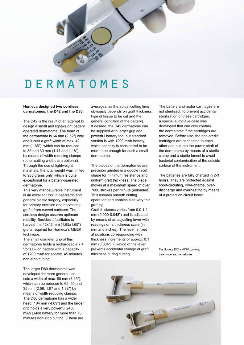
Humeca designed two cordless dermatomes, the D42 and the D80.
The D42 is the result of an attempt to design a small and lightweight battery operated dermatome. The head of the dermatome is 64 mm (2.52") only and it cuts a graft width of max. 42 mm (1.65"), which can be reduced to 36 and 30 mm (1.41 and 1.18") by means of width reducing clamps (other cutting widths are optional). Through the use of lightweight materials, the total weight was limited to 985 grams only, which is quite exceptional for a battery-operated dermatome.This very manoeuvrable instrument is an excellent tool in paediatric and general plastic surgery, especially for primary excision and harvesting grafts from curved surfaces. The cordless design assures optimum mobility. Besides it facilitates to harvest the 42x42 mm (1.65x1.65") grafts required for Humeca’s MEEK technique. The small diameter grip of this dermatome holds a rechargeable 7.4 Volts Li-Ion battery with a capacity of 1200 mAh for approx. 45 minutes non-stop cutting.
The larger D80 dermatome was developed for more general use. It cuts a width of max. 80 mm (3.15"), which can be reduced to 65, 50 and 35 mm (2.56, 1.97 and 1.38") by means of width reducing clamps. The D80 dermatome has a wider head (104 mm / 4.09") and the larger grip holds a very powerful 2400 mAh Li-Ion battery for more than 75 minutes non-stop cutting! (These are
averages, as the actual cutting time obviously depends on graft thickness, type of tissue to be cut and the general condition of the battery).If desired, the D42 dermatome can be supplied with larger grip and powerful battery too, but standard version is with 1200 mAh battery, which capacity is considered to be more than enough for such a small dermatome.
The blades of the dermatomes are precision grinded in a double facet shape for minimum resistance and uniform graft thickness. The blade moves at a maximum speed of over 7000 strokes per minute (unloaded). This assures smooth cutting operation and enables also very thin grafting. Graft thickness varies from 0.0-1.2 mm (0.000-0.048") and is adjusted by means of an adjusting lever with readings on a thickness scale (in mm and inches). The lever is fixed at positions corresponding with thickness increments of approx. 0.1 mm (0.004"). Fixation of the lever prevents accidental change of graft thickness during cutting.
The battery and motor cartridges are not sterilized. To prevent accidental sterilization of these cartridges, a special autoclave case was developed that can only contain the dermatome if the cartridges are removed. Before use, the non-sterile cartridges are connected to each other and put into the power shaft of the dermatome by means of a sterile clamp and a sterile funnel to avoid bacterial contamination of the outside surface of the instrument.
The batteries are fully charged in 2-3 hours. They are protected against short-circuiting, over-charge, over-discharge and overheating by means of a protection circuit board.
dermatomes
The Humeca D42 and D80 cordless, battery operated dermatomes
10

11
D42 and D80 features• Extremely small head of D42 allows precision cutting, especially in problematic zones and paediatric surgery• Cordless, battery operated and lightweight design offers optimum manoeuvrability and mobility• Precise thickness of the graft from 0.0 to 1.2 mm (0.000 – 0.048") in 0.1 mm (0.004") increments• Graft width of 42 mm (1.65") assures optimum performance in combination with the MEEK technique• The use of width-reducing clamps on the dermatome head allow cutting of smaller graft widths• Battery and motor of the instrument are not sterilized, thus guaranteeing optimum durability• Thickness adjustment can be fixed to prevent accidental change of graft thickness during cutting• Safe and quick blade replacement• Powerful Li-Ion batteries with no memory effect allow long time cutting without intermediate charging
D42 and D80 technical dataDermatome Charger for D42 / D80Total weight D42, small / large grip 985 / 1.115 g (35 / 39 oz) Power supply primary 100-240V / 50-60Hz Total weight D80, large grip 1.330 g (47 oz) Charge current for D42 / D80 400 / 1200 mA Weight motor cartridge 325 g (11.5 oz) Nominal output voltage 7.4 VDCWeight battery cartr. 1200 / 2400 mAh 149 / 240 g (5.3 / 8.5 oz) Time to charge empty battery 2.0 / 3.0 hLength dermatome with small / large grip 272 / 295 mm (10.7 / 11.6") Charger is supplied with 4 international adaptersWidth head type D42 / D80 64 / 104 mm (2.52 / 4.09") Maximum diameter small / large grip 40 / 45 mm (1.57 / 1.77") Disposable bladesStandard cutting width D42 / D80 42 / 80 mm (1.65 / 3.15") Type Double facet grindedCutting width using clamps D42 36 and 30 mm (1.41 / 1.18") Width / Thickness 19 / 0.38 mm (0.748 / 0.015”)Cutting width using clamps D80 65, 50 and 35 mm 2.56 / 1.97 / 1.38") Length D42 / D80 50 / 90 mm (1.97 / 3.54”)Graft thickness / increments 0.0-1.2 / 0.1 mm (0.000 – 0.048" / 0.004”) Material Stainless steel Stroke of blade 3.0 mm (0.118") Motor capacity / rpm 15 W / 7.030 min-1 Autoclave case Dimensions for D80 (lxwxh) 375x130x52 mm (14.6x5.12x2.05”)Battery Dimensions for D42 (lxwxh) 375x90x52 mm (14.8x3.54x2.05”)Voltage / capacity battery pack, small 7.4V / 1200 mAh Weight of case for D42 / D80 717 / 910 g (25 / 32 oz) Voltage / capacity battery pack, large 7.4V / 2400 mAh Battery chemistry Li-Ion (no memory effect)
Motor and battery cartridge coupled Charger with support unitWidth reducing clamp on a D80 dermatome

12
sober hand dermatome
The ability to cut a split-thickness skin graft free-handedly has been regarded as a skill acquired only by long experience. Several devices have been improvised to simplify this task. However, the size and elaborate arrangements of dermatomes often preclude their use, especially if the need is in an emergency room, a clinic or at the patient’s bedside. Besides sophisticated dermatomes are costly and more adequate for harvesting larger skin grafts.
In co-operation with the Dutch surgeon dr. Willem Nugteren, Humeca developed a dermatome for freehanded harvesting of a 30 mm (1¼") wide split skin graft with a pre-determined thickness of about 0.25 mm (0.01"). The product is called the ‘SOBER’ dermatome (from an English translation of the phonetic last name of the inventor).
Based on his experience with surgery in third world countries, it became clear that a portable, economical and simple, yet efficient dermatome would be a very useful tool for skin grafting.
As can be seen from the picture above, the shape of the SOBER dermatome was derived from a safety razor. The dermatome is held firmly against the tautly held skin at a pre-determined angle and a graft of the desired length is quickly cut. No lateral movements are required.
The blade in the dermatome is a double facet grinded type, similar to the Humeca® D42 and D80 blades. It is inserted in the SOBER dermatome by loosening a screw. The tool to do this is integrated in the grip, so it can never be lost and it is always ready at hand.
The SOBER dermatome offers the surgeon a low-cost alternative to the more elaborate mechanical dermatomes, whenever small grafts are needed. The low price and robust, durable construction make the instrument a very suitable tool for use in third world countries and remote clinics.
Donorsites on the upper leg of a child
Harvesting a graft with a Sober dermatome from the lower leg
Practizing SOBER dermatome on fruits
SOBER dermatome set with blades and extra key, as supplied by Humeca
13
dermatome blades
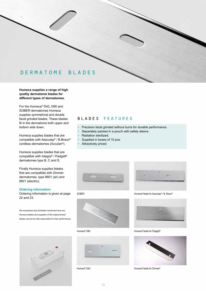
Humeca supplies a range of high quality dermatome blades for different types of dermatomes.
For the Humeca® D42, D80 and SOBER dermatomes Humeca supplies symmetrical and double facet grinded blades. These blades fit in the dermatome both upper and bottom side down.
Humeca supplies blades that are compatible with Aesculap® / B.Braun® cordless dermatomes (Acculan®).
Humeca supplies blades that are compatible with Integra® / Padgett® dermatomes type B, C and S.
Finally Humeca supplies blades that are compatible with Zimmer dermatomes, type 8801 (air) and 8821 (electric).
Ordering informationOrdering information is given at page 22 and 23.
We emphasize that all blades mentioned here are
Humeca blades and suppliers of the original brand
blades cannot be held responsible for their performance.
blades features• Precision facet grinded without burrs for durable performance. • Separately packed in a pouch with safety sleeve. • Radiation sterilized.• Supplied in boxes of 10 pcs. • Attractively priced
Donorsites on the upper leg of a child
Harvesting a graft with a Sober dermatome from the lower leg
Practizing SOBER dermatome on fruits
SOBER Humeca® blade for Aesculap® / B. Braun®
Humeca® D80 Humeca® blade for Padgett®
Humeca® D42 Humeca® blade for Zimmer®

14
v-carriersHumeca introduces a new range of grooved skin graft carriers for expansion and perforation, called ‘V-carriers’.
The symmetrical V-shaped groove pattern of these carriers prevents unwanted sideward movement of the carrier in the mesher during cutting. The standard length of these carriers is 280 mm (11.0"), which is more than the standard length of existing carriers. Furthermore care has been taken to ensure that the groove pattern of a carrier connects exactly to that of another one. This enables cutting of extra long graft strips without any disturbance of the mesh pattern in the graft.
All V-carriers are compatible with the newly developed Humeca® skin graft mesher. V10-type carriers are also compatible with Zimmer® meshers and V15-type carriers are compatible with Aesculap® / B.Braun® meshers. V-carriers are available for expansions 1:1.5, 1:2 (type V10 only) and 1:3. These are used for conventional skin meshing, where expansion of the graft surface is the main goal.
For larger expansion ratios Humeca developed the modified MEEK technique. Especially in severe burns (large total burned surface area) the MEEK technique should be the method of choice because of faster epithelialization, more efficient use of skin (better graft take and smaller donorsites), easier handling of the graft and better final results.
In addition to the V-carriers for expansion, Humeca introduces a new type of a meshgraft carrier that only perforates the graft without the intention of expanding it: the 1:1 V-carrier. Perforations in a graft are intended to achieve sufficient drainage of the wound bed in case full sheet grafts are used, in order to prevent the occurrence of seroma or haematoma under the graft.
Full sheet grafts are frequently applied when skin grafting is required in cosmetically sensitive body parts, such as the face, the neck and the dorsal aspect of the hands, in order to avoid the appearance of an unaesthetic mesh pattern.
The development of the Humeca® V-carriers for skin grafting was sponsored by the Dutch Burns Research Institute (BRI) in Beverwijk, The Netherlands.
V-carrier 1:3
15
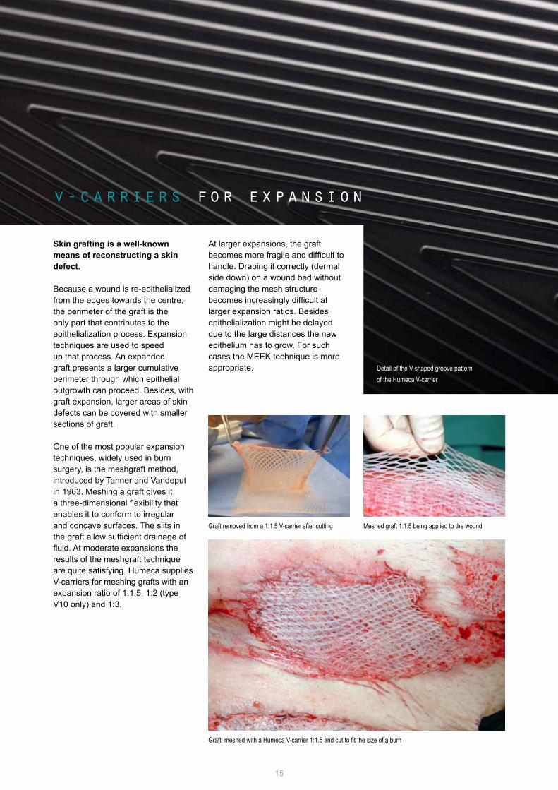
Skin grafting is a well-known means of reconstructing a skin defect.
Because a wound is re-epithelialized from the edges towards the centre, the perimeter of the graft is the only part that contributes to the epithelialization process. Expansion techniques are used to speed up that process. An expanded graft presents a larger cumulative perimeter through which epithelial outgrowth can proceed. Besides, with graft expansion, larger areas of skin defects can be covered with smaller sections of graft.
One of the most popular expansion techniques, widely used in burn surgery, is the meshgraft method, introduced by Tanner and Vandeput in 1963. Meshing a graft gives it a three-dimensional flexibility that enables it to conform to irregular and concave surfaces. The slits in the graft allow sufficient drainage of fluid. At moderate expansions the results of the meshgraft technique are quite satisfying. Humeca supplies V-carriers for meshing grafts with an expansion ratio of 1:1.5, 1:2 (type V10 only) and 1:3.
At larger expansions, the graft becomes more fragile and difficult to handle. Draping it correctly (dermal side down) on a wound bed without damaging the mesh structure becomes increasingly difficult at larger expansion ratios. Besides epithelialization might be delayed due to the large distances the new epithelium has to grow. For such cases the MEEK technique is more appropriate. Detail of the V-shaped groove pattern
of the Humeca V-carrier
Graft removed from a 1:1.5 V-carrier after cutting Meshed graft 1:1.5 being applied to the wound
Graft, meshed with a Humeca V-carrier 1:1.5 and cut to fit the size of a burn
v-carriers for expansion

16
v-carriers for perforation
The Humeca® V-carrier 1:1 is intended for perforation purposes, without expansion.
Why perforations?Perforation is frequently applied when sheet skin grafts (either split-thickness or full-thickness) are used. Sheet grafts are perforated for the following reasons:
DrainageThe slits in the graft allow adequate drainage of fluid. Drainage prevents separation of the graft from its wound bed by haematoma or seroma. Sheet grafts are often used on the face and hands. It is however these two sites that have an extremely vascular bed, making the chances of the appearance of haematoma higher. Collection of fluid under a graft is said to be one of the major causes of graft loss. Consequently perforation leads to increased graft survival.
Improved cosmetic resultsExpanded meshed grafts are frequently criticised because of the poor appearance of the residual mesh pattern, making surgeons reluctant to consider this technique. By using a 1:1 mesh, where the graft perforations are narrowed to slits instead of holes, it is possible to prevent such a pattern to appear and get results similar to a sheet graft. It is also reported that a perforated graft gives a more matt appearance than the shiny surface of a sheet graft, which makes it more suitable when grafting the forehead.
Infection controlTo reduce infection occurrence.
Methods to perforate graftsOne of the simplest methods often applied in the operation theatre is piercing a sheet graft with a scalpel blade by hand. This can be done either by piercing the graft directly or by placing a piece of graft on a grooved meshgraft carrier and cutting it with a scalpel at right angles to the grooves. These methods are labour intensive, and often the number of slits is insufficient to guarantee enough drainage. Nevertheless the method seems adequate in case of small grafts.
Another way of perforating is to use a 1:1.5 meshed graft without expanding it. After all theoretically the holes in the graft remain slits when the graft is not expanded. However it appears to be very difficult to apply such a graft to the wound bed without disturbing the pattern of slits. During transfer of the graft to the wound, due to internal friction the slits will become holes anyway and once this occurs it is almost impossible to regain the original shape of the graft. So this method is not recommended when the final cosmetic results of the perforated graft should be comparable to the results of a sheet graft.
Detail of the groove pattern of a 1:1 V-carrier
Finally, perforations in a graft can be achieved by a procedure sometimes referred to as “Sideways meshing” or “Reversed meshing”. This procedure was first described by Davison et.al. in 1986 and it concerns crosswise cutting a 1:1.5 meshgraft carrier. Such cutted parts of the carrier are then covered with graft and introduced in the mesher at a sideward direction (the grooves turned 90º from normal). This results in smaller slits that can be used as perforations. Disadvantage however is that the maximum length of the graft equals the width of the carrier (approx. 75 mm or 3 inches), while mostly larger grafts are required.
To overcome the difficulties of making perforations in grafts, Humeca introduced a carrier especially developed for this goal: the 1:1 V-carrier. When using this carrier, making perforations in a graft becomes as easy as meshing. Maximum length of the graft is 280 mm or 11 inches, but even longer graft strips can be perforated when using a second carrier, from which the grooves connect exactly to the first one. The Humeca 1:1 V-carriers are available in a V10-type for Humeca® and Zimmer® meshers and a V15-type for Humeca® and Aesculap® / B.Braun® meshers.The 1:1 perforation V-carrier was developed and clinically tested in cooperation with the burn centre of the University Hospital of Gent, Belgium.
Example of a graft, meshed on a 1:1 Humeca® V-carrier Example of a perforated piece of graft
•
•
•
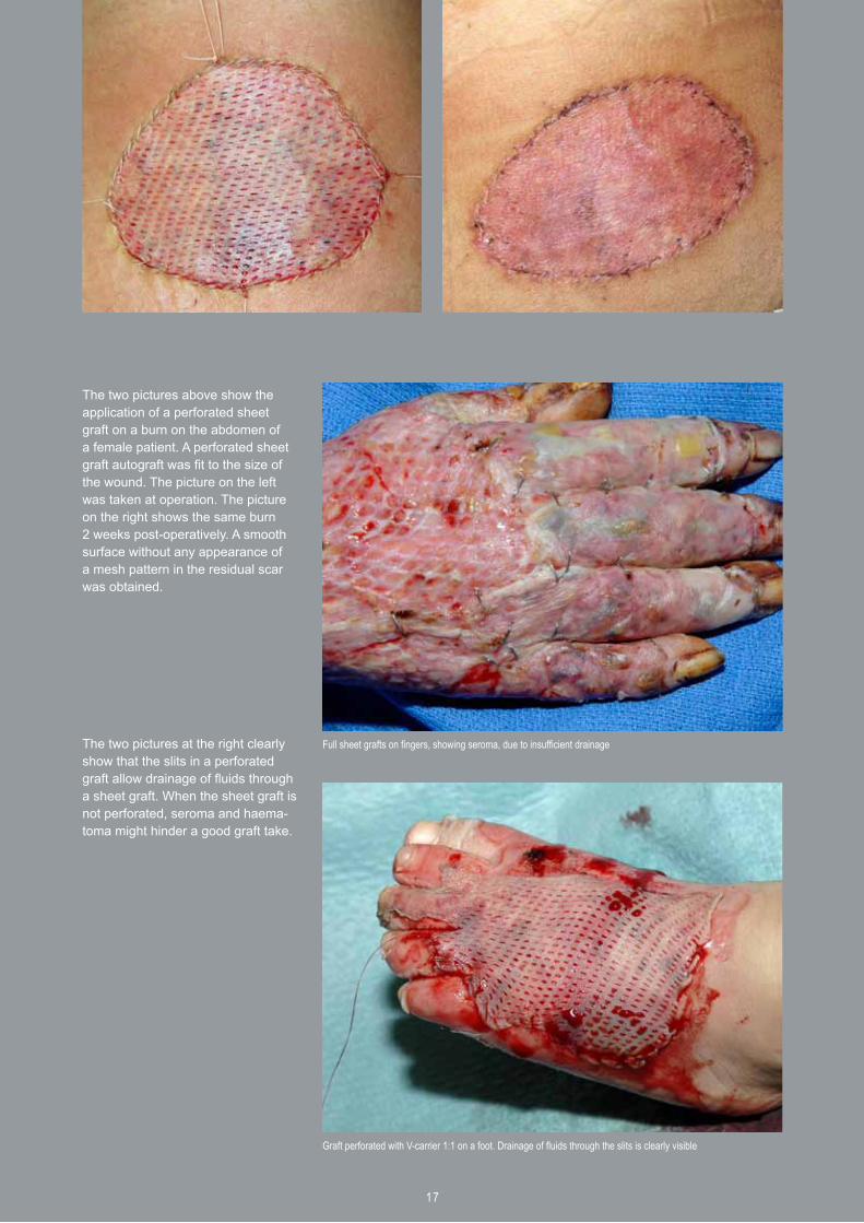
The two pictures above show the application of a perforated sheet graft on a burn on the abdomen of a female patient. A perforated sheet graft autograft was fit to the size of the wound. The picture on the left was taken at operation. The picture on the right shows the same burn 2 weeks post-operatively. A smooth surface without any appearance of a mesh pattern in the residual scar was obtained.
The two pictures at the right clearly show that the slits in a perforated graft allow drainage of fluids through a sheet graft. When the sheet graft is not perforated, seroma and haema-toma might hinder a good graft take.
Full sheet grafts on fingers, showing seroma, due to insufficient drainage
Graft perforated with V-carrier 1:1 on a foot. Drainage of fluids through the slits is clearly visible
17

18
Skin grafts consist of the entire epidermis and a dermal component of variable thickness. If the entire thickness of the dermis is included, an appropriate term is ‘full-thickness skin graft’ (FTSG). If less then the entire dermis is included, appropriate terms are partial or ‘split-thickness skin graft’ (STSG).
STSG’s are categorized further as thin (0.1-0.3 mm / 0.004-0.012"), medium-thickness (0.3-0.45 mm / 0.012-0.018") or thick (0.45-0.8 mm / 0.018-0.03"), based on the thickness of the harvested graft. The choice between FTSG and STSG depends on wound condition, location, size and aesthetic concerns. Split-thickness skin grafts require less ideal conditions for survival and have a much broader range of application than full-thickness skin grafts. They are for instance used to resurface large wounds, line cavities, resurface mucosal deficits, close flap donor sites and resurface muscle flaps. Full-thickness skin grafts are of limited availability. The ability to harvest them is restricted by the necessity of stitching up the donor site from which they were taken. FTSG’s are ideal for repairing small defects where good cosmetic matching or functional restoration is important. STSG’s do have some serious disadvantages compared to FTSG’s that have to be considered:
ContractionOne of them concerns contraction: a skin graft begins to shrink immediately after being harvested from its donor site. This ‘primary contraction’ is about 40% in a FTSG, 20% in a medium thickness STSG and about 10% in a thin STSG. So a STSG has less primary contraction than a FTSG, which is an advantage of STSG, because less tissue is needed for grafting. However, after transfer to a recipient site, the skin graft will shrink further as it heals in a process known as ‘secondary contraction’. FTSG’s tend to remain the same size (after primary contraction) and do not show any secondary contraction. STSG’s, on the other hand, contract whenever the circumstances allow. Unless STSG’s are fixed to underlying rigid structures and cannot move, they will contract secondary. When transplanted on soft tissue, contraction will be significant. A contracted wound is often tight and immobile and there is distortion of the surrounding tissue. If such a wound crosses a joint, contracture will lead to position abnormalities and inadequate motion. Therefore a FTSG is advised for such areas (hand, wrist, elbow, neck).
Graft growingOnce wound contraction ends, full-thickness skin grafts are able to grow with the individual, whereas split-thickness skin grafts tend to remain in a fixed contracted state and grow minimally, if at all.
FragilitySTSG’s are more fragile than FTSG’s, especially when placed over areas with little underlying soft tissue support.
Pigmentation and other cosmetic considerationsSplit-thickness skin grafts tend to be abnormally pigmented (either pale or white) or hyperpigmented, particularly in darker-skinned individuals. Their thinness, abnormal pigmentation and frequent lack of smooth texture and hair growth, make STSG’s more functional than cosmetic. When used to resurface large burns of the face, STSG’s may yield an undesirable mask like appearance.
Picture showing an example of the application of a 1:1.5 mesghraft on a burn of the hand, showing hypertrophic scarring and an early stage of wound contracture. The mesh pattern is clearly visible. A 1:1 perforated sheet graft would probably result in a better appearance and functionality.
full- and split- thickness skin grafts
19
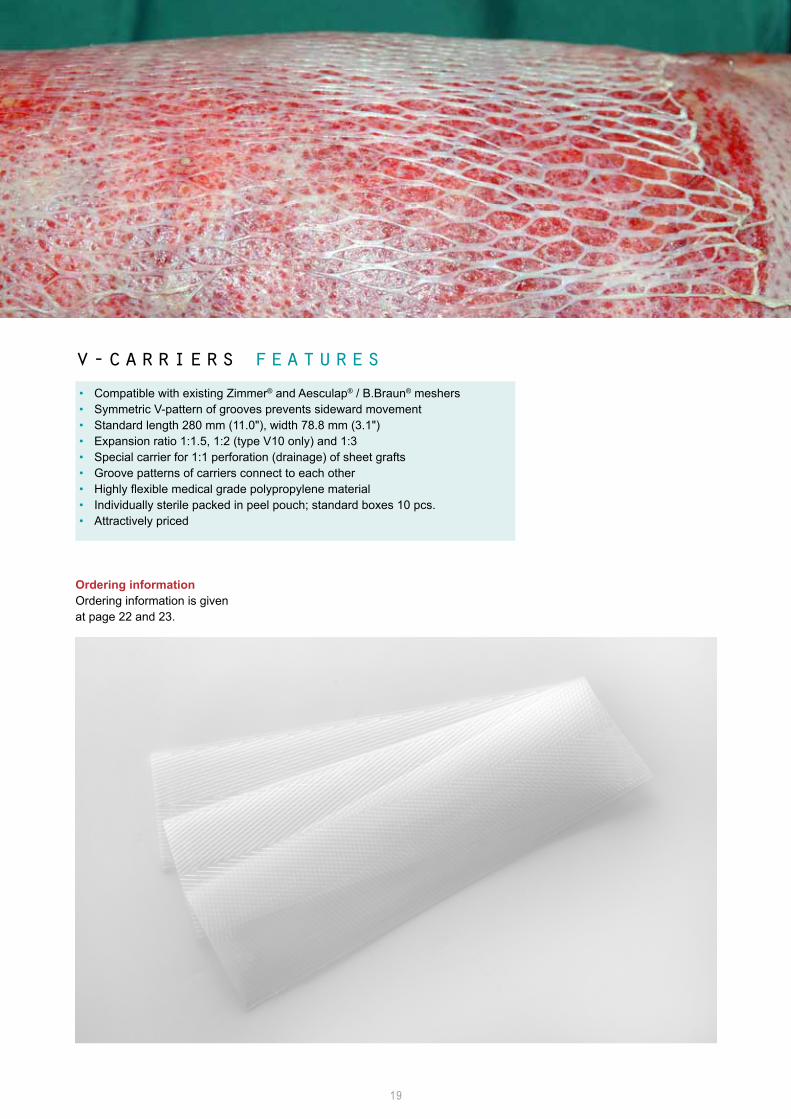
v-carriers features• Compatible with existing Zimmer® and Aesculap® / B.Braun® meshers• Symmetric V-pattern of grooves prevents sideward movement• Standard length 280 mm (11.0"), width 78.8 mm (3.1")• Expansion ratio 1:1.5, 1:2 (type V10 only) and 1:3• Special carrier for 1:1 perforation (drainage) of sheet grafts• Groove patterns of carriers connect to each other• Highly flexible medical grade polypropylene material• Individually sterile packed in peel pouch; standard boxes 10 pcs.• Attractively priced
Ordering informationOrdering information is given at page 22 and 23.

20
In addition to the V-carriers, Humeca developed a mesher to complete the product line for meshgrafting.
The Humeca® mesher is provided with a unique spring mechanism that prevents the blades from excessive pressure on the carrier during cutting. Due to their production process (injection molding) all meshgraft carriers have inevitable variations in thickness. When the thickness of a certain section of the carrier exceeds the maximum distance between the blades and the lower roller, the pressure exercised by the blades on the carrier surface will increase exponentially. Such high pressure manifests in increased friction and in the end it will damage the blades. On the other hand the mesher might not cut the graft completely at a thinner section of the carrier. To avoid such undesired phenomena, Humeca provided the mesher with springs that can meet with thickness variations in carriers. The mesher can be adjusted in two positions for the V10 or the V15- types of V-carriers.
During cutting the carrier is guided both at the left and the right side to assure straight movement, thus enabling exact connection of the grooves of a second carrier if applied.Unlike most conventional meshers, where the carrier is moved through the device by means of intermittent pulling a ratchet, the Humeca® mesher is driven by the continuous rotation of a handle. A gearwheels set limits the required force. The rotation makes the meshing procedure less time consuming and the design is far more ergonomic.
mesher
Side view of the Humeca® mesher

21
Bridge opened for cleaning and access to the cutting axis
mesher features • Robust and durable construction• Compatible with Humeca® V-carriers of all types (V10 and V15)• Compatible with Zimmer® and Aesculap® / B.Braun® carriers• Spring mechanism prevents blades damage• Continuous rotational drive; no intermittent pulling of a ratchet• Measures lxwxh: 220x212x183 mm (8.7x8.3x7.2"). Weight: 4.4 kg (9.7 lb)• Cutting axis can easily be replaced• Individual blades can be replaced• Compact st. steel sterilization case available, lxwxh: 277x232x197 mm (10.9x9.1x7.8")
The 50 blades (diameter 36 mm / 1.42") of the mesher are mounted on an axis, with an interspace of 1.5 mm (0.06") between the intersecting lines. Both the cutting axis and individual blades can be replaced if desired.
Opening the bridge of the mesher allows easy access to the cutting axis for cleaning and inspection.
The mesher can be washed and steam sterilized before use. A compact stainless steel sterilization case is available. A second small sterilization case can hold the cutting axis.
Ordering informationOrdering information is given at page 22 and 23.
Sterilization case for the Humeca mesher Sterilization case for the axis of the Humeca mesher
Cutting axis

22
ordering info
For ordering, please contact your local dealer. You can find dealers via the link in www.humeca.nlIf Humeca has no dealer in your territory, please contact Humeca for inquiry, preferably via email [email protected]
In order to provide Humeca quickly with brief and most adequate information and to assure soonest reply, the preferred method is to fill in the contact form that is displayed when using the contact link in the Humeca site www.humeca.nl
Humeca is a small organization with short communication lines. This allows very quick response and a high level of service.In case of ordering or request for quotation, please refer to the article codes given below.
meek micrografting Equipment3.HD/BLO MEEK cutting machine, hand driven (block model) without cutting block3.HD/CYL MEEK cutting machine, hand driven (cylindrical model) without cutting block3.MD/BLO MEEK cutting machine, motor driven (block model), without cutting block3.MD/CYL MEEK cutting machine, motor driven (cylindrical model), without cutting block3.MD/GW MEEK cutting machine, motor driven, gearwheels, without cutting block3.MD/AUT MEEK cutting machine, automatic version (two axis motor driven)3.BL38 MEEK circular blade, diameter 38 mm (1.50"). Required blade diameter depends on serial number machine3.BL39 MEEK circular blade, diameter 39 mm (1.54”). Required blade diameter depends on serial number machine3.CA01 MEEK cutting aid 41x41 mm (1.61x1.61")3.CB01 MEEK single cutting block with cork holder3.CB02 MEEK double cutting block with two cork holders3.CH01 MEEK cork holder3.CP4 MEEK pneumatic foot pedal with connectors and hose3.GWS MEEK gearwheel drive set3.KN13/38 MEEK cutting axis with 13 ceramic coated circular blades, diameter 38 mm (1.50")3.KN13/39 MEEK cutting axis with 13 ceramic coated circular blades, diameter 39 mm (1.54")3.MAC02 MEEK sterilization case 434x254x172 mm (17.1x10.0x6.8")3.MAC03 MEEK sterilization case 180x51x45 mm (7.1x2.0x1.8") for the cutting axis of the MEEK machine3.RM004 MEEK pneumatic block motor with connectors and coupling3.087 MEEK cylindrical pneumatic motor 2M123.SHD MEEK hand drive set3.SW01 MEEK serrated wedge (cam)
Disposables2.3/10 MEEK Micrograft gauze, expansion 1:3, with cork plate, box 10 pcs.2.4/10 MEEK Micrograft gauze, expansion 1:4, with cork plate, box 10 pcs.2.6/10 MEEK Micrograft gauze, expansion 1:6, with cork plate, box 10 pcs.2.9/10 MEEK Micrograft gauze, expansion 1:9, with cork plate, box 10 pcs. 2.3/40 MEEK Micrograft gauze, expansion 1:3, with cork plate, box 40 pcs.2.4/40 MEEK Micrograft gauze, expansion 1:4, with cork plate, box 40 pcs.2.6/40 MEEK Micrograft gauze, expansion 1:6, with cork plate, box 40 pcs.2.9/40 MEEK Micrograft gauze, expansion 1:9, with cork plate, box 40 pcs. 2.9190 Spray adhesive, bottle 200 ml (6.8 fl.oz).2.JG598 STERILIT® oil for surgical instruments, bottle 50 ml (1.7 fl.oz).
23
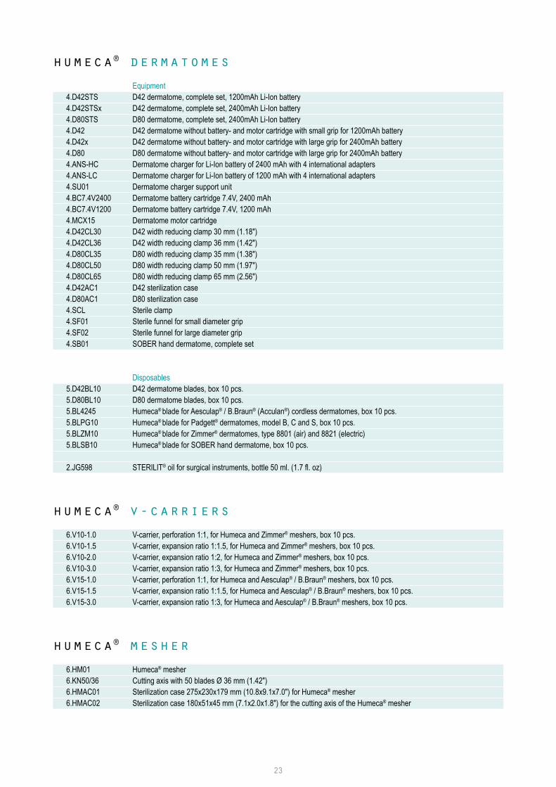
humeca® dermatomes Equipment 4.D42STS D42 dermatome, complete set, 1200mAh Li-Ion battery 4.D42STSx D42 dermatome, complete set, 2400mAh Li-Ion battery4.D80STS D80 dermatome, complete set, 2400mAh Li-Ion battery 4.D42 D42 dermatome without battery- and motor cartridge with small grip for 1200mAh battery4.D42x D42 dermatome without battery- and motor cartridge with large grip for 2400mAh battery4.D80 D80 dermatome without battery- and motor cartridge with large grip for 2400mAh battery 4.ANS-HC Dermatome charger for Li-Ion battery of 2400 mAh with 4 international adapters4.ANS-LC Dermatome charger for Li-Ion battery of 1200 mAh with 4 international adapters4.SU01 Dermatome charger support unit4.BC7.4V2400 Dermatome battery cartridge 7.4V, 2400 mAh4.BC7.4V1200 Dermatome battery cartridge 7.4V, 1200 mAh4.MCX15 Dermatome motor cartridge4.D42CL30 D42 width reducing clamp 30 mm (1.18")4.D42CL36 D42 width reducing clamp 36 mm (1.42")4.D80CL35 D80 width reducing clamp 35 mm (1.38")4.D80CL50 D80 width reducing clamp 50 mm (1.97")4.D80CL65 D80 width reducing clamp 65 mm (2.56")4.D42AC1 D42 sterilization case4.D80AC1 D80 sterilization case4.SCL Sterile clamp4.SF01 Sterile funnel for small diameter grip4.SF02 Sterile funnel for large diameter grip4.SB01 SOBER hand dermatome, complete set
Disposables5.D42BL10 D42 dermatome blades, box 10 pcs.5.D80BL10 D80 dermatome blades, box 10 pcs.5.BL4245 Humeca® blade for Aesculap® / B.Braun® (Acculan®) cordless dermatomes, box 10 pcs.5.BLPG10 Humeca® blade for Padgett® dermatomes, model B, C and S, box 10 pcs.5.BLZM10 Humeca® blade for Zimmer® dermatomes, type 8801 (air) and 8821 (electric)5.BLSB10 Humeca® blade for SOBER hand dermatome, box 10 pcs.
2.JG598 STERILIT® oil for surgical instruments, bottle 50 ml. (1.7 fl. oz)
humeca® v-carriers 6.V10-1.0 V-carrier, perforation 1:1, for Humeca and Zimmer® meshers, box 10 pcs.6.V10-1.5 V-carrier, expansion ratio 1:1.5, for Humeca and Zimmer® meshers, box 10 pcs.6.V10-2.0 V-carrier, expansion ratio 1:2, for Humeca and Zimmer® meshers, box 10 pcs.6.V10-3.0 V-carrier, expansion ratio 1:3, for Humeca and Zimmer® meshers, box 10 pcs.6.V15-1.0 V-carrier, perforation 1:1, for Humeca and Aesculap® / B.Braun® meshers, box 10 pcs.6.V15-1.5 V-carrier, expansion ratio 1:1.5, for Humeca and Aesculap® / B.Braun® meshers, box 10 pcs.6.V15-3.0 V-carrier, expansion ratio 1:3, for Humeca and Aesculap® / B.Braun® meshers, box 10 pcs.
humeca® mesher 6.HM01 Humeca® mesher6.KN50/36 Cutting axis with 50 blades Ø 36 mm (1.42")6.HMAC01 Sterilization case 275x230x179 mm (10.8x9.1x7.0") for Humeca® mesher6.HMAC02 Sterilization case 180x51x45 mm (7.1x2.0x1.8") for the cutting axis of the Humeca® mesher
24
Office addressHet Bijvank 251-aNL-7544 DB EnschedeThe Netherlands
Postal addressP.O.Box 40175NL-7504 RD EnschedeThe Netherlands
Humeca BVPhone +31 53 476 26 19Fax +31 53 477 19 05E-mail [email protected]
s k i n t r a n s p l a n t a t i o n t e c h n o l o g y
www.humeca.nl