skin layer mechanics - materials technology - tu eindhoven · skin layer mechanics the human skin...
TRANSCRIPT
Skin layer mechanics
MARION GEERLIGS

ISBN: 978-90-74445-92-4
Cover design: Marion Geerligs & Henny Herps
Printed by Universiteitsdrukkerij TU Eindhoven, Eindhoven, The Netherlands.
©Koninklijke Philips Electronics N.V. 2009
All rights reserved. Reproduction in whole or in part is prohibited without the
written consent of the copyright owner.
Skin layer mechanics
PROEFSCHRIFT
ter verkrijging van de graad van doctor aan de
Technische Universiteit Eindhoven, op gezag van de
rector magnificus, prof.dr.ir. C.J. van Duijn, voor een
commissie aangewezen door het College voor
Promoties in het openbaar te verdedigen
op donderdag 21 januari 2010 om 16.00 uur
door
Marion Geerligs
geboren te Hoogezand-Sappemeer

Dit proefschrift is goedgekeurd door de promotor:
prof.dr.ir. F.P.T. Baaijens
Copromotoren:
dr.ir. C.W.J. Oomens
en
dr.ir. G.W.M. Peters
Contents
Summary ................................................................................................ ix
Skin layer mechanics ............................................................................ ix
Chapter 1 General introduction ........................................................... 1
1.1 Introduction ............................................................................................................... 2
1.2 A mechanical view of skin anatomy and physiology ............................................... 4
1.2.1 Skin topography ................................................................................................. 4
1.2.2 Stratum corneum ................................................................................................ 5
1.2.3 Viable epidermis ................................................................................................ 6
1.2.4 Dermal-epidermal junction ................................................................................ 7
1.2.5 Dermis................................................................................................................ 8
1.2.6 Hypodermis........................................................................................................ 9
1.3 Review of skin layer mechanics ............................................................................. 10
1.3.1 In vivo vs in vitro experiments ........................................................................ 10
1.3.2 Mechanical behavior of the stratum corneum ................................................. 10
1.3.3 Mechanical behavior of the viable epidermis .................................................. 12
1.3.4 Hypodermis...................................................................................................... 12
1.4 Aim and Outline ..................................................................................................... 13
Chapter 2 Isolation and preservation methods for the epidermis
and stratum corneum ........................................................................... 15
2.1 Introduction ............................................................................................................. 16
2.2 Skin preparation and analyses ................................................................................ 17
2.2.1 Skin preparation ............................................................................................... 17
2.2.2 Histological examination ................................................................................. 18
2.2.3 Analyses of skin viability ................................................................................ 19
2.3 Epidermal isolation techniques ............................................................................... 19
Summary vi
2.3.1 Mechanical separation ..................................................................................... 19
2.3.2 Ionic change ..................................................................................................... 20
2.3.3 Heat .................................................................................................................. 21
2.3.4 Enzymatic digestion ........................................................................................ 21
2.3.5 Microwave irradiation ..................................................................................... 23
2.4 Isolation techniques for the stratum corneum......................................................... 23
2.4.1 Mechanical separation ..................................................................................... 24
2.4.2 Chemical separation ........................................................................................ 25
2.4.3 Enzymatic digestion ........................................................................................ 25
2.5 Preservation of the upper skin layers ...................................................................... 26
2.5.1 Short-term storage ........................................................................................... 27
2.5.2 Long-term storage ............................................................................................ 28
2.6 Discussion ............................................................................................................... 30
Chapter 3 Linear shear response of the upper skin layers .............. 33
3.1 Introduction ............................................................................................................. 34
3.2 Methods .................................................................................................................. 35
3.2.1 Sample preparation .......................................................................................... 35
3.2.2 Experimental set-up ......................................................................................... 36
3.2.3 Rheological methods ....................................................................................... 39
3.2.4 Experimental procedures ................................................................................. 40
3.3 Results ..................................................................................................................... 41
3.4 Discussion ............................................................................................................... 46
Chapter 4 A new indentation method to determine mechanical
properties of the epidermis ................................................................. 49
4.1 Introduction ............................................................................................................. 50
4.1.1 Sample preparation .......................................................................................... 51
4.1.2 Experimental procedure ................................................................................... 53
4.1.3 Determination of the Young‟s modulus .......................................................... 54
4.2 Results ..................................................................................................................... 55
4.3 Discussion ............................................................................................................... 56
Chapter 5 Linear viscoelastic behavior of subcutaneous adipose
tissue ...................................................................................................... 61
5.1 Introduction ............................................................................................................. 62
5.2 Methods and Materials ........................................................................................... 64
5.2.1 Sample preparation .......................................................................................... 64
5.2.2 Rheological methods ....................................................................................... 64
5.2.3 Testing procedure ............................................................................................ 65
5.2.4 Statistics ........................................................................................................... 66
5.3 Results ..................................................................................................................... 67
vii
5.3.1 Small oscillatory strain behavior ..................................................................... 67
5.3.2 Model application ............................................................................................ 68
5.3.3 Time-Temperature Superposition .................................................................... 69
5.3.4 Freezing effects ................................................................................................ 70
5.4 Discussion ............................................................................................................... 71
Chapter 6 Does subcutaneous adipose tissue behave as an
(anti-)thyxotropic material? ................................................................ 73
6.1 Introduction ............................................................................................................. 74
6.2 Materials & Methods .............................................................................................. 75
6.2.1 Sample preparation .......................................................................................... 75
6.2.2 Rheological methods ....................................................................................... 76
6.3 Results ..................................................................................................................... 78
6.3.1 Long term small strain behavior ...................................................................... 78
6.3.2 Large strain experiments ................................................................................. 79
6.4 Discussion ............................................................................................................... 81
Chapter 7 General discussion ............................................................. 85
7.1 Introductory remarks .............................................................................................. 86
7.2 In vitro model ......................................................................................................... 87
7.3 Mechanical methods ............................................................................................... 88
7.4 Main findings .......................................................................................................... 90
7.4.1 Small strain behavior of the epidermal layers ................................................. 90
7.4.2 Mechanical behavior of the subcutaneous adipose tissue ............................... 91
7.5 Implications for clinical and cosmetic applications ............................................... 91
7.6 Recommendations................................................................................................... 92
7.7 General conclusion ................................................................................................. 94
Samenvatting ........................................................................................ 95
Dankwoord ............................................................................................ 97
Curriculum Vitae ................................................................................. 99
References ........................................................................................... 100

Summary
Skin layer mechanics
The human skin is composed of several layers, each with an unique structure and
function. Knowledge about the mechanical behavior of these skin layers is important for
clinical and cosmetic research, such as the development of personal care products and
the understanding of skin diseases. Until today, most research was performed in vivo and
focused on the mid-layer, the dermis. However, clinical and cosmetic applications
require more detailed knowledge about the skin layers at the skin surface, the viable
epidermis and stratum corneum, and the deeper lying hypodermis. Studying these layers
in an in vivo set up is very challenging. The different length scales, ranging from μm for
the stratum corneum to cm for the hypodermis, the interwoven layered structure and the
inverse relation between penetration depth and resolution of non-invasive measurement
techniques form major problems. As a consequence, hardly any data are available for the
viable epidermis and hypodermis and reported data for stratum corneum are inconsistent.
The aim of this thesis was therefore to characterize the mechanical behavior of
individual skin layers in vitro and, for that, to develop the required experimental
procedures. It was considered essential to perform experiments with samples of
consistent quality in an accurate measurement set-up in a well-controlled environment.
Various isolation and preservation methods were investigated on tissue performance,
reproducibility and ease of handling.
Because of the inhomogeneous layered structure of the upper skin layers, mechanical
properties of the stratum corneum and viable epidermis were determined for various
loading directions. First, the stratum corneum and epidermis were subjected to shear
over a wide frequency range and with varying temperature and humidity. The typical
geometry of the upper skin layers required preliminary testing series in order to define
the right experimental conditions to ensure reliable results. Subsequently, micro-
indentation experiments were applied using a spherical tip with a relatively large
Summary x
diameter. The Young‟s moduli were derived via an analytical and numerical method.
Because of the complexity of measuring those skin layers, it was decided to focus on
small deformations first.
For both types of loading, result were highly reproducible. The shear tests demonstrated
that the shear modulus is influenced by humidity but not by temperature in the measured
range. If the skin is compressed with an indenter, the stiffness of the epidermis and
stratum corneum, which is about 1-2 MPa, is about a factor 100 higher than for shear. No
significant differences in stiffness between the stratum corneum and viable epidermis
were observed per loading type. The results of these tests prove that it is essential to take
into account the highly anisotropy of the tissue in numerical models.
Rheological methods were developed to study the mechanical response of the
subcutaneous adipose tissue. In the small linear viscoelastic strain regime, the shear
modulus showed a frequency- and temperature-dependent behavior and is about 7.5 kPa
at 10 rad/s and 37°C. Time-Temperature Superposition is applicable through shifting the
shear modulus horizontally. A power-law function model was able to describe the
frequency dependent behavior at constant temperature as well as the measured stress
relaxation behavior.
Prolonged loading at small strains results into a dramatic stiffening of the material.
Loading-unloading cycles showed that this behavior is reversible. In addition, various
large strain history sequences showed that stress-strain responses are reproducible up to
0.15 strain. When the strain further increases, the stress is decreasing for subsequent
loading cycles and, above 0.3 strain, the stress response becomes stationary. These
results showing time and strain effects indicate that adipose tissue likely behaves as an
(anti-)thixotropic material, meaning that a constitutive model should contain parameters
to describe the build-up and breakdown of the material structure. However, further
experimental research is needed to fully understand the thixotropic behavior before such
a model can be worked out in detail.
In conclusion, this thesis evaluates the mechanical behavior of stratum corneum,
epidermis and hypodermis using various in vitro set-ups. It was proven that for all skin
layers reproducible results can be obtained. The research was aimed at developing
reliable methods to determine the mechanical behavior of individual human skin layers.
Future work should be focused on the relationship between mechanical properties and
tissue deformation using imaging techniques and heading to the determination of the
skin‟s failure behavior in relation to clinical and cosmetic treatments.

Chapter 1
General introduction
2 Chapter 1
1.1 Introduction
The largest organ of the human body, the skin, has a major role in providing a barrier
against the hostile external environment. The skin prevents excessive water loss from the
aqueous interior, the ingress of foreign chemicals and micro-organisms and provides
strength and stiffness to resist mechanical loading. Other functions include insulation,
temperature regulation and sensation. To fulfill these functions, mechanical stability is
as important as mechanical flexibility. However, the mechanical balance of skin can be
threatened by diseases, trauma, medical or cosmetic treatments. In order to understand
the skin behavior following the onset of these conditions, knowledge of the mechanical
behavior of healthy skin in normal conditions is essential.
Human skin is composed of several layers, each with a unique structure and function, but
most research on its mechanical properties have ignored this non-uniform layered
structure. For many clinical and cosmetic applications, however, knowledge of the
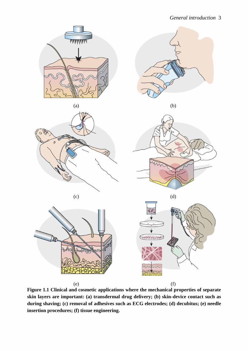
mechanical behavior of the various skin layers is indispensible (Figure 1.1). For
example, the benefit of transdermal drug delivery is that the microneedles exclusively
damage the pain-free outer skin layer, the epidermis. Its mechanical response is therefore
of particular interest. For needle insertion into the underlying dermal layer or for
diseases such as pressure ulcers, the combined mechanical response of all individual skin
layers is important. Although often not recognized, this is also the case during the
removal of skin adhesives or the use of consumer products such as shavers. For all these
applications, the subcutaneous fat layer contributes by attenuating or dispersing the
external pressures, even when those are very small [1]. In addition, mechanical
properties of the distinct skin layers are needed to grow them artificially, serving a wide
application field. These include the development of artificial outer skin to substitute
animal and clinical testing in evaluating drugs, cosmetics and other consumer products,
and engineered fatty tissue facilitates large volume soft tissue augmentation in plastic
surgery. Furthermore, the mechanical behavior of subcutaneous fat is critical for many
other clinical treatments beyond the scope of this thesis, such as liposuction surgery and
cellulite treatments.
To date, research on skin mechanics has mainly focused on full-thickness skin, the mid-
layer (dermis) and the top layer of the epidermis, the stratum corneum. The significance
of a proper understanding of the mechanical behavior of the other part of the epidermis,
the viable epidermis, and the subcutaneous fat tissue is not yet commonly felt. Indeed
very limited experimental data is available for those layers. In addition, there is no
consistency in data for the stratum corneum. Accordingly, the mechanical behavior of
individual skin layers could not have been yet incorporated in numerical models. This
thesis therefore focuses on the mechanical characterization of stratum corneum,
epidermis and the subcutaneous adipose tissue. Before the scope and outline of the thesis
is given, the anatomy of the skin and skin layer mechanics is shortly discussed.
General introduction 3
(a) (b)
(c) (d)
(e) (f)
Figure 1.1 Clinical and cosmetic applications where the mechanical properties of separate
skin layers are important: (a) transdermal drug delivery; (b) skin-device contact such as
during shaving; (c) removal of adhesives such as ECG electrodes; (d) decubitus; (e) needle
insertion procedures; (f) tissue engineering.
4 Chapter 1
1.2 A mechanical view of skin anatomy and physiology
Mechanical properties of skin vary considerably and depend on body site, age, race and
gender. Individual factors like exposure to UV irradiation, the use of creams and
individual health and nutritional status can also affect the mechanical properties.
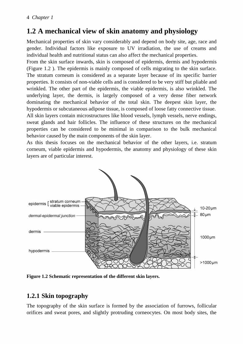
From the skin surface inwards, skin is composed of epidermis, dermis and hypodermis
(Figure 1.2 ). The epidermis is mainly composed of cells migrating to the skin surface.
The stratum corneum is considered as a separate layer because of its specific barrier
properties. It consists of non-viable cells and is considered to be very stiff but pliable and
wrinkled. The other part of the epidermis, the viable epidermis, is also wrinkled. The
underlying layer, the dermis, is largely composed of a very dense fiber network
dominating the mechanical behavior of the total skin. The deepest skin layer, the
hypodermis or subcutaneous adipose tissue, is composed of loose fatty connective tissue.
All skin layers contain microstructures like blood vessels, lymph vessels, nerve endings,
sweat glands and hair follicles. The influence of these structures on the mechanical
properties can be considered to be minimal in comparison to the bulk mechanical
behavior caused by the main components of the skin layer.
As this thesis focuses on the mechanical behavior of the other layers, i.e. stratum
corneum, viable epidermis and hypodermis, the anatomy and physiology of these skin
layers are of particular interest.
Figure 1.2 Schematic representation of the different skin layers.
1.2.1 Skin topography
The topography of the skin surface is formed by the association of furrows, follicular
orifices and sweat pores, and slightly protruding corneocytes. On most body sites, the
General introduction 5
main furrows, called primary lines, are 70-200 μm deep, and follow at least two
directions. The follicular orifices are located at the junction of the furrows, whereas the
sweat pores are mainly found in the plateaus or in more superficial furrows, called
secondary lines, being 20-70 μm deep. The third type of furrows separate groups of
corneocytes. The network of furrows varies with age and gender.
The main function of the furrows is considered to be mechanical. By (partially)
smoothing out, the skin surface and the epidermis can extend without loading the cells.
The deeper the furrows and the steeper their sides, the higher their physiological range of
extension. The direction of the higher extensibility is perpendicular to the direction of
the main furrows. As a consequence, the stratum corneum in vivo hardly experience
elongation stresses, but only unfolding. The furrows cannot be ignored when methods
are developed to mechanically characterize the stratum corneum and the epidermis.
1.2.2 Stratum corneum
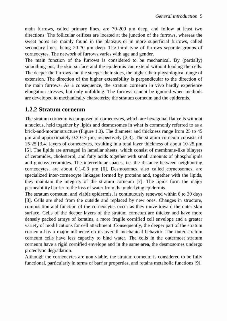
The stratum corneum is composed of corneocytes, which are hexagonal flat cells without
a nucleus, held together by lipids and desmosomes in what is commonly referred to as a
brick-and-mortar structure (Figure 1.3). The diameter and thickness range from 25 to 45
μm and approximately 0.3-0.7 μm, respectively [2,3]. The stratum corneum consists of
15-25 [3,4] layers of corneocytes, resulting in a total layer thickness of about 10-25 μm
[5]. The lipids are arranged in lamellar sheets, which consist of membrane-like bilayers
of ceramides, cholesterol, and fatty acids together with small amounts of phospholipids
and glucosylceramides. The intercellular spaces, i.e. the distance between neighboring
corneocytes, are about 0.1-0.3 μm [6]. Desmosomes, also called corneosomes, are
specialized inter-corneocyte linkages formed by proteins and, together with the lipids,
they maintain the integrity of the stratum corneum [7]. The lipids form the major
permeability barrier to the loss of water from the underlying epidermis.
The stratum corneum, and viable epidermis, is continuously renewed within 6 to 30 days
[8]. Cells are shed from the outside and replaced by new ones. Changes in structure,
composition and function of the corneocytes occur as they move toward the outer skin
surface. Cells of the deeper layers of the stratum corneum are thicker and have more
densely packed arrays of keratins, a more fragile cornified cell envelope and a greater
variety of modifications for cell attachment. Consequently, the deeper part of the stratum
corneum has a major influence on its overall mechanical behavior. The outer stratum
corneum cells have less capacity to bind water. The cells in the outermost stratum
corneum have a rigid cornified envelope and in the same area, the desmosomes undergo
proteolytic degradation.
Although the corneocytes are non-viable, the stratum corneum is considered to be fully
functional, particularly in terms of barrier properties, and retains metabolic functions [9].
6 Chapter 1
(a) (b)
Figure 1.3: Morphology of the stratum corneum. (a) schematic drawing (b) cryostat
section of normal human skin treated with Sorensen’s alkaline buffer and methylene blue.
Obtained from Marks [10].
The mechanical properties of both stratum corneum and viable epidermis are influenced
by environmental conditions such as relative humidity (RH) and temperature. In
addition, topical applications of either pure water, moisturizers or emollients alters the
hydration state of the stratum corneum, significantly modifying some of its mechanical
properties. Under normal conditions, the hydration in the stratum corneum conditions
varies from 5-10% near the surface up to 30% near to the transition with the viable
epidermis. Bound water associated with proteins and lipids accounts for 20-30% of the
total water volume. The total water content varies little between 30% and 60% RH,
although it increases considerably at higher values [11]. When fully hydrated, the
stratum corneum swells to twice its normal thickness. In an in vitro situation, however,
the stratum corneum can increase up to 400% of its original thickness [12]. This
highlights the constraints imposed on the stratum corneum in vivo.
1.2.3 Viable epidermis
The viable epidermis is a layered structure, consisting of three layers or „strata‟. The bulk
of epidermal cells are the keratinocytes, which migrate upwards to the skin surface
where they become non-viable. Other cell types within the viable epidermis include
melanocytes, Langerhans cells and Merkel cells.
Keratinocytes change their shape, size and physical properties when migrating to the
skin surface. Indeed the morhology of an individual keratinocyte correlates with its
position within the epidermis and its state of differentiation, which is reflected by the
different strata: the stratum basale, the stratum spinosum and the stratum granulosum
(Figure 1.4). The deepest layer is the stratum basale in which cell division occurs. It
consists of 1 to 3 layers of small cubic cells. In the next layer, the stratum spinosum, the
cells are larger and polyhedral in nature and are connected by desmosomes, which are
symmetrical laminated structures. The keratinocytes adopt a more flattened morphology
at higher layers of the stratum spinosum. In this layer, they are associated with lamellar
granules, which are lipid-synthesizing organelles that migrate toward the periphery of
the cell and eventually become extruded into the intercellular compartment in the next
layer, the stratum granulosum. At this stage of differentiation, the degradation of
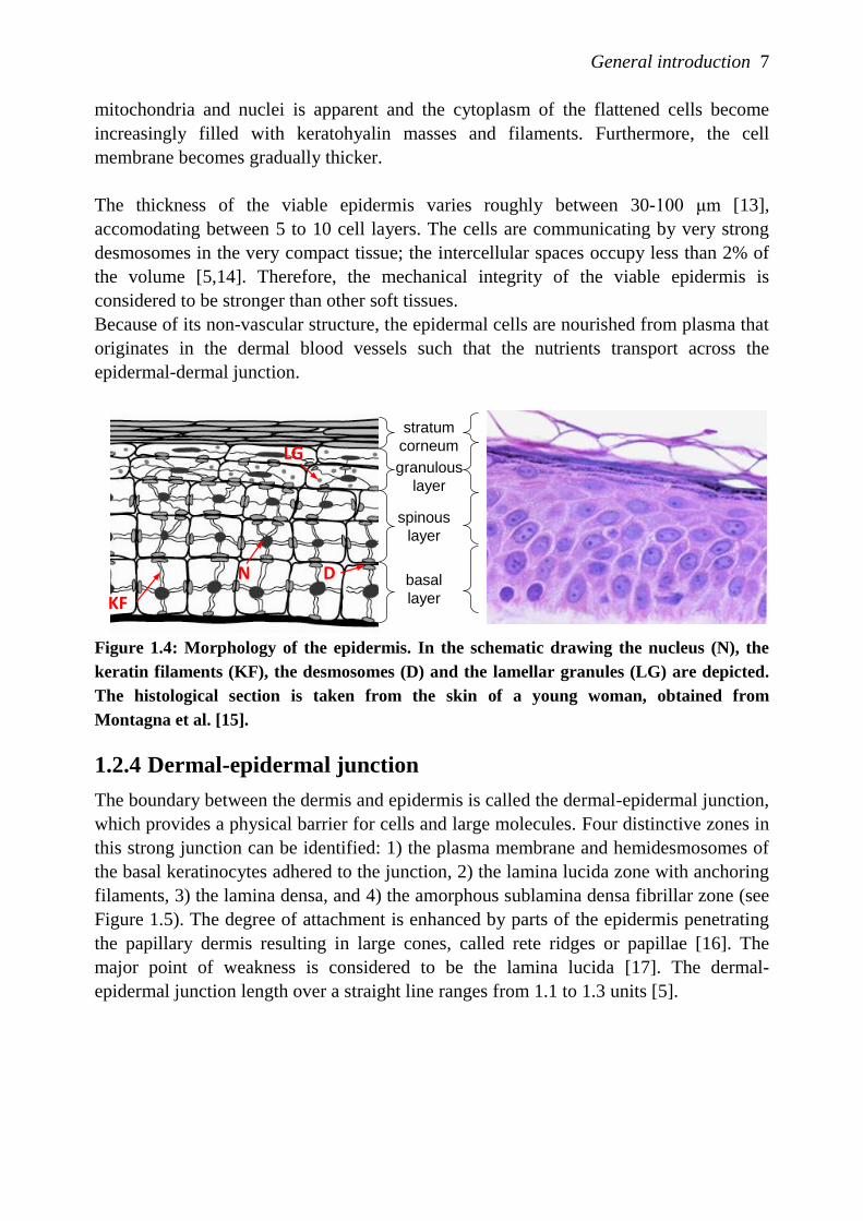
General introduction 7
mitochondria and nuclei is apparent and the cytoplasm of the flattened cells become
increasingly filled with keratohyalin masses and filaments. Furthermore, the cell
membrane becomes gradually thicker.
The thickness of the viable epidermis varies roughly between 30-100 μm [13],
accomodating between 5 to 10 cell layers. The cells are communicating by very strong
desmosomes in the very compact tissue; the intercellular spaces occupy less than 2% of
the volume [5,14]. Therefore, the mechanical integrity of the viable epidermis is
considered to be stronger than other soft tissues.
Because of its non-vascular structure, the epidermal cells are nourished from plasma that
originates in the dermal blood vessels such that the nutrients transport across the
epidermal-dermal junction.
Figure 1.4: Morphology of the epidermis. In the schematic drawing the nucleus (N), the
keratin filaments (KF), the desmosomes (D) and the lamellar granules (LG) are depicted.
The histological section is taken from the skin of a young woman, obtained from
Montagna et al. [15].
1.2.4 Dermal-epidermal junction
The boundary between the dermis and epidermis is called the dermal-epidermal junction,
which provides a physical barrier for cells and large molecules. Four distinctive zones in
this strong junction can be identified: 1) the plasma membrane and hemidesmosomes of
the basal keratinocytes adhered to the junction, 2) the lamina lucida zone with anchoring
filaments, 3) the lamina densa, and 4) the amorphous sublamina densa fibrillar zone (see
Figure 1.5). The degree of attachment is enhanced by parts of the epidermis penetrating
the papillary dermis resulting in large cones, called rete ridges or papillae [16]. The
major point of weakness is considered to be the lamina lucida [17]. The dermal-
epidermal junction length over a straight line ranges from 1.1 to 1.3 units [5].
stratum
corneum
basal
layer
granulous
layer
spinous
layer
N D
KF
LG
8 Chapter 1
Figure 1.5: Ultrastructure of the dermal-epidermal junction.
1.2.5 Dermis
The dermis can be divided into two anatomical regions: the papillary and reticular
dermis. The papillary dermis is the thinner outermost portion of the dermis, constituting
approximately 10% of the 1-4 mm thick dermis. It contains relatively small and loose
distribution of elastic and collagen fibrils within a significant amount of ground
substance. Its content in water and vascular volume show physiological variations that
can alter the mechanical behavior of skin as a whole. In addition, collagen and elastin
fibers are mostly vertically oriented in the papillary region and connect to the dermal-
epidermal junction. In the reticular dermis, fibers are horizontally oriented.
The dermis has a mainly mechanical function. The reticular dermis is able to extend up
to about 25% by stretching the collagen fibers, whereas it can be squeezed due to the
capacity to displace the ground substance laterally. The elastic fiber network ensures full
recovery of tissue shape and architecture after deformation. The amorphous ground
substance acts as a viscous gel-like material, which does not leak out of the dermis, even
under high pressure. The permanent tension in the reticular dermis generates the folding
of the overlying structures and hence, the skin surface. The fiber network in the papillary
dermis contributes to the protection of vessels and cells against mechanical insults.
In the papillary dermis, the microvasculature consists of papillary loops exchanging with
extravascular elements and a horizontal plexus in which the loops emerge. Although the
vascularization throughout the dermis appears relatively sparse, the supply of the
papillary loops is ensured by arterioles irrigated from the deep dermis.
General introduction 9
1.2.6 Hypodermis
The hypodermis is defined as the adipose tissue layer found between the dermis and the
aponeurosis and fasciae of the muscles. Its thickness varies with anatomical site, age,
sex, race, endocrine and nutritional status of the individual. The subcutaneous adipose
tissue is structurally and functionally well integrated with the dermis through nerve and
vascular networks and the continuity of epidermal appendages, such as hairs and nerve
endings.
The bulk of subcutaneous adipose tissue is a loose association of lipid-filled cells, the
white adipocytes, which are held in a framework of collagen fibers. However, only one
third of adipose tissue contains mature adipocytes [18], with the remainder being
stromal-vascular cells including fibroblasts, leukocytes, macrophages, and pre-
adipocytes [19]. Adipose tissue has little extracellular matrix compared to other
connective tissues.
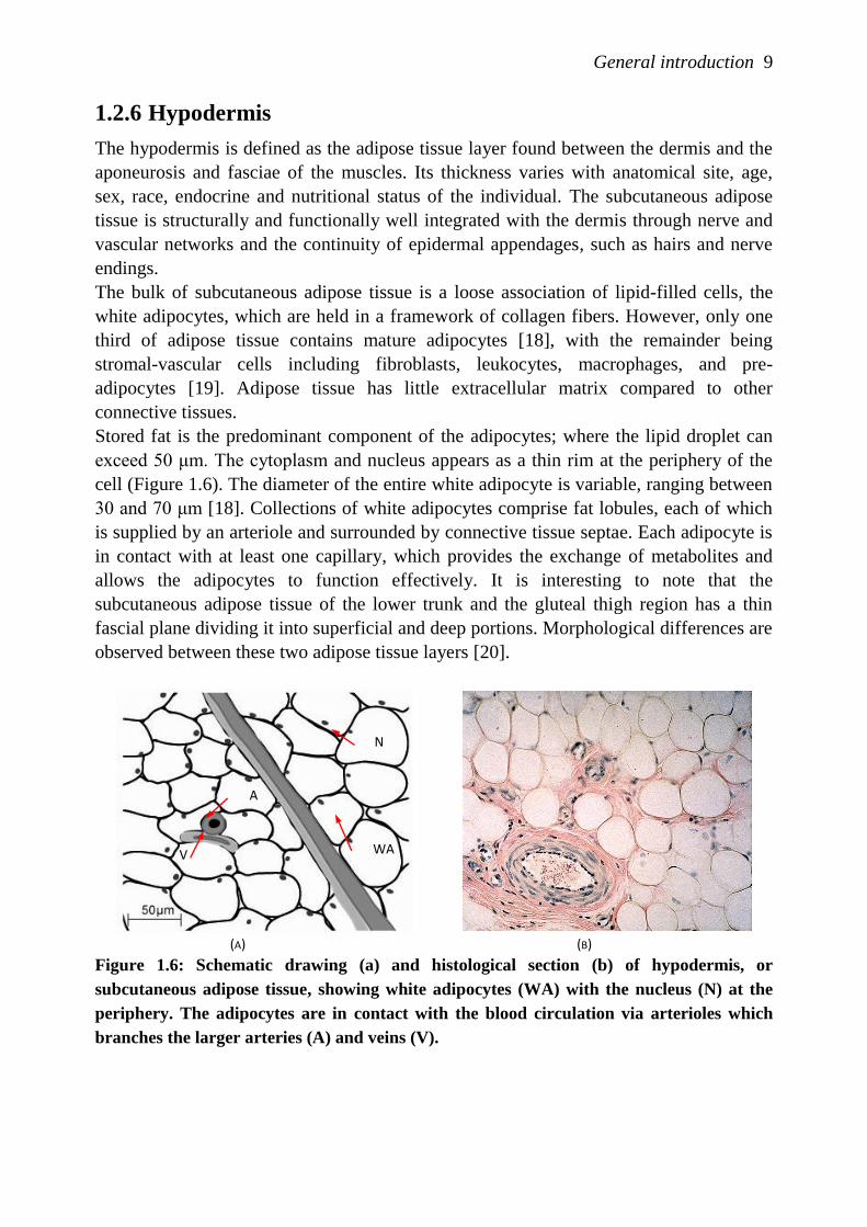
Stored fat is the predominant component of the adipocytes; where the lipid droplet can
exceed 50 μm. The cytoplasm and nucleus appears as a thin rim at the periphery of the
cell (Figure 1.6). The diameter of the entire white adipocyte is variable, ranging between
30 and 70 μm [18]. Collections of white adipocytes comprise fat lobules, each of which
is supplied by an arteriole and surrounded by connective tissue septae. Each adipocyte is
in contact with at least one capillary, which provides the exchange of metabolites and
allows the adipocytes to function effectively. It is interesting to note that the
subcutaneous adipose tissue of the lower trunk and the gluteal thigh region has a thin
fascial plane dividing it into superficial and deep portions. Morphological differences are
observed between these two adipose tissue layers [20].
(A) (B)
Figure 1.6: Schematic drawing (a) and histological section (b) of hypodermis, or
subcutaneous adipose tissue, showing white adipocytes (WA) with the nucleus (N) at the
periphery. The adipocytes are in contact with the blood circulation via arterioles which
branches the larger arteries (A) and veins (V).
A
V
N
WA
10 Chapter 1
The mechanical functions of the subcutaneous adipose tissue include allowing the
overlying skin to move as a whole, both horizontally and vertically, and the attenuation
and dispersion of externally applied pressure.
1.3 Review of skin layer mechanics
Measurement methods and mechanical properties of skin have been extensively
reviewed in the literature [5,21,22]. Therefore, given the focus of the present work, focus
will be limited to studies on the behavior of stratum corneum, viable epidermis and
hypodermis. More specifically, they include force-elongation data, either in vivo or in
vitro, and currently available constitutive models.
1.3.1 In vivo vs in vitro experiments
When measurements on skin mechanics are performed in vivo, the human skin exists in
its natural pre-stress and skin relief. The number of in vivo measurement methods is,
however, limited [22] and a numerical-experimental approach is usually adopted. In any
in vivo study, it is difficult to determine the contribution of each individual skin layer to
the overall skin response, whereas in vitro measurement methods offer the potential to
perform well-controlled experiments on individual skin layers. Another benefit of the
latter is that all forms of mechanical testing can be applied and a wide range of reliable
direct measurement methods becomes available. However, due to the limited availability
of skin grafts, the number of experiments, the variety of skin types, and the variety of
body sites can be problematic.
The appropriateness of in vitro experiments on the stratum corneum should be carefully
considered. In vivo, the stratum corneum partly unfolds when the total skin is stretched,
but does not elongate. Full extension of the stratum corneum occurs in critical, extra-
physiological situations due to disease, trauma, clinical or cosmetic applications.
1.3.2 Mechanical behavior of the stratum corneum
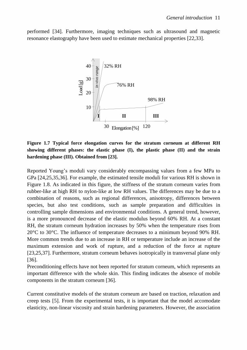
Force-elongation curves at constant elongation rate demonstrate one, two or three phases
depending on the hydration level in the in vitro experiment (Figure 1.7) [23]. The first
phase, up to a 10% extension, is considered to represent purely elastic behavior. The next
phase, absent at low RH, is an irreversible elongation with a low slope, with strains
ranging from 20-125%. In addition, fully hydrated stratum corneum exhibiting a final
phase, where strain hardening is observed before rupture, at approximately 200%
extension. The slope becomes steeper at increasing elongation rates, as would be
predicted of a viscoelastic response. Although the corneocytes are very elongated in
tensile testing, the final rupture is always extracellular and most likely at the
desmosomes [8].
From the 1970s, various authors have reported tensile testing [8,23-27]. Subsequently,
torsional techniques were developed to measure the stratum corneum behavior in vivo
[28-31]. More recently, indentation techniques were introduced to determine the
Young‟s modulus in vitro [32,33], and also in vivo indentation tests have been
General introduction 11
performed [34]. Furthermore, imaging techniques such as ultrasound and magnetic
resonance elastography have been used to estimate mechanical properties [22,33].
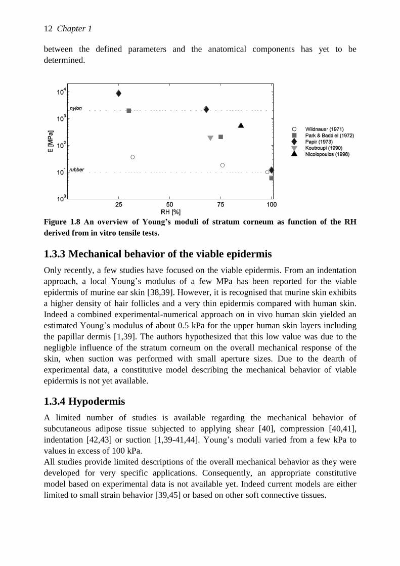
Reported Young‟s moduli vary considerably encompassing values from a few MPa to
GPa [24,25,35,36]. For example, the estimated tensile moduli for various RH is shown in
Figure 1.8. As indicated in this figure, the stiffness of the stratum corneum varies from
rubber-like at high RH to nylon-like at low RH values. The differences may be due to a
combination of reasons, such as regional differences, anisotropy, differences between
species, but also test conditions, such as sample preparation and difficulties in
controlling sample dimensions and environmental conditions. A general trend, however,
is a more pronounced decrease of the elastic modulus beyond 60% RH. At a constant
RH, the stratum corneum hydration increases by 50% when the temperature rises from
20°C to 30°C. The influence of temperature decreases to a minimum beyond 90% RH.
More common trends due to an increase in RH or temperature include an increase of the
maximum extension and work of rupture, and a reduction of the force at rupture
[23,25,37]. Furthermore, stratum corneum behaves isotropically in transversal plane only
[36].
Preconditioning effects have not been reported for stratum corneum, which represents an
important difference with the whole skin. This finding indicates the absence of mobile
components in the stratum corneum [36].
Current constitutive models of the stratum corneum are based on traction, relaxation and
creep tests [5]. From the experimental tests, it is important that the model accomodate
elasticity, non-linear viscosity and strain hardening parameters. However, the association
Figure 1.7 Typical force elongation curves for the stratum corneum at different RH
showing different phases: the elastic phase (I), the plastic phase (II) and the strain
hardening phase (III). Obtained from [23].
10
20
30
40
I II III
98% RH
76% RH
30 120
32% RH
Elongation [%]
Loa
d [g
]
in v
ivo
ra
ng
e
12 Chapter 1
between the defined parameters and the anatomical components has yet to be
determined.
Figure 1.8 An overview of Young’s moduli of stratum corneum as function of the RH
derived from in vitro tensile tests.
1.3.3 Mechanical behavior of the viable epidermis
Only recently, a few studies have focused on the viable epidermis. From an indentation
approach, a local Young‟s modulus of a few MPa has been reported for the viable
epidermis of murine ear skin [38,39]. However, it is recognised that murine skin exhibits
a higher density of hair follicles and a very thin epidermis compared with human skin.
Indeed a combined experimental-numerical approach on in vivo human skin yielded an
estimated Young‟s modulus of about 0.5 kPa for the upper human skin layers including
the papillar dermis [1,39]. The authors hypothesized that this low value was due to the
negligble influence of the stratum corneum on the overall mechanical response of the
skin, when suction was performed with small aperture sizes. Due to the dearth of
experimental data, a constitutive model describing the mechanical behavior of viable
epidermis is not yet available.
1.3.4 Hypodermis
A limited number of studies is available regarding the mechanical behavior of
subcutaneous adipose tissue subjected to applying shear [40], compression [40,41],
indentation [42,43] or suction [1,39-41,44]. Young‟s moduli varied from a few kPa to
values in excess of 100 kPa.
All studies provide limited descriptions of the overall mechanical behavior as they were
developed for very specific applications. Consequently, an appropriate constitutive
model based on experimental data is not available yet. Indeed current models are either
limited to small strain behavior [39,45] or based on other soft connective tissues.
General introduction 13
1.4 Aim and Outline
The objective of this thesis is to develop appropriate experimental techniques and
procedures, which will enable the characterization of the mechanical behavior of
individual skin layers in vitro. The focus is on those skin layers for which available data
is relatively scarce, i.e. the viable epidermis and hypodermis, and/or inconsistent as in
the case for the stratum corneum. The results should provide insight into the relationship
between the mechanical responses to the structure of the various skin layers and, hence,
provide better understanding of the way a treatment or disease affects the skin behavior.
Furthermore, the experimental data should provide suitable input for constitutive models.
Previous studies, such as the various in vitro tensile tests on the stratum corneum, have
indicated that differences in mechanical properties of the epidermis and stratum corneum
are not solely caused by variations in humidity and temperature, but are influenced test
conditions, anisotropy, sample preparation, etc. It is therefore essential to perform
experiments with samples of consistent quality in an accurate measurement system in a
well-controlled environment. This will be initially achieved in relatively simple small
strain experiments in various directions under different environmental conditions. If this
small strain behavior is reproducible and well-understood, then it is appropriate to extend
the work to examine the non-linear behavior.
In order to obtain in vitro samples of consistent quality, various isolation and
preservation treatments are first thoroughly investigated for both skin layers (Chapter 2).
Subsequently, a rheological measurement system has been designed to measure the shear
response of thin, soft tissues in a controlled environment (Chapter 3). A micro-
indentation method has been adapted to enable the measurement of loading
perpendicular to the skin surface (Chapter 4). Because viable epidermis cannot be
isolated as a single layer, a numerical model is introduced to predict its behavior from
the experiments on stratum corneum and whole epidermis.
Subsequently, rheological methods are developed to study the linear shear response of
subcutaneous adipose tissue (Chapter 5). From those results, a constitutive model
describing the linear viscoelastic behavior of subcutaneous adipose tissue at small strains
has been developed. Then, a set of experiments were designed to study both the large
deformation and time-dependent behavior (Chapter 6).
Finally (Chapter 7), a general discussion evaluates the selected measurement methods
for the skin layers and these outcomes, as well as the significance of the findings of this
work for various applications.

Chapter 2
Isolation and preservation methods for
the epidermis and stratum corneum
The contents of this chapter are based on M. Geerligs, D. Bronneberg, P.A.J.
Ackermans, C.W.J. Oomens, and D.L. Bader, Isolation and preservation methods for the
epidermis, submitted.
16 Chapter 2
2.1 Introduction
Ex vivo human skin grafts provide a cost-effective alternative to animal and clinical
testing. Various industries, such as the cosmetic, household product and pharmaceutical,
could benefit from in vitro studies to evaluate drugs and a range of consumer products.
Skin models are already used in many transdermal drug delivery and percutaneous
absorption studies, as well as in irritancy and toxicology studies. Studies on ex vivo skin
increase the fundamental knowledge on both structural and mechanical properties of
skin. In addition, studies on isolated skin layers, such as the epidermis or stratum
corneum, could provide an insight into the specific contribution of each layer to the
overall skin response. Skin models enable improved control of experimental conditions,
i.e. temperature, hydration level, and offer the potential to perform well-controlled in
vitro experiments. In order to obtain meaningful results, it is of utmost importance that
the structural integrity and viability of the skin are maintained.
The epidermis, the outermost skin layer, is directly contiguous to the external
environment and acts as a permeable barrier. It prevents excess water loss from the
aqueous interior and protects the internal tissue against mechanical insults, UV
irradiation and the ingress of foreign chemicals and micro-organisms. Due to the
extraordinary nature of the epidermis, its complete isolation while maintaining its
structural integrity remains a challenge. The keratinocytes are surrounded by a poor
extracellular matrix and lack the support of a fiber structure, which provides the strength
and stiffness of most biological tissues. Within the epidermis, the mechanical properties
are determined by the rigid tonofilament cytoskeleton and the numerous desmosomes to
which the filaments are anchored at the periphery of the keratinocytes. At the epidermal-
dermal junction hemidesmosomes anchor the epidermis to the dermis (see Figure 1.5).
These hemidesmosomes or the adjacent anchoring filaments need to be disrupted to fully
separate the epidermis from the dermis.
In order to maintain the complex structure of the stratum corneum during isolation, it is
important to preserve the curvature. The architecture of the stratum corneum is widely
established as a solid brick-and-mortar structure, with flat corneocytes surrounded by a
matrix of lipid enriched membranes strongly held together by desmosomes.
Due to the high number of plastic and cosmetic surgery procedures, such as
abdominoplasty and breast reduction, there is an increased availability of ex vivo human
skin. Whether a skin graft can be successfully used as skin model during in vitro
experiments depends on the nature of the tissue. The integrity of the skin tissue mainly
depends on the age of the subject, as well as on the donor body site. Furthermore, within
one skin graft, its structure might change as a result of disease or prior treatment. These
factors are usually reflected in tissue changes, such as convolutions of the epidermal-
dermal junction, thickness of epidermal strata, cell shape and surface folding, but may
also lead to qualitative and quantitative differences in the various epidermal components
Isolation and preservation methods for the epidermis and stratum corneum 17
[46]. To obtain the best experimental outcome from in vitro studies, it is important to use
structurally and functionally intact models.
In order to use the available intact skin grafts with optimal efficiency, factors such as
cleaning, preservation, and storage should be adequately addressed. In various studies,
such as transdermal drug delivery, percutaneous absorption studies, irritancy and
toxicology studies, an intact skin barrier is essential. Furthermore, adequate preservation
is crucial for maintaining the viability and integrity of the skin tissue. Tissue damage
such as the creation of vacuoles are easily induced and the selection of a proper tissue
storage method is therefore important.
Evaluation techniques to assess skin viability during storage have been extensively
described [47-49]. Common methods to assess viability include Trypan blue dye
exclusion, tetrazolium reductase activity, oxygen consumption rates, lactate and glucose
levels, and NMR spectroscopy. Structural integrity is usually assessed by histological
routines or imaging techniques.
This paper aims to critically review various isolation methods for the epidermis and
stratum corneum and preservation methods useful for in vitro research on split-thickness
skin, epidermis and stratum corneum. Existing reviews are considered to be out of date
and do not include recent work from the host laboratory [46,50-52]. No standards exist,
thus inter-study comparisons are problematic. In addition, much of the existing data may
have been influenced by the specific preparation technique, which have been employed.
Accordingly, the present paper describes mechanical, ionic change, heat, enzymatic
digestion and irradiation techniques for isolation of the skin layers. The advantages and
disadvantages of each technique are discussed in terms of maintaining the skin integrity
and ease of handling. In addition, the influence of various storage conditions on the skin
structure and viability are discussed.
2.2 Skin preparation and analyses
General steps in the preparation of skin samples used in the present experiments are
described below, as well as the analysis techniques used to study the skin structure and
viability.
2.2.1 Skin preparation
Human skin was obtained from female patients undergoing abdominoplasty. The
research proposal for our studies was approved by the Medical Ethics Committee of the
Catharina Hospital, Eindhoven, the Netherlands. Immediately after excision, the skin is
brought to the laboratory for further processing. Here, the skin is placed on a stainless
steel plate covered with paper towels to absorb body fluids. The skin surface is cleaned
with pure water. Using multiple forceps, the skin graft is stretched and fixed to the
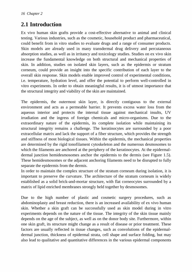
stainless steel plate (Figure 2.1a). Subsequently, split-thickness skin samples, varying in
thickness from 100-400 µm, are produced using a commercial dermatome (D42,
Humeca, The Netherlands) (Figure 2.1b).
18 Chapter 2
(a) (b)
Figure 2.1: Skin is stretched using forceps (a) and dermatomed (b).
(b)
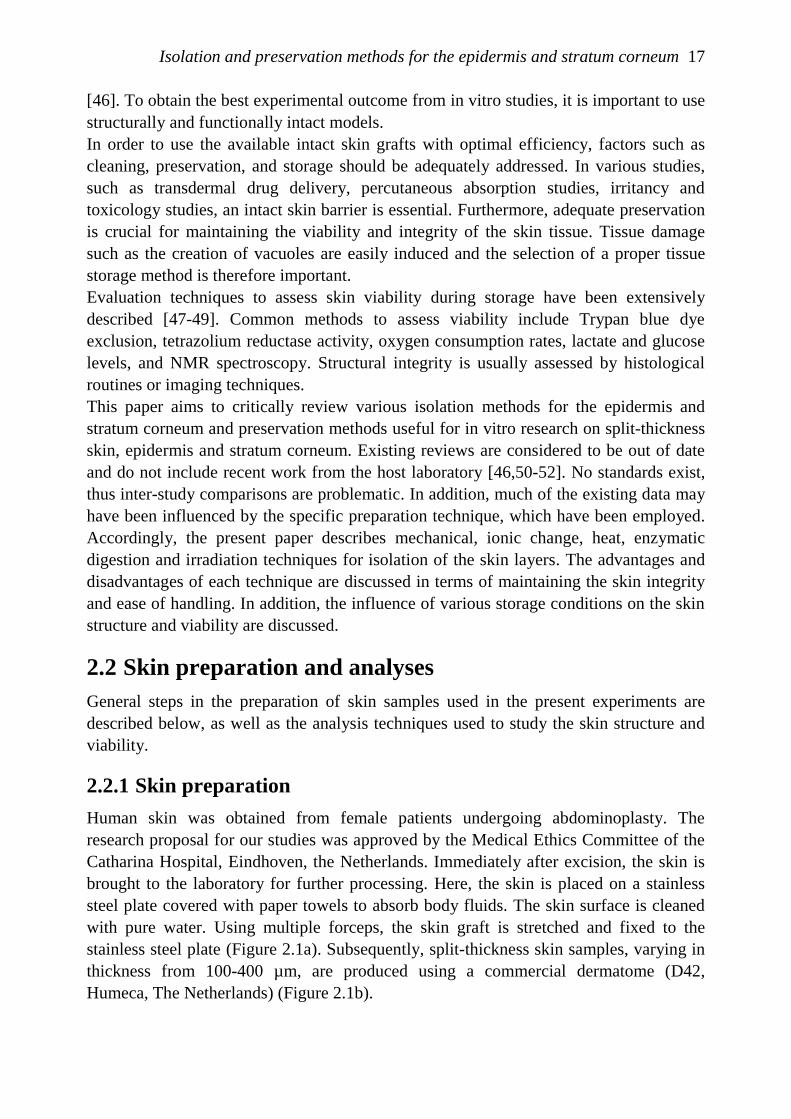
(a) (c)
Figure 2.2. (a) Full thickness skin stained with aldehyde-fuchsin to visualize the stratum
corneum (SC), viable epidermis (VE), papillar dermis (PD) and reticular dermis (RD);
(b) Dermatomed skin with a set thickness of 100 μm consists of the epidermal layer only;
(c) In some cases, however, some papillar dermis is still attached.
2.2.2 Histological examination
In order to examine tissue structure, samples were fixated in 10% phosphate-buffered
formalin and processed for conventional paraffin embedding. The sections were cut into
5 μm slices and stained with aldehyde-fuchsin and yellow green SF (Merckx) or standard
heamotoxilyn and eosin (H&E) staining. The tissue morphology was studied by light
microscopy. The aldehyde-fuchsin staining is used to clearly identify the different skin
layers, namely the stratum corneum, viable epidermis, papillar dermis and reticular
dermis (Figure 2.2a). The structural integrity is examined by using the H&E staining.
SC
VE
PD
RD
SC
VE
SC
VE
PD
Isolation and preservation methods for the epidermis and stratum corneum 19
2.2.3 Analyses of skin viability
Skin viability was studied by using the colorimetric MTT (Thiazolyl Blue Tetrazolium
Bromide) assay. Skin samples with a diameter of 8 mm were placed in a 24 wells-plate
containing 300 µl of 1 mg/ml MTT solution in PBS in a well (Phosphate Buffered
Saline). The plates were incubated at 37C and 5% CO2 for a period of 3 hours. After
incubation, the skin samples were removed and gently blotted with tissue paper, before
completely submerging them in 2 ml 2-propanol per well. The extraction plates were
placed in sealed bags to reduce evaporation and were gently shaken for 2 hours at room
temperature to extract the reduced MTT. The absorption of the extractant was measured
at 570 nm, using plain extractant as blank.
2.3 Epidermal isolation techniques
Isolation techniques for the epidermis can be divided into the following categories:
mechanical, ionic change, heat, enzymatic digestion and irradiation techniques. The
effectiveness of each is summarized in Table 2.1 at the end of the section in terms of
actual cleavage plane, maintaining of both cell viability and tissue integrity.
2.3.1 Mechanical separation
Cutting by using a dermatome
Van Scott et al. [53] recommended a stretching method for separating the epidermis
from the dermis. The method involves manually stretching the skin to its limit over a
slightly convex wooden surface, and anchoring it in place by means of thumbtacks. A
razor blade or scalpel is used to scrape off the epidermis. Subsequently, the epidermis is
grasped by tweezers to gently detach a continuous sheet. However, damage can be easily
induced in the epidermis using this relatively crude stretching technique. The severity of
this damage depends on the vigour of scraping and the degree of stretching. The
development of keratomes, either handheld devices or as part of a mechanical device,
has improved the reproducibility of this stretching technique.
In the present study, a cordless, battery operated dermatome was used. As previously
mentioned, ex vivo skin was mounted on a stainless steel plate to facilitate the cutting
process. When the dermatome was set to 100 μm, samples of the epidermis could be
obtained. In some cases, however, some papillar dermis was still attached to the
epidermal specimens (Figure 2.2). Due to the presence of rete ridges, it was highly
unlikely that the cutting plane went through the dermal-epidermal junction only.
However, the number of skin layers present in the separated tissue can be assessed
visually; with the yellowish translucent epidermis being easily distinguishable from the
white opaque dermis. A MTT-test demonstrated that the dermatomed skin retained its
viability for 100%, which is in agreement with Wester et al. [54].
The defined geometric shape of the specimen is very convenient for assessing its
mechanical properties. It is assumed that the mechanical properties of the present
papillary dermis are similar to the surrounding epidermal tissue, because no differences
20 Chapter 2
in shear properties were found between 100 and 200 μm thick split-skin samples (see
Chapter 3).
Suction device
Suction blisters can be produced by applying suction cups on the skin, in both in vivo
and in vitro experiments. In vivo separation of the human epidermis was first reported in
1964 [55]. Kiistala et al.(1968) found that a blister could be induced within 130 minutes
with a suction gap of 25 mm. The diameter of a suction cup may vary from 15-50 mm
depending on body site. To avoid tissue damage, the pressure within the cup had to be
maintained at 200 mm Hg or above. The cleavage occurs in the plane through the lamina
lucida, leaving the lamina densa on the dermis and retaining an intact, viable basal cell
layer. However, enlargement of intercellular spaces due to considerable stretching might
cause large vacuoles in keratinocytic cytoplasm [50,56].
Suction blister time depends on factors such as suction pressure, individual variation and
regional differences as well as temperature, but does not depend on cup size. Because of
the low reproducibility caused by individual variations that cannot be controlled, this
method is considered to be unfavourable.
2.3.2 Ionic change
An earlier method to isolate the epidermis involved its maceration in dilute acetic acid.
Cowdry [57] described that dilute acetic acid causes swelling of collagen fibers which
decreases their cohesive strength and, therefore, the binding of epidermis to dermis. In
addition, it was found that collagen fibers also swell in an alkaline environment. These
methods, however, are toxic to epidermal cells and are therefore no longer used [58].
In addition, EDTA (ethylenediamine tetraacetic acid) has been used to obtain epidermal
sheets [59]. The location of the split changes according to the duration of the treatment.
For example, after 30 min incubation in 0.01 M EDTA at pH 7.4 the split occurred in the
lower granular layer, whereas after 45 min it was in a spinous-suprabasilar location and
after 60 min or more it occurred at the dermal–epidermal junction. In adition,
intracellular oedema increases with time. Accordingly, this is not considered to be a
favourable method for epidermal separation.
After prolonged incubation in 1 M NaCl at 4°C, the epidermis can also be easily
removed from the dermis with forceps. The split occurs through the lamina lucida.
Nevertheless, mitochondrial swelling within the keratinocytes was noted [50]. Although
no other degenerative features have been reported, epidermal components may have been
diminished or modified during the long incubation times of 24 to 96 hours [60].
Prolonged incubation in PBS is also known to separate the epidermis from the dermis.
Indeed after 72-96 hours at 37°C, the epidermis can be readily peeled off [61]. In
contrast to the above techniques, where the split occurs through the lamina lucida, the
split is closer to the epidermal site of the dermal-epidermal junction [61].
Since no intact viable epidermal sheets can be obtained using any of the techniques
based on ionic change, they are not considered suitable for epidermal isolation.
Isolation and preservation methods for the epidermis and stratum corneum 21
2.3.3 Heat
Separating the epidermis from the dermis using a hot plate is a simple and rapid method
[58]. It was reported that the skin is heated up to 50 to 60C for 30 s. To maintain
enzyme activity, mild heat treatment at 52C for 30 s is required. Separation occurs at
the basal cell layer. Depending on the exact conditions, release of enzymes, cytolysis and
cell separation may occur. However, it has been claimed that heat does not modify
fibrous proteins within isolated epidermis [62]. Although heating can easily cause tissue
dehydration, this can be minimized by increasing the humidity of the environment or by
placing the skin in a sealed bag in hot water, instead of using a hot plate. After heating,
the epidermis can be gently peeled from the dermis.
In the present studies, human skin samples were heated on either a hot plate and in a
sealed bag. The former process appeared to flatten the undulating epidermal structure,
while the papillae remained intact after heating in a sealed bag in hot water. Much longer
heating times were needed than mentioned in literature. The epidermis could be peeled
from the dermis after more than 5 minutes.
For both heat separation techniques, structural tissue damage occured as evidenced by
the presence of vacuoles and a disrupted basal layer (Figure 2.3). It has been previously
reported that heat treated skin (60°C for 1 minute) and heat-separated epidermis and
dermis significantly lose viability [63]. Furthermore, some practical problems arose
when using a hot plate, such as curling of the dermal tissue and uneven separation of the
epidermis over the complete skin surface due to gradual thermal diffusion.
(a) (b)
Figure 2.3. Histological sections of epidermis isolated using heat by means of a hot plate (a)
or placing the epidermis in a sealed bag in hot water (b). A standard H&E staining has
been used.
2.3.4 Enzymatic digestion
Trypsin
Epidermal separation by means of trypsin has been widely used, although some
conflicting results have been published. For example, Briggeman et al. [64] reported that
the epidermis is isolated by the cleaving effect of trypsin, whereas other authors reported
that many basal cells remain loosly attached to the basement membrane after trypsin
treatment [65,66]. The epidermis can be easily peeled from the dermis using 0.1-0.3%
22 Chapter 2
trypsin in a saline solution supplemented with calcium and magnesium at 4°C. However,
these conditions also induce a high level intra-epidermal split at the spinous-granular
interface [46]. Inconsistencies within the reported findings seem to be related to various
factors such as size and thickness of the skin sample, enzymatic concentration and its
solvent, incubation time and temperature. In addition some side-effects are noted
following trypsin treatment such that recovery may take up to a few days [46]. All these
factors lead to inconsistent epidermal separation following treatment.
Thermolysin
The epidermis can easily be separated from the dermis following incubation at 4C for 1
h in a solution containing 250-500 g/ml thermolysin, a proteolytic enzyme more
generally used for protein analysis [65]. Thermolysin can be dissolved in sterile
magnesium free PBS containing 1 mM CaCl2 at pH 7.8. However, to ensure complete
penetration of the enzyme, it is advisable to remove the subcutaneous fat and the lower
dermis from the specimen. Light and electron microscopy revealed that the separation
occurred at the lamina lucida and that the hemidesmosomes were selectively disrupted
[65]. By contrast, Willsteed et al.[50] noticed an intraepidermal split, without any lamina
lucida separation.
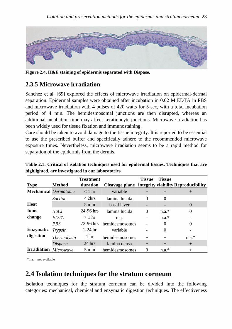
Dispase
Dispase II (Roche Diagnostics) has proven to be a rapid, effective, but gentle agent for
separating intact epidermis from the dermis [67,68]. This proteolytic enzyme is able to
cleave the basement membrane zone region while preserving the viability of the
epithelial cells.
Based on recommendations from the supplier, 2.4 U/ml dispase in 50 mM HEPES/KOH
buffer pH 7.4 with 150 mM NaCl was used in the present studies to separate the
epidermis from the dermis. Fresh skin samples of various sizes were placed on top of
sterile gauzes in 6 cm diameter petri dishes containing 5 ml of 2.4 U/ml Dispase II. The
stratum corneum of the skin samples was not exposed to the enzymatic solution during
the separation process to minimize loss of the skin barrier integrity. After overnight
incubation at 4C and thereafter 10 min at 37C, the epidermis was gently peeled from
the dermis using tweezers. In agreement with literature, the present study demonstrated
that the bottom surface of the separated epidermal sheet retained its rete-ridges and hair
follicles with sebaceous glands and the eccrine sweat glands retained their undistorted
shape [68] (Figure 2.4). The cleavage occurred in the lamina densa.
This isolation method is very suitable for generating intact epidermal sheets. The best
results were obtained when split-thickness skin samples of roughly 300 µm, which were
then enough to facilitate enzyme diffusion. Therefore, it is recommended to dermatome
skin grafts prior to performing the enzyme treatment.
Isolation and preservation methods for the epidermis and stratum corneum 23
Figure 2.4. H&E staining of epidermis separated with Dispase.
2.3.5 Microwave irradiation
Sanchez et al. [69] explored the effects of microwave irradiation on epidermal-dermal
separation. Epidermal samples were obtained after incubation in 0.02 M EDTA in PBS
and microwave irradiation with 4 pulses of 420 watts for 5 sec, with a total incubation
period of 4 min. The hemidesmosomal junctions are then disrupted, whereas an
additional incubation time may affect keratinocyte junctions. Microwave irradiation has
been widely used for tissue fixation and immunostaining.
Care should be taken to avoid damage to the tissue integrity. It is reported to be essential
to use the prescribed buffer and specifically adhere to the recommended microwave
exposure times. Nevertheless, microwave irradiation seems to be a rapid method for
separation of the epidermis from the dermis.
Table 2.1: Critical of isolation techniques used for epidermal tissues. Techniques that are
highlighted, are investigated in our laboratories.
2.4 Isolation techniques for the stratum corneum
Isolation techniques for the stratum corneum can be divided into the following
categories: mechanical, chemical and enzymatic digestion techniques. The effectiveness
Type Method
Treatment
duration Cleavage plane
Tissue
integrity
Tissue
viability Reproducibility
Mechanical Dermatome < 1 hr variable + + +
Suction < 2hrs lamina lucida 0 0 -
Heat 5 min basal layer - - 0
Ionic NaCl 24-96 hrs lamina lucida 0 n.a.* 0
change EDTA > 1 hr n.a. - n.a.* -
PBS 72-96 hrs hemidesmosomes - 0 0
Enzymatic Trypsin 1-24 hr variable - 0 -
digestion Thermolysin 1 hr hemidesmosomes + + n.a.*
Dispase 24 hrs lamina densa + + +
Irradiation Microwave 5 min hemidesmosomes 0 n.a.* +
*n.a. = not available
24 Chapter 2
of each technique is summarized in Table 2.2, in terms of maintaining both cell viability
and tissue integrity.
2.4.1 Mechanical separation
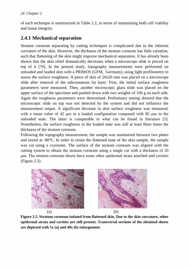
Stratum corneum separating by cutting techniques is complicated due to the inherent
curvature of the skin. However, the thickness of the stratum corneum has little variation,
such that flattening of the skin might improve mechanical separation. It has already been
shown that the skin relief dramatically decreases when a microscope slide is placed on
top of it [70]. In the present study, topography measurements were performed on
unloaded and loaded skin with a PRIMOS (GFM, Germany), using light profilometry to
assess the surface roughness. A piece of skin of 20x20 mm was placed on a microscope
slide after removal of the subcutaneous fat layer. First, the initial surface roughness
parameters were measured. Then, another microscopic glass slide was placed on the
upper surface of the specimen and pushed down with two weights of 100 g on each side.
Again the roughness parameters were determined. Preliminary testing showed that the
microscopic slide on top was not detected by the system and did not influence the
measurement output. A significant decrease in skin surface roughness was measured,
with a mean value of 42 μm in a loaded configuration compared with 85 μm in the
unloaded state. The latter is comparable to what can be found in literature [5].
Nonetheless, the surface roughness in the loaded state was still at least three times the
thickness of the stratum corneum.
Following the topography measurement, the sample was maintained between two plates
and stored at -80°C. In order to retain the flattened state of the skin sample, the sample
was cut using a cryotome. The surface of the stratum corneum was aligned with the
cutting system to obtain the stratum corneum using a single cut with a thickness of 20
μm. The stratum corneum sheets have some other epidermal strata attached and cavities
(Figure 2.5).
(a) (b)
Figure 2.5. Stratum corneum isolated from flattened skin. Due to the skin curvature, other
epidermal strata and cavities are still present. Transversal sections of the obtained sheets
are depicted with 5x (a) and 40x (b) enlargement.
Isolation and preservation methods for the epidermis and stratum corneum 25
2.4.2 Chemical separation
Cantharidin blister procedure
This method, however, has only been reported up to the early seventies [8,23].
Cantharidin was impregnated into 1 cm diameter disks of filter paper and placed under
occlusive patches rather than applied directly to the skin surface in a volatile solvent.
The disks were removed after 4 hours and protective caps were placed over the forming
blisters to prevent damage to the samples. The blister tops were surgically excised and
the loose underlying wet cells removed by gentle swabbing. Since the discovery that
cantharidin is toxic, it is not permitted to use it for skin treatments anymore.
Ammonia vapour
In the sixties and seventies, it was common to isolate stratum corneum through exposure
to ammonia vapour. The latest protocols reported around 30 min exposure to separate the
dermis and epidermis [71,72]. Adherent wet cells are subsequently removed with a
cotton swab such that the stratum corneum sheet remains [73]. Thereafter, the stratum
corneum sheet was allowed to dry on silicone-coated paper at ambient conditions. In
addition, it was noticed that the success of this treatment is variable. Since more
consistent techniques causing less damage became available, this method is no longer
used.
2.4.3 Enzymatic digestion
Trypsin
The working of trypsin throughout the epidermal strata has been extensively studied
[73]. It appeared that the architecture of the stratum corneum remains unaffected by
trypsinization. Corneodesmosomes and composite desmosomes shared by corneum and
granular cells are normal. Tonofilaments attached to these junctions also appear
unchanged [73]. However, concentrations of trypsin above 0.125% might damage the
stratum corneum such that its elastic properties change [5].
In order to enable the working of trypsin on the epidermal cells, the subcutaneous fat
layer and the lower dermis has to be removed. In our laboratories, the remaining skin
was immersed in a porcine 0.1% trypsin (SV30037.01, Hyclone) solution in PBS
(Phosphate Buffer Saline). For quick processing, the samples were then placed for over 2
hours in an incubator at 37°C. For this study, dermatomed skin of approximately 300 μm
thick and a surface area of 2 cm2 was placed in 3 ml trypsin. Similar results can be
obtained through an overnight culture at 4°C and 15 min at 37°C. Due to the lipids
within the stratum corneum, the thin layer floats to the surface while the remaining
epidermis sinks to the bottom. In order to prevent post trypsinization effects, stratum
corneum is rinsed with distilled water a few times to wash out trypsin and treated with
anti-trypsin. The overnight protocol can be considered as the golden standard, which is
frequently described and commonly used within several research fields.
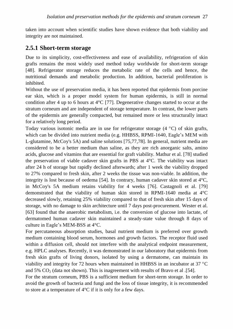
26 Chapter 2
Figure 2.6. (a) After staying overnight at 4°C, the extracellular matrix of the viable
epidermis is still attached to the stratum corneum; (b) Only stratum corneum is obtained
after leaving the skin sample for 1 hour at 37°C.
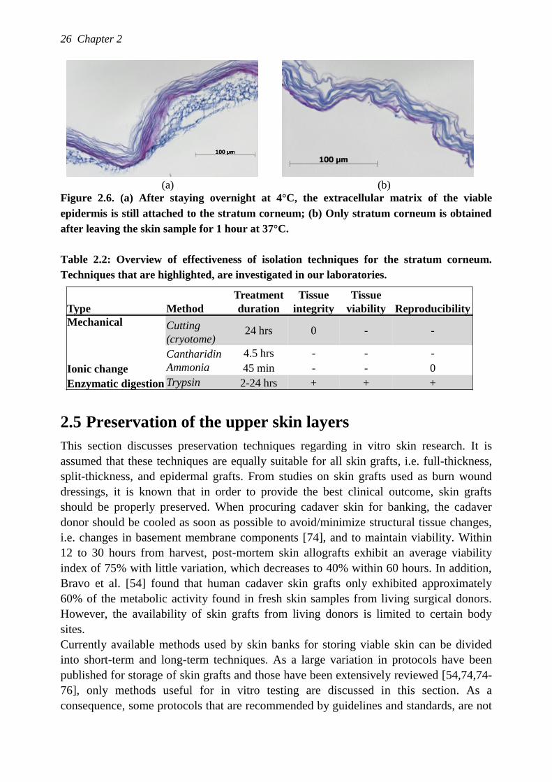
Table 2.2: Overview of effectiveness of isolation techniques for the stratum corneum.
Techniques that are highlighted, are investigated in our laboratories.
2.5 Preservation of the upper skin layers
This section discusses preservation techniques regarding in vitro skin research. It is
assumed that these techniques are equally suitable for all skin grafts, i.e. full-thickness,
split-thickness, and epidermal grafts. From studies on skin grafts used as burn wound
dressings, it is known that in order to provide the best clinical outcome, skin grafts
should be properly preserved. When procuring cadaver skin for banking, the cadaver
donor should be cooled as soon as possible to avoid/minimize structural tissue changes,
i.e. changes in basement membrane components [74], and to maintain viability. Within
12 to 30 hours from harvest, post-mortem skin allografts exhibit an average viability
index of 75% with little variation, which decreases to 40% within 60 hours. In addition,
Bravo et al. [54] found that human cadaver skin grafts only exhibited approximately
60% of the metabolic activity found in fresh skin samples from living surgical donors.
However, the availability of skin grafts from living donors is limited to certain body
sites.
Currently available methods used by skin banks for storing viable skin can be divided
into short-term and long-term techniques. As a large variation in protocols have been
published for storage of skin grafts and those have been extensively reviewed [54,74,74-
76], only methods useful for in vitro testing are discussed in this section. As a
consequence, some protocols that are recommended by guidelines and standards, are not
Type Method
Treatment
duration
Tissue
integrity
Tissue
viability Reproducibility
Mechanical Cutting
(cryotome)24 hrs 0 - -
Cantharidin 4.5 hrs - - -
Ionic change Ammonia 45 min - - 0
Enzymatic digestion Trypsin 2-24 hrs + + +
(a) (b)
Isolation and preservation methods for the epidermis and stratum corneum 27
taken into account when scientific studies have shown evidence that both viability and
integrity are not maintained.
2.5.1 Short-term storage
Due to its simplicity, cost-effectiveness and ease of availability, refrigeration of skin
grafts remains the most widely used method today worldwide for short-term storage
[48]. Refrigerator storage reduces the metabolic rate of the cells and hence, the
nutritional demands and metabolic production. In addition, bacterial proliferation is
inhibited.
Without the use of preservation media, it has been reported that epidermis from porcine
ear skin, which is a proper model system for human epidermis, is still in normal
condition after 4 up to 6 hours at 4°C [77]. Degenerative changes started to occur at the
stratum corneum and are independent of storage temperature. In contrast, the lower parts
of the epidermis are generally compacted, but remained more or less structurally intact
for a relatively long period.
Today various isotonic media are in use for refrigerator storage (4 C) of skin grafts,
which can be divided into nutrient media (e.g. HHBSS, RPMI-1640, Eagle‟s MEM with
L-glutamine, McCoy's 5A) and saline solutions [75,77,78]. In general, nutrient media are
considered to be a better medium than saline, as they are rich anorganic salts, amino
acids, glucose and vitamins that are essential for graft viability. Mathur et al. [78] studied
the preservation of viable cadaver skin grafts in PBS at 4°C. The viability was intact
after 24 h of storage but rapidly declined afterwards; after 1 week the viability dropped
to 27% compared to fresh skin, after 2 weeks the tissue was non-viable. In addition, the
integrity is lost because of oedema [54]. In contrary, human cadaver skin stored at 4°C,
in McCoy's 5A medium retains viability for 4 weeks [76]. Castagnoli et al. [79]
demonstrated that the viability of human skin stored in RPMI-1640 media at 4°C
decreased slowly, retaining 25% viability compared to that of fresh skin after 15 days of
storage, with no damage to skin architecture until 7 days post-procurement. Wester et al.
[63] found that the anaerobic metabolism, i.e. the conversion of glucose into lactate, of
dermatomed human cadaver skin maintained a steady-state value through 8 days of
culture in Eagle‟s MEM-BSS at 4°C.
For percutaneous absorption studies, basal nutrient medium is preferred over growth
medium containing blood serum, hormones and growth factors. The receptor fluid used
within a diffusion cell, should not interfere with the analytical endpoint measurement,
e.g. HPLC analyses. Recently, it was demonstrated in our laboratory that epidermis from
fresh skin grafts of living donors, isolated by using a dermatome, can maintain its
viability and integrity for 72 hours when maintained in HHBSS in an incubator at 37 C
and 5% CO2 (data not shown). This is inagreement with results of Bravo et al .[54].
For the stratum corneum, PBS is a sufficient medium for short-term storage. In order to
avoid the growth of bacteria and fungi and the loss of tissue integrity, it is recommended
to store at a temperature of 4°C if it is only for a few days.
28 Chapter 2
2.5.2 Long-term storage
Cryopreservation
Long-term storage of skin is possible via cryopreservation. In general, the success rate of
freezing tissue depends on various factors, i.e. conducting medium, the cooling rate, the
number of cell types in the tissue, the addition of a cryoprotective agent, storage
temperature, the cooling rate and thawing rate. The viability of the epidermis (and
dermis) can be well-retained when cooling to ultralow temperature by using
cryoprotective agents (CPA‟s), without the formation of ice crystals. Cryporeservation is
likely the most routinely used method for long-term storage of skin, because the skin can
then be stored for months to years [80].
Any cell type has its optimum cooling rate producing maximum cell survival. If the
cooling rate is higher than the optimum, intracellular ice appears, causing the cell to die.
In contrast, if the cooling rate is slow, free water is removed from solution to form
extracellular ice crystals increasing the salt concentrations in the tissue. The cells also
shrink because of osmosis. It is unlikely that each cell type within a tissue will exhibit
the same optimum cooling rate. Although epidermis mainly consists of keratinocytes,
maintaining high viability for all epidermal cells would be challenging. This can be
achieved, however, if cryoprotective chemicals are added before freezing. The most
common CPA‟s are glycerol and dimethyl sulfoxide (DMSO). These cryoprotective
agents act as solvents for the salts. In addition, their presence within the cells prevents
excessive shrinkage of the cells during the cooling phase. Therefore in the presence of
CPA‟s, it is possible to use very slow cooling rates that minimize intracellular ice
formation while protecting the cells against solution effects. High viabilities of all cell
types can be achieved using this slow cooling rate: a cooling rate of -30°C per minute
was shown to maintain the viability of keratinocytes [74].
When skin tissue cryopreserved with 15% glycerol in PBS or nutrient medium has been
cooled by a controlled-rate process to at least -80°C, it can be transferred for long-term
storage into the vapor phase of liquid nitrogen (below -130°C). Once the skin is at a
temperature lower than -130°C, i.e. the glass transition temperature of water, no further
loss of cell viability is incurred.
The optimum thawing procedure is a rapid warming method. This can be achieved by
plunging the skin into a 37°C water bath until the tissue is just thawed. Prolonged
storage at 37°C in the presence of CPA would be detrimental. Because the cells contain
high concentrations of CPA, they are hyperosmotic compared with normal saline. To
avoid osmotic lysis of the cells, either the saline can be added gradually or an
impermeant solute such as sucrose can be added to the saline to reduce the difference in
osmolarity. It has been reported that viability declines rapidly after thawing of the skin,
even if the epidermis is stored in nutrient media [54].
It should be noted that it is prefered to use glycerol rather than DMSO, because it has a
lower toxicity to the cells and is more effective [78,81]. Nevertheless, the skin viability
might be somewhat lower after cryopreservation with glycerol [54].
Isolation and preservation methods for the epidermis and stratum corneum 29
Although CPA‟s are relatively non-toxic at low temperatures, the toxicity can become
significant at higher temperatures. However, structurally intact skin tissue is relatively
resistant to cryogenic damage compared to single cells. In addition, the rate at which
CPA‟s enter the cell depends on the temperature and the CPA, being faster at higher
temperatures. CPA can be best dissolved in a HEPES or TES buffer, because those
zwitterionic buffers do not lose their buffering capacity at lower temperatures.
Many different methods are in use for the packaging of frozen skin, ranging from rolls of
skin within a tube to the use of flat pack bags in metal laminated pouches. The latter are
preferred, in that the greater surface area to volume ratio ensures more even cooling
across the skin tissue, and the metal laminates are good heat conductors [74].
Snap freezing
Snap freezing in a well conducting medium, e.g. salt water, isopentane or hexane,
provides an effectice, rapid storage method without causing structural damage due to
water phase transitions. In practice, skin samples packed in a metal pouch can be
emerged in a 2-methylbuthane, which is cooled down by liquid nitrogen to -80°C. The
skin samples will immediately freeze and can then be stored at a -80°C freezer until use.
Since this is above the glass transition temperature of water, the slow progressive decline
in viability limits the maximum storage time to months.
After slowly thawing at room temperature, there is no need to thaw in a buffer before
using the tissue.
Although the tissue is not viable anymore, the tissue integrity is well maintained. As
Foutz et al. [82] showed that the mechanical properties of human skin are not affected by
freezing as well, it might be sufficient to snapfreeze samples for mechanical
characterization. Snapfrozen tissue is used for penetration and permeation studies as
well.
Drying stratum corneum
The routinely used method of drying stratum corneum is presumably the best method to
store isolated stratum corneum. According to the protocol of Bouwstra et al. [83], drying
and storage should take place in a cool dark room under an atmosphere of argon or
krypton. Because of possible detoriation of the lipid organization, it is recommended to
adhere to a maximum storage period of approximately three months.
Drying stratum corneum facilitates handling of the specimen. Most commonly is to dry
the stratum corneum on filter paper, but damage may occur to the fragile sheet upon
removal from the filter paper. The use of a sieve instead of the filter paper solves this
problem, since the stratum corneum can be removed even dried. Before the stratum
corneum sheet can be assessed, it needs to be immersed in pure water or PBS.
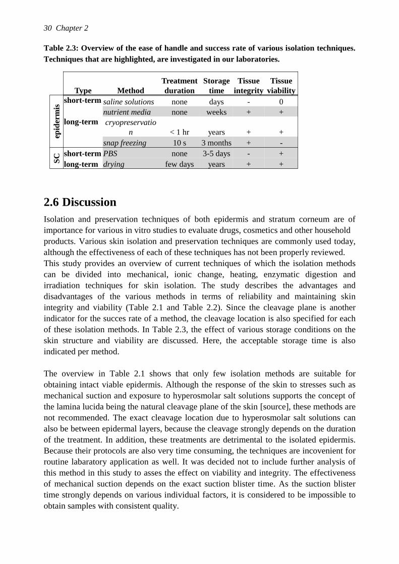
30 Chapter 2
Table 2.3: Overview of the ease of handle and success rate of various isolation techniques.
Techniques that are highlighted, are investigated in our laboratories.
2.6 Discussion
Isolation and preservation techniques of both epidermis and stratum corneum are of
importance for various in vitro studies to evaluate drugs, cosmetics and other household
products. Various skin isolation and preservation techniques are commonly used today,
although the effectiveness of each of these techniques has not been properly reviewed.
This study provides an overview of current techniques of which the isolation methods
can be divided into mechanical, ionic change, heating, enzymatic digestion and
irradiation techniques for skin isolation. The study describes the advantages and
disadvantages of the various methods in terms of reliability and maintaining skin
integrity and viability (Table 2.1 and Table 2.2). Since the cleavage plane is another
indicator for the succes rate of a method, the cleavage location is also specified for each
of these isolation methods. In Table 2.3, the effect of various storage conditions on the
skin structure and viability are discussed. Here, the acceptable storage time is also
indicated per method.
The overview in Table 2.1 shows that only few isolation methods are suitable for
obtaining intact viable epidermis. Although the response of the skin to stresses such as
mechanical suction and exposure to hyperosmolar salt solutions supports the concept of
the lamina lucida being the natural cleavage plane of the skin [source], these methods are
not recommended. The exact cleavage location due to hyperosmolar salt solutions can
also be between epidermal layers, because the cleavage strongly depends on the duration
of the treatment. In addition, these treatments are detrimental to the isolated epidermis.
Because their protocols are also very time consuming, the techniques are incovenient for
routine labaratory application as well. It was decided not to include further analysis of
this method in this study to asses the effect on viability and integrity. The effectiveness
of mechanical suction depends on the exact suction blister time. As the suction blister
time strongly depends on various individual factors, it is considered to be impossible to
obtain samples with consistent quality.
Type Method
Treatment
duration
Storage
time
Tissue
integrity
Tissue
viability
epid
erm
is
short-term saline solutions none days - 0
nutrient media none weeks + +
long-term cryopreservatio
n < 1 hr years + +
snap freezing 10 s 3 months + -
SC short-term PBS none 3-5 days - +
long-term drying few days years + +
Isolation and preservation methods for the epidermis and stratum corneum 31
Compared to the methods discussed above, isolation using heat or irradiation is much
less time consuming. However, although frequently used, heat treatment does not result
in an intact viable epidermis. The cleavage disrupts the basal layer, the viability declines
and structural changes like cell separation have been observed. In our own lab, for gently
removing the epidermis from the dermis, much longer heating times were needed than
reported in the literature. This is probablyly due to specimen type or experimental
conditions such as the humidity level. Due to the longer heating time, the susceptibility
to structure changes and loss of viability are increased so this method is considered
unfavourable as well. Isolation using microwave irradiation has been explored with
satisfying results regarding tissue viability, but it is not commonly used yet. To fully
assess the usefullness of microwave irradiation, more studies are needed.
Three enzymes are known to induce the dermal-epidermal split. The obvious advantage
of enzymatic digestion is that isolation takes place because of differences between cells
meaning that the undulating structure of the epidermis and stratum corneum is followed.
Trypsin, however, can cause cleavage at various planes in the epidermis making the
treatment using trypsin unreliable. Thermolysin might be an alternative, but practicle
studies with this enzyme are rare. What is widely used and extensively studied is
enzymatic digestion using dispase. This method is very robust compared to other
isolation methods: the epidermis has a consistent quality and the viability and integrity
can be fully maintained. The cleavage plane is the lamina densa, so also the basal layer is
completely intact. The duration of the treatment might be considered as a limitation as it
is an overnight procedure. However, the number of handling steps is small and an
additional advantage is that the cleavage plane is fairly independent of the treatment
duration.
Although it is sure that enzymatic digestion is by far the best method to isolate the
epidermis, sometimes additional reasons might lead to the choice for another isolation
method for certain applications. For example, the benefit of a sample with nearly perfect
geometry can be more important in an in vitro set-up than experiments on epidermis
only. Then, cutting slices of skin using a dermatome is an attractive quick method.
Applications such as mechanical characterization benefits from the fact that the natural
pre-stress in the skin is better retained. It was also demonstrated that the viability is
retained.
When looking at the options to isolate the stratum corneum from the viable epidermis,
fewer methods are available from which only enzymatic digestion using trypsin gives
satisfying results. It should be no surprise that both cantharadin and ammonia are
harmful to the skin. Furthermore, it is without doubt that enzymatic digestion by trypsin
is the common method to isolation the stratum corneum for any application field. The
robustness of the method and, hence, consistent quality does not give the incentive to
investigate new techniques.
As it is evident that there are means to obtain intact viable sheets of epidermis or stratum
corneum alone, the next challenge is to retain these properties over time. In literature,
32 Chapter 2
however, skin storage is mostly considered in relation to skin grafts used as burn wound
dressings. As a consequence, the focus is on split-thickness skin instead of isolated skin
layers, although the requirements in terms of viability and integrity are likely to be more
strict for in vitro testing than for the use of burn grafts, .
Preservation methods can be classified either based on technique, temperature or on
storage time. The latter was chosen here because in the case of in vitro testing one can
either immediately do the testing or needs to have a large batch available over a longer
time. Short-term storage can usually be done in a refrigerator. Storage in an incubator at
37°C is also satisfactory. Saline solutions certainly induce tissue damage, while various
nutrient media can keep the tissue viable and intact for at least a whole week before
degradation slowly begins. For the stratum corneum, it makes sense to use a saline
buffer. However, it is advised to do this only when using the samples within a week. For
longer storage, stratum corneum should be dried under the right conditions, as infections
and fungi may easily grow.
In the long-term, cryopreservation is a routine laboratory technique which can produce
large batches which can be beneficial for years. There is a risk of inconsistent quality of
the samples due to the sensitivity for tissue damage during thawing. When viability is
not requirement, snap freezing is a convenient and reliable method for long-term storage.
Although the variety of topics for in vitro skin research is enormous, this review has
shown that the isolation and storage protocols can be identical. Future in vitro research
should make use of isolated epidermis, which is separated by the enzyme dispase or cut
using a dermatome because of its convenient geometry. When the epidermal samples are
subsequently stored for just a short period and tissue growth is not the goal, it is advised
to use a nutrient media such as HHBSS. For long-term storage, the only option for intact
viable tissue is cryopreservation. Regarding the stratum corneum, trypsin and drying
remain by far the best methods to isolate and preserve this skin layer.
Acknowledgements
First of all, we would like to thank professor Bouwstra for her contribution in the
discussions. We are also very grateful to professor Hagisawa for providing the aldehyde-
fuchsin staining procedure. Last, we would like to thank the plastic surgery department
of the Catharina hospital in Eindhoven for providing the skin tissue.
Chapter 3 Linear shear response of the upper skin
layers
The contents of this chapter are based on M. Geerligs, G.W.M. Peters, P.A.J.
Ackermans, C.W.J. Oomens, and F.P.T. Baaijens, Linear shear response of the upper
skin layers, submitted.
34 Chapter 3
3.1 Introduction
Knowledge about the mechanical behavior of human skin is of great importance for
various clinical and cosmetic treatments. The human skin is composed of a non-uniform
layered structure and the mechanical behavior of all the layers is highly complex: i.e.
anisotropic, inhomogeneous, non-linear and viscoelastic. Therefore, the most appropriate
approach seems to be to determine the mechanical properties of each individual skin
layer in all loading directions in order to understand the full skin response.
The present study focuses on the contribution of the outer skin layer, the epidermis,
when in-plane forces are applied to the skin surface. Because of the anisotropic nature of
the epidermis, the response in tensile and shear are most probably different. Usually,
tensile properties are addressed in research studies. However, the shear component plays
a key role in applications such as the development of pressure ulcers, the removal of skin
adhesives and skin-device contact such as with prosthetic limbs and shavers. All these
applications could benefit from an improved knowledge of the mechanical response of
the epidermis to shear.
As the epidermis provides the chemical and physical barrier between the human body
and its environment, it possesses extraordinary structural properties. It is a stratified
epithelium, consisting of four different layers, defined by position, shape, morphology
and state of differentiation of the keratinocyte, the main cell type. The epidermal tissue is
renewed constantly: cells are lost from the skin surface by desquamation and this loss is
balanced by cell division and growth in the basal layer [84]. The most superficial layer,
the stratum corneum, has a thickness of typically 10-20 μm, and is considered as a
separate layer because of its specific barrier function. The stratum corneum has a „brick-
and-mortar‟ structure with the corneocytes, which are differentiated non-viable
keratinocytes, as „bricks‟ in a „mortar‟ of lipid membranes and desmosomes. The
thickness of the remaining part of the epidermis, the viable epidermis, ranges from 30-
100 μm. To strengthen the attachment of the epidermis to the dermis, the junction has an
undulating shape resulting in large cones of epidermal tissue penetrating the dermis. The
properties of both viable epidermis and stratum corneum are influenced by
environmental conditions, such as the temperature, 𝑇, and relative humidity, 𝑅𝐻.
Usually, load-bearing soft tissues are composed of a fiber network, providing strength
and elasticity to the tissue, but this is not the case for the epidermis. Its extensibility is
mainly due to the potential for smoothing out the skin surface, while the strength and
cohesiveness are due to the rigid tonofilament cytoskeleton and the numerous
desmosomes at the periphery of the keratinocytes. Furthermore, the viable epidermis is a
very compact tissue; the intercellular spaces occupy less than 2% of the volume [5,14].
Consequently, the viable epidermis is suspected to be more rigid than other soft tissues.
In the stratum corneum, the cellular membranes are thickened, the water content is
decreased and a larger amount of keratin is present and thus, its mechanical stiffness and
strength are suggested to be even higher.
Linear shear response of the upper skin layers 35
Due to the complex skin structure, the mechanical response of the epidermis cannot be
easily distinguished from that of the dermis in an in vivo experiment. This results into
two important implications for mechanical characterization of epidermis: 1) skin layers
need to be measured individually, and 2) in vitro measurements are required. Regarding
the first issue, the stratum corneum and the entire epidermis can be isolated from other
skin layers, but there are no means to isolate viable epidermis. So both isolated and
combined skin layers need to be characterized to assess the mechanical response of the
viable epidermis. Furthermore, in vitro measurements offer a broad range of reliable
standard techniques used in mechanical testing. Nevertheless, these methods need to be
adapted to enable the measurement of thin layers of soft materials. Moreover, issues
regarding the complex sample geometry, the heterogeneous tissue composition and the
sensitivity to environmental conditions have to be accomodated.
Currently, there is a paucity of papers describing the mechanical properties of the entire
epidermis or viable epidermis only. Studies to date were either on a small-sized scale
[38,85], not reproducible [86] or included the total papillar dermis [39] and none of them
investigated the shear response. Mechanical properties of the stratum corneum have been
studied and reviewed more extensively [5,22,35,87], although very few have examined
its shear response. Consequently, quantitative shear data for the upper skin layers is
sparse or not existent. It is hypothesized that the shear modulus of the epidermal layers is
considerably less than the broad range of tensile moduli found in literature, because of
the anisotropic structure of epidermis.
We measured the mechanical behavior of various human skin layers subjected to shear
over a wide frequency range and with varying environmental conditions, i.e. temperature
and relative humidity (RH). Because of the complexity, we limit ourselves in this study
to determine the small strain behavior of stratum corneum and viable epidermis. To
validate the experimental approach, tests with silicone rubbers are also performed.
3.2 Methods
3.2.1 Sample preparation
Skin
Skin was obtained from patients undergoing abdominoplastic surgery, who gave
informed consent for use of their skin for research purposes, under a protocol approved
by the ethics committee of the Catharina Hospital, Eindhoven, The Netherlands. Only
abdominal skin of Caucasian women from an age group between 35 and 55 years old
was used. Abdominal skin with stria, cellulite, damage due to UV exposure or
excessively hairy skin was excluded from the study.
Immediately after excision, the skin is transferred to the laboratory and processed within
4 hours. Skin slices are obtained using an electric dermatome (D42, Humeca, The
Netherlands) of which the prescribed thickness was refined for this purpose by the
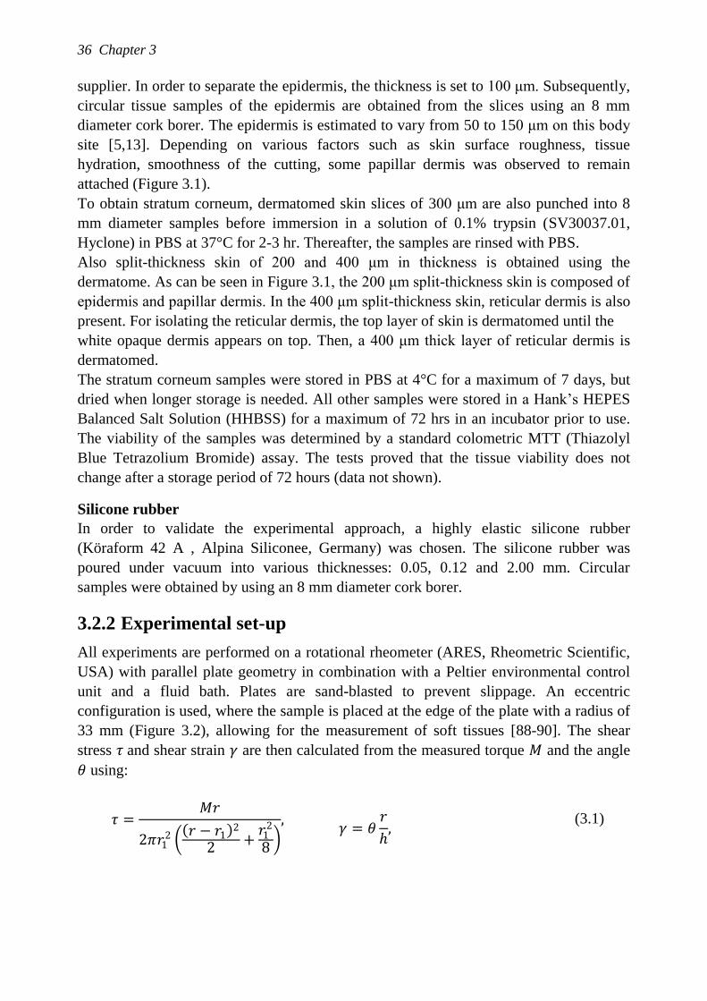
36 Chapter 3
supplier. In order to separate the epidermis, the thickness is set to 100 μm. Subsequently,
circular tissue samples of the epidermis are obtained from the slices using an 8 mm
diameter cork borer. The epidermis is estimated to vary from 50 to 150 μm on this body
site [5,13]. Depending on various factors such as skin surface roughness, tissue
hydration, smoothness of the cutting, some papillar dermis was observed to remain
attached (Figure 3.1).
To obtain stratum corneum, dermatomed skin slices of 300 μm are also punched into 8
mm diameter samples before immersion in a solution of 0.1% trypsin (SV30037.01,
Hyclone) in PBS at 37°C for 2-3 hr. Thereafter, the samples are rinsed with PBS.
Also split-thickness skin of 200 and 400 μm in thickness is obtained using the
dermatome. As can be seen in Figure 3.1, the 200 μm split-thickness skin is composed of
epidermis and papillar dermis. In the 400 μm split-thickness skin, reticular dermis is also
present. For isolating the reticular dermis, the top layer of skin is dermatomed until the
white opaque dermis appears on top. Then, a 400 μm thick layer of reticular dermis is
dermatomed.
The stratum corneum samples were stored in PBS at 4°C for a maximum of 7 days, but
dried when longer storage is needed. All other samples were stored in a Hank‟s HEPES
Balanced Salt Solution (HHBSS) for a maximum of 72 hrs in an incubator prior to use.
The viability of the samples was determined by a standard colometric MTT (Thiazolyl
Blue Tetrazolium Bromide) assay. The tests proved that the tissue viability does not
change after a storage period of 72 hours (data not shown).
Silicone rubber
In order to validate the experimental approach, a highly elastic silicone rubber
(Köraform 42 A , Alpina Siliconee, Germany) was chosen. The silicone rubber was
poured under vacuum into various thicknesses: 0.05, 0.12 and 2.00 mm. Circular
samples were obtained by using an 8 mm diameter cork borer.
3.2.2 Experimental set-up
All experiments are performed on a rotational rheometer (ARES, Rheometric Scientific,
USA) with parallel plate geometry in combination with a Peltier environmental control
unit and a fluid bath. Plates are sand-blasted to prevent slippage. An eccentric
configuration is used, where the sample is placed at the edge of the plate with a radius of
33 mm (Figure 3.2), allowing for the measurement of soft tissues [88-90]. The shear
stress 𝜏 and shear strain 𝛾 are then calculated from the measured torque 𝑀 and the angle
𝜃 using:
𝜏 =𝑀𝑟
2𝜋𝑟12
𝑟 − 𝑟1 2
2+
𝑟12
8
, 𝛾 = 𝜃
𝑟
ℎ,
(3.1)
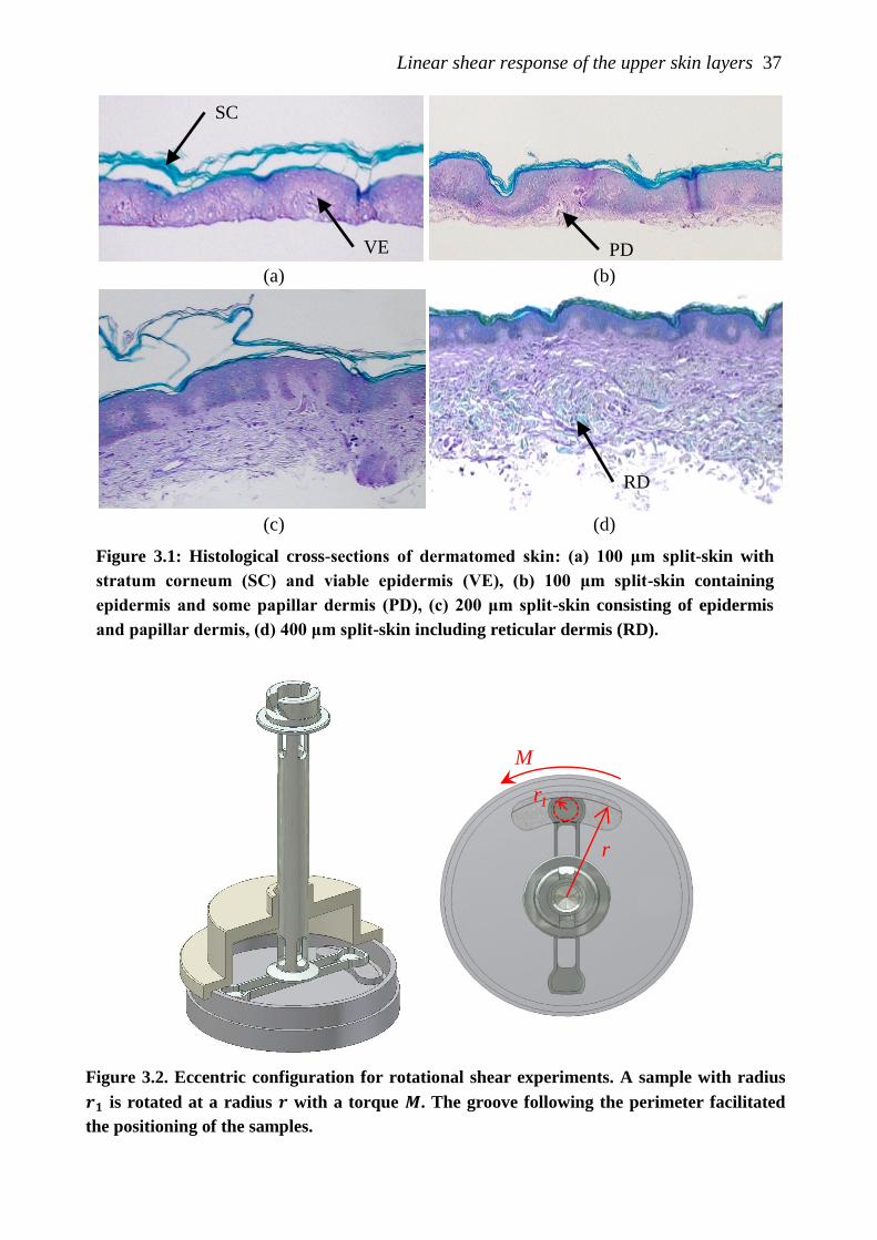
Linear shear response of the upper skin layers 37
Figure 3.2. Eccentric configuration for rotational shear experiments. A sample with radius
𝒓𝟏 is rotated at a radius 𝒓 with a torque 𝑴. The groove following the perimeter facilitated
the positioning of the samples.
r
r1
M
(a) (b)
(c) (d)
Figure 3.1: Histological cross-sections of dermatomed skin: (a) 100 μm split-skin with
stratum corneum (SC) and viable epidermis (VE), (b) 100 μm split-skin containing
epidermis and some papillar dermis (PD), (c) 200 μm split-skin consisting of epidermis
and papillar dermis, (d) 400 μm split-skin including reticular dermis (RD).
VE
SC
PD
RD
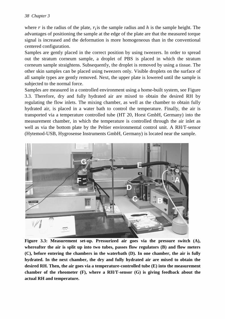
38 Chapter 3
where 𝑟 is the radius of the plate, 𝑟1is the sample radius and ℎ is the sample height. The
advantages of positioning the sample at the edge of the plate are that the measured torque
signal is increased and the deformation is more homogeneous than in the conventional
centered configuration.
Samples are gently placed in the correct position by using tweezers. In order to spread
out the stratum corneum sample, a droplet of PBS is placed in which the stratum
corneum sample straightens. Subsequently, the droplet is removed by using a tissue. The
other skin samples can be placed using tweezers only. Visible droplets on the surface of
all sample types are gently removed. Next, the upper plate is lowered until the sample is
subjected to the normal force.
Samples are measured in a controlled environment using a home-built system, see Figure
3.3. Therefore, dry and fully hydrated air are mixed to obtain the desired RH by
regulating the flow inlets. The mixing chamber, as well as the chamber to obtain fully
hydrated air, is placed in a water bath to control the temperature. Finally, the air is
transported via a temperature controlled tube (HT 20, Horst GmbH, Germany) into the
measurement chamber, in which the temperature is controlled through the air inlet as
well as via the bottom plate by the Peltier environmental control unit. A RH/T-sensor
(Hytemod-USB, Hygrosense Instruments GmbH, Germany) is located near the sample.
Figure 3.3: Measurement set-up. Pressurized air goes via the pressure switch (A),
whereafter the air is split up into two tubes, passes flow regulators (B) and flow meters
(C), before entering the chambers in the waterbath (D). In one chamber, the air is fully
hydrated. In the next chamber, the dry and fully hydrated air are mixed to obtain the
desired RH. Then, the air goes via a temperature-controlled tube (E) into the measurement
chamber of the rheometer (F), where a RH/T-sensor (G) is giving feedback about the
actual RH and temperature.
C
D A
B
E G
F
Linear shear response of the upper skin layers 39
3.2.3 Rheological methods
Linear viscoelastic material behavior is described by a multi-mode Maxwell model:
where 𝜏𝑖 is the shear stress contribution of mode 𝑖 with the relaxation time 𝜆𝑖 and
modulus 𝐺𝑖 . The applied strain rate is denoted with 𝛾 . The total stress (𝜏) is the sum of
the stress contributions of all modes:
𝜏 = 𝜏𝑖 𝑛𝑖=1 . (3.3)
A frequency (𝜔) dependent input 𝛾 = 𝛾0 sin 𝜔𝑡 will lead, for linear viscoelastic
behavior, to a sinusoidal shear stress:
𝜏 = 𝐺𝑑𝛾0 sin 𝜔𝑡 + 𝛿 , (3.4)
where 𝐺𝑑(𝜔, 𝑇) is the dynamic modulus and 𝛿(𝜔, 𝑇) the phase shift. The response can
be written in an in-phase and out-of-phase wave:
𝜏 = 𝜏 ′ + 𝜏′′ = 𝜏0′ sin 𝜔𝑡 + 𝜏0
′′ cos 𝜔 𝑡 . (3.5)
From this, the moduli can be computed:
𝐺 ′ = 𝜏0′ 𝛾0 = 𝐺𝑖
𝜆𝑖2𝜔2
1 + 𝜆𝑖2𝜔2
;
𝑛
𝑖=1
(3.6)
𝐺 ′′ = 𝜏0′′ 𝛾0 = 𝐺𝑖
𝜆𝑖𝜔
1 + 𝜆𝑖2𝜔2
𝑛
𝑖=1
, (3.7)
where 𝐺 ′ is the storage modulus, representing the elastic part of the behavior and 𝐺 ′′ is
the loss modulus, representing the viscous behavior. The two moduli, 𝐺 ′ and 𝐺 ′′ , form
the dynamic shear modulus:
𝐺𝑑 = 𝐺′2+𝐺′′2 . (3.8)
The phase shift 𝛿
to 𝐺′ and 𝐺" via:
tan 𝛿 =𝐺 ′′
𝐺′ . (3.9)
𝜏𝑖 +1
𝜆𝑖𝜏𝑖 = 𝐺𝑖𝛾 ; 𝑖 𝜖 1, 𝑛 , (3.2)
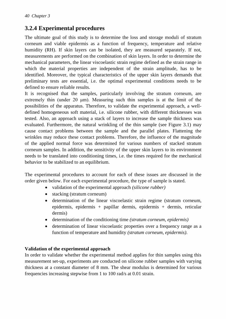
40 Chapter 3
3.2.4 Experimental procedures
The ultimate goal of this study is to determine the loss and storage moduli of stratum
corneum and viable epidermis as a function of frequency, temperature and relative
humidity (RH). If skin layers can be isolated, they are measured separately. If not,
measurements are performed on the combination of skin layers. In order to determine the
mechanical parameters, the linear viscoelastic strain regime defined as the strain range in
which the material properties are independent of the strain amplitude, has to be
identified. Moreover, the typical characteristics of the upper skin layers demands that
preliminary tests are essential, i.e. the optimal experimental conditions needs to be
defined to ensure reliable results.
It is recognised that the samples, particularly involving the stratum corneum, are
extremely thin (under 20 μm). Measuring such thin samples is at the limit of the
possibilities of the apparatus. Therefore, to validate the experimental approach, a well-
defined homogeneous soft material, i.e. silicone rubber, with different thicknesses was
tested. Also, an approach using a stack of layers to increase the sample thickness was
evaluated. Furthermore, the natural wrinkling of the thin sample (see Figure 3.1) may
cause contact problems between the sample and the parallel plates. Flattening the
wrinkles may reduce these contact problems. Therefore, the influence of the magnitude
of the applied normal force was determined for various numbers of stacked stratum
corneum samples. In addition, the sensitivity of the upper skin layers to its environment
needs to be translated into conditioning times, i.e. the times required for the mechanical
behavior to be stabilized to an equilibrium.
The experimental procedures to account for each of these issues are discussed in the
order given below. For each experimental procedure, the type of sample is stated.
validation of the experimental approach (silicone rubber)
stacking (stratum corneum)
determination of the linear viscoelastic strain regime (stratum corneum,
epidermis, epidermis + papillar dermis, epidermis + dermis, reticular
dermis)
determination of the conditioning time (stratum corneum, epidermis)
determination of linear viscoelastic properties over a frequency range as a
function of temperature and humidity (stratum corneum, epidermis).
Validation of the experimental approach
In order to validate whether the experimental method applies for thin samples using this
measurement set-up, experiments are conducted on silicone rubber samples with varying
thickness at a constant diameter of 8 mm. The shear modulus is determined for various
frequencies increasing stepwise from 1 to 100 rad/s at 0.01 strain.
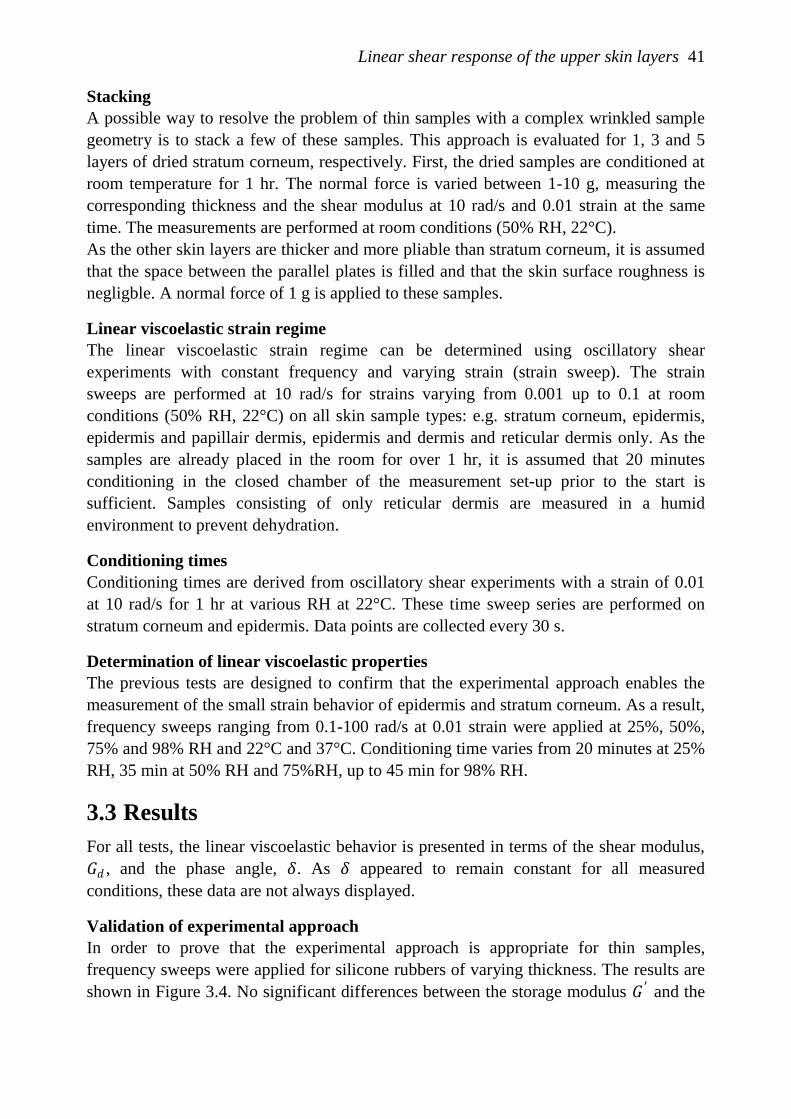
Linear shear response of the upper skin layers 41
Stacking
A possible way to resolve the problem of thin samples with a complex wrinkled sample
geometry is to stack a few of these samples. This approach is evaluated for 1, 3 and 5
layers of dried stratum corneum, respectively. First, the dried samples are conditioned at
room temperature for 1 hr. The normal force is varied between 1-10 g, measuring the
corresponding thickness and the shear modulus at 10 rad/s and 0.01 strain at the same
time. The measurements are performed at room conditions (50% RH, 22°C).
As the other skin layers are thicker and more pliable than stratum corneum, it is assumed
that the space between the parallel plates is filled and that the skin surface roughness is
negligble. A normal force of 1 g is applied to these samples.
Linear viscoelastic strain regime
The linear viscoelastic strain regime can be determined using oscillatory shear
experiments with constant frequency and varying strain (strain sweep). The strain
sweeps are performed at 10 rad/s for strains varying from 0.001 up to 0.1 at room
conditions (50% RH, 22°C) on all skin sample types: e.g. stratum corneum, epidermis,
epidermis and papillair dermis, epidermis and dermis and reticular dermis only. As the
samples are already placed in the room for over 1 hr, it is assumed that 20 minutes
conditioning in the closed chamber of the measurement set-up prior to the start is
sufficient. Samples consisting of only reticular dermis are measured in a humid
environment to prevent dehydration.
Conditioning times
Conditioning times are derived from oscillatory shear experiments with a strain of 0.01
at 10 rad/s for 1 hr at various RH at 22°C. These time sweep series are performed on
stratum corneum and epidermis. Data points are collected every 30 s.
Determination of linear viscoelastic properties
The previous tests are designed to confirm that the experimental approach enables the
measurement of the small strain behavior of epidermis and stratum corneum. As a result,
frequency sweeps ranging from 0.1-100 rad/s at 0.01 strain were applied at 25%, 50%,
75% and 98% RH and 22°C and 37°C. Conditioning time varies from 20 minutes at 25%
RH, 35 min at 50% RH and 75%RH, up to 45 min for 98% RH.
3.3 Results
For all tests, the linear viscoelastic behavior is presented in terms of the shear modulus,
𝐺𝑑 , and the phase angle, 𝛿. As 𝛿 appeared to remain constant for all measured
conditions, these data are not always displayed.
Validation of experimental approach
In order to prove that the experimental approach is appropriate for thin samples,
frequency sweeps were applied for silicone rubbers of varying thickness. The results are
shown in Figure 3.4. No significant differences between the storage modulus 𝐺 ′ and the
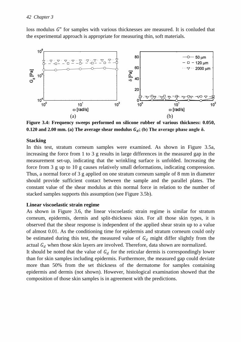
42 Chapter 3
loss modulus 𝐺" for samples with various thicknesses are measured. It is conluded that
the experimental approach is appropriate for measuring thin, soft materials.
(a) (b)
Figure 3.4: Frequency sweeps performed on silicone rubber of various thickness: 0.050,
0.120 and 2.00 mm. (a) The average shear modulus 𝑮𝒅; (b) The average phase angle δ.
Stacking
In this test, stratum corneum samples were examined. As shown in Figure 3.5a,
increasing the force from 1 to 3 g results in large differences in the measured gap in the
measurement set-up, indicating that the wrinkling surface is unfolded. Increasing the
force from 3 g up to 10 g causes relatively small deformations, indicating compression.
Thus, a normal force of 3 g applied on one stratum corneum sample of 8 mm in diameter
should provide sufficient contact between the sample and the parallel plates. The
constant value of the shear modulus at this normal force in relation to the number of
stacked samples supports this assumption (see Figure 3.5b).
Linear viscoelastic strain regime
As shown in Figure 3.6, the linear viscoelastic strain regime is similar for stratum
corneum, epidermis, dermis and split-thickness skin. For all those skin types, it is
observed that the shear response is independent of the applied shear strain up to a value
of almost 0.01. As the conditioning time for epidermis and stratum corneum could only
be estimated during this test, the measured value of 𝐺𝑑 might differ slightly from the
actual 𝐺𝑑 when those skin layers are involved. Therefore, data shown are normalized.
It should be noted that the value of 𝐺𝑑 for the reticular dermis is correspondingly lower
than for skin samples including epidermis. Furthermore, the measured gap could deviate
more than 50% from the set thickness of the dermatome for samples containing
epidermis and dermis (not shown). However, histological examination showed that the
composition of those skin samples is in agreement with the predictions.
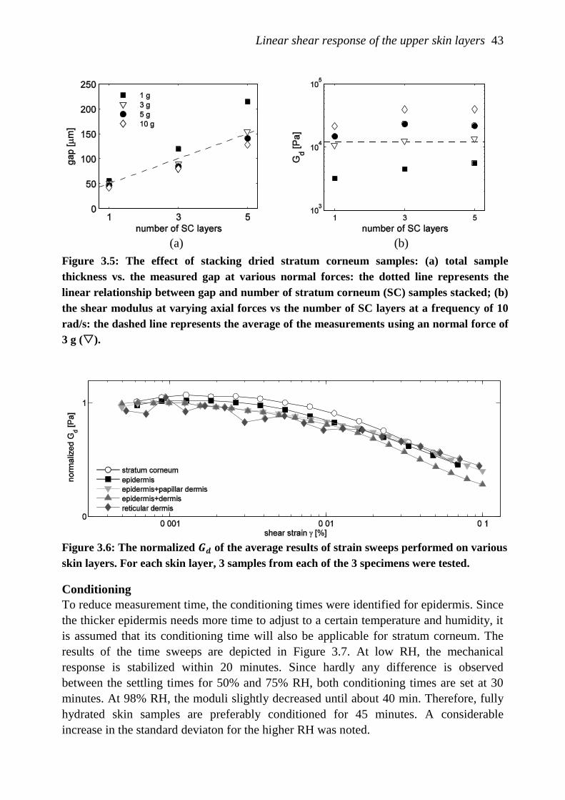
Linear shear response of the upper skin layers 43
Figure 3.6: The normalized 𝑮𝒅 of the average results of strain sweeps performed on various
skin layers. For each skin layer, 3 samples from each of the 3 specimens were tested.
Conditioning
To reduce measurement time, the conditioning times were identified for epidermis. Since
the thicker epidermis needs more time to adjust to a certain temperature and humidity, it
is assumed that its conditioning time will also be applicable for stratum corneum. The
results of the time sweeps are depicted in Figure 3.7. At low RH, the mechanical
response is stabilized within 20 minutes. Since hardly any difference is observed
between the settling times for 50% and 75% RH, both conditioning times are set at 30
minutes. At 98% RH, the moduli slightly decreased until about 40 min. Therefore, fully
hydrated skin samples are preferably conditioned for 45 minutes. A considerable
increase in the standard deviaton for the higher RH was noted.
(a) (b)
Figure 3.5: The effect of stacking dried stratum corneum samples: (a) total sample
thickness vs. the measured gap at various normal forces: the dotted line represents the
linear relationship between gap and number of stratum corneum (SC) samples stacked; (b)
the shear modulus at varying axial forces vs the number of SC layers at a frequency of 10
rad/s: the dashed line represents the average of the measurements using an normal force of
3 g ().
44 Chapter 3
(a) (b)
(c) (d)
Figure 3.7: Average values of 3 measurements for 𝑮𝒅(𝝎 = 𝟏𝟎𝒓𝒂𝒅/𝒔, 𝑻 = 𝟐𝟎°𝑪) and the
standard deviation over time (dotted lines) for the epidermis at various RH. The vertical
grey band indicates the necessary conditioning time.
Determination of G’ and G”
The dependency on RH and temperature were measured for both the epidermis and
stratum corneum. For each RH/T combination, the tests were designed to measure 3
samples per subject. However, the test sequence could not be completed for subjects 2
and 4 within 72 hr. As only three meausurements could be performed on epidermis from
subject 4, subject 4 was totally excluded from this part of the study.
For both stratum corneum and epidermis, the modulus was found to be slightly
frequency dependent (see Figure 3.8). However, the phase angle is not significantly
different for the various RH. As similar results were obtained for epidermis and stratum
corneum, only results from the latter are shown in Figure 3.8. Because of the small
frequency dependency, a comparison between the different environmental conditions
was performed at one frequency only. In this case, 10 rad/s was chosen (see Figure 3.9
and Figure 3.10). The results for stratum corneum show a decrease in modulus with
increasing humidity, but no apparent change with temperature. For the epidermis, data
were more variable, especially at 20°C. The mechanical parameters appeared
independent of the two temperatures.
Linear shear response of the upper skin layers 45
(a) (b)
Figure 3.8: Linear viscoelastic behavior of stratum corneum from one subject for various RH
at 20°C. (a) The average shear modulus 𝑮𝒅; (b) The average phase angle δ.
Figure 3.9: Linear viscoelastic behavior of the stratum corneum for various RH at 20°C and
37°C. The average values and standard deviations are shown for 𝑮𝒅 and δ per subject.
46 Chapter 3
Figure 3.10: Linear viscoelastic behavior of the epidermis for various RH at 20°C and 37°C.
The average values and standard deviations are shown for 𝑮𝒅 and δ per subject.
3.4 Discussion
In the past, mechanical behavior of epidermis has only been described qualitatively due
to the lack of experimental data. The skin curvature and the undulating dermal-epidermal
junction cause inherent difficulties during mechanical characterization of the epidermis
in vivo. In addition, in vivo measurement methods for shear, such as elastography,
cannot be applied due to limitations in resolution. Therefore, this study presents an in
vitro measurement method to determine shear properties of the epidermis. Preliminary
testing was essential to validate the methods applied and to obtain the optimal
experimental conditions to ensure reliable final measurements.
In order to measure shear properties of a soft biological material, measurement methods
have been developed for muscle, brain and thrombus [88-90]. However, pre-testing was
needed to prove that the experimental approach is also appropriate for samples in the
order of a few micrometers while retaining a relative large diameter to avoid the effect of
local properties. As there are inherent difficulties in determining the actual thickness of
stratum corneum from histology, the sample thickness was defined by the measured gap
between the plates. Although the thickness of the stratum corneum on the abdomen is
reported to be 14±4 μm [5], the measured gap at a normal force of 3g varied from 15 to
Linear shear response of the upper skin layers 47
60 μm due to local variations and skin surface roughness. Applying a higher normal
force causes compression of the sample, which influences the measured shear modulus.
The skin surface roughness becomes less significant for other samples involving thicker
skin layers. In addition, these layers are more pliable than the stratum corneum. Whether
stratum corneum, epidermis only or epidermis and dermis together are measured, the
shear response does not differ significantly for the strain sweeps. It is hypothesized that
loading in shear causes cell deformation in the epidermis whithout affecting the
desmosomes. In the dermis, the shear response will be determined by the ground
substance, because the collagen and elastin fibers are mainly oriented transversally. It is
likely that this substance has a lower shear resistance than the highly organized
epidermis. By contrast, the tissue response to in-plane tensile loading will be determined
by the mechanical integrity of the desmosomes, the elasticity of the dermal fibers and the
direction of the Langer lines.
Recently, the linear viscoelastic response on oscillatory shear strains of human intact
skin and dermis-only was measured [91,92]. The increase of the moduli was more
pronounced for the dermis-only at higher frequencies, so the authors concluded that the
epidermis is only slightly frequency dependent. At lower frequencies, 𝐺𝑑 ,𝑑𝑒𝑟𝑚𝑖𝑠 was of
the order of kPa. In accordance to this study, we observed in our frequency sweeps that
the epidermis is indeed slightly frequency-dependent. Our strain sweeps also resulted in
a value for 𝐺𝑑 ,𝑑𝑒𝑟𝑚𝑖𝑠 of a few kPa.
For the stratum corneum, the values of 𝐺𝑑 are similar to those of the epidermis, although
some corrections are needed to account for uncertainties in the sample thickness. The
results for epidermis and stratum corneum suggest that the small strain shear properties
of viable epidermis and stratum corneum are very similar. Currently, our shear moduli
can only be compared with in-plane tensile properties of stratum corneum from
literature. Accordingly, current values for shear moduli are one order of magnitude lower
than those in dry conditions and up to two orders of magnitude when fully hydrated,
based on the lowest reported values for tensile moduli [23,25,93]. This clearly supports
the highly anisotropic behavior of stratum corneum and epidermis.
A decrease in stiffness of the stratum corneum could be observed with increasing RH. In
accordance with the present observations, delamination studies with stratum corneum,
which also showed the pre-failure mechanical response, showed no dependence over the
identical temperature range [94].
No clear relationship between the mechanical properties of the epidermis and RH could
be established. Time sweeps showed that moduli stabilize to an equilibrium after a
certain conditioning time. However, both time sweeps and frequency tests for epidermis
showed larger variations per RH and per subject compared to stratum corneum. This
might be related to the less well-defined tissue composition. For example, the direction
of Langer lines or irregularities, such as sweat pores and hair follicles, can have a more
substantial role in the mechanical behavior in fully hydrated epidermis than for stratum
corneum. Future experiments should clarify the variance in these results.
Longer conditioning times and larger variations were observed in fully hydrated
epidermal samples than for less humid samples. Examination of fully hydrated stratum
48 Chapter 3
corneum structure has revealed swollen corneocytes and water pools in the extracellular
spaces after storage in PBS [95]. Furthermore, water disrupts the lipid lamellae to
varying degrees and causes degradation of intercellular corneosomes [84,84,96]. It is
likely that the desmosomes in the viable epidermis are also highly susceptible to damage.
However, histological examination did not show any sign of degradation in the present
samples. The prolonged time of conditioning for the sample at higher RH limited the
number of experiments that could be performed with epidermis from one donor within
72 hours.
The present study demonstrated that reproducible results can be obtained for the shear
properties of epidermis in an in vitro experimental system. Viable epidermis could not be
measured as an isolated skin layer, but its properties can be derived from the other skin
samples. The 𝐺𝑑 for stratum corneum approximately ranges from 4 to 12 kPa, decreasing
with increasing RH. The values are considerably lower than the shear modulus value
based on tensile Young‟s moduli (i.e. 𝐸 = 𝐺𝑑) in literature, assuming anisotropic
material behavior. Results for the epidermis were of the same order of magnitude, but
were less consistent possibly due to a less well-defined tissue composition. Therefore, it
would be interesting to combine mechanical testing with real-time imaging techniques to
monitor changes in tissue deformation. It has already been shown by histological
examination after 2 days of loading that shear forces induce cell displacement in skin,
and particularly in the epidermis [97].
Furthermore, electron microscope imaging techniques could support histological
examination in assessing tissue damage due to preparation, storage or handling. In
addition, it is important to correlate the shear response both with tensile testing and with
the effects of perpendicular loading, as accomplished with indentation or compression
testing.
Chapter 4
A new indentation method to determine
mechanical properties of the epidermis
The contents of this chapter are based on M. Geerligs, L.C.A.v. Breemen, G.W.M.
Peters, P.A.J. Ackermans, C.W.J. Oomens, and F.P.T. Baaijens, A new indentation
method to determine mechanical properties of the epidermis, submitted.
50 Chapter 4
4.1 Introduction
The outer skin layer possesses important characteristics that make it a favorable site for
pain-free drug delivery with minimal damage. Indeed it presents a rich population of
immunologically sensitive cells as well as the lack of blood vessels and sensory nerve
endings. The development of drug delivery using microneedles or microjets is a
challenging task because of the lack of understanding of the mechanical behavior of the
human skin layers. In particular, the key mechanical properties of the outer skin layer,
i.e. the epidermis composed of stratum corneum and viable epidermis, should be better
understood.
The structure and function of this layer are well-known [12]. The outer layer, the stratum
corneum, is an effective physical barrier of dead cells in a „brick-and-mortar‟ structure:
the anucleate corneocytes form „bricks‟ and the intercellular lipid membranes and
corneosomes are considered to represent the mortar (see Figure 1.3). The viable
epidermis mainly consists of keratinocytes migrating towards the stratum corneum,
continuously changing in composition, shape and function. The junction with the
underlying dermis is strengthened by its undulating pattern, such that large cones of
epidermal tissue penetrate the dermis (see Figure 4.1). Furthermore, epidermal properties
are influenced by environmental factors such as temperature, humidity and UV radiation.
In order to deliver drugs transdermally, the microneedle or microjet should penetrate the
stratum corneum to deliver the drug 100-150 μm below the skin surface, e.g. in the
viable epidermis or papillar dermis. Penetration is preceeded by indentation of the skin
layers, especially the epidermis. So a full understanding of the delivery path requires also
the understanding of this indentation phase and, therefore, the knowledge of the
mechanical behavior of the epidermis. The modeling of tissue repair and remodeling in
future work benefit from a well determined mechanical behavior.
Recently, Kendall et al. [38] were the first to report on the mechanical properties of the
(viable) epidermis during penetration, using modified standard tips on murine skin. They
observed a decrease in storage modulus when the 2 μm probe penetrates through the
stratum corneum, which is in accordance with studies on stratum corneum alone [98,99].
The authors argued that this is because of an increasing moisture content with depth. In
the viable epidermis, the storage modulus remained nearly constant. By contrast,
penetration of the 5 μm probe showed a negligible decrease in storage modulus
throughout the stratum corneum and a gradual increase in the viable epidermis, although
the values for the shear modulus were less than for the corresponding the 2 μm probe.
A variety of in vivo and in vitro indentation techniques were developed to measure the
stratum corneum. In the eighties, Hendley et al. developed an indentation device to
measure force variations in vivo due to age, sex and body site [99]. A needle with a tip
radius of 11 μm at the tip was held perpendicular to the surface and moved rapidly into
the skin. They claimed that the speed of the indentation ensured that the test was
predominantly confined to the properties of the stratum corneum [34]. Measured forces
were typically of the order of 3.0 N. Recently, a limited number of nano-indentation
studies have been performed on isolated stratum corneum [32,98,100,101,101]. The tips
A new indentation method to determine mechanical properties of the epidermis 51
used varied between 1-10 μm, while corneocytes have a diameter ranging from 26-45
μm [3,102,103]. As a consequence, very local properties were determined in these
experiments. Furthermore, in some of the studies, the three-sided Berkovich tip that has
a sharp three-sided point, is used. This tip easily induce damage on the sample‟s surface,
which interferes with the load-displacements results of the indentation. Three of the
nano-indentation studies were based on continuous stiffness measurements (CSM)
protocols [38,98,101]. The drawback of CSM is that the results are influenced by the
selected amplitude and frequency for viscoelastic materials. Combining the nano-
indentation studies on stratum corneum reveals a measured Young‟s moduli, which
varies from 10 MPa [104] for wet porcine samples up to 1 GPa for dried human samples
[98]. This broad range is likely caused by the differences in testing apparatus and
protocols, differences between species and body sites, and the heterogeneity of the
material. A reliable method to determine mechanical properties of the stratum corneum
only at the tissue level is therefore also required.
The aim of the present study is to present such an indentation method and to use it to
determine the Young‟s modulus of the epidermis, i.e. the stratum corneum and viable
epidermis. The typical complex geometry, a variable thickness between 30 to 150 μm,
and the porosity of the epidermis places high demands on this mechanical
characterization. Therefore, isolated epidermis and isolated stratum corneum were tested
using equipment that is known for its accuracy and reliablity. The device is originally
designed for solid materials of which well-defined samples can be obtained and
therefore, the testing protocol needs to be adapted to epidermal samples. To validate that
the testing protocol holds for thin materials with a low stiffness, tests have been
performed with silicone rubber. Moreover, indentation experiments require a model for
the interpretation of the measured results. Next to the analytical model used, which
assumes a homogeneous linear elastic material behavior upon unloading, we also
adopted a numerical method that allows for taking into account geometrical details and
different material properties for different layers.
4.1.1 Sample preparation
Skin
Indentation tests have been carried out on ex vivo abdominal skin of Caucasian women
aging 43±4 years old undergoing abdominoplasty surgery. All patients gave informed
consent for use of their skin for research purposes under a protocol approved by the
ethics committee of the Catharina Hospital, Eindhoven, The Netherlands. Abdominal
skin with striae markers, cellulite, damage due to UV exposure or excessively hairy skin
is excluded from the study.
Immediately after excision, the skin was brought into the laboratory and processed
within 4 hours. Epidermal sheets were obtained using a dermatome (D42, Humeca), in
which the prescribed thickness was refined for this purpose by the supplier. The
dermatomed slices of 100 μm thickness were cut in pieces of approximately 1 cm2.
Depending on various factors such as skin surface roughness, tissue hydration, and the
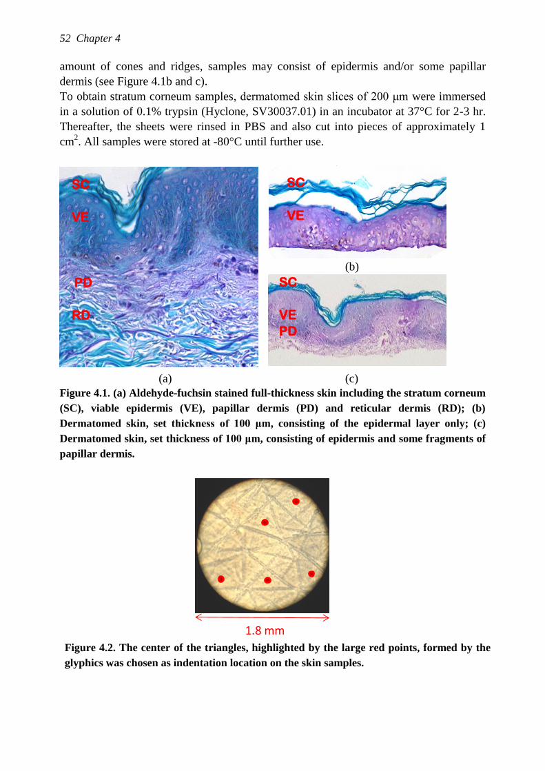
52 Chapter 4
amount of cones and ridges, samples may consist of epidermis and/or some papillar
dermis (see Figure 4.1b and c).
To obtain stratum corneum samples, dermatomed skin slices of 200 μm were immersed
in a solution of 0.1% trypsin (Hyclone, SV30037.01) in an incubator at 37°C for 2-3 hr.
Thereafter, the sheets were rinsed in PBS and also cut into pieces of approximately 1
cm2. All samples were stored at -80°C until further use.
SC
VE
PD
RD
(b)
SC
VE
PD
(a) (c)
Figure 4.1. (a) Aldehyde-fuchsin stained full-thickness skin including the stratum corneum
(SC), viable epidermis (VE), papillar dermis (PD) and reticular dermis (RD); (b)
Dermatomed skin, set thickness of 100 μm, consisting of the epidermal layer only; (c)
Dermatomed skin, set thickness of 100 μm, consisting of epidermis and some fragments of
papillar dermis.
Figure 4.2. The center of the triangles, highlighted by the large red points, formed by the
glyphics was chosen as indentation location on the skin samples.
1.8 mm
SC
VE
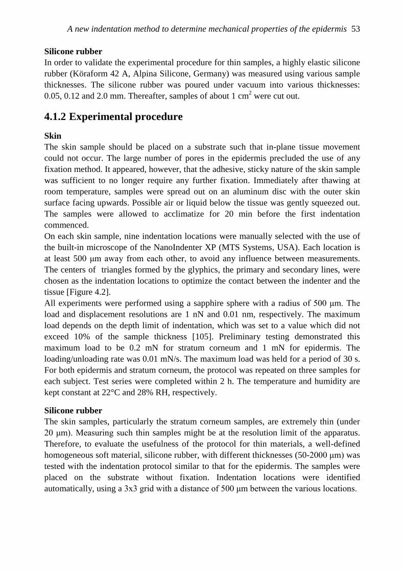
A new indentation method to determine mechanical properties of the epidermis 53
Silicone rubber
In order to validate the experimental procedure for thin samples, a highly elastic silicone
rubber (Köraform 42 A, Alpina Silicone, Germany) was measured using various sample
thicknesses. The silicone rubber was poured under vacuum into various thicknesses:
0.05, 0.12 and 2.0 mm. Thereafter, samples of about 1 cm2 were cut out.
4.1.2 Experimental procedure
Skin
The skin sample should be placed on a substrate such that in-plane tissue movement
could not occur. The large number of pores in the epidermis precluded the use of any
fixation method. It appeared, however, that the adhesive, sticky nature of the skin sample
was sufficient to no longer require any further fixation. Immediately after thawing at
room temperature, samples were spread out on an aluminum disc with the outer skin
surface facing upwards. Possible air or liquid below the tissue was gently squeezed out.
The samples were allowed to acclimatize for 20 min before the first indentation
commenced.
On each skin sample, nine indentation locations were manually selected with the use of
the built-in microscope of the NanoIndenter XP (MTS Systems, USA). Each location is
at least 500 μm away from each other, to avoid any influence between measurements.
The centers of triangles formed by the glyphics, the primary and secondary lines, were
chosen as the indentation locations to optimize the contact between the indenter and the
tissue [Figure 4.2].
All experiments were performed using a sapphire sphere with a radius of 500 μm. The
load and displacement resolutions are 1 nN and 0.01 nm, respectively. The maximum
load depends on the depth limit of indentation, which was set to a value which did not
exceed 10% of the sample thickness [105]. Preliminary testing demonstrated this
maximum load to be 0.2 mN for stratum corneum and 1 mN for epidermis. The
loading/unloading rate was 0.01 mN/s. The maximum load was held for a period of 30 s.
For both epidermis and stratum corneum, the protocol was repeated on three samples for
each subject. Test series were completed within 2 h. The temperature and humidity are
kept constant at 22°C and 28% RH, respectively.
Silicone rubber
The skin samples, particularly the stratum corneum samples, are extremely thin (under
20 μm). Measuring such thin samples might be at the resolution limit of the apparatus.
Therefore, to evaluate the usefulness of the protocol for thin materials, a well-defined
homogeneous soft material, silicone rubber, with different thicknesses (50-2000 μm) was
tested with the indentation protocol similar to that for the epidermis. The samples were
placed on the substrate without fixation. Indentation locations were identified
automatically, using a 3x3 grid with a distance of 500 μm between the various locations.
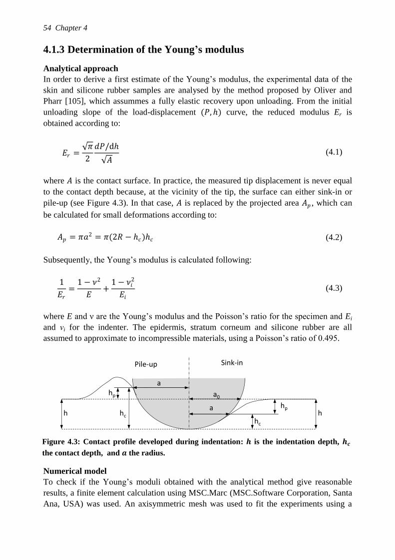
54 Chapter 4
4.1.3 Determination of the Young’s modulus
Analytical approach
In order to derive a first estimate of the Young‟s modulus, the experimental data of the
skin and silicone rubber samples are analysed by the method proposed by Oliver and
Pharr [105], which assummes a fully elastic recovery upon unloading. From the initial
unloading slope of the load-displacement (𝑃, ℎ) curve, the reduced modulus Er is
obtained according to:
𝐸𝑟 = 𝜋
2
𝑑𝑃/dℎ
𝐴 (4.1)
where 𝐴 is the contact surface. In practice, the measured tip displacement is never equal
to the contact depth because, at the vicinity of the tip, the surface can either sink-in or
pile-up (see Figure 4.3). In that case, 𝐴 is replaced by the projected area 𝐴𝑝 , which can
be calculated for small deformations according to:
𝐴𝑝 = 𝜋𝑎2 = 𝜋(2𝑅 − ℎ𝑐)ℎ𝑐 (4.2)
Subsequently, the Young‟s modulus is calculated following:
1
𝐸𝑟
=1 − 𝜈2
𝐸+
1 − 𝜈𝑖2
𝐸𝑖
(4.3)
where E and ν are the Young‟s modulus and the Poisson‟s ratio for the specimen and Ei
and νi for the indenter. The epidermis, stratum corneum and silicone rubber are all
assumed to approximate to incompressible materials, using a Poisson‟s ratio of 0.495.
Figure 4.3: Contact profile developed during indentation: 𝒉 is the indentation depth, 𝒉𝒄
the contact depth, and 𝒂 the radius.
Numerical model
To check if the Young‟s moduli obtained with the analytical method give reasonable
results, a finite element calculation using MSC.Marc (MSC.Software Corporation, Santa
Ana, USA) was used. An axisymmetric mesh was used to fit the experiments using a
hp
hchc
hp
a0
a
ah h
Pile-up Sink-in
A new indentation method to determine mechanical properties of the epidermis 55
Neo-Hookean model with different material parameters when modeling stratum corneum
and viable epidermis, assuming incompressible material behavior. The mesh consisted of
4329 linear quad4 elements, using full integration. The size of the mesh was chosen such
that the edges do not influence the stress distribution, contact between the indenter and
the sample was assumed to be frictionless.
For silicone rubber, the Young‟s modulus, 𝐸𝑆𝑅 , was estimated by fitting the average
load-displacement curve of the 50 μm thick samples. This value for 𝐸𝑆𝑅 was then used to
calculate the unloading curves of the 120 and 2000 μm thick samples. These unloading
curves are compared with the experimental data.
Since the deformations were small, linear elastic behavior was assumed for the skin
samples too. First, the Young‟s modulus for the stratum corneum, 𝐸𝑆𝐶 , was derived by
fitting the average load-displacement curve of the stratum corneum samples of 20 μm.
This modulus was used to describe the experimental data of the epidermis, such that the
modulus for the viable epidermis, 𝐸𝑉𝐸 , could be derived. The thickness of the stratum
corneum was varied from 10 to 20 μm, the thickness of the viable epidermis was kept
constant at 80 μm. In order to assess the sensitivity of this fitting approach, the effect of
increasing 𝐸𝑆𝐶 or decreasing 𝐸𝑉𝐸 by a factor 2 on the maximum indentation depth was
studied.
4.2 Results
Silicone rubber
The load-displacement curves obtained for the silicone rubber samples are shown in
Figure 4.4. The results were highly reproducible for each thickness. The maximum
indentation depth decreases with a decrease in sample thickness. Consequently, the slope
of the initial unloading curve decreases, which is reflected in the average values for the
Young‟s moduli namely, 3.67±0.20, 2.22±0.10 and 1.69±0.04 MPa for a sample
thickness of 50, 120 and 2000 μm, respectively. It is evident that the assumption of a
linearly elastic half space from Oliver & Pharr [105] is not valid for these thin samples.
Using the FE model, the Young‟s modulus was estimated to be 2.16 MPa. When using
this value to describe the unloading curves for the 120 and 2000 μm thick sample, it is
shown that the unloading curves and maximum indentation depth for all thicknesses are
in good agreement to the experimental data (Figure 4.5).
Epidermis and stratum corneum
An example of the results from one subject is shown in Figure 4.6. Data that displayed
significant measurement errors or deviated from the general response, were ignored. In
generally, 2 or 3 tests out of a series of 9 measurements were excluded from subsequent
calculations. Figure 4.7 clearly shows that the average curves overlap for different
subjects. Estimates for the Young‟s moduli were derived via the analytical approach and
found to be 2.6±0.6 MPa and 1.1±0.2 MPa for the stratum corneum and epidermis,
respectively.
56 Chapter 4
Figure 4.4. All force-indentation (𝑷, 𝒉) curves for silicone rubbers with different thicknesses.
Figure 4.5: The unloading curves obtained with the FE model (x) are depicted together
with the mean curves of the experimental data.
The fitting results using the FE-model are shown in Figure 4.8. From the stratum
corneum experiments, 𝐸𝑆𝐶 was calculated to be 0.6 MPa. For a 20 μm thick stratum
corneum and 80 μm thick viable epidermis, 𝐸𝑉𝐸 equals 𝐸𝑆𝐶 with a value of 0.6 MPa.
Decreasing the thickness of the stratum corneum to 10 μm minimally affects the
unloading curve. Also increasing the stiffness of the stratum corneum did not have a
noticeable effect. However, a reduction in the stiffness of the much thicker viable
epidermis caused an increase in indentation depth, from approximately 8 to 12 μm.
4.3 Discussion
The major problem in performing indentation experiments on skin is probably the skin‟s
surface roughness. In order to have a surface as smooth as possible, we used a large
spherical indenter (ø=500 μm) such that the contact area was much larger than the
diameter of individual cells and also more homogeneous. During preliminary tests that
were performed close to the glyphics, it was observed that the poor contact definition in
those areas resulted in an unacceptably high variability per subject. When positioning the
indenter at the highest point between a triangle formed by the glyphics, establishing a
well-defined contact between indenter and the tissue was not a problem. In addition, the
use of a spherical tip minimizes plastic deformations and stress concentrations and avoid
A new indentation method to determine mechanical properties of the epidermis 57
Figure 4.8. Unloading curves for the epidermius obtained with the FE model are depicted
together with the experimental indentation curve. The thickness of the stratum corneum is
varied from 10 (dashed lines) to 20 μm (dotted lines).
(a) (b)
Figure 4.6. Indentation curves of subject 2 for stratum corneum (a) and epidermis (b).
Dotted curves were not included in calculating the average curve.
(a) (b)
Figure 4.7 Average indentation curves for stratum corneum (a) and epidermis (b).
58 Chapter 4
damaging the sample [106]. Using this measurement protocol, highly reproducible data
could be obtained for all subjects and the variance between the subjects was negligbly
small.
In order to obtain meaningful, reproducible data from in vitro experiments, a correct
sample preparation is essential. In this study, the epidermal samples were isolated using
a dermatome. Although this method does not allow for separating the epidermis at the
basal membrane only, its benefit is that the bottom side of the sample with this obtained
geometry is in full contact with the substrate. As only small deformations were applied,
the results are not influenced by the possible fragments of papillar dermis in the sample.
Current tests were performed with epidermis that was thawed and immediately used in a
dry environment. As an increasing moisture content in the epidermis decreases the
stiffness, it becomes more difficult to define the initial contact surface at higher
humidities in the future.
The analytical method of Oliver and Pharr provides an easy method to asses the order of
magnitude of the Young‟s modulus from the experimental data. However, the theory
holds for homogeneous materials responding fully elastically upon unloading. In the case
of soft tissues, this assumption is not valid because of material responses like piling-up
and sinking-in. Due to piling up of the tissue, the projected contact area is bigger then
used in the calculations (see Figure 4.3). In the present study, the deviation is mild,
because the use of a large spherical indenter reduces the boundary effects.
The introduction of a numerical model should result in a better approach. The results
show that the stiffness of the viable epidermis is comparable to that of the stratum
corneum. For both epidermal layers, the stiffness of the two layers is approximately 1
MPa, which shows that the viable epidermis considerably contributes to the mechanical
response of skin at this length scale and load direction. In comparison with literature
using indentation test, current values for stratum corneum are on the low side of the
published range [35,98,104]. This can be explained by the fact that the local properties
studied in literature were mainly determined by the stiffness of individual corneocytes,
while our studies focused on the tissue level. In comparison with values for full-
thickness skin stiffness from in vivo indentation tests, our values are two orders of
magnitude higher [98,107,108].
Extending the Neo-Hookean model to a multimode Maxwell model would be a logical
step forward. However, the relaxation spectrum and corresponding low shear moduli that
were derived from rheological experiments (see Chapter 3) do not influence the fitting
on the load-displacement curve. The short relaxtion times that are ranging from 0.002 up
to 2 s, are only relevant at higher loading rates than those used in the present experiments
and are in accordance with the observed small viscoelastic plateau at the maximum
applied force in the indentation experiments (see Figure 4.6 and Figure 4.7).
To conclude, the small deformation behavior of epidermis was studied. We have
introduced a reliable experimental approach to evaluate the mechanical behavior of
A new indentation method to determine mechanical properties of the epidermis 59
epidermal tissue. The results demonstrated that the stiffness of the viable epidermis is
comparable to that of the stratum corneum for perpendicular direction at a length scale
relevant for clinical and cosmetic treatments. The applied load in this study covers the
physiologically relevant range. For clinical applications such as transdermal drug
delivery, the large deformations and, the ultimate goal, the failure behavior of the
epidermal layer need to be understood. The methods presented in this study are
considered to be a suitable tool that can be extended for these purposes.
Acknowledgments
We would like to thank the plastic surgery department of the Catharina hospital in
Eindhoven for providing the skin tissue. Furthermore, we are gratefully to dr. Hagisawa
providing the protocol for the histological examination.


Chapter 5
Linear viscoelastic behavior of
subcutaneous adipose tissue
The content of this chapter is based on M. Geerligs, G.W.M. Peters, P.A.J. Ackermans,
C.W.J. Oomens, and F.P.T. Baaijens (2008), Linear viscoelastic behavior of
subcutaneous adipose tissue, Biorheology; 45(6): pp 677-688.
62 Chapter 5
5.1 Introduction
The mechanical behavior of subcutaneous adipose tissue, also called hypodermis, is a
widely ignored topic in the biomechanics literature. A plethora of papers can be found on
properties of skin and skeletal muscle, but only few papers have addressed the properties
of the layer in between [39,40,44,109,110]. This is noteworthy, because adipose tissue
plays an important role in the load transfer between different structures in the body
during breathing, body movements or exercise, or when exposed to therapeutic
stretching during physiotherapy and massage. It is well recognized that the subcutaneous
fat experiences larger strains than the dermis during suction and that its stiffness is likely
to be a few orders less than that of the dermis [1,111]. However, it is still not common
practice to take the adjacent adipose layer into account when the combined mechanical
behavior of skin, fat and muscle tissue is modeled. Currently it would be difficult to do
so, because values for mechanical parameters of adipose tissue are limited and
inconsistent in the literature. Thus, there is a need to develop a parametric and
constitutive model of subcutaneous adipose tissue, which can be implemented in
numerical models of the whole skin as well as in multilayer models including skin, fat
and muscle. Numerical models including the subcutaneous fat layer are needed in a wide
field of applications, e.g. studying skin device contact, needle insertion procedures and
the removal of skin adhesives. Rheological experiments are accepted to be a good
starting point to develop such a constitutive model.
For a meaningful interpretation of the mechanical behavior of the adipose tissue, it is
essential to know the tissue composition. The present paper is focused on subcutaneous
adipose tissue, which is a type of connective tissue throughout the body found between
the dermis and the aponeurosis and fasciae of the muscles. However, the fat pads on the
palm of the hand and foot are considered to be different, since they contain a much
higher ratio of unsaturated versus saturated fatty acids and are therefore morphologically
different. Relatively small differences in tissue composition exist at the other body sites.
Subcutaneous adipose tissue is a loose association of lipid-filled cells called white
adipocytes, of which 90-99% is triglyceride, 5-30% water and 2-3% protein. Lipids
within the white adipocytes are organized in one droplet. The diameter of the white
adipocytes ranges from 30 to 70 μm, depending on the site of deposition [18].
Collections of white adipocytes comprise fat lobules, each of which is supplied by an
arteriole and surrounded by connective tissue septae. Each adipocyte is in contact with at
least one capillary. In healthy adults, only one third of the subcutaneous adipose tissue
contains mature adipocytes [18]. The remaining two thirds consists of blood vessels,
nerves, fibroblasts, and adipocyte precursor cells.
The subcutaneous adipose tissue of the lower trunk and the gluteal-thigh region is further
divided into two distinct layers: the superficial and deep subcutaneous adipose tissue
[20,112]. Both morphological and metabolic differences were found between those two
layers [112-114], but it is not clear if these layers differ in terms of the mechanical
properties.
Linear viscoelastic behavior of subcutaneous adipose tissue 63
To our knowledge, only a few authors studied the mechanical properties of the
subcutaneous adipose tissue. Of those, focus has been associated with breast tissue,
particularly in the early detection of cancerous tissues [42,44,45,92,115,116]. These
studies have generally utilized indirect and non-invasive measurements. The largest
study involving 70 samples of breast fat tissue using ex vivo indentation experiments
yielded a mean Young‟s modulus of 3.21 kPa [19]. Linear viscoelastic behavior was
shown up to 50% strain during uniaxial tension for abdominal subcutaneous tissue of rats
when applying incremental displacement steps of 1 mm followed by a 1 second
relaxation period [109]. Patel et al. [40] measured the storage and loss moduli of
subcutaneous fat tissue, also from the abdomen, for strains up to 20%. The results
showed a frequency-dependent shear moduli decreasing, which decreased with
increasing strain. These data, however, involved measurements outside the linear
viscoelastic strain range. Recently, the mechanical behavior of subcutaneous adipose
tissue of the buttock was measured in relation to pressure ulcers by performing confined
compression tests, but no mechanical parameters for modeling could be derived from the
results [41,117].
All the above-mentioned studies only give limited descriptions of the mechanical
behavior, either because the focus was only on the differences between breast tissue
types, or on long term quasi-static behavior [109,117], or because the authors were only
interested in a comparison of properties between human fat and a mimicking material
[40].
Our ultimate goal is to develop a skin model that includes the mechanical properties of
all skin layers separately, and can be used in a numerical model. Since it may be
predicted that the mechanical behavior of adipose tissue contributes considerably to the
overall skin behavior, there is a need to develop a thoroughly tested constitutive model
describing the mechanical behavior for large strains. The formulation of such a model
will be based on rheological experiments in vitro. The first step is to investigate the
material bulk properties within the linear viscoelastic strain region, which is defined as
the range of strain amplitudes where the material properties are independent of the
applied strain. The types of experiments are relatively simple to perform and hence, it is
appropriate to design experimental procedures as well as to identify experimental
problems. The linear viscoelastic parameters obtained will form the basis for a non-linear
viscoelastic model in future work. The concept will be developed for porcine
subcutaneous adipose tissue because of the availability and minimal biological
variability among specimens. The objective of the current study is to use dynamic
mechanical thermal analysis (DMTA) in combination with Time Temperature
Superposition (TTS) to determine the small oscillatory strain behavior of subcutaneous
adipose tissue in vitro. DMTA is performed through oscillatory shear experiments up to
100 rad/s at various temperatures. Next, the linear viscoelastic power-law memory
function, commonly used for soft-solids, will be introduced to describe the small strain
viscoelastic behavior of this tissue.
64 Chapter 5
5.2 Methods and Materials
5.2.1 Sample preparation
Porcine subcutaneous fat tissues were obtained from a local slaughterhouse (Ballering,
Son, The Netherlands), where they were cut into transverse slices of 1.5-2.0 mm thick
and stored at 4°C. In porcine species, the back fat is divided in an outer, middle and
inner layer of subcutaneous tissue because the adipocyte features of these layers differ
with respect to size, number and metabolic activity. The porcine middle layer, which is
used in the present study, is comparable to the deep subcutaneous layer in the abdominal
region of humans [118]. All pigs were Landrace, having a dressed carcass weight of
approximately 83 kilograms, and were 14-18 weeks old at necropsy.
Within 48 hr of collection, circular tissue samples were obtained from the slices with an
8 mm diameter cork borer. Next the samples were stored ice-cooled in a PBS solution
and tested within the subsequent 4 hours. An overview of the number of specimens and
the number of samples from each specimen per test is given in Table 5.1.
Methods of tissue preservation may change the mechanical properties of tissue due to
changes in tissue quality [82]. Rapid freezing, which has not been demonstrated to
change the fatty acid composition compared to fresh tissues [119], is an attractive
solution for storing tissue for prolonged periods. Thus, in order to assess whether snap
freezing preserves mechanical properties, adipose tissue was snap-frozen by immersion
in 2-methylbutane cooled by liquid nitrogen and stored at –80°C until use for mechanical
testing. Thawing of the samples was done slowly within an ice-cooled box. In order to
assess these storage conditions, histological sections were examined by light
microscopy. For that, the specimens were fixed in 10% phosphate-buffered formalin and
processed for conventional paraffin embedding. The specimens were cut into 5-μm thick
sections and stained with hematoxylin and eosin (H&E). Since all lipids were extracted
out of the adipocytes by using the conventional paraffin embedding technique, other
specimens were embedded in O.C.T. compound (TISSUE-TEC) and frozen for lipid
staining. These specimens were cut into 8-μm thick sections at –20°C, stained with oil
Red O (Sigma) and counterstained with hematoxylin.
5.2.2 Rheological methods
To determine the linear viscoelastic properties, oscillatory shear experiments were
performed using a rotational rheometer (Advanced Rheometric Expansion System
(ARES), Rheometrics Scientifc, USA) with a controlled strain mode, and parallel plate
geometry in combination with a Peltier environmental control unit. Sand-blasted plates
were used to prevent slippage. An oscilloscope was used to ascertain that the shape of
the torque signal was indeed sinusoidal. Samples were compressed between the plates by
lowering the upper plate until an axial force of 0.1 g was reached.
Linear viscoelastic behavior of subcutaneous adipose tissue 65
In the experiments a sinusoidal strain γ(t) insteady
state and within the range of linear viscoelastic behavior, resulted in a sinusoidal shear
rate, g(t), and shear stress,τ(t) with a phase shift δ:
𝛾 𝑡 = 𝛾0 sin 𝜔𝑡 , (5.1)
𝜏 𝑡 = 𝐺𝑑 sin 𝜔𝑡 + 𝛿 . (5.2)
The dynamic shear modulus Gd(ω,T) and the phase shift δ(ω,T) are both a function of the
angular frequency ω and temperature T. It is common to separate the dynamic shear
modulus into a storage modulus, G', representing the elastic behavior since this describes
the stress in phase with the strain, and a loss modulus, G'', representing the viscous
behavior, 1
2𝜋 out of phase with the strain, i.e. in phase with the strain rate:
𝐺𝑑 = 𝐺′2+𝐺"2 . (5.3)
The phase shift d
to (5.3):
tan 𝛿 =𝐺"
𝐺′ . (5.4)
The Time-Temperature Superposition (TTS) principle is applicable when data can be
shifted to and from a reference temperature T0 to form a master curve [120]. The
advantage of this principle is that the frequency domain can be extended beyond the
measurement limits as well as that data can be shifted to other working temperatures. A
smooth master curve is obtained by shifting frequency sweep curves obtained at different
temperatures horizontally and vertically on the curve obtained at the reference
temperature, until all the curves overlap. Normally the horizontal shift factor aT is
applied to the phase angle δ. Subsequently, the dynamic shear modulus Gd, and also G'
and G'', can be shifted along the horizontal and vertical axis to a reference temperature
with the horizontal shift factor aT and a vertical shift factor bT:
tan 𝛿 𝜔, 𝑇 = tan 𝛿 𝑎𝑇𝜔, 𝑇0 , (5.5)
𝐺𝑑 𝜔, 𝑇 =1
𝑏𝑇𝐺𝑑(𝑎𝑇𝜔, 𝑇0) . (5.6)
5.2.3 Testing procedure
Test protocols were based on measuring the linear viscoelastic properties of other soft
biological tissues, such as brain [121], muscle [122] and thrombus [90]. The linear
viscoelastic regime was determined using oscillatory shear experiments with constant
frequency and varying strain. Strain sweeps were performed from 0.04% to 10% at

66 Chapter 5
frequencies of 1, 10 and 100 rad/s and 20°C. A constant strain within the determined
linear regime of 0.1% was chosen for the subsequent frequency sweep tests.
The frequency sweep was repeated three times to avoid tissue conditioning phenomena,
observed during preliminary testing. We did not carry out traditional preconditioning.
Instead we performed always three frequency sweeps, increasing the frequency stepwise
logarithmically from 1 to 100 rad/s and then performing the data analysis on the third
frequency sweep. This protocol was also used to examine the influence of snap freezing
and thawing on the mechanical properties of subcutaneous fat tissue. For this purpose,
samples from 3 pigs were tested, both fresh and after freezing and thawing. All tests
were performed at 20°C.
To investigate whether the TTS principle is applicable to subcutaneous adipose tissue,
frequency/temperature sweeps were successively performed at temperatures of 5, 20, 35
and 40˚C, at 0.1% strain and frequencies ranging from 1-100 rad/s. Again, two
successive frequency sweeps from 1-100 rad/s were performed prior to these
frequency/temperature sweep tests. The temperature range is bounded at the low end by
the phase transition temperature of water and above by temperatures at which protein
degradation is likely to occur. To check the possible influence of the order of heating or
cooling, 3 samples were also subjected to a frequency/temperature sweep with
decreasing temperatures.
As a control for the applied power-law model, a stress relaxation experiment additional
to the frequency sweep tests was performed. In these experiments a step strain of 0.1%
was applied during 100 s.
Table 5.1: Overview of number of specimens and number of samples per specimen used
for the experiments.
Test # Specimens (# samples per specimen)
Strain Sweep 3 (4,5,3)
Frequency Sweep 3 (3,3,3)
5x repeated
Model fit 3 (3,3,3)
Effect snapfreezing 3 (3,5,3)*
Frequency/Temperature Sweep 2 (3,3)
Increasing T
Decreasing T 2 (2,1)
Stress relaxation 1 (5)
* sample number per condition
5.2.4 Statistics
For the strain sweep, frequency sweep and stress relaxation tests with fresh tissue, the
average values and standard deviations were calculated for the mechanical parameters at
different testing strains or frequencies. In order to determine whether snap-freezing has a
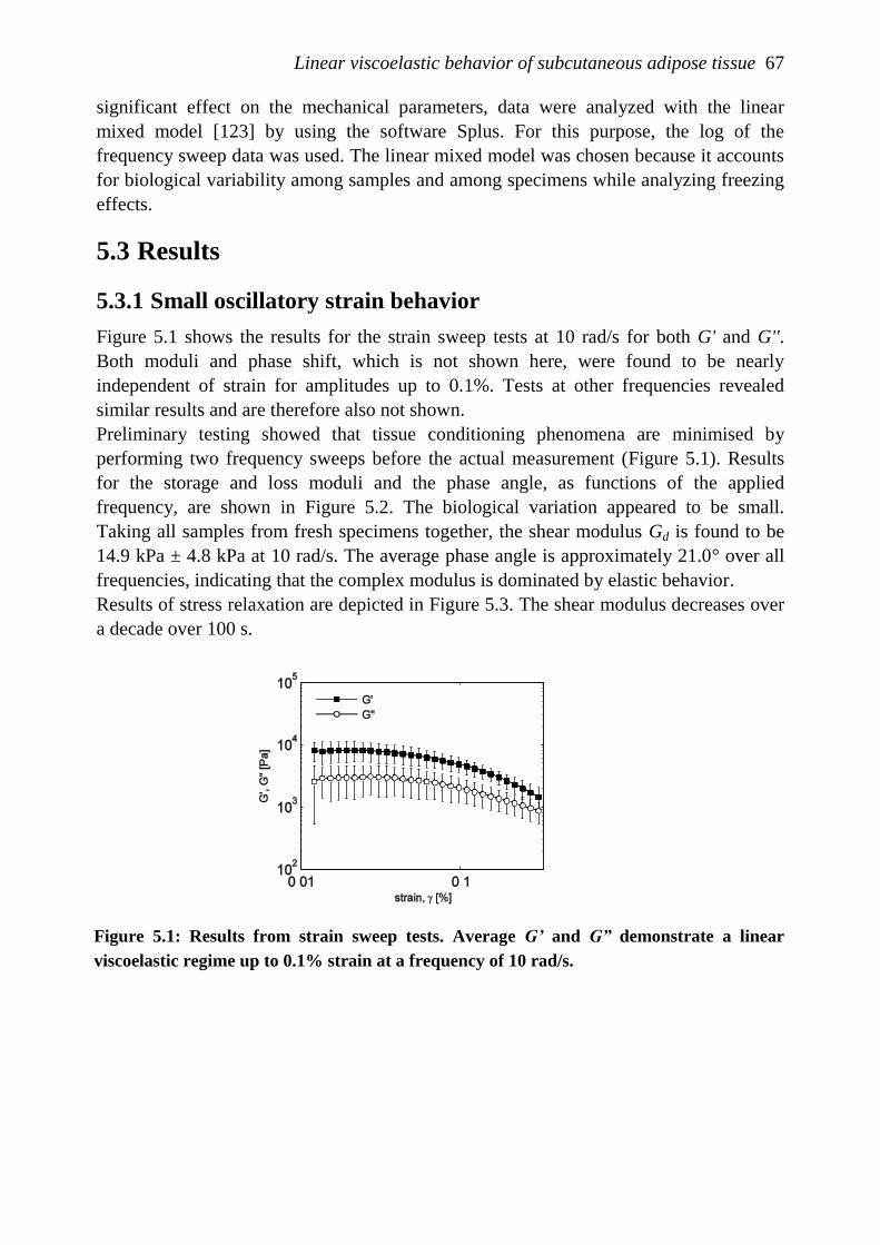
Linear viscoelastic behavior of subcutaneous adipose tissue 67
significant effect on the mechanical parameters, data were analyzed with the linear
mixed model [123] by using the software Splus. For this purpose, the log of the
frequency sweep data was used. The linear mixed model was chosen because it accounts
for biological variability among samples and among specimens while analyzing freezing
effects.
5.3 Results
5.3.1 Small oscillatory strain behavior
Figure 5.1 shows the results for the strain sweep tests at 10 rad/s for both G' and G''.
Both moduli and phase shift, which is not shown here, were found to be nearly
independent of strain for amplitudes up to 0.1%. Tests at other frequencies revealed
similar results and are therefore also not shown.
Preliminary testing showed that tissue conditioning phenomena are minimised by
performing two frequency sweeps before the actual measurement (Figure 5.1). Results
for the storage and loss moduli and the phase angle, as functions of the applied
frequency, are shown in Figure 5.2. The biological variation appeared to be small.
Taking all samples from fresh specimens together, the shear modulus Gd is found to be
14.9 kPa ± 4.8 kPa at 10 rad/s. The average phase angle is approximately 21.0° over all
frequencies, indicating that the complex modulus is dominated by elastic behavior.
Results of stress relaxation are depicted in Figure 5.3. The shear modulus decreases over
a decade over 100 s.
Figure 5.1: Results from strain sweep tests. Average G’ and G” demonstrate a linear
viscoelastic regime up to 0.1% strain at a frequency of 10 rad/s.
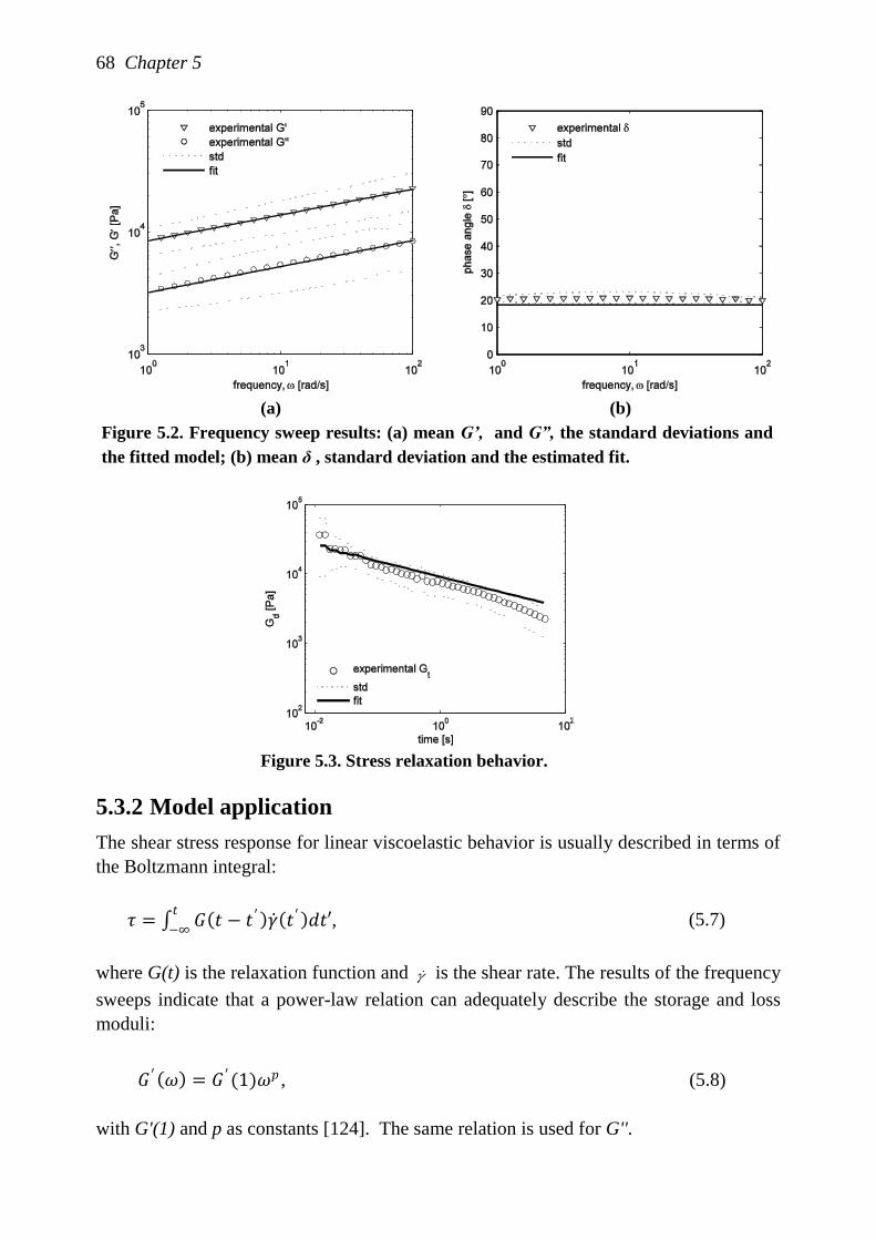
68 Chapter 5
(a) (b)
Figure 5.2. Frequency sweep results: (a) mean G’, and G”, the standard deviations and
the fitted model; (b) mean δ , standard deviation and the estimated fit.
Figure 5.3. Stress relaxation behavior.
5.3.2 Model application
The shear stress response for linear viscoelastic behavior is usually described in terms of
the Boltzmann integral:
𝜏 = 𝐺 𝑡 − 𝑡 ′ 𝛾 𝑡′ 𝑑𝑡′𝑡
−∞, (5.7)
where G(t) is the relaxation function and is the shear rate. The results of the frequency
sweeps indicate that a power-law relation can adequately describe the storage and loss
moduli:
with G'(1) and p as constants [124]. The same relation is used for G''.
𝐺 ′ 𝜔 = 𝐺 ′(1)𝜔𝑝 , (5.8)
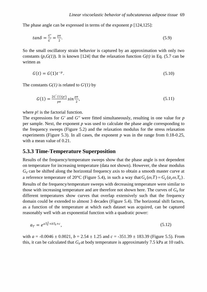
Linear viscoelastic behavior of subcutaneous adipose tissue 69
The phase angle can be expressed in terms of the exponent p [124,125]:
𝑡𝑎𝑛𝛿 =𝐺"
𝐺′=
𝑝𝜋
2. (5.9)
So the small oscillatory strain behavior is captured by an approximation with only two
constants (p,G(1)). It is known [124] that the relaxation function G(t) in Eq. (5.7 can be
written as
𝐺 𝑡 = 𝐺 1 𝑡−𝑝 . (5.10)
The constants G(1) is related to G'(1) by
𝐺 1 =2𝐺′ 1 (𝑝!)
𝑝𝜋𝑠𝑖𝑛
𝑝𝜋
2, (5.11)
where p! is the factorial function.
The expressions for G' and G'' were fitted simultaneously, resulting in one value for p
per sample. Next, the exponent p was used to calculate the phase angle corresponding to
the frequency sweeps (Figure 5.2) and the relaxation modulus for the stress relaxation
experiments (Figure 5.3). In all cases, the exponent p was in the range from 0.18-0.25,
with a mean value of 0.21.
5.3.3 Time-Temperature Superposition
Results of the frequency/temperature sweeps show that the phase angle is not dependent
on temperature for increasing temperature (data not shown). However, the shear modulus
Gd can be shifted along the horizontal frequency axis to obtain a smooth master curve at
a reference temperature of 20°C (Figure 5.4), in such a way that ),(),( 0TaGTG Tdd .
Results of the frequency/temperature sweeps with decreasing temperature were similar to
those with increasing temperature and are therefore not shown here. The curves of Gd for
different temperatures show curves that overlap extensively such that the frequency
domain could be extended to almost 3 decades (Figure 5.4). The horizontal shift factors,
as a function of the temperature at which each dataset was acquired, can be captured
reasonably well with an exponential function with a quadratic power:
𝑎𝑇 = 𝑒𝑎𝑇02+𝑏𝑇0+𝑐 , (5.12)
with a = -0.0046 ± 0.0021, b = 2.54 ± 1.25 and c = -351.39 ± 183.39 (Figure 5.5). From
this, it can be calculated that Gd at body temperature is approximately 7.5 kPa at 10 rad/s.
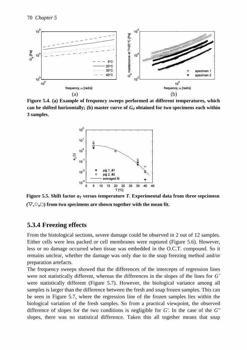
70 Chapter 5
(a) (b)
Figure 5.4. (a) Example of frequency sweeps performed at different temperatures, which
can be shifted horizontally; (b) master curve of Gd obtained for two specimens each within
3 samples.
Figure 5.5. Shift factor aT versus temperature T. Experimental data from three sepcimesn
(,○,□) from two specimens are shown together with the mean fit.
5.3.4 Freezing effects
From the histological sections, severe damage could be observed in 2 out of 12 samples.
Either cells were less packed or cell membranes were ruptured (Figure 5.6). However,
less or no damage occurred when tissue was embedded in the O.C.T. compound. So it
remains unclear, whether the damage was only due to the snap freezing method and/or
preparation artefacts.
The frequency sweeps showed that the differences of the intercepts of regression lines
were not statistically different, whereas the differences in the slopes of the lines for G’
were statistically different (Figure 5.7). However, the biological variance among all
samples is larger than the difference between the fresh and snap frozen samples. This can
be seen in Figure 5.7, where the regression line of the frozen samples lies within the
biological variation of the fresh samples. So from a practical viewpoint, the observed
difference of slopes for the two conditions is negligible for G'. In the case of the G''
slopes, there was no statistical difference. Taken this all together means that snap
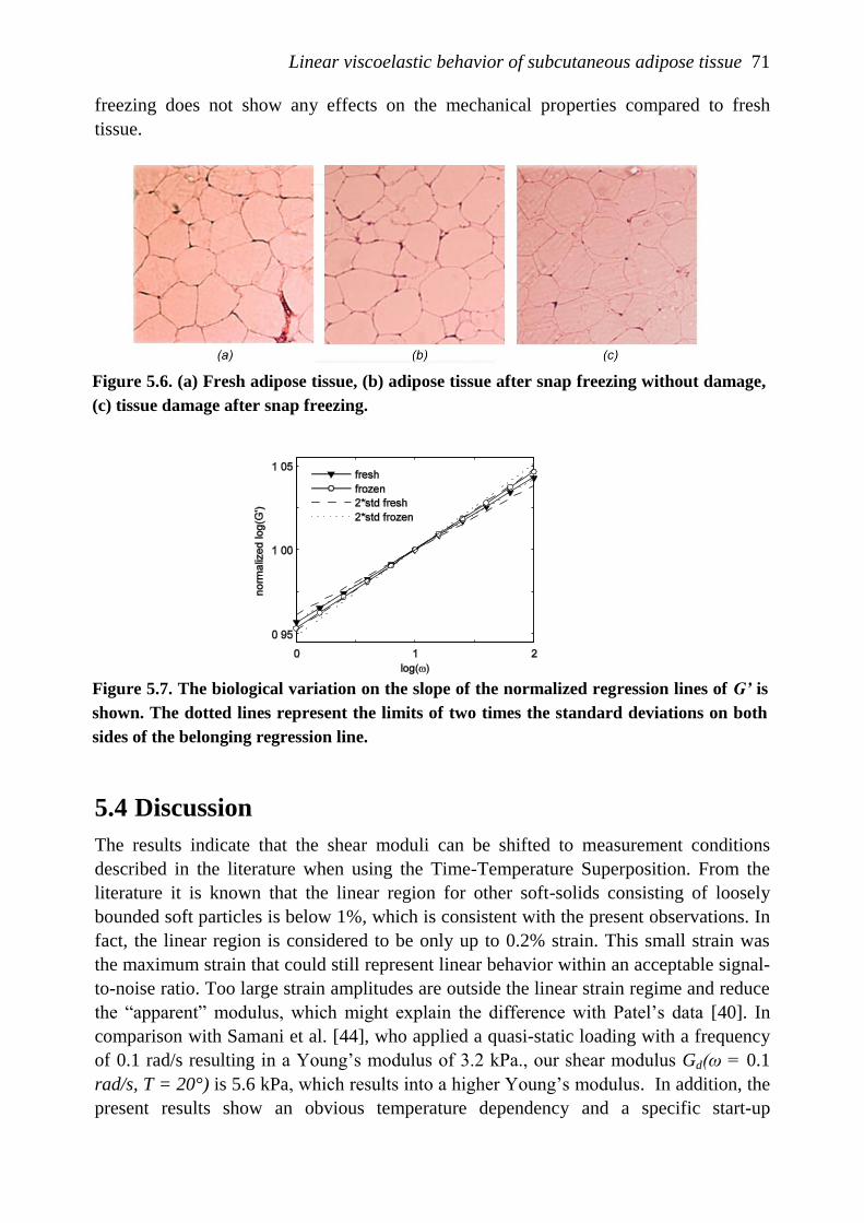
Linear viscoelastic behavior of subcutaneous adipose tissue 71
freezing does not show any effects on the mechanical properties compared to fresh
tissue.
Figure 5.6. (a) Fresh adipose tissue, (b) adipose tissue after snap freezing without damage,
(c) tissue damage after snap freezing.
Figure 5.7. The biological variation on the slope of the normalized regression lines of G’ is
shown. The dotted lines represent the limits of two times the standard deviations on both
sides of the belonging regression line.
5.4 Discussion
The results indicate that the shear moduli can be shifted to measurement conditions
described in the literature when using the Time-Temperature Superposition. From the
literature it is known that the linear region for other soft-solids consisting of loosely
bounded soft particles is below 1%, which is consistent with the present observations. In
fact, the linear region is considered to be only up to 0.2% strain. This small strain was
the maximum strain that could still represent linear behavior within an acceptable signal-
to-noise ratio. Too large strain amplitudes are outside the linear strain regime and reduce
the “apparent” modulus, which might explain the difference with Patel‟s data [40]. In
comparison with Samani et al. [44], who applied a quasi-static loading with a frequency
of 0.1 rad/s resulting in a Young‟s modulus of 3.2 kPa., our shear modulus Gd(ω = 0.1
rad/s, T = 20°) is 5.6 kPa, which results into a higher Young‟s modulus. In addition, the
present results show an obvious temperature dependency and a specific start-up
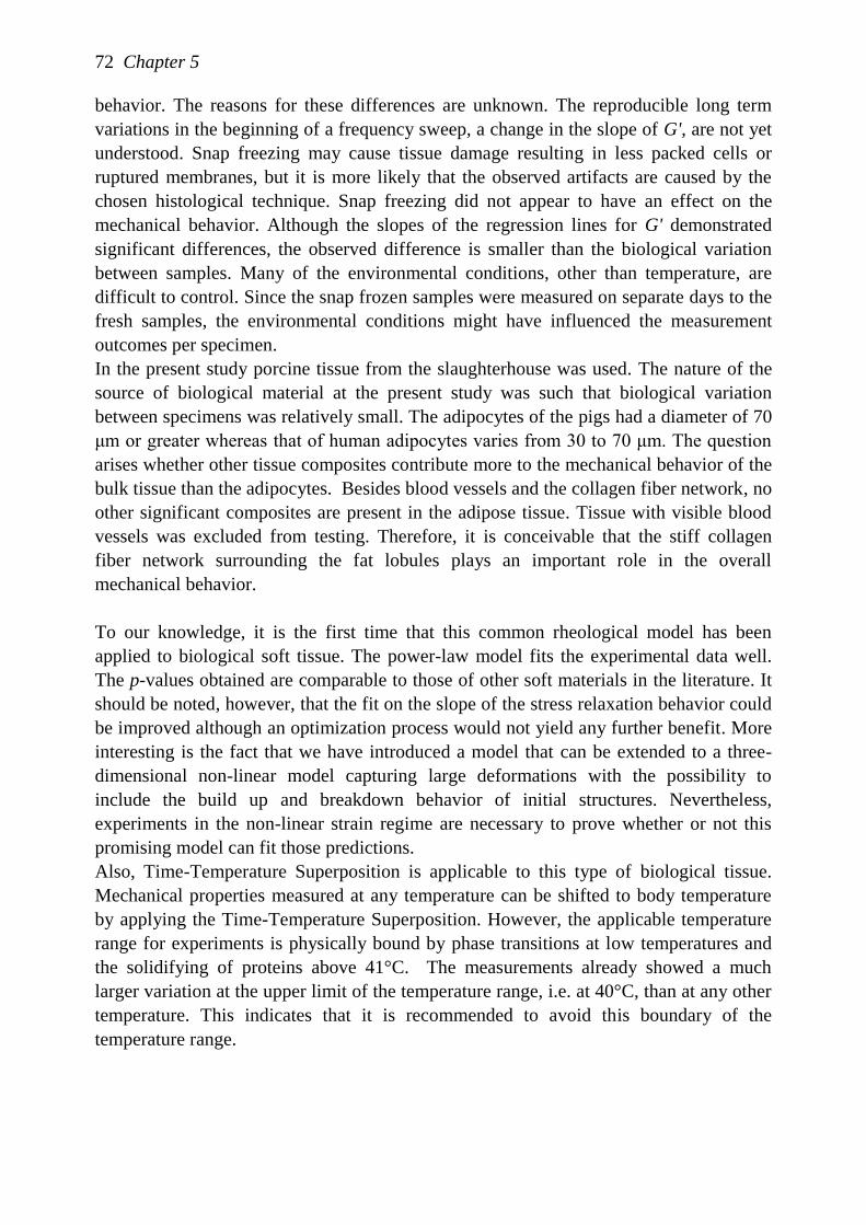
72 Chapter 5
behavior. The reasons for these differences are unknown. The reproducible long term
variations in the beginning of a frequency sweep, a change in the slope of G', are not yet
understood. Snap freezing may cause tissue damage resulting in less packed cells or
ruptured membranes, but it is more likely that the observed artifacts are caused by the
chosen histological technique. Snap freezing did not appear to have an effect on the
mechanical behavior. Although the slopes of the regression lines for G' demonstrated
significant differences, the observed difference is smaller than the biological variation
between samples. Many of the environmental conditions, other than temperature, are
difficult to control. Since the snap frozen samples were measured on separate days to the
fresh samples, the environmental conditions might have influenced the measurement
outcomes per specimen.
In the present study porcine tissue from the slaughterhouse was used. The nature of the
source of biological material at the present study was such that biological variation
between specimens was relatively small. The adipocytes of the pigs had a diameter of 70
μm or greater whereas that of human adipocytes varies from 30 to 70 μm. The question
arises whether other tissue composites contribute more to the mechanical behavior of the
bulk tissue than the adipocytes. Besides blood vessels and the collagen fiber network, no
other significant composites are present in the adipose tissue. Tissue with visible blood
vessels was excluded from testing. Therefore, it is conceivable that the stiff collagen
fiber network surrounding the fat lobules plays an important role in the overall
mechanical behavior.
To our knowledge, it is the first time that this common rheological model has been
applied to biological soft tissue. The power-law model fits the experimental data well.
The p-values obtained are comparable to those of other soft materials in the literature. It
should be noted, however, that the fit on the slope of the stress relaxation behavior could
be improved although an optimization process would not yield any further benefit. More
interesting is the fact that we have introduced a model that can be extended to a three-
dimensional non-linear model capturing large deformations with the possibility to
include the build up and breakdown behavior of initial structures. Nevertheless,
experiments in the non-linear strain regime are necessary to prove whether or not this
promising model can fit those predictions.
Also, Time-Temperature Superposition is applicable to this type of biological tissue.
Mechanical properties measured at any temperature can be shifted to body temperature
by applying the Time-Temperature Superposition. However, the applicable temperature
range for experiments is physically bound by phase transitions at low temperatures and
the solidifying of proteins above 41°C. The measurements already showed a much
larger variation at the upper limit of the temperature range, i.e. at 40°C, than at any other
temperature. This indicates that it is recommended to avoid this boundary of the
temperature range.
Chapter 6
Does subcutaneous adipose tissue behave
as an (anti-)thyxotropic material?
The contents of this chapter are based on M. Geerligs, G.W.M. Peters, P.A.J.
Ackermans, C.W.J. Oomens, and F.P.T. Baaijens (2010), Does subcutaneous adipose
tissue behave as an (anti-)thyxotropic material?, Journal of Biomechanics, accepted.
74 Chapter 6
6.1 Introduction
The mechanical load transfer from a skin contact area to deeper tissues involves several
tissue layers. On most body sites, the subcutaneous adipose tissue considerably
contributes to this load transfer. However, when numerical models are used to predict the
stress response due to external loading, the focus is either on the skin-device contact or
on the deeper tissue layers while the subcutaneous fat layer is often ignored. This
omission might be related to the lack of defined parameters, which describe the
mechanical behavior of adipose tissues. This is particularly surprising given the critical
roles for adipose tissues in the medical and cosmetic fields, involving, for example,
implantable drugs delivery, skin adhesive removal, deep tissue injury and needle
insertion procedures.
Recently, our previous work on the linear behavior of subcutaneous adipose tissue has
shown that the linear strain regime is valid for very small strains only, i.e. 0.001 [126]. In
most applications, however, much higher deformations occur in the adipose tissue for
prolonged periods. Indeed, for wheelchair or bedridden patients, for example, this might
lead to the development of deep tissue injury under bony prominences within a time
frame of minutes to hours, during which stress relaxation in the compressed tissue might
occur [41]. Numerical models based on experimental data are of indispensable value to
predict the onset and progression of such mechanical-induced damage.
Currently, there is a paucity of papers on the mechanical properties of the subcutaneous
adipose tissue found beneath hairy skin. Viscoelastic properties of single human
adipocytes have been recently characterized using AFM resulting in a relaxed modulus
and relaxation time for either load or deformation [127]. Few related in vitro studies on
tissue behavior exist. Of these, rheological measurements demonstrated a decrease in
viscosity with increasing shear rate [40]. In addition, the authors suggested that adipose
tissue loses firmness with increasing strain and frequency, a state which is not
recoverable. In a separate study, ovine subcutaneous tissue was subjected to ramp-and-
hold cycles during confined compression tests at various ramp rates [40,41]. The results
were given in the form of a transient aggregate modulus and short-term elastic moduli.
They also found a strong deformation rate dependency. Short-term moduli were in the
order of 20 kPa. In an alternative in vivo approach, a suction device yielded experimental
parameters which, when combined with numerical modeling, led to a first estimation of
non-linear material parameters for human skin [111]. To our knowledge, there are no in
vivo studies considering subcutaneous adipose tissue as a single layer. By contrast, some
in vivo studies have examined the mechanical properties for a compliant system
consisting of skin and subcutaneous adipose tissue [117,128].
The work mentioned above describes a range of loading conditions, often combining
techniques involving indentation, confined compression, stress relaxation and constant
shear responses. Clearly, this makes comparison of data from the studies problematic in

Does subcutaneous adipose tissue behave as an (anti-)thyxotropic material? 75
nature. For a general constitutive model for adipose tissue a more systematic approach is
required.
The material structure of subcutaneous adipose tissue does not relate conveniently to
other biological tissues. Its main component is the white adipocyte. The remaining
components are water (5-30% weight) and protein (2-3% weight). The white adipocytes
are filled with a large fat droplet imposing forces on both the nucleus and the small
cytoplasmic volume at the cell periphery. The composition of the white adipocytes
depends on the specific function and body site. As an example, differences throughout
the human body are known for the proportions of saturated fatty acids, monosaturated
versus polysaturated fat and the lipolysis rate [18]. White adipocytes are collected in a
surrounding fiber network. The adipose tissue is well-vascularized throughout with each
adipocyte in contact with at least one capillary. Hence, adipose tissue is susceptible to
ischemia and hypoxia, which influence its mechanical response.
Our previous work on the small strain behavior of adipose tissue has shown that
reproducible results are obtained in an in-vitro set-up using a rheometer with parallel
plate geometry and that the behavior can be described with a power-law model [126].
However, sometimes tissue samples were found to be much stiffer than the mean value
and early work at higher strains has suggested that (reversible) structural changes start to
play a role. In addition, earlier large strain studies formed the incentive for a more
systematic approach at higher strains to elucidate the phenomena that havealready been
described. Therefore, the present study aims to provide systematic data for long-term
small strain behavior as well as the effect of strain history, with the purpose of
contributing to the development of a constitutive model.
Accordingly, the work is divided in two parts. The first part contains long term
oscillatory tests at small strains to investigate temporal effects of the adipose tissue
samples. Subsequently, strain-dependency tests, comprising constant shear, stress
relaxation and constant strain rate, are applied. From these tests, non-linear parameters
can be obtained useful for constitutive modeling. Such an experimental approach is
designed to gain insight on the mechanical response of adipose tissue under shear where
the effect of strain history, strain level and duration is taken into account.
6.2 Materials & Methods
6.2.1 Sample preparation
In porcine species, the subcutaneous fat layer on the back is divided in an outer, middle
and inner layer. The porcine middle layer was selected for use, as it is considered to be
the most comparable with the deep subcutaneous layer in the abdominal region of
humans [118]. The tissue was obtained from a local slaughterhouse, where they were cut
into transverse slices of approximately 1.5 mm thick. In our laboratories, circular
samples were obtained from the slices with an 8 mm diameter cork borer. The samples
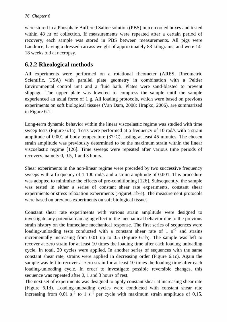
76 Chapter 6
were stored in a Phosphate Buffered Saline solution (PBS) in ice-cooled boxes and tested
within 48 hr of collection. If measurements were repeated after a certain period of
recovery, each sample was stored in PBS between measurements. All pigs were
Landrace, having a dressed carcass weight of approximately 83 kilograms, and were 14-
18 weeks old at necropsy.
6.2.2 Rheological methods
All experiments were performed on a rotational rheometer (ARES, Rheometric
Scientific, USA) with parallel plate geometry in combination with a Peltier
Environmental control unit and a fluid bath. Plates were sand-blasted to prevent
slippage. The upper plate was lowered to compress the sample until the sample
experienced an axial force of 1 g. All loading protocols, which were based on previous
experiments on soft biological tissues (Van Dam, 2008; Hrapko, 2006), are summarized
in Figure 6.1.
Long-term dynamic behavior within the linear viscoelastic regime was studied with time
sweep tests (Figure 6.1a). Tests were performed at a frequency of 10 rad/s with a strain
amplitude of 0.001 at body temperature (37°C), lasting at least 45 minutes. The chosen
strain amplitude was previously determined to be the maximum strain within the linear
viscoelastic regime [126]. Time sweeps were repeated after various time periods of
recovery, namely 0, 0.5, 1 and 3 hours.
Shear experiments in the non-linear regime were preceded by two successive frequency
sweeps with a frequency of 1-100 rad/s and a strain amplitude of 0.001. This procedure
was adopted to minimize the effects of pre-conditioning [126]. Subsequently, the sample
was tested in either a series of constant shear rate experiments, constant shear
experiments or stress relaxation experiments (Figure6.1b-e). The measurement protocols
were based on previous experiments on soft biological tissues.
Constant shear rate experiments with various strain amplitude were designed to
investigate any potential damaging effect in the mechanical behavior due to the previous
strain history on the immediate mechanical response. The first series of sequences were
loading-unloading tests conducted with a constant shear rate of 1 s−1
and strains
incrementally increasing from 0.01 up to 0.5 (Figure 6.1b). The sample was left to
recover at zero strain for at least 10 times the loading time after each loading-unloading
cycle. In total, 20 cycles were applied. In another series of sequences with the same
constant shear rate, strains were applied in decreasing order (Figure 6.1c). Again the
sample was left to recover at zero strain for at least 10 times the loading time after each
loading-unloading cycle. In order to investigate possible reversible changes, this
sequence was repeated after 0, 1 and 3 hours of rest.
The next set of experiments was designed to apply constant shear at increasing shear rate
(Figure 6.1d). Loading-unloading cycles were conducted with constant shear rate
increasing from 0.01 s−1
to 1 s−1
per cycle with maximum strain amplitude of 0.15.
Does subcutaneous adipose tissue behave as an (anti-)thyxotropic material? 77
Between two cycles, the sample was again left to recover for at least 10 times the loading
time.
Finally, stress relaxation experiments were composed of a series of ramp-and-hold tests
at different strain levels (Figure 6.1e). During the loading and unloading phase, a
constant strain rate of 1 s-1
was imposed. The maximum strain was held for 10 s during
which the relaxation of the material was recorded. The sample was left to recover for a
period of at least 100 s during which time the tissue response was monitored. The test
was repeated for four different strain levels, namely 0.01, 0.05, 0.1 and 0.15.
An overview of the number of specimens and the number of samples from each
specimen per test is given in Table 6.1.
Figure 6.1: Schematic illustration of test sequences. (a) Time sweep tests; (b) Constant
shear rate experiments with increasing shear strains; (c) constant shear rate experiments
with decreasing shear strains; (d) constant shear experiments with increasing shear rate;
(e) stress relaxation experiments.
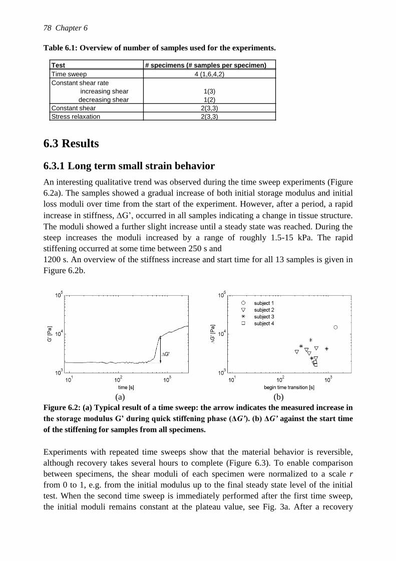
78 Chapter 6
Table 6.1: Overview of number of samples used for the experiments.
6.3 Results
6.3.1 Long term small strain behavior
An interesting qualitative trend was observed during the time sweep experiments (Figure
6.2a). The samples showed a gradual increase of both initial storage modulus and initial
loss moduli over time from the start of the experiment. However, after a period, a rapid
increase in stiffness, G‟, occurred in all samples indicating a change in tissue structure.
The moduli showed a further slight increase until a steady state was reached. During the
steep increases the moduli increased by a range of roughly 1.5-15 kPa. The rapid
stiffening occurred at some time between 250 s and
1200 s. An overview of the stiffness increase and start time for all 13 samples is given in
Figure 6.2b.
(a) (b)
Figure 6.2: (a) Typical result of a time sweep: the arrow indicates the measured increase in
the storage modulus G’ during quick stiffening phase (ΔG’). (b) ΔG’ against the start time
of the stiffening for samples from all specimens.
Experiments with repeated time sweeps show that the material behavior is reversible,
although recovery takes several hours to complete (Figure 6.3). To enable comparison
between specimens, the shear moduli of each specimen were normalized to a scale r
from 0 to 1, e.g. from the initial modulus up to the final steady state level of the initial
test. When the second time sweep is immediately performed after the first time sweep,
the initial moduli remains constant at the plateau value, see Fig. 3a. After a recovery
Test # specimens (# samples per specimen)
Time sweep 4 (1,6,4,2)
Constant shear rate
increasing shear 1(3)
decreasing shear 1(2)
Constant shear 2(3,3)
Stress relaxation 2(3,3)
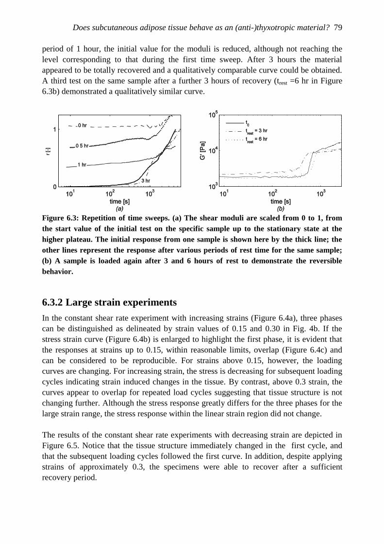
Does subcutaneous adipose tissue behave as an (anti-)thyxotropic material? 79
period of 1 hour, the initial value for the moduli is reduced, although not reaching the
level corresponding to that during the first time sweep. After 3 hours the material
appeared to be totally recovered and a qualitatively comparable curve could be obtained.
A third test on the same sample after a further 3 hours of recovery (trest =6 hr in Figure
6.3b) demonstrated a qualitatively similar curve.
Figure 6.3: Repetition of time sweeps. (a) The shear moduli are scaled from 0 to 1, from
the start value of the initial test on the specific sample up to the stationary state at the
higher plateau. The initial response from one sample is shown here by the thick line; the
other lines represent the response after various periods of rest time for the same sample;
(b) A sample is loaded again after 3 and 6 hours of rest to demonstrate the reversible
behavior.
6.3.2 Large strain experiments
In the constant shear rate experiment with increasing strains (Figure 6.4a), three phases
can be distinguished as delineated by strain values of 0.15 and 0.30 in Fig. 4b. If the
stress strain curve (Figure 6.4b) is enlarged to highlight the first phase, it is evident that
the responses at strains up to 0.15, within reasonable limits, overlap (Figure 6.4c) and
can be considered to be reproducible. For strains above 0.15, however, the loading
curves are changing. For increasing strain, the stress is decreasing for subsequent loading
cycles indicating strain induced changes in the tissue. By contrast, above 0.3 strain, the
curves appear to overlap for repeated load cycles suggesting that tissue structure is not
changing further. Although the stress response greatly differs for the three phases for the
large strain range, the stress response within the linear strain region did not change.
The results of the constant shear rate experiments with decreasing strain are depicted in
Figure 6.5. Notice that the tissue structure immediately changed in the first cycle, and
that the subsequent loading cycles followed the first curve. In addition, despite applying
strains of approximately 0.3, the specimens were able to recover after a sufficient
recovery period.
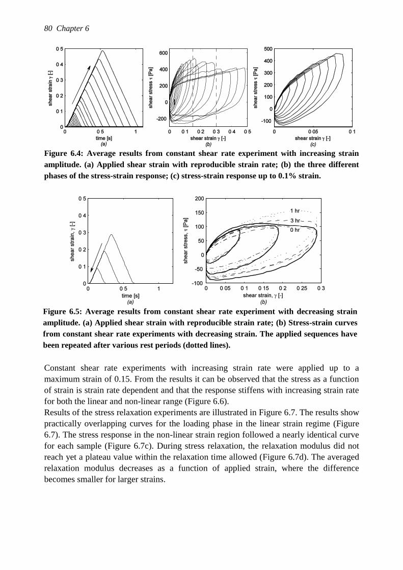
80 Chapter 6
Figure 6.4: Average results from constant shear rate experiment with increasing strain
amplitude. (a) Applied shear strain with reproducible strain rate; (b) the three different
phases of the stress-strain response; (c) stress-strain response up to 0.1% strain.
Figure 6.5: Average results from constant shear rate experiment with decreasing strain
amplitude. (a) Applied shear strain with reproducible strain rate; (b) Stress-strain curves
from constant shear rate experiments with decreasing strain. The applied sequences have
been repeated after various rest periods (dotted lines).
Constant shear rate experiments with increasing strain rate were applied up to a
maximum strain of 0.15. From the results it can be observed that the stress as a function
of strain is strain rate dependent and that the response stiffens with increasing strain rate
for both the linear and non-linear range (Figure 6.6).
Results of the stress relaxation experiments are illustrated in Figure 6.7. The results show
practically overlapping curves for the loading phase in the linear strain regime (Figure
6.7). The stress response in the non-linear strain region followed a nearly identical curve
for each sample (Figure 6.7c). During stress relaxation, the relaxation modulus did not
reach yet a plateau value within the relaxation time allowed (Figure 6.7d). The averaged
relaxation modulus decreases as a function of applied strain, where the difference
becomes smaller for larger strains.
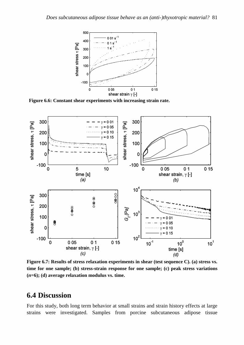
Does subcutaneous adipose tissue behave as an (anti-)thyxotropic material? 81
Figure 6.6: Constant shear experiments with increasing strain rate.
Figure 6.7: Results of stress relaxation experiments in shear (test sequence C). (a) stress vs.
time for one sample; (b) stress-strain response for one sample; (c) peak stress variations
(n=6); (d) average relaxation modulus vs. time.
6.4 Discussion
For this study, both long term behavior at small strains and strain history effects at large
strains were investigated. Samples from porcine subcutaneous adipose tissue
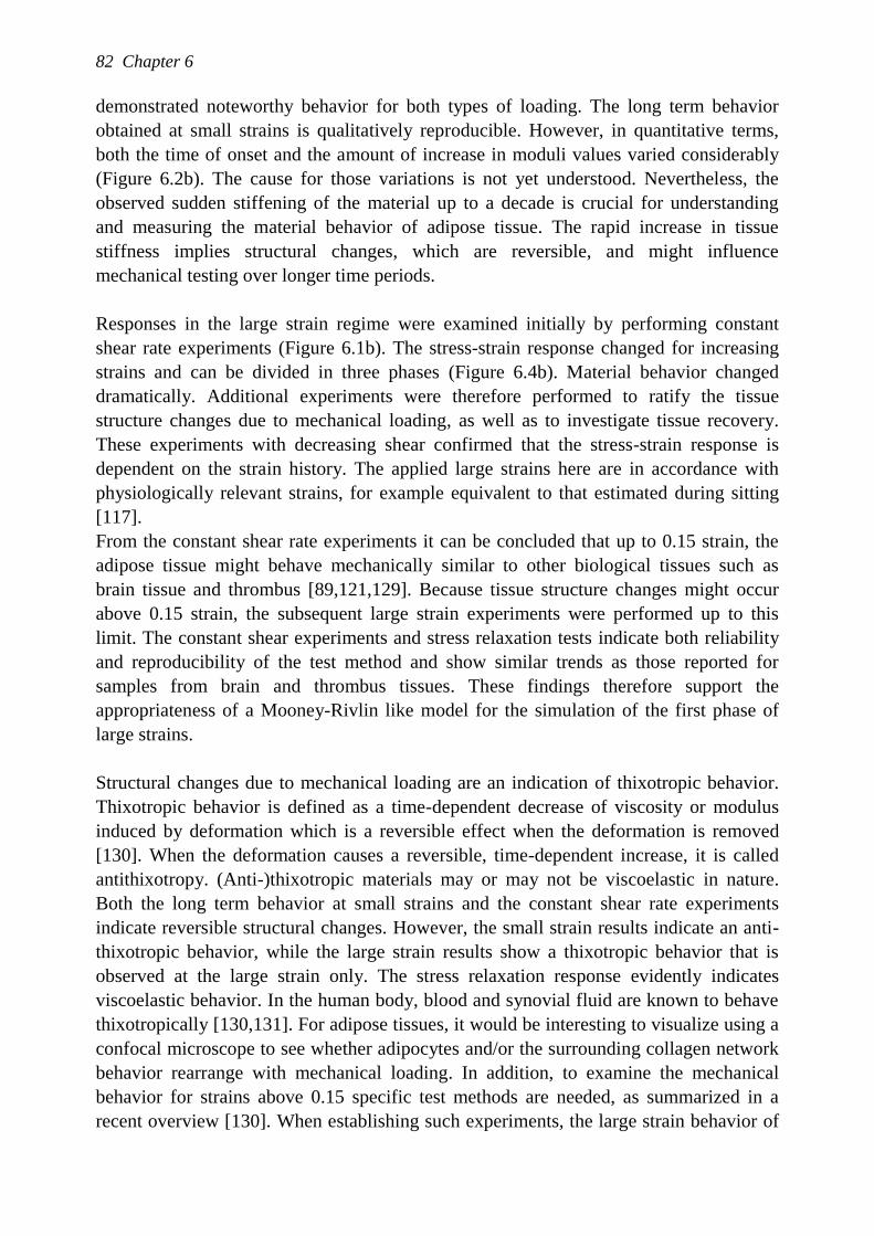
82 Chapter 6
demonstrated noteworthy behavior for both types of loading. The long term behavior
obtained at small strains is qualitatively reproducible. However, in quantitative terms,
both the time of onset and the amount of increase in moduli values varied considerably
(Figure 6.2b). The cause for those variations is not yet understood. Nevertheless, the
observed sudden stiffening of the material up to a decade is crucial for understanding
and measuring the material behavior of adipose tissue. The rapid increase in tissue
stiffness implies structural changes, which are reversible, and might influence
mechanical testing over longer time periods.
Responses in the large strain regime were examined initially by performing constant
shear rate experiments (Figure 6.1b). The stress-strain response changed for increasing
strains and can be divided in three phases (Figure 6.4b). Material behavior changed
dramatically. Additional experiments were therefore performed to ratify the tissue
structure changes due to mechanical loading, as well as to investigate tissue recovery.
These experiments with decreasing shear confirmed that the stress-strain response is
dependent on the strain history. The applied large strains here are in accordance with
physiologically relevant strains, for example equivalent to that estimated during sitting
[117].
From the constant shear rate experiments it can be concluded that up to 0.15 strain, the
adipose tissue might behave mechanically similar to other biological tissues such as
brain tissue and thrombus [89,121,129]. Because tissue structure changes might occur
above 0.15 strain, the subsequent large strain experiments were performed up to this
limit. The constant shear experiments and stress relaxation tests indicate both reliability
and reproducibility of the test method and show similar trends as those reported for
samples from brain and thrombus tissues. These findings therefore support the
appropriateness of a Mooney-Rivlin like model for the simulation of the first phase of
large strains.
Structural changes due to mechanical loading are an indication of thixotropic behavior.
Thixotropic behavior is defined as a time-dependent decrease of viscosity or modulus
induced by deformation which is a reversible effect when the deformation is removed
[130]. When the deformation causes a reversible, time-dependent increase, it is called
antithixotropy. (Anti-)thixotropic materials may or may not be viscoelastic in nature.
Both the long term behavior at small strains and the constant shear rate experiments
indicate reversible structural changes. However, the small strain results indicate an anti-
thixotropic behavior, while the large strain results show a thixotropic behavior that is
observed at the large strain only. The stress relaxation response evidently indicates
viscoelastic behavior. In the human body, blood and synovial fluid are known to behave
thixotropically [130,131]. For adipose tissues, it would be interesting to visualize using a
confocal microscope to see whether adipocytes and/or the surrounding collagen network
behavior rearrange with mechanical loading. In addition, to examine the mechanical
behavior for strains above 0.15 specific test methods are needed, as summarized in a
recent overview [130]. When establishing such experiments, the large strain behavior of
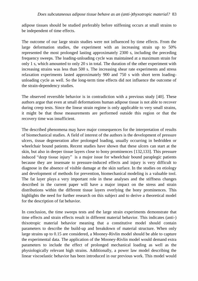
Does subcutaneous adipose tissue behave as an (anti-)thyxotropic material? 83
adipose tissues should be studied preferably before stiffening occurs at small strains to
be independent of time effects.
The outcome of our large strain studies were not influenced by time effects. From the
large deformation studies, the experiment with an increasing strain up to 50%
represented the most prolonged lasting approximately 2300 s, including the preceding
frequency sweeps. The loading-unloading cycle was maintained at a maximum strain for
only 1 s, which amounted to only 20 s in total. The duration of the other experiment with
increasing strains was less than 500 s. The increasing shear rate experiments and stress
relaxation experiments lasted approximately 900 and 750 s with short term loading-
unloading cycle as well. So the long-term time effects did not influence the outcome of
the strain-dependency studies.
The observed reversible behavior is in contradiction with a previous study [40]. These
authors argue that even at small deformations human adipose tissue is not able to recover
during creep tests. Since the linear strain regime is only applicable to very small strains,
it might be that those measurements are performed outside this region or that the
recovery time was insufficient.
The described phenomena may have major consequences for the interpretation of results
of biomechanical studies. A field of interest of the authors is the development of pressure
ulcers, tissue degeneration after prolonged loading, usually occurring in bedridden or
wheelchair bound patients. Recent studies have shown that these ulcers can start at the
skin, but also in deeper tissue layers close to bony prominences [132,133]. This pressure
induced “deep tissue injury” is a major issue for wheelchair bound paraplegic patients
because they are insensate to pressure-induced effects and injury is very difficult to
diagnose in the absence of visible damage at the skin surface. In the studies on etiology
and development of methods for prevention, biomechanical modeling is a valuable tool.
The fat layer plays a very important role in these analyses and the stiffness changes
described in the current paper will have a major impact on the stress and strain
distributions within the different tissue layers overlying the bony prominences. This
highlights the need for further research on this subject and to derive a theoretical model
for the description of fat behavior.
In conclusion, the time sweeps tests and the large strain experiments demonstrate that
time effects and strain effects result in different material behavior. This indicates (anti-)
thixotropic material behavior meaning that a constitutive model should contain
parameters to describe the build-up and breakdown of material structure. When only
large strains up to 0.15 are considered, a Mooney-Rivlin model should be able to capture
the experimental data. The application of the Mooney-Rivlin model would demand extra
parameters to include the effect of prolonged mechanical loading as well as the
physiologically relevant high strains. Additionally, a power law model describing the
linear viscoelastic behavior has been introduced in our previous work. This model would

84 Chapter 6
also be suitable for implementing a build-up and breakdown structure properties. We
believe, however, it is better to set-up more experiments to fully understand the material
behavior before continuing the building of a constitutive model.
This paper shows the high complexity of the material behavior and particularly
demonstrates more work is needed on this topic. The described effects should be taken
into account when setting up new experiments. The follow-up experiments should clarify
the effects of time and strain and the reversibility of the material.
Acknowledgements
We would like to thank Prof. Dan Bader for his valuable contribution to our discussions
during preparation of this article.

Chapter 7
General discussion
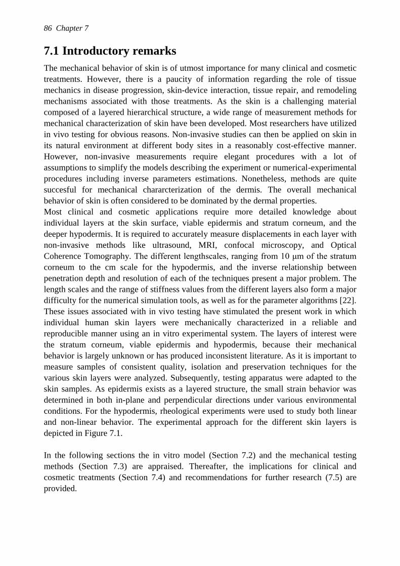
86 Chapter 7
7.1 Introductory remarks
The mechanical behavior of skin is of utmost importance for many clinical and cosmetic
treatments. However, there is a paucity of information regarding the role of tissue
mechanics in disease progression, skin-device interaction, tissue repair, and remodeling
mechanisms associated with those treatments. As the skin is a challenging material
composed of a layered hierarchical structure, a wide range of measurement methods for
mechanical characterization of skin have been developed. Most researchers have utilized
in vivo testing for obvious reasons. Non-invasive studies can then be applied on skin in
its natural environment at different body sites in a reasonably cost-effective manner.
However, non-invasive measurements require elegant procedures with a lot of
assumptions to simplify the models describing the experiment or numerical-experimental
procedures including inverse parameters estimations. Nonetheless, methods are quite
succesful for mechanical chararcterization of the dermis. The overall mechanical
behavior of skin is often considered to be dominated by the dermal properties.
Most clinical and cosmetic applications require more detailed knowledge about
individual layers at the skin surface, viable epidermis and stratum corneum, and the
deeper hypodermis. It is required to accurately measure displacements in each layer with
non-invasive methods like ultrasound, MRI, confocal microscopy, and Optical
Coherence Tomography. The different lengthscales, ranging from 10 μm of the stratum
corneum to the cm scale for the hypodermis, and the inverse relationship between
penetration depth and resolution of each of the techniques present a major problem. The
length scales and the range of stiffness values from the different layers also form a major
difficulty for the numerical simulation tools, as well as for the parameter algorithms [22].
These issues associated with in vivo testing have stimulated the present work in which
individual human skin layers were mechanically characterized in a reliable and
reproducible manner using an in vitro experimental system. The layers of interest were
the stratum corneum, viable epidermis and hypodermis, because their mechanical
behavior is largely unknown or has produced inconsistent literature. As it is important to
measure samples of consistent quality, isolation and preservation techniques for the
various skin layers were analyzed. Subsequently, testing apparatus were adapted to the
skin samples. As epidermis exists as a layered structure, the small strain behavior was
determined in both in-plane and perpendicular directions under various environmental
conditions. For the hypodermis, rheological experiments were used to study both linear
and non-linear behavior. The experimental approach for the different skin layers is
depicted in Figure 7.1.
In the following sections the in vitro model (Section 7.2) and the mechanical testing
methods (Section 7.3) are appraised. Thereafter, the implications for clinical and
cosmetic treatments (Section 7.4) and recommendations for further research (7.5) are
provided.
General discussion 87
Figure 7.1: Experimental approach for each skin layer.
7.2 In vitro model
An in vitro model enables improved control of the experimental conditions and offers the
potential of performing well-controlled mechanical experiments on a specific skin layer.
Skin obtained from plastic surgery, as opposed to cadaveric skin, provides higher
viability and is, moreover, available from a range of ages. However, the number of body
sites is limited. In this thesis, skin obtained from abdominoplastic surgery was used to
study the epidermis and stratum corneum. In obese people, the structure of subcutaneous
adipose tissue has undergone changes in comparison with healthy subjects [18,134].
Therefore, a porcine model was introduced for this tissue layer, which is comparable in
structure and function to human adipose tissue [118]. In addition, its availability and
reproducibility makes it an attractive option.
After harvesting the skin tissue, the necessary skin layer must be isolated. As the time
between harvesting and mechanical testing is usually too long to maintain the tissue
viability and intregrity, means of preservation were needed. It is essential to ensure that
preparation treatments do not have an effect on the mechanical properties. The use of ex
vivo human skin in percutaneous and absorption studies is well established. Current
standardized isolation and preparation protocols for skin [135] are mainly guided by
cost, time effectiveness and ease of use. However, it is widely known and demonstrated
that these ways of tissue preparation influence mechanical properties [136]. In particular,
the epidermal layers are known to be highly sensitive to chemical and physical changes
in the environment. Much knowledge is already available from skin grafting techniques
for burn wounds. However, the required tissue condition is different from in vitro testing
[54,74]. Therefore, available and new techniques to isolate and preserve epidermis and
stratum corneum were assessed as to their successfulness with reference to the
maintenance of tissue integrity and viability. Furthermore, both the ease of handling and
the reproducibility of the protocol were considered.
From the numerous techniques in use to isolate the epidermis, it was concluded that
many are limited when considering the maintenance of tissue integrity and viability.
slicer
isolation preservation
PBS(or -80°C)
HHBSS(or -80°C)
PBS(or drying)
dermatome
linear + non-linear
linear
linear
hp
hc
a
h
mechanical testing
linear
linear
indentation
shear
88 Chapter 7
However, cutting using a dermatome and enzymatic digestion with dispase fulfills both
requirements and also provides ease of handling and reproducibility. Cutting result into a
better defined sample geometry, which is convenient for most mechanical tests. As
shown in Chapters 3 and 4, when fragments of papillar dermis were present in the
epidermal samples (Figure 4.1), this did not lead to a measurable influence on the results
for small strain behavior. As established in these chapters, the mechanical behavior of
stratum corneum and viable epidermis are comparable and both have a higher stiffness
than the dermal layer. Thus, it can be assumed that the influence of fragments of papillar
dermis in the samples will also be minimal in large deformation studies. Regarding the
isolation of the stratum corneum, the gold standard represents enzymatic digestion with
0.1% trypsin. Some other techniques were analyzed but none comparable performance.
For the present studies, after separation the samples were preferrentially stored in
HHBSS in an incubator at 37°C and 5% CO2. It should be noted that a well-controlled
environment is much better achievable during storage in an incubator than during
transport and the mechanical tests. Usually, temperature control is built in a device but
implementation of a humidity control sytem remains difficult because it might influence,
for example, the sensitivity of fragile load cells.
The practical problems that have to be dealt with, emphasize the importance of careful
handling according to strict protocols for all skin layers. Although the dermatome was
refined by the supplier, the extent of stretching the skin to use the dermatome and its
intrinsic properties cause that the thickness of the separated epidermis sample was still
variable. Generally, handling of the sample might induce damage, which influences the
outcome of the mechanical test. The thin fragile stratum corneum easily tears during
transport and cannot be placed in a set-up without the addition of a drop water. Skin
samples including reticular dermis curl up and twist, which makes gentle treatment
challenging. With reference to adipose tissue, each touch causes geometric deformations,
which hinders the correct placement of the sample in the experimental set-up.
7.3 Mechanical methods
In this thesis, new protocols were developed for the mechanical characterization of
separate skin layers for available, reliable and accurate equipment. The standard
techniques for the in vitro mechanical characterization of skin layers are uniaxial and
biaxial testing. Uniaxial tensile tests are relatively easy to perform, cost-effective and
testing equipment is available in most biomechanical laboratories. Although uniaxial
tensile tests do not provide sufficient information for a full characterization of the in-
plane mechanical properties, it provides a means for direct comparison between
specimens, body sites, and the influence of environmental conditions for the various
treatments. Biaxial testing and its interpretation are more difficult and time-consuming to
perform. In addition, the equipment is more expensive and not widely available.
Disadvantages of both uniaxial and biaxial testing are that it is difficult to clamp the

General discussion 89
samples without influencing the measurement as well as to determine the width and
thickness of the sample due to the presence of the skin lines. In addition, it is also
difficult to define the unloaded initial configuration because of the natural pre-stress in
the skin.
Other techniques, such as indentation and rotational shear, are better able to
accommodate these issues and therefore provide an attractive alternative for axial testing.
In addition, smaller samples can be used. In order to perform these tests on our skin
samples, measurement methods known for their accuracy and reliability from
mechanical engineering were used: the ARES rheometer and MTS NanoIndenter XP.
The major measurement problems were due to the highly non-linear viscoelastic material
behavior, the relatively low stiffness, and the sample thickness and the rough surface of
epidermis and stratum corneum only. The newly developed protocols that were validated
with silicone rubber resulted into a set of repoducible data for all measured skin layers.
Only linear shear properties of the epidermis showed large variations (Figure 3.10).
In general, rheological experiments aim to characterize the viscoelastic response of soft
materials, requiring relatively large homogeneous samples. To be able to obtain a more
homogeneous strain field as well as to increase the accuracy, an eccentric configuration
that was especially designed for measuring soft tissues [88], was used for the upper skin
layers (see Figure 3.2). Temperature and humidity could be well regulated by a home-
built system. The measurement chamber with controlled environment could not be
closed completely, because it would then interfere with the applied shear. Accordingly,
the temperature and humidity sensors were placed close to the sample to ensure a stable
environment in that area. In addition, for the upper skin layers, the required settings were
close to the limitations of the apparatus. As the axial resolution is 1 μm, the rheometer
cannot be used to perform compression tests on stratum corneum, which is 10-20 μm in
thickness. In addition, there is some uncertainty about the shear data for the stratum
corneum, because of the thin, undulating geometry of the sample (Chapter 3).
Nonetheless, the results were reproducible, which indicates that the measurement itself is
reliable.
Many phenomena such as the frequency-dependency and the large deformation behavior
in adipose tissue could not have been measured in vivo and are also difficult to measure
with other in vitro testing techniques. Since the applied protocols did not give a
definitive answer on the non-linear behavior of adipose tissue, another set of experiments
designed to examine thixotropic behavior, would be appropriate. Although thixotropic
studies have been extensively discussed, appropriate protocols for biological tissues are
not available.
Indentation methods, such as the NanoIndenter XP (MTS Systems, USA), are
increasingly used to probe the mechanical response of biological materials. Because of
the variable probe size, indentation can be used to measure the mechanical properties of
biological samples ranging from cell membranes up to the global tissue level. In
addition, the method is appropriate for very thin, small and heterogeneous samples. This
90 Chapter 7
allows testing of tissue specimens that are unsuitable for traditional mechanical testing
techniques. Compared to the rheological tests on epidermis and stratum corneum, a small
region of the sample is loaded with a relatively large spherical indenter to obtain a good
contact during indentation. Because of the sensitivity of the load cells, it is challenging
to regulate humidity. Another related problem could be the definition of the initial
sample height, because the role of adhesive forces increases in the contact definition
problem. In addition, visualization of the experiment is not yet possible. Therefore,
alternative indentation set ups as developed by Cox et al. [137] might be more
appropriate for future work.
From a mechanical point of view, the Nanoindenter XP is a very interesting technique
for further research involving both the non-linear behavior of the upper skin layers and
failure behavior. Indeed Wu et al. [35] has already developed methods to determine
properties, such as fracture behavior, from load-displacements curves of the stratum
corneum. When a direct coupling between structure and loading is essential, other
methods need to be considered.
7.4 Main findings
7.4.1 Small strain behavior of the epidermal layers
In the present study, the stratum corneum and viable epidermis were measured in various
loading directions. The variations between studies were very small, emphasizing the
reproducibility and reliability of the experimental approach. The Young‟s moduli
derived from shear (in-plane) and indentation (perpendicular) studies are compared with
the tensile Young‟s moduli from the literature as indicated in Table 7.1. For the shear
experiments, the Young‟s modulus was derived from the shear modulus assuming Neo-
Hookean material behavior, such that 𝐸 = 3𝐺𝑑 . Although some authors have assessed
the stiffness of the (viable) epidermis in combination with intact papillar dermis
[38,39,138], the present study provides the first data obtained from epidermis within a
small strain regime. As shown in Chapter 4, analytical methods are not able to describe
the indentation experiments, such that a finite element model was used to obtain a value
for the stratum corneum and viable epidermis from the indentation experiments.
According to the highly anisotropic structure of the epidermis with the keratinocytes and
corneocytes, which change shape with depth, enormous differences in values exist
between loading directions. The differences can be further explained by the fact that
different structural components play a dominant role with various loading modalities.
The resistance of the keratinocytes mainly determine the mechanical response during
shear, while the tensile stiffness is determined by the connections between the
keratinocytes, i.e. the desmosomes. As indentation is a mixture of compression, tensile
and shear forces, it is difficult to identify which of the structural components is the most
dominant factor. The variability in stiffness for the various loading directions
emphasizes the need for an anisotropic model based on a set of experimental data in all
loading directions.

General discussion 91
Another important finding is that the stiffness of the viable epidermis is similar in shear
and indentation as the stratum corneum. This implies that the mechanical behavior of the
viable epidermis cannot be ignored in the measured lengthscales. In addition, it was
observed that the shear moduli decreased with increasing humidity, but was minimally
influenced by temperature and frequency.
Table 7.1: Overview of Young’s moduli for all skin layers. Those determined in this work
are given in bold.[139]
7.4.2 Mechanical behavior of the subcutaneous adipose tissue
For the adipose tissue, the shear modulus is about 8 kPa at 10 rad/s and 20°C and
changes with temperature and frequency. This value is in good agreement with literature
data [40,43]. Prolonged loading results in a dramatic stiffening of the material. This
behavior is reversible with a recovery time of about 3 hours (Figure 6.3).
The studies on its non-linear behavior suggest tissue structure changes with increasing
strains. Up to 0.15 strain, the adipose tissue behaves as a Mooney-Rivlin material.
Thereafter, the stress response decreases with increasing strain until a strain of 0.3.
Higher strains result in the same maximum stress level. In addition, this behavior appears
to be reversible in nature as well.
The present data suggest that adipose tissue behaves like a thixotropic material. Before
numerical models can be developed, more experiments are required to fully describe its
non-linear behavior.
7.5 Implications for clinical and cosmetic applications
The research presented in this thesis is part of larger research programmes being pursued
jointly within Philips Research and Eindhoven University of Technology (TU/e). The
relevance of the work in this thesis has already been indicated in Chapter 1. In this
section, the implications for some of these applications are discussed.
SHEAR INDENTATION TENSILE
Eshear [kPa] Eindent [kPa] Euniaxial [kPa]
Stratum corneum 25% RH 30 600 0.04-10∙106
98% RH 10 n.a.* 6-10∙104
(Viable) epidermis 25% RH 30 600 n.a *
98% RH 10 n.a.* n.a.*
Dermis 8 1-10[100,110] 1-20∙103 [5,139]
Hypodermis 24 20-30[41] n.a.*
*n.a =not available
92 Chapter 7
In Philips Research, part of the innovation is related to consumer products that are in
contact with skin, like electric shavers. During shaving, the skin penetrates the slits of a
shaver, a process known as doming. To enhance shaving performance, the hairs must be
cut as close as possible to the skin surface without causing irritation or other damage to
the skin. The small length scale of the skin doming requires that the top layers are
included in numerical simulations. To date, however, the influence of the top layer on
doming has been difficult to incorporate. During shaving, the underlying tissue can be
soft tissues, such as adipose tissue, or bone. The material parameters of the different skin
layers obtained in this study are useful to improve numerical models predicting shaver
performance. Moreover, the use of hydrating additives might affect the mechanical
behavior of the top layers and thereby affect skin doming.
At TU/e, an ongoing research programme is focused on the early detection and
evaluation of (deep) presssure ulcers. Pressure ulcers are defined as areas of soft tissue
breakdown that result from sustained mechanical loading, involving both compression
and shear, of skin and underlying tissues. To date, this work was mainly focused on early
markers in skin [140,141] and the mechanisms associated with muscle injury [142-145].
The poor understanding of the mechanical behavior of adipose tissue has limited its
corporation into the current research. Thus, in pressure ulcer research, the mechanical
behavior of adipose tissue has been largely ignored. The thixotropic behavior observed
in the present thesis would undoubtedly have an influence on the load distribution in soft
tissues during prolonged loading and thus it must play a role in the aetiology of pressure
ulcers.
The mechanical behavior of the top layers of the skin is important especially in the case
of high shear forces of the surface where friction also plays a role. Therefore, the
observed large differences in shear stiffness and compression stiffness are relevant. It is
evident that failure studies are required both for adipose tissue as well as epidermis.
7.6 Recommendations
Some important questions remained unanswered. The research described in this thesis
was focused on a reliable in vitro mechanical characterization of separate skin layers.
However, to fully understand the mechanical behavior of a heterogeneous sample, it is
necessary to understand how mechanical damage affect the tissue composites. For
instance, the specific role of keratinocytes and desmosomes in the epidermis and the role
of collagen fibers in the adipose tissue needs to be fully unraveled. In addition, real-time
imaging techniques can give additional information for the interpretation of
measurements, such as those involving the epidermis at high humidities.
Although a variety of imaging techniques are available, factors such as the depth of
imaging, resolution, field of view and the sample rate frequency limit the visualization of
the epidermis during mechanical testing. Therefore, it would be interesting to track cell
shape deformations by multiphoton laser scanning microscopy, allowing visualization of
General discussion 93
cellular and subcellular structures of the epidermis and upper dermis [146,147]. In
addition, confocal imaging techniques are able to track the cell nuclei with more than 10
images per second [148,149]. Both techniques have the advantage that images can be
obtained from intrinsic tissue properties only, thus making them appropriate for in vivo
imaging. Another imaging technique involves the combination of a second-harmonic
signal and 2-photon imaging as developed by Palero et al. [150]. They demonstrated
with both in vivo and ex vivo epidermal tissue from mice that the viability of cells and
the structure of the cell membranes could be measured simultaneously. In the longer
term, this technique is very attractive for failure studies.
Before visualization of the mechanical tests on adipose tissue can be performed, it is
recommended to study the fiber network surrounding groups of adipocytes. The relative
large structures, i.e. adipocytes have a diameter up to 70 μm, limit the number of
possible techniques. For instance, histological examination and confocal microscopy
cannot visualize the three-dimensional structure of the collagen fiber network. Another
problem is that current staining probes cannot enter thick native tissue. If this can be
resolved, then three-dimensional techniques such as optical projection tomography can
prove useful. However, in the meantime, the geometric deformations of the adipose
tissue samples can be examined during mechanical behavior. In particular, the response
on stiffening during prolonged loading and the different phases with increasing strains
have to be studied.
In this thesis, only the small deformation behavior of the upper skin layers was studied.
For clinical and cosmetic applications, it is essential to study the large deformation
behavior of those layers as well. In principle, the experimental approaches presented in
this thesis can be used to develop testing appropriate for the non-linear region.
Ultimately, experimental studies on the failure behavior are necessary, which might
incorporate models for transport and structural damage. Therefore, it would also be
desirable to perform those studies on in vitro human skin.
Current tests proved that the non-linear behavior of adipose tissue is rather complex and,
as yet, can not be captured in a constitutive model. Therefore, a new set of experimental
data have to be designed which are appropriate for input into such a constitutive model.
An overview of these type of tests is described by Dullaert et al. [151,152]. Mechanical
tests in other loading directions should also be performed. Compression tests are most
relevant to clinical and cosmetic applications and can also be performed on a rheometer.
It should be noted that the present work was conducted on abdominal skin from
Caucasian women in the age group of 35-55 years. Skin with cellulite, UV damage or
excessively hairy skin was excluded from the study. Other studies should include other
skin types, other body sites with a high density of hairs or UV exposure, ageing effects,
etc.
Ultimately, a full-thickness constitutive model consisting of individual skin layers may
be developed not only to study damage development, but also to serve as a model for

94 Chapter 7
investigating new prevention and treatment strategies. For applications, such as pressure
ulcers and transdermal drug delivery, it would be advisable to incorporate transport
models.
7.7 General conclusion
This thesis presents methods to determine mechanical properties of individual skin layers
in vitro. The two main findings are:
1) the stratum corneum and viable epidermis behave highly anisotropically in the
small strain regime and the stiffness of the viable epidermis is equivalent to that
of the stratum corneum in each loading direction,
2) the hypodermis initially shows typical small strain behavior for soft tissues, but
appears to behave thixotropically during prolonged deformation and for larger
strains.
These two main findings highlight the importance of mechanical characterization of
individual skin layers, as well as the need for anisotropic models involving separate skin
layers in numerical simulations. The experimental methods, which have been developed,
represent valuable tools for studying the mechanical properties of skin in relation to
disease and treatments in future.
Samenvatting
De mechanische eigenschappen van de menselijke huid zijn van belang voor vele
klinische en cosmetische toepassingen. Vaak wordt de huid beschouwd als één geheel,
maar inmiddels is gebleken dat het voor diverse toepassingen van belang is het
mechanische gedrag van de afzonderlijke huidlagen te begrijpen. Voorbeelden hiervan
zijn: het toedienen van medicijnen door de huid, de interactie tussen de huid en een
(scheer)apparaat en de preventie en behandeling van doorligwonden. Tot nu toe is veel
onderzoek naar de mechanische eigenschappen van de huid uitgevoerd door middel van
in vivo experimenten, waarbij werd aangenomen dat de middelste huidlaag met zijn
vezelstructuur representatief is voor de huid. Het doel van dit promotieonderzoek was
om de mechanische eigenschappen van de afzonderlijke huidlagen te karakteriseren.
Hierbij is de aandacht specifiek gericht op die huidlagen, waarvan nog nauwelijks
literatuur beschikbaar is of de resultaten in de literatuur inconsistent zijn.
Allereerst is onderzocht wat de beste methoden zijn om de verschillende huidlagen van
elkaar te scheiden en levensvatbaar te houden in een in vitro omgeving.
Aandachtspunten hierbij waren het effect van een methode op de weefselstructuur en de
levensvatbaarheid en daarnaast de betrouwbaarheid, duur en de uitvoerbaarheid. Hieruit
kon geconcludeerd worden dat voor dit onderzoek de epidermis het best geïsoleerd kan
worden met een dermatoom. Vervolgens is de epidermis bewaard in HHBSS of
ingevroren volgens een specifiek protocol. Het stratum corneum kan van de epidermis
geisoleerd worden door gebruik te maken van het enzym trypsine en vervolgens bewaard
in PBS of in gedroogde vorm.
Vervolgens zijn er verschillende methoden ontwikkeld om de mechanische reactie van
de afzonderlijke huidlagen te meten. Voor de bovenste huidlagen, de epidermis en
stratum corneum, zijn in vitro meetopstellingen gebouwd om de mechanische respons bij
kleine rekken te kunnen meten. Onder fysiologische omstandigheden worden grote
rekken in principe opgevangen door het ontvouwen van het huidoppervlak en dus alleen
bij niet-fysiologische omstandigheden, zoals een naald door de huid prikken, zal de
epidermis grote rekken ondergaan. Omdat schuif en druk sterk aan elkaar gerelateerd
zijn, en het bekend is dat de opperhuid een inhomogene gelaagde structuur heeft, is er
gekozen voor het opleggen van zowel een schuif- als indentatiebelasting. Voor beide
soorten belasting is aangetoond dat er geen significant verschil is tussen de mechanische
96 Samenvatting
eigenschappen van de epidermis en stratum corneum. Verder bleken deze huidlagen bij
een schuifrek wel gevoelig voor vochtigheid maar niet voor temperatuuur. Als de kracht
loodrecht op de huid staat, gedraagt de opperhuid zich veel stijver dan bij het opleggen
van een schuifrek. De uitkomsten van deze experimenten tonen aan dat het essentieel is
het anisotrope gedrag van deze afzonderlijke huidlagen mee te nemen in numerieke
huidmodellen.
De onderhuidse vetlaag is belast met kleine en grote schuifrekken gedurende korte en
lange tijd. De frequentie- en temperatuurafhankelijkheid van de mechanische parameters
zijn gemeten bij kleine rekken. Het is gebleken dat al bij zeer kleine rekken de
onderhuidse vetlaag ernstig gaat vervormen na langdurige belasting, maar dat na een
rustperiode het gedrag reversibel is. Dit duidt erop dat er veranderingen in de
weefselstructuur optreden door mechanische belasting maar zonder blijvende schade.
Ook het opleggen van grote schuifrekken resulteert in veranderingen in de
weefselstructuur die reversibel bleken. Tot zekere schuifrekken is het gedrag van
onderhuids vet vergelijkbaar met andere zachte lichaamsweefsels. Bij zeer hoge
schuifrekken wordt het materiaalgedrag complexer. Om dit goed te kunnen begrijpen,
zijn er eerst meer experimenten nodig voordat er numerieke modellen gebouwd kunnen
worden die ook deze grote schuifrekken kunnen beschrijven. Een goede basis voor een
numeriek model zou een Mooney-Rivlin of power-law model kunnen zijn.
In dit proefschrift zijn mechanische eigenschappen van individuele huidlagen bepaald in
een in vitro omgeving met behulp van nauwkeurige apparatuur, resulterend in
reproduceerbare resultaten. Het wordt aanbevolen om in de toekomst de relatie tussen de
weefselstructuur en het mechanisch gedrag te bestuderen met behulp van
visualisatietechnieken. Daarnaast zal het onderzoek uitgebreid moeten worden met
studies naar het faalgedrag van de individuele huidlagen in relatie tot klinische en
cosmetische toepassingen.
Dankwoord
Graag wil ik iedereen bedanken die (in)direct een bijdrage heeft geleverd aan de
totstandkoming van dit proefschrift. Een aantal mensen wil ik specifiek bedanken.
Allereerst wil ik Frank en Paco bedanken voor het mogelijk maken van mijn project
binnen deze bijzondere constructie tussen Philips en TU/e. Door deze samenwerking heb
ik gebruik kunnen maken van de faciliteiten van beide zijden alsook van de kennis over
de huid als van de (bio)mechanica. Cees, bedankt voor het vertrouwen en je positieve
relativerende kijk op zaken. Zonder jou en Sigi had ik de stap om te gaan promoveren
nooit genomen. Gerrit, bedankt voor het kijkje in de wereld van de polymeren en
rheologie. Hoewel ik je kunstzinnige hierogliefen tegenwoordig lees alsof het
geschreven is in Times New Roman is, zal ik ze toch gaan missen! Paul, ik vind je
enthousiasme, vertrouwen, en kritische blik altijd erg bijzonder. Bedankt dat je altijd
voor me klaar stond! Dear Dan, I really appreciate your contribution to my thesis.
Daarnaast is er nog een aantal mensen die me op praktisch vlak vooruit hebben
geholpen. Hoewel al een poosje terug, wil ik Matej en Evelyne bedanken voor de
kennismaking met het meten aan zachte weefsels aan de rheometer. Ik wist toen nog niet
dat het rheohok mijn huiskamer zou gaan worden! Henk en ook de mannen van de TU
werkplaats, bedankt voor de mooie verzameling rheometer hulpstukken. Lambert, we
hadden samen een voorbeeldig MaTe-project met jouw W en mijn BMT achtergrond, en
dan ook nog experimenteel en numeriek. Jan, ik ben zeer blij dat mijn
statistiekproblemen voor jouw een wetenschappelijke uitdaging waren. Sjoerd, zullen we
nog een keer een speklapje opeten, terwijl je de kurkboor scherp maakt? Sarita, bedankt
voor het snij- en kleurwerk dat je voor me gedaan hebt. Henny, jouw tekeningen hebben
dit boekje aanzienlijk opgefleurd. Ik wil de stagaires Francois, Roman Ditmar en
Suzanne Stolk en verscheidene derdejaars projectgroepjes bedanken voor hun bijdrage in
het onderzoek. Lisette, Debbie, Roel en Susanne, fijn dat er ook andere mensen met ex
vivo huid bezig waren. Anke, jij bent ook zeker een bedankje waard.
In een samenwerkingsverband tussen Philips en TU/e heb ik veel dubbel mogen beleven.
Het is erg bijzonder om te werken in twee groepen met zoveel collegae. Ik zou mijn
kamergenootjes bij Philips alsook op de TU/e specifiek willen bedanken voor hun
gezelschap. Rachel, goed bezig! I‟m glad that someone invented Facebook!:-) Anke en
98 Dankwoord
Maria, ik blijf het leuk vinden om af en toe het vijfde wiel aan de wagen te zijn en hoop
dan ook dat er nog veel etentjes komen!
Ik wil het personeel van de afdelingen plastische chirurgie en de operatiekamers in het
Catharina Ziekenhuis in Eindhoven bedanken voor alle emmertjes met huid. In het
bijzonder de plastisch chirurgen Van Rappard en Hoogbergen die deze samenwerking
mogelijk hebben gemaakt alsook Marjolein (en je directe collega‟s) en de OK-receptie
voor alle telefoongesprekken.
Lieve OLT en andere scoutingvriendjes, het is erg relativerend om een potje te koken en
een biertje te drinken in het bos, bij een kampvuur, in de disco of in de kroeg. Na al die
jaren en kampen blijft het gezellig en voor mij erg waardevol! Vrouwenweekendjes (en
de autorit heen en terug, Margo!) ben ik ook gaan waarderen. Daarnaast is het erg leuk
om in de wachttijd van een experiment over de scoutingorganisatie na te denken:
regiegroep, grote kampen, Georgie, enz., enz. Peter, mutsen en onderbroeken staan
garant voor leuke herinneringen. Ik ben benieuwd welke kledingstukken we de komende
jaren er nog bij weten te verzamelen.
Frank, Pe, Xander en Elizabeth, Nicole, en alle anderen bedankt voor jullie interesse in
mijn onderzoek. Lieve Iksiks, zonder Betty Boo en mijn roze kledingset was mijn
promotietijd toch een stuk minder vrolijk geweest! Nicole en Jannet, ik heb weer tijd
voor onze etentjes en bezoekjes aan ons wereldwijde vriendennetwerk (sorry!). Lieve
Rianne, ik heb weer zeeën van tijd voor onzinnige projectjes. Ook mijn wandelstokken
en bergschoenen staan te popelen (wordt het een graad 4?). Lieve papa en mama,
dankjullie wel voor jullie geduld. Het komt wel goed. Gerrie, Dick en Sebas, het is erg
ontspannend om met zo‟n gezellige schoonfamilie op stap te zijn!
Lieve Martijn, altijd komt toch alles goed? Maar eerlijk is eerlijk, zonder jouw luisterend
oor (ergens in een auto), je relativerende woorden en onvoorwaardelijke steun had ik het
nooit gered. Ga je mee naar Nice?
Marion Geerligs,
Eindhoven, november 2009.
Curriculum Vitae
Marion Geerligs is geboren op 21 juni 1979 in Hoogezand-Sappemeer. In 1998 behaalde
zij haar Gymnasium diploma aan het CSG Vincent van Gogh in Assen. Aansluitend
studeerde zij een jaar Bewegingswetenschappen aan de Vrije Universiteit Amsterdam.
Na een jaar besloot zij over te stappen naar de studie Biomedische Technologie aan de
Technische Universiteit Eindhoven. Als onderdeel van deze studie liep zij stage in het St.
Mary Hospital in Mumias (Kenia), waar zij onderzoek deed naar de preventie van
doorligwonden bij aan bedgebonden patienten. Haar afstudeerwerk richtte zich op het
ontwerpen van een testobject voor geautomatiseerd bloed prikken waarin de
mechanische en ultrasoundeigenschappen van de huid, vet, vaatwand en bloed werden
nagebootst. Dit onderzoek werd uitgevoerd binnen de groep Care & Health Applications
van Philips Research. Vanwege haar interesse in het onderzoek naar de biomechanica
van zachte weefsels besloot zij in 2005 verder te gaan met een promotieonderzoek bij
dezelfde groep in een samenwerkingsverband met de Technische Universiteit
Eindhoven. Vanaf 1 december 2009 is zij werkzaam bij Philips Consumer Lifestyle te
Drachten.
References
[1] Berson, M., Vabre, V., Karlsson, B., Gregoire, J. M., Yvon, C., Patat, F., Auriol, F.,
Vaillant, L., Diridollou, S., Black, D., and Gall, Y., (1998). An in vivo method for
measuring the mechanical properties of the skin using ultrasound. Ultrasound in
Medicine and Biology, 24(2), 215-224.
[2] Plewig, G. and Marples, R. R., (1970). Regional differences of cell sizes in the human
stratum corneum. I. British Journal of Dermatology, 54(1), 13-18.
[3] Holbrook, K. A. and Odland, G. F., (1974). Regional differences in the thickness (cell
layers) of the human stratum corneum: an ultrastructural analysis. Journal of
Investigative Dermatology, 62(4), 415-422.
[4] Brody, I., (1977). Ultrastructure of the stratum corneum. International Journal of
Dermatology, 16(4), 245-256.
[5] Agache, P. and Humbert, P., (2004). Measuring the skin, Springer Verlag, Berlin.
[6] Plewig, G., (1970). Regional differences of cell sizes in the human stratum corneum. II.
Effects of sex and age. British Journal of Dermatology, 54(1), 19-23.
[7] Rawlings, A. V., Scott, L. R., Harding, C. R., and Bowser, P. A., (1994). Stratum corneum
moisturization at the molecular level. Journal of Investigative Dermatology, 103(5),
731-740.
[8] Agache, P., Boyer, J. P., and Laurent, R., (1973). Biomechanical properties and
microscopic morphology of human stratum corneum incubated on a wet pad in vitro.
Archives of Dermatological Research, 246(3), 271-283.
[9] Elias, P. M., (2005). Stratum corneum defensive functions: an integrated view. British
Journal of Dermatology, 125(2), 183-200.
[10] Marks, R., (2004). The stratum corneum barrier: The final frontier. Journal of Nutrition,
134(8 SUPPL.), 2017-2021.
References 101
[11] Blank, I. H., Moloney, J., III, Emslie, A. G., Simon, I., and Apt, C., (1984). The diffusion
of water across the stratum corneum as a function of its water content. Journal of
Investigative Dermatology, 82(2), 188-194.
[12] Elias, P. M. and Feingold, K. R., (2005). Skin Barrier, Taylor & Francis Group, New
York.
[13] Sandby-Moller, J., Wulf, H. C., and Poulsen, T., (2003). Epidermal Thickness at Different
Body Sites: Relationship to Age, Gender, Pigmentation, Blood Content, Skin Type and
Smoking Habits. Acta Dermato-Venereologica, 83(6), 410-413.
[14] Silver, F. H., Siperko, L. M., and Seehra, G. P., (2003). Mechanobiology of force
transduction in dermal tissue. Skin Research and Technology, 9(1), 3-23.
[15] Montagna, W., Kligman, A. M., and Carlisle, K. S., (1992). Atlas of Normal Human Skin, Springer Verlag, New York.
[16] Odland, G., (1991). Structure of skin. In: Goldsmith, L. A. (eds), Physiology,
biochemistry, and molecular biology of the skin, Oxford University Press, Oxford.
[17] Briggaman, R. A. and Wheeler, C. E., Jr., (1975). The epidermal-dermal junction. British
Journal of Dermatology, 65(1), 71-84.
[18] Avram, A. S., Avram, M. M., and James, W. D., (2005). Subcutaneous fat in normal and
diseased states: 2. Anatomy and physiology of white and brown adipose tissue. Journal
of the American Academy of Dermatology, 53(4), 671-683.
[19] Albright, A. L. &. S. J. S., (2005). adipose tissue, http://sportsci.org/encyc/adipose/adipose.html
[20] Shen, W., Wang, Z, Punyanita, M, Lei, J., Sinav, A, Kral, J. G., Imielinska, C., Ross, R.,
and Heymsfield, S. B., (2005). Adipose tissue quantification by imaging methods.
Obesity research, 11(1), 5-11.
[21] Elsner, P., Berardesca, E., Wilhelm, K. P., and Maibach, H. I., (2002). Bioengineering of
the skin; Skin Biomechanics, CRC Press.
[22] Hendriks, F. M., (2005). Mechanical Behaviour of Human Epidermal and Dermal Layers.
Technische Universiteit Eindhoven.
[23] Wildnauer, R. H., Bothwell, J. W., and Douglass, A. B., (1971). Stratum corneum
biomechanical properties. I. Influence of relative humidity on normal and extracted
human stratum corneum. Journal of Investigative Dermatology, 56(1), 72-78.
[24] Park, A. C. and Baddiel, C. B., (1972). Rheology of stratum corneum-I: A molecular
interpretation of the stress-strain curve. Jourbal of Society Cosmetical Chemistry, 233-
12.
102 References
[25] Papir, Y. S., Hsu, K. H., and Wildnauer, R. H., (1975). The mechanical properties of
stratum corneum. I. The effect of water and ambient temperature on the tensile
properties of newborn rat stratum corneum. Biochimica et Biophysica Acta, 399(1),
170-180.
[26] Wolfram, M. A., Wolejsza, N. F., and Laden, K., (1972). Biomechanical properties of
delipidized stratum corneum. British Journal of Dermatology, 59(6), 421-426.
[27] Van Duzee, B. F., (1978). The influence of water content, chemical treatment and
temperature on the rheological properties of stratum corneum. Journal of Investigative
Dermatology, 71(2), 140-144.
[28] de Rigal, J. and Leveque, J. L., (1985). In vivo measurement of the stratum corneum
elasticity. Bioengineering and the Skin, 1(1), 13-23.
[29] Agache, P. G., Monneur, C., Leveque, J. L., and de Rigal, J., (1980). Mechanical
properties and Young's modulus of human skin in vivo. Archives of Dermatological
Research, 269(3), 221-232.
[30] Leveque, J. L., de Rigal, J., Agache, P. G., and Monneur, C., (1980). Influence of ageing
on the in vivo extensibility of human skin at a low stress. Archives of Dermatological
Research, 269(2), 127-135.
[31] Pierard, G. E., Nikkels-Tassoudji, N., and Pierard-Franchimont, C., (1995). Influence of
the test area on the mechanical properties of skin. Dermatology, 191(1), 9-15.
[32] Yonghui, Y. and Verma, R., (2003). Mechanical properties of stratum corneum studied by
nano-indentation. Spatially Resolved Characterization of Local Phenomena in Materials
and Nanostructures.Symposium (Mater.Res.Soc.Symposium Proceedings Vol.738), 265-
272.
[33] Gardner, T. N. and Briggs, G. A. D., (2001). Biomechanical measurements in
microscopically thin stratum corneum using acoustics. Skin Research and Technology,
7(4), 254-261.
[34] Graves, C. J. and Edwards, C., (2002). Hardware and Measuring Principles: The
Microindentometer. In: Elsner, P., Berardesca, E., Wilhelm, K. P., and Maibach, H. I.
(eds), Bioengineering of the skin: skin biomechanics, CRC Press LLC, Boca Raton.
[35] Wu, K. S., van Osdol, W. W., and Dauskardt, R. H., (8-8-2005). Mechanical properties of
human stratum corneum: Effects of temperature, hydration, and chemical treatment.
Biomaterials, 27(5), 785-795.
[36] Koutroupi, K. S. and Barbenel, J. C., (1990). Mechanical and failure behaviour of the
stratum corneum. J.Biomech., 23(3), 281-287.
References 103
[37] Middleton, J. D., (1969). The effect of temperature on extensibility of isolated corneum
and its relation to skin chapping. Br.J.Dermatol., 81(10), 717-721.
[38] Kendall, M. A., Chong, Y. F., and Cock, A., (2007). The mechanical properties of the skin
epidermis in relation to targeted gene and drug delivery. Biomaterials, 28(33), 4968-
4977.
[39] Hendriks, F. M., Oomens, C. W. J., Bader, D. L., Baaijens, F. P. T., and Brokken, D.,
(2006). The relative contributions of different skin layers to the mechanical behavior of
human skin in vivo using suction experiments. Medical Engineering and Physics, 28(3),
259-266.
[40] Patel, P. N., Smith, C. K., and Patrick, C. W., (2005). Rheological and recovery properties
of poly(ethylene glycol) diacrylate hydrogels and human adipose tissue. Journal of
Biomedical Materials Research Part A, 73A(3), 313-319.
[41] Gefen, A. and Haberman, E., (2007). Viscoelastic properties of ovine adipose tissue
covering the gluteus muscles. Journal of Biomechanical Engineering-Transactions of the
Asme, 129924-930.
[42] Samani, A. and Plewes, D., (21-9-2004). A method to measure the hyperelastic
parameters of ex vivo breast tissue samples. Phys.Med.Biol., 49(18), 4395-4405.
[43] Samani, A., Zubovits, J., and Plewes, D., (21-3-2007). Elastic moduli of normal and
pathological human breast tissues: an inversion-technique-based investigation of 169
samples. Physics in Medicine and Biology, 52(6), 1565-1576.
[44] Samani, A., Zubovits, J., and Plewes, D., (2007). Elastic moduli of normal and
pathological human breast tissues: an inversion-technique-based investigation of 169
samples. Physics in Medicine and Biology, 52(6), 1565-1576.
[45] Van Houten, E. E., Doyley, M. M., Kennedy, F. E., Weaver, J. B., and Paulsen, K. D.,
(2003). Initial in vivo experience with steady-state subzone-based MR elastography of
the human breast. Journal of Magnetic Resonance Imaging, 17(1), 72-85.
[46] Skerrow, D. and Skerrow, S. J., (1985). A Survey of Methods for the Isolation and
Fractionation of Epidermal Tissue and Cells. In: D.Skerrow and C.J.Skerrow (eds),
Methods in Skin Research, John Wiley & Sons Ltd., New York.
[47] Messager, S., Hann, A. C., Goddard, P. A., Dettmar, P. W., and Maillard, J. Y., (2003).
Assessment of skin viability: is it necessary to use different methodologies? Skin
Research and Technology, 9(4), 321-330.
[48] Basaran, O., Ozdemir, H., Kut, A., Sahin, F. I., Deniz, M., Sakallioglu, E. A., and Haberal,
M. A., (2006). Effects of different preservation solutions on skin graft epidermal cell
viability and graft performance in a rat model. Burns, 32(4), 423-429.
104 References
[49] Collier, S. W., Sheikh, N. M., Sakr, A., Lichtin, J. L., Stewart, R. F., and Bronaugh, R. L.,
(1989). Maintenance of skin viability during in vitro percutaneous
absorption/metabolism studies. Toxicology and Applied Pharmacology, 99(3), 522-533.
[50] Willsteed, E. M., Bhogal, B. S., Das, A., Bekir, S. S., Wojnarowska, F., Black, M. M., and
McKee, P. H., (1991). An ultrastructural comparison of dermo-epidermal separation
techniques. Journal of Cutaneous Pathology, 18(1), 8-12.
[51] Steinstrasser, I. and Merkle, H. P., (1995). Dermal metabolism of topically applied drugs:
pathways and models reconsidered. Pharmaceutica Acta Helvetiae, 70(1), 3-24.
[52] Ogura, R., Knox, J. M., and GRIFFIN, A. C., (1960). Separation of epidermis for the
study of epidermal sulfhydryl. Journal of Investigative Dermatology, 35239-243.
[53] VAN SCOTT, E. J., (1952). Mechanical separation of the epidermis from the corium.
British Journal of Dermatology, 18(5), 377-379.
[54] Bravo, D., Rigley, T. H., Gibran, N., Strong, D. M., and Newman-Gage, H., (2000). Effect
of storage and preservation methods on viability in transplantable human skin
allografts. Burns, 26(4), 367-378.
[55] Kiistala, U., (1968). Suction Blister Device for Separation of Viable Epidermis from
Dermis. Journal of Investigative Dermatology, 50(2), 129-137.
[56] Beerens, E. G., Slot, J. W., and van der Leun, J. C., (1975). Rapid regeneration of the
dermal-epidermal junction after partial separation by vacuum: an electron microscopic
study. Journal of Investigative Dermatology, 65(6), 513-521.
[57] COWDRY, E. V., CARRUTHERS, C., and SUNTZEFF, V., (1948). Influence of age on
the copper and zinc contest in the epidermis of mice undergoing carcinogenesis with
methylcholanthrene and a note on the role of calcium. J.Natl.Cancer Inst., 8(5-6), 209-
213.
[58] Baumberger, J. P., Suntzeff, V., and Cowdry, E. V., (1965). Methods for the Separation of
epidermis from Dermis and Some Physiologic and Chemical Properties of Isolated
Epidermis. Journal of the National Cancer Institute, 413-423.
[59] Dimond, R. L., Erickson, K. L., and Wuepper, K. D., (1976). The role of divalent cations
in epidermolysis. Br.J.Dermatol., 95(1), 25-34.
[60] Scaletta, L. J., Occhino, J. C., MacCallum, D. K., and Lillie, J. H., (1978). Isolation and
immunologic identification of basement membrane zone antigens from human skin. Lab
Invest, 39(1), 1-9.
[61] Woodley, D., Sauder, D., Talley, M. J., Silver, M., Grotendorst, G., and Qwarnstrom, E.,
(1983). Localization of basement membrane components after dermal-epidermal
junction separation. Journal of Investigative Dermatology, 81(2), 149-153.
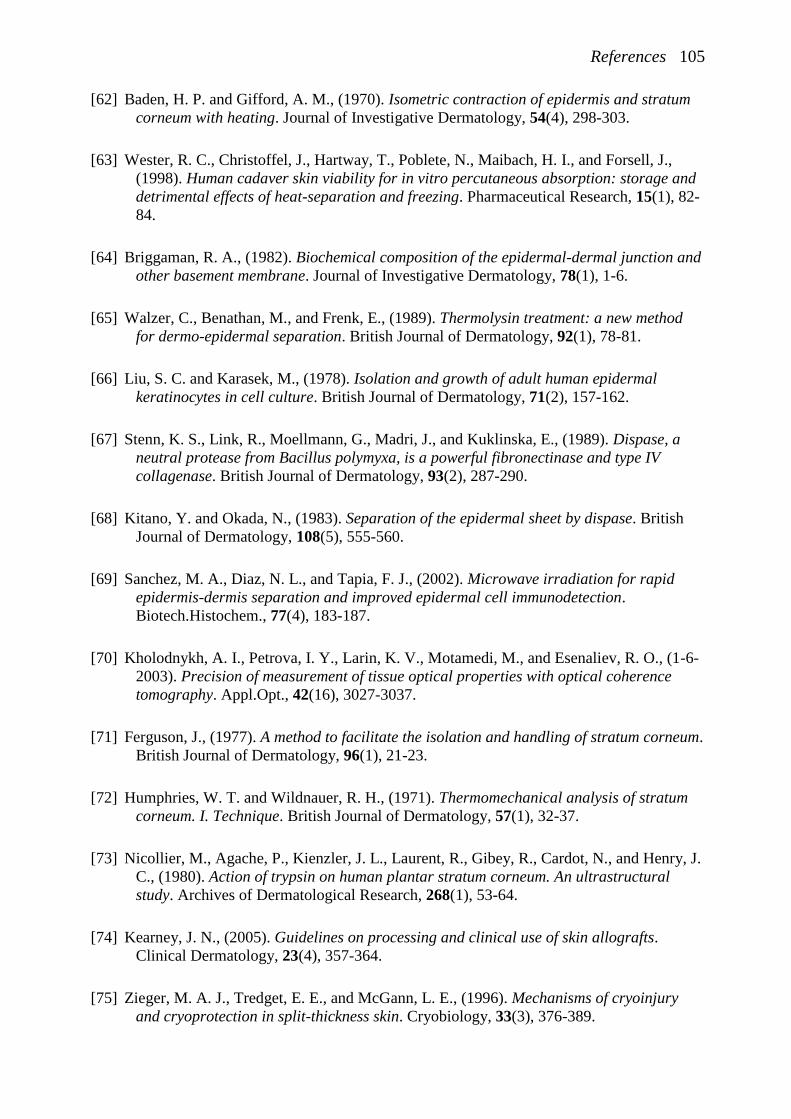
References 105
[62] Baden, H. P. and Gifford, A. M., (1970). Isometric contraction of epidermis and stratum
corneum with heating. Journal of Investigative Dermatology, 54(4), 298-303.
[63] Wester, R. C., Christoffel, J., Hartway, T., Poblete, N., Maibach, H. I., and Forsell, J.,
(1998). Human cadaver skin viability for in vitro percutaneous absorption: storage and
detrimental effects of heat-separation and freezing. Pharmaceutical Research, 15(1), 82-
84.
[64] Briggaman, R. A., (1982). Biochemical composition of the epidermal-dermal junction and
other basement membrane. Journal of Investigative Dermatology, 78(1), 1-6.
[65] Walzer, C., Benathan, M., and Frenk, E., (1989). Thermolysin treatment: a new method
for dermo-epidermal separation. British Journal of Dermatology, 92(1), 78-81.
[66] Liu, S. C. and Karasek, M., (1978). Isolation and growth of adult human epidermal
keratinocytes in cell culture. British Journal of Dermatology, 71(2), 157-162.
[67] Stenn, K. S., Link, R., Moellmann, G., Madri, J., and Kuklinska, E., (1989). Dispase, a
neutral protease from Bacillus polymyxa, is a powerful fibronectinase and type IV
collagenase. British Journal of Dermatology, 93(2), 287-290.
[68] Kitano, Y. and Okada, N., (1983). Separation of the epidermal sheet by dispase. British
Journal of Dermatology, 108(5), 555-560.
[69] Sanchez, M. A., Diaz, N. L., and Tapia, F. J., (2002). Microwave irradiation for rapid
epidermis-dermis separation and improved epidermal cell immunodetection.
Biotech.Histochem., 77(4), 183-187.
[70] Kholodnykh, A. I., Petrova, I. Y., Larin, K. V., Motamedi, M., and Esenaliev, R. O., (1-6-
2003). Precision of measurement of tissue optical properties with optical coherence
tomography. Appl.Opt., 42(16), 3027-3037.
[71] Ferguson, J., (1977). A method to facilitate the isolation and handling of stratum corneum.
British Journal of Dermatology, 96(1), 21-23.
[72] Humphries, W. T. and Wildnauer, R. H., (1971). Thermomechanical analysis of stratum
corneum. I. Technique. British Journal of Dermatology, 57(1), 32-37.
[73] Nicollier, M., Agache, P., Kienzler, J. L., Laurent, R., Gibey, R., Cardot, N., and Henry, J.
C., (1980). Action of trypsin on human plantar stratum corneum. An ultrastructural
study. Archives of Dermatological Research, 268(1), 53-64.
[74] Kearney, J. N., (2005). Guidelines on processing and clinical use of skin allografts.
Clinical Dermatology, 23(4), 357-364.
[75] Zieger, M. A. J., Tredget, E. E., and McGann, L. E., (1996). Mechanisms of cryoinjury
and cryoprotection in split-thickness skin. Cryobiology, 33(3), 376-389.
106 References
[76] Ben Bassat, H., (2005). Performance and safety of skin allografts. Clinical Dermatology,
23(4), 365-375.
[77] Meyer, W., Zschemisch, N. H., and Godynicki, S., (2003). The porcine ear skin as a
model system for the human integument: influence of storage conditions on basic
features of epidermis structure and function--a histological and histochemical study.
Polish Journal of Veterinary Sciences, 6(1), 17-28.
[78] De, A., Mathur, M., and Gore, M. A., (2008). Viability of cadaver skin grafts stored in
skin bank at two different temperatures. Indian Journal of Medical Research, 128(6),
769-771.
[79] Castagnoli, C., Alotto, D., Cambieri, I., Casimiri, R., Aluffi, M., Stella, M., Alasia, S. T.,
and Magliacani, G., (2003). Evaluation of donor skin viability: fresh and cryopreserved
skin using tetrazolioum salt assay. Burns, 29(8), 759-767.
[80] Han, B. and Bischof, J. C., (2004). Engineering Challenges in Tissue Preservation. Cell
Preservation Technology, 2(2), 91-112.
[81] Lawrence, J. C., (1972). Storage and skin metabolism. British Journal of Plastic Surgery,
25(4), 440-453.
[82] Foutz, T. L., Stone, E. A., and Abrams, Jr, (1992). Effects of freezing on mechanical
properties of rat skin. American journal of veterinary research, 53(5), 788-792.
[83] Bouwstra, J. A., (2009).
[84] Bouwstra, J. A., de Graaff, A., Gooris, G. S., Nijsse, J., Wiechers, J. W., and van Aelst, A.
C., (2003). Water distribution and related morphology in human stratum corneum at
different hydration levels. Journal of Investigative Dermatology, 120(5), 750-758.
[85] Chistolini, P., De Angelis, G., De Luca, M., Pellegrini, G., and Ruspantini, I., (1999).
Analysis of the mechanical properties of in vitro reconstructed epidermis: preliminary
results. Medical & Biological Engineering & Computing, 37(5), 670-672.
[86] Rochefort, A., Druot, P., Leduc, M., Vassalet, R., and Agache, P., (1986). A New
Technique for the Evaluation of Cosmetics Effect on Mechanical-Properties of Stratum-
Corneum and Epidermis Invitro. International Journal of Cosmetic Science, 8(1), 27-36.
[87] Geerligs, M., (2007). Mechanical behaviour of human skin layers: stratum corneum,
living epidermis and hypodermis. A literature review.PR-TN 2006/00450
[88] van Turnhout, M., Peters, G., Stekelenburg, A., and Oomens, C., (2005). Passive
transverse mechanical properties as a function of temperature of rat skeletal muscle in
vitro. Biorheology, 42(3), 193-207.
References 107
[89] Hrapko, M., van Dommelen, J. A., Peters, G. W., and Wismans, J. S., (2006). The
mechanical behaviour of brain tissue: large strain response and constitutive modelling.
Biorheology, 43(5), 623-636.
[90] van Dam, E. A., Dams, S. D., Peters, G. W., Rutten, M. C., Schurink, G. W., Buth, J., and
van de Vosse, F. N., (2006). Determination of linear viscoelastic behavior of abdominal
aortic aneurysm thrombus. Biorheology, 43(6), 695-707.
[91] Holt, B., Tripathi, A., and Morgan, J., (28-8-2008). Viscoelastic response of human skin to
low magnitude physiologically relevant shear. J.Biomech., 41(12), 2689-2695.
[92] Gennisson, J. L., Baldeweck, T., Tanter, M., Catheline, S., Fink, M., Sandrin, L.,
Cornillon, C., and Querleux, B., (2004). Assessment of elastic parameters of human skin
using dynamic elastography. Ieee Transactions on Ultrasonics Ferroelectrics and
Frequency Control, 51(8), 980-989.
[93] Park, A. C. and Baddiel, C. B., (1972). Rheology of Stratum Corneum .2. Physicochemical
Investigation of Factors Influencing Water-Content of Corneum. Journal of the Society
of Cosmetic Chemists, 23(1), 13-21.
[94] Wu, K. S., Li, J., Ananthapadmanabhan, K. P., and Dauskardt, R. H., (2007). Time-
dependant intercellular delamination of human stratum corneum. Journal of Materials
Science, 42(21), 8986-8994.
[95] vanHal, D. A., Jeremiasse, E., Junginger, H. E., Spies, F., and Bouwstra, J. A., (1996).
Structure of fully hydrated human stratum corneum: A freeze fracture electron
microscopy study. Journal of Investigative Dermatology, 106(1), 89-95.
[96] Warner, R. R., Stone, K. J., and Boissy, Y. L., (2003). Hydration disrupts human stratum
corneum ultrastructure. Journal of Investigative Dermatology, 120(2), 275-284.
[97] Ohura, T., Takahashi, M., and Ohura, N., Jr., (2008). Influence of external forces
(pressure and shear force) on superficial layer and subcutis of porcine skin and effects
of dressing materials: are dressing materials beneficial for reducing pressure and shear
force in tissues? Wound Repair and Regeneration, 16(1), 102-107.
[98] Pailler-Mattei, C., Pavan, S., Vargiolu, R., Pirot, F., Falson, F., and Zahouani, H., (2007).
Contribution of stratum corneum in determining bio-tribological properties of the
human skin. Wear, 263(7-12 SPEC. ISS.), 1038-1043.
[99] Hendley, A., Marks, R., and Payne, P. A., (1982). Measurement of forces for point
indentation of the stratum corneum in vivo: The influences of age, sex, site delipidisation
and hydration. Bioengineering and the Skin, 234-240.
[100] Pailler-Mattei, C. and Zahouani, H., (2004). Study of adhesion forces and mechanical
properties of human skin in vivo. Journal of Adhesion Science and Technology, 18(15-
16), 1739-1758.
108 References
[101] Potter, A., Luengo, G., Baltenneck, C., Pavan, S., and Loubet, JL, (1-7-2007). Measuring
mechanical properties of stratum corneum and isolated corneocytes at sub-micron
length scale
[102] Plewig, G. and Marples, R. R., (1970). Regional differences of cell sizes in the human
stratum corneum. I. Journal of Investigative Dermatology, 54(1), 13-18.
[103] Plewig, G., (1970). Regional differences of cell sizes in the human stratum corneum. II.
Effects of sex and age. Journal of Investigative Dermatology, 54(1), 19-23.
[104] Yuan, Y. and Verma, R., (2006). Measuring microelastic properties of stratum corneum.
Colloids and Surfaces B: Biointerfaces, 48(1), 6-12.
[105] Oliver, W. C. and Pharr, G. M., (1992). An Improved Technique for Determining
Hardness and Elastic-Modulus Using Load and Displacement Sensing Indentation
Experiments. Journal of Materials Research, 7(6), 1564-1583.
[106] Ebenstein, D. M. and Pruitt, L. A., (2006). Nanoindentation of biological materials.
Nano Today, 1(3), 26-33.
[107] Jachowicz, J., McMullen, R., and Prettypaul, D., (2007). Indentometric analysis of in
vivo skin and comparison with artificial skin models. Skin Research and Technology,
13(3), 299-309.
[108] Boyer, G., Laquize, L., Le Bot, A., Laquize, S., and Zahouani, H., (2009). Dynamic
indentation on human skin in vivo: ageing effects. Skin research and technology : official
journal of International Society for Bioengineering and the Skin (ISBS) [and]
International Society for Digital Imaging of Skin (ISDIS) [and] International Society for
Skin Imaging (ISSI), 15(1), 55-67.
[109] Iatridis, J. C., Wu, J., Yandow, J. A., and Langevin, H. M., (2003). Subcutaneous tissue
mechanical behavior is linear and viscoelastic under uniaxial tension. Connect.Tissue
Res., 44(5), 208-217.
[110] Haberman, E. and Gefen, A., (2007). Viscoelastic properties of ovine adipose tissue
covering the gluteus muscles as related to pressure ulcer modeling
[111] Hendriks, F. M., Brokken, D., van Eemeren, J. T. W. M., Oomens, C. W. J., Baaijens, F.
P. T., and Horsten, J. B. A. M., (2003). A numerical-experimental method to
characterize the non-linear mechanical behaviour of human skin. Skin Research and
Technology, 9(3), 274-283.
[112] Smith, S. R., Lovejoy, J. C., Greenway, F., Ryan, D., deJonge, L., de la Bretonne, J.,
Volafova, J., and Bray, G. A., (2001). Contributions of total body fat, abdominal
subcutaneous adipose tissue compartments, and visceral adipose tissue to the metabolic
complications of obesity. Metabolism-Clinical and Experimental, 50(4), 425-435.
References 109
[113] Lovejoy, J. C., Smith, S. R., and Rood, J. C., (2001). Comparison of regional fat
distribution and health risk factors in middle-aged white and African American women:
The Healthy Transitions Study. Obes.Res., 9(1), 10-16.
[114] Bray, G. A., Lovejoy, J. C., Smith, S. R., Delany, J. P., Lefevre, M., Hwang, D., Ryan,
D. H., and York, D. A., (2002). The influence of different fats and fatty acids on obesity,
insulin resistance and inflammation. Journal of Nutrition, 132(9), 2488-2491.
[115] Krouskop, T. A., Wheeler, T. M., Kallel, F., Garra, B. S., and Hall, T., (1998). Elastic
moduli of breast and prostate tissues under compression. Ultrasonic Imaging, 20(4),
260-274.
[116] sarvazyan, A. P., Skovoroda, A., and Emelianov, S, (1995). Biophysical bases of
elasticity imaging. Acoustic Imaging, 21223-240.
[117] Linder-Ganz, E., Shabshin, Noga, Itzchak, Yacov, and Gefen, Amit, (2007). Assessment
of mechanical conditions in sub-dermal tissues during sitting: A combined experimental-
MRI and finite element approach. Journal of Biomechanics, 40(7), 1443-1454.
[118] Klein, J., Permana, P. A., Owecki, M., Chaldakov, G. N., Bohm, M., Hausman, G.,
Lapiere, C. M., Atanassova, P., Sowinski, J., Fasshauer, M., Hausman, D. B., Maquoi,
E., Tonchev, A. B., Peneva, V. N., Vlachanov, K. P., Fiore, M., Aloe, L., Slominski, A.,
Reardon, C. L., Ryan, T. J., and Pond, C. M., (2007). What are subcutaneous adipocytes
really good for ...? Experimental Dermatology, 16(1), 45-70.
[119] Beynen, A. C. and Katan, M. B., (1985). Rapid Sampling and Long-Term Storage of
Subcutaneous Adipose-Tissue Biopsies for Determination of Fatty-Acid Composition.
American Journal of Clinical Nutrition, 42(2), 317-322.
[120] Tirrel, M., (1994). Rheology of Polymeric Liquids. In: Macosko, C. W. (eds), Rheology:
principles, measurements, and applications, Wiley-VCH, Inc, New york.
[121] Hrapko, M., Dommelen, J. A. W. v., Peters, G. W. M., and Wismans, J. S. H. M., (2005).
The mechanical behaviour of brain tissue: large strain response and constitutive
modeling59-69.
[122] van Turnhout, M., Peters, G., Stekelenburg, A., and Oomens, C., (2005). Passive
transverse mechanical properties as a function of temperature of rat skeletal muscle in
vitro. Biorheology, 42(3), 193-207.
[123] Verbeke, G. and Molenberghs, G., (2000). Linear Mixed Models for Longitudinal data, Springer, New York.
[124] Pipkin, A. C., (1986). Lectures on Viscoelasticity, Springer-Verlag, New York.
[125] Tanner, R. I., (2000). Engineering Rheology, Oxford University Press,
110 References
[126] Geerligs, M., Peters, G. W., Ackermans, P. A., Oomens, C. W., and Baaijens, F. P.,
(2008). Linear viscoelastic behavior of subcutaneous adipose tissue. Biorheology, 45(6),
677-688.
[127] Darling, E. M., Topel, M., Zauscher, S., Vail, T. P., and Guilak, F., (2008). Viscoelastic
properties of human mesenchymally-derived stem cells and primary osteoblasts,
chondrocytes, and adipocytes. J.Biomech., 41(2), 454-464.
[128] Then, C., Menger, J., Benderoth, G., Alizadeh, M., Vogl, T. J., Hubner, F., and Silber,
G., (2007). A method for a mechanical characterisation of human gluteal tissue.
Technology and Health Care, 15(6), 385-398.
[129] van Dam, E. A., Dams, S. D., Peters, G. W., Rutten, M. C., Schurink, G. W., Buth, J.,
and van de Vosse, F. N., (2008). Non-linear viscoelastic behavior of abdominal aortic
aneurysm thrombus. Biomech.Model.Mechanobiol., 7(2), 127-137.
[130] Mewis, J. and Wagner, N. J., (2009). Thixotropy. Advances in Colloid and Interface
Science, 147-48214-227.
[131] O'Neill, P. L. and Stachowiak, G. W., (1996). The inverse thixotropic behaviour of
synovial fluid and its relation to the lubrication of synovial joints. Journal of
Orthopaedic Rheumatology, 9(4), 222-228.
[132] Linder-Ganz, E. and Gefen, A., (2009). Stress analyses coupled with damage laws to
determine biomechanical risk factors for deep tissue injury during sitting. Journal of
Biomechanical Engineering, 131(1), 011003.
[133] Stekelenburg, A., Gawlitta, D., Bader, D. L., and Oomens, C. W., (2008). Deep tissue
injury: how deep is our understanding? Archives of Physical Medicine and
Rehabilitation, 89(7), 1410-1413.
[134] Avram, M. M., Avram, A. S., and James, W. D., (2005). Subcutaneous fat in normal and
diseased states: 1. Introduction. Journal of the American Academy of Dermatology,
53(4), 663-670.
[135] OECD, (13-4-2009). OECD Guideline for the testing of chemicals. Skin absorption: in
vitro MethodTest Guideline 428
[136] Garo, A., Hrapko, M., van Dommelen, J. A., and Peters, G. W., (2007). Towards a
reliable characterisation of the mechanical behaviour of brain tissue: The effects of
post-mortem time and sample preparation. Biorheology, 44(1), 51-58.
[137] Cox, M. A. J., Driessen, N. J. B., Boerboorn, R. A., Bouten, C. V. C., and Baaijens, F. P.
T., (2008). Mechanical characterization of anisotropic planar biological soft tissues
using finite indentation: Experimental feasibility. Journal of Biomechanics, 41(2), 422-
429.
References 111
[138] Hendriks, F. M., Oomens, C. W. J., Baaijens, F. P. T., and Brokken, D., (2004). Influence
of hydration and experimental length scale on the mechanical response of human skin in
vivo, using optical coherence tomography. Skin Research and Technology, 10(4), 231-
241.
[139] Bader, D. L. and Bowker, P., (1983). Mechanical characteristics of skin and underlying
tissues in vivo. Biomaterials, 4(4), 305-308.
[140] Bronneberg, D., (2007). Biochemical markers for early detection of superficial pressure
ulcers. Technische Universiteit Eindhoven.
[141] Cornelissen, L.H., (2008). Modeling the transport of biochemical markers in skin:
towards pressure ulcer assessment. Technische Universiteit Eindhoven.
[142] Gawlitta, D., (2006). Compression-induced factors influencing the damage of engineered
skeletal muscle. Technische Universiteit Eindhoven.
[143] Bosboom, E. M. H., (2001). Deformation as a trigger for pressure sore related muscle
damage. Technische Universiteit Eindhoven.
[144] Stekelenburg, A., (2005). Mechanisms associated with deep tissue injury induced by
sustained compressive loading. Technische Universiteit Eindhoven.
[145] Ceelen, K. K., (2008). Modeling the development of in vitro and in vivo pressure-induced
muscle damage, Technische Universiteit Eindhoven.
[146] Hendriks, R. and Lucassen, G., (2001). Two-photon fluorescence and confocal video
microscopy of in-vivo human skin4262287-293.
[147] Ericson, M. B., Simonsson, C., Guldbrand, S., Ljungblad, C., Paoli, J., and Smedh, M.,
(2008). Two-photon laser-scanning fluorescence microscopy applied for studies of
human skin. Journal of biophotonics, 1(4), 320-330.
[148] Branzan, A. L., Landthaler, M., and Szeimies, R. M., (2007). In vivo confocal scanning
laser microscopy in dermatology. Lasers in Medical Science, 22(2), 73-82.
[149] varez-Rom+ín, R., Naik, A., Kalia, Y. N., Fessi, H., and Guy, R. H., (2004).
Visualization of skin penetration using confocal laser scanning microscopy. European
Journal of Pharmaceutics and Biopharmaceutics, 58(2), 301-316.
[150] Palero, J. A., de Bruijn, H. S., van der Ploeg van den Heuvel, Sterenborg, H. J., and
Gerritsen, H. C., (1-8-2007). Spectrally resolved multiphoton imaging of in vivo and
excised mouse skin tissues. Biophysical Journal, 93(3), 992-1007.
[151] Dullaert, K. and Mewis, J., (2005). A model system for thixotropy studies. Rheologica
Acta, 45(1), 23-32.

112 References
[152] Dullaert, K. and Mewis, J., (2005). Thixotropy: Build-up and breakdown curves during
flow. Journal of Rheology, 49(6), 1213-1230.