skin infections viruses bacteria fungi. viral infections
TRANSCRIPT

SKININFECTIONS
• VIRUSES• BACTERIA• FUNGI

VIRAL INFECTIONS
VIRAL WARTS/VERRUCA
ETIOLOGY• Human papilloma virus-DNA virus• HPV-1, 2 and 4- common warts• HPV-3- plane warts• HPV-6, 11, 16 and 18- genital warts
Epidemiology• Age :
Nongenital wart: children and young adults Anogenital warts: adolescents and adults
• Transmission:Nongenital :direct skin to skin contact Anogenital: sexual transmission
vertical transmission-laryngeal papillomas in infant
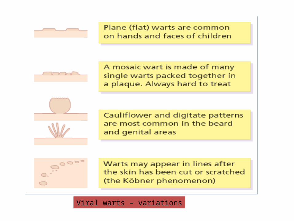
Viral warts – variations

PRESENTATIONCommon wart/verruca vulgaris• The first sign is a smooth skin-colored papule, • Often more easily felt than seen. • As the lesion enlarges, its irregular hyperkeratotic
surface give it the classic ‘warty’ appearance. • Common warts usually occur on the hands but
are also often on the face and genitals. • They are more often multiple than single. • Pain is rare.

Typical common warts on the fingers
MOSAIC WARTS/SUPERFICIAL PALMOPLANTAR WARTS
• Rough marginated plaques• Are made up of many small, tightly
packed but discrete individual warts. • They are most common on the soles but
are also seen on palms and around finger nails.
• Usually they are not painful.
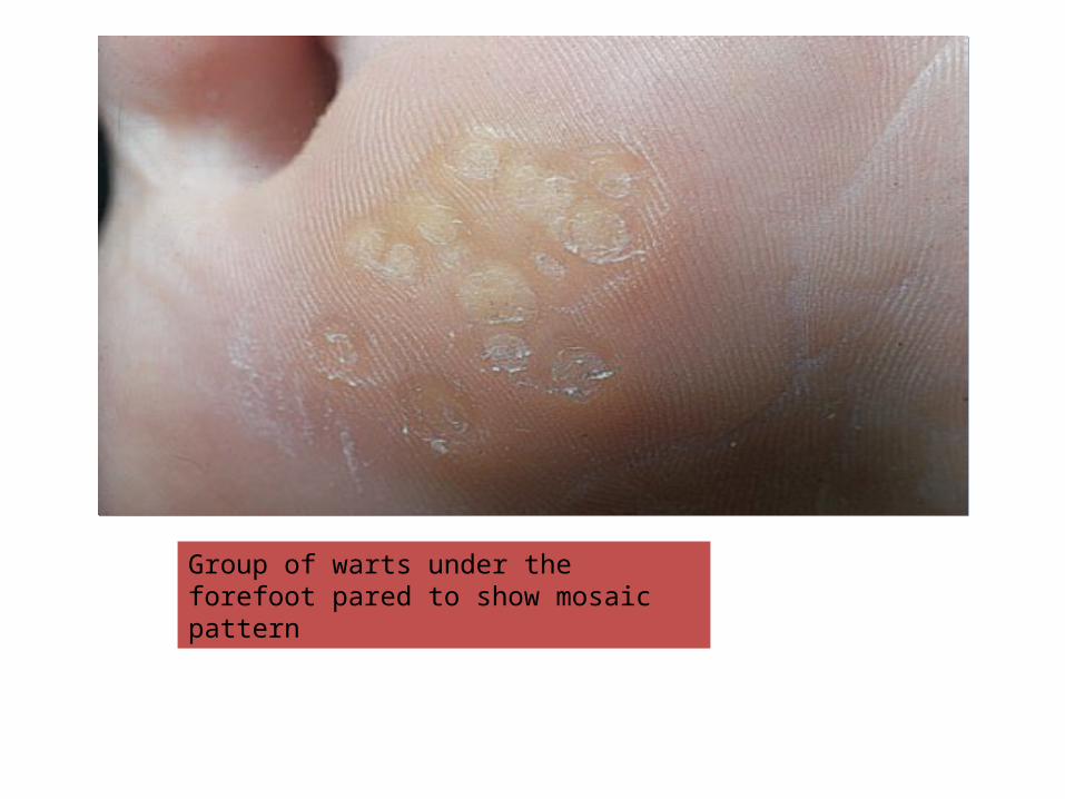
Group of warts under the forefoot pared to show mosaic pattern

DEEP PALMOPLANTAR WARTS
• These have a rough surface• Protrudes only slightly from the skin • Is surrounded by a horny collar • On paring, punctate black dots representing
thrombosed capillary loops are seen which allows plantar warts to be distinguished from corns.
• Often multiple• Plantar warts can be painful.

Solitary plantar wart on the heel

PLANE WARTS/VERRUCA PLANA
• These are smooth flat-topped papules • Are most common on the face and brow,
on the backs of the hands and on the shaven legs of women.
• Usually skin-colored or light brown• Lesions are multiple, painless and are
sometimes arranged along a scratch line(koebner’s phenomenon)
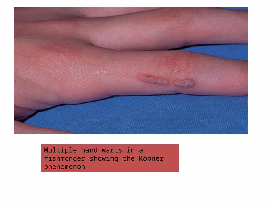
Multiple hand warts in a fishmonger showing the Köbner phenomenon
Filiform warts
• Asymptomatic thin dlongated firm projections arising from a horny base• Freqently over face (inoculation
by shaving )and scalp
EPIDERMODYSPLSSIA VERRUCIFORMIS
• Rare inherited disorder• Defective cell mediated immunity to certain
types of HPV(3,5,8,9)• Two types• Planter warts like lesion-confluent on face
and acral parts• Ptyriasis versicolor like irregular scaly
macules on trunk

ANOGENITAL WARTS (CONDYLOMA ACUMINATA)
• Sexually transmitted disease• Papillomatous cauliflower-like lesions• Can appear anywhere in this area. • They may coalesce to form huge fungating
plagues causing discomfort and irritation. • The vaginal and anorectal mucosae may be
affected.
COURSE• Warts resolve spontaneously in the healthy as the
immune response overcomes the infection. • This happens within 6 months in some 30% of patients,
and within 2 years in 65%. • Such spontaneous resolution, sometimes heralded by a
punctate blackening caused by capillary thrombosis• Leaves no trace. • Mosaic warts are notoriously slow to resolve and often
resist all treatments. • Warts persist and spread in immunocompromised
patients • Seventy per cent of renal allograft recipients will have
warts 5 years after transplantation.

COMPLICATIONS
• Pain• Malignant change is otherwise rare,
although infection with HPV types 16 and 18 predisposes to cervical carcinoma. HPV infections in immunocompromised patients have also been linked with skin cancer, especially on light-exposed areas.

DIFFERENTIAL DIAGNOSIS• Molluscum contagiosum are smooth, domeshaped and pearly, with central umbilication.
• Plantar corns are found on pressure areas; there is no capillary bleeding on paring. They have a central keratotic core and are painful.
• Granuloma annulare lesions have a smooth surface, as the lesions are dermal, and their outline is often annular.
• Condyloma lata are seen in syphilis. They are rare but should not be confused with condyloma acuminata (warts). The lesions are flatter, greyer and less well defined. If in doubt, look for other signs of secondary syphilis and carry out serological tests.
• Amelanotic melanomas, squamous cell carcinomas and other epithelial malignancies can present as verrucose nodules

TREATMENT
• Many warts give no trouble, need no treatment and go away by themselves.
• Destruction by cryotherapy is less likely to cause scars • Excision or electrosurgery.• Verruca vulgaris: cryotherapy, electrocautery, wart
paint(salicylic acid +lactic acid),mechanical removal ,RFA( radiofrequency abalation)
• Palmoplantar: wart paint(used daily for 3 mths), cryotherapy, formalin soaks(4%)
• Filiform: electric cautery, RFA• Epidermodysplasia verruciformis: oral acitretin• Anogenitalwarts: cryotherapy• Plane warts : retinoic acid topically (keratolytic )

HERPES ZOSTER• Shingles is caused by the herpes virus varicella-
zoster.• An attack is a result of the reactivation of virus
that has remained dormant in a sensory root ganglion since an earlier episode of chickenpox (varicella)
• Predisposiing factors for reactivation are:old age, lymphoreticular malignancies(hodgkin’s diseade,lumphoma), HIV, sometimes without apparent cause.
PRESENTATION AND COURSE
• Attacks usually start with a burning pain (excruciating)• Followed by erythema and grouped, sometimes blood-filled, vesicles scattered over a dermatome. • The clear vesicles quickly become purulent• After few days burst and crust. Crust fall off in abt a
fortnight and lesions heal with no or minimal scarring• Mucosa within affected dermatome may be involved• Draining lymph node are often enlarged.• Zoster is characteristically unilateral• Thoracic intercostal nerves,opthalmic division of the
trigeminal nerve and other spinal nerves are most frequently affected.
COMPLICATIONS• Secondary bacterial infection• Motor nerve involvement is uncommon, but
paralysis of ocular muscles, the facial muscles, the diaphragm and the bladder has been seen.
• Zoster of the ophthalmic division of the trigeminal nerve can lead to corneal ulcers and scarring.eye involvement is indicated when vesicles are present on the side of the nose(Hutchison’s sign)
• Post herpetic neuralgia:Persistent neuralgic pain, after the acute episode is over, is most common in the elderly.

INVESTIGATIONS• Biopsy or Tzanck smears show multinucleated
giant cells and a ballooning degeneration of keratinocytes, indicative of a herpes infection.
• Cultures are of little help as they take 5–7 days, and are only positive in 70% of cases.
• Rule out underlying immunodeficiency
TREATMENT• Systemic treatment should start within the first 5
days of an attack• Famciclovir and valaciclovir are as effective as
aciclovir (using them early cuts down the chance of post-herpetic neuralgia)
• If diagnosed late in the course of the disease, systemic treatment is not likely to be effective – treatment should be supportive with rest, analgesics and
bland applications such as calamine.
• Secondary bacterial infection should be treated with antibiotics.
• Prevention is better than cure.
• Vaccination of elderly patients with a live attenuated vaccine to the varicella-zoster virus has been shown to reduce the incidence of –herpes zoster and –postherpetic neuralgia
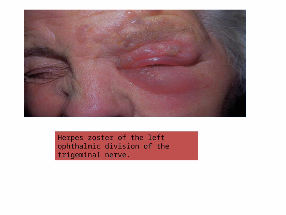
Herpes zoster of the left ophthalmic division of the trigeminal nerve.
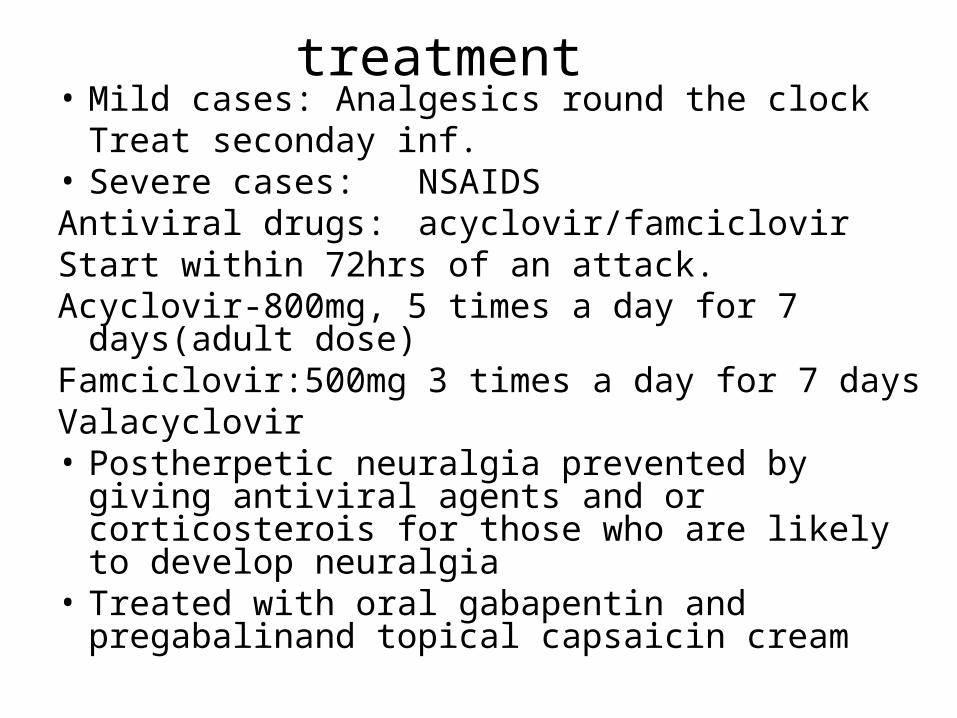
treatment• Mild cases: Analgesics round the clock
Treat seconday inf. • Severe cases: NSAIDSAntiviral drugs: acyclovir/famciclovirStart within 72hrs of an attack.Acyclovir-800mg, 5 times a day for 7 days(adult dose)Famciclovir:500mg 3 times a day for 7 days Valacyclovir• Postherpetic neuralgia prevented by giving
antiviral agents and or corticosterois for those who are likely to develop neuralgia
• Treated with oral gabapentin and pregabalinand topical capsaicin cream

HERPES SIMPLEXCause• Herpesvirus hominis
Two types • The lesions caused by type II virus occur
mainly on the genitals• those of type I are usually
extragenital(herpes labialis and other infection above the waist)
• Transmission• Direct contact or sexual contact• After the episode associated with the
primary infection– the virus may become latent, possibly within
sensory nerve ganglia– still capable of giving rise to recurrent bouts
of vesication–Viral shedding also occurs in the absence of
clinical lesions(asymptomatic shedding)

PRESENTATION• First episode• Primary type I infection in children asymptomatic or
an acute gingivostomatitis accompanied by malaise, headache, fever and enlarged cervical nodes.
• Vesicles, soon turning into ulcers, can be seen scattered over the lips and mucous membranes.
• The illness lasts about 2 weeks.• Primary type II virus infections, usually transmitted
sexually, asymptomatic or cause multiple and painful genital or perianal blisters which rapidly ulcerate.
• Constitutional symptoms and lymph node enlargement (inguinal) may be present
RECURRENT (RECRUDESCENT) INFECTIONS
• These strike in roughly the same place each time.
• may be precipitated by respiratory tract infections (cold sores), ultraviolet radiation, menstruation or even stress.
• Common sites include the face, the lips (type I) and the genitals (type II), but lesions can occur anywhere.
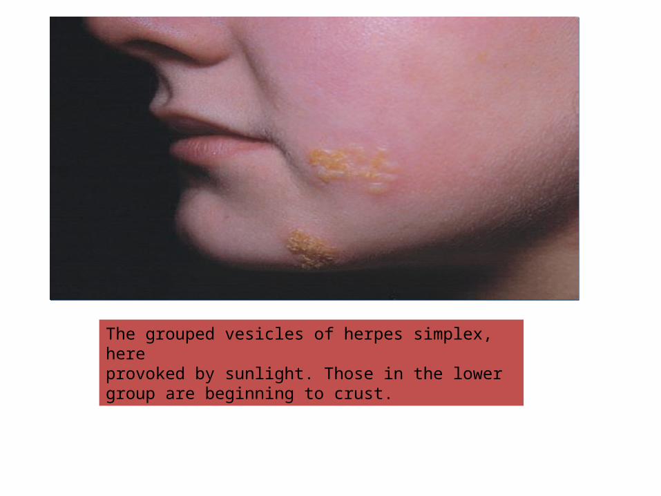
The grouped vesicles of herpes simplex, hereprovoked by sunlight. Those in the lower group are beginning to crust.
COURSE
• Tingling• Burning• Pain• Followed within a few hours by the
development of erythema and clusters of tense vesicles.
• Crusting occurs within 24–48 h • The whole episode lasts about 12 days.

COMPLICATIONS
• Herpes encephalitis or meningitis• Disseminated herpes simplex• Eczema herpeticum• Recurrent dendritic ulcers leading to
corneal scarring• Erythema multiforme
INVESTIGATIONS• None are usually needed. • Tzanck smear shows multinucleated giant
cells• Doubts over the diagnosis can be dispelled
by culturing the virus from vesicle fluid. • Antibody titres rise with primary, but not
with recurrent infections.• D/D• Herpes zoster

TREATMENT• Aciclovir cream, applied five or six times a day
for the first 4 days of the episode, may cut down the length of attacks.
• Aciclovir 200 mg five times daily for 5 or 7 days, this is usually reserved for those with widespread or systemic involvement.
• Famciclovir250mg 3times daily for 7days and valaciclovir1gm twice daely for 7 days are metabolized by the body into aciclovir and are as effective as aciclovir
• Reccurent disease: episodic Aciclovir 200 mg five times daily for 5 days or suppressive treatment if >6episodes per year with acyclovir 400mg twice daily for 12mths
• If complication rx with parenteral acyclovir
MOLLUSCUM CONTAGIOSUM
Cause• Common pox virus infection• Spread by• direct contact or sexually or by
sharing a towel at the swimming bath-fomites).

PRESENTATION AND COURSE• Common site: any part of body, anogenital region or
wide spread and several lesions seen in atopis and immunocompromised patients
• Individual lesions are shiny, white or pink, and hemispherical
• They grow slowly up to 0.5 cm in diameter.• A central punctum, which may contain a cheesy core,
gives the lesions their characteristic umbilicated look.• Multiple lesions• Untreated lesions usually clear in 6–9 months, often
after a brief local inflammation.• Large solitary lesions may take longer. Some leave
depressed scars.
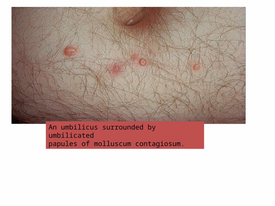
An umbilicus surrounded by umbilicatedpapules of molluscum contagiosum.
• Complication• Secondary Inf.
• Differential diagnosis• Boil• Keratocanthoma• Intradermal naevus• Cystic basal cell carcinoma
INVESTIGATIONS
• None
• Diagnosis can be confirmed by looking under the microscope for large eosinophilic intracytoplasmic inclusion bodies.
• Extensive mollusa may suggest need for HIV testing

TREATMENT
• Few lesion may resolve spontaneously• Liquid nitrogen, wart paints and topical
imiquimod may be helpful.• Mecanical removal after local anesthetics• Mechanical expression followed by
chemical cautery• Cryotherpay