ske ieta:! sys;t,em:th sku iikgobalet/files/bio351/kardong 2006 ch 7 skeletel... · components, the...
TRANSCRIPT
Kardong (2006) Chapter 7: 234-288 . ,.,
Ske Ieta:! Sys;t,em:Th e"Sku II . _ ' "~P' '.
\ .- t -';..--: '-."....... .....
INTRODU CTION
CHONDROCRAN1UM
Embryology
SPLANCHNOCRANIUM
Embryology Origin of Jaws Types of Jaw Attachments
DERMATOCRANIUM
Pans of the Dermatocranium Dermal Bone Series
OVERV1EW OF SKULL MORPHOLOGY
Braincase laws Hyoid Apparatus
CRAN1AL K1NES1S
PHYLOGENY O F THE SKULL
Agnathans Ostracoderms Cyclostomes
The skeleton gives the vertebrate body shape, supports its weight, offers a system of levers that together with muscles produces movement, and protects soft parts such as nerves, blood vessels, and other viscera. Because it is hard, bits of the skeleton often survive fossilization better than does soft tissue anatomy; so our most direct contact with long-extinct animals is often through their skeletons. The story of vertebrate function and evolution is written in the architecture of the skeleton.
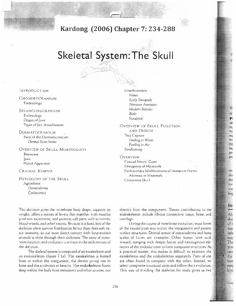
The skeletal system is composed of an exoskeleton and an endoskeleton (figure 7.1a) . The exoskeleton is formed from or within the integument, the dermis giving rise to bone and the epidermis to keratin. The endoskeleton forms deep within the body from mesoderm and other sources, not
Gnathostomes Fishes Early Tetrapods Primitive Amniotes
Modem Reptiles Birds Synapsids
ve OVERVIEW OF SKULL FUNCTION to,
AND DESIGN m;
Prey Capture SUI
Feeding in Water thl
Feeding in Air (b) sefSwallowing po:
OVERVIEW an(
Cranial Neural Crest Emergence of Mammals Evolutionary Modifications of Immature Forms: un
Akinesis in Mammals skI Composite Skull thE
tur ioe
bef
Int directly from the integument. Tissues contributing to the endoskeleton include fibrous connective tissue, bone, and cartilage. sku
During the course of vertebrate evolution, most bones of t of the exoskeleton stay within the integument and protect sep, surface structures. Dermal armor of osrracoderms and bony spla scales of fishes are examples. Other bones have sunk inward, merging with deeper bones and cartilaginous elements of the endoskeleton to form composite structures. As a practical matter, this makes it difficult to exa mine the exoskeleton and the endoskeleton separately. PartS of one are often found in company with the other. Instead, we select composite structural units and follow their evolution. This way of dividing the skeleton for study gives us two
234
---------------
---------------
Skeleton
Exoskeleton Endoskeleton (within the integument) (deep, within the body)
' ~ Bo~chord / ~ endoskeleton endoskeleton
Bony exoskeleton
(from dermis)
Skeleton
~ Postcranial
Axial Appendicular skeleton skeleton ~ ~
Notochord limbs Girdle
Dermatocranium
Organization of skeletal tissues in !itiibr;ates. Components of the skeletal system function
as a unit but, as a convenience. they can be divided into parts for closer analysis. (a) As a protective and
system. the skeleton can be divided into structures on (eXOSkeleton) and inside (endoskeleton) of the body
the basis of position. the skeleton can be treated as two
components, the cranial skeleton (skull) and the
ial skeleton. The postcranial skeleton includes the axial skeletons.
the skull, or cranial skeleton, and the postcranial (figure 7.1b). The postcranial skeleton includes
Wp,rtpi)r<l' column, limbs, girdles, and associated strucsuch as ribs and shells. In chapters 8 and 9, we exam
postcranial skeleton. Our discussion of the skeleton with the skull.
merged into a harmonious unit, the vertebrate cranium, is actually a composite structure formed
t parts. Each part of the skull arises from a phylogenetic source. The most ancient part is the
rn"'()r,r~nium (visceral cranium), which first arose to I slits in protochordates (figure 7.2a).
part, the chondrocranium, underlies and sup-brain and is formed of endochondral bone or of or both (figure 7 .2b). The third part of the skull is
a contribution that in later vertemost of the outer casing of the skull. As its
the dermatocranium is composed of dermal 7.2c) .
Endochondral and dermal bone (p. 179)
In addition to these formal components, two general terms apply to parts of the cranium. The braincase is a collective term that refers to the fused cranial components immediately surrounding and encasing the brain . Structures of the dermatocranium, the chondrocranium, and even the splanchnocranium can make up the braincase, depending on the species. The neurocranium is used as an equivalent term for the chondrocranium by some morphologists. Others expand the term to include the chondrocranium along with fused or attached sensory capsules-the supportive nasal, optic, and otic capsules. Still others consider the neu
rocranium to be only the ossified parts of the chondrocra-j n,ium. Be prepared for slightly different meanings in the literature. Although we use the term neurocranium sparingly, neurocranium is understood to include the braincase (ossi fied or not) plus associated sensory capsules.
Chondrocranium
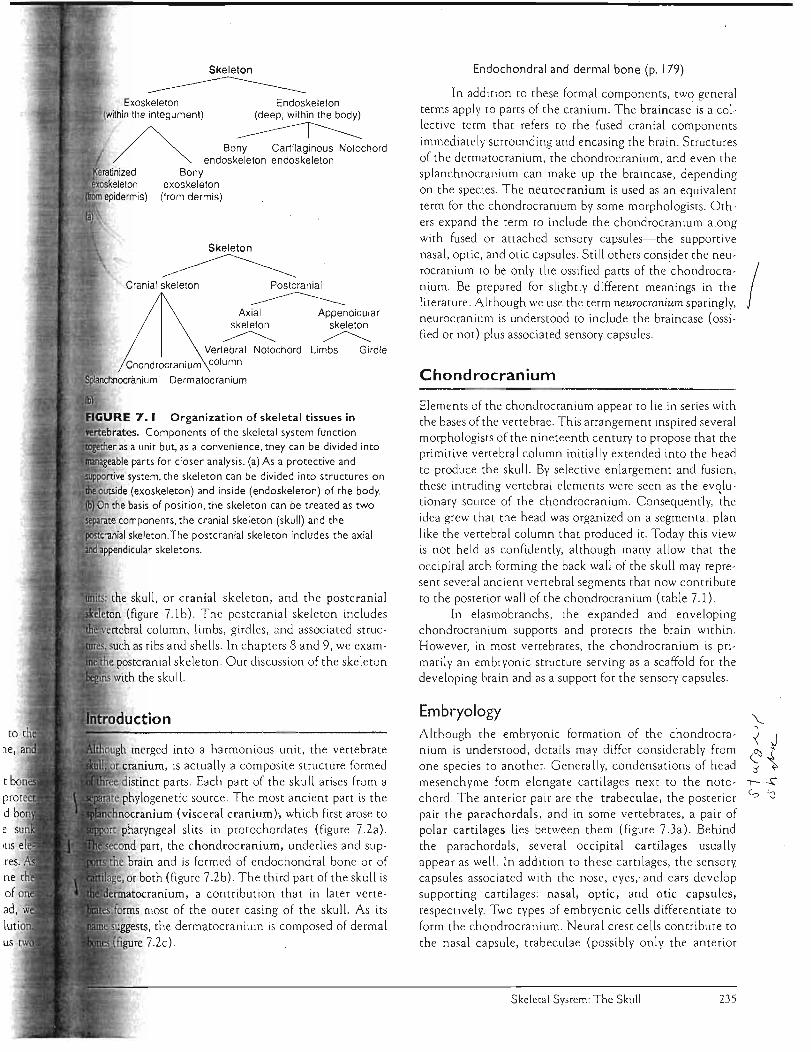
Elements of the chondrocranium appear to lie in series' with the bases of the vertebrae. This arrangement inspired several morphologists of the nineteenth century to propose that the primitive vertebral column initially extended into the head to produce the skull. By selective enlargement and fusion, these intruding vertebral elements were seen as the ev«lutionary source of the chondrocranium. Consequently, the idea grew that the head was organized on a segmental plan like the vertebral column that produced it. Today this view is not held as confidently, although many allow that the occipital arch forming the back wall of the skull may repre sent several ancient vertebral segments that now contribute to the posterior wall of the chondrocranium (table 7.1).
In elasmobranchs, the expanded and enveloping chondrocranium supports and protects the brain within, However, in most vertebrates, the chondrocranium is primarily an embryoniC structure serving as a scaffold for the developing brain and as a support for the sensory capsules.
Embryology Although the embryonic formation of the chondrocranium is understood, details may differ considerably from one species to another. Generally, condensations of head mesenchyme form elongate cartilages next to the notochord. The anterior pair are the trabeculae, the posterior pair the parachordals, and in some vertebrates, a pair of polar cartilages lies between them (figure 7.3a). Behind the parachordals, several occipital cartilages usually appear as well. In addition to these cartilages, the sensory capsules associated with the nose, eyes ,. and ears develop supporting cartilages: nasal, optic, and otic capsules, respectively. Two types of embryonic cells differentiate to form the chondrocranium. Neural crest cells contribute to the nasal capsule, trabeculae (possibly only the anterior
Skeletal System: The Skull 235

Suprabranchialis
Epibranchials
-----"--- PalatineHyomandibula Sympletic -__.=1
. Basipterygoid Nasal process opening
(b) Chondrocranium
Prearticular Angular
(a) Splanchnocranium ./Hyomandibula
Opercular
Subopercular Dentary
Supratemporal Postorbital Intertemporal
Parietal Poslfrontal
Prefrontal Frontal Nasal Premaxilla
Preopercular
Lacrimal JugalSubopercular Squamosal
Quadratojugal Surangular
Submandibular branchiostegal plate
(c) Dermatocranium
FIGURE 7.2 Composite skull. The skull is a mosaic composed of three primary contributing parts: the chondrocranium, the splanchnocranium, and the dermatocranium. Each has a separate evolutionary background. The skull of Eusthenopteron. a Devonian rhipidistian fish, illustrates how parts of all three phylogenetic sources contribute to the unit. (a) The splanchnocranium (yellow) arose first and is shown in association with the chondrocranium (blue) and parts of the dermatocranium (red).The right mandible is lowered from its point of articulation better to reveal deeper bones. (b) The chondrocranium in Eusthenopteron is formed by the union between the
anterior ethmosphenoid and the posterior oticooccipital units. (c) The superficial wall of bones composes the dermatocranium. The central figure depicts the relative position of each contributing set of bones brought together in the composite skull. (Sac: nasal series)
Chapter Seven 236
Amphibians
Supraoccipital Supraoccipital Exoccipital Exoccipital Basioccipital Basioccipital
Mesethmoid' Absent (internasal)
Ossified Unossified
Sphenethmoid Sphenethmoid Orbitosphenoid Orbitosphenoid [Basisphenoidt Basisphenoid Pleurosphenoid ?
{ Prootic Prootic Epiotic OPisthotic Sphenotic
(rom the splanchnocranium contributes.
Nasal capsule
Trabecula ~ _A ,
OPtIC~~~ capsu~e_~ " ~
Polar cartilage ---='----<> ~ Otic capsule ~~.• ·. ~o
Parachordal . . e
(a)
Occipitals
Notochord
(b)
Reptiles/Birds
Supraoccipital Exoccipital . Basioccipital
Absent
Unossified
Sphenethmoid Orbitosphenoid Basisphenoid Pleurosphenoid
(crocodilians. amphisbaenians)
Laterosphenoid (snakes)
Prootic }Opisthotic EPiotic
(absent in birds)
(c)
Mammals
Supraoccipital } Occipital bone Exoccipital
Basioccipital
Mesethmold
I,b,,", '" p,;m'''' ) mammals. ungulates) Ethmoid
Turbinals (ethmo-. naso-. maxilio-)
Presphenoid } Orbitosphenoid Sphenoid' Basisphenoid Absent
-Absent
Petrosal with mastoid process
~=-, Basisphenoid
--If---+--+- BaSioccipital
Supraoccipital
Exoccipital
Embryonic development of the chondrocranium. Cartilage (blue) appears first but in most vertebrates is
by bone (white) later in development.The chondrocranium includes these cartilaginous elements that form the base and back of
together with the supportive capsules around sensory organs . Early condensation of mesenchymal cells differentiates into
(a) that grows and fuses together to produce the basic ethmoid. basal. and occipital regions (b) that later OSSify (c) . forming basic
and perhaps to part of the otic capsule (figure 7 Aa). ",p'T1cn Vrnf> of mesodermal origin contributes to the rest
chondrocranium (figure 7Ab). As development pro, these cartilages fuse. The region between the nasa l
formed by the fusion of the anterior tips of the trais the ethmoid plate. The parachordals grow
r across the midline to form the basal plate between capsules. The occipitals grow upward and around
the nerve cord to form the occipital arch (figure 7.3b). Collectively, all of these expanded and fused cartilages constitute the chondrocranium. .
In elasmobranchs, the chondrocranium does not ossify. Instead the cartilage grows st ill farther upward and over the brain to complete the protective walls and roof of the braincase. In most other vertebrates, the chondrocranium becomes partly or entirely ossified (figu re 7.3c).
Skeletal System: The Skull 237
Neural crest
Myelencephalon
mesenchyme Neural crest- Neural crest Mesodermal
Sphenoid Lacrimal
Nasal e::?'~~'irl~'v Jugal
§
Maxilla Premaxilla
(c) (d)
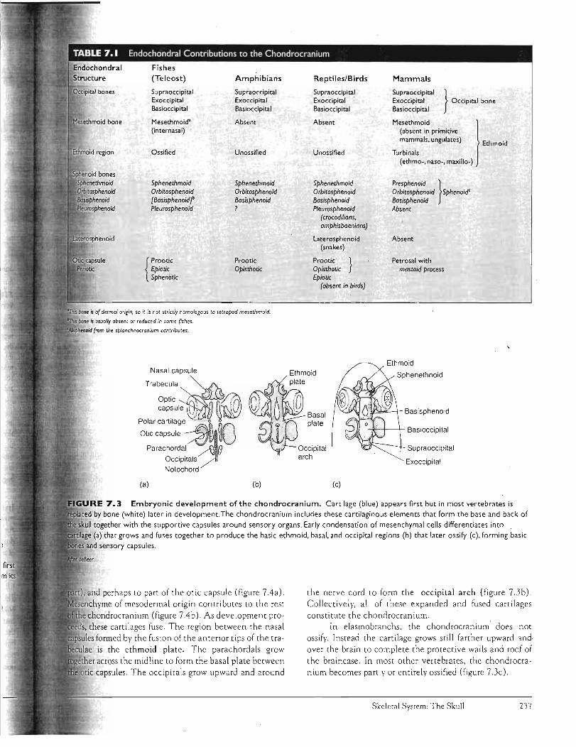
FIGURE 7.4 Neural crest contributions to the skull. (a) Salamander embryo illustrating the sequential spread of neural crest cells. During early embryoniC development, neural crest cells contribute to the head mesenchyme, which is called the ectomesoderm because of its neural crest origin. (b) Also contributing to the head mesenchyme are cells of mesodermal origin, the mesodermal mesenchyme. The position of the mesodermal (stippled) and the neural crest (shaded) mesenchyme, and the approximate interface between them, are indicated in the chick embryo. Skull of a chick (c) and a human fetus (d) show bones or portions of bones derived from neural crest cells (shaded).Abbreviations: angular (An), basibranchial (8b), basihyal (Bh), basisphenoid (Bs), ceratobranchial (Cb), dentary (D), epibranchial (Eb), entoglossum (Eg), exoccipital (Eo), ethmoid (Eth), frontal (F), jugal 0), nasal (N), cartilage nasal capsule (Nc), parietal (P), palatine (PI), premaxilla (Pm), postorbital (Po), prefrontal (Prf), parasphenoid (Ps), pterygOid (Pt), quadrate (Q), scleral ossicle (Sci),
~.
OQpha~",
Q (a) (b)
Po
supraoccipital (Soc), squamosal (Sq), stapes (Stp).
After Noden.
Splanchnocranium
The splanchnocranium is an ancienr chordate srructure. In amphioxus, the splanchnocranium, or at least its forerunner, is associated with the filter-feeding surfaces.
Among vertebrates, the splanchnocranium generally supports the gills and offers attachment for the respiratory muscles. Elements of the splanchnocranium contribute to th.e jaws and h.yoid apparatus of gnathostomes.
Embryology The mistaken view that the splanchnocranium developed from the same embryonic source as the walls of the digestive tract inspired the name "visceral" cranium, a name that unfortunately has stuck despite being a misnomer. Embryologically, the splanchnocranium arises from neural crest cells, not from lateral plate mesoderm like the smooth muscle in the walls of the digestive rract. In protochordates, neural crest cells are absent. Pharyngeal bars, composed of fibrous connective tissue, but never bone or cartilage, arise from mesoderm and form the unjointed branchial basket,
Chapter Seven
mesenchyme
Mesencephalon
Diencephalon
=
the phylogenetic predecessor of the vertebrate splanchnocranium. In vertebrates, cells of the neu'ral crest depart from the sides of the neural tube and move inro the walls of the pharynx between successive pharyngeal slits to differentiate into the respective pharyngeal arches. Ph.a arches of aquatic vertebrates usually are associated with respiratory gill system. Because of this association, they referred to as branchial arches, or gill arches.
Each arch can be composed of a series of up to
articulated elements per side, beginning with the p gobranchial element dorsally and then, in order, the epibranchial, ceratobranchial, hYPoDlranlCnllal"Jl and basibranchial elements (figure 7.5). One or more these anterior branchial arches may come ro border mouth, support soft tissue, and bear teeth. Branchial arc that suppOrt the mouth are called jaws, and each tributing arch is numbered sequentially or named. The fully functional arch of the jaw is the mandibular arch, largest and most anrerior of the modified series of a The mandibular arch is composed of the dorsally and Meckel's cartilage (mandibular cartilage)
238

Branchial arches
Hyomandibula Palatoquadrate
Meckel's cartilage
Hyoid Mandibular arch arch . j
Primitive splanchnocranium. Seven
are shown. Up to five elements compose an arch on each
beginning with the pharyngobranchial dorsally and in .[t~qUence to the basibranchials most ventrally. The first two
~!co"nDlete arches are named: mandibular arch for the first and
arch for the second that supports it. The characteristic five
elements are reduced to just two in the mandibular arch: the
rate and Meckel's cartilage. The large hyomandibula, from an epibranchial element, is the most prominent
nt of the next arch, the hyoid arch. Behind the hyoid variable numbers of branchial arches I, II , and so on.
The hyoid arch, whose most prominent element is hyomandibula, follows the mandibular arch. A varying
of branchial arches, often designated with roman , follow the hyoid arch (figure 7.5).
agnathans, the mouth is neither defined nor supported Instead, the sp lanchnocranium supports the roof of
pharynx and lateral pharyngea l slits. Lacking jaws, would have been restricted to a diet of small,
te food. The c iliary-mucous feeding surfaces of pro",,-"V"",1l'CO probably continued to playa large part in the
technique of ostracoderms. In some gro ups, teeth I ike structures, derived from surface scales, sur
the mouth. Perhaps ostracoderms used these rough to scrape rock surfaces and dislodge encrusted algae r organisms. As these food particles became susin water, ostracoderms drew them into their mouth
the incurrent flow of water. The mucus-lined walls of collected these dislodged food particles from
stream. Jaws appear first in acanthodian and placoderm fishes
used them as food traps to grab whole prey or take bites large prey. Within some groups, jaws also served as
or chewing devices to process food in the mouth. the advent of jaws, these fishes became more free-
predators of open waters. Jaws arose from one of the anterior pair of gill arches.
supporting this comes from several sources. First, of sharks suggests that Jaws and branchial
. ~
~(j~ {] ...~
~%~~~ l '( )
Branchial arches
Ch
Pq
Mk
FIGURE 7.6 Shark embryo, the dogfish Scyllium, Jaws
appear to be in series with the branchial arches. The mandibular
arch is first, followed by the hyoid and then several branchial
arches. Such a position of the jaws, in series with the arches, is taken as evidence that the jaws derive from the most anterior
branchial arch. Abbreviations: ceratohyal (Ch), hyomandibula (Hy), Meckel's cartilage (Mk), neural arch (Ne), occipital arch (Oa),
orbital cartilage (Oc), polar cartilage (Pc), palatoquadrate (Pq),
trabecula (Tr). Labial cartilages are not included.
After deBeer.
arches develop similarly in series (figure 7.6) and both'arise from neural crest. The spiracle appears to have once been a full-sized gill slit, but in modern sharks it is crowded and much reduced by the enlarged hyoid arch next in series. Furthermore , nerves and blood vessels are distributed in a pattern similar to branchial arches and jaws. Finally, the musculature of the jaws appears to be transformed and mod ified from branchial arch musculature.
So it seems reasonable to conclude that branchial arches phylogenetically gave rise to jaws. But the specifics remain controversial. For example, we are not sure whether jaws represent derivatives of the first, second , third, or even fourth branchial arches of primitive ancestors. Derivation of the mandibular arch also excites some controversy. The serial theory is the simplest view and holds that the first or perhaps second ancient branchial arch gave rise exclusively to the mandibular arch, the next branchial arch exclusively to the hyoid arch, and the rest of the arches to the branchial arches of gnathostomes (figure 7.7a).
Erik ]arvik, a Swedish paleontologist, proposed the composite theory, a more complex view based on his examination of fossil fish skulls and embryology of living forms (figure 7.7b). He hypothesized that ten branchial arches were present in primitive species, the first and following arches being named terminal , premandibular, mandibular, hyoid, and six branchial arches. Rather than the "one arch, one mandible" view, he envisioned a complex series of losses or fusions between selective partS of several arches that came together to produce the single composite mandible.
Skeletal System: The Skull 239

Gill slit
/
1. Contributions t,o
Otic shelf Neurocranium
Branchial Hyoid Mandibular Branchial Hyoid . Mandibular arches arch arch . arches arch arch
(a) Serial theory (b) Composite theory
FIGURE 7.7 Serial and composite theories of jaw development. (a) The serial theory holds that jaws arise completely from one of the anterior branchial arches. Elements may be lost within it, but othe{' elements from other arches do not contribute. (b) In the composite theory, the mandibular arch is formed from elements of several adjacent arches that also
contribute to the neurocranium.
According to his theory, the mandibular arch of gnathostomes is formed by fusion of parts of the premandibular arch and parts of the mandibular arch of jawless ancestors. The palatoquadrate forms from the· fusion of the epibranchial of the premandibular arch with the epibranchial and one pharyngobranchial of the mandibular arch. Meckel's cartilage arises from the expanded ceratobranchial element. Next, the hyoid arch arises phylogenetically from the epibranchial, ceratobranchial, and hypobranchial elements of the third primitive gill arch. The remaining branchial arches persist in serial order. The other elements of the primitive arches are lost or fused to the neurocranium.
Descriptive embryology provides much of the evidence put forth in these theories. However, descriptive embryology alone cannot trace arch components from embryo to adult structures with complete confidence. We can look forward to the use of more modern techniques to help settle this. For example, populations of cells can be marked with chemical or cellular markers early in embryonic development and followed to eventual sites of residence in: the adult. These markers would permit us to detect the contributions of gill arches to jaws or chondrocranium.
Modified hyostyly (teleosts)
Hyostyly T (some fish)
~ . ~ . ' ..... Quadrate
. ~. ( (' Stapes
~ ~~~*~~
''&~;~::i~~I~,") , /
Hyomandibula
Euautostyly 1(placoderms, acanthodians)
Meckel's cartilage
P"'"'''' 1(agnathans) ~--====---
~ a n ~.. . ~~~ .
FIGURE 7.8 Jaw suspension. The points at which jaws attach to the rest of the skull define the type of jaw suspension. Note the mandibular arches (yellow, crosshatched areas) and hyoid arches (yellow areas).The dermal bone (whi
areas) of the lower jaw is the dentary.
Nevertheless, even though some argue over details, we in general that vertebrate Jaws are derivatives of ancient arches (table 7.2).
Types of Jaw Attachments
Because of the mandible's prominence, evolution of the J is often traced through how the mandible is attached ( its suspensorium) to the skull (figure 7.8). Agnathans resent the earliest paleostylic stage in which none of arches attach themselves directly to the skull. The jawed condition is euautostylic, found in placoderms
Chapter Seven 240
Reptiles/Birds Mammals
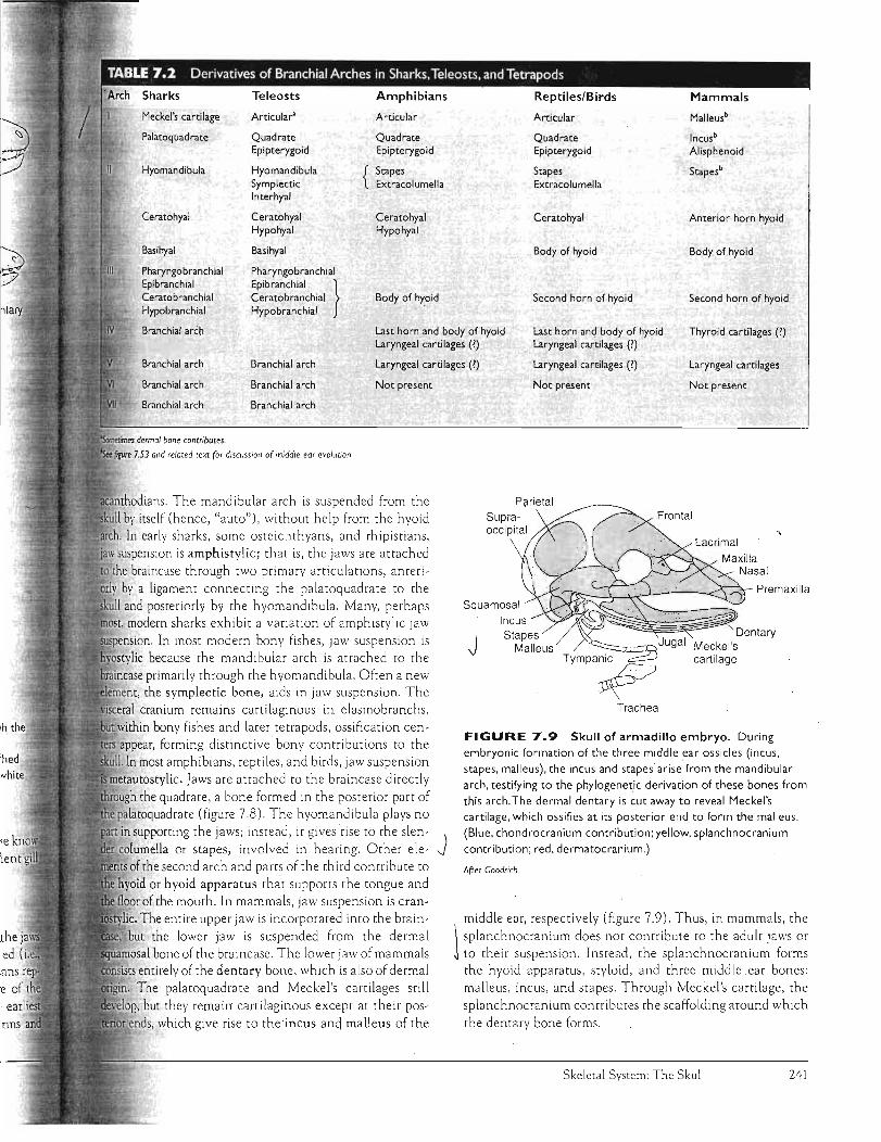
Meckel's cartilage Articular' Articular Articular Malleusb
Palatoquadrate Quadrate Quadrate Quadrate Incus' Epipterygoid Epipterygoid Epipterygoid Alisphenoid
Hyomandibula Hyomandibula { Stapes Stapes Stapesb
Symplectic Extracolumella Extracolumella Interhyal
Ceratohyal Ceratohyal Ceratohyal Ceratohyal Anterior horn hyoid Hypohyal Hypohyal
Basihyal Basihyal Body of hyoid Body of hyoid
Pharyngobranchial Pharyngobranchial Epibranchial Epibranchial }Ceratobranchial Ceratobranchial Body of hyoid Second horn of hyoid Second horn of hyoid Hypobranchial Hypobranchial
Branchial arch Last horn and body of hyoid Last horn and body of hyoid Thyroid cartilages (') Laryngeal cartilages (1) Laryngeal cartilages (1)
Branchial arch Branchial arch Laryngeal cartilages (1) Laryngeal cartilages (1) Laryngeal cartilages
Branchial arch Branchial arch Not present
Branchial arch Branchial arch
7.53 and related text for disClJssion of middle ear evolution.
ians. The mandibular arch is suspended from the by itself (hence, "auto"), without help from the hyoid
In early sharks, some osteichthyans, and rhipisti:ms,
sllspension is amphistylic; that is, the jaws are attached .....,"~·, 'hQ braincase through two primary articulations, anteri
by a ligament connecting the palatoquadrate to the
and posteriorly by the hyomandibula. Many, perhaps , modern sharks exhibit a variation of amphistylic Jaw
ion. In most modern bony fishes, jaw suspension is
lie because the mandibular arch is attached to the
primarily through the hyomandibula. Often a new the symplectic bone, aids in jaw suspension. The
cranium remains cartilaginous in elasmobranchs,
in bony fishes and later tetrapods, ossification cenappear, forming distinctive' bony contributions to the
In most amphibians, reptiles, and birds, jaw suspension
tylic. Jaws are attached to the braincase directly
the quadrate, a bone formed in the posterior part of latoquadrate (figure 7.8). The hyomandibula plays no
in supporting the jaws; instead, it gives rise to the slen- J columella or stapes, involved in hearing. Other ele
the second arch and parts of the third contribute to
. or hyoid apparatus that supports the tongue and
of the mouth. In mammals, jaw suspension is cran
The entire upper jaw is incorporated into the brain
but the lower jaw is suspended from the dermal
mosal bone of the braincase. The lower jaw of mammals entirely of the dentary bone, which is also of dermal
The palatoquadrate and Meckel's cartilages still
but they remain cartilaginous except at their pos
which give rise to the incus ane) malleus of the
Not present
Squamosal
Incus ----
J Stapes
Malleus -....-::::::::=:=x;::,Jugal
TYmp,~
Trachea
Not present
Premaxilla
FIGURE 7.9 Skull of armadillo embryo. During
embryonic formation of the three middle ear ossicles (incus,
stapes, malleus), the incus and stapes arise from the mandibular
arch, testifying to the phylogenetic derivation of these bones from
this arch. The dermal dentary is cut away to reveal Meckel's
cartilage, which ossifies at its posterior end to form the malleus.
(Blue, chondrocranium contribution; yellow, splanchnocranium
contribution; red, dermatocranium.)
After Goodrich.
middle ear, respectively (figure 7.9). Thus, in mammals, the
\ splanchnocranium does not contribute to the adult jaws or j to their suspension. Instead, the splanchnocranium forms
the hyoid apparatus, styloid, and three middle ear bones:
malleus, incus, and stapes. Through Meckel's cartilage, the
splanchnocranium contributes the scaffolding around which
the dentary bone forms.
Skeletal System: The Skull 241

Dermatoc\'·anium
Dermal bones that contribute to the skull belong to the dermatocranium. Phylogenetically, these bones arise from the bony armor of the integument of early fishes and sink inward to become applied to the chondrocranium and splanchnocranium. Bony elements of the armor a lso become associated with the endochondral elements of the pectoral girdle to give rise to the dermal components of [his girdle.
Dermal girdle (p. 330)
J
Dermal bones first become associated with the skull in ostracoderms. In later groups, additional dermal bones of the overlying integument also contribute. The dermatocranium forms the sides and roof of the skull to complete the protective bony case around the brain; it forms most of the bony lining of the roof of the mouth, and encases much of the sp lanchnocranium. Teeth that arise within the mouth usually rest on dermal bones.
As the name suggests, bones of the dermatocranium arise directly from mesenchymal and ectomesenchymal tissues of the dermis. Through the process of intramembranous ossification, these tissues form dermatocrania I bones.
Parts of the Dermatocranium
Derma l elements in modern fishes and living amphibians have tended to be lost o r fused so that the number of bones present is reduced and the skull Simplified. In amniotes, bones of the dermatocranium predominate,
J forRling most of the braincase and lower jaw. The dermal skull may contain a considerab le series of bones joined firmly at sutures in order to box in the brain and other skull e lements. As a convenience, we can group these series and recognize the most common bones in each (figure 7.10; table 7.3).
Dermal Bone Series Facial Series The facial series encircles the external naris . and collectively forms the snout. The maxilla and premaxilla (incisive) define the margins of the snou t and usually bear teeth . The nasal lies medial ro the naris. The septomaxilla is a small dermal bone of the facial series that is often absent. When present, it is usually sunken below the surface bones and aids in forming the nasal cavity.
oJ Orbital Series· The dermal bones encircle the eye to define the orbit superficia lly. The lacrimal takes its name from the nasolacrima1.( tear) duct of tetrapods that passes through or near this bone. The prefrontal, postfrontal, and postorbital continue the ring of bones above and behind the orbit. The jugal usually completes the lower rim of the orbit. Not to be confused with these dermal bones are the scleral ossicles of neural crest origin that, when presen t, reside within the orbit defined by the ring of dermal bones.
Dorsal Pa latal Facial series
Palatal series
FIGURE 7.10 Major bones ofthe dermatocranium. Sets of dermal bones form the facial series surround ing the nostril. The orbital series encircles the eye, and the temporal series composes the lateral wall behind the eye. The vault series, the roofing bones, run across the top of the skull above the brain. Covering the top of the mouth is the palatal series of bones. Meckel's cartilage (not shown) is encased in the mandibular series of the lower jaw Abbreviations : angular (An), dentary (D), ectopterygoid (Ec), frontal (F), intertemporal (It), jugal 0), lacrimal (l), maxilla (M), nasal (N), parietal (P), prearticular (Pa), palatine (PI), premaxilla (Pm), postorbital (Po), postparietal (Pp), prefrontal (Prf), parasphenoid (Ps), pterygOid (Pt), quadratojugal (OJ), I'
psurangular (Sa), splenial (Sp), squamosal (Sq), su pratemporal (St), tabular (T), vomer (V). n
n It
/ ai Temporal Series The temporal series lies behind the orbit, completing the posterior wall of the braincase . In many primitive tetrapods, this series is indented posteriorly by a temporal notch. Once thought in life to suspend an eardrum, this notch was named accordingly an otic notch. This now seems unlike ly, and instead the notch perhaps accommodated a spiracle, a respiratory tube. Openings called fenestrae (sing., B fenestra) arise within this region of the outer braincase in
Inmany tetrapods in association with the jaw muscularure. A
til row of bones , the intertemporal, supratemporat and tabu
al:lar, make up the medial part of the temporal series . This row
tHis reduced in early tetrapods and usually lost in later species.
illLaterally, the squamosal and quadratojugal complete the
se' j empOral series and fo rm the "cheek."
of m(
. Vault Series The vault, or roofing bones, run across the (fi
top of the skull and cover the brain beneath. These include sel
the frontal anteriorly and the postparietal (interparietal) posteriorly. Between them is the large parietal, occupying
en thi
the center of the roof and defining the small parietal foramen if it is present. The parieta l foramen is a tiny sky light in
nu tal
the skull roof that exposes the pinea l gland , an endocrine gland, to direct sunlight.
Chapter Seven 242
c

Orbital Series Temporal Series Vault Series Palatal Series Mandibular Series
Lacrimal Intertemporal Frontal Vomer Lateral bones:
Prefrontal Supratemporal Parietal Palatine Dentary (teeth)
Postfrontal Tabular Postparietal Ectopterygoid SpleniaIs (2) Postorbital
Jugal Squamosal Pterygoid Angular Quadratojugal Parasphenoid Surangular
(unpaired) Medial bones:
Prearticular Coronoids
Series The dermal bones of the primary palate Otic capsule
·.....orn"pr much of the roof of the mouth. The largest and most OPisthO\~__7ootic ' is the pterygoid. Lateral to it are the vomer, palatine,m. Vertebrae (-Js:j ----------___ _
ectopterygoid. Teeth may be present on any or all four these palatal bones. In fishes and lower tetrapods, there
!s, "-;-" 1.>LJ is an unpaired medial dermal bone, the parasphenoid. --- J Basi~X~~ \ ----~ 3in. Supraoccipitals ,Orbitosphenoid 'presphenoid Mesethmoid,
lar Series Meckel's cartilage is usually encased in Basisphenoid Sphenethmoid (lower tetrapods) bones of the mandibular series. Laterally, the wall of
series includes the tooth-bearing dentary and one or splenials, the angular at the posterior corner of the HYOmandib~~- Ro ------------ible and the surangular above. Many of these bones
ltal around the medial side of the mandible and meet the J D~~_)
and one or several coronoids to complete the), ial mandibular walL Left and right mandibles usually 3d-5th ~ ~ anteriorly at the midline in a mandibular symphysis. arches HYOid Meckel's cartilage Palatoquadrate
arch ' • Mandibular arch fi rm, the mandibular symphysis unites them into an
__ 'orr r1P(1 unit. Most notably in snakes, the mandibular symphbi ~, composed of soft tissues, permitting· independent /',. ... ---------- --im t of each mandible. \~'"J b u u 1;\'-______________"--')
Columella (stapes) ~rview of Skull Morphology
chondrichthyan fishes, the braincase is an elaborate carcase around the brain. The dermatocranium is
reflecting the elimination of almost all bone from skeleton. However, in most bony fishes and tetrapods,
Wi_rhl' braincase is extensively ossified with contributions from ..... _ "'''prol sources. For descriptive purposes, it is useful to think
braincase as a box with a platform of endoskeletal ele
,;.;;r;;~~ "'''cu supporting the brain, all encased in exoskeletal bones 7.11). The endoskeletal platform is assembledJrom a
of sphenoid bones. The occipital bones, which apparare derived from anterior vertebrae, form the end of
sphenoid platform. These occipital bones, up to four in (basioccipital, supraoccipital, and paired exoccipi
, close the posterior wall of the braincase except for a
hole they define, the foramen magnum, through the spinal cord runs. Articulation of the skull with
Quadrate (incus Arti~ular Epipterygoid of mammals) (malleus of
mammals)
J V'"It''';''~. ~~_F";"Orbital series / series Temporal series
Q -Palatal I:::::,,~\~S>-\\>......~_ series Hyoid '~-Mandlbular apparatus series
FIGURE 7. I I Contributions to the skull. The
chondrocranium (blue) establishes a supportive platform that is joined by contributions from the splanchnocranium (yellow), in particular the
epipterygoid. Other parts of the splanchnocranium give rise to the
articular; quadrate, and hyomandibula, as well as to the hyoid apparatus.
The dermatocranium (red) encases most of the chondrocranium together with contributions from the splanchnocranium.
Skeletal System: The Skull 243
·' .I" }
'.. ,
, BOX:,ESSAY 7.1 ' , ~;.'~~~:~~, '. ~ :,'
T he idea that the skull is derived from
serial compacted vertebrae dates to the
eighteenth century. The German naturalist
and poet, W. Goethe (1749-1832), was
apparently the first to think of but not the
first to pUblish this idea. Goethe gave us the
word morphology, which meant to him the
search for underlying meaning in organic
design or form. Among his discoveries was
the observation that plant flowers are
modified stem petals compacted together.
His venture into vertebrates and vertebrate skulls in particular occurred in 1790 whilst he was strolling in an old cemetery
in Venice. He spied a dried ram's skull disin
tegrated at its bony sutures but held in
sequence by the soil. The separated bones of the ram's skull seemed to be the fore
shortened anterior vertebrae of the back
bone, but Goethe did not publish this idea until about 1817. Public credit for this idea
and for elaborating it goes to another Ger
man naturalist, L. Oken (1779-185 I) . In 1806, Oken was strolling in a forest and
came upon a dried sheep skull. He was similarly struck by its serial homology with the
. vertebrae, and shortly thereafter published
the idea (box figure I a).
Next, the vertebral theory of skull origin
fell mto the hands of Richard Owen and
became part of his much embellished theo
retical view on animal archetypes (box fig
ure I b). Because of Owen's prominence in
early nineteenth-century science, the idea of skull from vertebrae became a central issue within European scientific communi
ties. One of the most persuasive dissenters
from this view of a vertebral source for the skull was T. H. Huxley, who based his cri
tique upon a detailed comparative study of
vertebrate skulls and their development.
This came to a head (no pun intended) in an invited lecture, the Croonian lecture of
1858, in which Huxley argued that the
development of the skull showed that it was
not composed of vertebrae. He suggested that the "skull was no more derived from
vertebrae, than vertebrae are derived from
(a)
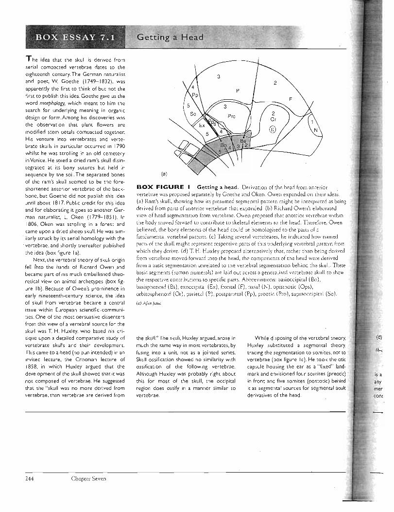
BOX FIGURE Getting a head, Derivation of the head from anterior vertebrae was proposed separately by Goethe and Oken. Owen expanded.on their ideas: (a) Ram's skull, showing how its presumed segmental pattern might be interpreted as being
derived from pans of anterior vertebrae that expanded. (b) Richard Owen's elaborated view of head segmenta tion from vertebrae. Owen proposed that anterior vertebrae within
the hody moved forward to contribute to skeletal e lements to the head. Therefore, Owen
believed, the bony elements of the head could be homologized to the ·parts of a fundamental vertebral pattern . (c) Taklng several vertebr<ltes, he indicated how named
pam of the skull might represent respective partS of this underlying vertebral pattern from
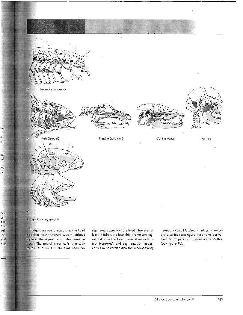
which they derive. (d) T. H. Huxley proposed alternatively that, rather than being derived
from vertebrae moved forward into the head, the components of the head were derived
from a basic segmentation unrelated to the vertebral segmentation behind the sku ll. These basic segments (roman numerals) are laid out across a generalized vertebrate skull to show
the respective contributions to specific parts. Abbreviations: basioccipital (Bo),
bas isphenoid (Bs), exoccipital (Ex), frontal (F), nasal (N), opisthotic (Ops), orbitosphenoid (Or), parietal (P), postparieta l (Pp), prootic (Pro), supraoccipital (So).
(a) N'c'T JolI,,! .
the skull." The skull, Huxley argued, arose in
much the same way in most vertebrates, by
fusing into a unit, not as a jointed series. Skull ossification showed no similarity with ossification of the following vertebrae.
Although Huxley was probably right about
this for most of the skull, the occipital
region does .ossify in a manner similar to vertebrae.
While disposing of the vertebral theory, (d)
Huxley substituted a segmental theory,
traCing the segmentation to somites, not to Ib-<:
vertebrae (box figure I c). He took the otic
capsule housing the ear as a "fixed" land
mark and envisioned four somites (preotic) is a in front and five somites (postotic) behind any it as segmental sources for segmental adult mer derivatives of the head. cont·
Chapter Seven 244
111
n
lm
ed
)11'
some would argue that the head
unique developmental system without
tie to the segmental somites (somito-
The neural crest cells that also
to parts of the skull show no
Reptile (all igator)
segmental pattern in the head. However, at
least in fishes , the branchial arches are seg
mental , as is the head paraxial mesoderm
(somitomeres) , and segmentation appar
ently can be carried into the accompanying
Canine (dog) Human
neurocranium. Matched shading in vertebrate series (box figure I c) shows deriva
tives from parts of theoretical ancestor
(box figure I b) .
Skeletal System: The Skull 245

the vertebral column is established through the occipital condyle, a single or double surface produced primarily within the basioccipital but with contributions from the exoccipitals in some species.
The otic capsule rests on the posterior part of the endoskeletal platform and encloses the sensory organs of the ear. The splanchnocranium contributes the epipterygoid (alisphenoid of mammals), to the endoskeletal platform and gives rise to one (columella/stapes) or more (malleus and incus of mammals) of the middle ear bones housed in the otic capsule.
In most vertebrates, these endoskeletal elements, along with the brain and sensory organs they support, are enclosed by the exoskeletal elements, derivatives of the dermis, to complete the braincase.
Jaws The upper jaw consists of the endoskeleta l palatoquadrate in primitive vertebrates. The palatoquadrate is fully functional in the Jaws of chondrichthyans and primitive fishes, but in bony fishes and tetrapods, the palatoquadrate usually makes limited contribut ions to the skull through its two derivatives: the epipterygoid, which fuses to tne neurocranium, and the quadrate, ·,which suspends the lower jaw except in mammals. The dermal maxilla and premaxilla replace the pala toquadrate as the upper jaw.
The lower jaw, or mandible, consists on ly of Meckel's cartilage in chondrichthyans. In most fishes and tetrapods, Meckel's cartilage persists but is enclosed in exoskeletal bone of ~he dermatocranium, which also supports teeth. MeC'kel's cartilage, encased in dermal bone, usually remains unossified, except in some tetrapods where its anterior end ossifies as the mental bone. In most fishes and tetrapods (except mammals), the posterior end of Meckel's cartilage can protrude from the exoskeletal case as an ossified articu
lar bone. In mammals, the lower jaw consists of a single bone,
the dermal dentary. The anterior tooth-bearing part of the dentary js its ramus. Jaw-closing muscles are inserted on the coronoid process, an upward extension of the dentary. Posteriorly, the dentary forms the transversely expanded
) mandibular condyle, a rounded process that articu lates with J the glenoid fossa, a depression within the temporal bone of
the braincase. Thus, in mammals, the mandibular condyle of the dentary replaces the articular bone as the surface of the lower jaw through which is established mandibular articulation with the braincase.
Hyoid Apparatus The hyoid or hyoid apparatus is a ventral derivative of the splanchnocranium behind the jaws. In fishes, it supports the floor of the mouth . Elements of the hyoid apparatus are derived from the ventral parts of the hyoid arch and from parts of the first few branchial arches. In larval and paedo
morphic amphibians, the branchial bars persist but form a
reduced hyoid apparatus that supports the floor of the mouth and functional gills. In adults, the gills and the associated part of the hyoid apparatus are lost, although elements per-' sis t within the floor of the mouth usually to support the tongue. Typically, the hyoid apparatus includes a main body, the corpus, and extensions, the cornua ("horns"). In many mammals, including humans, the distal end of the hyoid horn fuses with the otic region of the braincase to form the styloid process.
J C rama'I K'mesis,
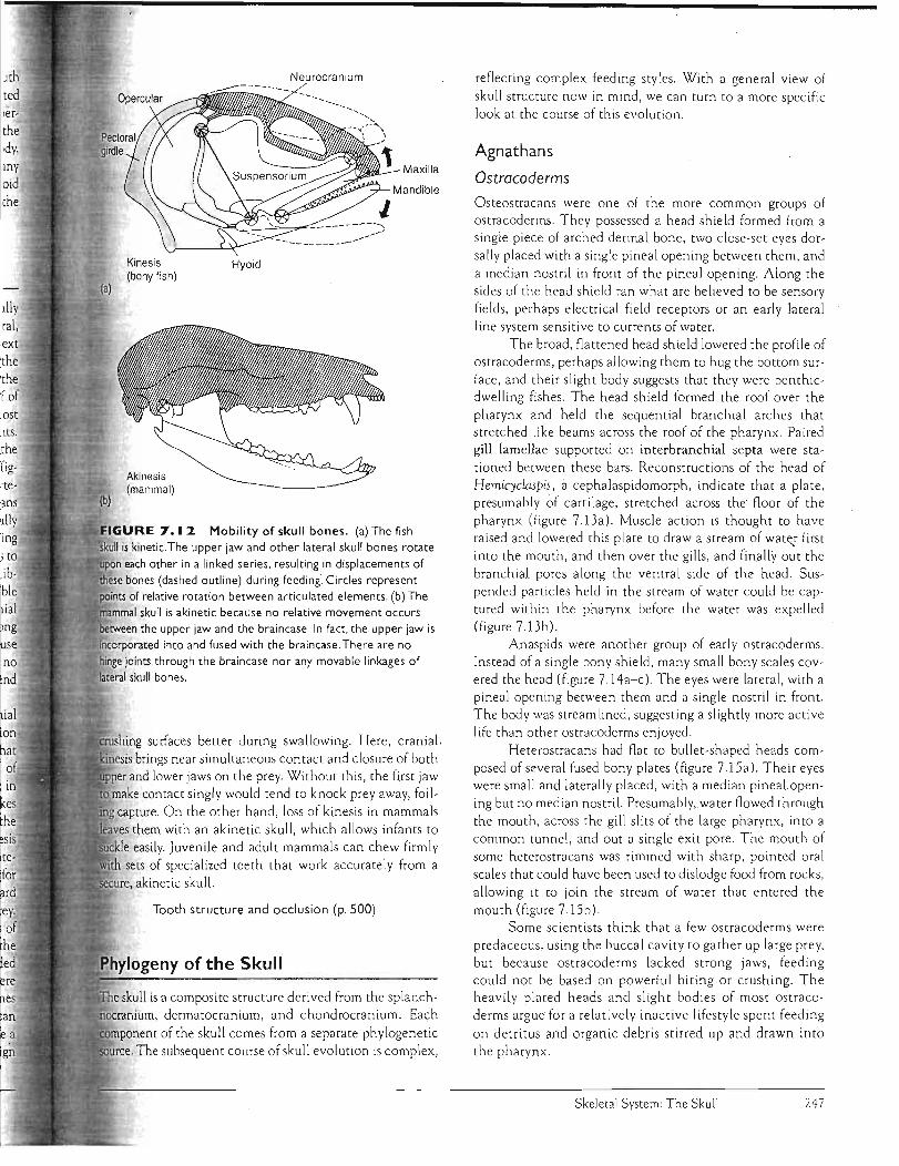
Kinesis means movement. Cranial kinesis refers literally then to movement within the skull. But if left this general, the definition becomes too broad to provide a useful context in which to discuss sku ll function. Some authors restrict the term to skulls with a transverse, hingelike joint across the skull roof and a transverse, sliding basal Joint in the roof of the mouth. But this restricted definition precludes most teleost fishes, despite their highly mobile skull elements. Here, we use cranial kinesis to mean movement between the upper jaw and the braincase about joints be tween them (figure 7.12a). Such kinetic skulls characterize most verte· brates. They are found in ancient fishes (crossopterygians and probably palaeoniscoids), bony fishes (especially teleosts), very early amph ibians, most reptiles (including most Mesozoic forms), birds, and early therapsid ancestors to
mammals. Kinetic sku lls are not present in modern amphibians, turtles, crocodiles, and mammals (with the possible exception of rabbits). The widespread presence of cranial kinesis among vertebrates, but its essential absence among t mammals, seems ro create a problem for humans. Because il we, like moSt other mammals, have akinetic skulls with no h such movement between upper jaw and braincase, we tend la
to underestimate its importance (figure 7.12b). Kinesis and akinesis each have advantages. Cranial
kinesis provides a way ro change the size and configuration of the mouth rapidly. In fishes and other vertebrates that
cr ki
feed in water, rapid kinesis creates a sudden reduction of pressure in the buccal cavity so that the animal can suck in 1I~
a surprised prey. This method of prey capture, which takes to
in:advantage of a sudden vacuum to gu lp in water carrying the Ie<
intended food , is known as suction feeding. Cranial kines is Su(
also allows rooth-bearing bones ro move quickly inro Stratewil
gic positions during rapid feeding. Some teleost fishes, for instance, swing their anterior rooth-bearing bones forward
sec
at the last moment ro reach out quickly at the intended prey. In many venomous snakes, linked bones along the sides of the skull can rotate forward. The venomous viper erects the maxillary bone bearing the fang and swings it from a folded Ph position along its upper lip ro the front of the mouth, where it can more easily deliver venom into prey. In many fishes The and reptiles with kinetic skulls , teeth on the upper jaw can noc be reoriented with respect to the prey in order to assume a com more favorable position during prey capture or ro align SOur
Chapter Seven 246
ociy, my Maxilla aid the
ost 1tS.
the rig-
Akinesis ·te (mammal) ans
Mobility of skull bones. (a) The fish is kinetic.The upper jaw and other lateral skull bones rotate
i:tE"lpon each other in a linked series. resulting in displacements of bones (dashed outline) during feeding~ Circles represent of relative rotation between articulated elements. (b) The
skull is akinetic because no relative movement occurs .......,,""""'...'n the upper jaw and the braincase. In fact. the upper jaw is ...:''':.inr"rn"...,r"rt into and fused with the braincase. There are no
binge joints through the braincase nor any movable linkages of lateral skull bones.
surfaces better during swallowing. Here, cranial, brings near simultaneous contact and closure of both
and lower jaws on the prey. Without this, the first jaw make contact singly would tend to knock prey away, foil-capture. On the other hand, loss of kinesis in mammals
them with an akinetic skull, which allows infants to easily. Juvenile and adult mammals can chew firmly
sets of specialized teeth that work accurately from a ,akinetic skull.
Tooth structure and occlusion (p. 500)
skull is a composite structure derived from the splanchdermatocranium, and chondrocranium. Each
of the skull comes from a separate phylogenetic subsequent course of skull evolution is complex,
reflecting complex feeding styles. With a general view of skull structure now in mind, we can turn to a more specific look at the course of this evolution.
Agnathans
Ostracoderms
Osteostracans were one of the more common groups of ostracoderms. They possessed a head shield formed from a single piece of arched dermal bone, two close-set eyes dorsally placed with a single pineal opening between them, and a median nostril in front of the pineal opening. Along the sides of the head shield ran what are believed to be sensory fields, perhaps electrical field receptors or an early lateral line system sensitive to currents of water.
The broad, flattened head shield lowered the profile of ostracoderms, perhaps allowing them to hug the bottom surface, and their slight body suggests that they were benthicdwelling fishes . The head shield formed the roof over the pharynx and held the sequential branchial arches that stretched like beams across the roof of the pharynx. Paired gill lamellae supported on interbranchial septa were stationed between these bars. Reconstructions of the head of Hemicyclaspis, acephalaspidomorph, indicate that a plate, presumably of cartilage, stretched across the floor of the pharynx (figure 7.13a). Muscle action is thought to have raised and lowered this plate to draw a stream of wate,!" first into the mouth, and then over the gills, and finally out the branchial pores along the ventral side of the head . Suspended particles held in the stream of water could be captured within the pharynx before the water was expelled (figure 7 .13b) .
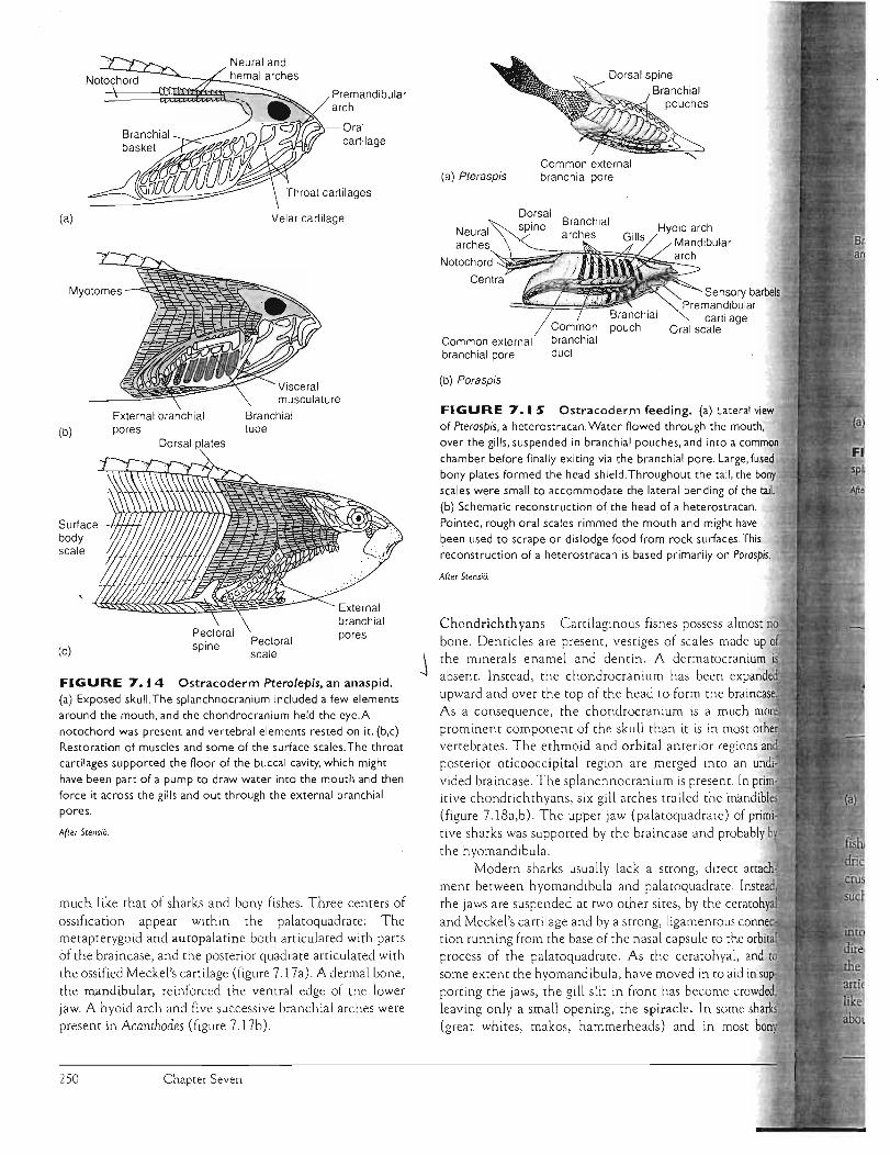
Anaspids were another group of early ostracoderms . Instead of a single bony shield, many small bony sca les covered the head (figure 7.14a-c). The eyes were lateral, with a pineal opening between them and a single nostril in front. The body was streamlined, suggesting a slightly more active life than other ostracoderms enjoyed.
Heterostracans had flat to bullet-shaped heads composed ofseveral fused bony plates (figure 7.15a). Their eyes were small and laterally placed, with a median pineal opening but no median nostril. Presumably,. water flowed through the mouth, across the gill slits of the large pharynx, into a common tunnel, and out a single exit pore. The mouth of some heterostracans was rimmed with sharp, pointed oral scales that could have been used to dislodge food from rocks, allowing it to join the stream of water that entered the mouth (figure 7.15b).
Some scientists think that a few os tracoderms were predaceous, using the buccal cavity to gather up large prey, but because ostracoderms lacked strong jaws, feeding could nOt be based on powerful biting or crushing. The heaVily plated heads and slight bodies of most ostracoderms argue for a relatively inactive lifestyle spent feeding
on detritus and organic debris stirred up and drawn into the pharynx.
Skeletal System: The Skull 247

(c)
I e o.., II
n IT
r e;
t!al
Fi
PI pI b, of pL t el
pa bo it
na
. .
. BO~. ~f ·S.SAY 7. '2 ..' • _, :-.11:;"'_~""!' .r, • .", ....
In hares or "jac krabbi ts" (but not in dis
tantly related pikas or in their fossil
ancestors), a suture between regions of
the fetal braincase remains open in the
adult, forming an intracranial joint (box
figure I ).This intracranial joint runs along the sides and base of the adult braincase
and hinges across the top via the post
parieta l. The joint permits relative motion
between anterior and posterior parts of
the braincase. It has been hypothesized
that this joint helps absorb the impact forces sustained as the forelimbs strike the ground when a rabbit runs. Upon impact, mechanical deformation of the
joint would absorb some kinetic energy
as the hinge is strained. This deformation
and absorption would reduce the shock
sustained by the anterior part of the
braincase. Additionally, the impact forces
would tend to drive blood from intracra
nial sinuses into a complex association of
venous channels and spaces within the
skull. This would 'help dissipate these
kinetic forces further as they acted against resistance offered by the walls of
the blood vascular system.
The external ears (pinnae) of hares
radia~e heat generated during strenuous
activity, but apparently only after locomo
tor exercise ceases. During locomotion, the ears are usually held erect by strong mus
cles at their bases. It has been hypothesized
that these erect ears help reopen the intracranial jOint as the hare pushes off on
another leap to accelerate again, thus in a sense "resetting" this cranial mechanism
and preparing it to act as a shock-absorbing
device when the forelimbs again strike the ground (box figure I c).
The functional significance of the
intracranial joint is still debated. However, if
such hypotheses are confirmed, this spe
cialized joint in hares, together with their
projecting ears, might also serve to reduce
Cyclostomes
Lampreys and hagfishes are the only surviving agnat hans and heirs of the ostracoderms. However, subsequent speC ia lizations have left cyclostomes with anatomies quite unlike those of the early ostracode rms. Cyclostomes lack bone
entirely and are specialized for parasi tic or scavenging lives
Chap te r Seven
ranial Kinesis in Hares?
Impact c
"00Ol . tt
"00- 0 ~ '" COl co.c 0. .s:: Ol 0.~o. -0. I ::J::JLL ro '" <D ::J '"'" '"
(a)
(b)
BOX FIGURE Possible cranial kinesis in hares. (a) Phases dur ing a
running str id e are illustrated. Note that the forelimbs receive the initia l impact upon
landing. (b) Posterior regions of the skull of the jackrabbit Lepus. The intracrania l joint extends along the sides o f the skull between squamosal (Sq) and otic reg ions and then a long the base of the sku ll. The interparietal bone forms the hinge across the top o f the
skull. (c) External ears held erect and attached to the posterior part of the skull may help
to reposition the posterior part of the skull relative to the anterior part during the extended suspension phase of running. The presumed motion (s lightly exaggerated) of the
anterior braincase relative to the posterior braincase is indicated. Fa is the force vec tor due to acce leration resulting from thrust, and Fd is th e force vector due to drag of the ears in
the oncoming wind. Abbreviations: bulla (B), postparietal (Pp), jugal (J), parietal (P), petrosal (Pet), supraoccipi tal (So), squamosal (Sq).
jarring of the eyes carried in the anterior
braincase. Among mammals, rabbit kinesis
represents an independent and apparently
unique condition that did not evolve from
that depend on a rasping tongue to scrape up tissue for a meal. Lampreys have a Single medial nostril and a pineal open ing. Branchial pouches are present. The braincase is cartilaginous. Branch ial arches, a lthough present, form an
unjointed branchial basket. Hagfi shes have a median nostril
bu t no external pineal opening.
Extended suspension .. ..
Intracranial jOint
therapsid kinesis . Further, it evolved not for
its advantages during feeding but rather for
its advantages during rapid locomotion.
(Based on the research of D. Bramble.)
Front s upport
248

n
in the pharynx and then passed to the esophagus.
lvertebrates, except agnathans, have jaws and form the group gna thostomes ("jaw mouth"). Some biolo
mark the advent of vertebrate jaws as one of the most transitions in their evolution. Powerfu l closing
, derivatives of the branchial arch musculature, the jaws strong biting or grasping devices. It is not sur
then, that with the advent of jaws, gnathostomes 'Ynf'r\p,nrp a dietary shift away from suspension feeding of
to large r food items. With a change in diet a more active lifestyle .
As much as a third to a half of the anterior ~.cOd€~rm body was composed of heavy plates of dermal
that also enclosed the pharynx and braincase. The rest body was covered with small bony scales. The dermal of the head were thick and tightly joined into a unit
the cranial shield (figure 7.16a,b). Although the of these dermal plates has been compared to scales of
fishes, their arrangement was suffiCiently different 'that best to follow the convention of using different
until so~ agreement is reached on their homologies.
Feeding on sill
Cranial cavity
Interbranchial
GillBranchial pouch and pore
Buccal cavity
(b)
Ostracoderm Hemicyclaspis, a cephalaspidomorph. (a) Ventral view showing branchial pores. the presumed
of exit for water moving through the pharynx. (b) Cross section through the pharynx illustrating respiratory gill lamellae and branchial arch. Presumably, the noor of the pharynx could be raised and lowered to actively draw water into the mouth al'ld
it out through the several branchial pores. The current crossed the respiratory gills before exiting. Suspended food may have been
The braincase was heavily ossified, and the upper jaws attached to it. In most, a well-defined joint existed between the braincase and the first vertebra. A spiracle was apparen tly abse nt. Water departing from the mouth exited posteriorly at the open junction between cranial and trunk shields. Most placoderms were 1 m in length , although one species possessing strong jaws reached nearly 6 m overall.
Acanthodians The gnathostomes with the earliest surviving fossil record are the acanthodians. Most were small, several centimeters in length , with streamlined bodies, suggesting an ac tive swimming lifesty le . Their bodies were covered with nonoverlapping, diamond-shaped, dermal bony sca les. The bony sca les of the head region were enlarged into small plates. The pattern of cranial dermal sca les resembled bony fishes, but as with placoderms, these · are usually given their own names. Some species ' had an operculum, a bony flap that covered the exit gill slits. Eyes were large, suggesting that visual information was especially important to these fishes. Acanthodes (early Permian) possessed a lateral cranial fissure, a gap that partially divided the posterior braincase. This fissure is an important fixture in actinopterygian fishes, where it allows exit of the tenth cranial nerve. The mandibular arch that formed the jaws was
Skeletal System: The Skull 249
Neural arches
Notochord~~~~r~~fi;~~~~~
Common
Premandibular cartilage
Oral scale
Sensory barbels
tail.
most bon.,
musculature
Neural and _-:n,--..:..:h.:::-emal arches
Premandibular arch
Oral cartilage
(a) Velar cartilage
Myotomes
External branchial Branchial (b) pores tube
Dorsal plates
Surface body scale
poresPectoralspine(c) scale
FIGURE 7.14 Ostracoderm Pterolepis, an anaspid. (a) Exposed skull. The splanchnocranium included a few elements around the mouth, and the chondrocranium held the eye.A notochord was present and vertebral elements rested on it. (b,c) Restoration of muscles and some of the surface scales. The throat cartilages supported the floor of the buccal cavity, which might
have been part of a pump to draw water into the mouth and then force it across the gills and out through the external branchial pores.
After Stens;o.
much like that of sharks and bony fishes. Three centers of ossification appear within the palatoquadrate: The metapterygoid and autopalatine borh articulated with parts of the braincase, and the posterior quadrate articulated with the ossified Meckel's cartilage (figure 7 .17a). A dermal bone, the mandibular, reinforced the ventral edge of the lower jaw. A hyoid arch and five successive branchial arches were present in Acanthodes (figure 717b).
Chapter Seven
Common external branchial pore
Dorsal spine Branchial
arches
(a) Pteraspis
Common external branchial branchial pore duct
(b) Poraspis
FIGURE 7. IS Ostracoderm feeding. (a) Lateral view of Pteraspis, a heterostracan. Water flowed through the mouth, over the gills, suspended in branchial pouches, and intO a common chamber before finally exiting via the branchial pore. Large, fused bony plates formed the head shield. Throughout the tail, the bony scales were small to accommodate the lateral bending of the ·(b) Schematic reconstruction ofthe head of a heterostracan.
Pointed, rough oral scales rimmed the mouth and might have been used to scrape or dislodge food from rock surfaces.This reconstruction of a heterostracan is based primarily on Poraspis.
After Stens;o.
Chondrichthyans Cartilaginous fishes possess almost no bone. Denticles are present, vestiges of scales made up
j the minerals enamel and dentin. A dermatocranium absent. Instead, the chondrocranium has been pv,..,~r,ri",i'.!
upward and over the top of the head to form the brai As a consequence, the chondrocranium is a much prominent component of the skull than it is in most vertebrates. The ethmoid and orbital anterior regions posterior oticooccipital region are merged into an und vided braincase. The splanchnocranium is present. In pri itive chondrichthyans, six gill arches trailed the mand' (figure 7.l8a,b). The upper jaw (palatoquadrate) of tive sharks was supported by the braincase and probably by the hyomandibula .
Modern sharks usually lack a strong, direct attach· ment between hyomandibula and palatoquadrate. the jaws are suspended at two other sites, by the ce and Meckel's cartilage and by a strong, ligamentous r""npr.......
tion running from the base of the nasal capsule to the process of the palatoquadrate. As the ceratohyal, and to some extent the hyomandibula, have moved in to aid in supporting the jaws, the gill slit in front has become crowded, leaving only a small opening, the spiracle. In some sharks (great whites, makos, hammerheads) and in
250

Orbital
Pineal
Hyomandibula
Fenestra
Nasal capsule
Meckel's cartilage
Palatoquadrate
Articular
(b)
Paranuchal plate
Nuchal plate
Postpineal
Lateral plate
Prelateral plate
Lateral plate
Placoderm skull. Bothrio{epis was about 15 cm long and lived in the middle Devonian. (a) Lateral view of
m and chondrocranium. (b) Skull with overlying dermatocranium in place. Note the dermal plates.
Pharyngobranchial Hyomandibula
Braincase
terminal mouth 11;
,/ ..--"""..:·e ~Ceratohyal
FIGURE 7.17 Acanthodian skull, Acanthodes. (a) Lateral"
view with mandibular arch
shown in its natural position.
(b) Mandibular arch is
removed to better reveal the
chondrocranium, hyoid arch,
and five successive branchial
arches. (Red, dermal bone;
yellow, splanchnocranium; blue,
chondrocranium)
Endoskeletal Ceratobranchials shoulder girdle
(b)
the spiracle has vanished altogether. In chonsuch as holocephalans, the jaws mechanically
hard shells of prey, but in 'active chondrichthyans, as predaceous sharks, the jaws capture prey. Sharks may use suction to draw small prey toward or the mouth, but more commonly, they attack prey
approaching it head-on. As sharks raise their head, jaw descends (figure 7.19a). Upper and lower jaws with each other, and both in turn are suspended
a pendulum from the hyoid arch. The hyoid arch swings "its attachment to the braincase, which permits the
After jarvik.
jaws to descend and shift downward and forward over the prey (figure 7 .19b). Teeth along the upper (palatoquadrate) and lower (Meckel's cartilage) jaws are often oriented with their points in an erect position to engage the surface of the prey. Occasionally a nictitating membrane , a movable flap of opaque skin, is drawn protectively across each eye.
Jaw protrusion may also ass ist the synchronized meeting of upper and lower jaws on the prey. If the lower jaw alone was responsible for closing the mouth, it might prematurely strike the prey before the upper jaw was suitably positioned to assist. Protracting the mandibles away from
Skeletal System: The Skull 251

Mandibular arch
'Vi
Pectoral fin
Cladoselache
FIGURE 7.18 ' Mandibles were followed by a complete hyoid arch and five branchial arches. Full gill slits were present between each arch. (b) Modern shark Squa/us, the dogfish shark.The hyoid arch, second in series, is modified to support the back of the mandibular arch.As the hyoid
moves forward to help suspend the jaw, the gill slit in front is crowded and reduced to the small spiracle. Although fused into one unit, the
three basic regions of the chondrocranium are ethmoid, orbital. and oticooccipital.Abbreviations: basihyal (Bh), ceratohyal (Ch),
hyomandibula (Hy), Meckel's cartilage (Mk), palatoquadrate (Pq).
(a) After Zanger/.
(b)
Shark skull. (a) Primitive shark Cladoselache, a late Devonian shark that reached perhaps SS cm in length,
(b)
Feeding in sharks. (a) Sketches of shark with jaws retracted (top) and manually protracted (bottom),
the he, metric ously a! clamp ( near th violent swallo\\
body sil Retract dynami back UI=
Actinol large e) extendi ous teel hyoid al ' gies of ( assign, especial be man ' rostral, shown i Notice i
gians th \V
occurre( eralize a ied spec radiatio
with and cular bOI pensoriu different
(a)
(a)
FIGURE 7.19 (b) Interpreted positional changes in the mandibular arch as it rides forward on its suspension from the ceratohyal. Po'sition depicted is
near the completion of jaw closure on the prey. Arrow indicates ventral and forward shift of the jaws.
Based on, and simplified from, the research of T H. Frazzet(Q,
Chapter Seven 252
head allows the jaws to assume a more favorable geo, configuration so that they meet the prey simultane
and avoid deflecting it when they close, As the jaws
on the prey, the mandibular arch often is protracted the end of closure. If the prey is large, the shark may
shake its head to cut free a section of the prey and it.
When protracted, the jaws disrupt the streamlined
silhouette characteristic of an acti ve, open-water fish. [\~[ac[lon of the jaws following feeding restores the hydro
ic, streamlined shape of the fish and tucks the jaws
up against the chondrocranium.
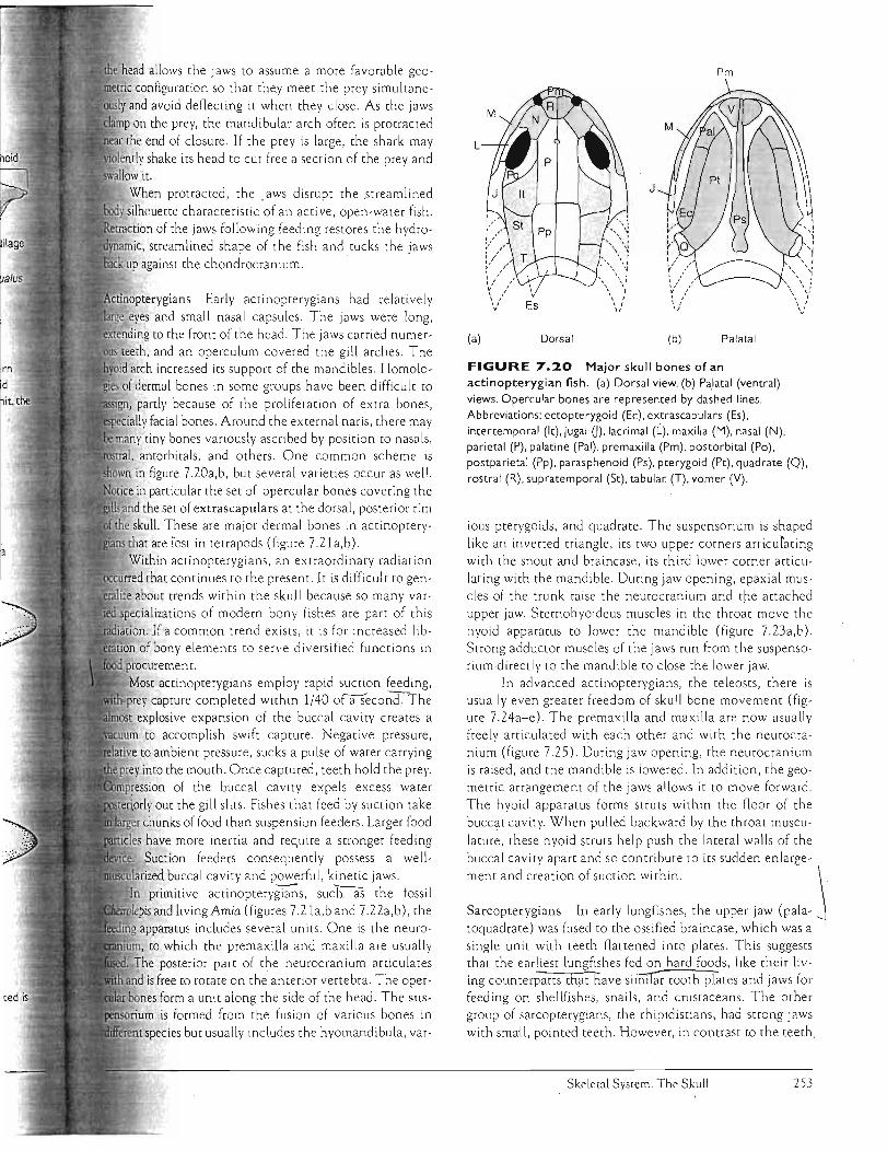
Early actinopterygians had relatively
eves and small nasal capsules. The jaws were long, "","pn';"", to the front of the head. The jaws carried numer
and an operculum covered the gill arches. The arch increased its support of the mandibles. Homolo
of dermal bones in some groups have been difficult to
partly because of the proliferation of extra bones, , facial bones. Around the external naris, there may
. tiny bones variously ascribed by position to nasals, antorbitals, and others. One common scheme is
in figure 7.20a,b, but several varieties occur as well.
in particular the set of opercular bones covering the the set of extrascapulars at the dorsal, posterior rim
skull. These are major dermal bones in actinoptery
that are rost in tetrapods (figure 7.21a,b). \Vithin actinopterygians, an extraordinary radiation
rred that continues to the present. It is difficult to genabout trends within th.e skull because so many var
wecializations of modem bony fishes are part of this · If a common trend exists, it is for increased lib
of bony elements to serve diversified functions in procure men t.
Most actinopterygians employ rapid suction feeding, · capture completed with.in 1/40 ofaSecond. The
explosive expansion of the buccal cavity creates a to accomplish swift capture. Negative pressure,
to ambient pressure, sucks a pulse of water carrying into the mouth. Once captured, teeth hold the prey.
of the buccal cavity expels excess water """"rlc'rh, out the gill slits. Fishes that feed by suction take
chunks of food than suspension feeders. Larger food
have more inertia and require a stronger feeding Suction feeders consequently possess a well
buccal cavity and powerful, kinetic jaws.
primitive actinoptery~s, such as the fossil · and living Amia (figures 7.21a,b and 7.22a,b), the
apparatus includes several units . One is the neuro
to which the premaxilla and maxilla are usually The posterior part of the neurocranium articulates
and is free to rotate on the anterior vertebra. The operform a unit along the side of the head. The sus
""..,-'..- is formed from the fusion of various bones in
species but usually includes the hyomandibula, var-
Pm
L
J
(a) Dorsal (b) Palatal
FIGURE 7.20 Major skull bones of an
actinopterygian fish. (a) Dorsal view. (b) Pa,latal (ventral)
views. Opercular bones are represented by dashed lines.
Abbreviations: ectopterygoid (Ee), extrascapulars (Es), intertemporal (It), jugal 0), lacrimal (L), maxilla (M), nasal (N), parietal (P), palatine (Pal), premaxilla (Pm), postorbital (Po),
postparietal (Pp), parasphenoid (Ps), pterygOid (Pt), quadrate (Q), rostral (R), supratemporal (St), tabular (T), vomer (V).
ious pterygoids, and quadrate. The suspensorium is shaped
like an inverted triangle, its two upper comers articulating with the snout and braincase, its third lower comer articlllating with the mandible. During jaw opening, epaxial muscles of the trunk raise the neurocranium and the attached upper jaw. Stemohyoideus muscles in the throat move the hyoid apparatus to lower the mandible (figure 7.23a,o). Strong adductor muscles of the jaws run from the suspensoriumdirectly to the mandible to close the lower jaw.
In advanced actinopterygians, the teleosts, there is usually even greater freedom of skull bone movement (fig ure 7.24a-e). The premaxilla and maxilla are now usually freely articulated with each other and with the neurocranium (figure 7.25). During jaw opening, the neurocranium is raised, and the mandible is lowered. In addition, the geometric arrangement of the jaws allows it to move forward.
The hyoid apparatus forms struts with.in the floor of the buccal cavity. When pulled backward by the throat muscu
lature, these hyoid struts help push the lateral walls of the buccal cavity apart and so contribute to its sudden enlarge
ment and creation of suction within. \
Sarcopterygians In early lungfishes, the upper jaw (pala - j toquadrate ) was fused to the ossified braincase, which was a single unit with teech flattened into plates. This suggests that the earliest lungfishes fed on hard foods, like their living counterparts thpt have sii1illar tooth. plates and jaws for feeding on shellfishes, snails, and crustaceans. The other group of sarcopterygians, the rhipidistians, had strong jaws with small , pointed teeth. However, in contrast to the teeth.
Skeletal System: The Skull 253
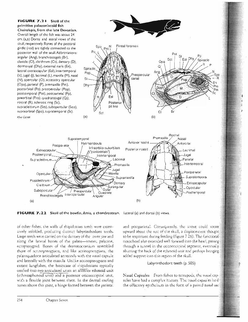
FIGURE 7.21 Skull ofthe primitive palaeoniscoid fish Cheirolepis, from the late Devonian. Overall length of the fish was about 24 cm. (a,b) Dorsal and lateral views of the skull. respectively. Bones of the pectoral girdle (red) are tightly connected to the posterior wall of the skull.Abbreviations: angular (Ang). branchiostegals(Br). clavicle (CI). cleithrum (Ct). dentary (D) . dermohyal (Dhy). external naris (En). lateral extrascapular (Esl). intertemporal (It). jugal 0). lacrimal (L). maxilla (M). nasal (N). opercular (0). accessory opercular (Opa), parietal (P). premaxilla (Pm). postorbital (Po), preopercular (Pop), posttemporal (Pot), postparietal (Pp), pre orbital (Pro), quadratojugal (Qj) , rostral (R), sclerotic ring (Sc), supracleithrum (SCt). subopercular (Sop), supraorbital (Spo), supratemporal (St).
Afr.er Carroll.
Spiracle Pop
St
Dhy
(a)
Supratemporal Hyomandibula
Pineal foramen
Preopercular canal
CI
(b)
Anterior nostril --.,,;;;;...:;;:c.;
Br
Infraorbito-su borbitals
Supracleithrum-.
Supratemporal
(a) (b)
FIGURE 7.22 Skull of the bowfin,Amia, a chondrostean. Lateral (a) and dorsal (b) views.
of other fishes , the walls of rhipid ist ian teeth were extensively infolded, producing distinct labyrinthodont teeth. Large teeth were carried on the dentary of the lower jaw and along the lateral bones of the palate-vomer, palatine, ectopterygoid. Bones of the dermatocranium resembled those of ac tinopterygia ns, and like actinopterygians, the palatoquadrate articulated anteriorly with the nasal capsule and laterally with the maxilla. Unlike actinopterygians and ex tant lungfishes, the braincase of rhipidistians typically ossified into two ar ticulated units: an anterior ethmoid unit (ethmosphenoid unir) and a posterior oticooccipital unit, with a flexible joint between them. In the dermal roofing bones above this Joint, a hinge formed between the parietal
254 Chapter Seven
and postparietal. Consequen tly, the snout could rotate upward abo ut the rest of the sku ll, a displacement thought to be important during feeding (figure 7.26). The functional notochord also extended well forward into the head, passing through a tunnel in the oticooccip ital segment, eventually abutting the back of the ethmoid unit and perhaps bringing added support into this region of the skull.
Labyrinthodont teeth (p.505)
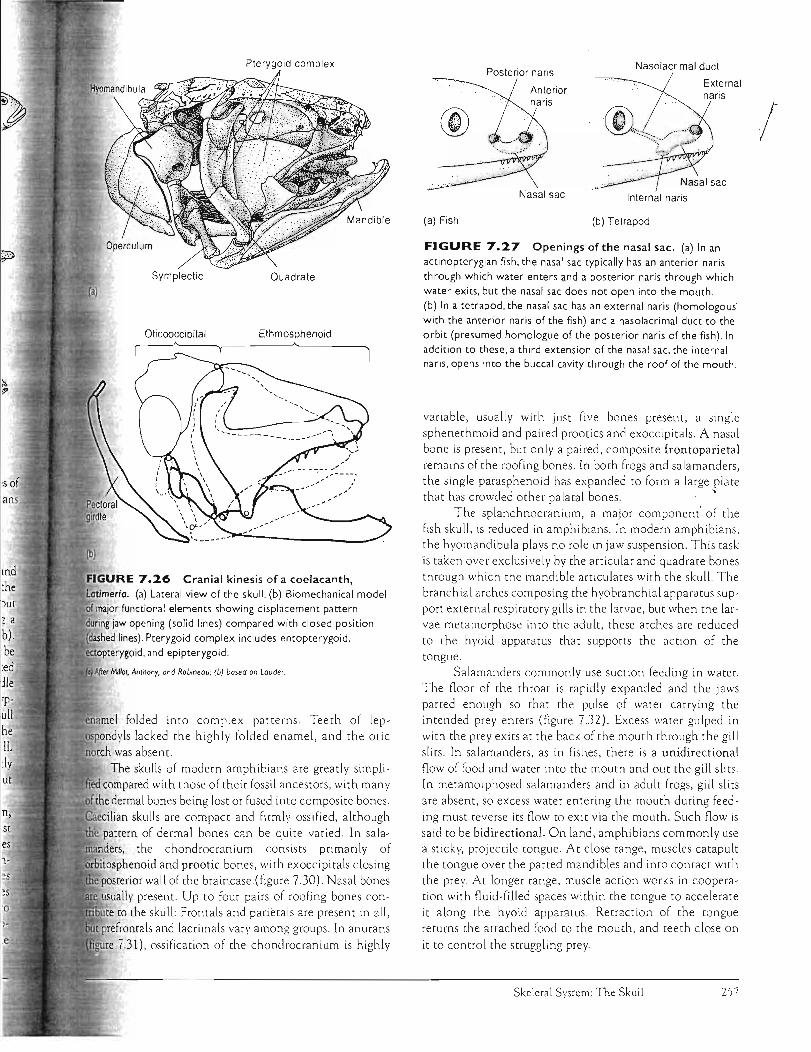
Nasal Capsules From fishes to tetrapods, the nasal capsules have had a complex history. The nasal capsules hold the olfactory epithelium in the form of a paired nasal sac
Sct

Neurocranium
Pectoral girdle
Opercular bones
:--::;;~~::.-- Ste rnohoideus po ..._~__~.)IF' muscle R Hypaxial muscle
\ (b) Open ro jaws 1)1
Jaw opening in a primitive actinopterygian fish. (a) Jaws are closed. (b) Jaws are open. The mandible rotates on its articulation with the suspensorium, which in turn is articulated with the opercular bones. The pectoral girdle remains relatively fixed
1nposition, but the neurocranium rotates on it to lift the head. Lines of action of major muscles are shown by arrows.
(b) Sheepshead fish
Ie) Needlefish
FIGURE 7.24 Teleost skulls.
Despite the great diversification of teleosts in many habits, the basic pattern
of skull bones is preserved. (a) Small
mouth bass (Micropterus dolomieu). (b) Sheepshead fish (Archosorgus probotocepholus). (c) Butterfly fish (Choetodon oce/latus) . (d) Cutlass fish
(Trichiurus lepturus) . (e) Needlefish (Tylosurus morinus).
Skeletal System: The Skull 255

Premaxilla Maxilla
EpaxialI muscles Neurocranium
Mandible
FIGURE 7.2S Suction feeding of a teleost fish. Top series are traces
from a high-speed film of jaw opening (food not shown). Note changes in position of the jaws. Lateral and ventral views, respectively, of the major kinetic bones of the skull are shown when jaws
are closed (left) and when they are open (right). Note the forward movement of the jaws (stippled areas) and outward expansion of the buccal cavity. Lines of muscle action are shown by arrows.
Pectoral Hyobranchial Hyoid
glcdl, ~L""""'W
After Uem.
(figure 7.27a). In actinopterygians, the nasal sac typ ically does not open directly into the mouth. Instead, its anterior (incurrent) and posterior (excurrent) narial openings es tablish a route for one-way water flow across tne olfactory epithelium, delivering to it fresh chemical odors. By co~trast, each nasal sac of tetrapods opens directly into the mouth via an internal naris, or choana (figure 7.27b). Each nasal sac also opens to tne exterior by way of an external naris (nostril), thus establishing a respiratory route for airflow in and o ut of the lungs. [n addition to internal and external nares, a third opening within the nasa l sac begins as a tube, the nasolacrimal duct, that runs toward the orbi t in order to drain away excess secretions of the adjoi ning lacrimal gland after helping to moisten tne surface of the eye.
Olfactory organs (p. 666)
Among sarcopterygians, the nasa l capsules of rhipidistlans are similar to those of tetrapods. In rhipidistians, the nasolacrimal duct is an adaptation that benefits surface fishes that poke tneir eyes and nostrils Out of the water. The lacrima l gland moistens exposed sensory o rgam that are subJected to drying. Tne nasolacrimal duct is probably homologous to the posterior (excurrent) naris of actinopterygian fishes. Rhipidistians (but not coelacanths) also possess internal nares, apparently representing a new derivative of the nasal sac connecting it with the mouth. However, lungfishes probably lack internal nares, a lthough tnis is still debated. In lungfishes, the posterior (excurrent) naris opens near the
Ventral view
margin of the mouth but does not pierce the palatal series of dermal bones as does the true internal nar is of rhipidistians and tetrapods.
Early Tetrapods The earliest tetrapods arose from rhipidistian ancestors and retained many of their skull fe a tures, including most of the bones of the dermatocranium. Numerous bones in the snour were reduced, leaving a distinct nasal bone occupying a position medial to the external naris (figure 7.Z8a,b). Beginning in tetrapods, the hyomandibula ceases to be involved in jaw suspension and instead becomes dedicated to hearing as the stapes (or columella) within the middle ear. The opQ£1llaLS~of bones covering the gills are typo ically lost. Extrascapulars across the back'of the fish skull also dis'3ppear in primitive tetrapods. Along with this, (he
'\ ' pectoral girdle loses its agachG1_ent tO.lhe back of the skull. Roofi~gbo~~~- and chondrocranium b~me inore ttghdy associated, reducing the neuroc ran ial mobility of the snout in comparison with rhipidistians.
The lateral line system, an aquatic sensory system, is evident in skulls of the earliest tetrapods, at least among the juveniles that were presumably aquatic stages (figure 7.Z9a,c). The skull is fl a ttened, and in some a tern · poral notch at the back of the sku ll is presenr. The stapes conveys sound vibrations to the inner ear. But tne stapes in early tetrapods is still a robust bone that also seenis to
be a buttress between the braincase and the palata· quadrate. Teeth were conical in labyr inthodonts, with the
o (, e
Ie
e.
o
n
fi,
01 C d: m
or tho
rri bu (fi
Chapter Seven 256
I
Symplectic Quadrate
Ethmosphenoid ~__~~______~A~________~
functional elements showing displacement pattern " ..mu lrmo jaw opening (solid lines) compared with closed position
lines). Pterygoid complex includes entopterygoid. ~~'OD!:~rv'90i,d and epipterygoid.
folded into complex patterns. Teeth of lep_,osDonllvls lacked the highly folded enamel, and the otic
was absent. The skulls of modem ampnibians are greatly simpli
compared with those of their fossil ancestors, with many the dermal bones being lost or fused into composite bones.
Kv~.,.<tcl..,·lian skulls are compact and firml y ossified, although pattern of dermal bones can be quite varied. In sala
the chondrocr;mium consists primarily of orb'ltosptlenloid and prootic bones, with exoccipitals closing rne ,IXJsterior wall of the braincase (figure 7.30). Nasal bones
usually present. Up to four pairs of roofing bones conto the shill: Frontals and parietals are present in all,
prefrontals and lacrimals vary among groups. In anurans
i.31), ossification of the cnondrocranium is highly
Nasolacrimal ductPosterior naris ExternalAnterior
I J
Internal naris
(a) Fish (b) Tetrapod
FIGURE 7.27 Openings of the nasal sac. (a) In an actinopterygian fish. the nasal sac typically has an anterior naris through which water enters and a posterior naris through which water exits. but the nasal sac does not open into the mouth. (b) In a tetrapod. the nasal sac has an external naris (homologous with the anterior naris of the fish) and a nasolacrimal duct to the orbit (presumed homologue of the posterior naris of the fish). In addition to these, a third extension of the nasal sac, the internal naris. opens into the buccal cavity through the roof of the mouth.
variable, usually with JUSt five bones present, a single sphene thmoid and paired prootics and exoccipitals. A nasa l bone is present, but only a paired, composite frontoparietal remains of the roofing bones. In both frogs and salamanders, the single parasphenoid has expanded to form a large plate that has crowded other palatal bones. ~
The splanchnocranium, a major component of th.e fish skull, is reduced in amphibians. In modern amphibians, the hyomandibula plays no role in jaw suspension. This task is taken over exclusively by the articular and quadrate bones through wnich the mandible articulates with the skull. The branchial arches composing the hyobranchial apparatus support external respiratory gills in tne larvae, but when the larvae metamorphose into the adult, these arches are reduced to the hyoid apparatus that supports the action of the tongue.
Salamanders commonly use suction feeding in water. The noor of the throat is rapidly expanded and the jaws parted enough so that the pulse of water carrying the intended prey enters (figure 7.32). Excess water gulped in with the prey exits at the back of the mouth through the gill sl its. In salamanders, as in fishes, there is a unidirectional now of food and water into the mouth and out the gill slits. In metamorphosed salamanders and in adult frogs, gill slits are absent, so excess water entering the mouth during feeding must reverse its now to exit via the mouth. Such now is said to be bidirectional. On land, amphibians commonly lise a sticky, projectile tongue. At close range, muscles catapult the tongue over the parted mandibles and into contact with the prey. At longer range, muscle action works in cooperation with nuid-filled spaces within the tongue to accelerate it along the hyoid apparatus. Retraction of the tongue returns the attached food to the mouth , and teeth close on
it to control the struggling prey.
Skeletal Sysrem: The Skull 257
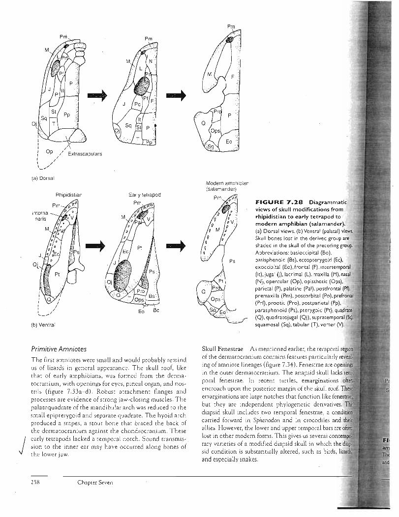
/
Op / Extrascapulars/
//
Pm
OJ
(a) Dorsal
Internal naris
J
J, J,
Rhipidistian
Eo BoJ / J /'-_ .....
(b) Ventral
Primitive Amniotes
J
The first amniotes were small and would probably remind us of lizards in general appearance. The skull roof, like tha t of early amphibians, was formed from the dermatocranium, with openings for eyes, pineal organ, and nostrils (figure 7.33a-d). Robust attachment flanges and processes are evidence of strong jaw-closing muscles. The palatoquadrate of the mandibular arch was reduced to the small epipterygoid and separate quadrate. The hyoid arch produced a stapes, a stout bone that braced the back of the dermatocranium against the chondrocranium. These early tetrapods lacked a temporal notch. Sound transmission to the inner ear may have occurred along bones of the lower jaw.
Chapter Seven
Pm
Modern amphibian (salamander)
FIGURE 7.28 Diagrammatic views of skull modifications from rhipidistian to early tetrapod to modern amphibian (salamander). (a) Dorsal views. (b) Ventral (palatal) views. Skull bones lost in the derived group are shaded in the skull of the preceding group. Abbreviations: basioccipital (Bo), basisphenoid (Bs), ectopterygoid (Ec), exoccipital (Eo), frontal (F) , intertemporal (It), jugal U),lacrimal (L), maxilla (M), nasal (N), opercular (Op), opisthotic (Ops), parietal (P), palatine (Pal), postfrontal (PI), premaxilla (Pm), postorbital (Po), prefrontal (Prf), prootic (Pro), postparietal (Pp), parasphenoid (Ps), pterygoid (Pt), quao~te (Q), quadratojugal (Qj), supratemporal (Stl, squamosal (Sq), tabular (T), vomer (V).
Skull Fenestrae As mentioned earlier, the temporal region of the dermatocranium contains features particularly revealing of amniote lineages (figure 7.34). Fenestrae are openings in the outer dermatocranium. The anapsid skull lacks teO)poral fenestrae. In recent turtles, emarginations encroach upon the posterior margin of the skull roof. emargiI1ations are large notches that function like tenestrlle:.!Ii but they are independent phylogenetic derivatives. The diapsid skull includes two temporal fenestrae, a condition carried forward in Sphenodon and in crocodiles and their allies. However, the lower and upper temporal bars are often lost in other modern forms. This gi ves us several contemp<>c rary varieties of a modified diapsid skull in which the diapsid condition is substantially altered, such as birds, lizards; and especially snakes.
258

Skull of Ichthyostega, a primitive
od of the late Devonian . Dorsal (a), ventral (b), and -".I'_'~__ I (e) views. Parallel tracks of dashed lines indicate course of
the aquatic lateral line system on the skull bones. Abbreviations:
(An), dentary (D), ectopterygoid (Eet) , frontal (F),
I (Itn), jugal OJ,lacrimal (L), maxilla (M), nasal (N), parietal
palatine (Pal), postfrontal (Pf), premaxilla (Pm), postorbital
preopercular (Pop), postparietal (Pp). prefrontal (Prf),
I3ra~;DhE!noid (Ps), pterygoid (Pt), quadrate (Q), quadratojugal surangular (Sa), septomaxilla (Sm), splenial (Sp),
·.''="~ID[,ran~mDor~1 (St). squamosal (Sq), tabular (T). vomer (V).
magnum
Trabecular horn
Ethmoid plate
Fenestra
Antorbital cartilage
Trabecular cartilage
Quadrate bone
Quadrate cartilage
Prootic
Synotic tectum Exoccipital
Basal plate
Skull of Necturus, a modern phibian. Superficial skull bones are indicated on the left.
bones have been removed to reveal the chondrocranium
Pro Sq II
Pm
Hyoid apparatus
(a) Qj
Eo
Sq
Q
(b)
FIGURE 7.3 I Frog skull. Lateral (a) and ventral (b)
views. Abbreviations: articular (Ar) , angulosphenoid (As), basihyal
(Bh), dentary (D), exoccipital (Eo), frontoparietal (Fp). maxilla (M), mento-Meckel ian (Mm), nasal (N) , naris (Na), palatine (Pal) , premaxilla (Pm), prootic (Pro), parasphenoid (Ps) , pterygoid (Pt). quadrate (Q), quadratojugal (Qj), sphenethmoid (Se), squamosal
(Sq), stapes (Stp), vomer (V). Roman numerals indicate foramina
serving specific cranial nerves.
After Marshall.
• (}-Food -;f)J
~, , , "~, ~.:, ~ Quadrate
Pectoral girdle
Closed ,Open
FIGURE 7.32 Suction feeding by an aquatic salamander. Before, during, and after suction feeding traced
from a high-speed film feeding sequence (top series). Note the
interpreted positions of the skull elements when the jaws are
closed (bottom left) and open (bottom right).
After Lauder.
Skeletal System: The Skull 259
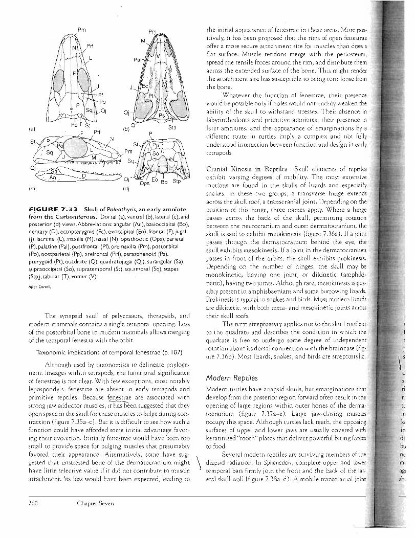
.
SI
tI n~
tc m
ki in di bu ne
Pm
OJ
T St(a~) Stp
.. . . .. Pp F Prl N (b)O P
StP 0 . ".
.·····Sq L· . Pm
St
.
( ' ~ ,
. J .. M . ~Sq
Oj~a ···.~' Oj
(c) (d)
FIGURE 7.33 Skull of Paleothyris, an early amniote from the Carboniferous. Dorsal (a), ventral (b), lateral (c), and posterior (d) views,Abbreviations: angular (An). basioccipital (Bo), dentary (D), ectopterygoid (Ec), exoccipital (Eo), frontal (F), jugal U).lacrimal (L), maxilla (M), nasal (N),opisthootic (Ops), parietal (P), palatine (Pal), postfrontal (Pf), premaxilla (Pm), postorbital (Po), postparietal (Pp), prefrontal (PIi), parasphenoid (Ps), pterygoid (Pt), quadrate (Q), quadratojugal (Qj), surangular (Sa), ~upraoccipital (So), supratemporal (St), squamosal (Sq), stapes (Stpl. tabular (T), vomer (Y),
After Carrol/,
The synapsid skull of pelycosaurs, therapsids, and modern mammals contains a single temporal opening, Loss of the postorbital bone in m,odern mammals allows merging of the tempora l fenestra with the orbit.
Taxonomic implications of temporal fenestrae' (p, 107)
Although used by taxonomists to delineate phylogenetic lineages within tetrapods, the functional significance of fenestrae is not clear, With few exceptions, most notably lepospondyls, fenestrae are absent in early tetrapods and primitive reptiles, Because fenestrae are associated with
strong jaw adductor muscles, it ha'0;een suggested that they open space in the skull for these muscles to bulge during contraction (figure 7,35a-c), But it is difficult to see how such a function could have afforded some initial advantage favoring their evolution, Initially fenestrae would have been too small to provide space for bulging muscles that presumably favored their appearance, Alternatively, some have sug
gested that unstressed bone of the dermatocranium might \ have little selective value if it did not contribute to muscle
attachment. Its loss would have been expected, leading to
the initial appearance of fenestrae in these areas, More positively, it has been proposed that the rims of open fenestrae offer a more secure attachment site for muscles than does a flat surface, Muscle tendons merge with the. periosteum, spread the tensile forces around the rim, and disrribute them
across the extended surface of the bone, This might render the attachment site less susceptible to being torn loose from the bone,
'Whatever the function of fenestrae, their presence would be possible only if holes would not unduly weaken the ability of the skull to withstand stresses, Their absence in labyrinthodonts and primitive amniotes, their presence in
later amniores, and the appearance of emarginations by a different route in turtles imply a complex and nbt fully
understood interaction between function and design in early tetrapods,
Cranial Kinesis in Reptiles Skull elements of reptiles exhibit varying degrees of mobility, The most extensive motions are found in the skulls of lizards and especially snakes, In these two groups, a transverse hinge extends across the skull roof, a transcranial joint. Depending on the position of this hinge, three names apply, Where a hinge passes across the back of the skull, permitting rotation between the neurocranium and outer dermatocranium, the skull is said to exhibit metakinesis (figure 7.36a), If a joint passes through the dermatocranium behind the eye, the skull exhibits mesokinesis. If a joint in the dermatocranium passes in front of the orbits , the skull exhibits prokinesis. Depending on the number of hinges, the skull may be monokinetic, having one joint, or dikinetic (amphiki
netic), having twO jo ints, Although rare, mesokinesis is possibly present in amphisbaenians and some burrowing lizards, Prokinesis is typical in snakes and birds, Most modern lizards
are dikinetic, with both meta- and mes9kinetic joints across their skull roofs,
The term streptostyly applies not to the skull roof but to the quadrate and describes the condition in which the quadrate is free to undergo some degree of independent rotation about its dorsal connection with the braincase (figure 7,36b), Most lizards, snakes, and birds are streptostylic.
Modern Reptiles Modern turtles have anapsid skulls, but emarginations that develop from the posterior region forward often result in the
opening of large regions within outer bones of the dermatocranium (figure 7,3 7a-e), Large jaw-closing muscles occupy this space, Although turtles lack teeth, the opposing surfaces of upper and lower Jaws are usua lly covered with keratinized "tooth" plates that deliver powerful biting forces to food,
Several modern reptiles are surviving members of the diapsid radiation, In Sphenodon, complete upper and lower temporal bars firml y Joi n the front and the back of the lat
eral sku II wall (figure 7.38a-d) A mobile transcranial joint
260 Chapter Seven

~S~Sq~s~ FIGURE 7.34 Major lineages of dermatocranium evolution within amniotes. The anapsid skull occurs in cotylosaurs and their modern descendants, turtles and tortoises. Two major groups, the diapsids and synapsids, independently evolved from the anapsids. Sphenodon and crocodilians retain the primitive diap~id skull. but it has been modified in diapsid derivatives such as snakes, lizards, and birds. Shading indicates positions of temporal fenestrae and orbit.Abbreviations: jugal 0), parietal (P), postorbital (Po), quadratojugal (OJ), squamosal (Sq) .
Modified Modified Modified Modified Modified anapsid testudines diapsid snake diapsid lizard diapsid bird synapsid mammal
" \.ppoj pJ PPo Sq~ Sq~
S ~~ Diapsid Synapsid
q Eu~apsid T /'
Lower jaw
P Po orr:;/
Sq~ OJ J Anapsid
1
Primitive amniote Therapsid
(a) (b)
Mammal
(e)
Temporal fenestrae. The shift in jaw muscle attachment to the skull is shown. (a) Anapsid skull. In early amniotes, temporal muscles run from the neurocranium to the lower jaw. Such a skull is retained in modern turtles. (b) Perforation in the
m opens fenestrae. and attachment of jaw muscles expands to the edges of these openings. (c) Extensive attachment of jaw to the surface of the dermatocranium. Such development of fenestrae characterizes the diapsid and synapsid radiations .
• ....... ,...._ movable palate are absent. Consequently, no significant 1ilII"-.",e,h,· 'ty is permitted within the dermatocranium. Howe ver,
mandible slides back and forth on the fixed quadrate
which it is suspended. The single row of teeth of the
ible moves between a double row of teeth on the upper
an action that seems to be important in slicing through
types of prey.
Loss of the lower temporal bar produces the modified
skull of lizards (figure 7.39). Loss of this lower bony
laterally liberates the posterior part of the skull from
t, thereby permitting streptostyly, and hence pro
the mesokinetic part of lizard dikinesis. Lizard ances
the younginiformes, apparently possessed a single
joint across the back of the skull. A second
joint, the mesokinetic .ioint , has been added to this
modern lizards, making the skulls of most lizards
UUl. II Ll<:lI·'L. Although skulls of some specialized lizards, such as
rs, anteaters, and some herbi vores , seem monoki
, this is likely a secondary condition. This kinetic
of lizard jaws has been modeled as a four-bar link
System (figure 7.40a,b). One unit is the triangular
snout. Its posterior wall forms one of the four
(a) Metakinesis Mesokinesis Prokinesis
Quadrate
(b) Streptostyly
FIGURE 7.36 Cranial kinesis in squamates. (a) There are three types of cranial kinesis based largely on the position at which the hinge (X) lies across the top of the skull. The hinge may run across the back of the skull roof (metakinesis). behind the orbit (mesokinesis). or in front of the orbit where the
snout articulates (prokinesis). (b) The ability of the quadrate to
rotate about its dorsal end is called streptostyly.
J
Skeletal System: The Skull 261
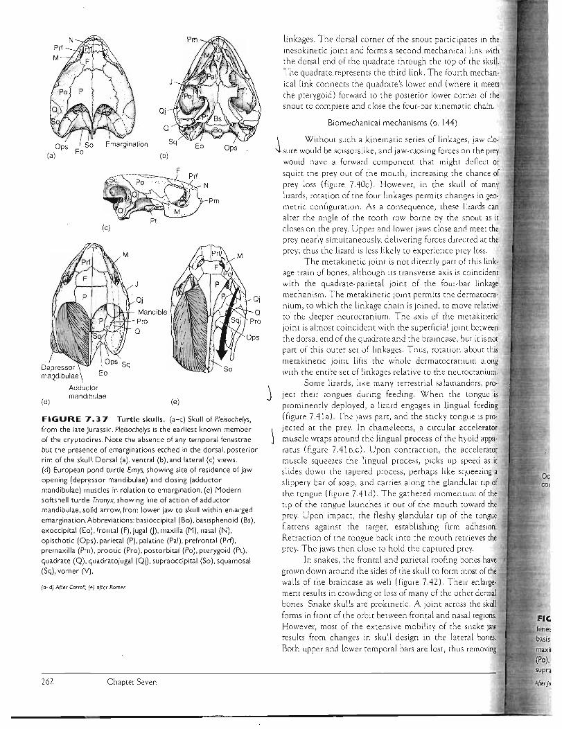
I
OJ o
Pro
linkage
tongue is
OJ
Ops
linkages. The dorsa l corner of the snOut participates in the mesokinetic jOint and forms a second mechanical link wirh the dorsal end of the quadrate through the tOp of (he skull.
The quadrate represents the third link. The fourth mechan
ical link connects the quadrate's lower end (where it meets (he pterygoid) forward to the posterior lower corner of the snout to complete and close the four-bar kinematic chain.
Biomechanical mechanisms (p. 144)
\ Without such a kinematic series of linkages, jaw cloOps I So Emargination
-.J sure would be scissors like, and jaw-closi ng forces on the prey(a) Eo (b) would have a forward component that might deflect or
squirt the prey Out of the mouth, increasing the chance of prey loss (figure 7.40c). However, in the skull of many
lizards, rotation of the four linkages permits changes in geometric configuration. As a consequence, these lizards can alter the angle of the tOoth row borne by the snout as it closes on the prey. Upper and lower jaws close and meer the prey nearly Simultaneously, delivering forces di rec ted at the prey; thus the lizard is less likely to experience prey loss.
The meta kinetic joint is not directly part of this linkage train of bones, although its transverse axis is coincidem
J with the quadrate-parietal joint of the four-bar mechanism. The metakinetic joint permits the dermarocra
OJ nium, to which the linkage chain is joined, to move relative
Mandible to the deeper neurocranium. The axis of the metakineric
Pro joint is almost coincident with the superficial joint betweeno the dorsal end of the quadrate and the braincase, but it is not part of this outer set of linkages. Thus, rotation about this metakinetic joint lifts the whole dermatocranium along with the entire set of linkages relative to the neurocranium.
Some lizards, like many terrestrial salamanders, pro-j ject their tongues during feeding. When the
(d) (e) prominently deployed , a lizard engages in lingual feeding (figure 7.41a). The jaws part, and the sticky tongue is proFIGURE 7.37 Turtle skulls. (a-c) Skull of Pleisochelys,
from the late Jurassic. Pleisochelys is the earliest known member Jected at the prey. In chameleons, a circular accelerator
of the cryptodires. Note the absence of any temporal fenestrae ) muscle wraps around the lingual process of the hyoid appabut the presence of emarginations etched in the dorsal, posterior ratus (figure 7.41b,c). Upon contraction, the accelerator rim of the skull. Dorsal (a), ventral (b), and lateral (c) views. muscle squeezes the lingual process, p iCks up speed as it (d) European pond turtle Emys, showing site of residence of jaw slides down the tapered process, perhaps like squeezing a opening (depressor mandibulae) and closing (adductor slippe ry bar of soap, and carries along the glandu lar rip of mandibulae) muscles in relation to emargination. (e) Modern the tongue (figure 7.41 d) . The gathered momentum of the softshell turtle Trionyx, showing line of action of adductor
tip of the tongue launches it out of the mouth toward the mandibulae, solid arrow, from lower jaw to skull within enlarged
prey. Upon impact, the fleshy glandular tip of the tongueemargination.Abbreviations: basioccipital (Bo), basisphenoid (Bs),
fla[[ens against the target, establishing firm adhesion.exoccipital (Eo), frontal (F), jugal 0), maxilla (M), nasal (N), Retraction of the tongue back into the mouth retrieves theopisthotic (Ops), parietal (P), palatine (Pal), prefrontal (Prf),
premaxilla (Pm), prootic (Pro), postorbital (Po), pterygOid (Pt), prey. The jaws then close to hold the captured prey.
quadrate (Q), quadratojugal (Qj), supraoccipital (So), squamosal In snakes, the frontal and parietal roofing bones have (Sq), vomer (Y). grown down around the sides of the skull to form most of the
walls of the braincase as well (figure 7.42). Their enlarge(a-d) Afrer Carroll; (e) afrer Romer.
ment results in crowding or loss of many of the other dermal bones . Snake skulls are prokinetic. A joint across the skull
forms in front of the orbit between frontal and nasal regions. However, most of the extensive mobility of the snake jaw results from changes in skull design in the lateral bones.
Both upper and lower temporal bars are lost, thus removing
262 Chapter Seven
Depressor f maQdibulae \
M
Adductor mandibulae

(b)
Eo
Qj-a...........~. ,'.~.....
SIp
(d)
VII PI
Ops
Bs--~'
Basiplerygoid process
Pm
Hypoglossal foramen
Medial
FIGURE 7.38 Living rhynchocephalian. The two temporal
fenestrae are still bounded by bone in
Sphenodon, a living diapsid. Dorsal (a),
ventral (b). lateral (c), and posterior (d)
views.Abbrevi;ltions: angular (An),
articular (Ar). basioccipital (Bo),
basisphenoid (Bs). coronoid (C), dentary
(D). ectopterygoid (Ec), exoccipital (Eo). epipterygOid (Ept), frontal (F) , jugal 0), maxilla (M). nasal (N), opisthotic (Ops),
parietal (P). prearticular (Pa), palatine (Pal). postfrontal (Pf), premaxilla (Pm).
postorbital (Po). prefrontal (Prf), prootic (Pro), parasphenoid (Ps), pterygoid (Pt),
quadrate (Q). quadratojugal (Qj). surangular (Sa). supraoccipital (Soc).
squamosal (Sq). stapes (Stp). vomer (V).
After Carroll.
Ec M
Foramen for Jacobson's organ
Pm
Foramen magnum
Lizard skull. Lizards are modified diapsids.Two fenestrae are present, but the ventral bony border of the lower
is absent. a result of changes serving increased cranial kinesis. Abbreviations: angular (An), articular (Ar). baSioccipital (Bo),
il<i<I,h~rlf'li'd (Bs). coronoid (C), dentary (D) , ectopterygoid (Ee), exoccipital (Eo). epipterygoid (Ept), frontal (F), jugal 0), lacrimal (L),
(M). nasal (N). opisthotic (Ops), orbitosphenoid (Osp). parietal (P), prearticular (Pa), postfrontal (Pf), premaxilla (Pm), postorbital
prefrontal (Prf), parasphenoid (Ps) . prootic (Pro). pterygoid (Pt). quadrate (Q), surangular (Sa). septomaxilla (Sm), splenial (Sp),
. ital (So). squamosal (Sq). supratemporal (St), stapes (Stp). vomer (V).
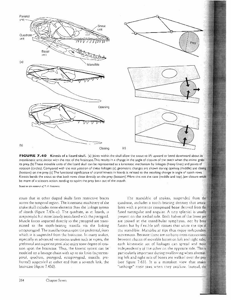
Parietal unit -=-~/~:::;::::::;:;::
~:~><::~~:-::
(a)
(b) Closing (c)
FIGURE 7.40 Kinesis of a lizard skull. mesokinetic articulation with the rest of the braincase. This results in a change in the angle of closure of the teeth when the animal grabs
its prey. (b) These movable units of the lizard skull can be represented as a kinematic mechanism by linkages (heavy lines) and points of
rotation (circles). Compared with the rest position of these linkages (a), geometric changes are shown during opening (middle) and closing (bottOm) on the prey. (c) The functional significance of cranial kinesis in lizards is related to the resulting change in angle of tooth rows.
Kinesis bends the snout so that both rows close directly on the prey (bottOm).Were this not the case (middle and top), jaw closure would be more of a scissors action, tending to squirt the prey back out of the mouth .
Based on the research or T. H Frazze[{a.
struts that in other diapsid skulls form restrictive braces across the temporal region . The kinematic machinery of the snake skull includes more elements than the linkage system of lizards (figure 7.43a-c). The quadrate, as in lizards, is streptostylic but more loosely articulated with the pterygoid. Muscle forces imparted directly to the pterygoid are transmitted to the tooth-bearing maxilla via the linking ectopterygoid. The maxilla rotates upon the prefrontal, from which it is suspended from the braincase . In many snakes, especially in ad vanced venomous snakes such as vipers, the prefrontal and supratemporal a lso enjoy some degree of rotation upon the braincase. Thus, the kinetic system can be · modeled on a linkage chain with up to six links (supratemporal, quadrate, pterygoid, ectopterygoid, maxilla, prefrontal) suspended at either. end from a seventh link , the braincase (figure 7.43d).
Chapter Seven
o
(a) joints within the skull allow the snout to lift upward or bend downward about its
The mandible of snakes, suspended from the quadrate, includes a tooth -bearing dentary that articulates with a posterior compound bone derived from the fused surangular and angular. A tiny splenial is usually present on the medial side. Both halves of the lower jaw are joined at the mandibular symp hysis, not by bony fusion but by flexible soft tissues that unite the tips of the mandible. Mandibular tips thus enjoy independent movement. Because there are no bony cross connection) between chains of movable bones on left and right sides, each kinematic set of lin kages can spread and move independently of the other on the opposite side. This is particularly important during swa llowing when al ternating left and right sets of bones are walked over the prey (see figure 7.63) . It is a mistaken view that snake) "u nhinge" their jaws when they swa llow. Instead, the
264
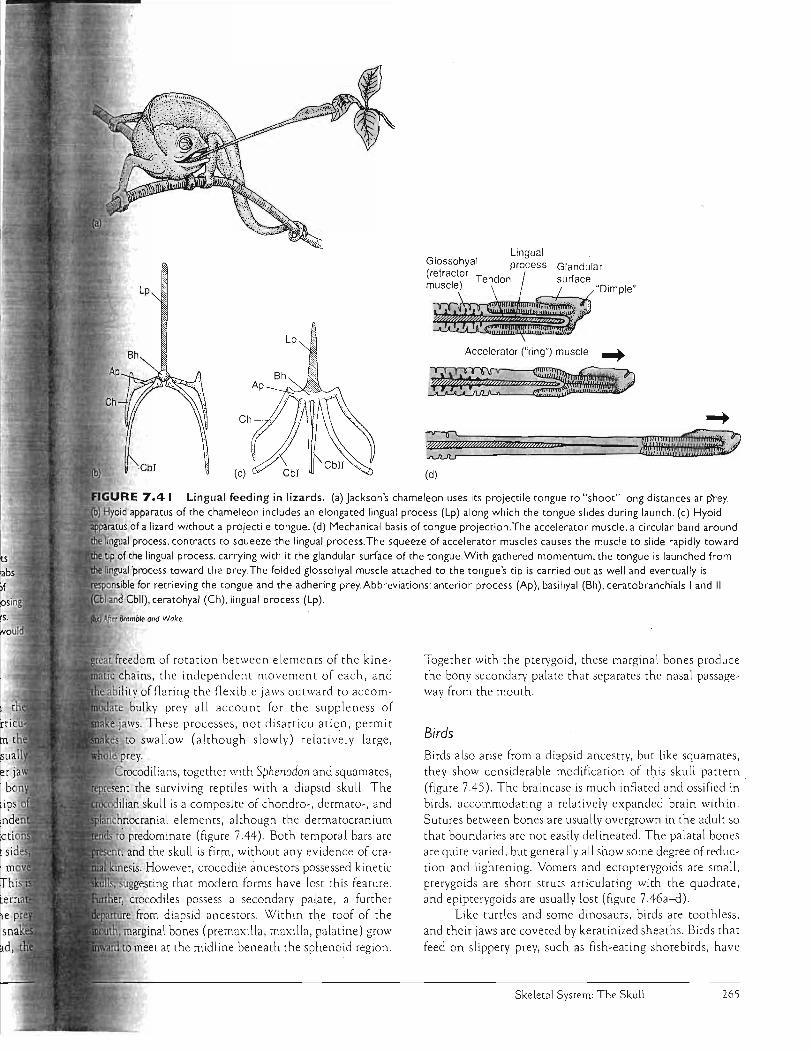
Lp
Lp Accelerator ("ring") muscle -+Bh
~fRJ .... $M'@N;:~
(d)
Lingual feeding in lizards. (a) Jackson's chameleon uses its projectile tongue to "shoot" long distances at p'rey. Hyoid apparatus of the chameleon includes an elongated lingual process (Lp) along which the tongue slides during launch. (c) Hyoid
of a lizard without a projectile tongue. (d) Mechanical basis of tongue projection.The accelerator muscle, a circular band around )ingual process. contracts to squeeze the lingual process. The squeeze of accelerator muscles causes the muscle to slide rapidly toward I of the lingual process. carrying with it the glandular surface of the tongue. With gathered momentum, the tongue is launched from
'process toward the prey. The folded glossohyal muscle attached to the tongue's tip is carried out as well and eventually is 'e$o<onSIIOle for retrieving the tongue and the adhering prey. Abbreviations: anterior process (Ap), basihyal (Bh), ceratobranchials I and II
Cbll), ceratohyal (Ch), lingual process (Lp).
freedom of rotation between elements of the kinechains, the independent movement of each, and i!ity of f1aring the f1exible jaws outward to accom
bulky prey all account for the suppleness of ws. These processes, not disarticulation, permit to swallow (although slow ly) relatively large,
Crocodilians, together with Sphenodon and squamates, t the surviving reptiles with a diapsid skull. The
lian skull is a composite of chondro-, dermato-, and ial elements, although the dermatocranium
to predominate (figure 7.44). Both temporal bars are and the skull is firm, without any evidence of cra-
However, crocodile ancestors possessed kinetic uggesting that modern forms have lost this feature.
crocodiles possess a secondary palate, a further from diapsid ancestors. Within rhe roof of the
marginal bones (premaxilla, maxilla, palatine) grow meet at the midline beneath the sphenoid region.
Together with the pterygoid, these marginal bones produce the bony secondary palate that separates the nasal passageway from the mouth.
Birds Birds also arise from a diapsid ancestry, but like squamates, they show considerable modification of t\:lis skull pattern . (figure 745). The braincase is much inflated and ossified in birds, accommodating a relatively expanded brain within. Sutures between bones are usually overgrown in the adult so that boundaries are not easily delineated . The palatal bones are quite varied, but gene.rally all show some degree of reduction and lightening. Vomers and ectopterygoids are small, pterygoids are short struts articulating with the quadrate, and epipterygoids are usually lost (figure 7.46a-d).
Like turtles and some dinosaurs, birds are toothless, and their jaws are covered by keratinized sheaths. Birds that
feed on slippery prey, such as fish-eating shorebirds, have
Skeletal System: The Skull 265

Pm
FIGURE 7.42
OJ
St T Pp
Diagrammatic comparison of a derived modern snake skull with an amniote skull. Bones lost in
the modern snake are indicated by
shading in the primitive
captorhinomorph.Abbrevia tions: exoccipital (Eo). frontal (F), jugal 0), lacrimal (L), maxilla (M), nasal (N), parietal (P), postfrontal (Pf), premaxilla (Pm). postorbital (Po). postparietal (Pp). prefrontal (Prf),
pterygOid (Pt). quadrate (Q), quadratojugal (Qj), supraoccipital
(50). squamosal (5q). supratemporal (5t), tabular (T). Primitive amniole
FIGURE 7.43 Kinematic model of movable skull bones in a venomous snake, the water moccasin. Whole head (a) with successive removal of skin and muscles (b) reveals bones of the skull (c). linkage bones movable relative to the braincase are in color; lower jaw is crosshatched.
(d) Biomechanical model of movable bones rotatable about pin
connections. Movable bones include the ectopterygoid (Ec), maxilla (M). pterygOid (Pt), prefrontal (Prf), quadrate (Q). supratemporal (5t). Location of the main venom gland (Vg) is shown as well.
After Kardong.
266 Chapter Seven
(b)
(c)
(d)
Derived reptile (viperid snake)
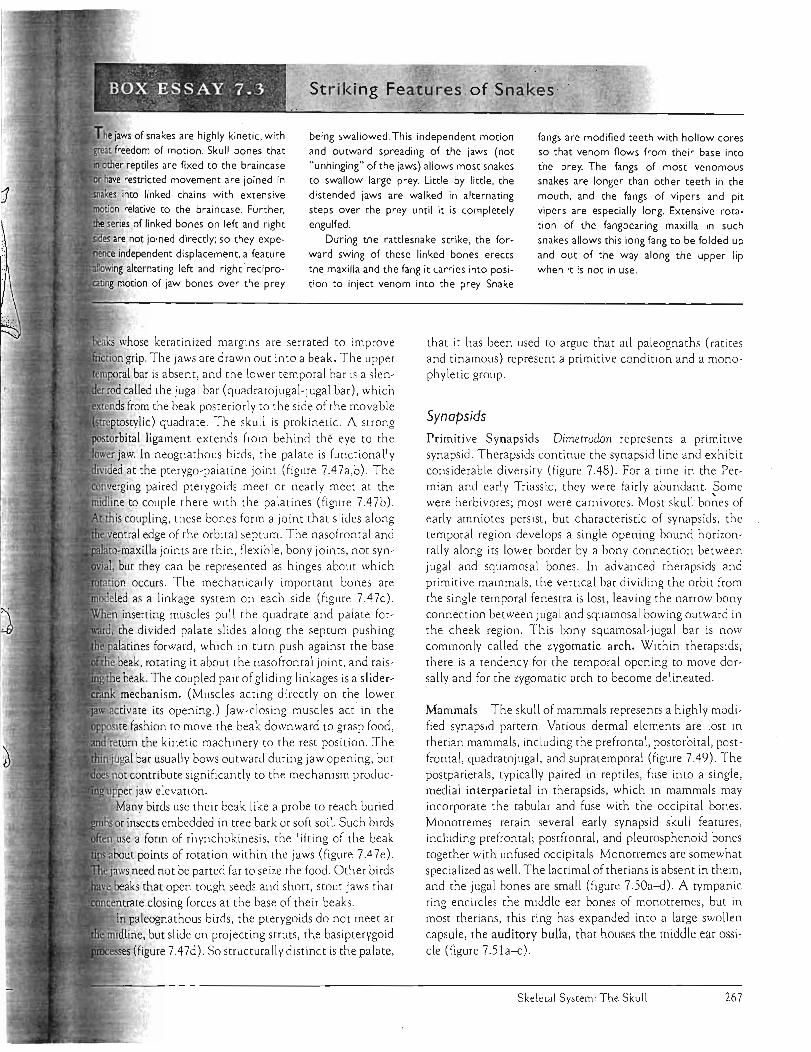
Stri ki ng . Eeatures of Snakes· ~"
he jaws of snakes are highly kinetic. wi[h freedom of motion. Skull bones that
reptiles are fixed to the braincase restricted movement are joined in
into linked chains with extensive
relative to the braincase. Funher. series of linked bones on left and right
not joined directly; so they expe
independent displacement. a feature
alternating left and right ·recipromotion of jaw bones over the' prey
being swallowed. Th is independent motion
and outward spreading of the jaws (not "unhinging" of the jaws) allows most snakes to swallow large prey. Little by little. the
distended jaws are walked in alternating steps over [he prey until it is completely
engulfed.
During the rattlesnake strike. the for
ward swing of these linked bones erects
the maxilla and the fang it carries into position to inject venom into the prey. Snake
fangs are modified teeth with hollow cores so that venom flows from their base into
the prey. The fangs of most venomous snakes are longer than other teeth in the
mouth. and the fangs of vipers and pit vipers are especially long. Extensive rota
tion of the fangbearing maxilla in such
snakes allows this long fang to be folded up
and out of the way along the upper lip when it is not in use .
whose keratinized margins a re se rrated to improve grip. The Jaws are drawn ou t into a beak. The upper I bar is absent, and the lower temporal bar is a slen
called the jugal bar (quadratojugal-jugal bar), which from the beak posteriorly to the side of the movable
;trel}to:;tV.1ic) quadrate. The skull is prokinetic. A strong ligament extends from behind the eye to the
jaw. In neognathous birds, the palate is functionally at the pterygo-paLatine joint (figure 7A7a,b). The 'ng paired pterygoids meet or nearly meet at the to couple there with the palatines (figure 7,47b).
this coupling, these bones form a .joint that slides along edge of the orbital septum. The nasofrontal and 'lla joints are thin, f1exible, bony joints, not syn
but they can be represented as hinges about which occurs . The mechanically important bones are as a linkage system on each side (figure 7,47c).
inserting muscles pull the quadrate and palate forthe divided palate slides along the septu m pushing
pa,ac",,,, forward, which in turn push against the base beak, rotating it about the nasofrontal joint, and raisbeak. The coupled pair of gliding linkages is a slidermechanism. (Muscles acting directly on the lower vate its opening.) Jaw-closing muscles act in the fashion to move the beak downward to grasp food,
return the kinetic machinery to the rest pOSition. The jugal bar usually bows outward during jaw opening, but not contribute significantly to the mechanism produc
jaw elevation. Many birds use their beak like a probe to reach buried
insects embedded in tree bark or soft soil. Such birds use a form of rhynchokinesis, the lifting of the beak
points of rotation within the jaws (figure 7,47e). need not be parted far to seize the food. Other birds
that open tough seeds and short. stout jaws that ~~J'l.1L'."U: closing forces at the base of their beaks.
In paleognathous birds, the pterygoids do not meet at but slide on projecting struts , the basipterygoid
(figure 7,47d). So structurally distinct is the palate,
that it has been used to argue that all paleognaths (ratites and tinamous) represent a primitive condition and a monophyletic group.
Synapsids Primitive Synapsids Dimetrodon represents a pnmltlve synapsid. Therapsids continue the synaps id line and exhibit consider~ble diversity (figure 7,48). For a time in the Permian and early Triassic, they were fairly abundant. Some were herbivores; most were carnivores. Most skull ' bo~es of early amniotes persist, but characteristic of synapsids. the temporal region develops a single opening bound horizontally along its lower border by a bony connection between jugal and squamosal bones. In advanced therapsids and primitive mammals, the vertical bar dividing the orbit from the single temporal fenestra is lost. leaving the narrow bony connection between jugal and squamosal bowing outward in the cheek region. This bony squamosal-jugal bar is now commonly called the zygomatic arch. Within therapsids, there is a tendency for the temporal opening to move dorsally and for the zygomatic arch to become delineated.
Mammals The sku ll of mammals represents a highly modified synapsid pattern. Various dermal elements are lost in therian mammals, including the prefrontal, postorbital, postfrontal, quadratojugal. and supratemporal (figure 7,49). The postparietals, typically paired in reptiles, fuse into a single, medial interparietal in the raps ids, which in mammals may incorporate the tabular and fuse with the occipital bones. Monotremes retain several early synapsid sku ll features. including prefrontal; postfrontal, and pleurosphenoid bones together with unfused occipitals. Monotremes are somewhat specialized as well. The lacrimal of therians is absent in them, and the jugal bones are small (figure 7.50a-d). A tympanic ring encircles the middle ear bones of monotremes, but in most therians, this ring has expanded into a large swollen capsule, the auditory bulla, that houses the middle ear ossicle (figure 7.51a-<:).
Skeleral Sys[em: The Skull 267
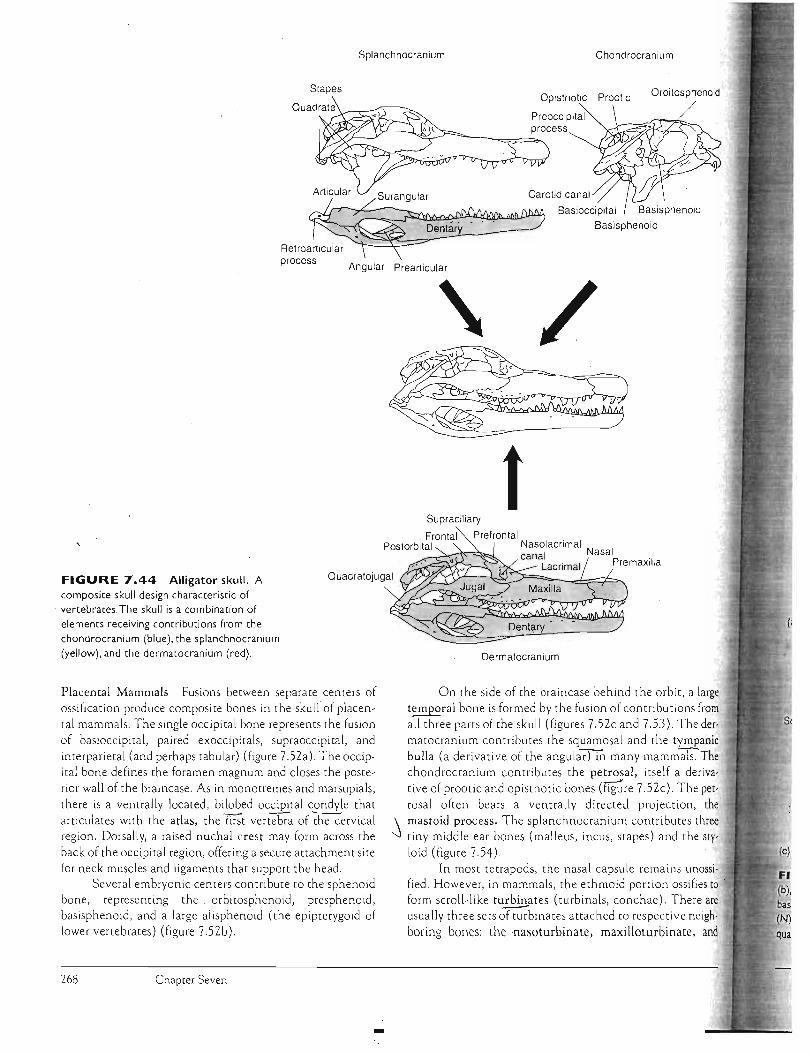
Splanchnocranium Chondrocranium
Stapes Opisthotic Prootic
Preoccipital process
Orbitosphenoid
Basioccipital Basisphenoid Basisphenoid
process Angular Prearticular
\
tSupraciliary
Frontal Prefrontal Postorbital Nasolacrimal
canal Nasal . Lacrimal PremaXilla
FIGURE 7.44 Alligator skull. A composite skull design characteristic of
Quadratojugal
, vertebrates, The skull is a combination of elements receiving contributions from the chondrocranium (blue), the splanchnocranium (yellow), and the dermatocranium (red).
Placental Mammals Fusions between separate centers of
ossification produce composite bones in the skull of placen
tal mammals. The single occipital bone represents the fusion
of basioccipital, paired exoccipitals, supraoccipital, and
interparietal (and perhaps tabular) (figure 7.52a). The occip
ital bone defines the foramen magnum and closes the poste
rior wall of the braincase. As in monotremes and marsupials,
there is a ventrally located, bitobed occipital condyle that
articulates with the atlas, the fust vertebra of cl1eC"ervical region. Dorsally, a raised nuchal crest may form across the
back of the occipital region, offering a secure attachment site
for neck muscles and ligaments that support the head. Several embryonic centers contribute to the sphenoid
bone, representing the. orbitosphenoid, presphenoid,
basisphenoid, and a large alisphenoid (the epipterygoid of
lower vertebrates) (figure 7.52b).
Dermatocranium
On the side of the braincase behind the orbit, a large
temporal bone is formed by the fusion of contributions from
all three pam of the skull (figures 7.S2c and 7.53). The der
matocranium contributes the squamosal and the tympanic
bulla (a derivative of the angularrtn many mammals. The
chondrocranium contributes the petrosal, itself a deriva
tive of prootic and opisthotic bones (rrg;:re 7.52c). The pet
rosal often bears a ventrally directed prOJection, the
\ mastoid process. The splanchnocranium contributes three -.j tiny middle ear bones (malleus, incus, stapes) and the sty
loid (figure 7.54).
In most tetrapods, the nasal capsule remains unossified. However, in mammals, the ethmoid portion ossifies to .
form scroll-like turbinates (turbinals, conchae). There are usually three sets ~binates attached to respective neigh
boring bones: the ·nasoturbinate, maxilloturbinate, and
Chapter Seven 268

So
FIGURE 7.4S Diagrammatic comparison of a derived bird skull with a primitive amniote. Bones lost in birds are shaded in the primitive reptile. Abbreviations: frontal (F). jugal 0), lacrimal (L), maxilla (M), nasal (N), parietal (P), postfrontal (P£), premaxilla (Pm), postorbital (Po), postparietal (Pp), prefrontal (Pr/), quadratojugal (Qj), supraoccipital (So) , squamosal (Sq),
Primitive amniote Bird supratemporal (St), tabular (T).
Sq PI
Pm
Bs OJ I
I
Jugal bar (b)
Sq
Bo Eo
An
(d)
In the adult bird, sutures between skull bones fuse to obliterate identifiable borders. Dorsal (a), ventral (c), and posterior (d) views of the skull of a young gosling (Anser) before bones fuse. Abbreviations: angular (An) , articular (Ar),
(Bo). basisphenoid (Bs), dentary (D), exoccipital (Eo), frontal (F), jugal 0) , lacrimal (L),laterosphenoid (Ls), maxilla (M) , nasal (Ops), parietal (P), palatine (Pal), premaxilla (Pm) , postorbital (Po), parasphenoid (Ps), pterygoid (Pt), quadrate (Q),
(Qj). surangular (Sa), supraoccipital (So), squamosal (Sq), vomer (Y).
Skeletal Sysrem: The Skull 269

--
process
(d)
(e)
(c)
FIGURE 7.47 Cranial kinesis in the crow skull (Corvus). (a) Lateral view. (b) Ventral view. (c) Linkage model of cranial kinesis. slider-crank mechanism. From the rest position (solid lines). the point of coupling between pterygoids and palatines slides forward along the orbital septum to a new position (dashed lines). which lifts the upper jaw about the prokinetic joint (nasofrontal hinge). (d) Paleognath. Ostrich palate. (e) Rhynchokinesis. Flexions within the beak permit the tips of the upper and lower jaws to part without
Maxilla
Palatine
Jugal bar
Pterygoid
Basipterygoid
Quadrate
ary transition undeniable. One of these changes is the the postdentary bones of the lower jaw. The other is presence ofrnree ~le ear bones. In vertebrates, the ' ear is embedd-ed deep wlthtn the otic capsule and holds sensory apparatus responsive to sounds. The hyo or its derivatives deliver sound vibrations to the sens .
tetrapods, the hyomandibula tends become reduced to a slender, light bone called the (= columella). Sometimes there is a second hyomandi derived bone, the extracolumella. The stapes is usually
middle ear cavity where damping restrictive attachments is minimized. As sounds set the panum into motion, these vibrations are imparted to
small, responsive stapes. Its opposite end often expands reach the sensitive inner ear appararus that responds to
In mammals, two tiny, additional bones join the in the middle ear. Together these bones transmit sound the inner ear. Specifically, these three middle ear bones
Nasofrontal hinge
(a)
(b)
--
............
opening of the entire mouth.
ethmoturbinate. The coiled walls of the rurbinates support the mucous membrane within the nasal passage. Air entering these passages is warmed and moistened before reaching the lungs, functions that are especially important in endotherms. Absent in ungulates but present in most other orders, such as rodents, carnivores, and primates, is another region of the nasal capsule, the mammal mesethmoid. This element forms the septum between the nasal capsules and usually remains cartilaginous. Between the nasal area and cranial cavity stands the transverse and finely perforated cribriform plate (figure 7.54). Olfactory nerves originating in the olfactory epithelium of the nasal capsule pass through this plate to reach the olfactory bulb of the brain.
Middle Ear Bones Two profound changes in the lower jaw mark the transition from therapsid to mammal (figure 7.55). Both changes go hand in glove. They result in such an alteration in skull design that some anatomists doubted them until the surprisingly good fossil record made the evolution-
Chapter Seven
) ~ J
inner ear. In all
pended in the
vibrations the stapes delivers.
270
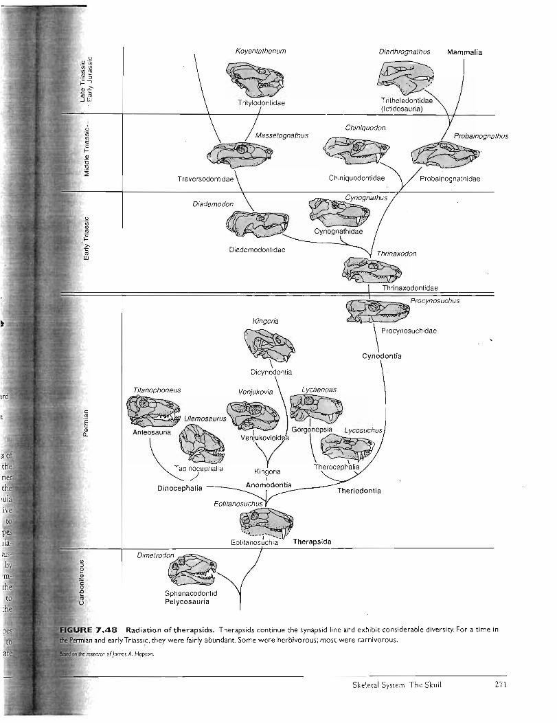
Dinocephalia
Eolitano5uchu5
Theriodontia
D;m"md~
.uuo..,"" 7.48
Koyenlalhenum Diarthrognalhu5 Mammalia
T ritheledontidae (Ictidosauria)
Probainognathidae
~"h",
Cynognathidae
Diademodontidae
Kingoria
~ .~ \ .
Dicynodontia
' : :'i ... .. .~ "'.:.::. : ~ :( . . , .' . ~ .. ~ .,
~.J"'-" - 'i-" """" _ / ~ Eotitanosuchia Therapsida
Sphenacodontid Pelycosauria
Radiation of therapsids. Therapsids continue the synapsid line and exhibit considerable diversity. For a time in
and early Triassic. they were fairly abundant. Some were herbivorous; most were carnivorous.
Skeletal System: The Skull 27 1

(a) Sq
Nuchal Temporalcrest crest
So
OCCipitalEo condyle
(c) (d) Ecv
Pm
OJ
T Pp
Primitive amniote
Pp (Ip)
Mammal (opossum)
Foramen ovaIe
FIGURE 7.49 Diagrammatic comparison of a derived mammal skull with a primitive amniote skull. Bones lost in the derived mammal are
shaded in the primitive amniote. In
mammals, orbital and temporal openings
merge.Abbreviations: frontal (F), jugal 0), interparietal (Ip), lacrimal (L), maxilla (M),
nasal (N), parietal (P), postfrontal (PI), premaxilla (Pm), postorbital (Po),
postparietal (Pp), prefrontal (Prf),
quadratojugal (OJ), squamosal (5q),
supratemporal (5t) , tabular (T).
Foramen rotundum
FIGURE 7.S0 Monotreme, skull of the duckbill platypus Ornithorhynchus. Dorsal (a),ventral (b), lateral (c) , and
posterior (d) views. Abbreviations: dentary (D), ectopterygOid (Ec), exoccipital (Eo), frontal (F), jugal 0), maxilla (M), nasal (N),
orbitosphenoid (Osp), parietal (P) , palatine (Pal), premaxilla (Pm), supraoccipital (50), squamosal (5q), vomer (V).
Chapter Seven 272

II.
:s I). 1),
Single opening for external nares
Inflected angle of dentary
Sq
J
Incisors Incisive foramen
Canine
Sq
Palatel foramina
Internal nares
Foramen [I::'::i-----;n~ ovale
.~~*~"-- ·Carotid canal
Glenoid
ear
Pm
Orbitosphenoid Presphenoid
Alisphenoid
Lingula
Basisphenoid Pterygoid
(b) Sphenoid
FIGURE 7.5 I Marsupial, skull of the opossum Didelphis. Dors~1 (a),
palatal (b), and lateral (c) views. Abbreviations: basioccipital (Bo), basisphenoid (Bs), dentary (D), frontal
(F), jugal 0), lacrimal (L), maxilla (M), nasal (N), parietal (P), palatine (Pal), premaxilla (Pm), parasphenoid (Ps) , pterygoid (Pt),
squamosal (Sq).
After Carroll.
Squamosal
Tympanic
(c) Temporal
Composite bones of the placental mammal skull during embryonic development, Homo sapiens. Occipital bone has centers of ossification that include the interparietal (postparietal), supraoccipital, paired exoccipital, and the
;:,.altt~i()Ccipital. (b) The sphenoid bone is a fusion of the orbitosphenoid, presphenoid, basisphenoid, pterygoid, and alisphenoid (epipterygoid).
many mammals, these fused bones are joined by parts of the pterygoid and lingula. (c) The temporal bone results primarily from the ·~''''~.n~. of the squamosal, tympanic, and petrosal (prootic plus opisthotic).
J
Skeletal System: The Skull 273
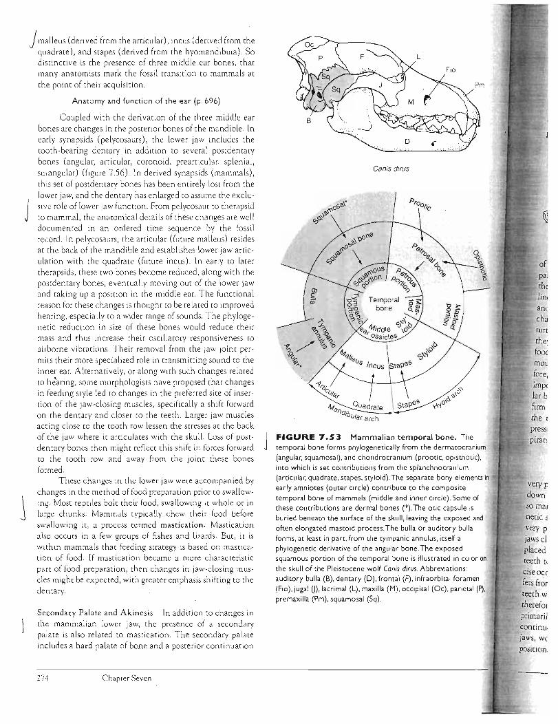
Jmalleus (derived from dle articular), incus (derived from the quadrate), and stapes (derived from the hyomandibula). So distinctive is the presence of three middle ear bones, that many anatomists mark the fossil transition to mammals at
the point of their acquisition.
Anatomy and function of the ear (p. 696)
Coupled with the derivation of the three middle ear bones are changes in the posterior bones of the mandible. In early synapsids (pelycosaurs), the lower jaw includes the tooth-bearing dentary in addition to several postdentary bones (angular, articular, coronoid, prearticular, splenial,
surangular) (figure 7.56). In derived synapsids (mammals),
this set of postdenta ry bones has been entirely lost from the lower jaw, and the dentary has enlarged to assume the exclusive role of lower jaw function. From pelycosaur to therapsid
J to mammal, the anatomical details of these changes are well documented in an ordered time sequence by the fossil reco rd. In pelycosaurs , the articular (future malleus) resides at the back of the mandible and establishes lower jaw articulation with the quadrate (future incus). In early to later therapsids, these twO bones become reduced, along with the postdentary bones, eventually moving Out of the lower jaw and taking up a position in the middle ear. The functional reason for these changes is thought to be related to improved hearing, especia lly to a wider range of sounds. The phylogenetic reduction in size of these bones would reduce their mass and thus increase their oscillatory responsiveness to airborne vibrations. Their removal from the jaw joint per
mits their more spec ialized ro le in transmitting sound to the inner ear. Alternatively, or along with such changes related to he'aring, some morphologists have proposed that changes in feeding style led to changes in the preferred site of insertion of the jaw-clOSing muscles, specifically a shift forward on the dentary and closer to the teeth. Larger jaw muscles acting close to the tooth row lessen the stresses a t the back of the jaw where it articulates with the skull. Loss of postdentary bones then might reflect this shift in forces forward to the tooth row and away from the joint these bones formed.
These changes in the lower jaw were accompanied by
changes in the method of fo od preparation prior to swallowing. Most reptiles bolt their food, swallowing it whole or in large chunks. Mammals typically chew their food before swallowing it, a process termed mastication. Mastication also occurs in a few groups of fishes and lizards. But, it is within mammals that feeding strategy is based on mastication of food. If mastication became a more characteristic part of food preparation, then changes in jaw-closing muscles might be expected, with greater emphasis shi fting to the dentary.
Secondary Palate and Akinesis In addition to changes in the mammalian lower jaw, the presence of a secondary palate is also related to mastication. The second ary palate includes a hard palate of bone and a posterior continuation
Canis dirus
4tq Ouadrate f)C/ib
lJlar arch
FIGURE 7. S 3 Mammalian temporal bone. The
J temporal bone forms phylogenetically from the dermatocranium (angular. squamosal). and chondrocranium (prootic. opisthotic).
into which is set contributions from the splanchnocranium (articular. quadrate. stapes. styloid). The separate bony elements in
early amniotes (outer circle) contribute to the composite
temporal bone of mammals (middle and inner circle). Some of these contributions are dermal bones (*). The otic capsule is
buried beneath the surface of the skull, leaving the exposed and
often elongated mastoid process.The bulla or auditory bulla
forms. at least in part.from the tympanic annulus. itself a phylogenetic derivative of the angular bone. The exposed squamous portion of the temporal bone is illustrated in color on
the skull of the Pleistocene wolf Canis dirus. Abbreviations:
auditory bulla (B). dentary (D). frontal (F). infraorbital foramen
(Fio). jugal U).lacrimal (L). maxilla (M). OCCipital (Oc). parietal (P). premaxilla (Pm), squamosal (Sq).
of pal the lin, an(
cha tun
the· fooe
m OL
tore, imp< lar b firm the
very r: down
so mal
netic ~ very p jaws cl placed teeth t, eise oc(
fers fr~r teeth w theref01
primarii eoncinu.
jaws, we
position
274 Chaprer Seven
I

Postparietal
supraOCCiPital~Barietal petrosal~~
tJ vll
Exoccipital 0 ~
xn Stapes :.£> IX . x, XI --0 Incu~
.. ~~eu~Tympamc nng~_ tal~
.,"·..r. ".'~'V"~'fJ' -------.; Basisphenoid
uamosal Tympanohyold· ~
StylOhyoidi\ Epihyoid EPi9lOltis~ V
Thyrohyoid <--~. (f~G , "O,,,IOhYO;d
Arytenoid Basihyoid
Ill , IV. .J/ Cribriform plate Alisphe- opv, 6~Lacrrmal
noid JVI 8 ~ 0 l::l g Nasal
~~ V. m V. max § ~ Meselhmold ~phenOiCIl ~
-.::::J Premaxilla ("omer >
ptery-GDala-~ 'd Maxilla gOI hne
FIGURE 7.54 Diagram ofa dog's skull, Sources of the various bones are outlined: dermatocranium (red), chondrocranium (blue), and splanchnocranium (yellow).
Meckel's cartilage
of fleshy tissue, the soft palate (figure 7.5 7a,b). The h ard
galate is f~rmed from the inward growth of bony processes of
premaxilla, maxilla, and palatine tha t meet at the mid as a bony platform (figure 7.58a-c). This hard palate
its fleshy continuation effectively separate the food below from the respiratory passage above. Some
and also crocodilians have a secondary palate, and
they benefi t from the advantages of separation of routes for
food and air. But chewing requ ires that food stays in the mouth for an extended period of time in mammals; there-)
Jore, sepmation of respiratory and oral passages is especially
important. Mastication can proceed without impeding regu
lar breathing. Similarly, the secondary palate completes the
firm roof of the food chamber, so that the pumping acti on of the throat of a suckling infant creates effective negative
pressure within the mouth without intelfering with the respiratory passage.
Mastication (p.529)
Mastication in mammals has been accompanied by
precise tooth occlusion to serve the mechanical break
oHood. Precise, strong occlusion requires a firm skull,
mammals have lost cranial kinesis, leav ing them an aki
_...... ,." .. _ skull. The mammalian mandibular condyle fits into a
very precise articulation with the squamosal bone. When
jaws close about this joint, upper and lower rows of teeth are placed in very precise alignment. This allows specialized
teeth to function properly. As a further consequence of pre
cise occlusion , the pattern of tooth eruption in mammals differs from that of most other vertebrates. In lower vertebrates, J
"";..'~~'" wear and are replaced continuously (polyphyodonty); therefore, the tooth row is always changing. If teeth function primarily to snag prey, this causes little difficulty. H owever, '
us replacement means that at some location in the
worn teeth are missing or new ones are moving into
To avoid disruptio~ of occlusion, teeth in most
After Evans.
mammals are not continuously replaced. Mammals exhibit
.J diphyodonty. Only two sets of teeth erupt during the life
time of a mammal, the "milk teeth" of the young and the "permanent" teeth of the adults.
Tooth types and their development (p. 500)
The chain of events leading from mastication to aki
nesis and to diphyodonty should not be viewed as inevi~ble. Some fishes chew their food but retain kinetic skulls and
polyphyodonty. But evolutionary events that produced the
mammalian skull underscore the importance of examining
anatomical changes in partnership with functional changes
that must accompany phylogenetic modification of verte
brate design. Form and function necessarily go together, an issue to which we next tum.
Overview of Skull Function and Design
The skull performs a variety of functions. It protects and
supports the brain and its sensory r~ceptors. It may func
tion to cool the brain during sustained activi ty or during a
rise in environmental temperature. In many active terres
trial mammals, the nasal epithelium lining the nasal passages dissipates excess heat by evaporation as a ir moves
across this moist lining. A similar function has been proposed for the elaborate air passageway in some groups of
hadrosaurs, the duck-billed dinosaurs (figure 7.59). Air
entering their nostrils would have coursed through intri cate passageways formed within premaxilla and nasal
bones to provide evaporative cooling. The skull of many animals also su pports the voice box and occasionally
se rves as a sound resonator to deepen or amplify an ani
mal's call. The Weddell seal takes advantage of its jaws to
open and maintain its breathing holes in surface ice (figure 7.60).
Skelecal Syscem: The Skull 275
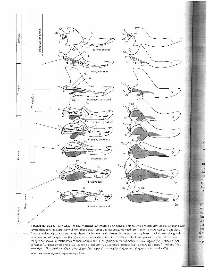
FI tn syl
are art
bee
oss
the
pos ref! tior
ign,
can
rior
feec ieeci or il
opr'
c ro § Q)
Cl.
Ul Cii E E 1-----ro E Q)
.~
Ar
Ar
Ar
Early mammal
Morganucodon
Pa
OJ
o
--~
o
Ul
Ul "0. ~ Q) .r: f-
C
Sa -----~-- "-""_,A<~ ~ Sp
Primitive synapsid
FIGURE 7. S S Evolution of the mammalian middle ear bones. Left column a medial view of the left mandibular ramus; right column lateral view of right mandibular ramus and quadrate. No teeth are shown to make compariso~s clear.
From primitive pelycosaurs, to therapsids, to the first mammals, changes in the postdentary bones are indicated along with
incorporation of the quadrate (incus) and articular (malleus) into the middle ear.The fossil species used to follow these
changes are shown in relationship to their occurrence in the geological record. Abbreviations: angular (An), articular (Ar),
coronoid (C), anterior coronoid (Ca), condyle of dentary (Co), coronoid process (Cp), dentary (D), incus (I), malleus (Ma),
prearticular (Pa), quadrate (0), quadratojugal (OJ), stapes (5), surangular (Sa) , splenial (5p), tympanic annulus (Ty).
Based on the research o(James A. Hopson and Edgar F. Allin.

Primary palateEarly mammal
~-------"""'- ..............
/ ----sqUam~al,I. \ .... __._ -.....""
M ~.)\~ ' ...... I ~ /.... '---_._-------/
joint
jaw joint
Dentary
Therapsid
Dentary
Pelycosaur
----
Postdentary bones
--....." .\ , , \
'f)
I/-, /---_/ /
Dentary
Changes in jaw articulation during t",,,,.ition from early (pelycosaurs) to late (mammals)
rJ"'·~~sYTlapsid's. In mammals. the postdentary bones of the lower jaw mostly rost and the dentary enlarges. Bones involved in jaw
.lIIii"" tirt,lotion in pelycosaurs. the articular and the quadrate. _'''''''~"M reduced and move in to contribute to the inner ear
of mammals. Jaw articulation in mammals is taken over by
dentary and squamosal. The stapes is not shown.
These examples remind uS that the sku II is a multipur1U,.,.V\,p "tool" involved in a great variety of functions. Its design
and incorporates these multiple roles. Generaliza ,,....... ;}."< about skull design and function can be misleading if we
~··m;' nn " ro its multiple function s. However, if we are cautious, we
understand how skull design reflects fundamenwl func problems. The skul l pr imarily functions as pan of the
ng system of vertebrates. How it addresses problems of .ii«~(~f:dirlg depends largely upon whether feeding occurs in air
water. Each medium presencs differenc limitations and . The viscosity of water and the buoyancy of
(a) Pelycosaurs
Secondary palate
(b) Mammals
FIGURE 7.57 Secondary palate. (a) In most r€ptiles , J air entering the nostrils and food entering the front of the ~outh share a common passage . although each exits differently at the back. Air moves down the trachea, and food travels down the
esophagus. (b) In mammals, the routes of air and food are separated from the beginning of their travel by the secondary
palate, a structure of bone (hard palate) and soft tissue (soft palate) .White arrows indicate the path of air; dark arrows indicate the path of food .
tiny organisms within it mean that water, much more than thin air, contains a richer com munity of floating planktonic organisms. Suspension feeding and harvesting of these tiny organisms becomes economical, and filter-feeding devices enjoy some adap tive favor. Generally, feeding proceeds in two steps, food capture and then swallowing. We look a t each of these in turn .
Prey Capture
Feeding in Water The first step in feeding is food capture, which depends generally on the medium in which feeding takes place. The
higher viscosity of water presents both problems and opportunities for the animal feeding in water. Feeding in wate~ poses a disadvancage in that water easily carries shock or pressure waves ("bow waves") immediately in front of the preda
tor approaching its food. These pressure waves can arrive an
instant before the advancing prc;dator and alert or deflect the intended prey. On the other hand, when a vertebrate quickly
Skeletal System The Skull 277

FIGURE 7.59 Air passage of duck-billed dinosaurs. The air passageway is formed by the premaxillae and nasal bones of the hadrosaur.Air that flowed through the nasal cavity on its way to the lungs cooled the nasal epithelial lining and hence the blood flow through it.Although the vascular system of the hadrosaur is not known, if it were similar to some mammals, then this cooled blood might have circulated in such a fashion as to
precool the blood flowing to the brain. In this way, the brain was protected from elevated temperatures. Alternatively or additionally, such an expanded air passage might have been a resonating chamber to amplify vocalizations.
After Wheeler.
Suspension Feeding Suspension feeding is a fe ed ing strar
egy confined largely, perhaps exclusively, to animals living
in water. Some, making an esoteric point, argue that bats
"filter" insects "suspended" in air, but this misses the point.
Air is too thin to h old suspended food for long. Bats catch or
grasp prey, but they do not really use a filtering appa ra tus nor
face the sa me mechanical problems as aquatic organisms in
a viscous medium; so they are not suspension feeders. Most
suspension feeders are benthic (bottom-dwelling) organisms
or are associa ted with a herbivorous/detritus feeding style.
Respiration and feeding are tightly coupled. Often the same
water currents support both activities.
Suspension feeders use several methods to intercept and
gather nutrients traveling within streams of water. Captured
particles are usually smaller than the pores of th.e filter. They
may collide directly with the filter (figure 7.61a), or because
of their ine rtia, they deviate from the streamlines to collide
with the mucus-covered surface of the filter (figure 7.61b).
Upon impac t, the particles cli ng to the sticky mucus and are rolled up in mucous cords, and then are passed by ci lia into the digestive tract.
Less commonly, a sieve can be used to strain sus
pended particles larger than the pores of the sieve. As the stream of water passes through the sieve, the pa rticles are
held back and then collected from the face of the se lective
filter (figure 7.61c). This method is rare a mong animals,
perhaps because the relatively large particles filtered tend to
a c
t
'2.1 p; th
at Sr ba
an
its
br: (h:
fee
ere
see
eili
of i tha
of mu,
Premaxilla Internal
~~ ~ Pterygoid
Primary palate
(a) Early tetrapod
process (b) Therapsid
Premaxilla
Primary Nasopharynxpalate
Secondary palate
(c) Mammal
FIGURE 7.58 Evolution of the secondary palate. (a) Early tetrapod with a primary palate in cross section (left) and ventral (right) views. (b) Therapsid with a partial secondary palate formed by the medial extension of the premaxilla and maxilla . (c) Mammal with a secondary palate that. in addition to extensions of the premaxilla and maxilla, includes part of the palatine bone.
After Smith.
gulps water into its mouth, the viscosi ty of the water drags
\ along the prey as well. This viscosity makes suction feeding,
used with relatively large prey, possible.
To capture sma ll foods, aquatic anima ls use suspension feeding. The density of water gives it the viscosity to
retard the fall of particulate material out of suspension.
Compared to air, water holds a flo a ting bounty of tiny
o rganic particles and microorganisms, a rich potential nutri
\ ti o nal resource fo r an organism with the equipment to har
J vest it. Cilia move and control currents of water (and
tra nsport captured food), and sticky mucus snatches sus
pended food from the current of water as it glides by.
Chapter Seven278
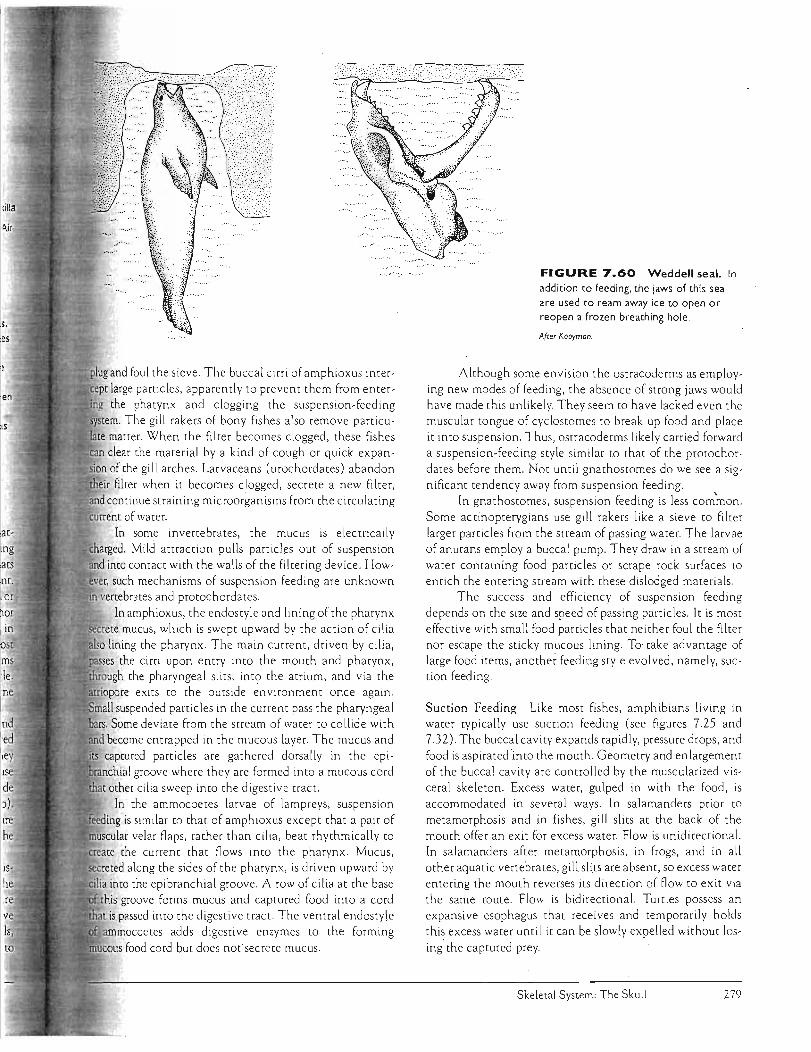
" " ~.'"
plug and foul the sieve. The buccal cirri of amphioxus intert large particles, apparently to preve nt them from enterthe phatynx and clogging the suspension-feeding
. The gill rakers of bony fishes also remove particumatter. When the filter becomes clogged, these fishes clear me material by a kind of cough or quick ex panof the gil l arches. Larvaceans (urochordates) abandon filter when it becomes clogged, secrete a new filter,
continue straining microorganisms from the circulating of water.
In some invertebrates, the mucus is electrically Mild attraction pulls particles out of suspension
into contact with the walls of the filtering device. Howsuch mechanisms of suspension feeding are unknown
vertebrates and protochordates. In amphioxus, the endostyle and lining of the pharynx
mucus, which is swept upward by the action of cilia lining the pharynx. The main current, driven by cilia,
the cirri upon entry into the mouth and pharynx, gh the pharyngeal slits, into the atrium, and via the
~'i'JI]QPiQ re exits to the outside environment once again. suspended particles in the current pass the pharyngeal
Some deviate from the stream of water to collide with become entrapped in the mucous layer. The mucus and captured particles are gathered dorsally in the epi
groove where they are formed into a mucous cord other cilia sweep into the digestive tract.
In me ammocoetes larvae of lampreys, suspension is similar to that of amphioxus except that a pair of
~".~'"~"W' velar flaps, rather than cilia, beat rhythmically to the current that flows into the pharynx. Mucus,
~=:r'~Na 'a'" along the sides of the pharynx, is driven upward by
imo the epibranchial groove. A row of cilia at the base . groove forms mucus and captured food into a cord is passed into the digestive tract. The ventral endostyle
adds digestive enzymes to the forming food cord but does not secrete mucus.
FIGURE 7.60 Weddell seal. In addition to feeding. the jaws of this seal are used to ream away ice to open or reopen a frozen breathing hole.
After Kooyman.
Although some envision the ostracoderms as employing new modes of fe eding, the absence of strong jaws would have made this unlikely. They seem to h ave lacked even the muscular tongue of cyclostomes to break up food and place it into suspension. Thus, ostracoderms likely carried forward a suspension-feeding style similar to that of the protochordates before them. Not until gnathostomes do we see a significant tendency away from suspension feeding.
In gnathostomes, suspension feeding is less com~on . Some actinopterygians use gill rakers like a sieve to filter large r particles from the stream of passing water. The larvae of anurans employ a buccal pump. They draw in a stream of water containing food particles or scrape rock surfaces to enrich the entering stream with these dislodged materia ls.
The success and efficiency of suspension feeding depends on the size and speed of passing particles. It is most effective with small food particles that ne ither foul the filter nor escape the sticky mucous lining. To~ take advantage of large food items, another feeding style evolved, namely, suction feeding.
Suction Feeding Like most fishes , amphibians living in water typically use suction feeding (see figures 7.25 and 7.32). The buccal cavity expands rapidly, pressure drops, and food is aspirated into the mouth. Geometry and enlargement of the buccal cavity are controlled by the muscularized visceral skeleton. Excess water, gulped in with the food, is accommodated in several ways. In salamanders prior to metamorphosis and in fishes, gill slits at the back of the mouth offer an exit for excess water. Flow is unidirectional. In salamanders after metamorphosis, in frogs , and in all other aquatic vertebrates, gill slits are absent, so excess water entering the mouth reverses its direction of flow to exit via the same route. Flow is bidirectional. Turtles possess an expansive esophagus that receives and temporarily holds this excess water until it can be slowly expelled without losing'the captured prey.
Skeletal System: The Skull 279
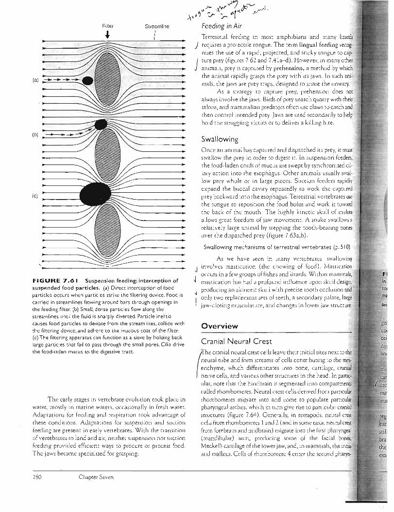
lizards
requires a projectile tongue. The term lingual feeding recog
nizes the use of a rapid, projected, a nd sticky tongue to cap
ture prey (figures 7.62 and 7.41a-d). H owever, in many ocher
animals, prey is captured by prehension, a method by which
the an imal ra pid ly grasps the prey with its jaws. In such ani
mals, the jaws are prey traps, deSigned to snare the unwary.
As a strategy to capture prey, prehension does nor
always involve the Ja ws. Birds of prey snatch quarry with their
talons, and mammalian predators often use claws to catch and
then control intended prey. Jaws are used secondarily to help
hold the Struggling victim or to deliver a killing bite.
Swallowing Once an animal has captured and dispatched its prey, it musr
swallow the prey in order to digest it. In suspension feeders,
the food-laden cmds of mucus are swept by synchronized ci!
ia ry action into the esophagus. Other animals usua lly swal
low prey whole or in large pieces. Suction feeders rapidh'
expand the buccal cavity repeatedly to work the captured
prey backward into the esophagus. Terrestrial velTebrates use
the tongue to reposition the food bolus and work it toward
the back of the mouth. The highly kinetic skull of snakes
allows great freedom of Jaw movement. A snake swallows a relatively large anima l by stepping the tooth-be'Hing bones
over the dispatched prey (figure 7 .63a,b).
Swallowing mechanisms of terrestrial vertebrates (p. 510)
As we have seen in marry venebrates , swa llowing
J involves masticmion (the chewing of food). Mastication
occurs in a few groups of fishes and lizards. Within mammals,
J mastication has had a profound influence upon sku ll design,
producing an akinetic skull with precise toOth occlusion and ~
only twO replacement sets of tee th , a secondary palate, large
jaw-clOSing musculature, and changes in lower Jaw structure.
Overview
Cranial Neural Crest
(
nhe cranial neural crest cells leave their initial si tes next to the
neural tube and form streams of cells contributing LO the mes
enchyme, which differentiates into bone, canilage, cranial
nerve cells, and various o ther st ructures in the head. In partic
ular, note that the hindbrain is segmented into compartments
called rhombomeres. Neural crest cells derived from particular
rhombomeres migrate into and come to populate particular
pharyngeal arches, IVhich in turn give rise to particular cranial
structures (figure 7.64) . Generally, in tetrapods, neural crest
cells from rhombomeres 1 arrd 2 (and in some taxa, rreural crest
from forebrain and midbrain) migrate into the first pharyngeal
(mandibular) arch, producing some of the facial bones,
Meckel's cartilage of the lower jaw, and, in mammals, the incus
and malleus. Cells of rhombomere 4 enter the second pharyn-
FI In tor
ma
ge:
ear sti!
bra
the
exa
A~~~ Filter Streamline
~ . ~~~; (a) -------- .~~~~
~ ~ ~ ..
(b)~~
~ (e)
•
FIGURE 7.61 Suspension feeding: interception of suspended food particles. (a) Direct interception of food particles occurs when particles strike the filtering device. Food is carried in streamlines flowing around bars through openings in the feeding filter. (b) Small, dense particles flow along the streamlines until the fluid is sharply diverted. Particle inertia causes food par ticles to deviate from the streamlines, collide with the filtering device, and adhere to the mucous coat of the filter. (c) The filtering apparatus can function as a sieve by holding back large particles that fail to pass through the small pores. Cilia drive the food-laden mucus to the digestive tract.
The early stages in venebrate evolution [Ook place in
wmer, mostly in marine waters, occasionally in fresh water.
Adaptations for feeding and respiration [Ook advantage of
these conditions. Adaptations for suspensio n and suctiorr
feeding are present in ea rly ver teb ra tes . With the transition
of ve n ebrates to land and air, neither suspenslon nor suction
feeding provided efficient ways [0 procure or process food.
The jaws became specialized for grasping.
Chapter Seven
.)7 ~ <>/- ,~ .... tP /.:r r p.(
Feeding in Air
Terrestrial feeding in most amph ibians and many
280

niy.
use ard kes 's a
0) .
Terrestrial feeding by a salamander. In this filmed sequence, the salamander's jaws open (top). Its
tongue starts to project (middle), approaches (bottom), and then
,-.,.,. , ...n~, contact with the prey.
(hyoid) arch, producing the stapes, styloid process, and a 'bution to the hyoid. Rhombomere 6 releases neural crest
,'=_.;,,_000 into pharyngeal arches} and 4, which contribute to the and to the thyroid cartilages. Cells from rhombomeres}
5 do not migrate. Somewhat surprisingly, in at least tetrapods both eleof the mandibular arch, the palatoquadrate and Meckel's
----..,.. , derive solely from a single condensation 0 eural
populating the first pharyn arc owever, the
, arch is not derived from the first pharyngeal arch but from several other neural crest sources of the head.
Hox genes expressed in various combinations and
of the neural crest (figure 7.64) instruct the cells quite as to which tissues they will form. Although details are
ming, many major evolutionary changes in vertejaws, teeth, and facial bones seen, based on changes in
placement or instructed fate of neural crest cells. For
a Hox gene is expressed in the mandibular arch of a
species of lamprey, but not in gnathostomes. This apparent loss of Hox gene expression may have been the genetic b8sis for facilitating the evolution of jaws in gnathostomes.
Neural crest (p. 238)
Emergence of Mammals Mammals brought many innovations to the vertebrate design; several involved the sku II. One change already noted is in the lower jaw. In early amniotes, as in gnathostomes generally, the jaws articulate with the braincase via the artic~lar-quadrate joint. In mammals, this is quite dif
ferent. The jaws articulate via the dentary-temporal joint. Seve ral postdentary bones becomelOst duri~ this t~ition to mammals, and a few move to the middle ear. The dentary expands posteriorly to form a new articulation with the skull, namely, via the dentary-temporal joint. Although the factors favoring these changes are disputed, the reality of these changes is not. Bones located at the back of the primitive amniote lower jaw were either lost or altered in function from jaw articulation to hearing. But this fact raises a new problem. How could bones involved in jaw suspension change function without disrupting the intermediate species? If postdentary bones moved to the middle ear, how could they abandon jaw suspension without producing an individual with no method of supporting the jaw against the ;kull? G. Cuvier, nineteenth-century French anatomist, would
have understood the dilemma. He argued that evolution could not occur for just such a reason, because a change in structure would disrupt function and stop evolution in its tracks before it had begun.
Diarthrognathus, a late cynodont close to primitive mammats,'"suggestsan answer. Its name means two (di-) sites of articulation (arthro-) of the jaw (gnathus). In addition to the articular-quadrate joint inherited from reptiles, a dentary-squamosal joint was apparently present. We do not know the feeding style of Diarthrognathus, so we cannot be sure of the biological role played by this second articulation
What do living vertebrates suggest? Some birds, such as the skimmer, for example, feed by holding their lower jaw just below the water's surface and flying swiftly along until they strike a fish. Then the jaws snap shut to snatch the fish. A secondary articulation seems to strengthen the lower jaw and help prevent its dislocation as it collides with the fish. Diarthrognathus did not feed on fish, but it may have wrestled with scruggling prey or fought with competitors. A second jaw articulation would make the jaw stronger. Whatever its advantages, a dentary-squamosal
joint was established before the postdentary bones . departed from the lower jaw; therefore, when the quadrate and articular bones departed, an alternative method of lower jaw-skull articulation was already in place. This is
significant because loss or movement of these bones to support hearing did not disrupt the function they abandoned,
Skeletal System: The Skull 281

T he largest animal alive today is the blue
whale . It makes its liv ing as a filter feeder.
The fi ltering device is the baleen , a brushlike specialization of the oral epithelium
occupying the site where teeth might otherwise be expected in the upper jaw.
"Whalebone" is a misnomer for baleen.
The term is inaccurate because no bone
occu rs within the baleen, Because baleen is
frayed , it acts like a strainer to hold back food from the stream of water pass ed
through it. Food preference depends a little
on species, but most baleen whales strain
small fishes or shrimplike crustaceans
called "krill :' which school or gather in dense swarms. Food collected in the baleen
is licked free by the tongue and swallowed.
Blue and humpback whales represent one subgroup of baleen whales called 'fin
whales ; or ro rquals. Right whales are the other subgroup. In both groups, the teeth are absent, the baleen is present, and the
skull is long and arched to hold the filter
feeding equipment.. To feed, right whales part their jaws
slightly and swim through swarms of krill. The stream of water enters the front of the
mouth and passes out the lateral suspended
wall of baleen. Here the krill become entangled In the frilly baleen and are licked up and
swallowed (box figure I a) . The blue whale
feeds differently. As it approaches a school of fishes or krill , it opens its mouth wide to
swim over and engulf the concentrated prey and accompanying water. Pleated furrows alo ng its neck and belly allow the throat to
inflate like a pouch and fill with this huge
mass of water (box figure I b). Up to 70 tons of water are temporarily held in the
(b)
BOX FIGURE Whales feeding. (a ) The right whale has long plaresofbaleen
suspended from its upper jaw, and it feeds by swimming through plankton with its mOllth
parted. Watet enters, passes along the sides of the tongue, and then departs through the curtain of baleen, leaVing the plankton entangled in the baleen. (b) As a fin whale approaches a concentration of planktoniC organisms, usually krill, it opens its mouth and engulfs these organisms together with the water in which they teside. [ts pleated throat
allows for cons iderable expansion of the mouth in order to accommodate the planhon· filled wate r. The whale lifts its throat to fotce wate r out through its baleen, which holds back the food but allows excess water to filter Ou t, Its tongue licks this food from the
jaw suspension. The existing dentary-squamosal articulation was in a sense "ready to serve," preadap ted for a new or expanded function.
Preadaptation (p. 18)
Probainognathus, anothe r late cynodont, like DianhrognQthus exhibits a posterior extension of (he dentary to es(ab· lish a secondary point of jaw articulation with the sku ll (figure 7.65 ). Probainognachus, Dianhrognathus, and several other late cynodonts with similar transitional double jaw articulat ions s~ggest how a harmonious transition in form and function might have occurred. They remind uS again that a series of anatomical changes alone are an incomplete
statement about evolutionary events. They must be with hypotheses about the accompanying functional se ' changes. Form and function go together, and both receive attention if we are to bring some understand' .the process of evolutionary change.
Evolutionary Modifications of Immature Forms: Akinesis in Mammals
We sometimes forget that an evolutionary modification debut in an em bryonic or infant stage and later incorporated or expanded in the adult. Such may have the case with akinesis in mammals. In all mammals, i_____.._"" _,~,
suckle milk from their mothers. Suckling requires a
Chapter Seven 282

o.';ie;q:ranejed throat. The whale then contracts
swollen pouch. forcing water through baleen where the food is strained. col
up by the tongue. and swallowed.
Humpback whales have been observed release air bubbles while circling a
of prey swimming above them. As 1ij·.·rlo· tI\p air bubbles rise. they form a "bubble
that may corral or drive the school
to the surface ahead of the whale. The
bubble cloud may also immobilize or confuse schools' of prey. causing them to clump
together. or it may disguise the whale as it
surges upward with its mouth open
through the center of the bubble cloud.
Some humpbacks begin feeding at the sur
face by lopping or slapping their tail against
the water as they dive. Just as the tail flukes
are about to reenter the water. a whale flexes its tail so that the flukes hit the
Eocene Oligocene Baleen cetacean whale whale
( Prozeuglodon) (Actiocetus)
IlII :)WallUl" ~ it. (c) Skulls of whales have been highly modified during their evolution.
the design of the face and the position of the nostrils. Andresarchus. a terrestri81 from the Eocene. may have belonged to a group from which early ce taceans arose .
. Eocene (Prozeuglodon). O ligocene (Acciocerus), and a modern baleen whale
!jIJ.ustJ·ated. Although not on a direct evolutionary line with each other. these comparisons changes in ce tacean skull design. especially in the facial region. Abbreviations:
L- (F). jugal 0), maxilla (M), nasal (N). pariml (P). premaxilla (Pm)
PiL:onmas: (e) after Olsen; Romer.
water. leaving a bubbling effervescence at the surface. This is thought to startle prey
and stimulate them to clump tightly
together in a school. The whale then
releases a bubble cloud as it dives. which it follows by a feeding lunge back up through
the bubble cloud to c?"ect the prey in its mouth.
The oldest fossil whales come from the
Oligocene and bear unmistakable resemblances to primitive terrestrial mammals.
Distinct incisor. canine. premolar. and molar teeth were present. From these earliest whales. two major modern lines soon
arose. One is the baleen whales. formally called the mysticetes . The other major line of whales is the toothed whales. or the odontocetes. including ' sperm. killer. and other whales with teeth.
In both baleen and toothed whales. the
skull is telescoped. Some bones are pushed
together and even overlap. yet a long snout persists (box figure I c).ln the odontocetes.
the backward lengthening of the facial
bones creates the snout. In the mystic~tes. the occipital bones are pushed forward.
Although achieved differently. the result is the same-to reposition the nostrils to a more central and dorsal position. When a whale surfaces to breathe. this position of the nostrils allows easy venting of the lungs and drawing in of fresh air without the
whale having to tip its entire head out of water.
seal. Fleshy lips provide the seal around the teat of the gland, the mouth is the chamber that receives the the up-and-down action of th.e tongue pumps th.e the mother to the infant's mouth and esophagus.
and feeding sh.ared a common chamber, as in the infant would have to interrupt nursing and
attachment to th.e nipple in order to breathe . A palate makes this inefficient interruption in feed
Ull""~,-,;,,ary. It separates feeding from breathing by septhe mouth from the nasal chambers. But a secondary
that separates the mouth from the nasal passages also left and right halves of the skull, thus preventing any
within or across the braincase. The result is an . skull.
Further changes in the adult evolved later. With loss of kinesis, the skull is firm and r.eady to serve strong jaw-closing muscles. Mastication, development of specialized teeth to serve chewing (accurately occlusal tooth rows), and a muscular tongue (to move food into position between tooth rows) might then find adaptive favor. Certainly there are other ways to chew food. Some fisnes with kinetic skulls and teeth that are continuously replaced chew their food. In mammals, the conditions seem especially favorable for mastication, and we find this adaptation in almost all mammalian species, including its appearance in some late th.erapsids. Analysis of evolutionary events often centers on adult stages, yet understanding of these events must come from a knowledge of the entire life history of species .
Skeletal System: The Skull 283

(a)
I I
I I I
(b)
FIGURE 7.63 Swallowing by a rat snake, Elaphe, as seen in dorsal view. (a) Oucline of snake's head during successive swallowing mocions, left co righc. Previous head posicion is indicaced by che dotted ouciine.Wich alcernacing left and righc advances, che jaws walk over che prey along a.line of progress, che axis of swallowing, unci I che jaws pass over che encire prey. These jaw-walking displacemencs place the prey at the back of the chroac where contraccions of neck muscles move che prey along co che scomach. (b) Movable bones of che skull (shaded) on one side swing oucward from the prey and advance farcher forward where they come co resc momentarily on che surface of che prey at a new posiCion. Movable bones of che opposice side now cake cheir turn. By such reciprocacing mocion, jaws walk along che prey. In addicion co jaw displacemenc, che skull itself swings oucward from che swallowing axis (arrow) chrough an angle (()) in che direccion of che advancing bones co place them farcher along the prey.Abbreviacions: eccopcerygoid (Ec) , maxilla (M), mandible (Md), palacine (Pal), pcerygoid (Pt), quad race (Q) , supratemporal (St).
After Kardong.
Stapes
Styloid process
(a)
Meckel's
Pharyngeal arch 1
cartilage
Pharyngeal arch 2
Pharyngeal arch 3
Pharyngeal arch 4
Pharyngeal arches
Mesenchyme
(b)
Nervous system
Cranial Neural ganglia ____ ___ ~~~~ _
Spinal cord
FIGURE 7.64 Cranial neural crest migration and Hox genes,generalized tetrapod. (a) Various cranial structures derived from parcicular pharyngeal arches. (b) In turn, these pharyngeal arches are themselves populaced by neural cresc migracing (arrows) ouc of che rhombomeres of the hindbrain. (c) Embryo showing locacion of pharyngeal arches and hindbrain. Hox expression paccerns in che neural crest show che boundaries of chese gene domains. Key co abbreviacions: r2-r8, rhombomeres 2-8.
After McGinnis and Krumlau(; Carls on.
284 Chapter Seven
rio
no
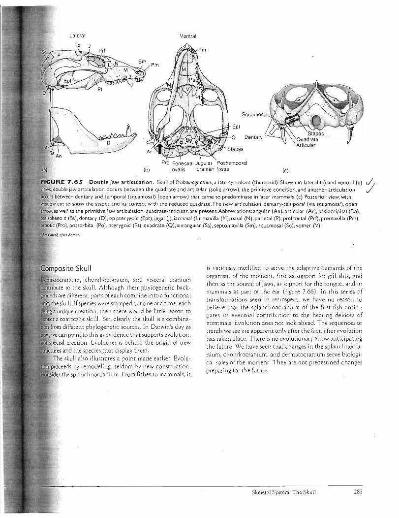
Lateral Ventral
Ept
Q Dentary
. ". J!_~ '.'''' ,'~.." .~
Pro Fenestra Jugular Posttemporal (b) ovalis foramen fossa (c)
Double jaw articulation. Skull of Probainognathus, a late cynodont (therapsid). Shown in lateral (a) and ventral (b) J/ Views, double jaw articulation occurs between the quadrate and articular (solid arrow), the primitive condition, and another arti.culation ,/ occurs between dentary and temporal (squamosal) (open arrow) that came to predominate in later mammals. (c) Posterior view, with window cut to show the stapes and its contact with the reduced quadrate. The new articulation, dentary-temporal (via squamosal), open arrow, as well as the primitive jaw articulation, quadrate-articular, are present. Abbreviations: angular (An), articular (Ar), basioccipital (Bo). basisphenoid (Bs), dentary (D), epipterygoid (Ept), jugal 0), lacrimal (L), maxilla (M), nasal (N), parietal (P), prefrontal (Prl), premaxilla (Pm), prootic (Pro), postorbital (Po), pterygoid (Pt), quadrate (Q), surangular (Sa), septomaxilla (Sm), squamosal (Sq), vomer (Y).
is variously modified to serve the adaptive demands of the organism of the moment, first as support for gill slits, and
rmatocranium, chondrocranium, and visceral cranium then as the source of jaws, as supporr for the tongue, and in
contribute to the skull. Although their phylogenetic backmammals as parr of the ear (figure 7.66). In this series of
grounds are different, parts of each combine into a functional transformations seen in retrospect, we have no reason to
the skull. If species were stamped out one at a time, each believe that the splanchnocranium of the first fish antici
a unique creation, then there would be little reason to pates its eventual contribution to the hearing devices of
a composite skull. Yet, clearly the skull is a combinamammals. Evolution does not look ahead. The sequences or
from different phylogenetic sources . In Darwin's day as trendswe see are apparent only after the fact, after evolution
we can point to this as evidence that supporrs evolution, has taken place. There is no evolutionary arrow anticipating
special creation. Evolution is behind the origin of new the future. We have seen that changes in the splanchnocra
tures and the species, that display them. nium, chondrocranium, and dermatocranium serve biologi
The skull also illust~ates a point made earlier. Evolucal roles of the moment. They are not predestined changes
proceeds by remodeling, seldom by new construction. preparing for the future.
11l/i,n<;,rl". the splanchnocranium. From fishes to mammals, it
Skeletal System: The Skull 285
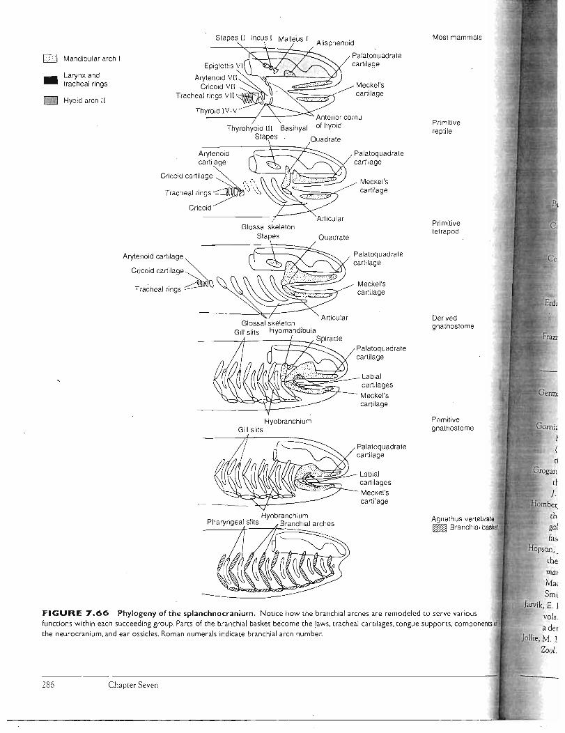
Most mammals
Primitive rept ile
Primitive tetrapod
Derived gnathostome
Primitive gnathostome
th gal fa:;,
Hopson, _
rhe
rna?
Ma( Smi
Jarvik, E. J
Zool.
FIGURE 7.66 Phylogeny of the splanchnocranium. Notice how the branchial arches are remodeled to serve various functions within each succeeding group. Parts of the branchial basket become the jaws, tracheal cartilages, tongue supports, components the neurocranium, and ear ossicles. Roman numerals indicate branchial arch number.
286 Chapter Seven
Stapes II Incus I Malleus I
Mandibular arch I
Larynx and tracheal rings
Hyoid arch II
Arytenoid cartilage
C,jooid ~rt;l,g,~ Tracheal rings
- ---71----;::::=1::-::::2'- Spiracle
Glossal skeleton Stapes
Glossal skeleton Gill slits Hyomandibula
Hyobranchium Gill slits
. Hyobranchium Pharyngeal sitts Branchial arches
Palatoquadrate
Meckel's cartilage
Palatoquadrate
Meckel's cartilage
Palatoquadrate cartilage
Meckel's cartilage
Palatoquadrate cartilage
Labial cartilages
Meckel's cartilage
Palatoquadrate cartilage
Labial cartilages Meckel's cartilage
Thyrohyoid III Basihyal Stapes
Arytenoid . cartilage
Cricoid cartilage ~ '. _
Tracheal ringS~\\':" Cricoid
Quadrate
cartilage
cartilage

SELECTED REFERENCES
E. E 1986. The auditory apparatus of advanced
mammal-like reptiles and early mammals. In The ecology and biolog)' of mammal-like reptiles, edited by N. Hotton,
111, P. D. MacLean, J. J. Roth, and E. C. Roth.
Washington, D.C.: Smithsonian Institution Press, pp.283-94.
n, E. E, and J. A. Hopson. 1992. Evolution of the aud itory
system in Synapsida ("mammal-like reptiles") and
primitive mammals as seen in the fossi l record. In The evolutionary biology of hearing, edited by D. B. Webster,
R. R. Fay, and A N. Popper. New York: Springer
Verlag, pp. 587-614.
D. M. 1989. Cranial specialtzation and locomotor
habit in the lagomorpha. Amer. Zool. 29:303-17.
J. A 1998. The neurocranium of Acanthostega gunnari ]arvik and the evolution of the otic region in tetrapods.
ZooL]. Linn. Soc. 122:61-97. R., P. Lwigale, R. Ericsson, D. Meulemans, H.-H.
Epperlein, and M. Bronner-Fraser. 2004. Developmental
origins and evolution of jaws: new interpretation of "maxillary" and "mandibular". Dev. BioI. 276:225-236.
S., and D. Cundall. 1984 The feeding apparatus of
the salamander Amphiuma r:ridactylum: Morphology and
behavior.}. Morph. 181:175-204. ,.,I(fr.m etta, T H. 1968. Adaptive problems and possibilities in
the temporal fenestration of tetrapod skulls.]. Morph. 125:145-57 .
1986. The origin of amphikinesis in lizards. Evol. BioI. 20:419-61.
R. Z., and A W. Crompton. 1996. Ontogeny of
suckling mechanisms in opossums (Didelphis virginiana). Brain Beliav. Evol. 48:157-64.
. G. C , H. l. Rosenberg, and C. Gans. 1982.
Mastication in the tuatara, Sphenodon punctatus (Reptilia: Rhynchocephalia): StructG-re and activity of
the motor system.]. Morph. 171:321-54. E. 0., R. Lund, and D. Didier. 1999. Description of
the chimaerid jaw and its phylogenetic origins.
J. Morph. 239:45-59. :lo1l'lberger,. D. G., and R. A Meyers. 1989. Morphology of
the lingual apparatus of the domestic chicken, Gallus gallus, with special attention to the structure of the
fasciae. Amer.]. Anat. 186:217-57. ]. A, and H. R. Barghusen. 1986. An analysis of
therapsid relationships. In The ecology and biology of mammal-like reptiles, edited by N. Hotton, III, P. D.
MacLean, J. ]. Roth, and E. C. Roth. Washington, D.C.:
Smithsonian Institution Press, pp. 83-106.
E. 1980. Basic structure and evolution of vertebrates. 2 vols. New York and London: Academic Press. Primarily.
a detailed study of certain extinct or primitive fishes.
M. 1977. Segmentation of the vertebrate head. Amer. 2001. L7:323-33.
Kardong, K. V 1977. Kinesis of the jaw apparatus during
swallowing in the cottonmouth snake, Agkistrodon piscivorus. Copeia 1977:338-48.
- --. 1986. Kinematics of swallowing in the yellow rat
snake, Elaphe obsoleta quadrivittata: A reappraisal. ]pn.]. Herpet. 11:96-109.
Kardong, K. V, P. Dullemeijer, and J. A. M. Fransen. 1986.
Feeding mechanism in the rattlesnake Crotalus durissus. Amph.Rept. 7:271-302.'
Larsen, J. H., Jr., J. 1.. Beneski, Jr. , and D. B. Wake. 1989.
Hyol ingual feeding systems of the Plethodontidae:
Comparative kinematics of prey capture by
salamanders with free and attached tongues.]. Exp. Zoo!. 252:25-33.
Larsen, J. H ., J r., and D. J. Guthrie. 1975. The feeding sys tem of terrestrial tiger salamanders (Ambyscoma tigrinum melanostictum Baird).). Morph. 147:137-54.
Lauder, G. V 1979. Feeding mechan ics in primitive teleosts
and in the halecomorph fish Amia calva. ] . Zoo!. (London) 187:543-78.
---. 1980. Evolution of the feeding mechanism in
primitive acti nopterygian fishes: A functional
anatomical analysis of PolYPterus, Lepisosteus, and Amia. ] . Morph. 163:283-317.
---. 1982. Patterns of evolution in the feeding mechanism
of actinopterygian fishes. Amer. Zoo!. 22:275-85.
---. 1985. Aquatic feeding in lower vertebrates. In
Functional vertebrate morphology, edited by M.
Hildebrand, D. M. Bramble, K. F. Liem, and D. B.
Wake. Cambridge: Harvard University Press,
pp.210-29,397-99.
Liem, K. F. 1980. Acquisition of energy by teleosts: Adaptive
mechanisms and evolutionary patterns. In
Environmental physiology offishes, edited by M. A. Ali .
N ew York: Plenum Press, pp. 299-334.
---. 1990. Aquatic versus terrestrial feeding modes:
Possible impacts on the trophic ecology of vertebrates.
Amer. Zool. 30:209-21. Lombard, R. E. , and D. B. Wake. 1977 . Tongue evolution in
the lungless salamanders, family Plethodontidae.
]. Morph. 153:39-80. Mallatt, J. 1996. Ventilation and the origin of jawed
vertebrates: A new mouth. Zool.]. Linn. Soc. 117:329-404.
---. 1998. Crossing a major morphological boundary: The
origin of jaws in vertebrates. Zoo!., Anal. Compo Sys. 100: 128-40.
McGinnis, w.,and R. Krumlauf. 1992. Homeobox genes and
axial patterning. Cell 68:283-302. Miller, B. 1.., and J. H. Larsen, Jr. 1990. Comparative
kinematics of terrestrial prey capture in salamanders and newts (Amphibia: Urodela: Salamandrida~). ]. Exp . Zool.256:135-53.
Skeletal System: The Skull 287

Nishikawa, K. C, and D. C Cannatella. 199 1. Kinematics of prey capture in the tailed frog Ascaphus truei (Anura: Ascaphidae) . Zoo1. ] Linn. Soc. 103:289-307 .
Noden, D. M. 1984. Craniofacial developmem: New views on old problems. Anat. Rec. 208: 1-13.
Rafferty, K. L, and S. W. Herri ng. 1999. Craniofacia l sutures: Morphology, growth, and in vivo masticatory srrains. ] Morph. 242:1 67-79.
Rieppel, O. 1980. The evolution of the ophidian feeding system. ZooL}. Anat. 103:5 51 -64.
Sande rson, S. L. , J. J. Cech, Jr., and M. R. Patterson. 1991. Fluid dynamics in suspension -feed ing blackfish. Science 251: 1346-48.
Sanderson, S. L, and R. Wassersug. 1990. Suspension-feeding vertebrates . Sci. Amer. (Ma rch ):96-101.
Schwenk , K. 2000. An introduction to tetrapod feeding. In Feeding, fo rm, function and evolution in tetrapod vertebrates , edited by K. Schwenk. San Diego: Academic Press, pp. 21-61.
So, K-K.]., P C Wainwright, and A. F Bennett. 1992. Kinematics of prey processing in Chamaeleo jacksonii: Conservation of fu nction with morphological specialization.). Zoo 1. (London) 226:47-64.
Vorobya, E., and H,-P Schultze. 1991. Description and systematics of panderichthid fishes with commems on their relationship to groups of tetrapods. In Origins of higher groups of tetrapods, edited by H.-P. Schu ltze and L. True b. Ithaca, NY: Comstock Pub. Assoc. , pp. 68-109.
Wainwflght, P C, D. M. Kraklau, and A. F Bennett. 1991. Kinematics of tOngue project ion in Chamaeleo oU5Wleti . ] Exp . BioI. 159:109-33.
Wainwright, P C, and A. F Bennett. 1992. The mechanism of tongue projection in chameleons. L Electromyographic tests of functional hypotheses. II. Role of shape change in a muscular hydrosrat. ] Exp. BioI. 168:1-21; 23-40.
Weinrich, M. T., M. R. Sc hilling, and C R. Belt. 1992. Evidence for acquisition of a novel fe eding behaviour: Lobtail fe eding in humpback whales , Megaptoa novaeangliae . Anim. Behav. 44:1059-72.
WEB LINKS
ViSIt this book's website at www.mhhe.comfKardong4 (click Vertebrates: Microscopic Anatomy of the Skeleton on this book 's title) to find Web links for further reading on Vertebrate Labora tory Exercises. these topics:
Skeletal Systems Vertebrates: Macroscopic Anatomy of the Skeleton
Chapter Seven 288