urogenital system - acads · vertebrates: comparative anatomy, function and evolution by kenneth v....
TRANSCRIPT

Urogenital System
Paulo Rafael Symaco Joson, M.Sc.(cand.)

The Urinary System
• Components: Kidneys and tubules
• Function: Waste elimination

Structure of the Kidney

Structure of the Kidney
Microscopic components:
Blood Vessels
Renal Corpuscle
Uriniferous Tubule:
Nephron Collecting tubules
Vertebrates: Comparative Anatomy, Function and Evolution by Kenneth V. Kardong
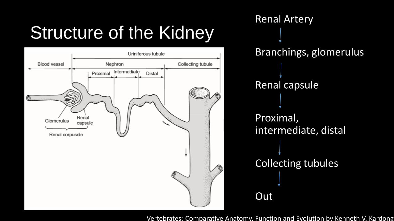
Structure of the Kidney Renal Artery
Branchings, glomerulus
Renal capsule
Proximal, intermediate, distal
Collecting tubules
Out
Vertebrates: Comparative Anatomy, Function and Evolution by Kenneth V. Kardong
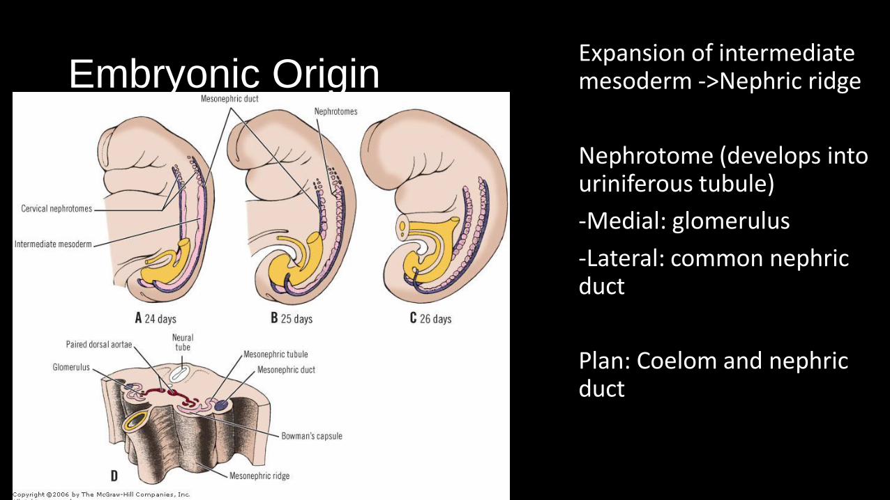
Embryonic Origin Expansion of intermediate mesoderm ->Nephric ridge
Nephrotome (develops into uriniferous tubule)
-Medial: glomerulus
-Lateral: common nephric duct
Plan: Coelom and nephric duct

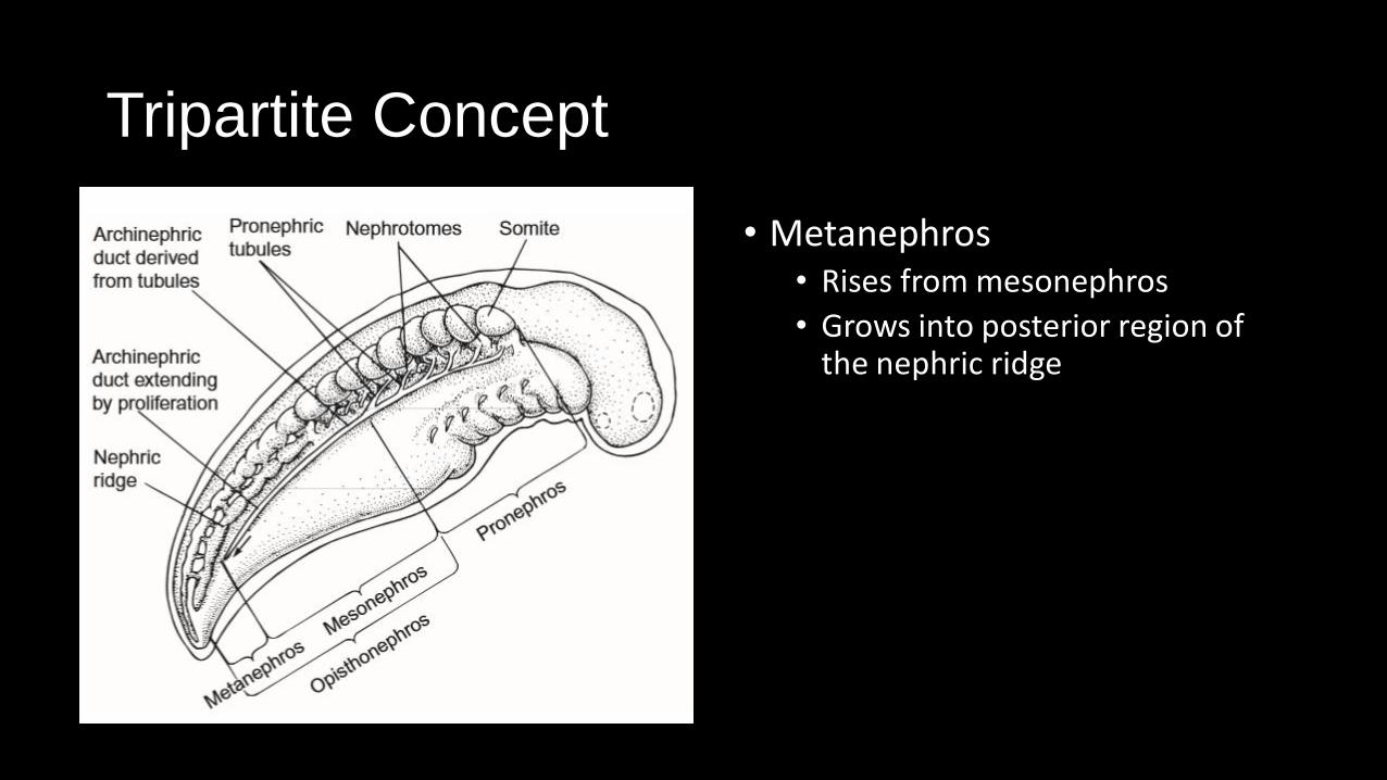
Tripartite Concept
• Three-part development
Vertebrates: Comparative Anatomy, Function and Evolution by Kenneth V. Kardong

Tripartite Concept
• Pronephros
• Mesonephros
• Metanephros
Vertebrates: Comparative Anatomy, Function and Evolution by Kenneth V. Kardong

Tripartite Concept
• Pronephros • Pronephric tubules, pronephric
duct
• Glomeruli extends from roof of coelom and filters into them
• Ususally transient
Vertebrates: Comparative Anatomy, Function and Evolution by Kenneth V. Kardong

Tripartite Concept
• Mesonephros • Taps into pronephric tubules
• Embryonically functional, can persist into adulthood
• Opisthonephros
Tripartite Concept
• Metanephros • Rises from mesonephros
• Grows into posterior region of the nephric ridge

Kidney Phylogeny: Fish
• Primitive: Pronephros
• Most teleosts: Mesonephros or opisthonephros
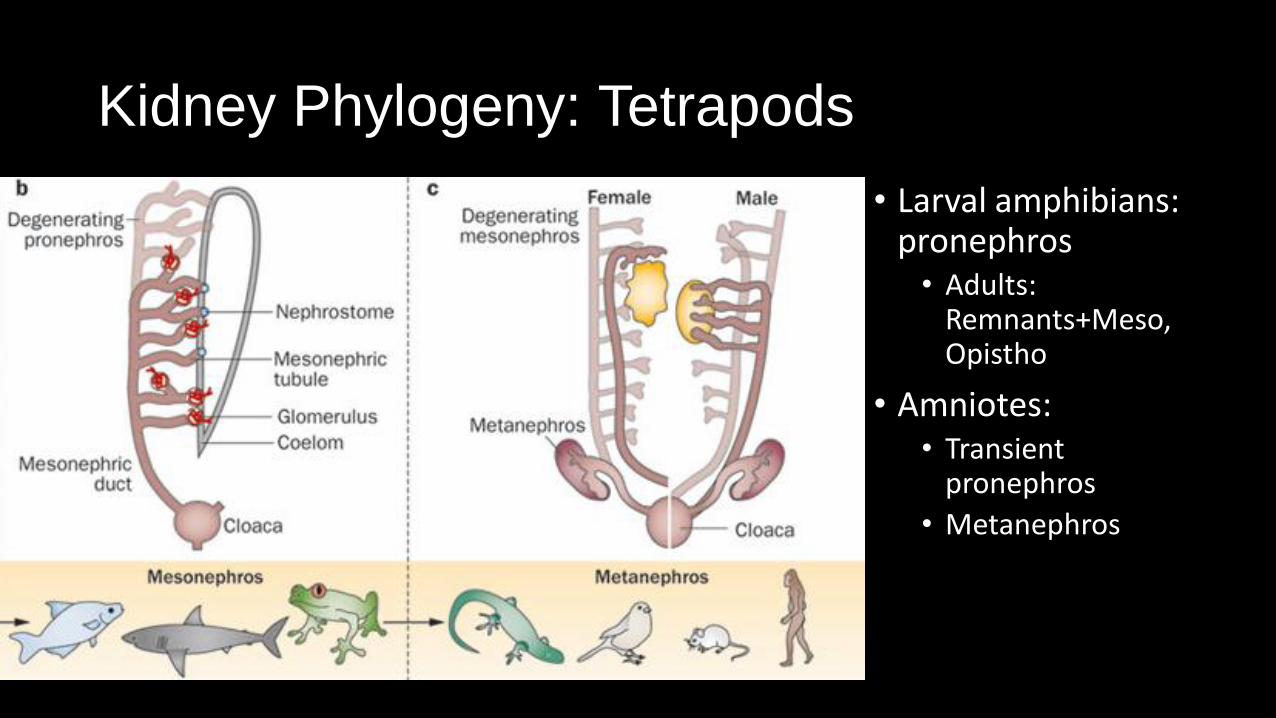
Kidney Phylogeny: Tetrapods
• Larval amphibians: pronephros • Adults:
Remnants+Meso, Opistho
• Amniotes: • Transient
pronephros
• Metanephros
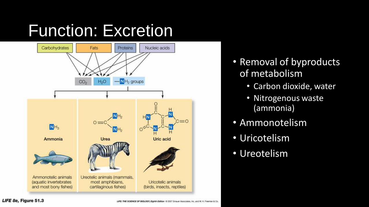
Function: Excretion
• Removal of byproducts of metabolism • Carbon dioxide, water
• Nitrogenous waste (ammonia)
• Ammonotelism
• Uricotelism
• Ureotelism

Function: Osmoregulation
• Regulation of body salt and water levels • Kidney
• Cloaca, large intestine
• Urinary bladder

Osmoregulation: Elimination and Conservation
• Filtration kidneys: glomerular filtrate, secretions along tubules, reabsorption
• Features: • Glomerulus size, presence of glomerulus
• Tubule length
• Loop of Henle
• Vasa recta

Osmoregulation: Elimination and Conservation
• Elimination: • Freshwater fish – large glomeruli, long distal tubule for more salt reabsorption
• Conservation – • Marine fish – no glomerulus, no distal tubules, secretion-dependent
• Terrestrial – loop of henle modification

Vertebrates: Comparative Anatomy, Function and Evolution by Kenneth V. Kardong
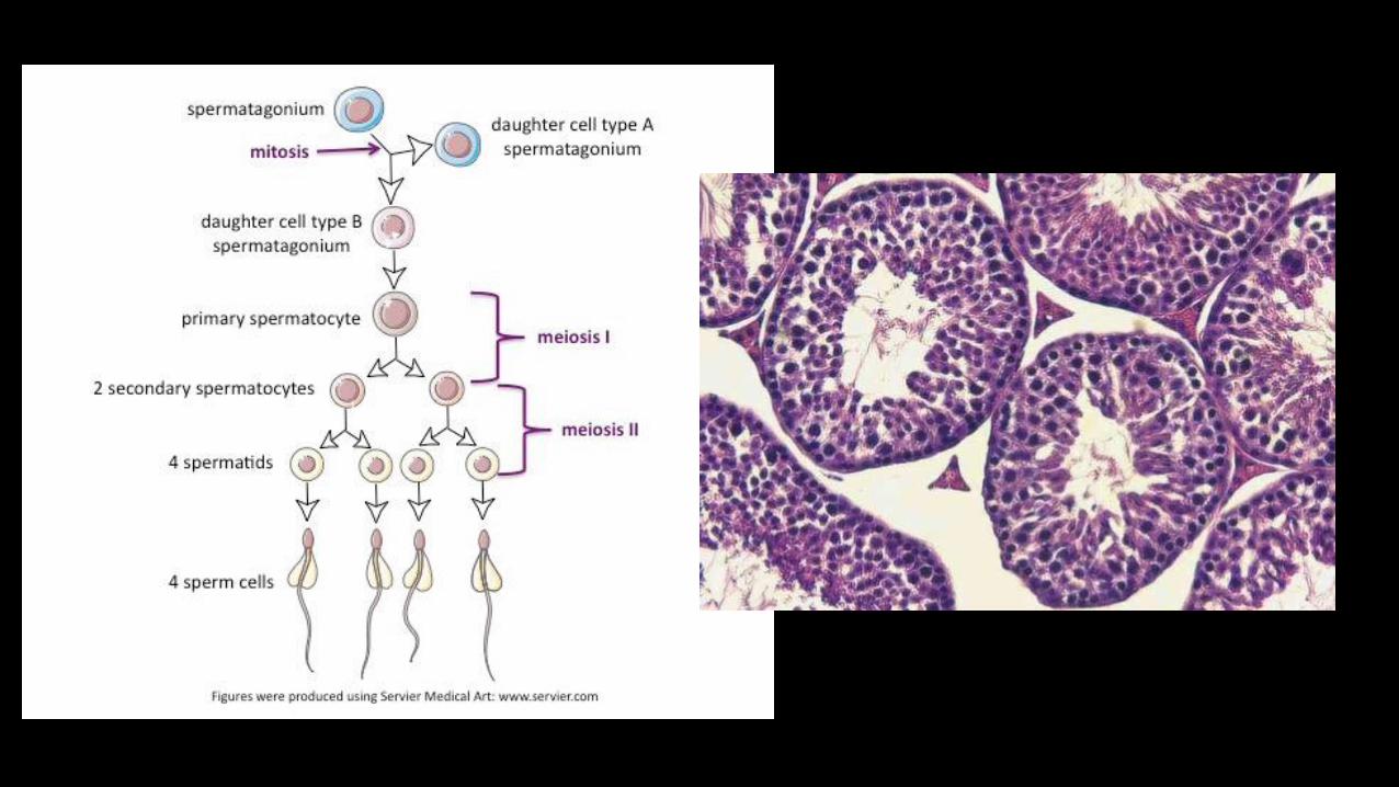
Reproductive System
• Gonads, secretions, gametes, and associated ducts

• Tunica albuginea
• Cortex
• Medulla
• Ova
• Follicle Cells


Embryonic Origin
• Genital ridge • Undifferentiated
• Extraembryonic: Germ cells • Females: Cortex
• Males: Medulla
• Organs: Salvaging from urinary

Embryonic Origin: Gonads
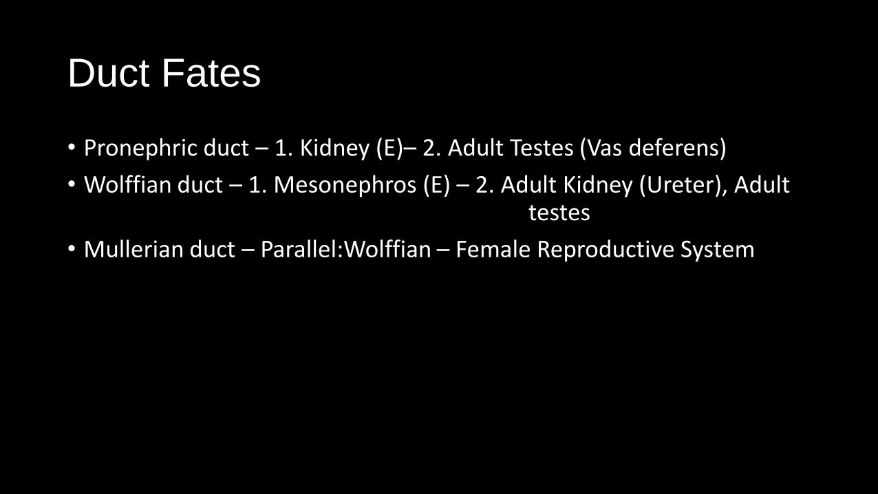
Duct Fates
• Pronephric duct – 1. Kidney (E)– 2. Adult Testes (Vas deferens)
• Wolffian duct – 1. Mesonephros (E) – 2. Adult Kidney (Ureter), Adult testes
• Mullerian duct – Parallel:Wolffian – Female Reproductive System


`
Vertebrates: Comparative Anatomy, Function and Evolution by Kenneth V. Kardong

Comparative Urogenital Anatomy - Females

Vertebrates: Comparative Anatomy, Function and Evolution by Kenneth V. Kardong
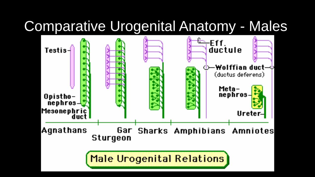
Comparative Urogenital Anatomy - Males
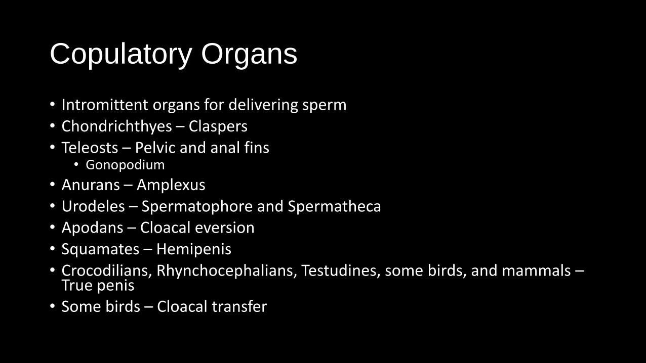
Copulatory Organs
• Intromittent organs for delivering sperm • Chondrichthyes – Claspers • Teleosts – Pelvic and anal fins
• Gonopodium
• Anurans – Amplexus • Urodeles – Spermatophore and Spermatheca • Apodans – Cloacal eversion • Squamates – Hemipenis • Crocodilians, Rhynchocephalians, Testudines, some birds, and mammals –
True penis • Some birds – Cloacal transfer

Images
• http://classconnection.s3.amazonaws.com/693/flashcards/619693/png/kidney1312481120499.png
• http://web.uni-plovdiv.bg/stu1104541018/docs/res/skandalakis'%20surgical%20anatomy%20-%202004/Chapter%2023_%20Kidneys%20and%20Ureters_fichiers/loadBinaryCA82AUH2.jpg
• http://www.nature.com/nrneph/journal/v9/n3/images/nrneph.2012.290-f1.jpg
• http://1.bp.blogspot.com/-pP4Y9Fhq8M0/TvCwDSHJi_I/AAAAAAAAABc/Mil2NYBoYac/s1600/f51003.jpg
• http://www.colorado.edu/intphys/iphy4480tsai/ovary.jpg
• http://www.melakafertility.com/images/drawings/anatomy/ant-001.png
• http://img.medscape.com/pi/emed/ckb/urology/435575-1350956-452831-1615775.jpg
• http://teleanatomy.com/Developmental%20Anatomy/Nutfah/Male%20gametes_files/image010.jpg
• http://www.nature.com/nrg/journal/v4/n12/images/nrg1225-i1.jpg
• https://www.ihcworld.com/imagegallery/images/he-stain/normal_Testis-ms-g.jpg
• http://www.repropedia.org/sites/repropedia/files/spermatogenesis.jpg
• http://education.med.nyu.edu/Histology/courseware/modules/fem-repro-sy/images/female.reproductive.02.gif
• http://celldivisionandreproduction.weebly.com/uploads/1/9/2/4/19240233/4000231_orig.png
• http://people.eku.edu/ritchisong/maleurogenitalducts.gif
• http://people.eku.edu/ritchisong/femaleurogenitalducts.gif
Diagrams and tables from Vertebrates: Comparative Anatomy, Function, Evolution 6th Edition by Kenneth Kardong. 2012. Published by McGraw-Hill, New York.