simultaneous reconstruction of the activity image and
TRANSCRIPT

Simultaneous Reconstruction of the Activity Image and Registration of the CT
image in TOF-PET
Ahmadreza Rezaei, Johan Nuyts

• In clinical PET/CT, – The CT image is adjusted to the photon energy of 511 KeV.– The projections of the “adjusted CT” is then used for attenuation correction.
– This “adjusted CT” is believed to be close enough to the true attenuation affecting the PET emission data.
• Between-scan motion and in-scan motion can violate this assumption.
• The emission data can be corrected for attenuation:– When the emission data is affected by an affine transform of a known attenuation
image.• F Natterer, 1993• A Welch, et. al., 1998, A Bromiley 2001• A Alessio, et. al., 2006
– Time-of-flight data are available• A Rezaei, 2011, 2012• M Defrise, 2012, 2013• Y Nuyts, 2012
2
Activity Reconstruction & Attenuation Registration– Attenuation Correction, Background

3
Joint Activity and Attenuation Reconstruction– MLAA
��� = ��������
�� = �� ∑ ������
Emission Image
Sensitivity of (TOF-bin t and LOR i)
to voxel jUn-attenuated TOF Emission Sinogram
Attenuation Image
Intersection of (LOR i)
and voxel j
Attenuation Factors
���� = ����� + ��Expected counts
Additive Contributions (Scatter and/or
Randoms)
�( �, � , �) = ���� ln������
− ����
� ��,���� = argmax,�
�( �, � , �)Poisson Log-likelihood for emission tomography:
1: ��, ���� = arginc�
�( �, �� ,�)2: ����, ���� = arginc
�
�( ��, � ,�)
TOF-MLEM
MLTR

4
��� = ��������
�� = �� ∑ �������
�
Emission Image
Sensitivity of (TOF-bin t and LOR i)
to voxel jUn-attenuated TOF Emission Sinogram
Deformed CT-converted Image
Intersection of (LOR i)
and voxel j
Attenuation Factors
���� = ����� + ��Expected counts
Additive Contributions (Scatter and/or
Randoms)
�( �,� , �) = ���� ln������
− ����
� ��,���� = argmax,�
�( �,� , �)Poisson Log-likelihood for emission tomography:
1:��, ���� = arginc�
�( �,�� ,�)2: ����,���� = arginc
�
�( ����,� ,�)
Activity Reconstruction & Attenuation Registration– Incorporating “side-information”
MLTR-Demons’
TOF-MLEM
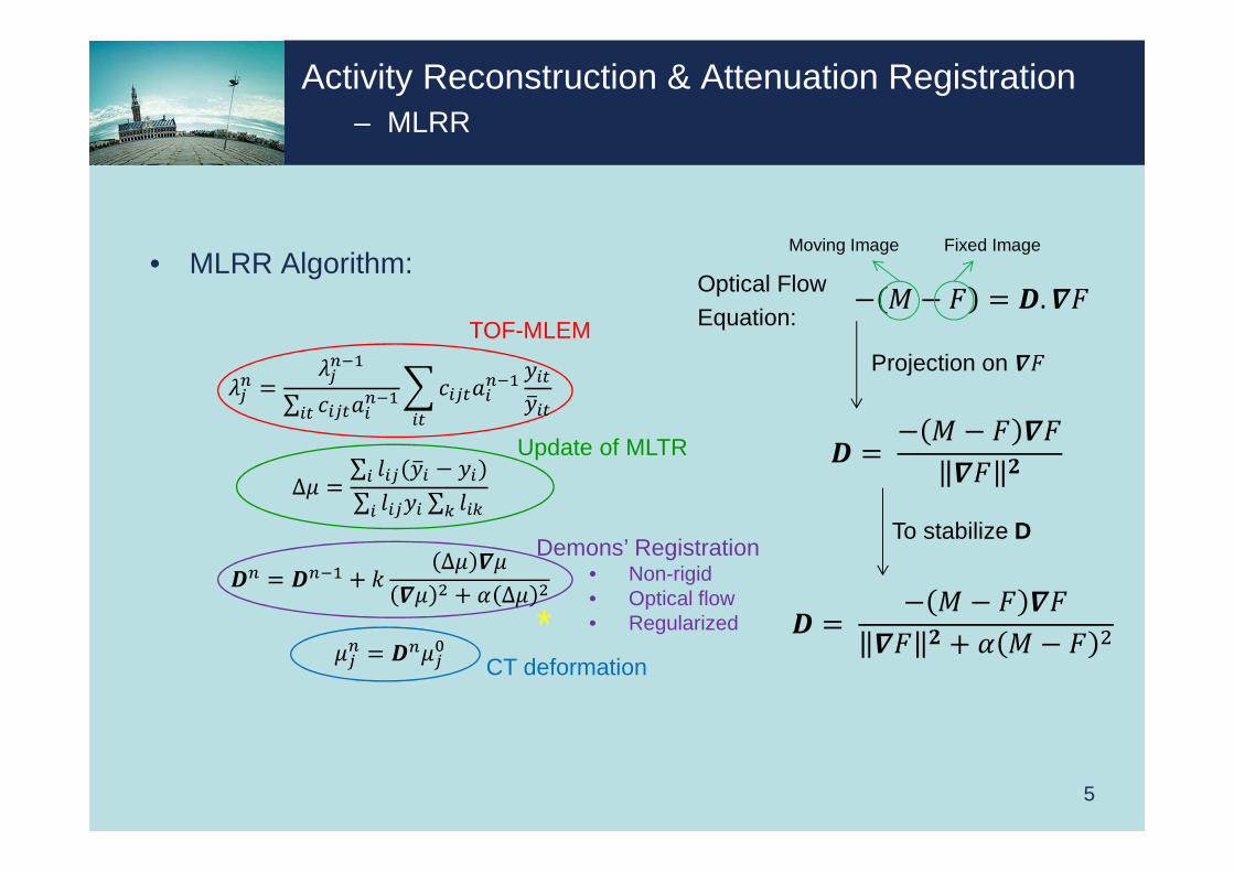
• MLRR Algorithm:
5
Activity Reconstruction & Attenuation Registration– MLRR
TOF-MLEM
Update of MLTR
Demons’ Registration• Non-rigid• Optical flow• Regularized
− � − � = �.��Moving Image Fixed Image
Projection on ��
� = − � − � ���� �
To stabilize D
� = − � − � ���� � + � � − �
Optical Flow Equation:
CT deformation
*
��� =����
∑ ������
��������� �
�
∆� =∑ ��(� − �)
∑ ��� ∑ ���
�� = ��� + � ∆� ���� + � ∆�
��� = �����

6
2D Simulations– Phantom & Demons’ Registration Method
CTactivity attenuation
True contourMismatched CT contour

7
MLEM (3:42)
MLAA – activity3:42
MLAA - attenuation
MLRR – activity15:42
MLRR - attenuation
2D Simulations– Noiseless data
CT
• Simulation specifications:– 200*200 thorax phantom– 168 projection angles over 180º– TOF resolution of 580ps – 13 TOF-bins of 312ps– Oversampling of 3 causing slight
mismatch
** (#iterations : #subsets)
True contourMismatched CT contour
8
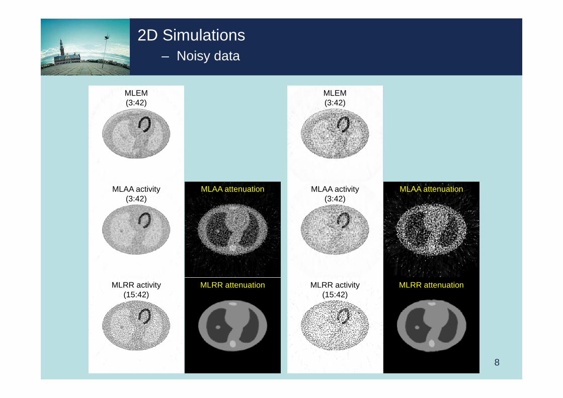
MLEM(3:42)
MLAA activity(3:42)
MLAA attenuation
MLRR activity(15:42)
MLRR attenuation
2D Simulations– Noisy data
MLEM(3:42)
MLAA activity(3:42)
MLAA attenuation
MLRR activity(15:42)
MLRR attenuation

9
2D Simulations– Noisy data
MLEM(3:42)
MLAA activity(3:42)
MLAA attenuation
MLRR attenuationMLRR activity(15:42)
FWHM=0.6cm
MLEM(3:42)
MLAA activity(3:42)
MLAA attenuation
MLRR attenuationMLRR activity(15:42)
FWHM=0.6cm

2D Simulations– Looking at displacements
10
Demons’
Horizontal Component Vertical Component
MLRR; noiseless
Horizontal Component Vertical Component
MLRR; Moderate-noise
Horizontal Component Vertical Component
MLRR; high-noise
Horizontal Component Vertical Component

• XCAT Phantom– # of frames: 8– Maximum Diaphragm motion: 2.0 cm– Max AP Expansion: 1.2 cm– Respiratory Motion: True– Heart Motion: False
11
3D Simulations– XCAT Phantom
activity
attenuationCT• Simulation specifications:
– 200*200*109 phantom– 7 oblique segments– Span 1*– 168 projection angles over 180º– TOF resolution of 580ps – 13 TOF-bins of 312ps
** (#iterations : #subsets)

12
3D Simulations– MLEM, MLAA & MLRR
MLAA attenuation
MLAA activity(3:42)
MLRR activity10:42
MLRR attenuationCT
TOF-MLEM(3:42)
MLEM (3:42)
13
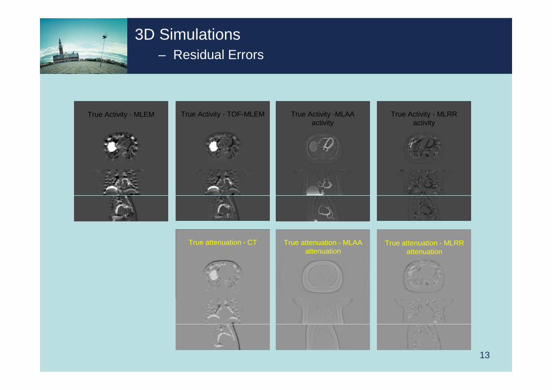
True Activity - MLEM True Activity - TOF-MLEM True Activity -MLAA activity
True Activity - MLRR activity
True attenuation - CT True attenuation - MLAA attenuation
True attenuation - MLRR attenuation
3D Simulations– Residual Errors

14
AbsoluteTrue Activity - MLRR
activity
AbsoluteTrue Activity -MLAA
activity
AbsoluteTrue Activity - TOF-MLEM
AbsoluteTrue Activity - MLEM
AbsoluteTrue attenuation - MLAA
attenuation
AbsoluteTrue attenuation - CT
AbsoluteTrue attenuation - MLRR
attenuation
3D Simulations– Absolute Residual Errors

15
Patient Data– MLEM, MLAA & MLRR
MLAA attenuation MLRR attenuationCT
MLEM(3:42)
TOF-MLEM(3:42)
MLAA activity(3:42)
MLRR activity(5:42)

16
Patient Data– MLEM; Gated data from listmode
CT MLEMgate 0
MLEMgate 2
MLEMgate 1
• Amplitude-based Gating– W van Elmpt 2011, Optimal gating compared to 3D and 4D PET reconstruction,
Eur J Nucl Med Mol Imaging, 38:843-855.

17
TOF-MLEMgate 0
TOF-MLEMgate 2
Patient Data– MLEM; Gated data from listmode
TOF-MLEMgate 1
CT

18
Patient Data– MLAA; Gated data from listmode
MLAA attenuation Gate 0
MLAA attenuation Gate 1
MLAA attenuation Gate 2
MLAA activity gate 0
MLAA activity gate 1
MLAA activity gate 2
CT

19
Patient Data– MLRR; Gated data from listmode
MLRR attenuation Gate 0
MLRR attenuationGate 1
MLRR attenuation Gate 2
MLRR activity gate 0
MLRR activity gate 1
MLRR activitygate 2
CT

20
Patient Data– Difference Images
CT - MLRR attenuationGate 0
CT - MLRR attenuation Gate 1
CT - MLRR attenuationGate 2
CT
Conclusions & Future Work
• MLRR is proposed to make use of high quality CT scans.• Our 2D/3D simulations indicate:
– MLRR is able to produce aligned activity and attenuation reconstructions similar to MLAA, with two advantages:
• The scale problem is solved!• High quality, noise-free attenuation reconstructions are made available.
• The clinical results show:– Good agreement of MLRR attenuation reconstructions and expected breathing
patterns.– More accurate activity reconstructions near boundaries affected by motion
(compared to MLEM activity reconstructions with the CT-derived attenuation).
• Future Work:– Incorporate physical properties of different tissue types.– Because the emission data from a moving attenuation is inconsistent, analyzing the gated
reconstructions seems to be the only way to quantitative reconstruction in the presence of motion.
21

• Our thanks to:
– Michel Defrise from the Vrije Universiteit Brussel, and Annemie Ribbens from KU Leuven for the very insightful discussions.
– Mike Casey, Judson Jones from Siemens Healthcare, Molecular Imaging for the data-based gating software and data processing.
– S Stroobants, S Staelens and M Lambrechts from the UniversiteitAntwerpen for the patient data.
– You! for your attention. ☺
22