silylated precision particles for controlled release of ... · silylated precision particles for...
TRANSCRIPT

Silylated Precision Particles for Controlled Release of ProteinsKhosrow Khodabandehlou,† Amar S. Kumbhar,‡ Sohrab Habibi,§ Ashish A. Pandya,§,∥
J. Christopher Luft,§,∥,⊥,# Saad A. Khan,† and Joseph M. DeSimone*,†,§,∥,⊥,#,¶,∇
†Department of Chemical and Biomolecular Engineering, North Carolina State University, Raleigh, North Carolina 27695, UnitedStates§Department of Chemistry, ∥Lineberger Comprehensive Cancer Center, ⊥Institute for Nanomedicine, #Eshelman School ofPharmacy, ¶Institute for Advanced Materials, and ‡Chapel Hill Analytical and Nanofabrication Laboratory, University of NorthCarolina, Chapel Hill, North Carolina 27599, United States∇Sloan-Kettering Institute for Cancer Research, Memorial Sloan-Kettering Cancer Center, New York, New York 10065, United States
*S Supporting Information
ABSTRACT: With the recent advances in the development ofnovel protein based therapeutics, controlled delivery of thesebiologics is an important area of research. Herein, we reportthe synthesis of microparticles from bovine serum albumin(BSA) as a model protein using Particle Replication in Non-wetting Templates (PRINT) with specific size and shape.These particles were functionalized at room temperature usingmultifunctional chlorosilane that cross-link the particles torender them to slowly-dissolving in aqueous media. Massspectrometric study of the reaction products of diisopropyldi-chlorosilane with individual components of the particlesrevealed that they are capable of reacting and forming cross-links. Energy dispersive spectroscopy (EDS) and X-rayphotoelectron spectroscopy (XPS) were also used to confirm the functionalization of the particles. Cross sectional analysisusing focused ion beam (FIB) and EDS proved that the functionalization occurs throughout the bulk of the particles and is notjust limited to the surface. Circular dichroism data confirmed that the fraction of BSA molecules released from the particlesretains its secondary structure thereby indicating that the system can be used for delivering protein based formulations whilecontrolling the dissolution kinetics.
KEYWORDS: silylation, protein delivery, controlled release, functionalization, bovine serum albumin, diisopropyldichlorosilane
■ INTRODUCTION
Controlled delivery of protein based formulations to the site ofinterest is of high practical importance, due to the developmentof novel biomolecules with therapeutic potential.1 With therecent advances in biotechnology, monoclonal antibodies(mAb) are also being used as protein based therapeutic agentsfor treating different diseases.2 However, low bioavailability ofthese molecules makes their systemic delivery through the oralroute quite challenging. This can be attributed to largemolecular weight and hydrophilicity of these macromoleculesthat makes their absorption through biological membranesdifficult.3,4 The ability to protect the active compound from theharsh conditions of the gastrointestinal tract and enzymaticbarrier are other challenges in oral delivery of proteins, sincethe delicate structure of these molecules is prone to differentmodes of instabilities.4,5 Due to less than 1% oral bioavailabilityof the initial dose, proteins are normally administered by eitherinjection or infusion in aqueous solutions.4,6−8 Local delivery toa specific site using intratumoral,9,10 intravitreal,11 epidural,12
and intra-articular13 routes is desirable as it significantly reducesthe therapeutic drug dose and adverse side effects associated
with the undesirable distribution in various tissues andorgans.14 Other noninvasive alternative routes suffer from thefact that, in the absence of a membrane permeation enhancer,the absorption is much less than that of parenteraladministration while toxicity is a major drawback of usingthese enhancers in the formulation.14,15
According to Almeida and Souto,1 the established method ofprotein delivery consists of attaching the drugs to a properparticulate carrier, where the fate of the drug molecule isdetermined by the properties of the carrier rather than those ofthe protein. Therefore, use of an appropriate delivery vehiclethat can protect proteins from enzymatic degradation andcontrol the release rate is a promising approach for prolongedretention while saving the biological activity of the drug.16
There are several studies that address different systems forprotein/antibody delivery. Lee and Yuk16 review bothpolymeric hydrogel carriers and liposomal systems for delivery
Received: December 3, 2014Accepted: February 25, 2015Published: March 5, 2015
Research Article
www.acsami.org
© 2015 American Chemical Society 5756 DOI: 10.1021/am508520zACS Appl. Mater. Interfaces 2015, 7, 5756−5767

of proteins. One of the obstacles in the development of asuitable polymeric carrier for protein drugs is low drugloading.10 A possible solution to this problem is preparationof particles that are made from a protein based carrier instead ofa polymer. Several researchers have tried to prepare particulatesystems that do not rely on a carrier polymer for delivering theprotein. Yang et al.17 have developed methods to producecrystalline suspensions of approved therapeutic mAbs. Miller etal.18 studied highly concentrated suspensions of lysozymepowder in a nonaqueous mixture of safflower oil and benzylbenzoate. α-Amylase crystals have been suspended in sucroseacetate isobutyrate plasticized with ethanol as a potential in situformable protein depot.6
Preparation of cross-linked albumin nano/microparticles hasbeen reported in the literature using suspension technol-ogy,19,20 layer-by-layer assembly,21,22 spray-drying,23 and desol-vation/coacervation.24,25 These methods rely on heat treatmentfor self-cross-linking through formation of interchain amidelinks or chemical treatment with a stabilizer such asglutaraldehyde (GA) and butanedione to confer dimensionalstability. GA reacts with amino groups of the protein whilebutanedione reacts with the guanidinyl moiety of arginineresidue to form uncleavable chemical cross-links. In addition tothe irreversible cross-linking reaction of these stabilizers, theshapes of the fabricated particles are limited to spheres,capsules, and rods. Bogdansky26 gives a detailed insight into theuse of albumin based particles for therapeutic applications. Theadvantage of the albumin based particulate system is the lack oftoxicity and degradation into natural products. Albumin basedparticles containing proteins have been examined as potentialdrug delivery systems by other researchers. Urokinase has beenimmobilized in GA cross-linked bovine serum albumin (BSA)particles by Bhargava and Ando,27 and GA cross-linked insulin-BSA microbeads were prepared by Goosen et al.28 Humanserum albumin (HSA) nanocapsules were prepared by Rollettet al.29 for targeted drug delivery to activated macrophages.Application of albumin microspheres (3−4 μm) to pro-inflammatory cytokine inhibition was reviewed by Oettingerand D’Souza,30 and versatility of albumin as a drug carrier wasevaluated by Kratz.31 Doshi and Mitragotri32 demonstrated thatboth size and shape of polymeric particles critically control theirelimination by macrophages. Therefore, precise control oversize and shape is desirable for controlling the clearance time ofthe particles upon local delivery.According to Lee et al.,33 injectable beads prepared by cross-
linking BSA under mild conditions using GA should yield anonimmunogenic and biodegradable device for drug deliveryand have been used for sustained release of progesterone.Furthermore, Ratcliffe et al.34 tested the in vivo biocompatibilityof poly(lactic acid), poly(butyl cyanoacrylate), gelatin, and GAcross-linked homologous albumin microspheres (1.9 ± 1.5 μm)with rabbit’s synovial tissues upon intra-articular injection. Theyconcluded that albumin microspheres are the most acceptablepolymer among the ones studied. The slight hyperplasiaobserved by the authors in the synovial membrane wasattributed to denaturation of albumin with GA.Numerous pH cleavable linkers such as acetals, ketals, ortho
esters,35,36 bis-pyridoxal polyphosphates,37 and the derivative of2-methylmaleic anhydride38 have been developed for cross-linking proteins. Silyl ethers have also been successfully used forreversibly cross-linking precision particles made from hydrox-yethyl acrylate and controlling the release kinetics based on the
pH of the environment and size of the substituents on thesilicon atom.39
The objective of this work was to fabricate suspensions ofprotein particles with controlled size and shape capable ofslowly releasing the protein based therapeutic in aqueous mediaexploiting the pH cleavability of the silyl ether bond in cross-linked particles. This carrier system can be used for localdelivery of the biomolecules to the site of interest whilemaintaining protein concentration within the therapeuticwindow. The outcome of this work demonstrates the abilityto effectively increase the retention time of the protein basedtherapeutics deliverable through the parenteral route by fine-tuning the silylation reaction and can potentially maximize theefficacy of the drug.
■ EXPERIMENTAL SECTIONMaterials. BSA, diisopropyldichlorosilane (DIDCS), and formic
acid (FA) were obtained from Sigma-Aldrich. Extra-dry acetonitrile(ACN), trifluoroacetic acid (TFA), α-D-lactose monohydrate (lactose),glycerol, and extra-dry isopropanol (IPA) were all ACROS productsobtained through Fisher Scientific. Standard buffers of pH 7.4 and10.0, as well as HPLC grade water and acetonitrile, were all obtainedthrough Fisher Scientific. The Alexa Fluor 555 conjugated BSA waspurchased from Life Technologies.
Particle Fabrication. Particle Replication in Non-wettingTemplates (PRINT) platform has been previously used to fabricatemonodisperse, shape-specific albumin particles.40 In this work, PRINTwas used to make particles from a mixture of BSA, lactose, and glycerol(40/40/20 wt %) in deionized water (18.2 MΩ·cm). The mixture wasrolled and dried into the form of a solid flat sheet on the surface of apoly(ethylene terephthalate) film using Mayer rod on EZ Coater EC-200 (ChemInstruments) which is then used to fill the donut-shapedmicron-sized molds. The filling process was carried out using acommercially available hot roll laminator (ChemInstruments) at 414kPa and 138 °C as measured by Thermax Range C (TMC Hallcrest)temperature indicator films. Lactose was added to the formulation tolower the softening temperature of BSA and to make it moldable in thePRINT process. Glycerol merely serves as a processing aid during thefilling stage. The particles were then transferred to a harvesting filmmade from Luvitec VA 64 (BASF) in a heated laminator (totallamination time of less than 30 s). Particles were freed from theharvesting film by dissolving the adhesive in extra-dry ACN. Due tothe fact that the solubility of BSA in ACN is minimal (0.02 mg/mL),41
it was possible to separate these particles from the Luvitec layerthrough consecutive washing without any recognizable changes to thestructural integrity of the particles. Furthermore, the secondarystructure of the protein was expected to remain intact in the absenceof water when using extra-dry ACN.42
Particle Composition. The composition of particles wasdetermined using a high-performance liquid chromatography(HPLC) instrument (1260 Infinity by Agilent) equipped with anevaporative light scattering detector. The HPLC method used wasbased on separating BSA from lactose and glycerol using an Ultracel-30K Amicon Ultra centrifugal filter (Millipore) and then running themon two different columns based on reversed phase chromatographyusing a Poroshell 300SB-C8 column (Agilent) for the BSA in themixture and ion exchange chromatography using a Hi-Plex Ca column(Agilent) for lactose and glycerol (Table S1, Supporting Information).
Functionalization (Silylation). In this work, silylation based onchlorosilanes was carried out on particles after harvesting them inACN. The mass of the harvested particles was determined usingthermogravimetric analysis (TGA Q5000-TA Instruments). Particles(5 mg) were dispersed in ACN (4.7 mL), and then DIDCS (1.27 mL)was added while allowing the reaction (rx) to proceed for 6, 12, and 24hour (hr) in separate vials. At the end of the treatment with thechlorosilane, the particles were washed with IPA which quenched thereaction at the desired time point and enabled the removal ofunreacted DIDCS. The silylation process on the particles and
ACS Applied Materials & Interfaces Research Article
DOI: 10.1021/am508520zACS Appl. Mater. Interfaces 2015, 7, 5756−5767
5757

introduction of a silyl group in place of the labile hydrogen atomsupon exposure to DIDCS result in the formation of cross-links (FigureS1, Supporting Information). The amino acid composition of BSA43,44
contains multiple residues with labile hydrogen atoms (e.g., Tyr, Ser)that can serve as an anchor point for attachment of diisopropyl groups.Therefore, it is expected that multiple residues participate in thereaction. The byproduct of the reaction is hydrochloric acid which isexpected to behave as a weak acid in pure ACN (acid dissociationconstant (pKHA) of 8.9).
45
Chemical Characterization. Mass Spectrometry (MS). Tounderstand the interaction of chlorosilane with the components ofthe particles, mass spectrometric analysis using electrospray ionization(ESI) was performed on the individual ingredients of the particles.BSA powder was pulverized in an agate pestle and mortar combinationand passed through a 400 mesh sieve (38 μm nominal opening) to geta fine dry powder. This powder (20 mg) was then dispersed in extra-dry ACN (12 mL), and DIDCS (5 mL) was later added to themixture. The silylation reaction was allowed to proceed for 12−24 hrand then neutralized with IPA at the end of the experiment. BSAcrystals were then spun down and washed with extra IPA to removeunreacted and physisorbed DIDCS. Intact and silylated samples wereall purified and isolated by centrifugal filtration of the aqueous solutionthrough Amicon Ultracel-30K filters. Silylated samples were dialyzed(Slide-A-Lyzer dialysis cassette, Pierce, 20 000 MWCO) overnight inHPLC grade water. Samples (5 mg/mL) were then directly injectedinto the LTQ-FT mass spectrometer (Thermo Fisher Scientific) and
analyzed in positive ion trap mode (600−2000 m/z). Typically, thesample (0.5 mL) was mixed with HPLC grade ACN (1 mL) and FA inHPLC grade water (3 v/v%, 0.5 mL). The instrument was externallycalibrated with a resolution setting of 100 000. ProMass (Novatia)software was used for deconvolution of the intact and modified proteinspectra to obtain the molecular weights. High baseline removal (1.2)was used in the deconvolution algorithm, and the output range was setto 40 000−100 000 Daltons (Da).
Reaction products of glycerol with DIDCS were analyzed using thesame mass spectrometer in Fourier transform (FT) mode. Glycerol (1mL) was mixed with DIDCS (1 mL), and the reaction was allowed toproceed for 3 days when it was finally neutralized with IPA (3 mL).The reaction mixture was then directly injected to the massspectrometer without addition of water to prevent desilylation of thereaction products.
EDS/FIB/XPS. The nature of functionalization at the individualparticle level was analyzed by energy dispersive spectroscopy (EDS)using INCA (Oxford Instruments) on the FEI Helios 600 NanolabDual Beam System which was also used for cross sectional study.Individual particle analysis was done by drying the particles over thesurface of copper tape and then coating them with 3 nm gold (Au)/palladium (Pd) (Cressington sputter coater 108 auto). Three differentrandom particles per each sample were analyzed to quantify theamount of silicon. For cross sectional analysis using focused ion beam(FIB), particles were dried over the surface of a carbon planchet(Electron Microscopy Services) and coated with carbon [10.5 nm] in a
Figure 1. (A) BSA powder ground and sieved through a 400 mesh screen. (B) SEM of the particles obtained on the harvesting layer. (C) SEM offree particles after washing with ACN. (D) Particle composition postfabrication (blue: lactose; red: glycerol; green: BSA). (E) TEM of the particles.(F) Particle size as measured by image analysis using ImageJ (n = 28). (G) Particles loaded with Alexa Fluor 555 conjugated BSA on the surface ofthe harvesting layer (Excitation: 555 nm; Long pass filter: 560 nm).
ACS Applied Materials & Interfaces Research Article
DOI: 10.1021/am508520zACS Appl. Mater. Interfaces 2015, 7, 5756−5767
5758
high resolution ion beam coater (Gatan 681) to prevent redeposition-ing of Au/Pd upon cutting the particle at 30.0 kV with 9.7 pA Galliumion (Ga+) current. Linescans at 5.0 kV were then performed on theparticle to measure the variation of elements across the particle crosssection.X-ray photoelectron spectroscopy (XPS) using the monochromatic
Al Kα source (1486.6 eV) of Kratos Axis Ultra (150 W) was used toinvestigate the structural changes and to follow the cross-linkingchemistry. Particles were deposited on Au [65 nm]/titanium (Ti) [15nm] covered silicon wafers, and two different points on each samplewere analyzed; then, the measured values were averaged. During theseexperiments, the charge neutralizer was on. Four sweeps of 800 msdwelling time were used for sulfur (S 2p), silicon (Si 2p, Si 2s), andchlorine (Cl 2p), while for carbon (C 1s), oxygen (O 1s), and nitrogen(N 1s) only one sweep of 800 ms was performed for each peak.Individual peaks were fitted with Gaussian−Lorentzian shape(GL[30]) curves, and locations of the components were comparedagainst the values reported in the literature after shifting the peaks for+0.24 eV to fix the aliphatic carbon peak at 285.0 eV.SEM/TEM/Optical Microscopy. Scanning electron microscopy
(SEM) images were obtained on a Hitachi S-4700 Cold CathodeField Emission Scanning Electron Microscope by coating the sampleswith 3 nm of Au/Pd alloy. Transmission electron microscopy (TEM)was carried by first staining the particles with osmium tetroxidethrough vapor deposition, and then, an image was obtained usingJEOL 2010F-FasTEM on a copper grid. Postfabrication particle sizemeasurements were carried on a dry sample by performing 28 differentmeasurements on SEM images using ImageJ.46 Optical images of theparticles were obtained with a LSM 700 (Zeiss) microscope influorescent channel.Circular Dichroism. Circular dichroism (CD) spectra were
obtained with Chirascan Plus spectrometer (Applied Photophysics)in a cell with a path length (d) of 0.1 cm at room temperature from
190 to 240 nm. Coomassie plus colorimetric assay (Thermo Scientific)was used to determine the protein concentration (c g/L). Solutions(15 μL) were mixed with assay reagent (300 μL), and the absorbancewas measured at 595 nm (25−500 μg/mL). Concentrations obtainedfrom the protein assay were then used to convert the CD data (θλmillidegrees at specific wavelength λ) to Δε molar differentialextinction coefficient per residue (M−1cm−1) assuming a mean residueweight (MRW) of 114.15 Da for BSA prior to and postmodification(Δε = ((MRW × θλ)/(c × 10 × 3298 × d))). CDPro software(Colorado State University) was used to analyze the CD spectra andquantify the structure. The spectra were fitted with CONTINLLsubroutine using 7 bases, and the secondary structure of the proteinwas estimated by deconvoluting the peaks and calculating thepercentage of helical, beta, turn, and random components of themolecule.47
Modified particles reacted for 24 h, and unmodified control particleswere dissolved in pH 7.4 buffer for a period of 18 h at 4 °C and thendiluted in 10 mM K2HPO4. For control particles (202 μg/mL) andthose reacted for 24 h (82.5 μg/mL), concentrations were determinedwith coomassie plus assay. Melting experiment of BSA was done byheating a solution (350 μg/mL) using QNW temperature controlfrom 25 to 95 °C to determine the structure of the heat denaturedmolecule.
Alexa Fluor 555 BSA Conjugate Loading. Alexa Fluor 555conjugated BSA (5 mg) was loaded into the preparticle solution (per250 mg of BSA) to fabricate fluorescent particles for studying thedissolution mechanism and kinetics. To understand the dissolution/desilylation process, particles were reacted with DIDCS for 6, 12, and24 h, and then, the resulting material was exposed to pH 7.4 buffer tomeasure the fractional release of Alexa Fluor 555 conjugated BSA.Several different aliquots containing particles (3.13 mg) were preparedfor each specific time point (15, 60, 150, 300, and 600 min). Eachaliquot was then reconstituted in pH 7.4 buffer (600 μL), and at each
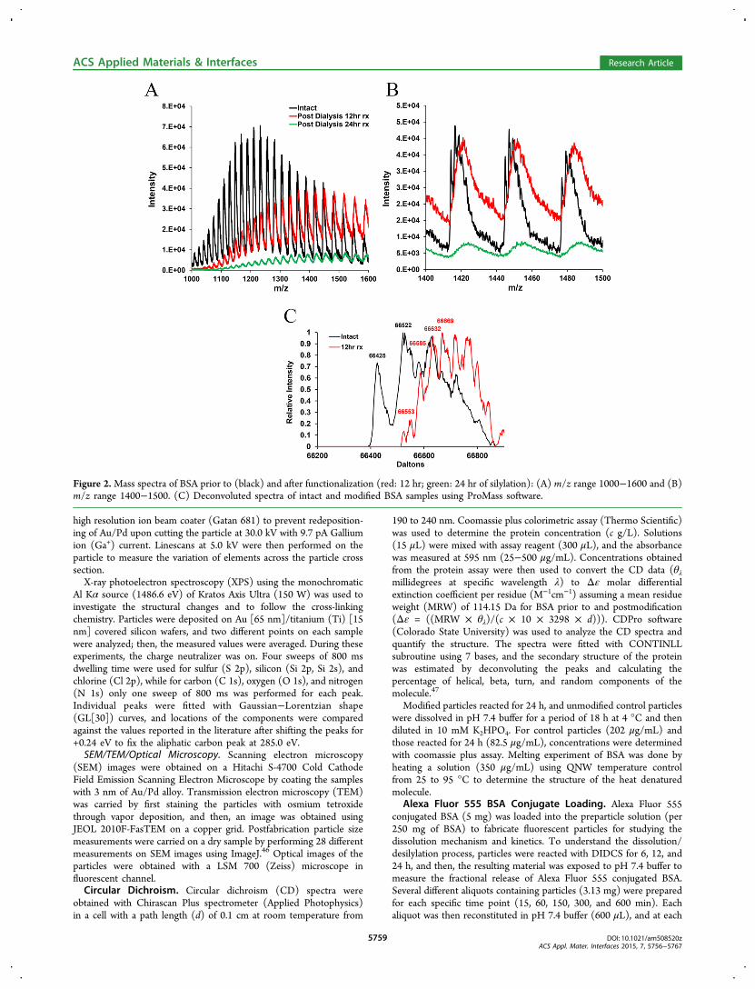
Figure 2.Mass spectra of BSA prior to (black) and after functionalization (red: 12 hr; green: 24 hr of silylation): (A) m/z range 1000−1600 and (B)m/z range 1400−1500. (C) Deconvoluted spectra of intact and modified BSA samples using ProMass software.
ACS Applied Materials & Interfaces Research Article
DOI: 10.1021/am508520zACS Appl. Mater. Interfaces 2015, 7, 5756−5767
5759
time point, the particles were spun down and the supernatant (100μL) was analyzed in triplicate by measuring the fluorescence intensity(Excitation: 545 nm; Emission: 575 nm) using SpectraMax M5(Molecular Devices) in Nunc 96 microwell plates (Thermo Scientific).The intensity of soluble unfunctionalized particles at 600 min served asthe control reference in calculation of the fractional dye release fromthe particles.Fitting and Statistical Analysis. All the model fittings were done
by JMP software (SAS Institute), and p values for statisticalsignificances were calculated by QuickCalcs (GraphPad) onlinestatistical calculator.
■ RESULTS AND DISCUSSION
Particle Fabrication and Composition. A SEM image ofsieved BSA powder is shown in Figure 1A while that of theparticles as fabricated with PRINT on the surface of theharvesting layer is displayed in Figure 1B. Harvested freeparticles of Figure 1C are obtained by washing them with ACN.A TEM image of the free particles is presented in Figure 1E.The composition of the particles postfabrication wasdetermined to be 55.7 wt % BSA, 43.1 wt % lactose, and 1.2wt % glycerol as schematically shown in Figure 1D. Theseparticles were further loaded with Alexa Fluor 555 conjugatedBSA as seen in Figure 1G. The mean external and internaldiameters plus the thickness of the particles were measured as2.88 ± 0.21, 0.7 ± 0.06, 0.56 ± 0.17 μm, respectively (Figure1F).
Component Reactivity. Silylation is extensively used inorganic chemistry to block certain reaction centers byintroducing a silyl group in place of a labile hydrogen atom.Silylation reactions are reversible and proceed via a mechanisminvolving bimolecular nucleophilic substitution at the siliconatom. In many cases, the mechanism and rate of silylation aredetermined by the steric factors associated with the silylatingagent or the compound being silylated.48 Rogozhin et al.49 havederivatized amino acids (Pro, Tyr, Trp, Val, Asp, Lys) anddipeptides (Gly-Ala, Val-Val, Gly-Tyr, Gly-Trp) with differentsilylating agents (trimethylsilyldiethylamine, bis(trimethylsilyl)acetamide, N-trimethylsilyl-N-methylacetamide, and hexame-thyldisilazane) in organic solvents at 20 °C.Silylation with chlorosilanes has been previously used by
several authors on nonprotein/peptide based materials to alterthe surface properties or incorporate drugs into polymericmatrices. Chlorosilanes have been used on multiwall carbonnanotubes to functionalize their surface.50 Gousse et al.51
modified cellulose whisker dispersed in organic solvents andconcluded that it is possible to partially silylate the whiskerswhere the surface of the whiskers is silylated but the core is keptalmost intact. Silylether chemistry has also been used forincorporating drugs into the matrix of particles as a pro-drugstrategy with hydroxyl ethyl acrylate.52
Figure 2A compares the spectra of dialyzed BSA post-functionalization (12−24 h) with intact material. After 12 hr offunctionalization, mass over charge (m/z) peaks for BSA are
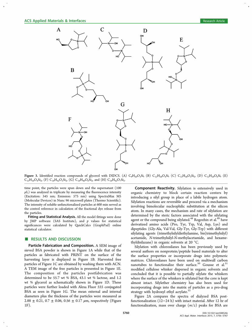
Figure 3. Identified reaction compounds of glycerol with DIDCS. (A) C9H20O3Si, (B) C12H28O4Si, (C) C12H30O3Si2, (D) C12H28O6Si, (E)C15H34O4Si2, (F) C18H42O5Si2, (G) C18H40O6Si2, and (H) C18H42O7Si2.
ACS Applied Materials & Interfaces Research Article
DOI: 10.1021/am508520zACS Appl. Mater. Interfaces 2015, 7, 5756−5767
5760
shifted toward higher values, which serves as the evidence offunctionalization with DIDCS (Figure 2B). Exposure of BSA toDIDCS for 24 h causes formation of visible precipitates.However, due to low signal intensity after 24 h of silylation, noconclusions could be drawn regarding this sample. Once thespectra for intact and 12 hr modified samples are deconvoluted,an increase in the molecular weight of the BSA due to silylationis observed as illustrated in Figure 2C.Liquid chromatography-tandem mass spectrometry (LC-
MS/MS) evaluation of the structure of trypsin digested 12 hrfunctionalized BSA molecules did not reveal any detectablemodification of the recovered peptidic sequence. This can beattributed to the susceptibility of the bonds formed tohydrolysis during the digestion period.Glycerol is capable of forming a variety of combinations with
DIDCS under the reaction conditions (Figure S2, SupportingInformation). The proposed structures are shown in Figure 3for compounds A through H. The most abundant component iscompound E formed from the reaction of two equivalents ofDIDCS with one equivalent of glycerol. Although less abundantcompared to compound E, multiple glycerol molecules can alsobe linked together due to the multifunctionality of both DIDCS
(2 × chlorine [Cl]) and glycerol (3 × hydroxyl [OH]) asconfirmed by the presence of compounds D, G, and H.Furthermore, the reaction conditions are conducive to theformation of larger fragments. Nevertheless, assigning struc-tures to these peaks is not straightforward due to a multitude ofcombinations. Errors involved between the observed andtheoretical masses of the identified peaks are presented inTable S2, Supporting Information.No evidence of reactivity of lactose with DIDCS could be
detected under the reaction conditions. Although lactose bearsmultiple hydroxyl groups, we believe that insolubility of thematerial makes it immune to solid state functionalizationcarried in extra-dry ACN. Lactose is not soluble in acetonitrile−water mixtures containing less than 15% water.53
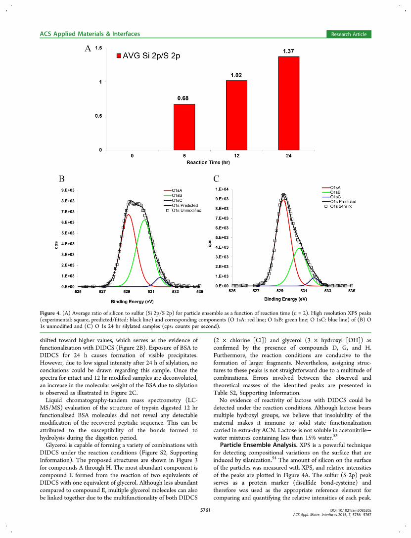
Particle Ensemble Analysis. XPS is a powerful techniquefor detecting compositional variations on the surface that areinduced by silanization.54 The amount of silicon on the surfaceof the particles was measured with XPS, and relative intensitiesof the peaks are plotted in Figure 4A. The sulfur (S 2p) peakserves as a protein marker (disulfide bond-cysteine) andtherefore was used as the appropriate reference element forcomparing and quantifying the relative intensities of each peak.
Figure 4. (A) Average ratio of silicon to sulfur (Si 2p/S 2p) for particle ensemble as a function of reaction time (n = 2). High resolution XPS peaks(experimental: square, predicted/fitted: black line) and corresponding components (O 1sA: red line; O 1sB: green line; O 1sC: blue line) of (B) O1s unmodified and (C) O 1s 24 hr silylated samples (cps: counts per second).
ACS Applied Materials & Interfaces Research Article
DOI: 10.1021/am508520zACS Appl. Mater. Interfaces 2015, 7, 5756−5767
5761
Both silicon (Si 2s) and (Si 2p) peaks increase with respect tosulfur (S 2p) as a function of reaction time. However, the datafor Si 2s is not shown for brevity. High resolution peaks ofsilicon (Si 2s, Si 2p) and sulfur (S 2p) for unmodified andreacted samples along with the survey scans are compared inFigure S3, Supporting Information. High resolution scans ofoxygen (O 1s), carbon (C 1s), and nitrogen (N 1s) peaks wereperformed to detect any chemical changes associated withfunctionalization. Figure 4B,C compares oxygen (O 1s) scansfor unmodified particles and the sample silylated for 24 hr. Theshifted values of the binding energies for the deconvolutedcomponents are listed in Table S3, Supporting Information.Lopez and co-workers55 reported the value of 532.8 eV for
the oxygen (O 1s) peak of 1,10-decanediol and 532.7 eV forpoly(vinyl alcohol) and poly(propylene glycol) with referenceto an aliphatic carbon peak at 285.0 eV. Therefore, it can beconcluded that the reduction in the intensity of the oxygen (O1s) peak (labeled B) is consistent with consumption ofhydroxyl groups on the surface due to the functionalizationafter 24 hr. The oxygen (O 1s) peak (labeled A) of 531.4 eV isvery close to peptidic oxygen of BSA as reported by Iucci etal.56 at 531.6 eV. Carbon (C 1s) and nitrogen (N 1s) scans forthe same samples are also presented in Figure S4, SupportingInformation.
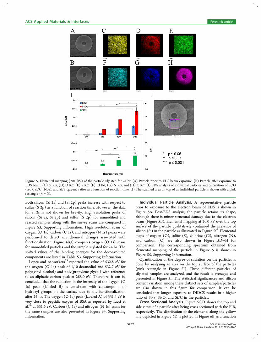
Individual Particle Analysis. A representative particleprior to exposure to the electron beam of EDS is shown inFigure 5A. Post-EDS analysis, the particle retains its shape,although there is minor structural damage due to the electronbeam (Figure 5B). Elemental mapping at 20.0 kV over the topsurface of the particle qualitatively confirmed the presence ofsilicon (Si) in the particle as illustrated in Figure 5C. Elementalmaps of oxygen (O), sulfur (S), chlorine (Cl), nitrogen (N),and carbon (C) are also shown in Figure 5D−H forcomparison. The corresponding spectrum obtained fromelemental mapping of the particle in Figure 5 is shown inFigure S5, Supporting Information.Quantification of the degree of silylation on the particles is
done by analyzing an area on the top surface of the particles(pink rectangle in Figure 5J). Three different particles ofsilylated samples are analyzed, and the result is averaged andpresented in Figure 5I. The statistical significances and siliconcontent variation among these distinct sets of samples/particlesare also shown in this figure for comparison. It can beconcluded that longer exposure to DIDCS results in a higherratio of Si/S, Si/O, and Si/C in the particles.
Cross Sectional Analysis. Figure 6C,D shows the top andside views of a particle after being cross sectioned with the FIB,respectively. The distribution of the elements along the yellowline depicted in Figure 6D is plotted in Figure 6B as a function
Figure 5. Elemental mapping (20.0 kV) of the particle silylated for 24 hr. (A) Particle prior to EDS beam exposure. (B) Particle after exposure toEDS beam. (C) Si Kα, (D) O Kα, (E) S Kα, (F) Cl Kα, (G) N Kα, and (H) C Kα. (I) EDS analysis of individual particles and calculation of Si/O(red), Si/C (blue), and Si/S (green) ratios as a function of reaction time. (J) The scanned area on top of an individual particle is shown with a pinkrectangle (n = 3).
ACS Applied Materials & Interfaces Research Article
DOI: 10.1021/am508520zACS Appl. Mater. Interfaces 2015, 7, 5756−5767
5762

of transverse distance. The sum spectrum of the linescan is alsocompared with the spectrum of the background to confirm thatno silicon signal is detected on the carbon background (Figure6A). No variation could be observed in the silicon contentalong the scanned line of Figure 6D, which suggests thatDIDCS penetrates all the way through the particle and thereaction is not limited only to the surface of the particle.Mechanism and Dissolution Profile. To study the
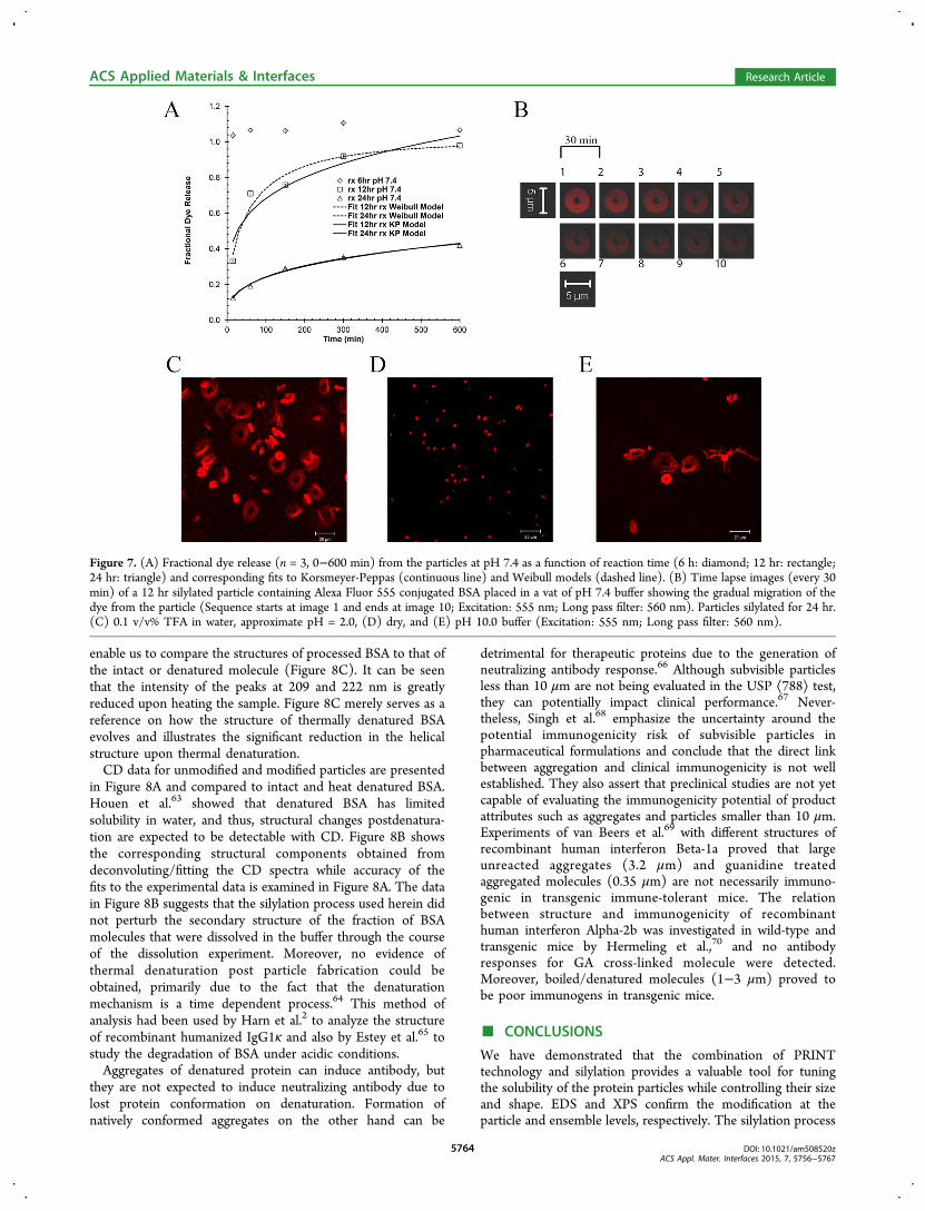
dissolution of particles, silylated particles were exposed to pH7.4 buffer and the fractional release of Alexa Fluor 555conjugated BSA was measured (Figure 7A, symbols). The datademonstrates that the longer the treatment of particles withDIDCS, the slower is the dissolution rate of the particles. Thisis consistent with the higher silicon content of the particles as afunction of reaction time, obtained from EDS and XPSexperiments.Numerous mathematical models exist in the literature for
describing the dissolution behavior.57,58 Weibull59 andKorsmeyer-Peppas60 models are fitted to the experimentaldata (Figure 7A, lines), and corresponding model parametersare listed in Table S4, Supporting Information. The diffusionalexponent in the Korsmeyer-Peppas model is indicative of arelease mechanism (mode of transport of solute) and is shapedependent.61
To visualize the dissolution process for particles post-functionalization, the behavior of a single 12 hr silylated particleis tracked in pH 7.4 buffer as a function of time. As observed inFigure 7B, the intensity of Alexa Fluor 555 conjugated BSA inthe particle gradually diminishes due to dissolution of the
fluorophore from the particle into the surrounding environ-ment. Effects of both acidic (0.1 v/v% TFA, approximate pH =2.0) and basic (pH 10.0 buffer) conditions on the size ofparticles are illustrated in Figure 7C,E for particles function-alized for 24 hr, respectively. At these pH extremes, particlessignificantly swell to 4−5 times their original size of Figure 7D.Shirai et al.62 have studied the effect of different pHs on thehydrolytic removal of silyl groups from trialkyl silyl ethers.Their results indicate that there exists a pH where the observedhydrolysis rate constants are minimized. They demonstratedthat this rate of hydrolysis is also affected by the electro-negativity and steric size of the substituents on both silicon andoxygen atoms. The extreme swelling of these particles in bothacidic and basic conditions suggests that the prevailingmechanism of dissolution is desilylation (removal of silylgroups).The extreme swelling of the particles in acidic pH as
observed in Figure 7C leaves the protein content of theparticles prone to degradation in the harsh acidic environmentof the gastrointestinal tract, and therefore, an oral delivery routefor these suspensions is not envisioned. A measure for thestability of the suspension of 24 hr functionalized particles inpH 7.4 buffer is presented in Figure S6, SupportingInformation.
Structural Evaluation. CD signal can determine structuralchanges associated with processing or functionalization of theprotein in the particles. An examination of the melting behaviorof intact BSA was done to evaluate the structural changesassociated with heat denaturation of the molecule, which will
Figure 6. (A) Sum of the elemental spectrum along the EDS linescan on the particle sectioned using FIB compared to the spectrum of the carbonbackground (5.0 kV). (B) Distribution of the elements along the linescan (red: silicon; blue: sulfur; green: chlorine; black: oxygen; burgundy:gallium). (C) Top view of the FIB sectioned particle. (D) Side view of the same particle (yellow line indicates the position of the line used toperform the EDS analysis).
ACS Applied Materials & Interfaces Research Article
DOI: 10.1021/am508520zACS Appl. Mater. Interfaces 2015, 7, 5756−5767
5763
enable us to compare the structures of processed BSA to that ofthe intact or denatured molecule (Figure 8C). It can be seenthat the intensity of the peaks at 209 and 222 nm is greatlyreduced upon heating the sample. Figure 8C merely serves as areference on how the structure of thermally denatured BSAevolves and illustrates the significant reduction in the helicalstructure upon thermal denaturation.CD data for unmodified and modified particles are presented
in Figure 8A and compared to intact and heat denatured BSA.Houen et al.63 showed that denatured BSA has limitedsolubility in water, and thus, structural changes postdenatura-tion are expected to be detectable with CD. Figure 8B showsthe corresponding structural components obtained fromdeconvoluting/fitting the CD spectra while accuracy of thefits to the experimental data is examined in Figure 8A. The datain Figure 8B suggests that the silylation process used herein didnot perturb the secondary structure of the fraction of BSAmolecules that were dissolved in the buffer through the courseof the dissolution experiment. Moreover, no evidence ofthermal denaturation post particle fabrication could beobtained, primarily due to the fact that the denaturationmechanism is a time dependent process.64 This method ofanalysis had been used by Harn et al.2 to analyze the structureof recombinant humanized IgG1κ and also by Estey et al.65 tostudy the degradation of BSA under acidic conditions.Aggregates of denatured protein can induce antibody, but
they are not expected to induce neutralizing antibody due tolost protein conformation on denaturation. Formation ofnatively conformed aggregates on the other hand can be
detrimental for therapeutic proteins due to the generation ofneutralizing antibody response.66 Although subvisible particlesless than 10 μm are not being evaluated in the USP ⟨788⟩ test,they can potentially impact clinical performance.67 Never-theless, Singh et al.68 emphasize the uncertainty around thepotential immunogenicity risk of subvisible particles inpharmaceutical formulations and conclude that the direct linkbetween aggregation and clinical immunogenicity is not wellestablished. They also assert that preclinical studies are not yetcapable of evaluating the immunogenicity potential of productattributes such as aggregates and particles smaller than 10 μm.Experiments of van Beers et al.69 with different structures ofrecombinant human interferon Beta-1a proved that largeunreacted aggregates (3.2 μm) and guanidine treatedaggregated molecules (0.35 μm) are not necessarily immuno-genic in transgenic immune-tolerant mice. The relationbetween structure and immunogenicity of recombinanthuman interferon Alpha-2b was investigated in wild-type andtransgenic mice by Hermeling et al.,70 and no antibodyresponses for GA cross-linked molecule were detected.Moreover, boiled/denatured molecules (1−3 μm) proved tobe poor immunogens in transgenic mice.
■ CONCLUSIONS
We have demonstrated that the combination of PRINTtechnology and silylation provides a valuable tool for tuningthe solubility of the protein particles while controlling their sizeand shape. EDS and XPS confirm the modification at theparticle and ensemble levels, respectively. The silylation process
Figure 7. (A) Fractional dye release (n = 3, 0−600 min) from the particles at pH 7.4 as a function of reaction time (6 h: diamond; 12 hr: rectangle;24 hr: triangle) and corresponding fits to Korsmeyer-Peppas (continuous line) and Weibull models (dashed line). (B) Time lapse images (every 30min) of a 12 hr silylated particle containing Alexa Fluor 555 conjugated BSA placed in a vat of pH 7.4 buffer showing the gradual migration of thedye from the particle (Sequence starts at image 1 and ends at image 10; Excitation: 555 nm; Long pass filter: 560 nm). Particles silylated for 24 hr.(C) 0.1 v/v% TFA in water, approximate pH = 2.0, (D) dry, and (E) pH 10.0 buffer (Excitation: 555 nm; Long pass filter: 560 nm).
ACS Applied Materials & Interfaces Research Article
DOI: 10.1021/am508520zACS Appl. Mater. Interfaces 2015, 7, 5756−5767
5764

renders the solubility of the particles to be tunable, eventuallydissolving in aqueous media while not perturbing the secondarystructure of the fraction of BSA released from the particles. Therate of dissolution can be controlled by the time particles spendin the functionalization media consisting of extra-dryacetonitrile and diisopropyldichlorosilane. Extreme swelling ofthe particles in low and high pH confirms that desilylation isthe mechanism of particle dissolution. To further understandthe nature of functionalization, EDS cross sectional analysisconfirmed that the chlorosilane diffuses all the way through theparticles and is not just limited to the surface.Lowering the temperature used in the filling/harvesting
stages of the particle fabrication enables us to better protect thesensitive contents of the particles against various modes ofinstabilities. Fabricating particles from homologous albumininstead of a heterologous one is recommended as it allows us toprepare particles that are made from nonimmunogenicingredients. Immunogenicity of our subvisible functionalizedparticles loaded with a therapeutic protein will be thoroughlyinvestigated in future, to elucidate the role of functionalizationand fabrication parameters. In vivo behavior of these function-alized particles and their potential in extending proteinretention is a subject of a separate study.
■ ASSOCIATED CONTENT
*S Supporting Information(1) HPLC analysis of the particles postfabrication. (2)Schematic of particle silylation. (3) FT-MS analysis of reactionproducts of glycerol and DIDCS. (4) Estimated error involvedin the structure assignment for the reaction products of glyceroland DIDCS. (5) High resolution XPS scans of silicon andsulfur. (6) High resolution XPS scans and peak locations. (7)
EDS elemental mapping performed on an individual particle.(8) Parameters for Korsmeyer-Peppas and Weibull models. (9)Optical density measurement for the suspension of function-alized particles. This material is available free of charge via theInternet at http://pubs.acs.org.
■ AUTHOR INFORMATIONCorresponding Author*E-mail: [email protected]. Tel: (919) 962-2166. Fax: (919)962-5467.
NotesThe authors declare the following competing financialinterest(s): Joseph M. DeSimone is a founder and maintainsa financial interest in Liquidia Technologies.
■ ACKNOWLEDGMENTSWe acknowledge the National Institutes of Health (NIH)Director’s Pioneer Award and Liquidia Technologies forsupport. Characterization of the particles was done in theChapel Hill Analytical and Nanofabrication Laboratory(CHANL), and microscopic evaluation of the particles wasdone at Microscopy Services Laboratory (MSL) of theUniversity of North Carolina at Chapel Hill.
■ REFERENCES(1) Almeida, A. J.; Souto, E. Solid Lipid Nanoparticles as a DrugDelivery System for Peptides and Proteins. Adv. Drug Delivery Rev.2007, 59, 478−490.(2) Harn, N.; Allan, C.; Oliver, C.; Middaugh, C. R. HighlyConcentrated Monoclonal Antibody Solutions: Direct Analysis ofPhysical Structure and Thermal Stability. J. Pharm. Sci. 2007, 96, 532−546.
Figure 8. (A) CD spectra for heat denatured BSA (circle), intact BSA (diamond), unfunctionalized control particles (square), and functionalizedparticles for 24 hr (triangle). Corresponding fits using CDPro software are also shown (lines). (B) Contribution of each structure afterdeconvolution (green: helical; yellow: beta; blue: turn; red: random). (C) CD signal at 222 nm (triangle) and 209 nm (circle) as a function oftemperature (°C) for BSA solution and the associated heat denaturation of the molecule.
ACS Applied Materials & Interfaces Research Article
DOI: 10.1021/am508520zACS Appl. Mater. Interfaces 2015, 7, 5756−5767
5765

(3) Antosova, Z.; Mackova, M.; Kral, V.; Macek, T. TherapeuticApplication of Peptides and Proteins: Parenteral Forever? TrendsBiotechnol. 2009, 27, 628−635.(4) Vermonden, T.; Censi, R.; Hennink, W. E. Hydrogels for ProteinDelivery. Chem. Rev. 2012, 112, 2853−2888.(5) Manning, M. C.; Chou, D. K.; Murphy, B. M.; Payne, R. W.;Katayama, D. S. Stability of Protein Pharmaceuticals: An Update.Pharm. Res. 2010, 27, 544−575.(6) Pechenov, S.; Shenoy, B.; Yang, M. X.; Basu, S. K.; Margolin, A.L. Injectable Controlled Release Formulations Incorporating ProteinCrystals. J. Controlled Release 2004, 96, 149−158.(7) Lee, V. H. L.; Dodda-Kashi, S.; Grass, G. M.; Rubas, W. In Peptideand Protein Drug Delivery; Lee, V. H. L., Ed.; Marcel Dekker: NewYork, NY, 1991; Chapter 16, pp 691−738.(8) Brown, L. R. Commercial Challenges of Protein Drug Delivery.Expert Opin. Drug Delivery 2005, 2, 29−42.(9) Riva, P.; Arista, A.; Sturiale, C.; Moscatelli, G.; Tison, V.; Mariani,M.; Seccamani, E.; Lazzari, S.; Fagioli, L.; Franceschi, G.; Sarti, G.;Riva, N.; Natali, P. G.; Zardi, L.; Scassellati, G. A. Treatment ofIntracranial Human Glioblastoma by Direct Intratumoral Admin-istration of I-131-Labeled Anti-Tenascin Monoclonal-Antibody BC-2.Int. J. Cancer 1992, 51, 7−13.(10) Kitamura, K.; Takahashi, T.; Kotani, T.; Miyagaki, T.; Yamaoka,N.; Tsurumi, H.; Noguchi, A.; Yamaguchi, T. Local-Administration ofMonoclonal Antibody-Drug Conjugate - A New Strategy to Reducethe Local Recurrence of Colorectal-Cancer. Cancer Res. 1992, 52,6323−6328.(11) Abrishami, M.; Ganavati, S. Z.; Soroush, D.; Rouhbakhsh, M.;Jaafari, M. R.; Malaekeh-Nikouei, B. Preparation, Characterization, andin Vivo Evaluation of Nanoliposomes-Encapsulated Bevacizumab(Avastin) for Intravitreal Administration. Retina 2009, 29, 699−703.(12) Ohtori, S.; Miyagi, M.; Eguchi, Y.; Inoue, G.; Orita, S.; Ochiai,N.; Kishida, S.; Kuniyoshi, K.; Nakamura, J.; Aoki, Y.; Ishikawa, T.;Arai, G.; Kamoda, H.; Suzuki, M.; Takaso, M.; Furuya, T.; Kubota, G.;Sakuma, Y.; Oikawa, Y.; Toyone, T.; Takahashi, K. Efficacy of EpiduralAdministration of Anti-Interleukin-6 Receptor Antibody onto SpinalNerve for Treatment of Sciatica. Eur. Spine J. 2012, 21, 2079−2084.(13) Nikas, S.; Temekonidis, T.; Zikou, A.; Argyropoulou, M.;Efremidis, S.; Drosos, A. Treatment of Resistant Rheumatoid Arthritisby Intra-Articular Infliximab Injections: A Pilot Study. Ann. Rheum. Dis.2004, 63, 102−103.(14) Jorgensen, L.; Nielsen, H. M. Delivery Technologies forBiopharmaceuticals: Peptides, Proteins, Nucleic Acids, and Vaccines, 1sted; John Wiley & Sons: West Sussex, 2009.(15) Banerjee, P. S.; Hosny, E. A.; Robinson, J. R. In Peptide andProtein Drug Delivery; Lee, V. H. L., Ed.; Marcel Dekker: New York,NY, 1991; Chapter 10, pp 487−543.(16) Lee, K. Y.; Yuk, S. H. Polymeric Protein Delivery Systems. Prog.Polym. Sci. 2007, 32, 669−697.(17) Yang, M. X.; Shenoy, B.; Disttler, M.; Patel, R.; McGrath, M.;Pechenov, S.; Margolin, A. L. Crystalline Monoclonal Antibodies forSubcutaneous Delivery. Proc. Natl. Acad. Sci. U.S.A. 2003, 100, 6934−6939.(18) Miller, M. A.; Engstrom, J. D.; Ludher, B. S.; Johnston, K. P.Low Viscosity Highly Concentrated Injectable Nonaqueous Suspen-sions of Lysozyme Microparticles. Langmuir 2010, 26, 1067−1074.(19) Arshady, R. Albumin Microspheres and Microcapsules -Methodology of Manufacturing Techniques. J. Controlled Release1990, 14, 111−131.(20) Patil, G. V. Biopolymer Albumin for Diagnosis and in DrugDelivery. Drug Dev. Res. 2003, 58, 219−247.(21) Tong, W.; Gao, C.; Moehwald, H. PH-Responsive ProteinMicrocapsules Fabricated via Glutaraldehyde Mediated CovalentLayer-by-Layer Assembly. Colloid Polym. Sci. 2008, 286, 1103−1109.(22) Zhou, Z.; Anselmo, A. C.; Mitragotri, S. Synthesis of Protein-Based, Rod-Shaped Particles from Spherical Templates Using Layer-by-Layer Assembly. Adv. Mater. 2013, 25, 2723−2727.(23) Nettey, H.; Haswani, D.; Oettinger, C. W.; D’Souza, M. J.Formulation and Testing of Vancomycin Loaded Albumin Micro-
spheres Prepared by Spray-Drying. J. Microencapsulation 2006, 23,632−642.(24) Langer, K.; Balthasar, S.; Vogel, V.; Dinauer, N.; Von Briesen,H.; Schubert, D. Optimization of the Preparation Process for HumanSerum Albumin (HSA) Nanoparticles. Int. J. Pharm. 2003, 257, 169−180.(25) Lin, W.; Coombes, A. G.; Davies, M. C.; Davis, S. S.; Illum, L.Preparation of Sub-100 nm Human Serum Albumin Nanospheresusing a pH-Coacervation Method. J. Drug Targeting 1993, 1, 237−243.(26) Bogdansky, S. In Biodegradable Polymers as Drug DeliverySystems; Chasin, M., Langer, R., Eds.; Marcel Dekker: New York, NY,1990; Chapter 7, pp 231−259.(27) Bhargava, K.; Ando, H. Y. Immobilization of Active Urokinaseon Albumin Microspheres: Use of a Chemical Dehydrant and ProcessMonitoring. Pharm. Res. 1992, 9, 776−781.(28) Goosen, M. F.; Leung, Y. F.; O’Shea, G. M.; Chou, S.; Sun, A.M. Slow Release of Insulin from a Biodegradable Matrix Implanted inDiabetic Rats. Diabetes 1983, 32, 478−481.(29) Rollett, A.; Reiter, T.; Nogueira, P.; Cardinale, M.; Loureiro, A.;Gomes, A.; Cavaco-Paulo, A.; Moreira, A.; Carmo, A. M.; Guebitz, G.M. Folic Acid-Functionalized Human Serum Albumin Nanocapsulesfor Targeted Drug Delivery to Chronically Activated Macrophages. Int.J. Pharm. 2012, 427, 460−466.(30) Oettinger, C. W.; D’Souza, M. J. Microencapsulated DrugDelivery: A New Approach to Pro-Inflammatory Cytokine Inhibition.J. Microencapsulation 2012, 29, 455−462.(31) Kratz, F. Albumin as a Drug Carrier: Design of Prodrugs, DrugConjugates and Nanoparticles. J. Controlled Release 2008, 132, 171−183.(32) Doshi, N.; Mitragotri, S. Macrophages Recognize Size andShape of Their Targets. PLoS One 2010, 5, No. e10051.(33) Lee, T. K.; Sokoloski, T. D.; Royer, G. P. Serum Albumin Beads:An Injectable, Biodegradable System for the Sustained Release ofDrugs. Science 1981, 213, 233−235.(34) Ratcliffe, J. H.; Hunneyball, I. M.; Smith, A.; Wilson, C. G.;Davis, S. S. Preparation and Evaluation of Biodegradable PolymericSystems for the Intra-Articular Delivery of Drugs. J. Pharm. Pharmacol.1984, 36, 431−436.(35) Wong, S. S. Chemistry of Protein Conjugation and Cross-Linking,1st ed; CRC Press: Boca Raton, FL, 1991.(36) Srinivasachar, K.; Neville, D. New Protein Cross-LinkingReagents that Are Cleaved by Mild Acid. Biochemistry 1989, 28, 2501−2509.(37) Benesch, R. E.; Kwong, S. Bis-Pyridoxal Polyphosphates - ANew Class of Specific Intramolecular Crosslinking Agents forHemoglobin. Biochem. Biophys. Res. Commun. 1988, 156, 9−14.(38) Blattler, W. A.; Kuenzi, B. S.; Lambert, J. M.; Senter, P. D. NewHeterobifunctional Protein Cross-Linking Reagent that Forms anAcid-Labile Link. Biochemistry 1985, 24, 1517−1524.(39) Parrott, M. C.; Luft, J. C.; Byrne, J. D.; Fain, J. H.; Napier, M. E.;DeSimone, J. M. Tunable Bifunctional Silyl Ether Cross-Linkers forthe Design of Acid-Sensitive Biomaterials. J. Am. Chem. Soc. 2010, 132,17928−17932.(40) Kelly, J. Y.; DeSimone, J. M. Shape-Specific, MonodisperseNano-Molding of Protein Particles. J. Am. Chem. Soc. 2008, 130,5438−5439.(41) Houen, G. The Solubility of Proteins in Organic Solvents. ActaChem. Scand. 1996, 50, 68−70.(42) Griebenow, K.; Klibanov, A. M. On Protein Denaturation inAqueous-Organic Mixtures but not in Pure Organic Solvents. J. Am.Chem. Soc. 1996, 118, 11695−11700.(43) Spahr, P.; Edsall, J. Amino Acid Composition of Human +Bovine Serum Mercaptalbumins. J. Biol. Chem. 1964, 239, 850−854.(44) Brown, J. R. Structure of Bovine Serum-Albumin. Fed. Proc.1975, 34, 591.(45) Kolthoff, I. M.; Bruckenstein, S.; Chantooni, M. K. Acid-baseEquilibria in Acetonitrile - Spectrophotometric and ConductometricDetermination of Dissociation of Various Acids. J. Am. Chem. Soc.1961, 83, 3927−3935.
ACS Applied Materials & Interfaces Research Article
DOI: 10.1021/am508520zACS Appl. Mater. Interfaces 2015, 7, 5756−5767
5766

(46) Schneider, C. A.; Rasband, W. S.; Eliceiri, K. W. NIH Image toImageJ: 25 Years of Image Analysis. Nat. Methods 2012, 9, 671−675.(47) Sreerama, N.; Woody, R. W. Estimation of Protein SecondaryStructure from Circular Dichroism Spectra: Comparison of CONTIN,SELCON, and CDSSTR Methods with an Expanded Reference Set.Anal. Biochem. 2000, 287, 252−260.(48) Kashutina, M. V.; Ioffe, S. L.; Tartakovskii, V. A. Silylation ofOrganic Compounds. Russ. Chem. Rev. 1975, 44, 733−747.(49) Rogozhin, S. V.; Davidovich, Y. A.; Andreev, S. M.; Mironova,N. V.; Yurtanov, A. I. Preparation of Trimethylsilyl Derivatives ofAmino-Acids and Peptides for Peptide-Synthesis. Bull. Acad. Sci. USSR,Div. Chem. Sci. (Engl. Transl.) 1974, 23, 1789−1792.(50) Vast, L.; Mekhalif, Z.; Fonseca, A.; Nagy, J. B.; Delhalle, J.Preparation and Electrical Characterization of a Silicone ElastomerComposite Charged with Multi-Wall Carbon Nanotubes Function-alized with 7-Octenyltrichlorosilane. Compos. Sci. Technol. 2007, 67,880−889.(51) Gousse, C.; Chanzy, H.; Excoffier, G.; Soubeyrand, L.; Fleury, E.Stable Suspensions of Partially Silylated Cellulose Whiskers Dispersedin Organic Solvents. Polymer 2002, 43, 2645−2651.(52) Parrott, M. C.; Finniss, M.; Luft, J. C.; Pandya, A.; Gullapalli, A.;Napier, M. E.; Desimone, J. M. Incorporation and Controlled Releaseof Silyl Ether Prodrugs from PRINT Nanoparticles. J. Am. Chem. Soc.2012, 134, 7978−7982.(53) Folkes, D. J.; Jordan, M. A. In Carbohydrates in Food; Eliasson,A. C., Ed.; Taylor & Francis: Boca Raton, FL, 2006; Chapter 1, pp 1−40.(54) Puglisi, O.; Torrisi, A.; Marletta, G. XPS Investigation of theEffects Induced by the Silanization on Real Glass Surfaces. J. Non-Cryst. Solids 1984, 68, 219−230.(55) Lopez, G. P.; Castner, D. G.; Ratner, B. D. XPS O 1s Binding-Energies for Polymers Containing Hydroxyl, Ether, Ketone and EsterGroups. Surf. Interface Anal. 1991, 17, 267−272.(56) Iucci, G.; Polzonetti, G.; Infante, G.; Rossi, L. XPS and FT-IRSpectroscopy Study of Albumin Adsorption on the Surface of a Pi-Conjugated Polymer Film. Surf. Interface Anal. 2004, 36, 724−728.(57) Arifin, D. Y.; Lee, L. Y.; Wang, C. Mathematical Modeling andSimulation of Drug Release from Microspheres: Implications to DrugDelivery Systems. Adv. Drug Delivery Rev. 2006, 58, 1274−1325.(58) Costa, P.; Manuel, J.; Lobo, S. Modeling and Comparison ofDissolution Profiles. Eur. J. Pharm. Sci. 2001, 13, 123−133.(59) Weibull, W. A Statistical Distribution Function of WideApplicability. J. Appl. Mech. 1951, 18, 293−297.(60) Korsmeyer, R. W.; Gurny, R.; Doelker, E.; Buri, P.; Peppas, N.A. Mechanisms of Solute Release from Porous Hydrophilic Polymers.Int. J. Pharm. 1983, 15, 25−35.(61) Ritger, P. L.; Peppas, N. A. A Simple Equation for Descriptionof Solute Release II. Fickian and Anomalous Release from SwellableDevices. J. Controlled Release 1987, 5, 37−42.(62) Shirai, N.; Moriya, K.; Kawazoe, Y. pH-Dependence ofHydrolytic Removal of Silyl Group from Trialkylsilyl Ethers.Tetrahedron 1986, 42, 2211−2214.(63) Houen, G.; Svaerke, C.; Barkholt, V. The Solubilities ofDenatured Proteins in Different Organic Solvents. Acta Chem. Scand.1999, 53, 1122−1126.(64) Nakagaki, M.; Sano, Y. Light Scattering Studies on the ThermalDenaturation of Bovine Serum Albumin. Bull. Chem. Soc. Jpn. 1973, 46,791−797.(65) Estey, T.; Kang, J.; Schwendeman, S. P.; Carpenter, J. F. BSADegradation under Acidic Conditions: A Model for Protein Instabilityduring Release from PLGA Delivery Systems. J. Pharm. Sci. 2006, 95,1626−1639.(66) Rosenberg, A. S. Effects of Protein Aggregates: An ImmunologicPerspective. AAPS J. 2006, 8, E501−E507.(67) Carpenter, J. F.; Randolph, T. W.; Jiskoot, W.; Crommelin, D.J.; Middaugh, C. R.; Winter, G.; Fan, Y. X.; Kirshner, S.; Verthelyi, D.;Kozlowski, S.; Clouse, K. A.; Swann, P. G.; Rosenberg, A.; Cherney, B.Overlooking Subvisible Particles in Therapeutic Protein Products:
Gaps That May Compromise Product Quality. J. Pharm. Sci. 2009, 98,1201−1205.(68) Singh, S. K.; Afonina, N.; Awwad, M.; Bechtold-Peters, K.; Blue,J. T.; Chou, D.; Cromwell, M.; Krause, H. J.; Mahler, H. C.; Meyer, B.K.; Narhi, L.; Nesta, D. P.; Spitznagel, T. An Industry Perspective onthe Monitoring of Subvisible Particles as a Quality Attribute forProtein Therapeutics. J. Pharm. Sci. 2010, 99, 3302−3321.(69) van Beers, M. M.; Sauerborn, M.; Gilli, F.; Brinks, V.;Schellekens, H.; Jiskoot, W. Oxidized and Aggregated RecombinantHuman Interferon Beta is Immunogenic in Human Interferon BetaTransgenic Mice. Pharm. Res. 2011, 28, 2393−2402.(70) Hermeling, S.; Aranha, L.; Damen, J. M.; Slijper, M.;Schellekens, H.; Crommelin, D. J.; Jiskoot, W. Structural Character-ization and Immunogenicity in Wild-Type and Immune Tolerant Miceof Degraded Recombinant Human Interferon Alpha2b. Pharm. Res.2005, 22, 1997−2006.
ACS Applied Materials & Interfaces Research Article
DOI: 10.1021/am508520zACS Appl. Mater. Interfaces 2015, 7, 5756−5767
5767