silver nano proof jbn
TRANSCRIPT
8/8/2019 Silver Nano Proof JBN
http://slidepdf.com/reader/full/silver-nano-proof-jbn 1/6

8/8/2019 Silver Nano Proof JBN
http://slidepdf.com/reader/full/silver-nano-proof-jbn 2/6
RESEARCH
AR
TICLE
Inherently Colored Antimicrobial Fibers Employing Silver Nanoparticles Sreekumar et al.
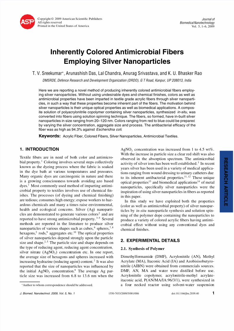
Ram
1st coag. bath 2nd coag. bath Washing cum
drawing bath
Rollers
To Winder
Heater plate
Fig. 1. Schematic diagram of the fiber spinning set up.
polymerization technique at 55 C under nitrogen atmo-
sphere. The reaction medium was a mixture of dimethylformamide and water. Azobisisobutyronitrile (1 wt% withrespect to monomer feed) was used as the initiator.The details of the synthesis procedures are reported
elsewhere.15
2.2. Synthesis of Silver Nanoparticles
AgNO3 and hydrazine hydrate (N2H4 ·H2O) were used asreceived from commercial sources. In a standard experi-ment 12 g of PAN copolymer was dissolved in 100 ml of DMF to obtain a transparent solution. In a separate vial
0.5% AgNO3 (w.r.t PAN copolymer) was dissolved in 5 mlof DMF. The two solutions were mixed together by con-stant stirring. In this procedure four dope solutions were
prepared with 0, 0.5, 1 and 3% AgNO3 with respect toPAN. The Ag+ ions of AgNO3 were reduced to metallic sil-
ver using equivalent amount of hydrazine hydrate (N2H4 ·
H2O). Thus, Ag contents in the solutions were approxi-
mately 0, 0.3, 0.6 and 1.9%.
2.3. Acrylic Fiber Spinning
The fibres were spun using solution spinning technique ona laboratory spinning machine (Fig. 1). The dope solutionswere converted to fibers by solution spinning process16
using the small scale spinning system manufactured by
Bradford University Research Ltd. DMF/water volumetricratios in the coagulation baths (baths 1 and 2) and drawing
bath (bath 3) were 60/40, 10/90, and 0/100, respectively.The temperature of the two coagulation baths was maintai-ned at 30 C. The drawing bath temperature was maintainedat 100 C. An in-line heater plate was used for fiber dryingwhich was maintained at 130 C. There was no fiber draw-
ing in the two coagulation baths. Fiber was drawn between2nd and 3rd rollers in boiling water, and was allowed to relaxduring the drying process over the heater plate. The first
take-up roller speed (1 m/min) and the winder speed(5 m/min) were set in such a way that the final draw ratiofor each composite fiber was 5. Fibers were also producedfrom PAN/AgNO3 (PAN:AgNO3 = 99:1) dope solutions
without adding hydrazine hydrate. The decomposition of
AgNO3 to form Ag in the fiber was carried out by heattreatment of the fiber at 150 C over the heater plate.
2.4. Fiber Characterization
Transmission Electron Microscopy (TEM) analysis wasperformed with Tecnai G2 TEM machine supplied by FieldEmission Instrument (FEI), USA, operated at 200 kV.
UV-Vis spectra of the acrylic fibres were recordedin the absorption mode using Perkin-Elmer Lamda 19UV/VIS/NIR spectrometer. The fiber mechanical properties
were measured using Favimat single fiber testing machinesupplied by Textechno, Germany. The gauge length and
testing speed used were 25 mm and 10 mm/min respec-tively. Antimicrobial tests were conducted at Bombay Tex-tile Research Association (BTRA), Mumbai, using ASTM:
E2149-01 test method. This test was conducted by shak-
ing samples in a concentrated bacterial suspension for aone hour contact time. The suspension was serially diluted
both before and after contact and cultured. The number of organisms in the suspension was determined and the per-cent reduction was calculated based on initial counts on
pure PAN control sample.
3. RESULTS AND DISCUSSION
3.1. Nanoparticle Formation
The in-situ synthesis of the Ag nanoparticle is based on
the fact that both AgNO3 and polyacrylonitrile dissolve inDMF which is a common solvent. Upon reduction withhydrazine hydrate AgNO3 will be converted to metallic Ag
particles. The viscous polymer gel (Dope) does not allowthe nanoparticles to coalesce easily and the particles getkinetically stabilized. The spinning of the fiber needs to
be conducted as soon as possible as the chances of coa-lescence can not be ruled out with time. Figure 2 showsthree typical TEM micrographs of PAN containing low
(0.6%) medium (3.0%) and high (6.0%), Ag concentra-tions. For transmission electron microscopy (TEM) analy-sis, the samples were prepared by directly depositing the
2 J. Biomed. Nanotechnol. 5, 1–6,2009

8/8/2019 Silver Nano Proof JBN
http://slidepdf.com/reader/full/silver-nano-proof-jbn 3/6
RE SEAR CH
ARTI CLE
Sreekumar et al. Inherently Colored Antimicrobial Fibers Employing Silver Nanoparticles
(a)
(b)
(c)
200 nm
200 nm
500 nm
Fig. 2. TEM images of PAN/Ag composites containing (a) 0.6%,
(b) 3% and (c) 6% Ag concentration.
dope solutions containing silver nanoparticles over coppergrids and further coagulating in a solution of 60:40 DMF
and H2O. The image consists of particles of various sizes
and shapes. The different shapes present in the PAN/Agcomposites are spheres, hexagons and a few rods. The
average size of a sphere ranges from 20 to 25 nm and the
larger spheres are aggregates of smaller ones. The size of the hexagons is about 120 nm and the length of the rod is
about 75 nm. Although, the number and aggregate size of
the silver particle have increased by increasing the concen-tration of AgNO3, the size of individual spherical particle
Fig. 3. EDX Spectrum of PAN/Ag composite. Electron diffraction spec-
trum is given in the inset.
seems to be unchanged. The presence of Ag was furtherconfirmed by EDX analysis which shows that the crystalsare made of silver (Fig. 3). EDX and electron diffractionwere carried out by concentrating the beam over a hexago-nal silver crystal. In the electron diffraction pattern (Fig. 3
inset), the angle between two shortest vectors connectingthe central sport is about 60, however, the expected anglefor crystalline silver is 70.5. Similar observations weremade by other researches also17 and mentioned that thistype of structure is still under investigation.
3.2. Colored Fiber FormationFigure 4 shows various colored fibers obtained after thewet spinning of PAN dopes containing silver nanoparti-
cles. Figures 4(a) to (d) are fibers obtained by changingthe concentration of the Ag in the fiber. Thus fibers with aseries of colors could be produced by incorporating differ-ent quantities of Ag. Figure 4(a) is the neat acrylic fiberwith no silver content which is white in color. By incor-porating 1.9% silver nanoparticles bluish grey fiber was
obtained (Fig. 4(b)). Above this Ag content, the color of the fiber did not change from bluish grey. However, withdecrease in the Ag content the color of the fiber shifted
from blue to red side of the spectrum. Figure 4(c) showsfiber produced form a PAN spinning dope solution contain-
ing 0.6% silver nanoparticles. This fiber has brown shade.Fiber containing 0.3% Ag is shown in Figure 4(d). Thisfiber has a yellow-orange shade. Figure 4(e) shows fiberproduced from PAN/AgNO3 (PAN:AgNO3 = 99:1) dopesolution. In this case the dope solution containing PAN
and AgNO3 was directly spun into the coagulation bathwithout reducing the salt with hydrazine hydrate, whilethe Ag nanoparticles were generated by thermal reduc-tion at 150 C over the heater plate. The residence timeover the heater plate was about 10 seconds. The reduction
J. Biomed. Nanotechnol. 5, 1–6, 2009 3

8/8/2019 Silver Nano Proof JBN
http://slidepdf.com/reader/full/silver-nano-proof-jbn 4/6
RESEARCH
AR
TICLE
Inherently Colored Antimicrobial Fibers Employing Silver Nanoparticles Sreekumar et al.
(e)(d)(c)(b)(a)
Fig. 4. Acrylic fibres containing silver nanoparticles. (a) PAN/0% Ag, (b) PAN/1.9% Ag, (c) PAN/0.6% Ag, (d) PAN/0.3% Ag, and (e) PAN/0.6% Ag
where silver nanoparticles are produced by the thermal reduction of AgNO 3.
takes place at this short interval. In this case the color of the fiber immediately changes from colorless to reddishover the heater plate. The fiber was colorless, similar toFigure 4(a), before heat treatment.
One of the most interesting aspects of metal nanopar-ticles is their optical properties associated with their size
and shape. These optical effects are due to the changes insurface plasmon resonance, the frequency at which con-duction electrons oscillate in response to the alternatingelectric field of incident electromagnetic radiation. It hasbeen reported that only metals with free electrons suchas Au, Ag, Cu and alkali metals possess plasmon reso-nance in the in the visible spectrum, which give rise todifferent colors.18 The optical properties not only dependon the individual particle size and shape but also dependstrongly on the surrounding medium and distance betweenneighboring metal nanoparticles.19 In our case this distancedepends on the concentration of silver nanoparticles usedduring the dope preparation. With increase in concentra-tion the particles come closer and the resonance energywill be different for different PAN/Ag compositions. Atlower concentrations (particles far apart) the resonanceenergy will be high which leads to interaction in the blueregion, resulting in to a fiber with color in the red sideof the spectrum. On the other hand at higher concentra-tions of silver nanoparticles due to low resonance energythe interaction will be with longer wave length (red side)allowing the eyes to see the blue region.
This is a first time report that colored acrylic fibers areprepared without adding any dye or pigment. In industrycolored acrylic yarns are prepared by dying the yarns using
cationic dyes. The positively charged dye ions attach them-selves with the negatively charged comonomers presentin the polyacrylonitrile copolymer molecules.20 Using ourmethod hazardous organic dyes and pigments could beavoided. In the present case, as silver nanoparticles couldbe prepared in situ in the spinning dope, colored fibrescould be produced in one step. This could reduce the totalproduction cost of the fiber.
As discussed in the TEM results the composite fiberconsists of a mixture of Ag nanoparticles of different sizeand shape. This is further confirmed by UV-Vis absorptionspectroscopy of these fibers (Fig. 5) where a wide hump is
observed in the visible region. In Figure 5, the curves due
to blue, brown and orange were obtained after subtract-ing the contribution of pure acrylic fiber from the original
curves so that the effect of base polymer is nullified. In the350–600 nm range of wave length no well resolved peak
is observed, except a very wide hump. Generally a single
sharp peak is observed for uniform particle size with nar-row distribution.5 Most authors have reported sharp peaks
due to spherical particles at ∼400 nm (Refs. [5, 21]) and
nano rods at ∼570 nm.9 In the present case no such sharppeak is observed in the visible region except two sharp
peaks in the UV region at ∼220 nm and ∼300 nm. It maybe noted that the absorbance intensity of colored fibers in
the visible region is higher than that of the control pure
PAN fiber.
3.3. Mechanical Properties
The mechanical properties of colored fibers were foundsimilar to that of pure acrylic fiber and were not signifi-cantly influenced by the presence of nanoparticles. Tenac-
ity of the fibers ranges from 3.0 to 3.5 g/den, modulus
of the fibers ranges from 40 to 80 g/den and elongationranges from 8 to 10%. Deformation starts at ∼1.5 g/den in
pure PAN and Ag impregnated acrylic fibers. These val-
ues are comparable to the commodity textile fibers. Gen-erally by incorporating fillers of micron size in the fiber,
mechanical properties, especially tenacity and elongation,
reduces. This is because these fillers act as weak linkages
–0.2
0
0.2
0.4
0.6
0.8
1
1.2
1.4
200 300 400 500 600 700 800 900
Wavelength (nm)
A b s o r b a n c e ( a u )
Pure PAN
Blue Orange
Brown
Fig. 5. UV-Vis spectra of various acrylic fibres containing Ag.
4 J. Biomed. Nanotechnol. 5, 1–6,2009

8/8/2019 Silver Nano Proof JBN
http://slidepdf.com/reader/full/silver-nano-proof-jbn 5/6
RE SEAR CH
ARTI CLE
Sreekumar et al. Inherently Colored Antimicrobial Fibers Employing Silver Nanoparticles
Fig. 6. Antimicrobial activity of fibres containing silver nanoparticles against Escherichia Coli. (a) PAN/0% Ag, (b) PAN/0.3% Ag, (c) PAN/0.6% Ag,
and (d) PAN/1.9% Ag.
and become points of stress concentration. However, in the
present case the particle sizes are in the order of nanome-ters which could well fit in between the aligned or orientedmolecules.
3.4. Antimicrobial Properties
Antimicrobial properties of the silver nanoparticle embed-ded fibers were investigated against Staphylococcus aureus
(Gram-positive), and Escherichia coli (Gram-negative)
using standard ASTM: E2149-01 test method for deter-mining antimicrobial activity of immobilized antimicrobialagents under dynamic contact conditions. Figure 6 shows
the growth of Escherichia Coli in the presence of normalfiber and in the presence of fibers containing Ag nanopar-ticles. Number of bacterial colonies gradually reduces with
increase in silver content in the fiber. The reducing trendfor Staphylococcus aureus is also similar to Escherichia
Coli (picture not shown). The results of the antimicrobial
study are illustrated in Table I. Staphylococcus aureus fre-quently live on the skin or in the nose of a person. Itcan cause a range of illnesses from minor skin infections,
such as pimples, boils, folliculitis, furuncles, carbuncles,scalded skin syndrome and abscesses, to life-threateningdiseases, such as pneumonia and septicemia. Its incidence
is from skin, soft tissue, respiratory, bone, joint, endovas-cular to wound infections. By incorporating 1.9% Ag morethan 90% of both the bacteria could be eliminated. Thus
clothing made out of this fiber could act as a bio-protectivewear for kids, patients and even for doctors. This work could give further opportunities in terms of water purifica-
tion, clothing against biological warfare agents and medi-cal textiles.
Table I. Antimicrobial efficacy of the fibers containing various silver
nanoparticle concentrations.
Bacterial reduction percentage
Ag content (%) Escherichia Coli Staphylococcus Aureus
0 52 29
0.3 54 36
0.6 56 43
1.9 94 92
4. CONCLUSIONS
Fibers with different colors and antimicrobial activity
could be produced by using silver metal in nano size along
with polymer matrix of the fiber rather than using haz-
ardous dyes and biocides. In this study, we have shown that
many shades are possible just by varying the Ag concen-
tration or by slightly changing the process of fiber produc-tion. No different dyes for different colors are required in
fibers and fabrics and this concept could make a difference
in textile dyeing and processing industry. The antibac-
terial property of the fiber is as high as 94.3% against
Escherichia coli. As silver nanoparticles get embedded
into polymer itself, they become inherent part of the fiber
and are expected to survive repeated wash cycles without
losing their efficacy. The mechanical properties of the fiber
were not altered by the incorporation of the nanoparticles.
Thus more than one functional property could be achieved
without sacrificing the mechanical properties of the fiber.
Acknowledgments: The authors acknowledge Dr. B.
Sandeep, Dr. K. Muraleedharan of DMRL, Hyderabad
for their help in TEM analysis and Mrs. Debarati
Bhattacharjee, Mrs. Sreeja Sreekumar for fruitful discus-
sions on fibers and antimicrobial properties.
References and Notes
1. K. V. Datye and A. A. Vaidya, Chemical processing of synthetic
fibres and blends, John Wiley & Sons (1984).
2. M. Maillard, S. Giorgio, and M. Pileni, Tuning the size of silver
nanodisks with similar aspect ratios: Synthesis and optical properties. J. Phys. Chem. B 107, 2466 (2003).
3. L. S. Nair and C. T. Laurencin, Silver nanoparticles: Synthesis and
therapeutic applications. J. Biomed. Nanotechnol. 3, 1 (2007).
4. A. Dove, R. Frederickson, and J. Hodgson, Bacteria with a silver
lining. Nat. Biotechnol. 18, 9 (2000).
5. S. Shanmugam, B. Viswanathan, and T. K. Varadarajan, A novel
single step chemical route for noble metal nanoparticles embedded
organic–inorganic composite films. Mat. Chem. Phys. 95, 51 (2006).
6. Y. Sun and Y. Xia, Shape-controlled synthesis of gold and silver
nanoparticles. Science 298, 2176 (2002).
7. Z. Zhang, L. Zhang, S. Wang, W. Chen, and Y. Lei, A convenient
route to polyacrylonitrile/silver nanoparticle composite by simulta-
neous polymerization–reduction approach. Polymer 42, 8315 (2001).
J. Biomed. Nanotechnol. 5, 1–6, 2009 5
8/8/2019 Silver Nano Proof JBN
http://slidepdf.com/reader/full/silver-nano-proof-jbn 6/6
RESEARCH
AR
TICLE
Inherently Colored Antimicrobial Fibers Employing Silver Nanoparticles Sreekumar et al.
8. Z. Zhang and M. Han, One-step preparation of size-selected and
well-dispersed silver nanocrystals in polyacrylonitrile by simultane-
ous reduction and polymerization. J. Mater. Chem. 13, 641 (2003).
9. Y. Sun, B. Gates, B. Mayers, and Y. Xia, Crystalline silver nanowires
by soft solution processing. Nano Lett. 2, 165 (2002).
10. L. A. Peyser, A. E. Vinson, A. P. Bartko, and R. M. Dickson, Photo-
activated fluorescence from individual silver nanoclusters. Science
291, 103 (2001).
11. M. Bosetti, A. Masse, E. Tobin, and M. Cannas, Silver coated mate-rials for external fixation devices: In vitro biocompatibility and geno-
toxicity. Biomaterials 23, 887 (2002).
12. L. L. Woodyard, T. L. Bowersock, J. J. Turek, G. P. McCabe, and
J. DeFord, A comparison of the effects of several silver-treated intra-
venous catheters on the survival of staphylococci in suspension and
their adhesion to the catheter surface. J. Control. Release 40, 23
(1996).
13. R. J. Gehr and R. W. Boyd, Optical properties of nanostructured
optical materials. Chem. Mater. 8, 1807 (1996).
14. F. Furno, K. S. Morley, B. Wong, B. L. Sharp, P. L. Arnold et al.,
Silver nanoparticles and polymeric medical devices: A new approach
to prevention of infection? J. Antimicrobial Chemotherapy 54, 1019
(2004).
15. P. Bajaj, T. V. Sreekumar, and K. Sen, Production of high tenacity
acrylic fibres. Chem Fibre Intern. 48, 308 (1998).
16. T. V. Sreekumar, T. Liu, B. G. Min, H. Guo, S. Kumar, R. H. Hauge,
and R. E. Smalley, SWNT/PAN composite fibres. Adv. Mater. 16, 58
(2004).
17. T. Klaus, R. Joerger, E. Olsson, and C. G. Gramqvist, Silver-
based crystalline nanoparticles, microbially fabricated. Appl. Phys.Sci./Microbiol. 96, 13611 (1999).
18. L. M. Liz-Marzan, Nanometals: Formation and color. Mater. Today
7, 26 (2004).
19. T. Ung, L. M. Liz-Marzan, and P. Mulvaney, Optical properties of
thin films of Au@SiO2 particles. J. Phys. Chem. B 105, 3441 (2001).
20. P. Bajaj, Manufactured fiber technology, edited by V. B. Gupta and
V. K. Kothari, Chapman & Hall, London (1997).
21. Y. Ditrix, C. Bastiaansen, W. Caseri, and P. Smith, Oriented pearl-
necklace arrays of metallic nanoparticles in polymers: A new route
toward polarization-dependent colour filters. Adv. Mater. 11, 223
(1999).
Received: 07 March 2008. Revised/Accepted: 06 June 2008.
6 J. Biomed. Nanotechnol. 5, 1–6,2009