shiga and shiga-like toxins - microbiology and molecular … · shiga and shiga-like toxins 207...
TRANSCRIPT

Vol. 51, No. 2MICROBIOLOGICAL REVIEWS, June 1987, p. 206-2200146-0749/87/020206-14$02.00/0Copyright © 1987, American Society for Microbiology
Shiga and Shiga-Like ToxinsALISON D. O'BRIEN* AND RANDALL K. HOLMES
Department of Microbiology, Uniformed Services University of the Health Sciences, Bethesda, Maryland 208144799
INTRODUCTION.............................. 206
SHIGA TOXIN IN SHIGELLA SPP .........................., 207
History........................... 207Purification and Structure.......................... 207
Mode ofAction........................... 208
Receptor binding and internalization............................ 208
Inhibition of protein synthesis .......................... 209Immunology and Immunochemistry.......................... , 209
Genetics................. 210
Role of Toxin in Disease................. 210
Dysentery..... , 210
Diarrhea.... ,, , 210
HUS .............................................. 211
SHIGA-LIKE TOXINS IN E. COLI ............................................ 211
History and Nomenclature .............................................. 211
Discovery that EPEC and EHEC make Shiga-like toxins .............................................. 211
Relationship of Shiga-like toxins to Vero toxins.............................................. 212
Purification and Structure .............................................. 212
Mode of Action ............................................... 212
Immunology and Immunochemistry ............................................... 213
Genetics and Regulation .............................................. 213
Role in Disease.............................................. 215
Epidemiological evidence................. 215
Animal models .............................................................................................................. 215SHIGA-LIKE TOXINS IN OTHER BACTERIA .................................... o . 216
CONCLUSION ..................................... 216
ACKNOWLEDGMENTS .................................... . 216
LITERATURE CITED .................................... 217
INTRODUCTION
Although Shigella dysenteriae serotype 1 (Shiga) toxinwas discovered more than 80 years ago (18) and has longbeen recognized as one of the most potent bacterial toxins(18, 116), early efforts to characterize its structure, biologicactivity, genetics, role in pathogenesis, and other propertieswere hindered by difficulties in preparing pure toxin, obtain-ing appropriate immunologic reagents, and conducting ge-netic studies with S. dysenteriae 1. The enterotoxicity ofShiga toxin was first reported in 1972 (59). Other importantadvances that have been made during the past decadeinclude the following: (i) purification of Shiga toxin tohomogeneity (7, 21, 81, 87, 120); (ii) identification of cellsurface receptors for toxin (8, 45; A. A. Lindberg, J. E.Brown, N. Stromberg, M. Westling-Ryd, J. E. Schultz, andK. A. Karlson, J. Bliol. Chem., in press; A. A. Lindberg,J. E. Schultz, M. Westling, J. E. Brown, S. W. Rothman,K. A. Karlsson, and N. Stromberg, Proc. FEMS Meet. Mol.Biol. Pathogenic Microorganisms, in press); (iii) demonstra-tion that the intracellular action of toxin causes an inhibitionof protein synthesis (7, 8a, 97, 111; T. G. Obrig, T. P. Moran,and J. E. Brown, J. Biol. Chem., in press); (iv) recognition
* Corresponding author.
that Escherichia coli and other enteric bacteria produceShiga-like toxins (82; A. D. O'Brien, M. E. Chen, R. K.Holmes, J. Kaper, and M. M. Levine, Letter, Lancet i:702,1983); (v) appreciation that Vero cytotoxins and Shiga-liketoxins are the same (A. D. O'Brien, T. A. Lively, M. E.Chen, S. W. Rothman, and S. B. Formal, Letter, Lanceti:702, 1983); (vi) purification of Shiga-like toxin to homoge-neity (80); (vii) demonstration that production of Shiga-liketoxins can be controlled by phage conversion (84, 108; S. M.Scotland, H. R. Smith, G. A. Willshaw, and B. Rowe,Letter, Lancet ii:216, 1983); (viii) cloning and characteriza-tion of the structural genes for Shiga-like toxin fromtoxinogenic coliphages (44, 78, 119); (ix) demonstration ofantigenic heterogeneity among Shiga-like toxins (110; S. M.Scotland, H. R. Smith, and B. Rowe, Letter, Lancetii:85-886, 1985; M. A. Karmali, M. Petric, S. Louie, and R.Cheung, Letter, Lancet i:164-165, 1986); and (x) associationof Shiga-like toxin-producing strains of E. coli with hemor-rhagic colitis (W. M. Johnson, H. Lior, and G. S. Bezanson,Letter, Lancet i:76, 1983; A. D. O'Brien, T. A. Lively,M. E. Chen, S. W. Rothman, and S. B. Formal, Letter,Lancet i:702, 1983), and hemolytic uremic syndrome (HUS)(52; M. A. Karmali, M. Petric, B. T. Steele, and C. Lim,Letter, Lancet i:619-620, 1983). The primary goal of thisreview is to provide an overview of the current status of therapidly evolving research on Shiga and Shiga-like toxins.
206
on June 20, 2020 by guesthttp://m
mbr.asm
.org/D
ownloaded from

SHIGA AND SHIGA-LIKE TOXINS 207
SHIGA TOXIN IN SHIGELLA SPP.
History
Shigellosis, the severest form of which was historicallycalled bacillary dysentery, is a disease limited to humans andcertain other primates (24). The bacteria that causeshigellosis belong to the genus Shigella and are classifiedinto four species and over 40 serotypes on the basis of thecharacteristics of their 0 antigens. The species of Shigellaare S. dysenteriae (serogroup A), S. flexneri (serogroup B),S. boydii (serogroup C), and S. sonnei (serogroup D).Shigella spp. colonize the large bowel and must penetrateand multiply within colonic epithelial cells to cause diseasein volunteers (25, 70). The pathophysiology of shigellosis isbeyond the scope of this review and is discussed in detailelsewhere (57, 58). Typical symptoms of shigellosis includecramps, painful defecation, fever, diarrhea, dysentery(blood and mucus in stools), or diarrhea and dysentery (24).All four species of Shigella have been shown to contain largeplasmids (120 to 140 megadaltons) that are required for theorganism to invade epithelial cells (101-104). At least threechromosomally encoded determinants must also be ex-pressed for shigellae to be fully virulent in animal models(102), but the specific gene products made by them have notbeen established.
Shigella spp. also elaborate a toxin, designated as Shigatoxin, that may contribute to the development of necroticlesions of the colon. Several detailed reviews on Shiga toxinhave been published over the last 50 years (12, 30, 57, 58,113, 116). The Shiga toxin was first described in 1903 byConradi, who reported that intravenous inoculation ofautolysates of Shiga's bacillus (Shigella dysenteriae 1) par-alyzed and killed rabbits (18). It was subsequently shownthat various animal species exhibit differential susceptibili-ties to the lethal effects of Shiga toxin, but only the rabbitand mouse display neurological symptoms (14). Studies inthe 1950s suggested that Shiga toxin does not act directly onneurons (i.e., is not actually a neurotoxin) but that it cancause secondary neurological disorders by its action on thevascular system of the brain and spinal cord (6, 43). Morerecent data concerning the interaction of Shiga toxin withneurons are controversial. Wiley et al. (118) reported thatpurified Shiga toxin can be axonally transported to rat vagalsensory neurons and can kill those neurons. In contrast,Brown et al. failed to demonstrate axonal transport ofpurified Shiga toxin, nor were they able to show cytotoxicityor specific binding of toxin to neuroblastoma hybrid cells(J. E. Brown, W. H. Habig, J. G. Kenimer, and M. C.Hardegree, Abstr. Annu. Meet. Am. Soc. Microbiol. 1985,B109, p. 36). The reason for the diverse results of Wiley etal. and Brown et al. is not apparent.Although the paralytic-lethal effects of crude Shiga toxin
were originally demonstrated in the early part of this cen-tury, it was not until the 1940s that such preparations werereported to be cytotoxic for selected mammalian cells (69,115). Subsequently, Keusch et al. (59) showed that partiallypurified preparations of Shiga toxin could elicit fluid accu-mulation, i.e., were enterotoxic, in ligated segments ofrabbit ileum. When purified Shiga toxin became available inthe 1980s, several groups independently demonstrated thatthe paralytic-lethal, cytotoxic, and enterotoxic activitiesdescribed for extracts or culture filtrates of Shiga's bacilluscould be ascribed to a single toxin (7, 21, 28, 81). Thesevaried biological properties appear to reflect the molecularaction of Shiga toxin, i.e., inhibition of protein synthesis in
susceptible target cells (9, 10, 97, 111), as discussed in asubsequent section of this review.
Purification and StructureThe following two points relevant to all purification
schemes should be emphasized: (i) Shiga toxin is a cell-associated toxin that may be located in the periplasmic space(20, 37) and released into the culture medium after cell death(23, 114); and (ii) the yield of Shiga toxin is increased if thebacteria are cultured in a medium with a reduced ironcontent or a medium that has been adjusted to a high pH (23,79, 114). Purification of the Shiga toxin to homogeneity wasfirst reported in 1980 (81, 87), and several new and modifiedpurification procedures have subsequently been published(7, 21, 80, 88, 120). The protocol for toxin purification nowused in our laboratory (80) is representative of the variousschemes. The shigellae are grown in an iron-depleted, mod-ified glucose syncase broth (79, 82). The organisms areharvested by centrifugation and lysed in a French pressurecell, and the lysates are then clarified by ultracentrifugationand passed over an Affi-Gel Blue (Bio-Rad Laboratories,Richmond, Calif.) column. Toxin which binds to the Affi-GelBlue is eluted from the column and subsequently purified bychromatofocusing and antitoxin affinity chromatography.About 60% of the cytotoxin activity present in the startingcell lysates is recovered, but the amount of purified proteinis low (100 to 200 pLg of toxin per 8 liters of culture). Theyield of purified toxin per 3 liters of culture as reported byOlsnes et al. (88) was 150 ng with a recovery of 5%. Bycontrast, Donohue-Rolfe et al. (21) reported yields of up to800 1tg of purified protein toxin from 4 liters of culture(recovery, 47%). In addition to the different yields of purifiedShiga toxin obtained by various investigators, a range ofvalues has been reported for the specific activity of purifiedShiga toxin as measured in 50% cytotoxic doses (CD50) permilligram of toxin or 50% protein synthesis-inhibitory doses(ID50) per milligram of toxin. The specific activities ofpurified Shiga toxin published to date are 4 x 10⁷ CD50/mg(21), 3 x 10⁸ CD50,/mg (7), 5 x 10⁸ to 1 x 10⁹ CD50/mg (79,80), and 1 x 10⁹ ID50/mg (88). These differences in specificactivity could reflect differences in sensitivity of the HeLacell sublines used to detect cytotoxin, variations amonglaboratories in the method used to measure cytotoxicity, ordifferences in the purity or extent of denaturation of thevarious toxin preparations.
Past estimates of the molecular weight (MW) of purifiedShiga holotoxin have ranged from 58,000 to 70,000 (7, 21, 80,88). The 70,000 value appears to be correct as calculatedfrom recent data on subunit MW and subunit stoichiometry.The newest fidings indicated that Shiga toxin comprises oneA subunit of MW ca. 32,000 (21) and five copies (21) of a Bsubunit of MW 7,700 (106). The mature B subunit consists of69 amino acids (106) which agree exactly in composition andsequence with the 69 amino acids inferred from the nucleo-tide sequence (47a) of the gene for the B subunit of E. coliShiga-like toxin type I (SLT-I). The A subunit must benicked by a proteolytic enzyme and its disulfide bonds mustbe reduced for it to form the enzymatically active A1fragment of MW 27,000 (88). The subunit structure ofpurified Shiga toxin is reminiscent of those of cholera toxinand E. coli heat-labile toxin, but no antigenic cross-reactivityis demonstrable between Shiga toxin and those toxins (81).Nonetheless, Seidah et al. (106) have proposed on the basisof amino acid sequence data that the B subunits of E. coliheat-labile toxin and cholera toxin are distantly related to theB subunit of Shiga toxin.
VOL. 51, 1987
on June 20, 2020 by guesthttp://m
mbr.asm
.org/D
ownloaded from

208 O'BRIEN AND HOLMES
Although some strains of S. flexneri and S. sonnei (61, 85)produce very low levels of Shiga-like toxin that is neutraliz-able by anti-Shiga toxin, this toxin has not yet been obtainedin pure form. O'Brien and LaVeck used a solid-phaseradioimmunoassay to measure the amounts of toxin antigenproduced by Shigella spp. other than S. dysenteriae 1 (79).Their results indicated that the variations in levels ofcytotoxin produced by the different shigellae reflect differ-ences in toxin yields rather than variations in cytotoxicityper unit of toxin antigen. The question remains whether theShiga-like toxins of some S. flexneri and S. sonnei isolatesand the Shiga toxin of S. dysenteriae 1 are identical toxins ordifferent but closely related toxins. Until amino acid ornucleotide sequence data are available for comparison of theproteins or structural genes of the Shigella toxins, it isappropriate to continue to use the term Shiga-like to describetoxins neutralized by anti-Shiga toxin that are produced byShigella serotypes other than S. dysenteriae 1.
Mode of Action
Each subunit of Shiga toxin plays an essential role in theintoxication of sensitive mammalian cells. The B subunit isresponsible for the binding of toxin to the mammalian cellreceptor, whereas the A subunit, after it is proteolyticallynicked and reduced to the Al fragment, is the componentresponsible for inhibiting protein synthesis in the target cell.With respect to the localization on different subunits ordomains of the functions that mediate toxic activity and cellbinding, Shiga toxin is similar to many other bifunctionalbacterial toxins such as cholera toxin, E. coli heat-labiletoxin, diphtheria toxin, Pseudomonas exotoxin A, andpertussis toxin (75). Details of the mode of action of Shigatoxin are given below and are represented diagramatically inFig. 1.
Receptor binding and internalization. Several reviews havebeen published in which the receptor-mediated binding andinternalization of Shiga toxin by mammalian cells is dis-cussed (26, 56, 75, 89). Keusch and Jacewicz (62) firstdemonstrated the specific binding of toxin to rat liver mem-branes and to HeLa cells by means of an indirect assaywhich measures the reduction of cytotoxicity in cell culturemedium. In the same study (62), the chemical nature of theShiga toxin receptor was analyzed. The receptor was de-stroyed by proteolytic enzymes, phospholipases, andlyzozyme but not by neuraminidase or galactose oxidase.Chitin-containing compounds competitively inhibited Shigatoxin binding to HeLa cells, and wheat germ agglutinin,which is known to possess specific binding affinity forN,N',N"'-triacyl chitotriose, blocked toxin uptake as as-sessed by an indirect toxin consumption assay. Keusch andJacewicz (62) surmised from these data that the receptor forShiga toxin on mammalian cells may be a glycoprotein. Itshould be noted that Lindberg et al. (J. Biol. Chem., inpress) were unable to confirm that chiotriose derivativesinhibit the binding of Shiga toxin to HeLa cells when theyused a direct binding assay. Recently, Keusch et al. (63)reexamined the chemical nature of the Shiga toxin receptorwith purified 1251I-labeled toxin as a probe to measure toxinbinding to HeLa cells. They concluded that the functionalreceptor (defined as the receptor that mediates the cytotoxiceffect of Shiga toxin) is an N-linked glycoprotein on the basisof a series of experiments in which they examined the effectsof trypsin, tunicamycin, ,-galactosidase, ,3-N-acetylglucosaminidase, and various sugars and lectins on toxinbinding and cytotoxicity. In that report (63), Keusch et al.
proposed that two other binding sites for Shiga toxin exist onHeLa cells (one tunicamycin sensitive and one tunicamycinresistant), but neither is a functional receptor. A subsequentpublication (45) from that laboratory reported that anonfunctional, glycolipid receptor (presumably thetunicamycin-resistant receptor) for HeLa cells had beenisolated and identified as a globotriaosylceramide. Lindberg
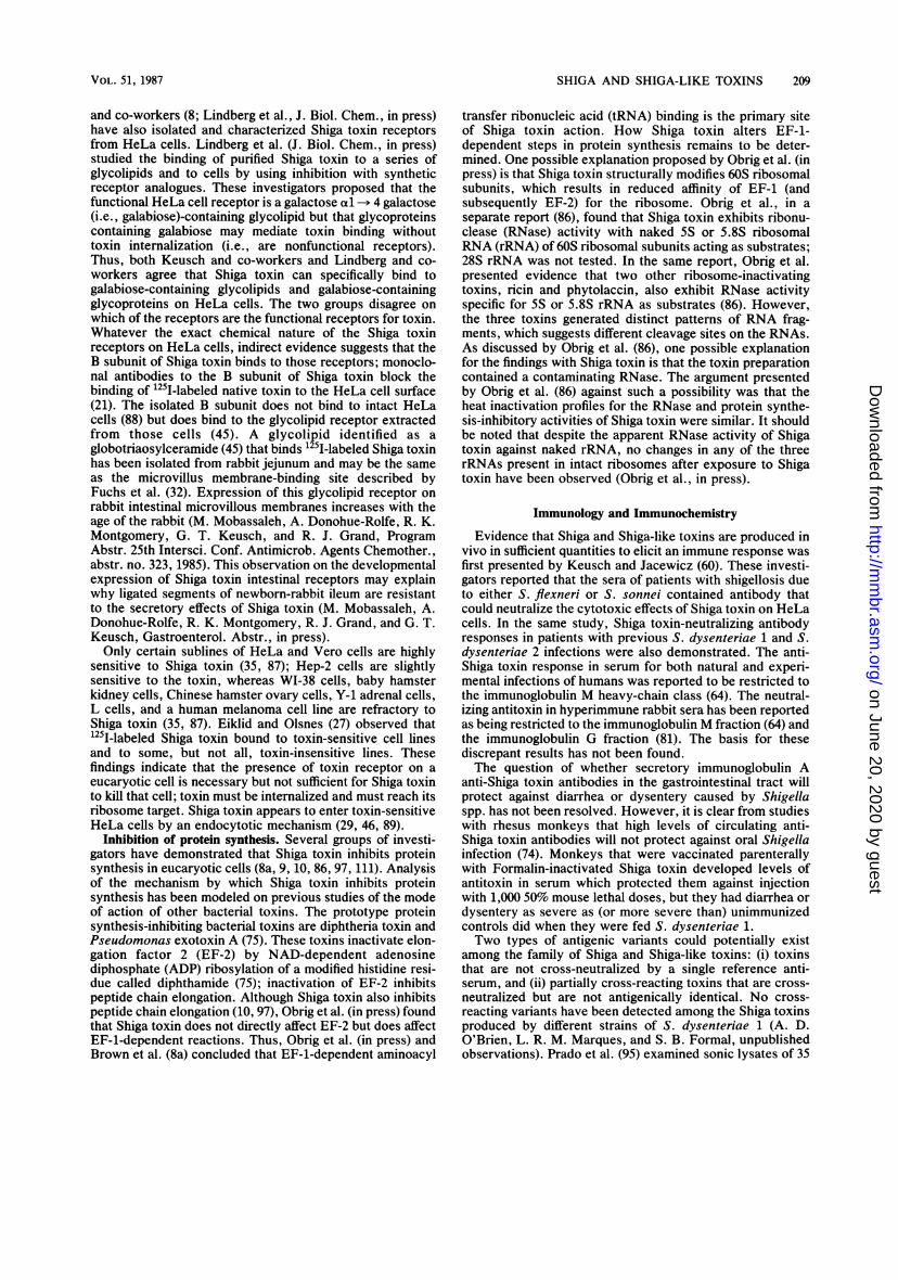
BD EXTERNAL TOXIN
FIG. 1. Model for the receptor-mediated endocytic entry ofShiga toxin and processing of Shiga toxin in a mammalian cell(adapted from reference 57, Fig. 6). Shiga toxin enters the cell byreceptor-mediated endocytosis. The B subunit of the toxin binds tothe mammalian cell receptor. The clathrin-coated pit is pinched off,and the coated vesicle is formed. The vesicle is acidified, and it mayfuse with lysosomes. The mechanism by which the enzymaticallyactive A1 fragment of Shiga toxin is generated and reaches thecytosol is not known but is presumed to involve proteolytic nickingand reduction of disulfide bonds of the A subunit. The A1 fragmentwithin the cytosol binds to the 60S ribosome, leading to inhibition ofprotein synthesis and cell death.
MICROBIOL. REV.
on June 20, 2020 by guesthttp://m
mbr.asm
.org/D
ownloaded from
SHIGA AND SHIGA-LIKE TOXINS 209
and co-workers (8; Lindberg et al., J. Biol. Chem., in press)have also isolated and characterized Shiga toxin receptorsfrom HeLa cells. Lindberg et al. (J. Biol. Chem., in press)studied the binding of purified Shiga toxin to a series ofglycolipids and to cells by using inhibition with syntheticreceptor analogues. These investigators proposed that thefunctional HeLa cell receptor is a galactose al -* 4 galactose(i.e., galabiose)-containing glycolipid but that glycoproteinscontaining galabiose may mediate toxin binding withouttoxin internalization (i.e., are nonfunctional receptors).Thus, both Keusch and co-workers and Lindberg and co-workers agree that Shiga toxin can specifically bind togalabiose-containing glycolipids and galabiose-containingglycoproteins on HeLa cells. The two groups disagree onwhich of the receptors are the functional receptors for toxin.Whatever the exact chemical nature of the Shiga toxinreceptors on HeLa cells, indirect evidence suggests that theB subunit of Shiga toxin binds to those receptors; monoclo-nal antibodies to the B subunit of Shiga toxin block thebinding of "251-labeled native toxin to the HeLa cell surface(21). The isolated B subunit does not bind to intact HeLacells (88) but does bind to the glycolipid receptor extractedfrom those cells (45). A glycolipid identified as aglobotriaosylceramide (45) that binds 125I-labeled Shiga toxinhas been isolated from rabbit jejunum and may be the sameas the microvillus membrane-binding site described byFuchs et al. (32). Expression of this glycolipid receptor onrabbit intestinal microvillous membranes increases with theage of the rabbit (M. Mobassaleh, A. Donohue-Rolfe, R. K.Montgomery, G. T. Keusch, and R. J. Grand, ProgramAbstr. 25th Intersci. Conf. Antimicrob. Agents Chemother.,abstr. no. 323, 1985). This observation on the developmentalexpression of Shiga toxin intestinal receptors may explainwhy ligated segments of newborn-rabbit ileum are resistantto the secretory effects of Shiga toxin (M. Mobassaleh, A.Donohue-Rolfe, R. K. Montgomery, R. J. Grand, and G. T.Keusch, Gastroenterol. Abstr., in press).Only certain sublines of HeLa and Vero cells are highly
sensitive to Shiga toxin (35, 87); Hep-2 cells are slightlysensitive to the toxin, whereas WI-38 cells, baby hamsterkidney cells, Chinese hamster ovary cells, Y-1 adrenal cells,L cells, and a human melanoma cell line are refractory toShiga toxin (35, 87). Eiklid and Olsnes (27) observed that125I-labeled Shiga toxin bound to toxin-sensitive cell linesand to some, but not all, toxin-insensitive lines. Thesefindings indicate that the presence of toxin receptor on aeucaryotic cell is necessary but not sufficient for Shiga toxinto kill that cell; toxin must be internalized and must reach itsribosome target. Shiga toxin appears to enter toxin-sensitiveHeLa cells by an endocytotic mechanism (29, 46, 89).
Inhibition of protein synthesis. Several groups of investi-gators have demonstrated that Shiga toxin inhibits proteinsynthesis in eucaryotic cells (8a, 9, 10, 86, 97, 111). Analysisof the mechanism by which Shiga toxin inhibits proteinsynthesis has been modeled on previous studies of the modeof action of other bacterial toxins. The prototype proteinsynthesis-inhibiting bacterial toxins are diphtheria toxin andPseudomonas exotoxin A (75). These toxins inactivate elon-gation factor 2 (EF-2) by NAD-dependent adenosinediphosphate (ADP) ribosylation of a modified histidine resi-due called diphthamide (75); inactivation of EF-2 inhibitspeptide chain elongation. Although Shiga toxin also inhibitspeptide chain elongation (10, 97), Obrig et al. (in press) foundthat Shiga toxin does not directly affect EF-2 but does affectEF-1-dependent reactions. Thus, Obrig et al. (in press) andBrown et al. (8a) concluded that EF-1-dependent aminoacyl
transfer ribonucleic acid (tRNA) binding is the primary siteof Shiga toxin action. How Shiga toxin alters EF-1-dependent steps in protein synthesis remains to be deter-mined. One possible explanation proposed by Obrig et al. (inpress) is that Shiga toxin structurally modifies 60S ribosomalsubunits, which results in reduced affinity of EF-1 (andsubsequently EF-2) for the ribosome. Obrig et al., in aseparate report (86), found that Shiga toxin exhibits ribonu-clease (RNase) activity with naked 5S or 5.8S ribosomalRNA (rRNA) of 60S ribosomal subunits acting as substrates;28S rRNA was not tested. In the same report, Obrig et al.presented evidence that two other ribosome-inactivatingtoxins, ricin and phytolaccin, also exhibit RNase activityspecific for 5S or 5.8S rRNA as substrates (86). However,the three toxins generated distinct patterns of RNA frag-ments, which suggests different cleavage sites on the RNAs.As discussed by Obrig et al. (86), one possible explanationfor the findings with Shiga toxin is that the toxin preparationcontained a contaminating RNase. The argument presentedby Obrig et al. (86) against such a possibility was that theheat inactivation profiles for the RNase and protein synthe-sis-inhibitory activities of Shiga toxin were similar. It shouldbe noted that despite the apparent RNase activity of Shigatoxin against naked rRNA, no changes in any of the threerRNAs present in intact ribosomes after exposure to Shigatoxin have been observed (Obrig et al., in press).
Immunology and Immunochemistry
Evidence that Shiga and Shiga-like toxins are produced invivo in sufficient quantities to elicit an immune response wasfirst presented by Keusch and Jacewicz (60). These investi-gators reported that the sera of patients with shigellosis dueto either S. flexneri or S. sonnei contained antibody thatcould neutralize the cytotoxic effects of Shiga toxin on HeLacells. In the same study, Shiga toxin-neutralizing antibodyresponses in patients with previous S. dysenteriae 1 and S.dysenteriae 2 infections were also demonstrated. The anti-Shiga toxin response in serum for both natural and experi-mental infections of humans was reported to be restricted tothe immunoglobulin M heavy-chain class (64). The neutral-izing antitoxin in hyperimmune rabbit sera has been reportedas being restricted to the immunoglobulin M fraction (64) andthe immunoglobulin G fraction (81). The basis for thesediscrepant results has not been found.The question of whether secretory immunoglobulin A
anti-Shiga toxin antibodies in the gastrointestinal tract willprotect against diarrhea or dysentery caused by Shigellaspp. has not been resolved. However, it is clear from studieswith rhesus monkeys that high levels of circulating anti-Shiga toxin antibodies will not protect against oral Shigellainfection (74). Monkeys that were vaccinated parenterallywith Formalin-inactivated Shiga toxin developed levels ofantitoxin in serum which protected them against injectionwith 1,000 50% mouse lethal doses, but they had diarrhea ordysentery as severe as (or more severe than) unimmunizedcontrols did when they were fed S. dysenteriae 1.Two types of antigenic variants could potentially exist
among the family of Shiga and Shiga-like toxins: (i) toxinsthat are not cross-neutralized by a single reference anti-serum, and (ii) partially cross-reacting toxins that are cross-neutralized but are not antigenically identical. No cross-reacting variants have been detected among the Shiga toxinsproduced by different strains of S. dysenteriae 1 (A. D.O'Brien, L. R. M. Marques, and S. B. Formal, unpublishedobservations). Prado et al. (95) examined sonic lysates of 35
VOL. 51, 1987
on June 20, 2020 by guesthttp://m
mbr.asm
.org/D
ownloaded from

210 O'BRIEN AND HOLMES
strains of shigellae and found that 86% of the lysates (otherthan S. dysenteriae 1) could not be neutralized by anti-Shigatoxin. Whether the cytotoxin(s) present in these strains arerelated to Shiga toxin but are not cross-neutralizable remainsto be determined.
Genetics
Little historical information is available on the genetics ofShiga toxin production. The only mutations reported toaffect the yield of shiga toxin are chlorate-resistant muta-tions in S. dysenteriae 1 (34) that reduce the level ofcytotoxin produced (64, 79). The mutations in those strainswere not mapped, nor was the mechanism by which theyreduced the yield of cytotoxin determined. In fact, thechlorate-resistant mutants were originally (34) classified asnontoxigenic rather than as low-level toxin producers, be-cause the cytotoxicity assay used in the early 1970s was lesssensitive than that now available.The genes for the Shiga toxin of S. dysenteriae 1 and the
Shiga-like toxin of S. flexneri appear to be located on thechromosome; no evidence for toxin genes on plasmids orbacteriophages of Shigella spp. has been found. Timmis etal. (112) reported the first in vivo cloning of the Shiga toxindeterminant. These investigators generated a series of R'plasmids in E. coli K-12 that carry sections of the S.dysenteriae 1 chromosome. The plasmids were constructedto carry portions of the S. dysenteriae 1 chromosomepreviously demonstrated (102) for S. flexneri to correlatewith the ability of the organism to provoke fluid secretion inrabbit ileal segments. The results of the study by Timmis etal. (112) localized the Shiga toxin genes (designated sht) nearthe "metB distal side of argE". The close linkage of sht toargE is in some disagreement with the report of Sansonetti etal. (102), who tentatively localized the region on the S.flexneri chromosome for fluid production in rabbits as beingnear the lysine decarboxylase (LDS)-negative locus and inthe rha-mtl region. Timmis et al. (112) offered two quiteplausible explanations for these differences in the localiza-tion of the toxin genes: the maps of S. dysenteriae 1 and S.flexneri may not be exactly the same, and S. dysenteriae 1may carry a second copy of sht. The latter possibility issupported by the mention (112) of an unpublished findingthat a second determinant for cytotoxin production is carriedon an R'-asn plasmid that also carries the LDS-negativelocus of Shigella spp. Timmis et al. (112) stated that they hadsuccessfully cloned the toxin determinants of the R'-argEplasmid in vitro. It will be of considerable interest tocompare the operon structure and nucleotide sequence of shtwith the structural genes of E. coli SLT-I and E. coli SLT-II(see below). Toward that end, the structural genes for Shigatoxin have recently been cloned in our laboratory (N. A.Strockbine, R. K. Holmes, and A. D. O'Brien, Abstr. Annu.Meet. Am. Soc. Microbiol. 1987, B-39, p. 78).
Role of Toxin in Disease
Dysentery. The subject of the possible role of Shiga andShiga-like toxins in the pathogenesis of several Shigella- andE. coli-associated diseases was recently reviewed by Cantey(12). There is no direct proof that Shiga and Shiga-like toxinsfunction as important virulence factors, but there are cir-cumstantial data to support that hypothesis. Dysentery involunteers fed an invasive, low-toxin-producing (64, 79),chlorate-resistant mutant of S. dysenteriae 1 designated
strain 725 was milder than the disease caused by the inva-sive, highly toxinogenic parent strain M131. The durationand height of the fever and severity of the dysentery wereless in volunteers fed strain 725 than in those fed strain M131(70). These clinical observations are in keeping with the ideathat the higher level of toxin produced by S. dysenteriae 1than other Shigella spp. correlates with the more severedisease caused by Shiga's bacillus; dysentery occurs morecommonly in patients infected with S. dysenteriae 1 than inthose infected with any other serotype of Shigella (57). Oneexplanation for how Shiga toxin promotes dysentery is thattoxin present free in the colonic lumen binds to colonicepithelia and ultimately kills those cells by inhibiting proteinsynthesis. Two lines of evidence support such a theory.First, measurable toxin is found in human fecal specimensfrom patients with S. dysenteriae 1 infection (19). Second,Shiga toxin binds to and is cytotoxic for primary cultures ofhuman colonic epithelial cells (M. P. Moyer, S. W. Roth-man, P. S. Dixon, and J. E. Brown, submitted for publica-tion; M. P. Moyer, S. W. Rothman, and J. E. Brown, Abstr.Annu. Meet. Am. Soc. Microbiol. 1985, B108, p. 36).
Diarrhea. The results of gut perfusion studies (100) donewith monkeys also provide evidence for a role of Shiga orShiga-like toxins in causing the watery diarrhea sometimesseen during shigellosis (24). All S. flexneri-infected animalsshowed decreased colonic absorption or net colonic secre-tion. However, animals with diarrhea also showed net se-cretion in the jejunum, whereas no jejunal secretion wasevident in monkeys with dysentery alone. No invasion ofjejunal tissues by Shigella spp. was found in any of theanimals, although invasion of the colonic epithelium oc-curred regularly. Subsequently, Kinsey et al. (65) showedthat monkeys inoculated intracecally with S. flexneri haddysentery but not diarrhea. Thus, in monkeys, the diarrheaof shigellosis appears to require a secretory response of thesmall bowel superimposed on the colonic dysfunction. Morerecent findings with humans (11) suggest that shigella diar-rhea results from colonic dysfunction without evidence foran increased small-bowel flow rate. Wherever the intestinalsite for the secretion responsible for shigella diarrhea, onemay postulate that the secretion is a consequence of toxinproduced by the Shigella spp.
If Shiga toxin is responsible for the diarrhea of shigellosis,a key question is how can a protein synthesis-inhibiting toxinelicit fluid secretion in the intestine? Shiga toxin does notappear to act like cholera toxin or E. coli heat-labile toxin.Although Shiga toxin can elevate the cyclic adenosinemonophosphate (cAMP) content of rabbit intestinal mucosa(22), as can cholera toxin and E. coli heat-labile toxin,Shiga-toxin-induced accumulation of cAMP does not occuruntil long after fluid secretion begins (H. T. Paulk, A. 0.Cardamone, G. S. Gotterer, and T. R. Hendrix, Gastroen-terology 72:1164, 1977). Shiga toxin also does not appear toact like E. coli heat-stable enterotoxin. Unpublished obser-vations from collaborative studies with R. Guerrant indicatethat Shiga toxin does not increase the cyclic guanosinemonophosphate (cGMP) content of the rabbit ileum. Analternative mechanism by which Shiga toxin could cause netfluid secretion is to block (or inhibit) fluid absorption.Indeed, Keenan et al. (55) recently reported that purifiedShiga toxin and purified E. coli Shiga-like toxin selectivelydestroyed the mature absorptive epithelial cells of the rabbitileum. One can therefore propose that Shiga toxin binds tomature absorptive epithelial cells through glycolipid recep-tors, inactivates protein synthesis in the host cells, andultimately causes the death of those cells. In this model,
MICROBIOL. REV.
on June 20, 2020 by guesthttp://m
mbr.asm
.org/D
ownloaded from

SHIGA AND SHIGA-LIKE TOXINS 211
TABLE 1. Characteristics of E. coli enterotoxinsa
Mass of No ofEntero- subunits A N Immuno-toxinb Structure MW (kilodaltsnis) A anit genic Bioassays Mode of action
(kilodaltons) A and B gei
LT-I Protein 86,000 28; 11.8 1; 5 Yes Y1 or CHO cells; rabbit ileal loops Adenylate cyclase productionLT-II Protein NDC 28; 11.8 ND Yes Y1 or CHO cells Adenylate cyclase productionSLT-I Protein 70,000 32; 7.7 1; 5 Yes HeLa or Vero cell cytotoxicity; rabbit Protein synthesis inactivation
ileal loops; lethality to miceSLT-II Protein ND ND ND Yes HeLa or Vero cell cytoxicity; rabbit ND
ileal loops; lethality to miceSTa Polypeptide 2,000 No Suckling mouse Guanylate cyclase productionSTb Polypeptide 5,000 No Weaned pig jejunal loops ND
a For recent reviews of the enterotoxins, see Guerrant et al. (39) and O'Brien and Nataro (83).b LT, Heat-labile toxins; ST, heat-stable toxins. For details about the recently characterized toxin LT-II, see Pickett et al. (93), Holmes et al. (42), and Guth
et al. (40, 41).c ND, Not determined.
diarrhea would result from inhibition of absorption ratherthan from active secretion.HUS. HUS was originally described by Gasser et al. (33)
as the association of hemolytic anemia, thrombocytopenia,and acute renal failure. HUS can occur after infection withS. dysenteriae 1 (36, 68, 96) or after infection with E. coliserotypes, such as E. coli 0157:H7, that produce high levelsof Shiga-like (Vero) toxin (52, 91; Karmali et al., Letter,1983). S. dysenteriae 1-associated HUS tends to be moresevere than E. coli-associated HUS.The signs of HUS after Shiga infection include dissemi-
nated intravascular coagulation, renal microangiopathy, andhemolytic anemia (68). In one study (68), 50% of the patientswith S. dysenteriae 1-associated HUS had endotoxemia asassessed by the Limulus assay. These symptoms presage apoor prognosis. The clinical manifestations of E. coli0157:H7-associated HUS are the classic triad described byGasser et al. (33). Karmali et al. (52) have suggested that theShiga toxin of S. dysenteriae 1 or the Shiga-like toxin of E.coli 0157:H7 may enter the bloodstream and cause damageto vascular endothelial cells such as those in the kidneys.This is an attractive hypothesis, because the damage tovascular epithelium seen on autopsy of patients who havedied of HUS (68) resembles the histopathological damage tosmall vessels in the central nervous systems of animalsinoculated with crude Shiga toxin (6, 14). An animal modelof HUS has not yet been developed, and the limited avail-ability of purified toxin has hindered studies on the effects ofpurified toxin in animals.
SHIGA-LIKE TOXINS IN E. COLI
History and Nomenclature
Several different toxins that act on the intestinal tract canbe produced by E. coli (Table 1). E. coli organisms thatcause diarrheal disease in humans and animals have beenclassified into four groups (71). Enterotoxigenic E. coli(ETEC) adhere to the mucosa of the small bowel andproduce a heat-labile toxin or heat-stable toxin or both.Enteroinvasive E. coli (EIEC) do not produce heat-labile orheat-stable toxin, but, like shigellae, they penetrate andmultiply within colonic epithelial cells. Enteropathogenic E.coli (EPEC) were originally incriminated epidemiologicallyas pathogens for infants and young children. EPEC belong toa limited number of serotypes (71), and some EPEC strainshave been shown to adhere avidly to the mucosa of the small
intestine and produce characteristic effacement of microvilli(1, 16, 77, 99). Enterohemorrhagic E. coli (EHEC) causehemorrhagic colitis and are of the serotype 0157:H7 (98).EPEC and EHEC are not enteroinvasive and do not producethe classical heat-stable and heat-labile enterotoxins (71, 98,117). We and others (12, 52, 73) have proposed that someEPEC and all EHEC strains may cause intestinal disease byproducing elevated levels of Shiga-like cytotoxins. In thissection, the discovery and nomenclature of E. coli Shiga-liketoxins, as well as the relationship of the toxins to Verotoxins, will be discussed.
Discovery that EPEC and EHEC make Shiga-like toxins.The initial search for a Shiga-like toxin of E. coli focused onEPEC that caused diarrhea in infant rabbits (13) or infanthumans, because no virulence mechanism was establishedfor EPEC, and, as mentioned above, they are neitherenteroinvasive nor able to produce heat-labile or heat-stableenterotoxins. The results of a preliminary survey of selectedE. coli strains for production of a Shiga-like toxin (defined atthat time as a cytotoxin for HeLa cells that could beneutralized by rabbit antitoxin raised against crude Shigatoxin) indicated that certain EPEC can make such a toxin(A. D. O'Brien, M. R. Thompson, J. R. Cantey, and S. B.Formal, Abstr. Annu. Meet. Am. Soc. Microbiol. 1977,B103, p. 32). This hypothesis was confirmed in 1982 (82),when monospecific antibody to Shiga toxin became avail-able. In the same investigation (82), it was also observed thatthe level of Shiga-like toxin produced by E. coli strainsvaried from trace amounts that were detectable only inbacterial lysates to levels equivalent to that produced by S.dysenteriae 1.That EHEC also make Shiga-like toxins was first reported
in 1983. O'Brien et al. (A. D. O'Brien, T. A. Lively, M. E.Chen, S. W. Rothman, and S. B. Formal, Letter, Lanceti:702, 1983) found that isolates of E. coli 0157:H7 isolatedduring outbreaks that had occurred in the United Statesmade high levels of a cell-associated cytotoxin for HeLa andVero cells that could be neutralized by rabbit anti-Shigatoxin. In the same study, it was reported that culturesupernatants of E. coli 0157:H7 strains 931, 932, and 933also contained a cytotoxin for HeLa and Vero cells thatcould be completely neutralized by rabbit anti-Shiga toxin.On further analysis of the 933 strain, it is now clear that thisorganism makes two kinds of cytotoxins, one of which canbe neutralized by anti-Shiga toxin (110; Scotland et al.,Letter, 1985). We have designated (110) the anti-Shiga toxinneutralizable cytotoxin as Shiga-like toxin I (SLT-I), and wehave named the other cytotoxin Shiga-like toxin II (SLT-II).
VOL. 51, 1987
on June 20, 2020 by guesthttp://m
mbr.asm
.org/D
ownloaded from

212 O'BRIEN AND HOLMES
It is important to note that SLT-I predominates in celllysates and SLT-II is the more active toxin in culture filtrate(110) when both toxins are produced by the same strain.
Relationship of Shiga-like toxins to Vero toxins. The firstdescription of an EPEC Vero toxin was made in 1977 byKonowalchuk et al. (67). The Vero toxin produced by thestrain used as a prototype in that classical study, EPEC026:H11 strain H-30, appears to be the same toxin as SLT-Iof E. coli (A. D. O'Brien, T. A. Lively, M. E. Chen, S. W.Rothman, and S. B. Formal, Letter, Lancet i:702, 1983).Konowalchuk et al. identified nine strains in addition to E.coli H-30 that contained a Vero toxin in culture filtrates ofthe organisms (67). Of these 10 strains, 7 were associatedwith diarrhea in human infants, 1 was associated withdiarrhea in a weanling pig, and 2 were isolated from cheese.None of the E. coli strains that made Vero toxin also madeE. coli heat-labile or heat-stable enterotoxin. Vero toxinfrom 8 of the 10 strains was neutralized by antiserum againstthe Vero toxin from E. coli H30; Vero toxins from an EPEC0128:B12 and from the pig isolate 0138:K81 were notneutralized by the antiserum. This was the earliest indicationthat E. coli strains produce more than one toxin activeagainst Vero cells.The first studies on E. coli isolated from animals Vero
toxin concentrated on EPEC and EPEC-like strains (seeabove) (15, 53, 54, 66, 105; W. G. Wade, B. T. Thom, and N.Evans, Letter, Lancet ii:1235-1236, 1979; S. M. Scotland,N. P. Day, G. A. Willshaw, and B. Rowe, Letter, Lanceti:90, 1980; M. W. Wilson and K. A. Bettelheim, Letter,Lancet i:201, 1980). Subsequently, Johnson et al. (Letter,1983) found that Canadian isolates of EHEC which had beenincriminated as the agents of hemorrhagic colitis made Verotoxin. As was the case with E. coli 0157:H7 strains isolatedduring two outbreaks of hemorrhagic colitis in the UnitedStates (98, 117), the Canadian strains were notenteroinvasive and did not make E. coli heat-labile orheat-stable enterotoxin. With the observation that EHECmake a Shiga-like toxin active on Vero cells (A. D. O'Brien,T. A. Lively, M. E. Chen, S. W. Rothman and S. B. Formal,Letter, Lancet i:702, 1983), O'Brien et al. concuded thatVero toxin and Shiga-like toxin were the same toxin. Whenit was subsequently realized that EHEC can make more thanone antigenically distinct Vero toxin (110; Scotland et al.,Letter, 1985), Scotland et al. adopted the nomenclature Verotoxin 1 (VT1) and Vero toxin 2 (VT2). VT1 is equivalent toSLT-I, and VT2 is synonymous with SLT-II.
Purification and Structure
SLT-Is from cell lysates of EPEC strain H-30 and fromcell lysates of EHEC strain 933 have been purified tohomogeneity in our laboratory (80; A. D. O'Brien, T. A.Lively, T. W. Chang, and S. L. Gorbach, Letter, Lancetii:573, 1983) by the same series of steps used to purify toxinfrom S. dysenteriae 1: i.e., French pressure cell disruption ofbacteria grown in iron-depleted medium, Affi-Gel Blue chro-matography, chromatofocusing, and anti-Shiga toxin affinitychromatography. The SLT-I from EHEC has also beenpurified by a method that includes ammonium sulfate frac-tionation, DEAE-cellulose column chromatography, andhigh-pressure liquid chromatography (M. Noda, N.Nakabayaski, T. Yutsudo, T. Hirayama, and Y. Takeda,Abstr. Toxicon 23:600, 1985). Each SLT-I purified fromthese EPEC and EHEC strains was comprised of an Asubunit and multiple copies of a B subunit.
The relative mobilities on sodium dodecyl sulfate-polyacrylamide gel electrophoresis of the A and B subunitsof the SLT-Is purified from EPEC strain H30 and EHECstrain 933 were indistinguishable from those of the purifiedShiga toxin of S. dysenteriae 1 strain 60R when all threesamples were run in parallel (O'Brien, et al., Letter, Lancetii:573, 1983). This observation, together with the data nowavailable on the subunit structure of Shiga toxin (see above)and the translated nucleotide sequence of SLT-I from EHECstrain 933 (see Genetics and Regulation), indicate that theSLT-I A subunit has a MW of 32,200 and the SLT-I Bsubunit has a MW of 7,700. The value for the A subunit MWis close to that of the original estimate obtained with purifiedtoxin of EPEC strain H30 (31,500 [80]), but the value for theB subunit is almost twice that estimated from cross-linkingstudies (80). By analogy with Shiga toxin, the most likelyA-to-B subunit ratio for SLT-I is 1:5. If that is indeed thecase, then the MW of holotoxin is ca. 70,000. Previousestimates of the MW of SLT-I have ranged from a low of10,000 to 30,000 (67; W. G. Wade, B. T. Thom, and N.Evans, Letter, Lancet ii:1235-1236, 1979) to a high of 48,000(80). The reason for this range in the reported MW of SLT-Iis not known.
SLT-II may have been purified to homogeneity by Padhyeet al., who recently reported (90) that they had purified aVero cell cytotoxin from culture filtrates of E. coli 0157:H7strain 932 by concentration of filtrates followed by molecularsieve and anion-exchange chromatography. Since the Verocell cytotoxin prepared by Padhye et al. (90) was notneutralized by anti-Shiga toxin, and since strain 932 pro-duces both SLT-I and SLT-II (73), it seems reasonable topresume that the material isolated by Padhye et al. is SLT-II.Neutralization studies with anti-SLT-II must be done toprove or disprove that hypothesis. One of the problems withisolating one type of SLT from E. coli strains that make bothSLT-I and SLT-II is that a toxin preparation could becontaminated with the variant toxin. Indeed, with the dis-covery by Scotland et al. (Letter, 1985) that E. coli 933makes not only VT1 (SLT-I) but also VT2 (SLT-II), wereexamined our SLT-I preparation from E. coli 933 forSLT-II. We found no evidence that our preparation of SLT-Iwas contaminated with SLT-II.
Mode of Action
Purified SLT-Is from E. coli H30 (80) and E. coli 933(O'Brien et al., Letter, Lancet ii:573, 1983) have the samebiological activities as and comparable specific activities topurified Shiga toxin: cytotoxicity for HeLa and Vero cells,enterotoxicity for ligated rabbit ileal segments, and lethalityat doses between 100 ng and 2 ,ug for mice. By analogy withShiga toxin, the molecular basis for these three seeminglydiverse biological activities is catalytic inactivation of 60Sribosomes in toxin-sensitive (receptor-expressing) cells. Toour knowledge, the experiments to test the validity of thisanalogy have not been done, although crude preparations ofE. coli SLT-I do inhibit protein synthesis in HeLa cells (81).The receptor for SLT-I has been identified (C. A. Lingwood,H. Law, S. Richardson, M. Petric, J. L. Brunton, and M.Karmali, J. Biol. Chem., in press) and is the same as forShiga toxin. Cell lysates of the lysogenic strain E. coli C600(933W) were recently used to assess the biological propertiesof SLT-II (110). The results of that study indicate thatSLT-II has the same three biological properties as SLT-I,but, per unit of protein in cell lysates, it is more lethal formice and less cytotoxic than SLT-I (110).
MICROBIOL. REV.
on June 20, 2020 by guesthttp://m
mbr.asm
.org/D
ownloaded from
SHIGA AND SHIGA-LIKE TOXINS 213
Immunology and Immunochemistry
Recent studies have established that SLT-I and VT1 arealternative names for the same toxin (110; Scotland et al.,Letter, 1985; Karmali et al., Letter, 1986). SLT-I was
purified to apparent homogeneity from both EPEC andEHEC strains (80; O'Brien et al., Letter, Lancet ii:573,1983). In immunodiffusion tests with anti-Shiga toxin, bothpreparations of purified SLT-I gave lines of identity withShiga toxin. Two monoclonal antibodies raised against E.coli SLT-I neutralized both homologous E. coli toxin andpurified Shiga toxin (109). Monoclonal antibodies againstShiga toxin have also been isolated, but their reactivitiesagainst purified SLT-I were not determined (21, 38). System-atic studies of cross-reactivity between Shiga toxin andSLT-I from various strains of E. coli in immunobindingassays were not performed, primarily because sufficientquantities of the purified toxins were not available. Furtherstudies are needed to determine whether Shiga toxin andSLT-I are immunochemically identical or whether there areunique epitopes on SLT-I that are not present on Shigatoxin, or vice versa.
Although the SLT-II of E. coli has cytotoxic, enterotoxic,and lethal activities similar to those of SLT-I and Shiga toxin(110), it is not neutralized by antiserum against purified Shigatoxin or monoclonal antibodies against SLT-I. In contrast, itis neutralized by antiserum prepared against a crude prepa-ration of SLT-II. Antigenic diversity among Vero cytotoxinsof E. coli has also been documented, and VT2 has beenrecognized that is not neutralized by anti-Shiga toxin but isneutralized by antiserum against crude VT2 (Scotland et al.,Letter, 1985; Karmali et al., Letter, 1986). Reference SLT-IIantiserum (110) neutralized the cytotoxicity of lysates fromE. coli E32511 (0157:H-), the prototype strain against whichthe reference VT2 antiserum was raised (Scotland et al.,Letter, 1985), indicating that SLT-II and VT2 are probablyalternate names for the same or closely related toxins. Notoxin that has been identified immunologically as SLT-II orVT2 has been purified to homogeneity or characterizedstructurally, nor has the mode of action of this toxin beeninvestigated (see preceding section). As noted above,Padhye et al. (90) have purified a toxin from E. coli 932 thatis distinct from SLT-I, but the immunologic identity of thistoxin with SLT-II has not yet been established. The possi-bility that SLT-I and SLT-II have shared epitopes that are
not involved in neutralization of toxicity by antibody has notyet been investigated.There is preliminary evidence that E. coli from a variety of
sources may produce Vero cytotoxins of additional antigenictypes. The Vero cytotoxins produced by strains of E. coliisolated from pigs with edema disease or neonatal pigs withdiarrhea were not neutralized by antisera that were activeagainst the cytotoxin(s) from E. coli strains from humans(67, 108). Karmali et al. (Letter, 1986) prepared a referenceantiserum against a toxoid made from purified VT from E.coli H-30 (which is known to produce VT1) and compared itsneutralizing activity with that of serum obtained from a
patient convalescing from HUS. In tests with culture filtratesfrom a collection of E. coli strains from human and animalsources, all of the toxins that were completely neutralized bythe antitoxoid (presumed to be VT1) were also neutralizedby the convalescent serum. However, the toxins that were
not neutralized by the antitoxoid could be divided into twogroups on the basis of their reaction with the convalescentserum: some that were partially neutralized (and presumablycontained VT2 exclusively or in highest titer) and others that
were not neutralized at all. Karmali et al. interpreted thedata as supporting the existence of antigenically distinct VT1and VT2 toxins produced by E. coli, a conclusion with whichwe agree, but they did not address the nature of the toxin(s)that were not neutralized by either of their antisera. Unpub-lished observations from our laboratory (L. R. M. Marques,J. S. M. Peiris, S. J. Cryz, and A. D. O'Brien) suggest thatat least one Vero toxin from E. coli strains isolated from pigswith edema disease is related to, but not identical with, theprototype SLT-II. Such strains contain an extracellular Verotoxin that, unlike SLT-II, is not active on HeLa cells but isneutralized by reference antisera against SLT-II. How thisvariant of SLT-II is related to the Vero toxins produced bythe strains described by Karmali et al. (Letter, 1986) andSmith et al. (108) remains to be determined.
Genetics and RegulationPhage conversion is responsible for controlling the pro-
duction of several important bacterial toxins, including diph-theria toxin, streptococcal erythrogenic toxin, botulinumtoxin, and staphylococcal enterotoxin A (2, 5, 72). Toxin-converting phages contain genes that are essential fortoxinogenesis, and susceptible bacteria that becomelysogenic for such tox+ phages acquire the capacity toproduce the corresponding toxin. Recently, production ofShiga-like toxins was also shown to be determined byspecific phages in selected strains of both EPEC (026, 0128)and EHEC (0157) serotypes isolated from humans (84, 108;Scotland et al., Letter, 1983; H. R. Smith, N. P. Day, S. M.Scotland, R. J. Gross, and B. Rowe, Letter, Lanceti:1242-1243, 1984). In contrast, production of Shiga-liketoxin in E. coli strains isolated from pigs was not found to becontrolled by phages (108).The phages that determine the production of Shiga-like
toxins in E. coli are of several different types that wereinitially distinguished on the basis of differences in proper-ties such as host range, immunity specificity, and suscepti-bility to thermal inactivation (84, 108). E. coli H19 (026) and933 (0157) are both lysogenic for two different toxin-converting phages. The phages, originally designated H19A(108) and H19J (108) are independent isolates of the samephage from strain H19 that are closely related to phage 933Jfrom strain 933 (108) in terms of their morphology, virionpolypeptides, DNA restriction fragments, and immunityspecificity, but phage H19B has a different immunity speci-ficity. Phages H19A/J, 933J, and H19B are all lambdoidphages that code for SLT-I. A 8.1-kilobase (kb) EcoRIfragment of the H19B genome was shown to be homologouswith the region of the gene of phage lambda that encodes thegenes cI, ninR, 0, P, and the origin of replication (44); theregions of homology between phage lambda and phagesH19A/J and 933J have not yet been mapped in detail. Phage933W from E. coli 933 has quite different characteristicsfrom the other phages described above and codes for SLT-II(84, 110). A converting phage was also isolated from an
0157:H- E. coli strain, E32511 (119), which was subse-quently shown to produce SLT-II (110; Scotland et al.,Letter, 1985).The organization and expression of the operon for SLT-I
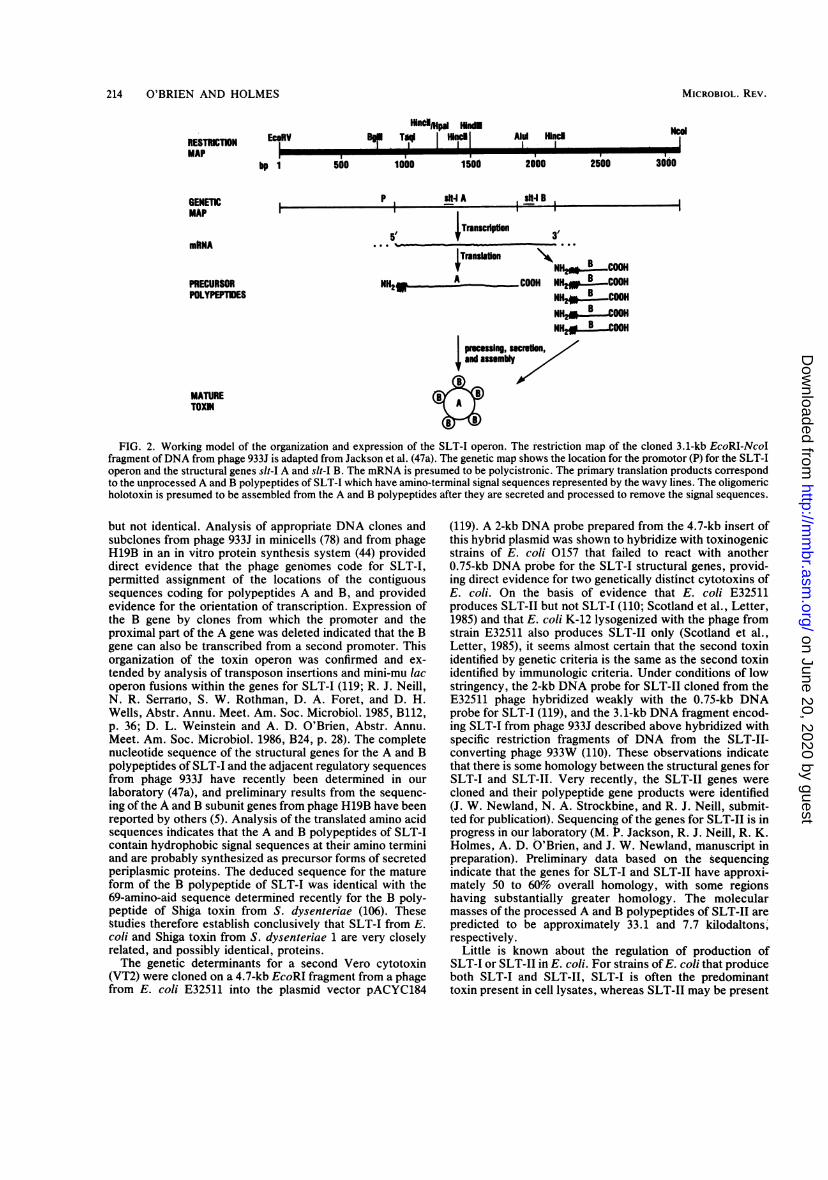
are shown schematically in Fig. 2. The structural genes thatencode SLT-I were cloned on a 2.5-kb DNA fragment froma phage from strain H19 (not characterized as H19A or
H19B) (119), on a 3.1-kb DNA fragment from phage 933J(78), and on a 1.7-kb DNA fragment from phage H19B (44).The restriction maps of phages 933J and H19B were similar
VOL. 51, 1987
on June 20, 2020 by guesthttp://m
mbr.asm
.org/D
ownloaded from
214 O'BRIEN AND HOLMES
lindMpl HindOmodBP, TdqI IEcoRV
bp 1I I I II
500 1000
P , I4IAI-
1500
lcdAld Hlncd
2000
, skt B ,
2500
-I - I I
5-
3000
-t
TranscdpIon
ns--TrmnIaon \NILCA
~~NH20 B COOHNH2R A COOH NH2 COOH
NH - COOHNH2*2....ooHNH2, B _OOH
FIG. 2. Working model of the organization and expression of the SLT-I operon. The restriction map of the cloned 3.1-kb EcoRI-NcoIfragment of DNA from phage 933J is adapted from Jackson et al. (47a). The genetic map shows the location for the promotor (P) for the SLT-Ioperon and the structural genes slt-I A and slt-I B. The mRNA is presumed to be polycistronic. The primary translation products correspondto the unprocessed A and B polypeptides of SLT-I which have amino-terminal signal sequences represented by the wavy lines. The oligomericholotoxin is presumed to be assembled from the A and B polypeptides after they are secreted and processed to remove the signal sequences.
but not identical. Analysis of appropriate DNA clones andsubclones from phage 933J in minicells (78) and from phageH19B in an in vitro protein synthesis system (44) provideddirect evidence that the phage genomes code for SLT-I,permitted assignment of the locations of the contiguoussequences coding for polypeptides A and B, and providedevidence for the orientation of transcription. Expression ofthe B gene by clones from which the promoter and theproximal part of the A gene was deleted indicated that the Bgene can also be transcribed from a second promoter. Thisorganization of the toxin operon was confirmed and ex-tended by analysis of transposon insertions and mini-mu lacoperon fusions within the genes for SLT-I (119; R. J. Neill,N. R. Serrano, S. W. Rothman, D. A. Foret, and D. H.Wells, Abstr. Annu. Meet. Am. Soc. Microbiol. 1985, B112,p. 36; D. L. Weinstein and A. D. O'Brien, Abstr. Annu.Meet. Am. Soc. Microbiol. 1986, B24, p. 28). The completenucleotide sequence of the structural genes for the A and Bpolypeptides of SLT-I and the adjacent regulatory sequencesfromn phage 933J have recently been determined in ourlaboratory (47a), and preliminary results from the sequenc-ing of the A and B subunit genes from phage H19B have beenreported by others (5). Analysis of the translated amino acidsequences indicates that the A and B polypeptides of SLT-Icontain hydrophobic signal sequences at their amino terminiand are probably synthesized as precursor forms of secretedperiplasmic proteins. The deduced sequence for the matureform of the B polypeptide of SLT-I was identical with the69-amino-aid sequence determined recently for the B poly-peptide of Shiga toxin from S. dysenteriae (106). Thesestudies therefore establish conclusively that SLT-I from E.coli and Shiga toxin from S. dysenteriae 1 are very closelyrelated, and possibly identical, proteins.The genetic determinants for a second Vero cytotoxin
(VT2) were cloned on a 4.7-kb EcoRI fragment from a phagefrom E. coli E32511 into the plasmid vector pACYC184
(119). A 2-kb DNA probe prepared from the 4.7-kb insert ofthis hybrid plasmid was shown to hybridize with toxinogenicstrains of E. coli 0157 that failed to react with another0.75-kb DNA probe for the SLT-I structural genes, provid-ing direct evidence for two genetically distinct cytotoxins ofE. coli. On the basis of evidence that E. coli E32511produces SLT-II but not SLT-I (110; Scotland et al., Letter,1985) and that E. coli K-12 lysogenized with the phage fromstrain E32511 also produces SLT-II only (Scotland et al.,Letter, 1985), it seems almost certain that th, second toxinidentified by genetic criteria is the same as the second toxinidentified by immunologic criteria. Under conditions of lowstringency, the 2-kb DNA probe for SLT-II cloned from theE32511 phage hybridized weakly with the 0.75-kb DNAprobe for SLT-I (119), and the 3.1-kb DNA fragment encod-ing SLT-I from phage 933J described above hybridized withspecific restriction fragments of DNA from the SLT-II-converting phage 933W (110). These observations indicatethat there is some homology between the structural genes forSLT-I and SLT-II. Very recently, the SLT-II genes werecloned and their polypeptide gene products were identified(J. W. Newland, N. A. Strockbine, and R. J. Neill, submit-ted for publication). Sequencing of the genes for SLT-II is inprogress in our laboratory (M. P. Jackson, R. J. Neill, R. K.Holmes, A. D. O'Brien, and J. W. Newland, mnanuscript inpreparation). Preliminary data based on the sequencingindicate that the genes for SLT-I and SLT-II have approxi-mately 50 to 60% overall homology, with some regionshaving substantially greater homology. The molecularmasses of the processed A and B polypeptides of SLT-II arepredicted to be approximately 33.1 and 7.7 kilodaltons,respectively.
Little is known about the regulation of production ofSLT-I or SLT-II in E. coli. For strains of E. coli that produceboth SLT-I and SLT-II, SLT-I is often the predominanttoxin present in cell lysates, whereas SLT-II may be present
RESTRCTIONMAP
GENETICMAP
mkNA
PRECURSORPOLYPEPTIDES
MATURETOXN
MICROBIOL. REV.
I 9
3'
on June 20, 2020 by guesthttp://m
mbr.asm
.org/D
ownloaded from

SHIGA AND SHIGA-LIKE TOXINS 215
in much higher titer than SLT-I in culture supernatants (16).This observation suggests that there may be significantdifferences in secretion or localization of SLT-I and SLT-IIin E. coli. Wild-type strains of E. coli have been classified ashigh-, moderate-, or low-toxin-producing strains on the basisof quantitative titrations of toxin produced by strains grownunder rigidly controlled conditions in deferrated medium(73). All strains of E. coli 0157:H7 associated with hemor-rhagic colitis have been characterized as highly toxinogenic,and a significant proportion of EPEC isolates are highlytoxinogenic. Most highly toxinogenic strains produce SLT-I,and some also produce SLT-II. Production of SLT-II only iscommon among strains classified as moderately toxinogenic(73, 110). All strains of E. coli K-12 examined to dateproduce low levels of Shiga-like toxin that can be neutralizedby polyclonal anti-Shiga toxin or monoclonal antibodiesagainst SLT-I, but the DNA probe for SLT-I from phage933J described above does not hybridize to any significantextent with DNA from E. coli K-12 strains. The reasons forthis apparent discrepancy between the results of immuno-logic and genetic tests for the SLT of E. coli K-12 strainshave not been resolved.
Role in Disease
Epidemiological evidence. As is the case with Shiga toxin,there is no direct proof that E. coli SLT-I or SLT-II plays a
role in disease. Some of the strongest circumstantial evi-dence comes from epidemiological studies ofE. coli strainsisolated from humans (17, 48a, 52, 73, 105, 108) and animals(15, 53, 54, 73, 76, 107). Most of the epidemiological studiesto date have exclusively examined culture filtrates of bacte-ria for Vero toxin. By contrast, Marques et al. (73) recentlytested culture supernatants and sonic lysates of 418 strains ofE. coli isolated from human or animal stools or from food forcytotoxic effects on HeLa cells. These investigators alsoasked whether cytotoxicity could be neutralized by anti-Shiga toxin. Marques et al. (73) classified strains accordingto the level of cell-associated toxin they produced. Thecategories were as follows: low, 2 x 10² to 6 x 10² CD50 perml of sonic lysate; moderate, 1 x 10³ to 1 x 10⁴ CD50 per mlof sonic lysate; and high, 1 x 10⁵ to 1 x 10⁸ CD50 per ml ofsonic lysate. Of the 418 strains analyzed, 107 had no detect-able cell-associated cytotoxicity. A total of 262 strains madelow levels of cytotoxin. The culture supernatants from thelow-level cytotoxin producers had no detectable activity.Strains that fell in the low-level toxin producer categoryincluded E. coli isolated from healthy adults, E. coli K-12substrains, EIEC, ETEC,E. coli strains of classical EPECserogroups isolated from patients with diarrhea, otherstrains isolated from patients with diarrhea, and three strains(not of the 0157:H7 serotype) isolated from patients withhemorrhagic colitis. Ten strains produced moderate levels ofcell-associated cytotoxin, and cytotoxicity could also bedetected in culture supernatants. All 10 strains were associ-ated with diarrhea, hemorrhagic colitis, or HUS, and the 10strains made only SLT-II (110). A total of 39 strains madehigh levels of cytotoxin, and 38 of the 39 were associatedwith diarrhea, hemorrhagic colitis, or HUS. All 39 strainsmade SLT-I, and 17 of the 39 strains also made SLT-II (110).The finding that moderate or high levels of cytotoxin werealmost exclusively (48 of 49) found in strains isolated frompeople with diarrhea, hemorrhagic colitis, or HUS suggeststhat such elevated levels of cytotoxin may play a role in thepathogenesis of these diseases. However, the mechanism bywhich elevated levels of SLT-I or SLT-II or both might
mediate these diseases has not been determined. It is alsonot clear whether one of the two Shiga-like toxins is moreimportant than the other in the pathogenesis of each type ofenteric disease. It should be emphasized that low levels ofcytotoxin might also damage host cells if toxin were pro-duced by an E. coli strain that could adhere avidly to orinvade intestinal epithelial cells.With the realization that moderate- and high-level Shiga-
like toxin-producing strains are strongly associated withenteric disease and HUS (48a, 52, 73) comes the need for anefficient and sensitive means of detecting such strains instools. Karmali et al. (51) have described a method fordetecting low numbers of Vero toxin-producing E. coli inmixed cultures by a combination of colony "sweeps" (loop-fuls of confluent bacterial growth) and polymyxin extractionof cell pellets. The advantages of this procedure are itssensitivity and its capacity to detect both SLT-I and SLT-II-producing strains of E. coli. The major disadvantage of theprocedure is the need to have tissue culture facilities avail-able in the clinical laboratory. This is a particular problemfor small laboratories and for nearly all laboratories indeveloping countries. A colony enzyme-linked immunosor-bent assay that permits direct detection of high-level SLT-I-producing E. coli on agar plates was recently described(109). In that assay, cultures are grown on iron-depleted agarsupplemented with subinhibitory doses of trimethoprim-sulfamethoxazole, an antimicrobial mixture that stimulatesShiga and Shiga-like toxin synthesis (50). This observationwas made when Karch et al. (48) tested several antimicrobialagents for their effects on toxin production. After samplesare inoculated onto the agar containing trimethoprim-sulfamethoxazole, plates are incubated overnight, and bac-terial colonies are then transferred to nitrocellulose byblotting. Toxin is released from the colonies on the nitrocel-lulose with polymyxin B, and the toxin bound to the mem-brane is detected by addition of monoclonal antibody to theB subunit of SLT-I, a horseradish peroxidase-coupled sec-ond antibody, and substrate. The advantages of the colonyenzyme-linked immunosorbent assay are its direct applica-tion to stool cultures and its sensitivity and specificity (109).The disadvantage of the assay is that it will detect only E.coli that produce high levels of SLT-I (109). A monoclonalantibody specific for SLT-II will have to be developed andincorporated into the assay to detect E. coli that make onlySLT-II.Animal models. Several animal models have been devel-
oped for studying the virulence mechanisms of EHEC0157:H7 (3, 4, 31, 92, 94; J. J. Farmer, M. E. Potter, L. W.Riley, T. J. Barrett, P. A. Blake, M. L. Cohen, G. K.Morris, B. M. Thompson, C. A. Bopp, A. Kaufmann, R. S.Remis, and J. G. Wells, Letter, Lancet i:702-703, 1983) andotherE. coli strains that produce elevated levels of Shiga-like toxins (92). The infant rabbit, the gnotobiotic pig, andthe gnotobiotic calf (D. Francis, personal communication)seem to be appropriate models for studying colonization andinduction of diarrhea after oral infection with E. coli0157:H7 strains. The mechanism of colonization of thegnotobiotic pig by E. coli 0157:H7 resembles the attach-and-efface mode of classical EPEC (reviewed in reference 71),except that colonization by EHEC of the gnotobiotic pig isapparently not plasmid mediated (S. Tzipori and R. Robins-Browne, personal communication). However, a large plas-mid of 60 to 70 megadaltons is present in most isolates ofE.coli 0157:H7 (117; Johnson et al., Letter, 1983), and thatplasmid is associated with the expression of a unique fimbrialantigen (49).
VOL. 51, 1987
on June 20, 2020 by guesthttp://m
mbr.asm
.org/D
ownloaded from

216 O'BRIEN AND HOLMES
In a very interesting study by Pai et al. (92), it wasobserved that 3-day-old rabbits inoculated intragastricallywith 108 E. coli 0157:H7 cells consistently developed diar-rhea. The damage to the mucosal epithelium was seenmainly in the mid- and distal colon and was characterized byapoptosis (defined by Pai et al. [92] as "individual celldeath") in the surface epithelium, increased mitotic activityin the crypts, mucin depletion, and a mild to moderateinfiltration of neutrophils in the lamina propria and epithe-lium. Pai et al. (92) also found that infant rabbits developeddiarrhea when given partially purified Vero toxin (isolatedfrom culture filtrates of E. coli 0157:H7 and thereforeprobably predominately SLT-II). Furthermore, these inves-tigators noted that the mucosal abnormalities of rabbitsinoculated with toxin alone mimicked those seen when theintact organism was used. It is of note that apoptosis was astriking feature of the histopathological damage to adultrabbit ileum after exposure to purified Shiga toxin or SLT-I(55).
SHIGA-LIKE TOXINS IN OTHER BACTERIA
Some strains of Vibrio cholerae and Vibrio parahae-molyticus make low levels of a cell-associated cytotoxin forHeLa cells that can be neutralized by polyclonal anti-Shigatoxin (A. D. O'Brien, M. E. Chen, R. K. Holmes, J. Kaper,and M. M. Levine, Letter, Lancet, i:77-78, 1984) andmonoclonal antibody to the B subunit of SLT-I (109). Wehave previously speculated (O'Brien et al., Letter, 1984) thata Shiga-like toxin might be responsible for the diarrheaoccasionally seen with volunteers fed V. cholerae strainsthat do not make cholera enterotoxin, such as JBK70 (47).One of the ways to test this hypothesis is to construct V.cholerae strains that no longer contain the Shiga-like toxingenes and to compare the diarrhea-evoking potential of themutant with that of its parent. Experiments to produce suchmutants are in progress (J. Kaper, personal communication).In addition to the Vibrio sp., a strain of Salmonellatyphimurium (82, 109) and several isolates of Campylobacterjejuni (M. M. Moore, M. J. Blaser, G. I. Perez-Perez, andA. D. O'Brien) have been found to produce low levels of acytotoxin neutralized by anti-Shiga toxin. The role, if any,that these toxins play in the pathogenesis of salmonellosisand campylobacteriosis remains to be determined. More-over, the frequency of Shiga-like toxin synthesis by S.typhimurium and C. jejuni is unknown. To date, no bacteriaother than E. coli have been shown to produce a cytotoxinneutralized by reference anti-SLT-II sera.
CONCLUSION
The main purpose of this review was to summarize theresults of recent studies leading to the purification andcharacterization of S. dysenteriae 1 toxin and the immuno-logically related E. coli Shiga-like toxin, as well as thecloning and characterization of the structural genes for E.coli SLT-I. Determination of the nucleotide sequences of thestructural genes and the corresponding amino acid se-quences for the structural polypeptides for all known mem-bers of the Shiga toxin family in the near future is technicallyfeasible. Such studies will provide unequivocal evidenceabout the extent of genetic and structural homology amongthese toxins, but additional studies are required for detailedknowledge concerning the three-dimensional structures ofthe toxins and the relationships between structure, receptor-
binding activity, mechanism(s) for inactivation of proteinsynthesis, and biologically important antigenic determi-nants. The availability of the cloned toxin genes, highlyspecific monoclonal antitoxic antibodies, and purified toxinsprovides a powerful set of tools for molecular analysis ofthese and other aspects of Shiga and Shiga-like toxins.At present, the role of Shiga toxin in the pathogenesis of
infectious diseases caused by Shigella spp. remains highlysuggestive but inconclusive. No strains of S. dysenteriae 1have been identified that are virulent but fail to produceShiga toxin. It should be feasible to produce geneticallyengineered, isogenic strains of S. dysenteriae 1 that differonly with respect to their toxinogenicity or nontoxinogenic-ity and to perform direct tests to determine whether the toxinis an essential or an accessory virulence factor for thepathogenesis of shigellosis. Analogous studies should also bedone with other Shigella species. All efforts to date toprevent shigellosis by antitoxic immunity have been unsuc-cessful, but reliable methods for inducing secretory antitoxicantibodies in the gut are not yet available.
Epidemiological studies have established that productionof high or moderate levels of Shiga-like toxins is associatedwith many EPEC and most EHEC strains that cause diseasein humans. Furthermore, E. coli or Shigella strains thatproduce high or moderate levels of toxin are frequentlyisolated from patients with HUS. Most strains of E. coli thatcause edema disease in pigs also produce elevated levels ofShiga-like toxin. Many of these studies do not differentiatebetween the antigenic variants of Shiga-like toxins; there-fore, additional studies are needed to define more preciselythe prevalence of SLT-I and SLT-II among such strains andto establish whether other serotypes or serovariants existamong the Shiga-like toxins of E. coli.The observation that active immunization against Shiga
toxin does not provide protection against shigellosis inexperimental animals (74) does not provide definitive evi-dence against a pathogenic role for Shiga or Shiga-liketoxins. The pathogenesis of such infections involves severaldifferent kinds of interaction between bacteria and host cells.Shigella spp. are able to invade and multiple within colonicepithelial cells, and EPEC become closely associated andefface the microvilli of enterocytes. Toxin could be producedin the intracellular environmnt or delivered to target cells bydirect contact with bacterial cells, and such toxin might notbe accessible to neutralization by antibodies in the extracel-lular milieu. The nature of the interaction between EHECand target cells in the intestines of infected humans is not yetclearly defined. For HUS and edema disease, it has beenpostulated that toxin may be disseminated hematogenouslyfrom the intestinal tract and reach putative target cells suchas the vascular endothelium in distant organs. If toxinreleased from bacterial cell has an essential or adjunctiverole in pathogenesis, then specific antitoxic immunity mightbe effective for prevention or treatment of some of thesediseases.
ACKNOWLEDGMENTS
Current work on Shiga and Shiga-like toxins in our laboratorieswas supported in part by Public Health Service grant AI20148 fromthe National Institutes of Health.We gratefully acknowledge the help of J. E. Brown, Thomas
Cleary, Gerald Keusch, David Francis, Michael Doyle, HelgeKarch, and Mohammed Karmali, who provided us with theirunpublished manuscripts. We thank Elsie Cabanas for excellentsecretarial assistance.
MICROBIOL . REV .
on June 20, 2020 by guesthttp://m
mbr.asm
.org/D
ownloaded from

SHIGA AND SHIGA-LIKE TOXINS 217
LITERATURE CITED1. Baldini, M. M., J. B. Kaper, M. M. Levine, D. C. A. Candy,
and H. W. Moon. 1983. Plasmid-mediated adhesion in entero-pathogenic Escherichia coli. J. Pediatr. Gastroenterol. Nutr.2:534-538.
2. Barksdale, L., and S. B. Arden. 1974. Persisting bacteriophageinfections, lysogeny and phage conversions. Annu. Rev. Mi-crobiol. 28:265-279.
3. Beery, J. T., M. P. Doyle, and N. A. Higley. 1984. Cytotoxicactivity of Escherichia coli 0157:H7 culture filtrate on themouse colon and kidney. Curr. Microbiol. 11:335-342.
4. Beery, J. T., M. P. Doyle, and J. L. Schoeni. 1985. Colonizationof chicken cecae by Escherichia coli associated with hemor-rhagic colitis. Appl. Environ. Microbiol. 49:310-315.
5. Betley, M. J., V. L. Miller, and J. J. Mekalonos. 1986. Geneticsof bacterial enterotoxins. Annu. Rev. Microbiol. 40:577-605.
6. Bridgwater, F. A. J., R. S. Morgan, K. E. K. Rowson, andG. P. Wright. 1955. The neurotoxin of Shigella shigae. Mor-phological and functional lesions produced in the centralnervous system of rabbits. Br. J. Exp. Pathol. 36:447-453.
7. Brown, J. E., D. E. Griffin, S. W. Rothman, and B. P. Doctor.1982. Purification and biological characterization of Shiga toxinfrom Shigella dysenteriae 1. Infect. Immun. 36:996-1005.
8. Brown, J. E., K. A. Karlsson, A. Lindberg, N. Stromberg, andJ. Thurin. 1983. Identification of the receptor glycolipid for thetoxin of Shigella dysenteriae, p. 678-679. In M. A. Chester, D.Heinegard, A. Lundblad, and S. Svensson (ed.), Glycocon-jugates. Proceedings of the 7th International Symposium onGlycoconjugates. Lund-Ronneby, Sweden.
8a.Brown, J. E., T. G. Obrig, M. A. Ussery, and T. P. Moran.1986. Shiga toxin from Shigella dysenteriae 1 inhibits proteinsynthesis in reticulocyte lysates by inactivation of aminoacyl-tRNA binding. Microb. Pathogenesis 1:325-334.
9. Brown, J. E., S. W. Rothman, and B. P. Doctor. 1980.Inhibition of protein synthesis in intact HeLa cells by Shigelladysenteriae 1 toxin. Infect. Immun. 29:98-107.
10. Brown, J. E., M. A. Ussery, S. H. Leppla, and S. W. Rothman.1980. Inhibition of protein synthesis by Shiga toxin. Activationof the toxin and inhibition of peptide elongation. FEBS Lett.117:84-88.
11. Butler, T., P. Speelman, I. Kabir, and J. Banwell. 1986. Colonicdysfunction during shigellosis. J. Infect. Dis. 154:817-823.
12. Cantey, J. R. 1985. Shiga toxin-an expanding role in thepathogenesis of infectious diseases. J. Infect. Dis. 151:766-771.
13. Cantey, J. R., and R. K. Blake. 1977. Diarrhea due to Esche-richia coli in the rabbit: a novel mechanism. J. Infect. Dis.135:454-462.
14. Cavanagh, J. B., J. G. Howard, and J. L. Whitby. 1956. Theneurotoxin of Shigella shigae. A comparative study of theeffects produced in various laboratory animals. Br. J. Exp.Med. 37:272-278.
15. Chen, C., T. Kume, T. Hohdatsu, and S. Tsubaki. 1984.Escherichia coli originated from diarrhea of suckling piglets inTaiwan. III. Ability to produce enterotoxin and Verocytotoxin. Kitasato Arch. Exp. Med. 57:221-226.
16. Clausen, C. R., and D. L. Christie. 1982. Chronic diarrhea ininfants caused by adherent enteropathogenic Escherichia coli.J. Pediatr. 100:358-361.
17. Cleary, T. G., J. J. Mathewson, E. Faris, and L. K. Pickering.1985. Shiga-like cyototoxin production by enteropathogenicEscherichia coli serogroups. Infect. Immun. 47:335-337.
18. Conradi, H. 1903. Ueber loslishe, durch aseptische Autolyse,erhaltene Giftstoffe von Ruhr- und Typhus bazillen. Dtsch.Med. Wochenschr. 29:26-28.
19. Donohue-Rolfe, A., N. A. Kelley, M. Bennish, and G. T.Keusch. 1986. Enzyme-linked immunosorbent assay for Shi-gella toxin. J. Clin. Microbiol. 24:65-68.
20. Donohue-Rolfe, A., and G. T. Keusch. 1983. Shigelladysenteriae 1 cytotoxin: periplasmic protein releasable bypolymyxin B and osmotic shock. Infect. Immun. 39:270-274.
21. Donohue-Rolfe, A., G. T. Keusch, C. Edson, D. Thorley-Lawson, and M. Jacewicz. 1984. Pathogenesis of Shigella
diarrhea. IX. Simplified high yield purification of Shigella toxinand characterization of subunit composition and function bythe use of subunit-specific monoclonal and polyclonal antibod-ies. J. Exp. Med. 160:1767-1781.
22. Donowitz, M., G. T. Keusch, and J. J. Binder. 1975. Effect ofShigella enterotoxin on electrolyte transport in rabbit ileum.Gastroenterology 69:1230-1237.
23. Dubos, R. J., and J. W. Geiger. 1946. Preparation and proper-ties of Shiga toxin and toxoid. J. Exp. Med. 84:143-156.
24. Dupont, H. L. 1985. Shigella species (bacillary dysentery), p.1269-1274. In G. L. Mandell, R. G. Douglas, Jr., and J. E.Bennett (ed.), Principles and practice of infectious diseases,2nd ed. John Wiley & Sons, Inc., New York.
25. Dupont, H. L., R. B. Hornick, A. T. Dawkins, M. J. Snyder,and S. B. Formal. 1969. The response of man to virulentShigellaflexneri 2a. J. Infect. Dis. 119:296-299.
26. Eidels, L., R. L. Proia, and D. A. Hart. 1983. Membranereceptors for bacterial toxins. Microbiol. Rev. 47:596-620.
27. Eiklid, K., and S. Olsnes. 1980. Interaction of Shirella shigaecytotoxin with receptors on sensitive and insensitive cells. J.Recept. Res. 1:199-213.
28. Eiklid, K., and S. Olsnes. 1983. Animal toxicity of Shigelladysenteriae cytotoxin: evidence that the neurotoxic,enterotoxic, and cytotoxic activities are due to one toxin. J.Immunol. 130:380-384.
29. Eiklid, K., and S. Olsnes. 1983. Entry of Shigella dysenteriaetoxin into HeLa cells. Infect. Immun. 42:771-777.
30. Engley, F. B., Jr. 1952. The neurotoxin of Shigella dysenteriae(Shiga). Bacteriol. Rev. 16:153-178.
31. Francis, D. H., J. E. Collins, and J. R. Duimstra. 1986.Infection of gnotobiotic pigs with an Escherichia coli 0157:H7strain associated with an outbreak of hemorrhagic colitis.Infect. Immun. 51:953-956.
32. Fuchs, G., M. Mobassaleh, A. Donohue-Rolfe, R. K. Montgom-ery, R. J. Grand, and G. T. Keusch. 1986. Pathogenesis ofShigella diarrhea: rabbit intestinal cell microvillus membranebinding site for Shigella toxin. Infect. Immun. 53:372-377.
33. Gasser, C., E. Gautier, A. Steck, R. E. Siebenmann, and R.Oechslin. 1955. Hamolytisch-uramische Syndrome: bilateraleniereninden-nekrosen bei akuten erworbenen hamolytischenAnamien. Schweiz. Med. Wochenschr. 85:905-909.
34. Gemski, P., Jr., A. Takeuchi, 0. Washington, and S. B.Formal. 1972. Shigellosis due to Shigella dysenteriae 1: rela-tive importance of mucosal invasion versus toxin production inpathogenesis. J. Infect. Dis. 126:523-530.
35. Gentry, M. K., and J. M. Dalrymple. 1980. Quantitativemicrotiter cytotoxicity assay for Shigella toxin. J. Clin. Micro-biol. 12:361-366.
36. Gianantonio, C., M. Vitacco, F. Mendilaharzu, A. Rutty, and J.Mendilaharzu. 1964. The hemolytic-uremic syndrome. J.Pediatr. 64:478-491.
37. Griffin, D. E., and P. Gemski. 1983. Release of shiga toxin fromShigella dysenteriae 1 by polymyxin B. Infect. Immun. 40:425-428.
38. Griffin, D. E., M. K. Gentry, and J. E. Brown. 1983. Isolationand characterization of monoclonal antibodies to Shiga toxin.Infect. Immun. 41:430-433.
39. Guerrant, R. L., R. K. Holmes, D. C. Robertson, and R. N.Greenberg. 1985. Roles of enterotoxins in the pathogenesis ofEscherichia coli diarrhea, p. 68-73. In L. Leive (ed.), Micro-biology-1985. American Society for Microbiology, Washing-ton, D.C.
40. Guth, B. E., C. L. Pickett, E. M. Twiddy, R. K. Holmes,T. A. T. Gomes, A. A. M. Lima, R. L. Guerrant, B. D. G. M.Franco, and L. R. Trabulsi. 1986. Production of type IIheat-labile enterotoxin by Escherichia coli isolated from foodand human feces. Infect. Immun. 54:587-589.
41. Guth, B. E. C., E. M. Twiddy, L. R. Trabulsi, and R. K.Holmes. 1986. Variation in chemical properties and entigenicdeterminants among type II heat-labile enterotoxins of Esche-richia coli. Infect. Immun. 54:529-536.
42. Holmes, R. K., E. M. Twiddy, and C. L. Pickett. 1986.Purification and characterization of type II heat-labile
VOL. 51, 1987
on June 20, 2020 by guesthttp://m
mbr.asm
.org/D
ownloaded from

218 O'BRIEN AND HOLMES
enterotoxin of Escherichia coli. Infect. Immun. 53:464 473.43. Howard, J. G. 1955. Observations on the intoxication pro-
duced in mice and rabbits by the neurotoxin of Shigella shigae.Br. J. Exp. Pathol. 36:439-446.
44. Huang, A., S. de Grandis, J. Friesen, M. Karmali, M. Petric, R.Congi, and J. L. Brunton. 1986. Cloning and expression of thegenes specifying Shiga-like toxin production an Escherichiacoli H19. J. Bacteriol. 166:375-379.
45. Jacewicz, M., H. Clausen, E. Nudelman, A. Donohue-Rolfe, andG. T. Keusch. 1986. Pathogenesis of Shigella diarrhea. XI.Isolation of a Shigella toxin-binding glycolipid from rabbitjejunum and HeLa cells and its identification as globo-triaosylceramide. J. Exp. Med. 163:1391-1404.
46. Jacewicz, M., and G. T. Keusch. 1983. Pathogenesis of Shigelladiarrhea. VIII. Evidence for a translocation step in thecytotoxic action of Shiga toxin. J. Infect. Dis. 148:844-854.
47. Kaper, J. B., H. Lockman, M. M. Baldini, and M. M. Levine.1984. Recombinant nontoxinogenic Vibrio cholerae strains asattenuated cholera vaccine candidates. Nature (London)308:655-658.
47a.Jackson, M. P., J. W. Newland, R. K. Holmes, and A. D.O'Brien. 1987. Nucleotide sequence analysis of the structuralgene for Shiga-like toxin I by bacteriophage 933J from Esche-richia coli. Microb. Pathogenesis 2:147-153.
48. Karch, H., P. Goroncy-Bermes, W. Opferkuch, H.-P. Kroll,and A. O'Brien. 1985. Subinhibitory concentrations of antibi-otic modulate amount of Shiga-like toxin produced by Esche-richia coli, p. 239-245. In D. Adam, H. Hahn, and W.Opferkuch (ed.), Host-parasite relationship II. Springer-VerlagKG, Berlin.
48a.Karch. H., J. Heesemann, and R. Laufs. 1987. Phage-associated cytotoxin production by and enteroadhesiveness ofenteropathogenic Escherichia coli isolated from infants withdiarrhea in West Germany. J. Infect. Dis. 155:707-715.
49. Karch, H., J. Heesemann, R. Laufs, A. D. O'Brien, C. 0.Tacket, and M. M. Levine. 1987. A plasmid of enterohemor-rhagic Escherichia coli 0157:H7 is required for expression of anew fimbrial antigen and for adhesion to epithelial cells. Infect.Immun. 55:455-461.
50. Karch, H., N. A. Strockbine, and A. D. O'Brien. 1986. Growthof Escherichia coli in the presence of trimethoprim-sulfamethoxazole facilitates detection of Shiga-like toxin pro-ducing strains by colony blot assay. FEMS Microbiol. Lett.35:141-145.
51. Karmali, M. A., M. Petric, C. Lim, R. Cheung, and G. S.Arbus. 1985. Sensitive method for detecting low numbers ofVero toxin-producing Escherichia coli in mixed cultures by useof colony sweeps and polymyxin extraction of Vero toxin. J.Clin. Microbiol. 22:614-619.
52. Karmali, M. A., M. Petric, C. Lim, P. C. Fleming, G. S. Arbus,and H. Lior. 1985. The association between idiopathic hemo-lytic uremic syndrome and infection by Verotoxin-producingEscherichia coli. J. Infect. Dis. 151:775-782.
53. Kashiwazaki, M., T. Ogawa, Y. Isayama, Y. Akaike, K.Tamura, and R. Sakazaki. 1980. Detection of Vero cytotoxicstrains of Escherichia coli isolated from diseased animals.Natl. Inst. Anim. Health Q. (Jpn.). 20:116-117.
54. Kashiwazaki, M., T. Ogawa, K. Nakamura, Y. Isayama, K.Tamura, and R. Sakazaki. 1980. Vero cytotoxin produced byEscherichia coli strains of animal origin. Natl. Inst. Anim.Health A (Jpn.). 21:68-72.
55. Keenan, K. P., D. D. Sharpnack, H. Collins, S. B. Formal, andA. D. O'Brien. 1986. Morphologic evaluation of the effects ofShiga toxin and E. coli Shiga-like toxin on the rabbit intestine.Am. J. Pathol. 125:69-80.
56. Keusch, G. T. 1981. Receptor-mediated endocytosis of Shi-gella cytotoxin, p. 95-112. In J. L. Middlebrook and L. D.Kohn (ed.), Receptor-mediated binding and internalization oftoxins and hormones. Academic Press, Inc., New York.
57. Keusch, G. T., A. Donohue-Rolfe, and M. Jacewicz. 1982.Shigella toxin(s): description and role in diarrhea and dysen-tery. Pharmacol. Ther. 15:403-438.
58. Keusch, G. T., A. Donohue-Rolfe, and M. Jacewicz. 1985.
Shigella toxin and the pathogensis of shigellosis, p. 193-214. InD. Evered and J. Whelan (ed.), Microbial toxins and diarrhoealdisease. Ciba Foundation Symposium 112. Pitman Publishers,London.
59. Keusch, G. T., G. F. Grady, L. J. Mata, and J. McIver. 1972.The pathogenesis of Shigella diarrhea. 1 Enterotoxin produc-tion by Shigella dysenteriae 1. J. Clin. Invest. 51:1212-1218.
60. Keusch, G. T., and M. Jacewicz. 1973. Serum enterotoxin-neutralizing antibody in human shigellosis. Nature New Biol.241:31-32.
61. Keusch, G. T., and M. Jacewicz. 1977. The pathogenesis ofShigella diarrhea. VI. Toxin and antitoxin in Shigella flexneriand Shigella sonnei infections in humans. J. Infect. Dis. 135:552-556.
62. Keusch, G. T., and M. Jacewicz. 1977. Pathogenesis of Shigelladiarrhea. VII. Evidence for a cell membrane toxin receptorinvolving P-4 linked N-acetyl-D-glucosamine oligomers. J.Exp. Med. 146:535-546.
63. Keusch, G. T., M. Jacewicz, and A. Donohue-Rolfe. 1986.Pathogenesis of Shigella diarrhea: evidence for an N-linkedglycoprotein Shigella toxin receptor and receptor modulationby P-galactosidase. J. Infect. Dis. 153:238-248.
64. Keusch, G. T., M. Jacewicz, M. M. Levine, R. B. Hornick, andS. Kochwa. 1976. Pathogenesis of shigella diarrhea. Serumanticytotoxin antibody response produced by toxigenic andnontoxigenic Shigella dysenteriae 1. J. Clin. Invest. 57:194-202.
65. Kinsey, M. D., S. B. Formal, G. J. Dammin, and R. A.Giannella. 1976. Fluid and electrolyte transport in Rhesusmonkeys challenged intracecally with Shigella flexneri 2a.Infect. Immun. 14:368-371.
66. Konowalchuk, J., N. Dickie, S. Stavric, and J. I. Speirs. 1978.Comparative studies of five heat-labile toxic products of Esch-erichia coli. Infect. Immun. 22:644-648.
67. Konowalchuk, J., J. I. Speirs, and S. Stavric. 1977. Veroresponse to a cytotoxin of Escherichia coli. Infect. Immun.18:775-779.
68. Koster, F., J. Levin, L. Walker, K. S. K. Tung, R. H. Gilman,M. M. Rahaman, M. A. Majid, S. Islam, and R. C. Williams,Jr. 1977. Hemolytic-uremic syndrome after shigellosis. Rela-tion to endotoxemia and circulating immune complexes. N.Engl. J. Med. 298:927-933.
69. Lasfarques, E., and A. Delaunay. 1946. Cultures de tissusappliquees a la solution de problemes immunologiques. I.Etude du pouvoir necrosant des toxines microbiennes in vitro.Ann. Inst. Pasteur (Paris) 72:39-43.
70. Levine, M. M., H. L. DuPont, S. B. Formal, R. B. Hornick, A.Takeuchi, E. J. Gangarosa, M. J. Snyder, and J. P. Libonati.1973. Pathogenesis of Shigella dysenteriae 1 (Shiga) dysentery.J. Infect. Dis. 127:261-270.
71. Levine, M. M., and R. Edelman. 1984. EnteropathogenicEscherichia coli of classic serotypes associated with infantdiarrhea: epidemiology and pathogenesis. Epidemiol. Rev.6:31-51.
72. Maas, W. K. 1981. Genetics of bacterial virulence, p. 341-360.In S. W. Glover and D. A. Hopwood (ed.), Genetics as a toolin microbiology. Thirty-first Symposium of the Society forGeneral Microbiology. Cambridge University Press, Cam-bridge.
73. Marques, L. R. M., M. A. Moore, J. G. Wells, I. K.Wachsmuth, and A. D. O'Brien. 1986. Production of Shiga-liketoxin by Escherichia coli. J. Infect. Dis. 154:338-341.
74. McIver, J., G. F. Grady, and S. B. Formal. 1977. Immunizationwith Shigella dysenteriae type 1: evaluation of antitoxic immu-nity in prevention of experimental disease in Rhesus monkeys(Macca mulatta). J. Infect. Dis. 156:416-421.
75. Middlebrook, J. L., and R. B. Dorland. 1984. Bacterial toxins:cellular mechanisms of action. Microbiol. Rev. 48:199-221.
76. Mohammad, A., J. S. M. Peiris, E. A. WUewanta, S. Mahal-ingam, and G. Gunasekara. 1985. Role of verocytotoxigenicEscherichia coli in cattle and buffalo calf diarrhoea. FEMSMicrobiol. Lett. 26:281-283.
77. Moon, H. W., S. C. Whipp, R. A. Arzenio, M. M. Levine, and
MICROBIOL. REV.
on June 20, 2020 by guesthttp://m
mbr.asm
.org/D
ownloaded from

SHIGA AND SHIGA-LIKE TOXINS 219
R. A. Gianelia. 1983. Attaching and effacing activities of rabbitand human enteropathogenic Escherichia coli in pig and rabbitintestines. Infect. Immun. 41:1340-1351.
78. Newland, J. W., N. A. Strockbine, S. F. Miller, A. D. O'Brien,and R. K. Holmes. 1985. Cloning of Shiga-like toxin structuralgenes from a toxin converting phage of Escherichia coli.Science 230:179-181.
79. O'Brien, A. D., and G. D. LaVeck. 1982. Immunochemical andcytotoxic activities of Shigella dysenteriae 1 (Shiga) and Shiga-like toxins. Infect. Immun. 35:1151-1154.
80. O'Brien, A. D., and G. D. LaVeck. 1983. Purification andcharacterization of a Shigella dysenteriae 1-like toxin pro-duced by Escherichia coli. Infect. Immun. 40:675-683.
81. O'Brien, A. D., G. D. LaVeck, D. E. Griffin, and M. R.Thompson. 1980. Characterization of Shigella dysenteriae 1(Shiga) toxin purified by anti-Shiga toxin affinity chromatogra-phy. Infect. Immun. 30:170-179.
82. O'Brien, A. D.0 G. D. LaVeck, M. R. Thompson, and S. B.Formal. 1982. Production of Shigella dysenteriae type 1-likecytotoxin by Escherichia coli. J. Infect. Dis. 1446:763-769.
83. O'Brien, A. D., and J. P. Nataro. 1985. New concepts in thepathogenesis of enteropathogenic Escherichia coli diarrhea, p.78-82. In L. Leive (ed.), Microbiology-1985. American Soci-ety for Microbiology, Washington, D.C.
84. O'Brien, A. D., J. W. Newland, S. F. Miller, R. K. Holmes,H. W. Smith, and S. B. Formal. 1984. Shiga-like toxin-converting phages from Escherichia coli strains that causehemorrhagic colitis or infantile diarrhea. Science. 226:694-696.
85. O'Brien, A. D., M. R. Thompson, P. Gemski, B. P. Doctor, andS. B. Formal. 1977. Biological properties of Shigella flexneri2A toxin and its serological relationship to Shigella dysenteriae1 toxin. Infect. Immun. 15:796-798.
86. Obrig, T. G., T. P. Moran, and R. J. Colinas. 1985. Ribonu-clease activity associated with the 60S ribosome-inactivatingproteins ricin A, phytolaccin and Shiga toxin. Biochem. Bio-phys. Res. Commun. 130:879-884.
87. Olsnes, S., and K. Eiklid. 1980. Isolation and characterizationof Shigella shigae cytotoxin. J. Biol. Chem. 255:284-289.
88. Olsnes, S., R. Reisbig, and K. Eiklid. 1981. Subunit structure ofShigella cytotoxin. J. Biol. Chem. 256:8732-8738.
89. Olsnes, S., and K. Sandvig. 1985. Entry of pblypeptide toxinsinto animal cells, p. 195-234. In I. Pastan and M. C. Willington(ed.), Endocytosis. Plenum Publishing Corp., New York.
90. Padhye, V. V., F. B. Kittell, and M. P. Doyle. 1986. Purificationand physiochemical properties of a unique Vero cell cytotoxinfrom Escherichia coli 0157:H7. Biochem. Biophys. Res. Com-mun. 139:424-430.
91. Pai, C. H., R. Gordon, H. V. Sims, and L. E. Bryan. 1984.Sporadic cases of hemorrhagic colitis associated with Esche-richia coli 0157:H7. Ann. Intern. Med. 101:738-742.
92. Pai, C. H., J. K. Kelly, and G. L. Meyers. 1986. Experimentalinfection of infant rabbits with Vero toxin-producing Esche-richia coli. Infectt. Immun. 51:16-23.
93. Pickett, C. L., E. M. T*iddy, B. W. Belisle, and R. K. Holmes.1986. Cloning of genes that encode a new heat-labileenterotoxin of Escherichia coli. J. Bacteriol. 165:348-352.
94. Potter, M. E., A. F. Kadifmani, B. M. Thoniason, P. A. Blake,and J. J. Farmer HI. 1985. Diarrhea due to Escherichia coli0157:H7 in the infant rabbit. J. Infect. Dis. 152:1341-1343.
95. Prido, D., T. G. Cleary, L. K. Pickering, C. D. Ericsson, A. V.Bartlett III, H. L. DuPont, and P. C. Johnson. 1986. Therelationship between cytotoxin production and clinical featuresin shigellosis. J. Infect. Dis. 154:149-155.
96. Raghupathy, P., A. Date, J. C. M. Shastry, A. Sudarsanam, andM. Jadhav. 1978. Haemolytic-uraemic syndrome complicatingshigella dysentery in south Indian children. Br. Med. J. 1:1518-1521.
97. Reisbig, R., S. Olsnes, and K. Eiklid. 1981. The cytotoxicactivity of Shigella toxin. Evidence for catalytic inactivation ofthe 60 S ribosomal subunit. J. Biol. Chem. 256:8739-8744.
98. Riley, L. W., R. S. Remis, S. D. Helgerson, H. B. McGee, J. G.Wells, B. R. Davis, R. J. Hebert, E. S. Olcott, L. M. Johnson,N. T. Hargrett, P. A. Blake, and M. L. Cohen. 1983. Hemor-
rhagic colitis associated with a rare Escherichia coli serotype.N. Engl. J. Med. 308:681-685.
99. Rothbaum, R., J. McAdams, R. Gianella, and J. C. Partin.1982. A clinicopathologic study of enterocyte-adherent Esch-erichia coli: a cause of protracted diarrhea in infants. Gastro-enterology 83:441-454.
100. Rout, W. R., S. B. Formal, R. A. Giannella, and G, J. Dammin.1975. Pathophysiology of Shigella diarrhea in the rhesus mon-key: intestinal transport, morphological, and bacteriologicalstudies. Gastroenterology 68:270-278.
101. Sansonetti, P. J., H. d'Hauteville, C. Ecobichon, and C.Pourcel. 1983. Molecular comparison of virulence plasmids inShigella and enteroinvasive Escherichia coli. Ann. Microbiol.(Inst. Pasteur) 134A:295-318.
102. Sansonetti, P. J., T. L. Hale, G. J. Dammin, C. Kapfer, H. H.Collins, Jr., and S. B. Formal. 1983. Alterations in the patho-genicity of Escherichia coli K-12 after transfer of plasmid andchromosomal genes from Shigella flexneri. Infect. Immun.39:1392-1402.
103. Sansonetti, P. J., D. J. Kopecko, and S. B. Formal. 1981.Shigella sonnei plasmids: evidence that a large plasmid isnecessary for virulence. Infect. Immun. 34:75-83.
104. Sansonetti, P. J., D. J. Kopecko, and S. B. Formal. 1982.Involvement of a plasmid in the invasive ability of Shigellaflexneri. Infect. Immun. 35:852-860.
105. Scotland, S. M., N. P. Day, and B. Rowe. 1980. Production ofa cytotoxin affecting Vero cells by strains of Escherichia colibelonging to traditional enteropathogenic serogroups. FEMSMicrobiol. Lett. 7:15-17.
106. Seidah, N. G., A. Donohue-Rolfe, C. Lazure, F. Auclair, G. T.Keusch, and M. Chretein. 1986. Complete amino acid sequenceof Shigella toxin B-chain. A novel polypeptide containing 69amnino acids and one disulfide bridge. J. Biol. Chem. 261:13928-13931.
107. Sherwood, D., D. R. Snodgrass, and A. D. O'Brien. 1985.Shiga-like toxin production from Escherichia coli associatedwith calf diarrhoea. Vet. Rec. 116:217-218.
108. Smith, W. H., P. Green, and Z. Parsell. 1983. Vero cell toxinsin Escherichia coli and related bacteria: transfer by phage andconjugation and toxic action in laboratory ahimals, chickensand pigs. J. Gen. Microbiol. 129:3121-3137.
109. Strockbine, N. A., L. R. M. Marques, R. K. Holmes, and A. D.O'Brien. 1985. Characterization of monoclonal antibodiesagainst Shiga-like toxin from Escherichia coli. Infect. Immun.50:695-700.
110. Stroekbine, N. A., L. R. M. Marques, J. W. Newland, H. W.Smith, R. K. Holmes, and A. D. O'Brien. 1986. Two toxin-converting phages from Escherichia coli 0157:H7 strain 933encode antigenically distinct toxins with similar biologicalactivities. Infect. Immun. 53:135-140.
111. Thompson, M. R., M. S. Steinberg, P. Genmski, S. B. Formal,and B. P. Doctor. 1976. Inhibition of in vitro proteinl synthesisby Shigella dysenteriae 1 toxin. Biochem. Biophys. Res.Commun. 71:783-788.
112. Timinis, K. N., C. L. Cliyton, and T. Sekizaki. 1985. Localiza-tion of Shiga toxin gene in the region of Shigella dysenteriae 1chromosome specifying virulence functions. FEMS Microbiol.Lett. 30:301-305.
113. vadi Heyningen, W. E. 1971. The exotoxin of Shigelladysenteriae, p. 255-269. In S. Kadis, T. C. Montie, and S. J.Ajl (ed.), Microbial toxins, vol IIA. Academic Press, Inc.,New York.
114. van Heyningen, W. E., and G. P. Gladstone. 1953. Theneurotoxin of Shigella shigae. 1. Production, purification andproperties of the toxin. Br. J. Exp. Pathol. 34:202-216.
115. Vicari, G., A. L. Olitzki, and Z. Olitzki. 1960. The action of thethermolabile toxin of Shigella dysenteriae on cells cultivated invitro. Br. J. Exp. Pathol. 41:179-189.
116. Weil, A. J. 1943. Progress in the study of bacillary dysentery.J. Immunol. 46:13-46.
117. Wells, J. G.+ B. R. Davis, I. K. Wachsmuth, L. W. Riley, R. S.Remis, R. Sokolow, and G. K. Morris. 1983. Laboratoryinvestigation of hemorrhagic colitis outbreaks associated with
VOL. 51, 1987
on June 20, 2020 by guesthttp://m
mbr.asm
.org/D
ownloaded from

220 O'BRIEN AND HOLMES
a rare Escherichia coli serotype. J. Clin. Microbiol. 18:512-520.
118. Wiley, R. G., A. Donohue-Rolfe, and G. T. Keusch. 1985.Axonally transported Shigella cytotoxin is neuronotoxic. J.Neuropathol. Exp. Neurol. 44:496-506.
119. Wilishaw, G. A., H. R. Smith, S. M. Scotland, and B. Rowe.
MICROBIOL. REV.
1985. Cloning of genes determining the production of Verocytotoxin by Escherichia coli. J. Gen. Microbiol. 131:3047-3053.
120. Yutsudo, T., T. Honda, T. Miwatani, and Y. Takeda. 1986.Characterization of purified Shiga toxin from Shigelladysenteriae 1. Microbiol. Immunol. 30:1115-1127.
on June 20, 2020 by guesthttp://m
mbr.asm
.org/D
ownloaded from