shape, size and arrangement of bacteria
TRANSCRIPT

SHAPE, SIZE AND ARRANGEMENT OF BACTERIA
Faculty: Dr. Rakesh Sharda

Shapes of Bacteria

Basic shapes
1. Coccus (pl. cocci)– round or spherical
2. Bacillus (pl. bacilli) – rod or cylindrical
3. Spirillum (pl. spirlli) – spiral

➢ Cocci – Spherical or ovoid cells, e.g. Staphylococcus .
➢ Bacilli –
➢ Straight and cylindrical rods, e.g. Bacillus spp.
➢ long, thin filamentous form, e.g. Actinomycetes
➢ Spirillum –
➢ comma-shaped (vibrio), e.g. Vibrio, Campylobacter
➢ spiral-shaped, loosely coiled (spirochete), e.g. Spirochetes,
➢ elongated, tightly coiled (spirillum), e.g. Azospirillum spp)
➢ Pleomorphic – variable shape
SHAPES OF BACTERIA

• Coccobacilli– cells in between round
and rod shape
• Vibrio– curved cell
• Spirillum– spirilla, plural
– rigid, wave-like shaped cell
• Spirochete– Corkscrew shaped cells
Other Common Shapes

▪ Bacteria are very small in size
➢cocci are approx. 0.5 to 1.0 μm in diameter.
➢ rods range from 2 to 5 μm in length by 0.5 to 1.0 μm in width
➢ Spirochetes are longer (up to 20 μm) and narrower (0.1 to 1.0 μm)
▪ varies with the medium and growth phase
▪ usually smallest in the logarithmic phase of growth.
SIZE OF BACTERIA

Bacteria
are very
small
compared
to cells
with
nuclei

Bacteria
compared
to a white
blood
cell that
is going
to eat itBacteria

Bacteria on pin-head
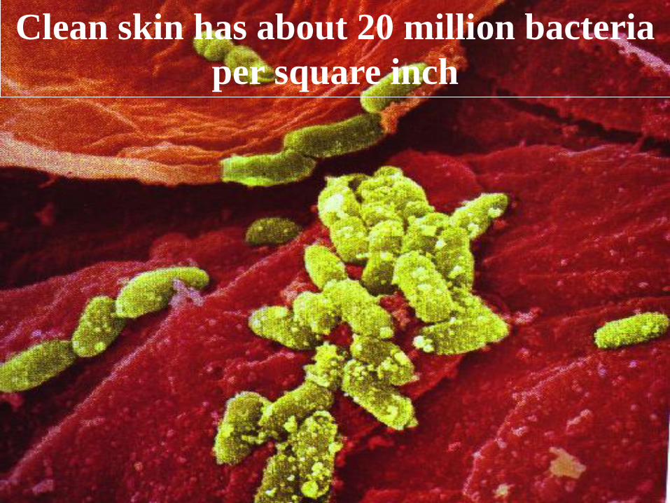
Clean skin has about 20 million bacteria
per square inch

The surface area/volume ratio of a spherical bacteria of
1 µm in diameter is high (6:1) as compared to a spherical
eukaryotic cell having a diameter of 20 µm (0.3:1).
Consequently:
• the intake of nutrients and removal of waste products
is quick - the bacteria has high rate of growth and
metabolism.
• no circulatory mechanism for nutrients is needed - the
cytoplasmic streaming is absent.
Surface area/volume ratio

ARRANGEMENT OF BACTERIAL CELLS
Cocci➢ Diplococci - Cells divide in one plane and remain attached predominately in pairs, e.g.
pneumococci.
➢Streptococci - Cells divide in one plain and remain attached to form chains, e.g Streptococcus
➢ Tetracocci - Cells divide in two planes and forms groups of four cells. (also called as‘tetrads’), e.g. Aerococcus.
➢Sarcinae - Cells divide in three planes, in a regular pattern producing a cubodial arrangementof cells.
➢Staphylococci - Cells divide in three planes, in an irregular pattern producing bunches ofcocci, e.g. Staphylococcus aureus

Spherical is called coccus.
Division along the same plane forms chains; 2 cocci together - Diplococcus
4 - 20 in chains - Streptococcus.
Division along 2 different planes - Tetrads
Division along 3 planes regularly - Sarcinae
Division along 3 planes irregularly - Staphylococci

Small cocci occurring singly or in small groups

A tetrad appears as a square of four cocci (arrows)

COCCI ARRANGED IN CLUSTERS

Bacilli➢ Single
➢ Diplobacilli - in pairs
➢ Streptobacilli – in chains, e.g. Bacillus subtilis
➢ Trichomes - rod-shaped bacteria arranged in chains with a larger area of contact between adjacent cells, e.g. Beggiatoa spp.
➢ Palisade – the cells are lined side by side as match sticks, e.g. Mycobacterium tuberculosis.
➢ Chinese letter like – e.g., Corynebacterium spp.
➢ Filamentous – long, mycelium like branching, mono-nuclear, e.g. Actinomycetes
➢ Hyphae – long, branched, multinucleate filaments, e.g. Streptomyces.

Rod shape is called Bacillus.
Two bacilli together - Diplobacilli
Chains of bacilli are called Streptobacilli
Palisades - Rods side by side or in X, V or Y figures

Long, thin rods

BACILLI ARRANGED IN LONG CHAINS
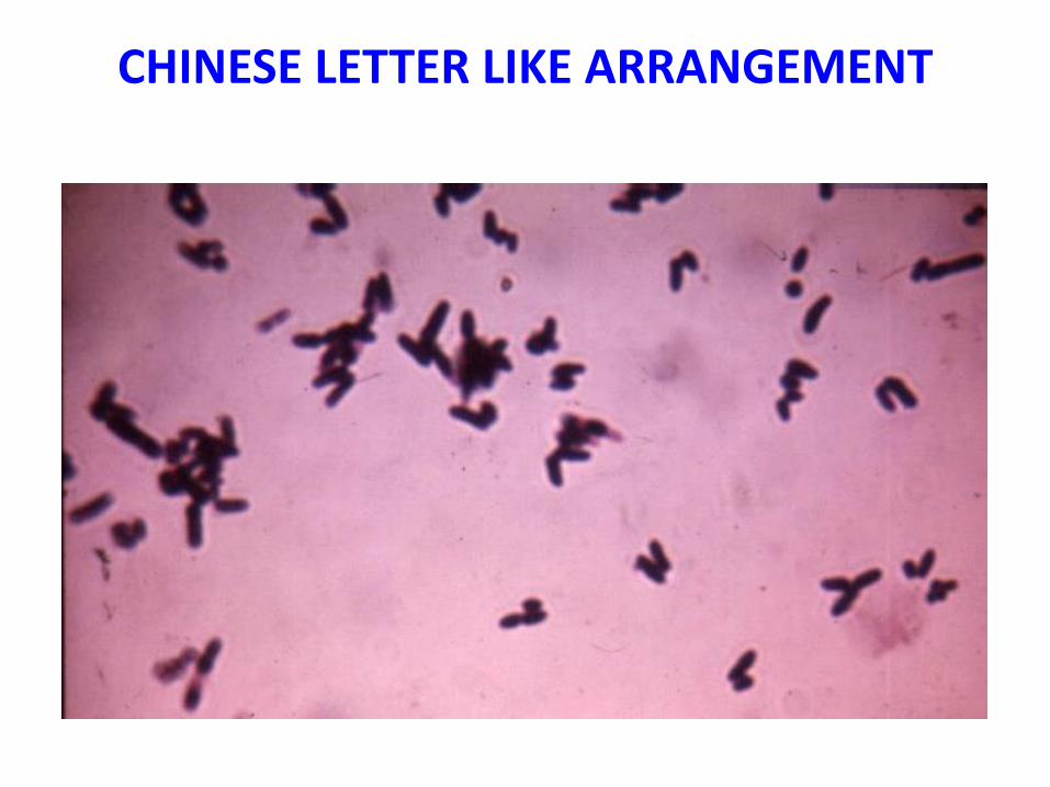
CHINESE LETTER LIKE ARRANGEMENT


25
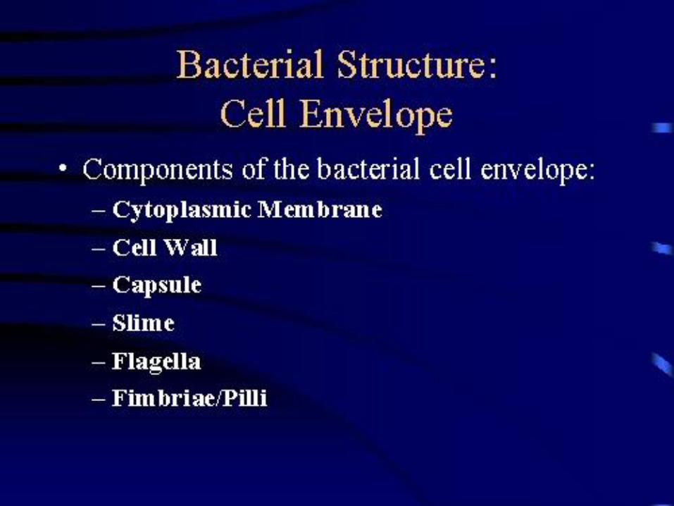
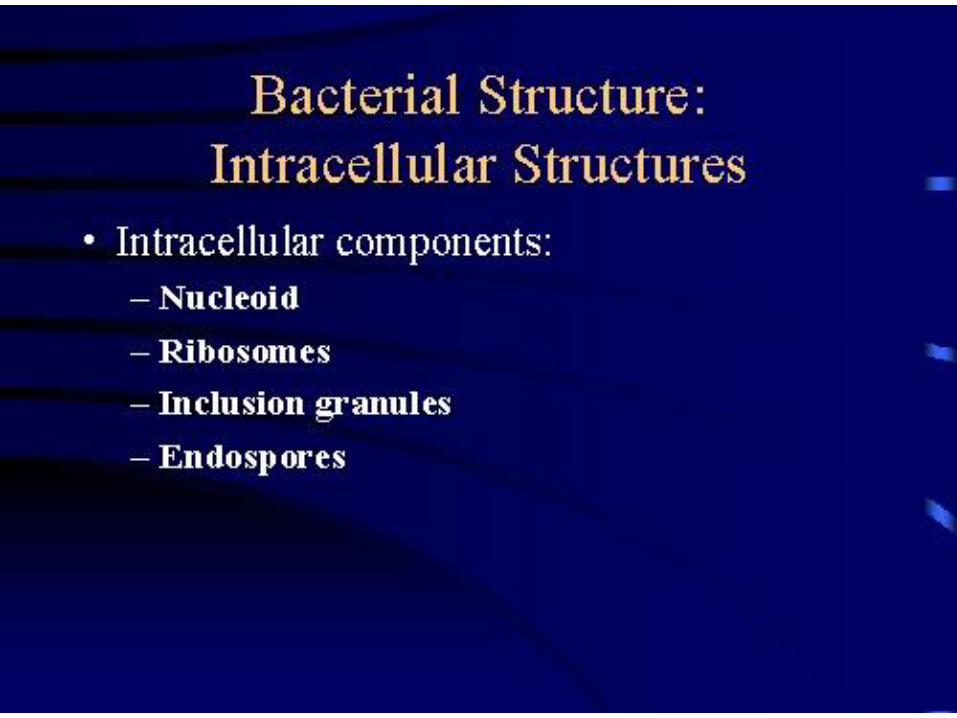
STRUCTURES OF BACTERIA
Structure Function
Cell Wall Protects and gives shape
Cell Membrane Regulates movement of materials, contains enzymes important to
cellular respiration
Cytoplasm Contains DNA, ribosomes, essential compounds
Chromosome Carries genetic information
Plasmid Contains some genes obtained through recomb.
Capsule & Slime
Layer
Protects the cell and assist in attaching cell to other surfaces
Endospore Protects cell agains harsh enviornments
Pilus Assists the cell in attaching to other surfaces
Flagellum Moves the cell

MYCOPLASMAS
(PPLO)
•naturally lack cell walls
•Gram-negative
•size ranges from 50-60 to 100-250 nm
•highly pleomorphic eubacteria
•five genera require sterols and three do not.
•no free-living Mycoplasma; strictly parasitic
• parasitize a wide range of organism including
humans, plants, animals, and insects.
MYCOPLASMAS
• facultative anaerobes and obligate anaerobes.
• growth on artificial media is slow with a generation
time ranging up to nine hours in some species.
• supplementation with other factors, such as serum,
may be required
• utilize glucose or arginine as the major source of
energy.
• ‘fried egg’ or ‘nipple shaped’ colonies, which can be
stained by Dienes’ stain.

RICKETTSIA AND CHLAMYDIA
•coccoid to rods in shape, with a diameter
of 0.3-0.7 μm.
•Gram-negative type cell walls
•except one rickettsia (Rochalimaea), all are
obligate intracellular parasites.
•contain both DNA and RNA.