severe open lisfranc injuries: one-stage operation · pdf filesevere open lisfranc injuries:...
TRANSCRIPT
RESEARCH ARTICLE Open Access
Severe open Lisfranc injuries: one-stageoperation through internal fixationassociated with vacuum sealing drainageWenqing Qu1, Shuqin Ni1, Zhenhai Wang1, Yong Zhao1, Shimin Zhang2, Yiheng Cheng1, Tong Liu1, Min Yu1
and Dan Wang1*
Abstract
Background: This study aimed to investigate the clinical feasibility of treating severe open Lisfranc injuries bymeans of one-stage internal fixation with k-wires associated with vacuum sealing drainage (VSD).
Methods: The clinical outcomes of 20 cases of severe open Lisfranc joint fracture-dislocation treated by using one-stage internal fixation with k-wires associated with VSD, after debridement and suturing during emergencytreatment, were reviewed.
Results: At 6 and 12 months after surgery, the American Orthopaedic Foot and Ankle Society midfoot scoreswere 69.2 and 78.2, the positive rates were 75 and 85 %, and the average visual analogue scale scores were 4.3and 1.3, respectively. The average time of internal fixation surgery was 47 min (30–70 min). There were three casesof wound-edge necrosis; however, there were no cases of skin necrosis around the incision, or deep infection.The mean time of first hospital stay was 16.1 days (10–23 days).
Conclusions: Treatment of severe open Lisfranc fracture and dislocation through one-stage internal fixation withk-wires in association with VSD led to fast anatomical reduction, stabilized bony structure, fast soft tissue recovery,and good short-term follow-up results.
Keywords: Lisfranc joints injury, Openness, k-wire internal fixation, VSD
BackgroundHigh-energy open Lisfranc injuries mainly result frombeing crushed under a heavy object, traffic accidents, orfalling from a height and are represented by severeLisfranc joint fracture-dislocation with serious soft tissueinjuries. Improper treatment of these fractures mightlead to negative outcomes such as soft tissue necrosis,posttraumatic arthritis, and arch abnormalities [1].Staged treatment has been widely applied in clinicalpractice; however, many patients were hospitalized for along time with unsatisfactory clinical outcomes, such aspoor recovery of soft tissue after temporary fixation withan external fixator, leading to a delay in performing openreduction internal fixation (ORIF) in the second stage,
resulting in difficulty or even failure in performing ORIF[2–5]. From March 2011 to January 2015, 30 cases of severeopen Lisfranc injuries were treated with one-stage k-wireinternal fixation associated with vacuum sealing drainage(VSD) at the Foot and Ankle Surgery Department of ourhospital, with satisfactory outcomes. In this report, 20 caseswith intact information are summarized and analyzed.
MethodsGeneral dataThis study included 17 male and 3 female patients with amean age of 49.5 years (range, 22–65 years). According tothe Gustilo classification for open injuries, there were 7type II cases and 13 type III cases. According to theMyerson classification for Lisfranc injuries, there were 4type B2 cases, 4 C1 cases, and 12 C2 cases. All injurieswere unilateral. The causes of injury were as follows: beingcrushed under a heavy object, 11 cases; traffic accident, 5
* Correspondence: [email protected] of Orthopaedics and Trauma, Yantaishan Hospital, Yantai264008, ChinaFull list of author information is available at the end of the article
© The Author(s). 2016 Open Access This article is distributed under the terms of the Creative Commons Attribution 4.0International License (http://creativecommons.org/licenses/by/4.0/), which permits unrestricted use, distribution, andreproduction in any medium, provided you give appropriate credit to the original author(s) and the source, provide a link tothe Creative Commons license, and indicate if changes were made. The Creative Commons Public Domain Dedication waiver(http://creativecommons.org/publicdomain/zero/1.0/) applies to the data made available in this article, unless otherwise stated.
Qu et al. Journal of Orthopaedic Surgery and Research (2016) 11:134 DOI 10.1186/s13018-016-0471-1

cases; and fall from a height, 4 cases. Anteroposterior,lateral, and oblique radiographs were obtained for allpatients before surgery. Computed tomography (CT)scanning and three-dimensional reconstruction wereperformed in nine cases. There were two cases with com-bined phalangeal fracture, five cases of articulatio tarsitransversa or articulatio cuneonavicularis fracture and dis-location, two cases of ankle fracture, one case of spinalfracture, and two cases of rib fracture. After excluding anycontraindication, emergency surgery was performed in allcases, and the average injury-to-surgery interval was 4.4 h(2–13 h). This study was conducted in accordance withthe Declaration of Helsinki and with approval from theEthics Committee of Yantaishan Hospital. Written in-formed consent was obtained from all patients.
Surgical methodsPreventative antibiotics were intravenously given to pa-tients 30 min before surgery. Eleven patients were anes-thetized with spinal or epidural anesthesia, and sevenpatients with general anesthesia with tracheal intubation.The patients were laid in the supine position. A tourniquetwas applied to the proximal end of the thigh. The woundwas washed first with hydrogen peroxide and then with alarge amount (>3000 mL) of normal saline (NS). Thewound, including the leg from below the knee, was soakedin povidone-iodine, followed with routine draping.Completely detached bone blocks were also soaked inpovidone-iodine for further surgery. In 18 cases, the openwound was located at the dorsum. Wound debridementwas performed from the surface to the deeper part, leavingas much skin tissue as possible, including peeled skin.Contused and devitalized soft tissue such as subcutaneoustissue, fascia, and tendon sheath were removed, whereaslarge bone blocks, but not small fragments, were kept.Important muscle tendons, blood vessels, and deep andsuperficial peroneal nerves were marked. Then, thesurface was again washed repeatedly with hydrogenperoxide, povidone-iodine, and NS.One-stage internal fixation with k-wires was
performed as definitive fixation for osteoarticular injury.Reduction was performed through the original wound inten cases. In eight cases with Myerson type C injuries,an extra incision was made to perform anatomicalreduction under direct vision. Reduction started fromthe cuneiforms to the base of metatarsals, from themedial, to central, to lateral columns. The mortise andtenon structure, formed by the cuneiforms and the base ofthe second metatarsal, and other undamaged bones wereused as anatomical markers. To maintain the reduction,the bones were clamped with towel forceps. The bonesand joints were fixed by using the k-wires through thewound or by penetrating the skin, as follows: in 1st ray, atleast two 2.0-mm intersecting k-wires were used for stable
fixation; in ray 2–4, a 1.5-mm k-wire was used for verticalfixation respectively; and in ray 5, another 2.0-mm k-wirewas used. Extra intersecting fixation was provided hori-zontally, if necessary, through the base of metatarsals orthe cuneiforms to achieve global Lisfranc stability. Antero-posterior and lateral radiographs were taken to examinethe positional relationship between the base of the firstand second metatarsals and the medial and intermediatecuneiforms, respectively, and 30° internal oblique radio-graphs were obtained to examine the positional relation-ship between the base of the fourth metatarsal and themedial border of the cuboid. If the bones could not be ac-curately restored, reduction of midfoot malalignment wasprioritized.To close the wound, the tourniquet was loosened and
a new incision was performed with interrupted, low-tension suture or exclusion after hemostasis. A low-tension suture was also performed on the originalwound with severe skin retraction or peeling, with theapplication of several interrupted stitches to draw theskin margin together, facilitate drainage, and avoidfurther skin necrosis. VSD accessories (Wuhan VSDMedical Science & Technology Co. Ltd., Wuhan, China)were used to cover the wound, seal the main film, con-nect the vacuum pump, and start suction (Figs. 1 and 2).
Treatment after surgeryThe patients were given routine antibiotics to preventinfection. The VSD kit was opened to evaluate woundsurface recovery after 5–7 days of suctioning. Anotherdebridement, skin graft, or skin flap surgery wasperformed on patients with skin or soft tissue colobomaor new skin necrosis after the initial debridement. Afterthe surgery, VSD was continued, and another evaluationand wound treatment were done after another 7 days.The k-wires through the lateral column were removed inthe outpatient clinic after 6–8 weeks, and the k-wiresthrough the medial and central columns were removedin the ward after 3–4 months. Partial-weight-bearingwalking and active and passive joint function training ofthe foot and ankle were started after removing allinternal fixators. The schedule for full-weight-bearingwalking depended on the radiographic findings (usuallyat 8 weeks after the removal of internal fixators).The radiographs of the patients (some of the patients were
evaluated with CT) were reviewed at 6 weeks, 12 weeks,6 months, and 1 year after the initial surgery. Moreover,functional scoring was performed by using the AmericanOrthopaedic Foot and Ankle Society (AOFAS) score.
ResultsSurgery detailsThe average time of debridement, internal fixation, andVSD wound closing was 47 min (30–70 min). The
Qu et al. Journal of Orthopaedic Surgery and Research (2016) 11:134 Page 2 of 7

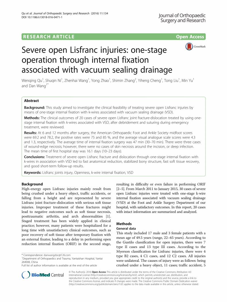
Fig. 1 Male patient, aged 55, with right foot severe open Lisfranc fracture-dislocation, was admitted to hospital 2 h after being crushed under heavy objects.One-stage internal fixation using k-wires associated with VSD was performed during emergency. a Severe open wound on the right foot with Lisfrancfracture-dislocation and badly contaminated, combined with ankle fracture shown on X-ray film. b One-stage reduction and multiple k-wire internal fixationwere performed via the open wound and dorsal incision, sealed with VSD kit. c The condition of original wound and incision was checked when VSD kit waschanged 5 days after surgery. d The patient was discharged from hospital 23 days after surgery, when the dorsal incision recovered well after suture, and theoriginal open wound also recovered well after postage stamp grafting. e One year after injury and 8 months after weight-bearing walking, with satisfyingappearance and function. f On X-ray film before surgery, fracture-dislocation was observed to the right Lisfranc joint, combined with distal fibula fracture.g Multiple k-wire cross-fixation was performed during surgery; the fracture-dislocation reached to anatomical reduction under X-ray. h AP, oblique, and lateralX-ray showing the condition of fracture-dislocation after one-stage internal fixation with good alignment. i Space broadening was not observed by CT reviewand X-ray film half a year after removing internal fixators
Qu et al. Journal of Orthopaedic Surgery and Research (2016) 11:134 Page 3 of 7
average number of k-wires for each case was 7.4 (5–13),and a total of 62 2.0-mm and 75 1.5-mm k-wires wereused. The original open wound was completely closed instage 1 in six cases, sutured in stage 2 in eight cases,recovered after skin graft in five cases, and repaired withskin flap in one case. The extra incision made duringsurgery (in eight cases in total) was completely closed instage 1 in six cases and sutured in stage 2 in two cases.VSD was used for 47 times in all cases. Because the frontfoot was not bearing load before the k-wires wereremoved, the k-wires did not lead to serious soft tissueirritation. The internal fixators were removed in stagesafter surgery. The fixators through the lateral columnwere removed after 6–8 weeks, and those throughthe medial and central columns were removed after10–16 weeks.
Follow-upThe average follow-up time for 18 cases was 16.4 months(12–26 months). No patient developed osteofascial com-partment syndrome, and no loosening or cracking of theinternal fixator was observed in any cases. Radiographicexaminations immediately and at 6 weeks, 12 weeks,6 months, and 1 year after surgery indicated that theaverage recovery time for bone fracture was 12 weeks(8–20 weeks). In two cases with cuneiform coloboma,autologous iliac crest graft was performed 12 weeks afterthe initial internal fixation, which healed after 20 weeks.Spontaneous partial joint fusion was observed in twocases, and wound-edge necrosis occurred in three cases;however, there were no cases of necrosis of the incisionedge, or deep infection. The mean time of first hospitalstay was 16.1 days (10–23 days).
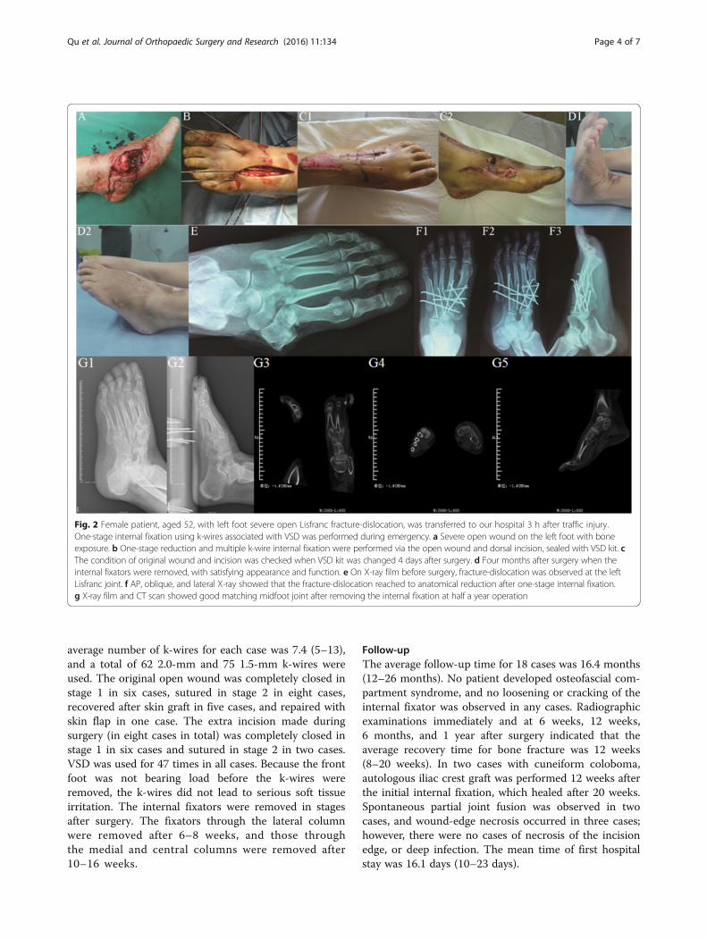
Fig. 2 Female patient, aged 52, with left foot severe open Lisfranc fracture-dislocation, was transferred to our hospital 3 h after traffic injury.One-stage internal fixation using k-wires associated with VSD was performed during emergency. a Severe open wound on the left foot with boneexposure. b One-stage reduction and multiple k-wire internal fixation were performed via the open wound and dorsal incision, sealed with VSD kit. cThe condition of original wound and incision was checked when VSD kit was changed 4 days after surgery. d Four months after surgery when theinternal fixators were removed, with satisfying appearance and function. e On X-ray film before surgery, fracture-dislocation was observed at the leftLisfranc joint. f AP, oblique, and lateral X-ray showed that the fracture-dislocation reached to anatomical reduction after one-stage internal fixation.g X-ray film and CT scan showed good matching midfoot joint after removing the internal fixation at half a year operation
Qu et al. Journal of Orthopaedic Surgery and Research (2016) 11:134 Page 4 of 7

The AOFAS midfoot scoring system was applied forfunctional evaluation at 6 and 12 months after surgery.The average scores at 6 and 12 months were 69.2 (55–86)and 88.2 (68–95), the positive rates were 75 and 85 %, andthe average visual analogue scale scores were 4.3 (2–6)and 1.3 (0–3), respectively.In four cases, loading resulted in foot pain in the lower
arch. The symptoms were improved after a customizedinsole treatment, and the patients did not undergo jointfusion surgery.
DiscussionLisfranc injuries are often difficult to diagnose and treat,causing long-term disability without appropriate man-agement [6–8]. The difficulty in the clinical treatment ofopen Lisfranc fracture and dislocation results from thesevere destruction of the bony structure, with poorcondition of local soft tissue. Thus, staged treatment hasbeen frequently performed [2–5], in which general align-ment and restoration of the length of the foot are firstperformed by using temporary external fixators, facilitat-ing soft tissue recovery, and then a final definitive in-ternal fixation or joint fusion is performed after thebody and local soft tissue condition has improved. Thismethod has been widely applied in the treatment ofcomplex tibial plateau and pilon fractures, with good re-sults. However, in many cases with severe Lisfranc jointfracture and dislocation, especially those with openwound, the long recovery time for soft tissue in stage 1delayed the internal fixation in stage 2. Sufficient inci-sion for exposing the injury area was not allowed owingto the soft tissue condition of the newly healed woundor fracture-dislocation area after skin graft or skin flapcoverage, and the formed cicatrix and inflammatorygranulation tissue caused difficulties in reduction duringsurgery. Application of an internal fixation plate, whichis firm but large, also required a good soft tissue condi-tion. Moreover, in some cases, ORIF could not beperformed, which could only be remedied with osteot-omy or fusion surgery. In one study, 123 cases withstaged treatment for high-energy midfoot fractures-dislocations, with an average time of 21.3 days for theapplication of external fixators, were reported, similar toour previous clinical experience [2]. Therefore, it is prac-tical to attempt a safe and effective one-stage definitiveinternal fixation method.
Soft tissue managementSatisfactory results can be achieved with open reductionfor Lisfranc injuries. However, despite this treatment,both the severity of the soft tissue injury and nonana-tomic reduction are negative prognostic factors in thetreatment of Lisfranc fractures-dislocations [9].
Soft tissue treatment is especially important for openLisfranc fracture-dislocation. VSD has been applied in manycases, leading to earlier wound closure, clear wound surfacedrainage, faster detumescence, accelerated tissue growth,and reduced clinical workload, compared with traditionalmethods [10, 11]. Moreover, wounds in most cases showedgood improvement when the VSD accessory was changedfor the first time at 5–7 days after surgery, and soft tissuewas fast repaired simply with direct suture, skin graft, orskin flap transplantation, owing to emergency measuressuch as washing with a large amount of NS, keeping asmuch skin as possible, avoiding high-tension suture, anddrilling on the exposed bone with a thin k-wire.
Final definitive internal fixation with one-stage anatomicreduction and k-wiresNithyananth performed this treatment in 22 cases and ob-tained satisfactory function scores during long-termfollow-ups with 16 days of average wound healing time,much shorter than our experience before using an exter-nal fixator (21.3 days). The exposure, reduction, and fix-ation of severe midfoot fracture-dislocation were normallyperformed through a dorsal approach: with high bonedensity, no obvious bone coloboma after severe bone in-jury was fixed with k-wires; the reduction of adjacentjoints can help estimate the reduction degree of targetjoints as they wedged with each other. The characteristicsof the soft tissue of this area allow extra auxiliary incisionas the tolerance of the tissues to violent injuries such asgrinding or tearing was high; for instance, midfoot skinand subcutaneous tissue gain tolerance from long-termfriction and extrusion from shoes, as well as the ligamentof joint dorsum and extensor digitorum longus tendon,but there was hardly any muscle tissue with low tolerance.Most of the fracture-dislocation area could be exposedthrough the open wound, as this injury was superficialwhen surgery was performed through the dorsal approach,or anatomical reduction could be performed under directvision through auxiliary incision, without increased risk ofskin necrosis. The k-wire was small enough for cross-fixation for providing high strength, particularly whenthree to four 2.0-mm k-wires were applied to the medialand lateral columns to form a “splint-like” fixation for thecentral column. Thinner k-wires were applied to thecentral column to decrease the damage area of the articu-lar cartilage surface and reduce bone infection risk signifi-cantly after surgery, compared with that using a screwspike or fixation plate.Anatomical reduction is the basic principle of treating
intra-articular fracture-dislocation. Many researchersindicated that rigorous anatomical reduction should beperformed, as functional recovery after surgery directlyrelates to reduction, regardless of performing a true percu-taneous technique or open methods [9, 12–14]. Previous
Qu et al. Journal of Orthopaedic Surgery and Research (2016) 11:134 Page 5 of 7

studies demonstrated that malreduction during surgeryled to poor function and imaging scores in up to 49.6 %cases [15, 16]. Nithyananth, Kadow, and Kuo demon-strated the standard to ensure anatomical reduction dur-ing surgery [1, 2, 17]: the tala axis and the first metatarsalaxis were nearly overlapping on the foot anteroposteriorfilm (the average angle between the two was 7.7°). Themedial and lateral borders of the first metatarsal, and themedial and lateral borders of the medial cuneiform, re-spectively, were in one line, as were those of the secondmetatarsal and the intermediate cuneiform, forming anevenly matched cross-joint space, with the width betweenthe medial cuneiform and medial border of the secondmetatarsal being <2 mm. On a 30° internal oblique radio-graph, the medial border of the fourth metatarsal boneand that of the cuboid were in one smooth line, and thecalcaneus axis and the fourth metatarsal axis were over-lapping. On the lateral film, no metatarsal shift was ob-served toward the metatarsal side or the dorsal side, andno collapse arch or high-arch deformity was observed; thetalo-first metatarsal angle (Meary’s angle) was between ±4°[18]. If anatomical reduction was difficult to perform be-cause of severe fracture, alignment recovery of the mid-foot and hindfoot was prioritized.
Comparison of the therapeutic strategy between one-stage internal fixation and joint fusionMidfoot fracture-dislocation has been treated with one-stage joint fusion, based on the theory that the medial col-umn is responsible for stabilization and support, and thelateral column for flexibility and cushioning stress [19].The method by Boffeli was the most typical, in which one-stage fusion was performed in the medial column, ORIFin the central column, and k-wire fixation in the lateralcolumn [20]. Some researchers demonstrated no statisti-cally significant difference in the functional scoring andsatisfaction degree between cases treated with one-stagejoint fusion and those treated with one-stage internal fix-ation, after a long-term follow-up [14, 19, 21]. The advan-tage of joint fusion was that the probability of removingthe internal fixator was low, whereas the disadvantageswere as follows: joint fusion requires skilled doctors and isdifficult to fulfill during emergencies or hospital nightshift, and fixation in standard joint fusion by using a screwspike through a joint or a fixation plate across a jointsignificantly increases the risk of bone infection in caseswith open injury. After medial column fusion of Lisfrancjoints, the load of adjacent joints, such as the centralcolumn, metatarsophalangeal joints, and transverse tarsaljoint, will increase, resulting in posttraumatic arthritis.In our study, Lisfranc posttraumatic arthritis was identi-
fied on the radiographs of seven cases 1 year after surgery,with mild clinical symptoms. Similar results showing thatarthritis observed on radiographs was not in accordance
with clinical symptoms were also reported in anotherstudy after a long-term follow-up [16]. We speculated thatthe medial and central columns of Lisfranc joints areamphiarthrodial joints with low abrasion. Although thestrength of internal fixation with k-wires is lower than thatwith a fixation plate, joint stability was enhanced as exten-sive soft tissue injury generated large amounts of fibrouscicatrices around the joints during recovery, and ankylosis,not arthrochalasis, occurred after surgery, similar to subta-lar joint ankylosis that occurs after treatment of calcanealfractures by means of internal fixation with a steel platethrough an L-shaped calcaneal lateral incision. In thecases in our short-term study, space broadening was notobserved in the joints with the original injury on CTreview observed after 6 months of full-weight-bearingwalking (Fig. 1h); however, long-term follow-up of jointstability requires further investigation. It can be speculatedthat although symptomatic Lisfranc instability, fallenarches, or traumatic flat foot was observed after surgery,this can be treated by using customized shoe pads or byperforming joint fusion after the soft tissue has fullyrecovered. Thus, except for joint dislocations withoutfracture, we prefer one-stage internal fixation for Lisfrancfracture-dislocation over joint fusion.
ConclusionsIn conclusion, the goal of the treatment of Lisfranc fracture-dislocation is to achieve a painless, functional plantigradefoot with a good appearance [22]. Although staged treat-ment for severe open Lisfranc fracture and dislocation hasbeen frequently reported and has shown acceptable results,one-stage internal fixation with k-wires associated with VSDled to faster anatomical reduction, stabilized bony structure,faster soft tissue recovery after surgery, and good short-termfollow-up results according to our research, although themedium- and long-term follow-up results concerning jointstability require further investigation.
AbbreviationsAOFAS: American Orthopaedic Foot and Ankle Society; CT: Computedtomography; NS: Normal saline; ORIF: Open reduction internal fixation;VSD: Vacuum sealing drainage
AcknowledgementsWe appreciate all the colleagues who provide help.
FundingNone.
Availability of data and materialsNot applicable.
Authors’ contributionsWQ, SN, ZW, and YZ reviewed the database and prepared the manuscript.SZ, YC, TL, and MY carried out the statistical analysis and assisted withpreparation of the manuscript. WQ and ZW carried out the surgeries. DWsupervised the writing of the whole paper. All authors read and approvedthe final manuscript.
Qu et al. Journal of Orthopaedic Surgery and Research (2016) 11:134 Page 6 of 7

Competing interestsThe authors declare that they have no competing interests.
Consent for publicationAll patients consent to publish patient identifiable information and theobtained data.
Ethics approval and consent to participateThis study was conducted in accordance with the Declaration of Helsinki. Thisstudy was conducted with approval from the Ethics Committee of YantaishanHospital. Written informed consent to participate was obtained from all theparticipants.
Author details1Department of Orthopaedics and Trauma, Yantaishan Hospital, Yantai264008, China. 2Department of Orthopaedics, Yangpu Hospital of TongjiUniversity, Shanghai 200090, China.
Received: 30 June 2016 Accepted: 23 August 2016
References1. Nithyananth M, Boopalan PR, Titus VT, Sundararaj GD, Lee VN. Long-term
outcome of high-energy open Lisfranc injuries: a retrospective study.J Trauma. 2011;70:710–6.
2. Kadow TR, Siska PA, Evans AR, Sands SS, Tarkin IS. Staged treatment of highenergy midfoot fracture dislocations. Foot Ankle Int. 2014;35:1287–91.
3. Coetzee JC, Ly TV. Treatment of primarily ligamentous Lisfranc joint injuries:primary arthrodesis compared with open reduction and internal fixation.Surgical technique. J Bone Joint Surg Am. 2007;89:S122–7.
4. Tarkin IS, Clare MP, Marcantonio A, Pape HC. An update on themanagement of high-energy pilon fractures. Injury. 2008;39:142–54.
5. Tarkin IS, Sop A, Pape HC. High-energy foot and ankle trauma: principles forformulating an individualized care plan. Foot Ankle Clin. 2008;13:705–23.
6. Welck MJ, Zinchenko R, Rudge B. Lisfranc injuries. Injury. 2015;46:536–41.7. Siddiqui NA, Galizia MS, Almusa E, Omar IM. Evaluation of the
tarsometatarsal joint using conventional radiography, CT, and MR imaging.Radiographics. 2014;34:514–31.
8. Laird RC. Acute forefoot and midfoot injuries. Clin Podiatr Med Surg. 2015;32:231–8.
9. Demirkale I, Tecimel O, Celik I, Kilicarslan K, Ocguder A, Dogan M. The effectof the Tscherne injury pattern on the outcome of operatively treatedLisfranc fracture dislocations. Foot Ankle Surg. 2013;19:188–93.
10. Li W, Ji L, Tao W. Effect of vacuum sealing drainage in osteofascialcompartment syndrome. Int J Clin Exp Med. 2015;8:16112–6.
11. Ko YS, Jung SW. Vacuum-assisted close versus conventional treatment forpostlaparotomy wound dehiscence. Ann Surg Treat Res. 2014;87:260–4.
12. Benirschke SK, Meinberg E, Anderson SA, Jones CB, Cole PA. Fractures anddislocations of the midfoot: Lisfranc and Chopart injuries. J Bone Joint SurgAm. 2012;94:1325–37.
13. Thompson MC, Mormino MA. Injury to the tarsometatarsal joint complex.J Am Acad Orthop Surg. 2003;11:260–7.
14. Seybold JD, Coetzee JC. Lisfranc injuries: when to observe, fix, or fuse.Clin Sports Med. 2015;34:705–23.
15. Stavlas P, Roberts CS, Xypnitos FN, Giannoudis PV. The role of reduction andinternal fixation of Lisfranc fracture-dislocations: a systematic review of theliterature. Int Orthop. 2010;34:1083–91.
16. Marín-Peña OR, Viloriarecio F, Sanz Gómez T, Larrainzargarijo R. Fourteenyears follow up after Lisfranc fracture-dislocation: functional and radiologicalresults. Injury. 2012;43:S79–82.
17. Kuo RS, Tejwani NC, Digiovanni CW, Holt SK, Benirschke SK, Hansen Jr ST,et al. Outcome after open reduction and internal fixation of Lisfranc jointinjuries. J Bone Joint Surg Am. 2000;82-A:1609–18.
18. Younger AS, Sawatzky B, Dryden P. Radiographic assessment of adultflatfoot. Foot Ankle Int. 2005;26:820–5.
19. Henning JA, Jones CB, Sietsema DL, Bohay DR, Anderson JG. Openreduction internal fixation versus primary arthrodesis for Lisfranc injuries:a prospective randomized study. Foot Ankle Int. 2009;30:913–22.
20. Boffeli TJ, Pfannenstein RR, Thompson JC. Combined medial columnprimary arthrodesis, middle column open reduction internal fixation, andlateral column pinning for treatment of Lisfranc fracture-dislocation injuries.J Foot Ankle Surg. 2014;53:657–63.
21. Ly TV, Coetzee JC. Treatment of primarily ligamentous Lisfranc joint injuries:primary arthrodesis compared with open reduction and internal fixation.A prospective, randomized study. J Bone Joint Surg Am. 2006;88:514–20.
22. Zonno AJ, Myerson MS. Surgical correction of midfoot arthritis with andwithout deformity. Foot Ankle Clin. 2011;16:35–47.
• We accept pre-submission inquiries
• Our selector tool helps you to find the most relevant journal
• We provide round the clock customer support
• Convenient online submission
• Thorough peer review
• Inclusion in PubMed and all major indexing services
• Maximum visibility for your research
Submit your manuscript atwww.biomedcentral.com/submit
Submit your next manuscript to BioMed Central and we will help you at every step:
Qu et al. Journal of Orthopaedic Surgery and Research (2016) 11:134 Page 7 of 7