sense organs handout - science olympiad · sense organs handout sensory receptors ... • note: for...
TRANSCRIPT
1
SENSE ORGANS HANDOUT Sensory Receptors - receive input, generate receptor potentials and with enough summation, generate action
potentials in the neurons they are part of or synapse with 5 Types of Sensory Receptors - based on the type of stimuli they detect:
1. Mechanoreceptors - pressure receptors, stretch receptors, and specialized mechanoreceptors involved in movement and balance.
2. Thermoreceptors - skin and viscera, respond to both external and internal temperature 3. Pain receptors - stimulated by lack of O2, chemicals released from damaged cells and inflammatory
cells 4. Chemoreceptors - detect changes in levels of O2, CO2, and H+ ions (pH) as well as chemicals that
stimulate taste and smell receptors 5. Photoreceptors - stimulated by light
Distribution of Receptors in the body:
Special Senses • mediated by relatively complex sense organs of the head, innervated by cranial nerves • vision, hearing, equilibrium, taste and smell
General (somesthetic, somatosensory)
• receptors widely distributed in skin, muscles, tendons, joints, and viscera • they detect touch, pressure, stretch, heat, cold and pain, blood pressure
Special Senses
Sensation and perception • Vision – Eye • Hearing – Ear • Equilibrium – Ear • Taste – Taste receptors • Smell – Olfactory system
General Senses
• Skin – Hot, cold, pressure, pain • Muscles, joints, and tendons – proprioceptors- stretch receptors respond to stretch or compression • Pain Receptors – somatic or visceral
2
SPECIAL SENSES Eye - Vision Processes • Light energy is transduced into neural activity • Neural activity is processed by the brain • Note: For an analogy, you can imagine taking a picture with a camera. The eye is the camera, the retina,
which is a specialized part of the brain at the back of the eye, is the film, and the parts of the brain that process visual information is the photoshop.
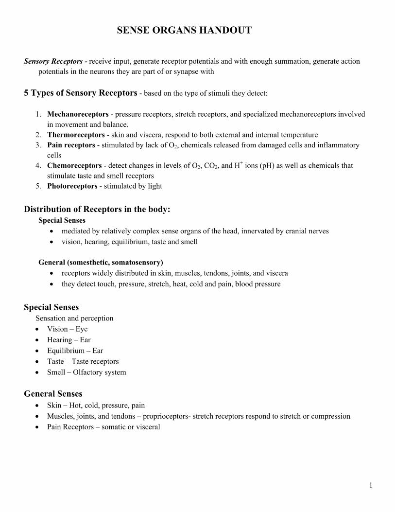
Human visual systems permit light reflected off distant objects to be: • Localized relative to the individual within his or her environment • Identified based on size, shape, color, and past experience • Perceived to be moving (or not) • Detected in a wide variety of lighting conditions Sequence of events • Light entering the eye is focused on the retina • Retina converts light energy into neuronal activity • Axons of the retinal neurons are bundled to form the optic nerves • Visual information is distributed to several brain structures that perform different functions Eye – the organ used to sense light
Three layers – 1. Outer layer consists of sclera and
cornea 2. Middle layer consists of choroid, ciliary
body and iris 3. Inner layer consists of retina
Extraocular muscles--attached to the eye and skull and allow movement
3
Anatomy of the Eye Gross anatomy Functions of the major parts of the eye: • Sclera or Scleroid Layer – (white of eye) the outermost layer that forms the eyeball- a tough protective
layer of connective tissue that helps maintain the shape of the eye and provides an attachment for the muscles that move the eye
• Conjunctiva--membrane inside the eyelid attached to the sclera • Cornea - the transparent surface covering the iris and pupil- a clear, dome-shaped part of the sclera
covering the front of the eye through which light enters the eye • Anterior Chamber – a small chamber between the cornea and the pupil • Aqueous Humor - fluid behind the cornea - the clear fluid that fills that anterior chamber of the eye and
helps to maintain the shape of the cornea providing most of the nutrients for the lens and the cornea and involved in waste management in the front of the eye
• Choroid Layer - middle layer of the eye containing may blood vessels • Ciliary Body - the ciliary body is a circular band of muscle that is connected and sits immediately
behind the iris- produces aqueous humor, changes shape of lens for focusing, and • Iris - circular muscle that controls the diameter of the pupil - the pigmented front portion of the choroid
layer and contains the blood vessels - it determines the eye color and it controls the amount of light that enters the eye by changing the size of the pupil (an albino only has the blood vessels – not pigment so it appears red or pink because of the blood vessels)
• Lens - a crystalline structure located just behind the iris - it focuses light onto the retina • Pupil - the opening in the center of the iris- it changes size as the amount of light changes (the more
light, the smaller the hole) and it allows light to reach the retina • Vitreous - a thick, transparent liquid that fills the center of the eye - it is mostly water and gives the eye
its form and shape (also called the vitreous humor) • Retina - axons of the retina leaving the eye - sensory tissue that lines the back of the eye. It contains
millions of photoreceptors (rods for black & white and cones for color ) that convert light rays into electrical impulses that are relayed to the brain via the optic nerve
• Optic nerve - the nerve that transmits electrical impulses from the retina to the brain
Opthalmoscopic appearance (Retina as seen through
the pupil) • Note: in photographs, the red appearance of the
eye is actually the retina photographed. Double flash camera causes the pupil to constrict.
• Optic disk (blind spot)--no vision is possible o Blood vessels originate here. The vessels
shadow the retina o Optic nerve fibers exit here o No photoreceptors
• Macula--area of the retina responsible for central vision (vs. peripheral)
• Fovea--center of the retina (where most of the cones are)
4
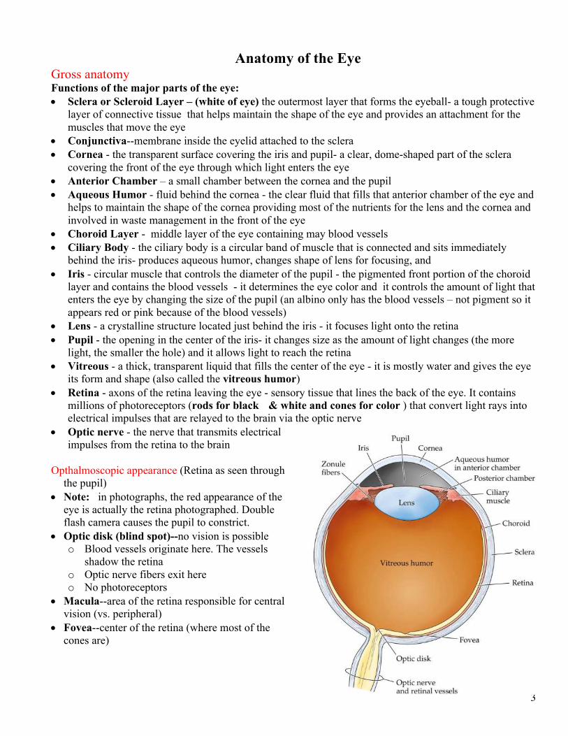
Common eye defects include • myopia or nearsightedness where the eyeball is too
long or the cornea is too steep; • hyperopia or far sightedness where the eyeball is
short or lens cannot become round enough: • presbyopia where the muscles controlling the
bulging of the lens become weak as we age; • cataracts where the lens becomes fogged; • nyctalopia or night blindness where vision is
impaired in dim light and in the dark due to pigment rhodospin in the rods not functioning properly External features of the eye
Cross sectional anatomy • Lens--transparent surface that contributes to the formation of images (w/i 9 meters) • Ciliary muscles--change the shape of the lens and allow focusing • Vitreous humor--more viscous than the aqueous humor - Lies between the lens and the retina and
provides spherical shape • Retina - inner most layer of cells at the back of the eye - Transduces light energy into neural activity
5
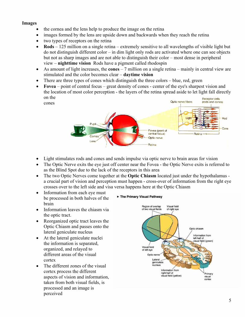
Images
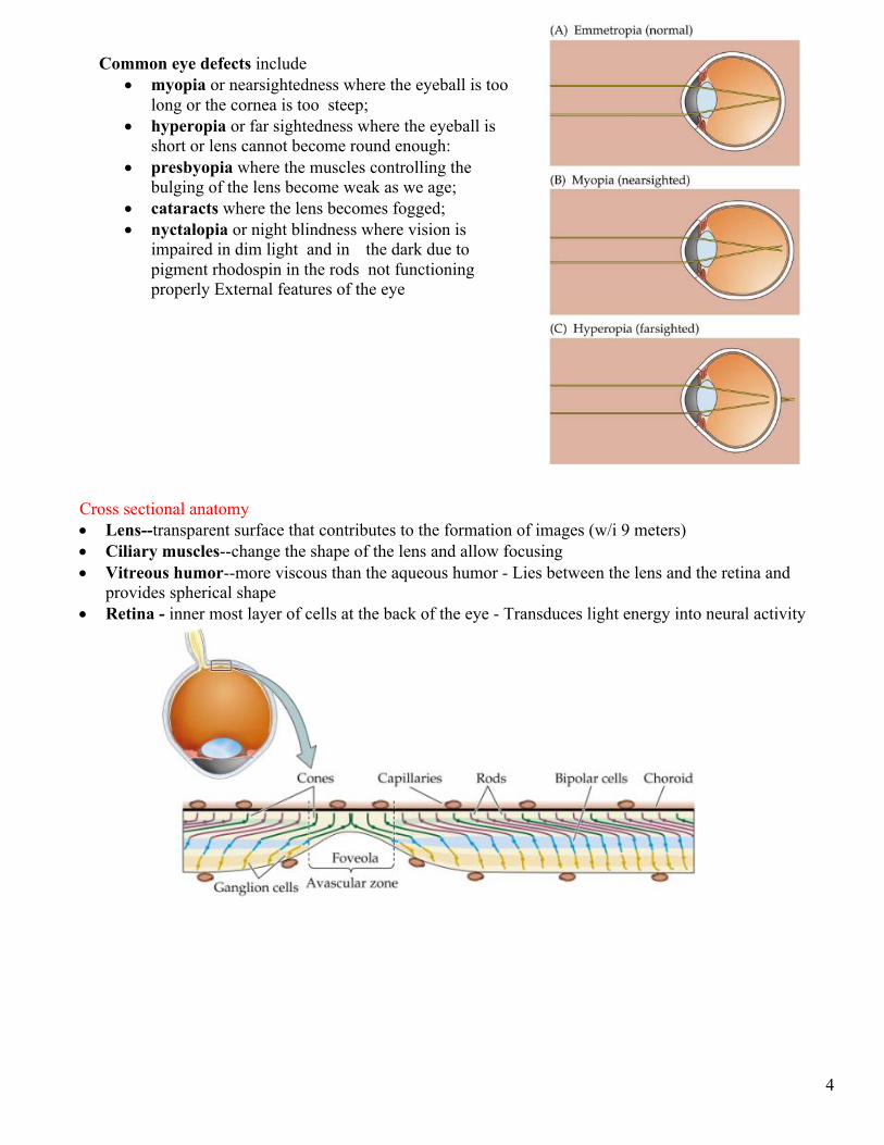
• the cornea and the lens help to produce the image on the retina • images formed by the lens are upside down and backwards when they reach the retina • two types of receptors on the retina • Rods – 125 million on a single retina – extremely sensitive to all wavelengths of visible light but
do not distinguish different color – in dim light only rods are activated where one can see objects but not as sharp images and are not able to distinguish their color – most dense in peripheral view – nighttime vision Rods have a pigment called rhodospin
• As amount of light increases, the cones – 7 million on a single retina – mainly in central view are stimulated and the color becomes clear – daytime vision
• There are three types of cones which distinguish the three colors – blue, red, green • Fovea – point of central focus – great density of cones - center of the eye's sharpest vision and
the location of most color perception - the layers of the retina spread aside to let light fall directly on the cones
• Light stimulates rods and cones and sends impulse via optic nerve to brain areas for vision • The Optic Nerve exits the eye just off center near the Fovea - the Optic Nerve exits is referred to
as the Blind Spot due to the lack of the receptors in this area • The two Optic Nerves come together at the Optic Chiasm located just under the hypothalamus -
a crucial part of vision and perception must happen - cross-over of information from the right eye crosses over to the left side and visa versa happens here at the Optic Chiasm
• Information from each eye must be processed in both halves of the brain
• Information leaves the chiasm via the optic tract.
• Reorganized optic tract leaves the Optic Chiasm and passes onto the lateral geniculate nucleus
• At the lateral geniculate nuclei the information is separated, organized, and relayed to different areas of the visual cortex
• The different zones of the visual cortex process the different aspects of vision and information, taken from both visual fields, is processed and an image is perceived
6
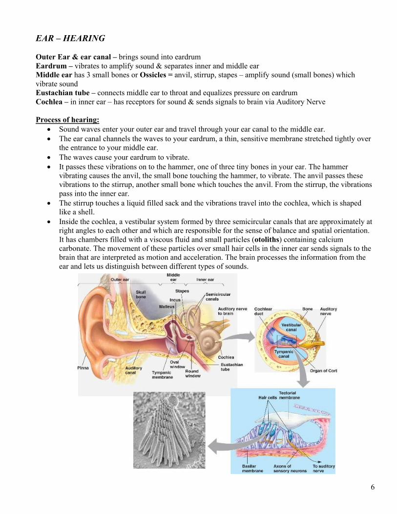
EAR – HEARING Outer Ear & ear canal – brings sound into eardrum Eardrum – vibrates to amplify sound & separates inner and middle ear Middle ear has 3 small bones or Ossicles = anvil, stirrup, stapes – amplify sound (small bones) which vibrate sound Eustachian tube – connects middle ear to throat and equalizes pressure on eardrum Cochlea – in inner ear – has receptors for sound & sends signals to brain via Auditory Nerve Process of hearing:
• Sound waves enter your outer ear and travel through your ear canal to the middle ear. • The ear canal channels the waves to your eardrum, a thin, sensitive membrane stretched tightly over
the entrance to your middle ear. • The waves cause your eardrum to vibrate. • It passes these vibrations on to the hammer, one of three tiny bones in your ear. The hammer
vibrating causes the anvil, the small bone touching the hammer, to vibrate. The anvil passes these vibrations to the stirrup, another small bone which touches the anvil. From the stirrup, the vibrations pass into the inner ear.
• The stirrup touches a liquid filled sack and the vibrations travel into the cochlea, which is shaped like a shell.
• Inside the cochlea, a vestibular system formed by three semicircular canals that are approximately at right angles to each other and which are responsible for the sense of balance and spatial orientation. It has chambers filled with a viscous fluid and small particles (otoliths) containing calcium carbonate. The movement of these particles over small hair cells in the inner ear sends signals to the brain that are interpreted as motion and acceleration. The brain processes the information from the ear and lets us distinguish between different types of sounds.
7
Ear – Equilibrium
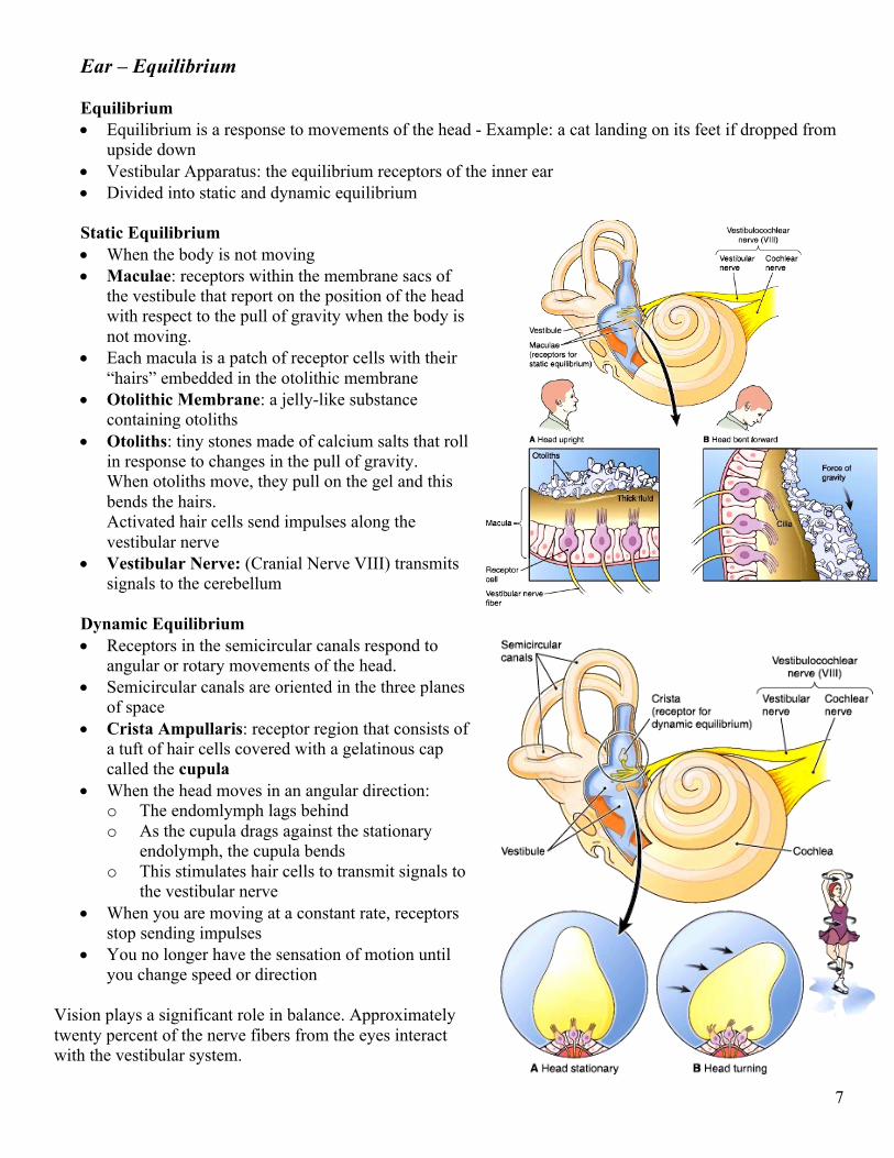
Equilibrium • Equilibrium is a response to movements of the head - Example: a cat landing on its feet if dropped from
upside down • Vestibular Apparatus: the equilibrium receptors of the inner ear • Divided into static and dynamic equilibrium
Static Equilibrium • When the body is not moving • Maculae: receptors within the membrane sacs of
the vestibule that report on the position of the head with respect to the pull of gravity when the body is not moving.
• Each macula is a patch of receptor cells with their “hairs” embedded in the otolithic membrane
• Otolithic Membrane: a jelly-like substance containing otoliths
• Otoliths: tiny stones made of calcium salts that roll in response to changes in the pull of gravity. When otoliths move, they pull on the gel and this bends the hairs. Activated hair cells send impulses along the vestibular nerve
• Vestibular Nerve: (Cranial Nerve VIII) transmits signals to the cerebellum
Dynamic Equilibrium • Receptors in the semicircular canals respond to
angular or rotary movements of the head. • Semicircular canals are oriented in the three planes
of space • Crista Ampullaris: receptor region that consists of
a tuft of hair cells covered with a gelatinous cap called the cupula
• When the head moves in an angular direction: o The endomlymph lags behind o As the cupula drags against the stationary
endolymph, the cupula bends o This stimulates hair cells to transmit signals to
the vestibular nerve • When you are moving at a constant rate, receptors
stop sending impulses • You no longer have the sensation of motion until
you change speed or direction Vision plays a significant role in balance. Approximately twenty percent of the nerve fibers from the eyes interact with the vestibular system.
8
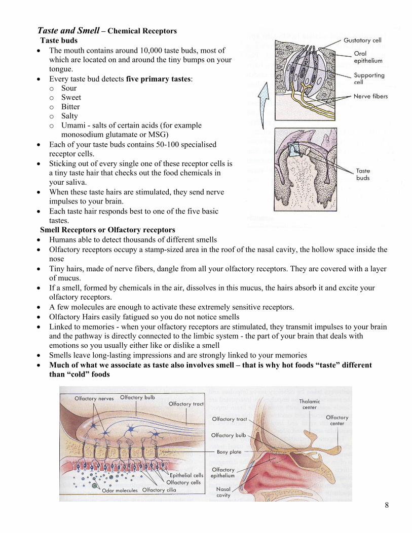
Taste and Smell – Chemical Receptors Taste buds • The mouth contains around 10,000 taste buds, most of
which are located on and around the tiny bumps on your tongue.
• Every taste bud detects five primary tastes: o Sour o Sweet o Bitter o Salty o Umami - salts of certain acids (for example
monosodium glutamate or MSG) • Each of your taste buds contains 50-100 specialised
receptor cells. • Sticking out of every single one of these receptor cells is
a tiny taste hair that checks out the food chemicals in your saliva.
• When these taste hairs are stimulated, they send nerve impulses to your brain.
• Each taste hair responds best to one of the five basic tastes.
Smell Receptors or Olfactory receptors • Humans able to detect thousands of different smells • Olfactory receptors occupy a stamp-sized area in the roof of the nasal cavity, the hollow space inside the
nose • Tiny hairs, made of nerve fibers, dangle from all your olfactory receptors. They are covered with a layer
of mucus. • If a smell, formed by chemicals in the air, dissolves in this mucus, the hairs absorb it and excite your
olfactory receptors. • A few molecules are enough to activate these extremely sensitive receptors. • Olfactory Hairs easily fatigued so you do not notice smells • Linked to memories - when your olfactory receptors are stimulated, they transmit impulses to your brain
and the pathway is directly connected to the limbic system - the part of your brain that deals with emotions so you usually either like or dislike a smell
• Smells leave long-lasting impressions and are strongly linked to your memories • Much of what we associate as taste also involves smell – that is why hot foods “taste” different
than “cold” foods
9
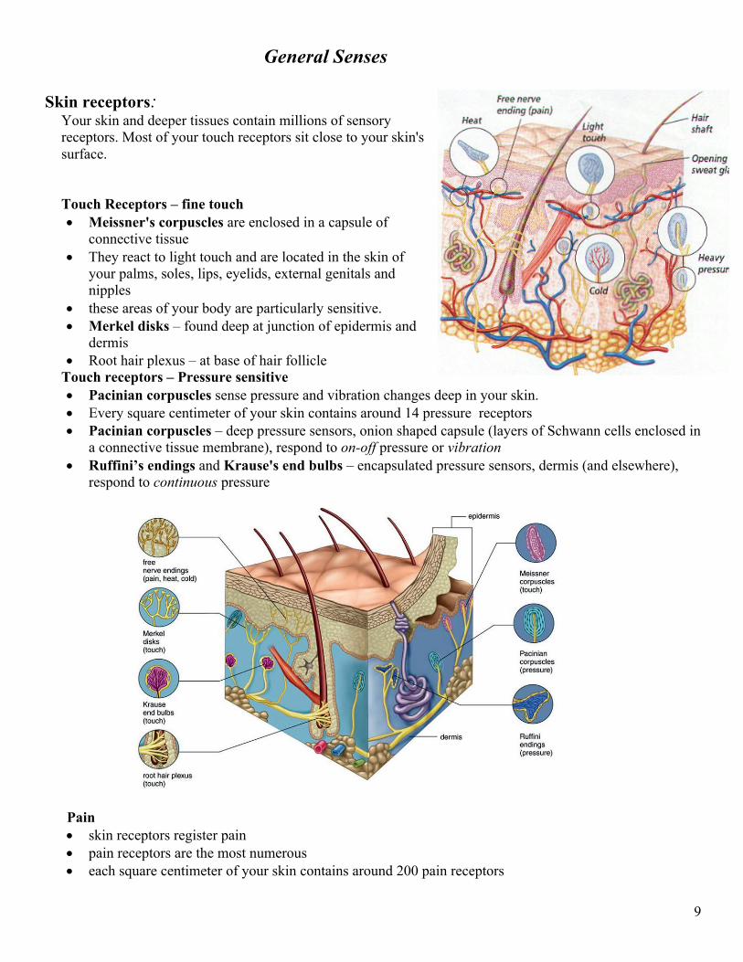
General Senses Skin receptors:
Your skin and deeper tissues contain millions of sensory receptors. Most of your touch receptors sit close to your skin's surface.
Touch Receptors – fine touch • Meissner's corpuscles are enclosed in a capsule of
connective tissue • They react to light touch and are located in the skin of
your palms, soles, lips, eyelids, external genitals and nipples
• these areas of your body are particularly sensitive. • Merkel disks – found deep at junction of epidermis and
dermis • Root hair plexus – at base of hair follicle
Touch receptors – Pressure sensitive • Pacinian corpuscles sense pressure and vibration changes deep in your skin. • Every square centimeter of your skin contains around 14 pressure receptors • Pacinian corpuscles – deep pressure sensors, onion shaped capsule (layers of Schwann cells enclosed in
a connective tissue membrane), respond to on-off pressure or vibration • Ruffini’s endings and Krause's end bulbs – encapsulated pressure sensors, dermis (and elsewhere),
respond to continuous pressure
Pain • skin receptors register pain • pain receptors are the most numerous • each square centimeter of your skin contains around 200 pain receptors
10
Temperature • skin receptors register warmth and cold • each square centimeter of your skin contains 6 receptors for cold and 1 receptor for warmth • Cold receptors start to perceive cold sensations when the surface of the skin drops below 95 º F. They
are most stimulated when the surface of the skin is at 77 º F and are no longer stimulated when the surface of the skin drops below 41 º F. This is why your feet or hands start to go numb when they are submerged in icy water for a long period of time.
• Hot receptors start to perceive hot sensations when the surface of the skin rises above 86 º F and are most stimulated at 113 º F. Beyond 113 º F, pain receptors take over to avoid damage being done to the skin and underlying tissues.
• thermoreceptors are found all over the body, but cold receptors are found in greater density than heat receptors – most of the time of our environment is colder than our body temperature
• The highest concentration of thermoreceptors can be found in the face and ears so your nose and ears always get colder faster than the rest of your body on a chilly winter day
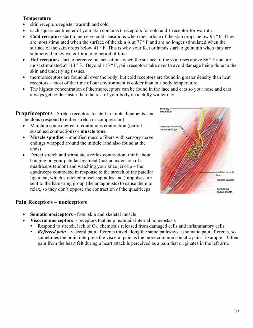
Proprioceptors - Stretch receptors located in joints, ligaments, and tendons (respond to either stretch or compression) • Maintain some degree of continuous contraction (partial
sustained contraction) or muscle tone • Muscle spindles – modified muscle fibers with sensory nerve
endings wrapped around the middle (and also found at the ends)
• Detect stretch and stimulate a reflex contraction; think about banging on your patellar ligament (just an extension of a quadriceps tendon) and watching your knee jerk up – the quadriceps contracted in response to the stretch of the patellar ligament, which stretched muscle spindles and ) impulses are sent to the hamstring group (the antagonists) to cause them to relax, so they don’t oppose the contraction of the quadriceps
Pain Receptors – nociceptors
• Somatic nociceptors - from skin and skeletal muscle • Visceral nociceptors - receptors that help maintain internal homeostasis
§ Respond to stretch, lack of O2, chemicals released from damaged cells and inflammatory cells. § Referred pain – visceral pain afferents travel along the same pathways as somatic pain afferents, so
sometimes the brain interprets the visceral pain as the more common somatic pain. Example – Often pain from the heart felt during a heart attack is perceived as a pain that originates in the left arm.