sem and tem for identification of capsular fibrosis and
TRANSCRIPT

METHODOLOGY ARTICLE Open Access
SEM and TEM for identification of capsularfibrosis and cellular behavior around breastimplants – a descriptive analysisBritta Kuehlmann1,2* , Isabel Zucal2, Clark Andrew Bonham1, Lydia-Marie Joubert3 and Lukas Prantl2
Abstract
Background: Capsular fibrosis (CF) is the most common long-term complication in implant-based breastaugmentation. It is well accepted that the foreign body response (FBR) instigates the development of fibroticdisease. Our study aims to compare murine and human samples of CF and describe the cellular and extracellularmatrix (ECM) composition using scanning and transmission electron microscopy (SEM and TEM).
Results: Miniature microtextured silicone breast implants were implanted in mice and subsequently harvested atdays 15, 30, and 90 post-operation. Isolated human capsules with the most aggravated form of CF (Baker IV) wereharvested post-operation. Both were analyzed with SEM and TEM to assess cellular infiltration and ECM structure.An architectural shift of collagen fiber arrangement from unidirectional to multidirectional was observed at day 90when compared to days 15 and 30. Fibrosis was observed with an increase of histiocytic infiltration. Moreover,bacterial accumulation was seen around silicone fragments. These findings were common in both murine andhuman capsules.
Conclusions: This murine model accurately recapitulates CF found in humans and can be utilized for futureresearch on cellular invasion in capsular fibrosis. This descriptive study helps to gain a better understanding ofcellular mechanisms involved in the FBR. Increases of ECM and cellularity were observed over time with SEM andTEM analysis.
Keywords: Breast implants, Foreign body response, Capsular fibrosis, Cellular behavior, Scanning electronmicroscopy, Transmission electron microscopy
BackgroundImplant-based breast augmentation is the number oneplastic surgery procedure performed worldwide, withover 50% of breast augmentation patients chose to sub-sequently replace their removed breast implants withnew ones [1]. Explantation is most often due to capsular
fibrosis (CF), a commonplace long-term complication. Infact, studies report a prevalence of CF of 2.8 to 20.4%[2–8]. The degree of severity is clinically classified by theBaker score system which includes four stages [9, 10].The first stage is the least severe and is characterized bya lack of clinical symptoms, while the fourth stage repre-sents the most severe form of CF characterized by breastdeformity, stiffness, and pain. Moreover, the severitygrade is histologically classified by Wilflingseder et al.:stage I is a thin and uncontracted capsule, stage II ischaracterized by constrictive fibrosis and absence ofgiant cells, stage III displays constrictive fibrosis with ac-cumulation of giant cells, and stage IV is characterized
© The Author(s). 2021, corrected publication 2021. Open Access This article is licensed under a Creative Commons Attribution4.0 International License, which permits use, sharing, adaptation, distribution and reproduction in any medium or format, aslong as you give appropriate credit to the original author(s) and the source, provide a link to the Creative Commons licence,and indicate if changes were made. The images or other third party material in this article are included in the article's CreativeCommons licence, unless indicated otherwise in a credit line to the material. If material is not included in the article's CreativeCommons licence and your intended use is not permitted by statutory regulation or exceeds the permitted use, you will needto obtain permission directly from the copyright holder. To view a copy of this licence, visit http://creativecommons.org/licenses/by/4.0/. The Creative Commons Public Domain Dedication waiver (http://creativecommons.org/publicdomain/zero/1.0/) applies to the data made available in this article, unless otherwise stated in a credit line to the data.
* Correspondence: [email protected]; [email protected] of Plastic and Reconstructive Surgery, Department of Surgery,Stanford University, Stanford, CA 94305, USA2University Center for Plastic, Reconstructive, Aesthetic and Hand Surgery,University Hospital Regensburg and Caritas Hospital St. Josef, 93053Regensburg, GermanyFull list of author information is available at the end of the article
BMC Molecular andCell Biology
Kuehlmann et al. BMC Molecular and Cell Biology (2021) 22:25 https://doi.org/10.1186/s12860-021-00364-8

by infiltration of inflammatory cells, foreign body granu-lomas, and neovascularization [11]. Studies found thatmacrophages and Staphylococcus epidermidis withincapsules were often associated with CF, though bacterialcolonization is likely a promoting factor rather than thecause of it [12].The pathogenesis of CF has been widely studied and it
is thought that the underlying mechanism is a fibroticforeign body reaction brought about by inflammatorysignaling. TNF-α production has been observed in theprogression of CF, where it is expressed primarily incollagen-depositing fibroblasts and macrophages aroundthe implant [13]. Conversely, less severe cases of CF ex-hibit far fewer bundles of collagen [14]. However, accur-ate implant and capsular surface analysis of bothcollagen fiber arrangement and cellular components arescarce. Implant surface texture and volume influencehost tissue responses and the development of CF [15,16]. Atlan et al. classified the surface texture intosmooth/nanotexture (80–100 mm2), microtexture (100 -200mm2), macrotexture (200 - 300mm2), andmacrotexture-plus (> 300mm2) when using scanningelectron microscopy (SEM) [17].In this descriptive study, capsules of a microtextured
interface were examined using SEM, TEM, and light mi-croscopy. We aimed to analyze the effect of commonlyused silicone implants on capsule formation. Humancapsules were taken from patients with aggravated CF(Baker IV). We sought to identify a murine model forCF that might be more suitable for the characterizationof human CF than current small animal models. Cus-tomized, miniature silicone implants for mice were usedto more accurately recapitulate human-like CF in a mur-ine setting.
ResultsSEM analysisHuman capsules were explanted at 5 to 9 years after im-plantation, while murine capsules were harvested at days60 and 90 to ensure adequate fibrotic development.Explanted implant surfaces displayed progressive fi-
brotic accumulation that increased with time Fig. 1a, b.Capsule accumulation was especially notable within theconcavities of textured implants (Fig. 1c, e). Interest-ingly, detached silicone particles and the presence ofcoccoid bacteria could be found as early as day 30 (Fig.1d, f). Finally, Fig. 1e shows accumulation of white bloodcells in the surface’s concavity. This observation maysupport the hypothesis that breast implant associatedanaplastic large cell lymphoma is associated with tex-tured implants and enhanced T-cell response [18].Further assessment revealed that murine capsules dis-
played pervasive fiber-like structures in a similar fashionto that of human samples. SEM images of the murine
capsules verified an increase in ECM over time, accom-panied by an architectural shift of collagen fibers fromunidirectional at days 15 and 30, to a more disorganizedfashion by day 90 (Fig. 2a, b). Further, erythrocytes wereobserved in greater numbers at day 90 when comparedto day 15, whereas fewer white blood cells were presentat day 90 compared to day 15, suggesting a dissolutionof acute inflammation over time (Fig. 2c, d).SEM images of both murine sections and human cap-
sules revealed a progressive increase in ECM and cellu-larity within the capsules, with the cellular gradientincreasing towards the implant surface at later timepoints. Cell morphology was comparable in both murineand human models, with the number of histiocytes in-creasing at later time points in both murine and humancapsules, correlating with severe fibrotic deposition(Fig. 3a, b). Furthermore, silicone fragments found incapsules at later stages of fibrosis were characterized byaccumulations of coccoid bacteria (Fig. 3c). Interestingly,a biofilm fragment of rod-shaped bacteria was foundamong multidirectional collagen fibers in a mouse modelat day 90 (Fig. 3d).
Light microscopyImmediately prior to TEM analysis, light microscopy ofhuman and murine capsular sections was performed.These images displayed a comparable histology of CF inhumans, Baker IV and in mice at day 90. Both displayeda collagenous connective tissue-core with several fibro-blasts and dispersed smooth muscle cells (Fig. 4). Bloodvessels were present in the capsules and the tissue layerat the inner surface of the capsule was characterized bylymphocytic infiltration (Fig. 4).
TEM analysisTEM images of murine sections showed an increaseof collagen concentration at day 90 compared to day15. Furthermore, at day 15, collagen fibers were orga-nized in bundles, whereas at day 90, fibers weremultidirectional (Fig. 5). An increase of collagen as anindicator of progressive fibrosis was observed at day30 compared to day 15 and collagen arrangement wascomparable to day 90.
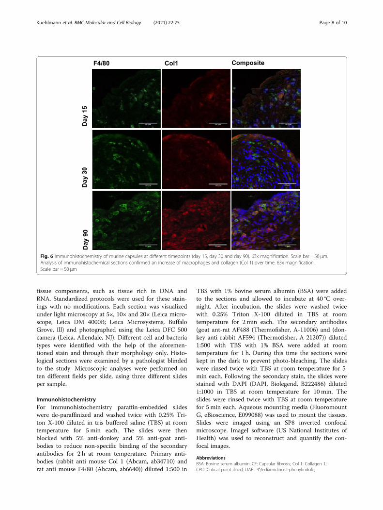
Immunohistochemistry analysisImmunohistochemistry (IHC) was performed to identifycellularity and ECM deposition in murine capsules atdifferent timepoints (days 15, 30 and 90). IHC confirmedan increase of macrophages and an increase of Collagendeposition (Col 1) over time (Fig. 6).
DiscussionThe implantable device market is rapidly expandingaround the world. Since their introduction, the
Kuehlmann et al. BMC Molecular and Cell Biology (2021) 22:25 Page 2 of 10

composition of breast implants has been ceaselessly al-tered in attempts to maximize biocompatibility and re-duce complications. In 1992, the FDA restricted the useof silicone implants for cosmetic purposes, but due toinsufficient evidence linking such implants to disease,their use in breast augmentation was reapproved in 2006[19]. CF persists as the most common long-term compli-cation of breast implants regardless of material compos-ition [2–7], bringing about a need for further studies todetermine its pathology.Implant surface texture has been shown to influence
the development of CF [11, 20]. In recent years, an em-phasis has been placed on developing biomimetic im-plant surfaces to reduce the foreign body reaction. Forexample, a novel polydimethysiloxane implant surface
was described in a previous study, imitating adipose tis-sue [21]. As a result, pro-inflammatory genes includingILβ1, TNF-α and IL6 were significantly downregulated.Furthermore, reduced fibroblast and macrophage infil-tration was observed in immunocytochemistry and SEM,indicative of a diminished inflammatory response to theforeign body. Here, macrophages were less prevalentcompared to original adipose and smooth surfaces.Moreover, fibroblasts were less aligned in the valleys ofthe surface in modelled adipose surface with secondarytexture compared to the original adipose surface withprimary texture [21, 22]. Our experiments confirm thatlarger implantable materials play a crucial role in elicit-ing a more dramatic and human-like fibrosis in a murinemodel than those that are smaller, thus making this
Fig. 1 SEM figures of explanted breast implants at day 15 and 30 in mice. a and b Explanted breast implants with surrounding capsular tissue inmurine models are shown at days 15 and 30. c and d Capsular fragments accumulated in the concavities of the textured implants are shown atdays 15 and 30, respectively. e White blood cell accumulation (especially macrophages and lymphocytes) in an implant’s pore is presented at day15. f Coccoid bacterial accumulation (white arrows) on a murine capsule of day 30 is shown
Kuehlmann et al. BMC Molecular and Cell Biology (2021) 22:25 Page 3 of 10

modified model more suitable than others to study themechanisms of human CF. Such comparisons betweenthis model and human CF are corroborated by similar-ities in cellular infiltrate and ECM composition.In our study, a descriptive analysis of cellularity and
collagen arrangement using SEM, light microscopy andTEM was performed. SEM permits topographicalvisualization and detailed surface examination of solidspecimens. Samples are bombarded with electrons thatbounce back once they reach their surface, providing in-depth, 3D models [23–25]. Transmission electron mi-croscopy (TEM) is generally used in diagnostic path-ology and provides high resolution images (up to 0.2nm). This allows for the visualization of small intra- andextracellular structures, such as cell organelles, cellularinclusions, microtubules, microfilaments, and intermedi-ate filaments, as well as collagen and amyloids [26].Microtextured implants were examined, displaying an
accumulation of capsular fragments, particularly withinthe implants’ concavities. Early presence of detached sili-cone pieces and bacteria at day 30 was observed. Thepresence of silicone fragments in capsular tissue hasbeen shown to correlate with greater capsular thickness[11]. In our study, we found significant accumulation ofcoccoid bacteria around these fragments, with rod
shaped bacteria found in one specimen. The presence ofbacteria in CF has been largely studied in the past, withstudies suggesting bacterial colonization could play animportant role in the pathogenesis of CF [12, 27, 28]and breast implant associated anaplastic large celllymphoma (BIA-ALCL) by stimulating an enhanced T-cell response [29–32].In the cellular analysis, our findings show an increase
in histiocytes over time, in agreement with previousstudies as an indicator of ongoing fibrosis [33]. Accord-ing to Siggelkow et al., histiocytic inflammation is morecommon in patients with clinical symptoms (p < 0.001)and around subglandular implants (p < 0.096) [34]. Inanother study, CD68 positive histiocytes were found tobe increased in the presence of siliconomas [35].Histological analysis of the capsular layers in a previ-
ous study further suggested the presence of synovialmetaplasia within the inner layer, particularly in healthy,uncontracted Baker I capsules, suggesting a protectivefactor [36]. The middle layer has been observed in linewith the capsular border, while the outer layer is looselyarranged [36]. In our histological analysis, we could con-firm this type of arrangement in three layers.Previous studies have identified notable collagen fiber
alignment in progressive instances of CF. Highly aligned
Fig. 2 SEM images on collagen arrangement and cellularity at day 15 and 90 in mice. a Collagen fiber alignment at day 15 is unidirectional andorganized in bundles. b At day 90, collagen arrangement appears to be disorganized with loose and multidirectional collagen fibers. c Day 15maintains poor cellularity and is characterized by the presence of white blood cells (white arrows). d Cellularity at day 90 shows an increase inerythrocytes (white triangles), whereas white blood cells (white arrows) appear to be reduced in comparison to day 15
Kuehlmann et al. BMC Molecular and Cell Biology (2021) 22:25 Page 4 of 10

fibers are found in more contracted capsules, whereasthey are loosely arranged and multidirectional in uncon-tracted capsules [37]. This, however, cannot be con-firmed by our findings that show more aligned fibers indays 15 and 30 when compared to day 90, in which thefibers are arranged in a disordered fashion. However,both in vivo and in vitro studies have determined thatcollagen organization is dependent on the substrate ma-terial [38, 39]. TEM analysis showed an increase of colla-gen fiber-concentration in the ECM at day 90 comparedto day 15. Also, previous studies reported enhanced col-lagen secretion by fibroblasts and myofibroblasts, afteracute and chronic inflammation characterized by neu-trophils’ and macrophages’ infiltration respectively,dissolved.
ConclusionsUsing this murine model, we were able to confirm thefindings of previous studies and further characterize thedevelopment of CF at a structural level. When analyzingCF at days 15, 30, and 90, we observed a progressive in-crease in collagen fibers underlying a structural shiftfrom unidirectional to multidirectional. Further, erythro-cytes and histiocytes accumulated over time, suggesting
ongoing fibrosis and vascularization of the capsule in ad-vanced stages. Finally, bacterial infiltration was presentat all stages in time and was observed to gather aroundsilicone particles and collagen fibers. These findings arerelevant as they provide an optimized murine model thatallows for better comparison of human CF and mayserve as a basis in future studies to develop enhancedbiocompatible materials and reduce CF.
MethodsThis experimental study was performed at the Divisionof Plastic and Reconstructive Surgery, Department ofSurgery, Stanford University, USA. Thirty murine andten human capsules (both of female gender, respectively)around microtextured breast implants were examinedwith SEM, light microscopy, and TEM at different pointsin time: day 15, day 30, and day 90 in murine capsulesand Baker IV in human capsules. All mice received cus-tomized textured, gel filled silicone implants (PolytechHealth and Aesthetic, Dieburg, Germany; gel filled, poresize range of 50–900 μm, 2 cm in diameter). Murine im-plants were placed in a subcutaneous pocket. Human cap-sules were explanted around microtextured breastimplants from an epipectoral pocket. Detailed information
Fig. 3 SEM images of human CF (Baker IV) and murine CF at day 60 and 90. a Human capsules, Baker IV (human capsules were extracted more than 5to 9 years after implantation) and b murine capsules are shown at days 60 and 90, respectively. Comparable cell infiltration is shown: Both capsulescontained coccoid bacteria and a comparable dispersion of blood cells (white blood cells are marked with white triangles), as well as histiocytes (whitearrows), that were not found in early stages of CF. c The presence of coccoid bacteria (white arrow) around dispersed silicone particles is shown. d Inone murine capsule, a biofilm-fragment of rod-shaped bacteria is shown in the middle of the multidirectional arranged fibers
Kuehlmann et al. BMC Molecular and Cell Biology (2021) 22:25 Page 5 of 10

about each implants’ manufacturer and generation wasnot available retrospectively which is a potential limitationof this study. Cell invasion and collagen fiber alignmentwere assessed.
AnimalsAll animals were treated humanely, and protocols usedwere approved a priori by Institutional Animal Care andUse Committee at Stanford University (IACUC) andStanford University’s Administrative Panel on Labora-tory Animal Care (Protocol No. 28410) according to Na-tional Institutes of Health and institutional guidelines.Six-week old female wild-type C57BL/6 mice were ob-tained from The Jackson Laboratory (Bar Harbor, ME,USA). All mice were housed in sterile micro-insulators.Food and water were provided ad libitum in accordancewith institutional guidelines of Stanford University ani-mal care. Ten mice per group were used per each ex-periment and the experiments were run three times toverify findings.
Human breast tissueHuman breast capsules were received in FFPE (FormalinFixed Paraffin Embedded) blocks from the Institute of
Pathology, University Hospital Regensburg, Germany.All human capsules examined were formed aroundmicrotextured implants to guarantee structural compari-son for this study. Approval was given by the local ethiccommittee in Regensburg (Reference No.: 15–101-0024).
Scanning electron microscopy (SEM)For SEM analysis, specimens (breast implants andexplanted capsular samples) were rinsed in PBS, fixedovernight in 4% Paraformaldehyde with 2% Glutaralde-hyde in 0.1M Sodium Cacodylate Buffer (pH 7.4), rinsedin the same buffer and post-fixed for 1 h with 1% aque-ous OsO4. After dehydration in an ascending ethanolseries (50, 70, 90, 100% (twice); 5 min each), small tissue(capsule) pieces were critical point dried (CPD) with li-quid CO2 in a Tousimis Autosamdri-815B apparatus(Tousimis, Rockville, MD), while implants whichexceeded the CPD chamber size were treated withHMDS (hexamethyldisilazane) for 30 min (2 × 15min),before overnight drying in a desiccator. All specimenswere mounted onto 15 - 50 mm circular aluminum stubs(Electron Microscopy Sciences, Hatfield, PA), andsputter-coated with 50 Å of gold-palladium using a Den-ton Desk II Sputter Coater (Denton Vacuum,
Fig. 4 Light microscopy images of human CF (Baker IV) and murine capsules at day 30 and 90. Light microscopy (toluidine blue staining) ofhuman capsular sections, Baker IV (a and c) are compared to murine capsular sections at day 90 (b and d). The capsular surface directed to theimplant (inner surface) is indicated with (i). Comparable histology was found: The capsules are characterized by collagenous connective tissuebuilding the core of the capsule and the presence of smooth muscle cells with cigar-shaped nuclei, as well as fibroblasts (arrows). Around theouter surface of the capsules, adipose tissue (crosses) and blood vessels (asterisks) can be noticed. Blood vessels are present within the capsule aswell (c). The capsular layer close to the inner surface is further characterized by lymphocytic infiltration (dark, round nuclei filled with chromatin)
Kuehlmann et al. BMC Molecular and Cell Biology (2021) 22:25 Page 6 of 10

Moorestown, NJ). Scanning Electron Microscopy (SEM)images captured with either a Hitachi S3400-N VariablePressure SEM (Hitachi High-Tech, Dallas, TX) operatedat 10 kV accelerating voltage and using secondary elec-tron detection or a Zeiss Sigma Field Emission SEM(Carl Zeiss Microscopy, Pleasanton, CA) operated at 3-5kV accelerating voltage and using InLens SecondaryElectron (SE) and Everhard Thornly (SE2) detection.While SEM procedure can cause dehydration artifactsthat could negatively impact the tissue processing, hist-ology with respective SEM images of the same sectionswas performed to ensure that the collagen alignmentand hydration state match the fibrotic profile observedin tissue.
Transmission electron microscopy (TEM)Samples were fixed in Karnovsky’s fixative: 2% Glutaral-dehyde (EMS Cat# 16000) and 4% Formaldehyde (EMSCat# 15700) in 0.1 M Sodium Cacodylate (EMS Cat#12300), pH 7.4, for 1 h, chilled, and sent to Stanford’sCSIF on ice. They were then warmed to roomtemperature (RT) in cold 1% Osmium tetroxide (EMSCat# 19100) for 1 h while rotating in a hood, washed 3xwith ultrafiltered water, then en bloc stained overnightin 1% Uranyl Acetate at 4 °C while rotating. Sampleswere then dehydrated in a series of ethanol washes for30 min each at 4 °C beginning at 50, 70, 95% where thesamples were then allowed to rise to RT, changed to
100% twice, then Propylene Oxide (PO) was applied for15 min. Samples were infiltrated with EMbed-812 resin(EMS Cat#14120) mixed 1:2, 1:1, and 2:1 with PO for 2h each and were subsequently left in 2:1 resin to POovernight, rotating at RT in a hood. Samples were thenplaced into EMbed-812 for 2 to 4 h before being placedinto molds with labels and fresh resin, oriented, andplaced into 65 °C oven overnight.Sections were taken between 75 and 90 nm, picked up
on formvar/Carbon coated slot Cu grids, stained for 30 sin 3.5% Uranyl Acetate/ 50% Acetone, followed by stain-ing in 0.2% Lead Citrate for 3 min. Sections were ob-served in the JEOL JEM-1400 120 kV with photos beingtaken with a Gatan Orius 832 4 k X 2.6 k digital camerawith 9 μm pixel size.
HistologyMice were sacrificed with CO2 asphyxiation and cervicaldislocation. Murine capsule (tissue) was harvested onday 15, day 30 and day 90 after placing the silicone im-plant and fixed in 4% paraformaldehyde overnight at40 °C. The samples were fixed in 4% paraformaldehydein phosphate buffered solution/saline (PBS) overnight,washed twice with PBS and dehydrated in a 30% sucrosesolution for 24 h. Samples were processed routinely andembedded in paraffin. Sections were cut at 1 μm and3 μm serially for histology staining. Sections were stainedwith toluidine blue, which has a high affinity for acidic
Fig. 5 TEM images about collagen fibers in murine capsules at day 15 and 90. TEM photos of murine capsules at day 15 (a and c) and day 90 (band d) are presented. a and c The collagen fibers around the fibroblast are not as concentrated and appear to be organized in bundles (blackdashed line indicated with arrows in (c)) at early stages of CF. b and d As fibrosis goes on, the concentration of collagen fibers seems to increasewith a random-pattern arrangement
Kuehlmann et al. BMC Molecular and Cell Biology (2021) 22:25 Page 7 of 10
tissue components, such as tissue rich in DNA andRNA. Standardized protocols were used for these stain-ings with no modifications. Each section was visualizedunder light microscopy at 5×, 10× and 20× (Leica micro-scope, Leica DM 4000B; Leica Microsystems, BuffaloGrove, Ill) and photographed using the Leica DFC 500camera (Leica, Allendale, NJ). Different cell and bacteriatypes were identified with the help of the aforemen-tioned stain and through their morphology only. Histo-logical sections were examined by a pathologist blindedto the study. Microscopic analyses were performed onten different fields per slide, using three different slidesper sample.
ImmunohistochemistryFor immunohistochemistry paraffin-embedded slideswere de-paraffinized and washed twice with 0.25% Tri-ton X-100 diluted in tris buffered saline (TBS) at roomtemperature for 5 min each. The slides were thenblocked with 5% anti-donkey and 5% anti-goat anti-bodies to reduce non-specific binding of the secondaryantibodies for 2 h at room temperature. Primary anti-bodies (rabbit anti mouse Col 1 (Abcam, ab34710) andrat anti mouse F4/80 (Abcam, ab6640)) diluted 1:500 in
TBS with 1% bovine serum albumin (BSA) were addedto the sections and allowed to incubate at 40 °C over-night. After incubation, the slides were washed twicewith 0.25% Triton X-100 diluted in TBS at roomtemperature for 2 min each. The secondary antibodies(goat ant-rat AF488 (Thermofisher, A-11006) and (don-key anti rabbit AF594 (Thermofisher, A-21207)) diluted1:500 with TBS with 1% BSA were added at roomtemperature for 1 h. During this time the sections werekept in the dark to prevent photo-bleaching. The slideswere rinsed twice with TBS at room temperature for 5min each. Following the secondary stain, the slides werestained with DAPI (DAPI, Biolegend, B222486) diluted1:1000 in TBS at room temperature for 10 min. Theslides were rinsed twice with TBS at room temperaturefor 5 min each. Aqueous mounting media (FluoromountG, eBioscience, E099088) was used to mount the tissues.Slides were imaged using an SP8 inverted confocalmicroscope. ImageJ software (US National Institutes ofHealth) was used to reconstruct and quantify the con-focal images.
AbbreviationsBSA: Bovine serum albumin; CF: Capsular fibrosis; Col 1: Collagen 1;CPD: Critical point dried; DAPI: 4′,6-diamidino-2-phenylindole;
Fig. 6 Immunohistochemistry of murine capsules at different timepoints (day 15, day 30 and day 90). 63x magnification. Scale bar = 50 μm.Analysis of immunohistochemical sections confirmed an increase of macrophages and collagen (Col 1) over time. 63x magnification.Scale bar = 50 μm
Kuehlmann et al. BMC Molecular and Cell Biology (2021) 22:25 Page 8 of 10

DNA: Deoxyribonucleic acid; ECM: Extracellular matrix; FBR: Foreign bodyresponse; FFPE: Formalin fixed paraffin embedded;HMDS: Hexamethyldisilazane; IHC: Immunohistochemistry; ILβ1: Interleukin 1beta; IL6: Interleukin 6; M: Molar mass; mm: Millimeter; nm: Nanometer;PBS: Phosphate buffered solution/saline; PO: Propylene oxide; RT: Roomtemperature; SE: Secondary electron; SEM: Scanning electron microscopy;TBS: Tris buffered saline; TEM: Transmission electron microscopy; TNF-α: Tumor necrosis factor alpha
AcknowledgementsThe authors thank John J. Perrino and Ibanri Phanwar-Wood for their helpwith processing the TEM samples.
Authors’ contributionsB.K. conceived the project, designed, and performed the experiments. B.K.analyzed and interpreted the presented data. B.K., L.M.J. and C.A.B. performedthe histopathological and microscopical examination of the samples. B.K. andI.Z. wrote the manuscript. L.P. contributed to oversight of and advice on theoverall project. All authors read and approved the final manuscript.
FundingThis research did not receive any specific grant from funding agencies in thepublic, commercial, or not-for-profit sectors. Open Access funding enabledand organized by Projekt DEAL
Availability of data and materialsThe datasets used and/or analyzed during the current study are availablefrom the corresponding author on reasonable request.
Declarations
Ethics approval and consent to participateAll animals were treated humanely, and protocols used were approved apriori by Institutional Animal Care and Use Committee at Stanford University(IACUC) and Stanford University’s Administrative Panel on Laboratory AnimalCare (Protocol No. 28410) according to National Institutes of Health andinstitutional guidelines. Human breast capsules were received in FFPE(Formalin Fixed Paraffin Embedded) blocks from the Institute of Pathology,University Hospital Regensburg, Germany. Approval was given by the localethic committee in Regensburg (Reference No.: 15–101-0024).We have obtained consent to publish from the participant to reportindividual patient data.
Consent for publicationEach participant has given written informed consent for publication.
Competing interestsThe authors declare that they have no competing interests.
Author details1Division of Plastic and Reconstructive Surgery, Department of Surgery,Stanford University, Stanford, CA 94305, USA. 2University Center for Plastic,Reconstructive, Aesthetic and Hand Surgery, University Hospital Regensburgand Caritas Hospital St. Josef, 93053 Regensburg, Germany. 3Stanford-SLACCryo-EM Center, Stanford University, Stanford, CA 94305, USA.
Received: 21 December 2020 Accepted: 15 April 2021
References1. ASAPS. Cosmetic Surgery National Data Bank Statistics for 2018. Available at:
https://www.surgery.org/media/statistics. Last update 14th May 2020.2. Spear SL, Murphy DK, A.S.B.I.U.S.C.C.S. Group. Natrelle round silicone breast
implants: core study results at 10 years. Plast Reconstr Surg. 2014;133(6):1354–61. https://doi.org/10.1097/PRS.0000000000000021.
3. Blount AL, Martin MD, Lineberry KD, Kettaneh N, Alfonso DR. Capsularcontracture rate in a low-risk population after primary augmentationmammaplasty. Aesthet Surg J. 2013;33(4):516–21. https://doi.org/10.1177/1090820X13484465.
4. Stutman RL, Codner M, Mahoney A, Amei A. Comparison of breastaugmentation incisions and common complications. Aesthet Plast Surg.2012;36(5):1096–104. https://doi.org/10.1007/s00266-012-9918-x.
5. Codner MA, Mejia JD, Locke MB, Mahoney A, Thiels C, Nahai FR, et al. A 15-year experience with primary breast augmentation. Plast Reconstr Surg.2011;127(3):1300–10. https://doi.org/10.1097/PRS.0b013e318205f41b.
6. Sevin A, Sevin K, Senen D, Deren O, Adanali G, Erdogan B. Augmentationmammaplasty: retrospective analysis of 210 cases. Aesthet Plast Surg. 2006;30(6):651–4. https://doi.org/10.1007/s00266-006-0076-x.
7. Gutowski KA, Mesna GT, Cunningham BL. Saline-filled breast implants: a PlasticSurgery Educational Foundation multicenter outcomes study. Plast ReconstrSurg. 1997;100(4):1019–27. https://doi.org/10.1097/00006534-199709001-00028.
8. Kuehlmann B, Burkhardt R, Kosaric N, Prantl L. Capsular fibrosis inaesthetic and reconstructive-cancer patients: a retrospective analysis of319 cases. Clin Hemorheol Microcirc. 2018;70(2):191–200. https://doi.org/10.3233/CH-170365.
9. Spear SL, Baker JL Jr. Classification of capsular contracture after prostheticbreast reconstruction. Plast Reconstr Surg. 1995;96(5):1119–23; discussion1124. https://doi.org/10.1097/00006534-199510000-00018.
10. de Bakker E, Rots M, Buncamper ME, Niessen FB, Smit JM, Winters HAH,et al. The Baker classification for capsular contracture in breast implantsurgery is unreliable as a diagnostic tool. Plast Reconstr Surg. 2020;146(5):956–62. https://doi.org/10.1097/PRS.0000000000007238.
11. Prantl L, Schreml S, Fichtner-Feigl S, Pöppl N, Eisenmann-Klein M, SchwarzeH, et al. Clinical and morphological conditions in capsular contractureformed around silicone breast implants. Plast Reconstr Surg. 2007;120(1):275–84. https://doi.org/10.1097/01.prs.0000264398.85652.9a.
12. Bachour Y, Verweij SP, Gibbs S, Ket JCF, Ritt MJPF, Niessen FB, et al. Theaetiopathogenesis of capsular contracture: a systematic review of theliterature. J Plast Reconstr Aesthet Surg. 2018;71(3):307–17. https://doi.org/10.1016/j.bjps.2017.12.002.
13. Tan KT, Wijeratne D, Shih B, Baildam AD, Bayat A. Tumour necrosis factor-αexpression is associated with increased severity of periprosthetic breastcapsular contracture. Eur Surg Res. 2010;45(3–4):327–32. https://doi.org/10.1159/000321009.
14. Moyer KE, Ehrlich HP. Capsular contracture after breast reconstruction:collagen fiber orientation and organization. Plast Reconstr Surg. 2013;131(4):680–5. https://doi.org/10.1097/PRS.0b013e31828189d0.
15. Dutta S, Sengupta P. Men and mice: relating their ages. Life Sci. 2016;152:244–8. https://doi.org/10.1016/j.lfs.2015.10.025.
16. Brigaud I, et al. Surface texturization of breast implants impacts extracellularmatrix and inflammatory gene expression in asymptomatic capsules. PlastReconstr Surg. 2020;145(3):542e–51e.
17. Atlan M, Nuti G, Wang H, Decker S, Perry TA. Breast implant surface textureimpacts host tissue response. J Mech Behav Biomed Mater. 2018;88:377–85.https://doi.org/10.1016/j.jmbbm.2018.08.035.
18. Turner SD, Inghirami G, Miranda RN, Kadin ME. Cell of origin andimmunologic events in the pathogenesis of breast implant-associatedanaplastic large-cell lymphoma. Am J Pathol. 2020;190(1):2–10. https://doi.org/10.1016/j.ajpath.2019.09.005.
19. Fardo D, Campos MS, Pensler JM. Breast Augmentation. In: StatPearls[Internet]. Treasure Island: StatPearls Publishing; 2021.
20. Poeppl N, Schreml S, Lichtenegger F, Lenich A, Eisenmann-Klein M, Prantl L.Does the surface structure of implants have an impact on the formation ofa capsular contracture? Aesthet Plast Surg. 2007;31(2):133–9. https://doi.org/10.1007/s00266-006-0091-y.
21. Barr S, Hill EW, Bayat A. Development, fabrication and evaluation of a novelbiomimetic human breast tissue derived breast implant surface. ActaBiomater. 2017;49:260–71. https://doi.org/10.1016/j.actbio.2016.11.052.
22. Kyle DJ, et al. Development and functional evaluation of biomimeticsilicone surfaces with hierarchical micro/nano-topographical featuresdemonstrates favourable in vitro foreign body response of breast-derivedfibroblasts. Biomaterials. 2015;52:88–102. https://doi.org/10.1016/j.biomaterials.2015.02.003.
23. Fischer ER, et al. Scanning electron microscopy. Curr Protoc Microbiol. 2012;Chapter 2:Unit 2B.2.
24. Nguyen JNT, Harbison AM. Scanning electron microscopy samplepreparation and imaging. Methods Mol Biol. 2017;1606:71–84. https://doi.org/10.1007/978-1-4939-6990-6_5.
25. Jones CG. Scanning electron microscopy: preparation and imaging for SEM.Methods Mol Biol. 2012;915:1–20. https://doi.org/10.1007/978-1-61779-977-8_1.
Kuehlmann et al. BMC Molecular and Cell Biology (2021) 22:25 Page 9 of 10

26. Erlandson R. Diagnostic transmission electron microscopy, withclinicopathological, immunohistichemical, and cytogenetic correlations. NeYork: Raven Press; 1994.
27. Bachour Y, Bargon CA, de Blok CJM, Ket JCF, Ritt MJPF, Niessen FB. Riskfactors for developing capsular contracture in women after breast implantsurgery: a systematic review of the literature. J Plast Reconstr Aesthet Surg.2018;71(9):e29–48. https://doi.org/10.1016/j.bjps.2018.05.022.
28. Ajdic D, Zoghbi Y, Gerth D, Panthaki ZJ, Thaller S. The relationship ofbacterial biofilms and capsular contracture in breast implants. Aesthet SurgJ. 2016;36(3):297–309. https://doi.org/10.1093/asj/sjv177.
29. Walker JN, Hanson BM, Pinkner CL, Simar SR, Pinkner JS, Parikh R, et al.Insights into the microbiome of breast implants and periprosthetic tissue inbreast implant-associated anaplastic large cell lymphoma. Sci Rep. 2019;9(1):10393. https://doi.org/10.1038/s41598-019-46535-8.
30. Hu H, Jacombs A, Vickery K, Merten SL, Pennington DG, Deva AK. Chronicbiofilm infection in breast implants is associated with an increased T-celllymphocytic infiltrate: implications for breast implant-associated lymphoma.Plast Reconstr Surg. 2015;135(2):319–29. https://doi.org/10.1097/PRS.0000000000000886.
31. Loch-Wilkinson A, Beath KJ, Knight RJW, Wessels WLF, Magnusson M,Papadopoulos T, et al. Breast implant-associated anaplastic large celllymphoma in Australia and New Zealand: high-surface-area texturedimplants are associated with increased risk. Plast Reconstr Surg. 2017;140(4):645–54. https://doi.org/10.1097/PRS.0000000000003654.
32. Adams WP, Culbertson EJ, Deva AK, R. Magnusson M, Layt C, Jewell ML,et al. Macrotextured breast implants with defined steps to minimizebacterial contamination around the device: experience in 42,000 implants.Plast Reconstr Surg. 2017;140(3):427–31. https://doi.org/10.1097/PRS.0000000000003575.
33. Bergmann PA, et al. Histological and immunohistochemical study ofcapsular contracture in an animal model--a comparison of two implantsaccording to a modification of Wilflingseder’s classification. HandchirMikrochir Plast Chir. 2012;44(4):220–6.
34. Siggelkow W, Faridi A, Spiritus K, Klinge U, Rath W, Klosterhalfen B.Histological analysis of silicone breast implant capsules and correlation withcapsular contracture. Biomaterials. 2003;24(6):1101–9. https://doi.org/10.1016/S0142-9612(02)00429-5.
35. Meza Britez ME, Caballero Llano C, Chaux A. Periprosthetic breast capsulesand immunophenotypes of inflammatory cells. Eur J Plast Surg. 2012;35(9):647–51. https://doi.org/10.1007/s00238-012-0728-9.
36. de Bakker E, van den Broek LJ, Ritt MJPF, Gibbs S, Niessen FB. Thehistological composition of capsular contracture focussed on the inner layerof the capsule: an intra-donor Baker-I versus Baker-IV comparison. AesthetPlast Surg. 2018;42(6):1485–91. https://doi.org/10.1007/s00266-018-1211-1.
37. Bui JM, Perry TA, Ren CD, Nofrey B, Teitelbaum S, van Epps DE. Histologicalcharacterization of human breast implant capsules. Aesthet Plast Surg. 2015;39(3):306–15. https://doi.org/10.1007/s00266-014-0439-7.
38. Curtis AS, Forrester JV, McInnes C, Lawrie F. Adhesion of cells to polystyrenesurfaces. J Cell Biol. 1983;97(5 Pt 1):1500–6. https://doi.org/10.1083/jcb.97.5.1500.
39. Stadelmann WK, Digenis AG, Tobin GR. Physiology and healing dynamics ofchronic cutaneous wounds. Am J Surg. 1998;176(2A Suppl):26S–38S. https://doi.org/10.1016/S0002-9610(98)00183-4.
Publisher’s NoteSpringer Nature remains neutral with regard to jurisdictional claims inpublished maps and institutional affiliations.
Kuehlmann et al. BMC Molecular and Cell Biology (2021) 22:25 Page 10 of 10