seizure protection by intrapulmonary delivery of propofol
TRANSCRIPT

JPET/2010/173591
1
Seizure Protection by Intrapulmonary Delivery of Propofol Hemisuccinate
Ashish Dhir, Dorota Zolkowska, Randall B. Murphy and Michael A. Rogawski
Department of Neurology, School of Medicine (A.D., D.Z., M.A.R.) and Center for Neuroscience
(M.A.R.), University of California, Davis, Sacramento, California; and Medkura Pharmaceuticals,
Fairfield, New Jersey (R.B.M.)
JPET Fast Forward. Published on September 13, 2010 as DOI:10.1124/jpet.110.173591
Copyright 2010 by the American Society for Pharmacology and Experimental Therapeutics.
JPET Fast Forward. Published on September 13, 2010 as DOI:10.1124/jpet.110.173591
Copyright 2010 by the American Society for Pharmacology and Experimental Therapeutics.
JPET Fast Forward. Published on September 13, 2010 as DOI:10.1124/jpet.110.173591
Copyright 2010 by the American Society for Pharmacology and Experimental Therapeutics.
This article has not been copyedited and formatted. The final version may differ from this version.JPET Fast Forward. Published on September 13, 2010 as DOI: 10.1124/jpet.110.173591
at ASPE
T Journals on January 7, 2019
jpet.aspetjournals.orgD
ownloaded from

JPET/2010/173591
2
Running title: “Intrapulmonary propofol hemisuccinate”
Address correspondence to: Michael A. Rogawski, M.D., Ph.D., Department of Neurology,
University of California, Davis, 4860 Y Street, Suite 3700, Sacramento, CA 95817. E-mail:
Number of text pages: 23
Number of tables: 1
Number of figures: 9
Number of references: 52
Number of words in Abstract: 287
Number of words in Introduction: 652
Number of words in Discussion: 1448
ABBREVIATIONS: PHS, propofol hemisuccinate
Recommended Section Assignment: Neuropharmacology
This article has not been copyedited and formatted. The final version may differ from this version.JPET Fast Forward. Published on September 13, 2010 as DOI: 10.1124/jpet.110.173591
at ASPE
T Journals on January 7, 2019
jpet.aspetjournals.orgD
ownloaded from

JPET/2010/173591
3
ABSTRACT
The lung provides a portal of entry for drug delivery that could be used to administer anticonvulsant
substances to prevent or abort seizures. Here we demonstrate that intrapulmonary propofol
hemisucinate (PHS) rapidly confers seizure protection in various rodent chemoconvulsant models.
Propofol is a powerful anticonvulsant substance at subanesthetic doses but it is a viscous, water-
immiscible oil that is not suitable for intrapulmonary administration. We found that PHS can be
formulated as an aqueous solution that is well tolerated when instilled into the lung. High dose
intraperitoneal PHS induced loss-of-righting reflex in rats and mice. The onset of action of PHS was
delayed in comparison with propofol, suggesting that conversion to propofol is required for activity.
A lower dose of PHS (40 mg/kg, i.p.) did not cause general anesthesia, but protected against
pentylenetetrazol (PTZ)-induced seizures in rats. Intrapulmonary administration of an aqueous PHS
solution via a tracheal cannula at lower doses (5 and 10 mg/kg) conferred equivalent seizure
protection without acute motor toxicity. In mice, intraperitoneal PHS (60–80 mg/kg) was associated
with an elevation in PTZ, bicuculline, picrotoxin, and kainic acid seizure thresholds. Intratracheal
PHS was markedly more potent, producing seizure threshold elevations at doses of 10–15 mg/kg. In
the PTZ threshold model in mice, PHS was active at 5 min, maximally active at 10 min and no longer
active at 20 min. Intratracheal PHS also prolonged the onset of 4-aminopyridine-induced convulsions
but did not affect the threshold for NMDA-induced convulsions. We conclude that intratracheal
administration of an aqueous solution of PHS, a putative propofol prodrug, provides potent seizure
protection of rapid onset and brief duration. Intrapulmonary PHS may be useful to prevent the spread
of seizures or to abort seizure clusters without causing prolonged sedation.
This article has not been copyedited and formatted. The final version may differ from this version.JPET Fast Forward. Published on September 13, 2010 as DOI: 10.1124/jpet.110.173591
at ASPE
T Journals on January 7, 2019
jpet.aspetjournals.orgD
ownloaded from

JPET/2010/173591
4
Inhaled therapeutic agents have been in clinical use for more than century, but the
intrapulmonary route has been applied nearly exclusively for the delivery of agents to treat
pulmonary diseases (Crompton, 2006). Only recently has it been recognized that the lung offers
certain advantages over conventional routes of administration as a portal of entry for systemic
therapies (Patton and Byron, 2007). These advantages include more rapid systemic delivery than with
oral, intranasal or rectal dosing; avoidance of first pass metabolism in the liver; and improved
bioavailability for certain agents that are poorly absorbed by the intestine (Schanker, 1978). The lung
is highly vascularized and the thin alveolar epithelium represents a large absorptive area estimated at
100 m2 that presents a minimal barrier to drug absorption (Patton, 1996). It is therefore feasible to
rapidly and with high efficiency deliver larger drug quantities through the lung than by other
nonconventional portals of entry such as the nose. The blood exiting the lungs is directly carried to
the brain via the carotid arteries. Consequently, pulmonary delivery may be of particular utility for
agents that are targeted to the central nervous system. Other than volatile anesthetics, however, no
drugs are marketed in a form suitable for pulmonary delivery although an abortive treatment for
migraine is in late stage development (Aurora et al., 2009).
Recognizing the potential of the lung as a route for the rapid delivery of therapeutic drugs to
the brain, we sought to investigate pulmonary delivery of treatments for epilepsy. Inhaled epilepsy
treatment approaches could be used in situations where speed of action is critical, such as to abort an
impending seizure or for the out-of-hospital treatment of seizure clusters or status epilepticus when
intravenous access is not readily available. In the present study, we have characterized the antiseizure
activity of a putative propofol (2,6-diisopropylphenol) prodrug delivered by the pulmonary route.
Propofol is commonly used as an intravenous sedative and for the induction and maintenance of
general anesthesia (Reves et al., 2009). In addition to its sedative and anesthetic properties, at low
(non-anesthetic) doses propofol confers seizure protection. The anticonvulsant activity of propofol
has been demonstrated in diverse animal seizure models (Lowson et al., 1990; Hasan et al., 1991;
This article has not been copyedited and formatted. The final version may differ from this version.JPET Fast Forward. Published on September 13, 2010 as DOI: 10.1124/jpet.110.173591
at ASPE
T Journals on January 7, 2019
jpet.aspetjournals.orgD
ownloaded from

JPET/2010/173591
5
Micklethwaite and Pleuvry, 1993) and in vitro preparations (Rasmussen et al., 1996; Ohmori et al.,
2004; Borowicz and Czuczwar, 2003). Propofol is especially active in animal models of status
epilepticus (De Riu et al., 1992; Ahuja and Germano, 1998; Lee and Cheun, 1999), and it is
commonly used in the treatment of refractory status epilepticus in humans (Prasad et al., 2001; Arif
and Hirsch, 2008). Propofol protects against seizures, at least in part, via its action as a powerful
GABAA receptor positive modulator with greater potency than barbiturates (Peduto et al., 1991;
Hales and Lambert, 1991; Hara et al., 1994; Lingamaneni and Hemmings, 2003).
Propofol is a challenging compound to formulate because it is an oil at room temperature that
is poorly miscible with water and because it does not form a pharmaceutically suitable salt (Baker
and Naguib, 2005). Neither pure propofol nor the soybean oil emulsion form that is used for
intravenous administration can be safely administered into the lung (unpublished observations). We
therefore have chosen to investigate a water-soluble prodrug form that combines the active propofol
moiety with a cleavable hemisuccinate group (Fig. 1). Only limited information is available
regarding the biological activity of propofol hemisuccinate (PHS) (Sagara et al., 1999; Vansant et al.,
2007) and to our knowledge the compound has not previously been used as a propofol prodrug or
administered by the pulmonary route. In the present study, we delivered an aqueous solution of PHS
directly into the lung by intratracheal instillation. Recognizing that the lung is rich in esterase
enzymes (Vatter et al., 1971), we hypothesized that PHS would be enzymatically cleaved to propofol
so that active propofol could be absorbed into the pulmonary circulation for delivery to the brain.
Materials and Methods
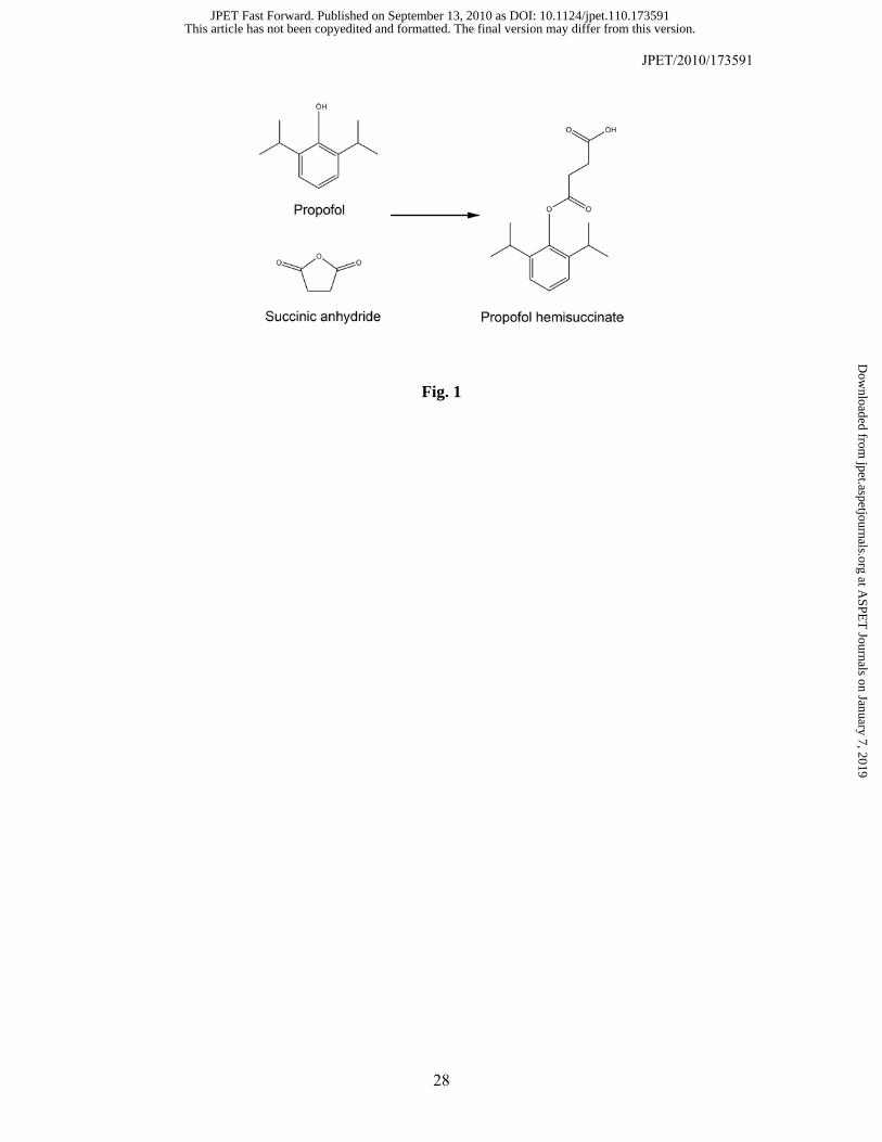
Preparation of Propofol hemisuccinate (PHS). PHS [4-(2,6-diisopropylphenoxy)-4-
oxobutanoic acid)] was synthesized according to the scheme shown in Fig. 1. In brief, 178 g (1.0
mole) of 2,6-diisopropylphenol (propofol; Sigma-Aldrich, St. Louis, MO, USA) was combined with
This article has not been copyedited and formatted. The final version may differ from this version.JPET Fast Forward. Published on September 13, 2010 as DOI: 10.1124/jpet.110.173591
at ASPE
T Journals on January 7, 2019
jpet.aspetjournals.orgD
ownloaded from
JPET/2010/173591
6
120 g (1.2 mole) of succinic anhydride in 300 ml of triethylamine under argon in a flask equipped
with a mechanical stirrer. A catalytic amount (100 mg) of 4-dimethylaminopyridine (Sigma-Aldrich)
was added and the material was stirred for a 48-h period. At the end of this time, triethylamine was
removed on the rotary evaporator to yield oil, which was taken up with 300 ml ethyl acetate. This
solution was washed with 3 × 300 ml of 1 M hydrochloric acid, 3 × 300 ml portions of saturated
sodium bicarbonate, and 3 × 300 ml portions of deionized water. The resultant solution was dried
with anhydrous magnesium sulfate, treated with activated characoal, and purified by flash
chromatography. The product was crystallized from a mixture of heptane and 2-propanol to yield 228
g (82%) of analytically pure material. The structure of propofol hemisuccinate was confirmed by
NMR.
Animals. Male Sprague Dawley rats (Taconic; 150–200g) and NIH Swiss mice (22–30 g)
were kept in a vivarium under controlled environmental conditions (temperature, 22–26°C; humidity,
40–50%) with an artificial 12-h light/dark cycle. Wood chips were used in all cages. Experiments
were performed during the light phase of the light/dark cycle after a minimum 30-min period of
acclimation to the experimental room. The animal facilities were fully accredited by the Association
for Assessment and Accreditation of Laboratory Animal Care. All studies were performed under
protocols approved by the Animal Care and Use Committee of the University of California, Davis in
strict compliance with the Guide for the Care and Use of Laboratory Animals of the National
Research Council (National Academy Press, Washington, D.C.;
http://www.nap.edu/readingroom/books/labrats/).
Test Substances and Chemoconvulsants. PHS was dissolved in slightly basic water (pH
7.5). Propofol was administered as a commercially available injectable emulsion (Diprivan 1%,
AstraZeneca) that contains soybean oil (100 mg/ml), glycerol (22.5 mg/ml), egg lecithin (12 mg/ml),
and disodium edetate (0.005%) with sodium hydroxide to adjust the pH 7–8.5. Pentylenetetrazol
(PTZ), picrotoxin, 4-aminopyridine, N-methyl-D-aspartate (NMDA), and kainic acid were dissolved
This article has not been copyedited and formatted. The final version may differ from this version.JPET Fast Forward. Published on September 13, 2010 as DOI: 10.1124/jpet.110.173591
at ASPE
T Journals on January 7, 2019
jpet.aspetjournals.orgD
ownloaded from

JPET/2010/173591
7
in 0.9% saline while bicuculline was dissolved in slightly acidic water (pH 5.0). All test substances
other than Diprivan were from Sigma-Aldrich. The volumes used for intraperitoneal injection were
10 ml/kg in the case of mice and 1 ml/kg in the case of rats. PHS was administered via the
intratracheal route in a volume of 0.25 ml/kg in mice and 0.75 ml/kg in rats. Intraperitoneal PHS was
administered 15 min before challenging the animals with convulsant agents; this interval corresponds
to the onset of loss-of-righting reflex in rats and 1.7 times this value in mice (Fig. 2). Intratracheal
PHS was administered 10 min (or in a few experiments with rats 7 min and in the time course
experiment with mice 5, 10 and 20 min) before beginning the challenge with convulsant agents; in
the threshold experiments the infusion typically required 20–35 s, so the exact time threshold values
were recorded is variably delayed from the nominal pretreatment time. A 10 min pretreatment
interval was routinely used to insure that the animals had fully recovered from the isoflurane
anesthesia required to permit intratracheal intubation as described below. Animals appeared to be
conscious at 3–5 min following isoflurane and there was no effect of isoflurane at 5 min on motor
function as assessed in the horizontal screen test (see Results) or on seizure threshold (Fig. 4). Each
experimental group consisted of a minimum of 6 animals.
Intratracheal Drug Delivery. Intratracheal administration of propofol hemiscuccinate was
carried out as described by Oka et al. (2006). In brief, rats and mice were briefly anesthetized using
isoflurane anesthesia (4% isoflurane). The animals were immediately placed on a surgical board held
at a 60º angle. The animal’s mouth was kept open by hanging the upper incisors on a hook to
facilitate detection of the epiglottis. An operating light was used to illuminate the view of the
pharynx after displacement of the tongue with a spatula. A syringe fitted with a blunted 18-gauge/50-
mm needle (in case of rats) and 24-gauge/25-mm (in case of mice) was pushed against the soft palate
to enter the trachea past the vocal cords. When the tracheal cartilage ring is felt, the needle is
considered properly placed within the tracheal lumen. The needle was inserted almost to the bottom
of the trachea. When the animals began to show signs that the anesthesia was waning, the liquid
This article has not been copyedited and formatted. The final version may differ from this version.JPET Fast Forward. Published on September 13, 2010 as DOI: 10.1124/jpet.110.173591
at ASPE
T Journals on January 7, 2019
jpet.aspetjournals.orgD
ownloaded from

JPET/2010/173591
8
sample was gently injected at a dose volume of 0.25 ml/kg (in case of mice) or 0.75 ml/kg (in case of
rats). After the injection of solution, air was blown inside the lungs to uniformly distribute the drug in
the lungs (2 ml air in case of rats and 1 ml in case of mice). Following the injection, the needle was
gently removed and animal was held vertically for 2 min to facilitate the downward movement of
liquid in the lungs. The animal was allowed 10 min to recover fully from the anesthesia as confirmed
by normal spontaneous exploratory behavior. Seizure testing then commenced. For each of the
seizure tests, we verified that there was no residual effect of the anesthetic on responsiveness.
Loss-of-righting Reflex Test. Loss-of-righting reflex, an indication of profound
sedation/anesthesia, was assessed in rats and mice following intraperitoneal injection of different
doses of either propofol or PHS (Ozbakis-Dengiz and Bakirci, 2009). Following injection, animals
were returned to their cage and when they started showing signs of ataxia, they were taken out of the
cage and placed on a filter paper stage. Animals were gently turned onto their backs every 10 s.
Untreated animals immediately regained normal posture. For drug treated animals, onset of the loss-
of-righting-reflex was the time after injection when the animal was unable to right itself. Recovery
from loss-of-righting reflex was the time after injection when the animal regained the righting reflex
(able to right itself three times within 30 sec). The total time for loss-of-righting reflex (“total sleep
time”) was calculated as the difference between the onset and recovery times.
PTZ-induced Convulsions in Rats. PTZ was administered intraperitoneally at a dose of
80 mg/kg, which causes clonic convulsions in >97% of rats (Dhir et al., 2006). Animals were
observed for a period of 30 min following injection. The time of onset of myoclonic jerks, clonus and
tonic extension; the severity of clonus; and the incidence of lethality was recorded. The severity of
the clonus was scored as either mild (1), moderate (2) or severe (3), where mild indicates brief and
transitory episodes of limb clonus, moderate indicates longer but not continuous episodes of limb
clonus where at least one episode lasts at least 10 s, and severe indicates continuous limb clonus.
This article has not been copyedited and formatted. The final version may differ from this version.JPET Fast Forward. Published on September 13, 2010 as DOI: 10.1124/jpet.110.173591
at ASPE
T Journals on January 7, 2019
jpet.aspetjournals.orgD
ownloaded from
JPET/2010/173591
9
Mean convulsion severity was calculated as the sum of the individual scores divided by the number
of animals.
PTZ, Bicuculline and Picrotoxin Seizure Threshold Tests in Mice. The thresholds for
various behavioral seizure stages induced by the GABA receptor antagonists PTZ, bicuculline and
picrotoxin were determined by infusing the convulsant drugs via a 27 gauge-¾ inch “butterfly”
needle inserted into the lateral tail vein. The needle was secured to the tail vein by a narrow piece of
adhesive tape and the animal was permitted to move freely inside an inverted 2 litre glass beaker with
free aeration from the top. PTZ (10 mg/ml) (Akula et al., 2008), bicuculline (0.1 mg/ml), and
picrotoxin (1 mg/ml) (Chan et al., 2006) was infused at a constant rate of 0.5 ml/min using a Beckton
Dickinson (1 ml) syringe mounted on an infusion pump (Model ‘11’ plus syringe pump; Harvard
Apparatus, Holliston, MA, USA). The syringe was connected to the needle by polyethylene tubing.
The infusion was stopped at 3 min or at the onset of tonic extension, whichever occurred first. The
thresholds to the following endpoints were determined: (i) the first myoclonic jerk; (ii) the onset of
generalized clonus with loss-of-righting reflex; and (iii) the onset of tonic extension. Latencies were
measured from the start of convulsant infusion to the onset of all these three events. The threshold
value (mg/kg) for each endpoint was determined according to the following formula: (infusion
duration [sec] infusion rate [ml/min] convulsant drug concentration [mg/ml] 1000)/(60 [sec]
weight of mouse [g]). Fractional change in threshold values were calculated by dividing each of the
threshold values for the three endpoints by the corresponding vehicle control threshold values.
N-Methyl-D-aspartate (NMDA) and Kainic Acid Seizure Threshold Test in Mice.
Threshold determinations for the excitatory amino acid agonists NMDA (8 mg/ml; infusion rate 0.5
ml/min) (Finn and Crabbe, 1999) and kainic acid (7.5 mg/ml; infusion rate 0.15 ml/min) (Kaminski
et al., 2005) when administered intravenously were obtained according to the same protocol as
described for the GABA receptor antagonists.
This article has not been copyedited and formatted. The final version may differ from this version.JPET Fast Forward. Published on September 13, 2010 as DOI: 10.1124/jpet.110.173591
at ASPE
T Journals on January 7, 2019
jpet.aspetjournals.orgD
ownloaded from

JPET/2010/173591
10
4-Aminopyridine (4-AP)-induced Convulsions in Mice. The 4-AP seizure test was carried
out as previously described (Yamaguchi and Rogawski, 1992). In brief, 4-AP was administered
intraperitoneally at a dose of 13 mg/kg, which causes tonic seizures in 100% of control mice. Onset
time of myoclonic jerks, clonus and tonic extension was noted.
Motor toxicity test. Motor toxicity was evaluated using a modification of the horizontal
screen test (Coughenour et al., 1977) that determines an animal’s ability to support its own body
weight by grasping an inverted grid for 1 min. Unless intoxicated, mice do not fall from the grid.
Data Analysis. Results are expressed as mean ± S.E.M.; the significance of the difference in
the responses of treatment groups with respect to control is based on one-way analysis of variance
(ANOVA) followed by specific post-hoc comparisons using Tukey’s test. Differences were
considered statistically significant when the probability of error was less than 0.05 (p < 0.05).
Control values in the threshold tests were the mean threshold values for vehicle-treated groups.
Results
Comparison of Loss-of-Righting Reflex Following Intraperitoneal Propofol and PHS in
Rats and Mice. To assess the central nervous system activity of systemically administered PHS, we
determined if intraperitoneal PHS influences the righting reflex; for comparison, we administered
aqueous propofol emulsion. In rats, 70 mg/kg PHS caused total sleep time (2163 ± 324 s) that was
roughly equivalent to the sleep time obtained with 60 mg/kg propofol (2116 ± 101 s) (Fig. 2A).
However, onset and recovery occurred earlier with propofol than PHS. In an additional experiment
with mice, we administered equimolar doses of the two agents. As shown in Fig. 2B, the onset of
loss-of-righting reflex occurred earlier with propofol (200 mg/kg) than PHS (312 mg/kg) but the total
sleep time was more prolonged with PHS (3842 ± 202 s) than with propofol (1367 ± 283 s).
This article has not been copyedited and formatted. The final version may differ from this version.JPET Fast Forward. Published on September 13, 2010 as DOI: 10.1124/jpet.110.173591
at ASPE
T Journals on January 7, 2019
jpet.aspetjournals.orgD
ownloaded from

JPET/2010/173591
11
Comparison of Intraperitoneal and Intratracheal PHS on PTZ-induced Seizures in
Rats. Intraperitoneal PTZ (80 mg/kg) induced clonic seizures, tonic seizures and mortality in all
vehicle pretreated animals (Table 1). When administered 15 min prior to PTZ, intraperitoneal PHS
(40 mg/kg) provided marked seizure protection against clonic seizures and completely prevented
tonic seizures and the mortality that invariably accompanies tonic seizures. At lower doses,
intratracheal PHS administered 10 and 7 min before PTZ provided similar seizure protection. The
effect of intratracheal PHS was dose-dependent: the 5 mg/kg dose failed to provide complete
protection against tonic seizures and mortality whereas the 10 mg/kg dose protected all animals in
both the 10 and 7 min pretreatment time experiments.
Comparison of Intraperitoneal and Intratracheal PHS in the PTZ Threshold Test in
Mice. Intravenous infusion of PTZ (10 mg/ml) produced a sequence of myoclonic jerks, clonus and
tonic extension followed by death. Pretreatment (15 min) with PHS by the intraperitoneal route at
doses of 60 and 80 mg/kg but not 40 mg/kg caused a significant elevation in the thresholds for all
seizure signs with respect to the values in vehicle-treated animals (Fig. 3A). In contrast, pretreatment
(10 min) with PHS by the intratracheal route was effective at lower doses of 10 and 15 mg/kg but not
5 mg/kg (Fig. 3B).
Having established that a 10 mg/kg intratracheal dose of PHS was effective with a 10 min
pretreatment time, we next sought to assess the time course of action of PHS administered by the
intratracheal route. As shown in Fig. 4, an elevation in threshold with respect to the corresponding
threshold values in vehicle pretreated animals was obtained with 10 mg/kg intratracheal PHS at 5 and
10 min but not 20 min. The fractional increase with respect to the corresponding threshold values in
vehicle-treated animals for all three seizure measures was numerically greater at 10 min than at 5 min
although there was no statistical significance between the 10 min and 5 min values (not shown). This
suggests that the maximal effect of intratracheal PHS occurs near 10 min although a significant
threshold change is observed as early as 5 min. It was not practical to assess shorter pretreatment
This article has not been copyedited and formatted. The final version may differ from this version.JPET Fast Forward. Published on September 13, 2010 as DOI: 10.1124/jpet.110.173591
at ASPE
T Journals on January 7, 2019
jpet.aspetjournals.orgD
ownloaded from
JPET/2010/173591
12
times due to the confounding effect of the isoflurane anesthesia that was required for intratracheal
cannulation. Indeed, the small but non-significant increase in mean threshold values obtained at the 5
min time point in the vehicle pretreatment group is likely due to residual anesthesia effects.
Motor toxicity testing. The horizontal screen test was used to assess the acute motor toxicity
of intratracheal PHS (10 mg/kg) in the time course experiment of Fig. 4. No motor toxicity was
observed with PHS at any time point.
Comparison of Intraperitoneal and Intratracheal PHS in the Bicuculline and Picrotoxin
Seizure Threshold Tests in Mice. Intravenous infusion of bicuculline (0.1 mg/ml) produced a
similar sequence of seizure signs as PTZ. Pretreatment (15 min) with PHS by the intraperitoneal
route at doses of 60 and 80 mg/kg caused a significant elevation in the thresholds for all seizure
signs; the 40 mg/kg dose only affected tonic extension significantly (Fig. 5A). In contrast,
pretreatment (10 min) with PHS by the intratracheal route was effective at lower doses of 15 and 20
mg/kg but not 10 mg/kg (Fig. 5B).
Intravenous infusion of picrotoxin (1 mg/ml) also produced the same sequence of seizure
signs. Pretreatment (15 min) with PHS by the intraperitoneal route at dose of 60 and 80 mg/kg but
not 40 mg/kg caused a significant elevation in the thresholds for all seizure signs (Fig. 6A). In
contrast, pretreatment (10 min) with PHS by the intratracheal route was effective at lower doses of
10, 15 and 20 mg/kg (Fig. 6B).
Effects of Intratracheal Administration of PHS in the NMDA and Kainic Acid Seizure
Threshold Tests in Mice. Intravenous infusion of the excitatory amino acid agonists NMDA (8
mg/ml) (Fig. 7) and kainic acid (7.5 mg/ml) (Fig. 8) induced a similar sequence of seizure signs as
did the GABAA receptor antagonists PTZ, bicuculline and picrotoxin. However, with NMDA
infusion, intratracheal pretreatment (10 min) with PHS at doses of 10 and 15 mg/kg did not modify
the thresholds for any of the seizure signs. In contrast, with kainic acid infusion, PHS at doses of 10
and 15 mg/kg but not 5 mg/kg did elevate the thresholds for all seizure signs.
This article has not been copyedited and formatted. The final version may differ from this version.JPET Fast Forward. Published on September 13, 2010 as DOI: 10.1124/jpet.110.173591
at ASPE
T Journals on January 7, 2019
jpet.aspetjournals.orgD
ownloaded from

JPET/2010/173591
13
Effects of Intratracheal Administration of PHS in the 4-AP Seizure Test in Mice.
Intraperitoneal 4-AP (13 mg/kg) induced a sequence of seizure signs consisting of myoclonic jerks,
clonus and tonic extension followed by death (Fig. 9). PHS pretreatment (10 min) at 10 mg/kg but
not 5 mg/kg delayed the onset of all seizure signs.
Discussion
This study for the first time demonstrates the anticonvulsant activity of intrapulmonary
(intratracheal) administration of PHS, a putative propofol prodrug. High systemic doses of PHS were
found to cause anesthesia as assessed by loss-of-righting reflex in rats and mice. In rats, PHS was
slightly less potent than propofol itself in inducing loss-of-righting reflex but the onset and recovery
of the righting reflex was delayed in comparison with propofol, consistent with the requirement for
PHS to be activated by deacetylation. Equimolar doses of PHS resulted in longer total sleep time than
with propofol presumably because it acts as a depot-like form that is released over a longer period of
time than propofol itself. The ability of PHS to cause loss-of-righting reflex demonstrated that it is
active as a propofol prodrug when administered systemically by the intraperitoneal route.
We next compared the activity of PHS when administered by the intraperitoneal and
intratracheal routes in various rodent seizure models. PHS was effective by both routes of
administration. However, the potency with intratracheal dosing was greater. Thus, in the maximal
PTZ test in rats (Table 1), intratracheal PHS at a dose of 10 mg/kg demonstrated efficacy similar to
that of a 40 mg/kg intraperitoneal dose. Similarly, in the PTZ seizure threshold model in mice, an
intraperitoneal dose of 60 mg/kg but not 40 mg/kg was associated with a significant fractional change
in threshold in all measures whereas when administered by the intratracheal route the 10 mg/kg but
not 5 mg/kg dose was associated with a change in threshold in all measures. The onset of the action
of intrapulmonary PHS was remarkably rapid. Maximal efficacy occurred at 10 min and a nearly
This article has not been copyedited and formatted. The final version may differ from this version.JPET Fast Forward. Published on September 13, 2010 as DOI: 10.1124/jpet.110.173591
at ASPE
T Journals on January 7, 2019
jpet.aspetjournals.orgD
ownloaded from

JPET/2010/173591
14
maximal effect was obtained by 5 min. It was not possible to determine the minimum onset time due
to the requirement for general anesthesia with intratracheal delivery but it may be substantially less
than 5 min. For comparison, it is noteworthy that the time to peak functional (central nervous system)
effect of propofol following bolus intravenous administration in humans is about 100 sec (Flaishon et
al., 1997; Schnider et al., 1999) and this corresponds with peak blood levels of 30–120 sec as
observed in other species (rat, dog and rabbit) (Simons et al., 1991; Shyr et al., 1993). Recovery was
similarly rapid, with a return to baseline seizure threshold values at 20 min. The rapid time course of
the anticonvulsant action of intrapulmonary PHS is consistent with the known pharmacokinetic
properties of propofol (Cockshott, 1985). The pharmacokinetics of propofol is generally described by
two-compartment or three-compartment models in which blood levels after a single bolus fall
according to 2 or 3 exponentials. The half-time of the first exponential component, representing
extensive redistribution from a large central compartment into a volume much greater than total body
volume, is 3.5 min in rats (Cockshott et al., 1992) and in the range of 1.6–4.2 min in humans (Saint-
Maurice et al., 1989). This initial redistribution likely accounts for the rapid recovery seen in the
present study. A second slow distribution phase may follow the first (with half-time of 33 min in rats
and 34–69 min in humans) followed by metabolism and clearance proceeding with a half-time of 6.4
h in rats and 8–14 h in humans (Cockshott, 1985; Saint-Maurice et al., 1989). At the minimum
intratracheal dose effective in raising seizure threshold (10 mg/kg), there was no apparent behavioral
effect and an objective measure of motor impairment (horizontal-screen test) failed to demonstrate
acute neurological toxicity. Consequently, there is a separation between the subanesthetic doses of
propofol that confer seizure protection and the higher doses that would induce neurobehavioral side
effects. Interestingly, a similar threshold dose of intravenous propofol (10 mg/kg) has been reported
to confer a transient analgesic effect and suppression of electroencephalographic activity with
maintained cardiovascular performance (Shyr et al., 1993).
This article has not been copyedited and formatted. The final version may differ from this version.JPET Fast Forward. Published on September 13, 2010 as DOI: 10.1124/jpet.110.173591
at ASPE
T Journals on January 7, 2019
jpet.aspetjournals.orgD
ownloaded from
JPET/2010/173591
15
As was the case for PTZ, PHS raised the threshold for seizures induced by bicuculline and
picrotoxin, which like PTZ act by blocking GABAA receptors. With all three convulsant agents, PHS
was 6-fold more potent when administered by the intratracheal route than by intraperitoneal
injection. The potency difference may relate to a high capacity of the lungs to activate PHS by
deacetylation, given the high abundance of non-specific esterase enzymes in the bronchiolar mucosa
and alveolar walls (Chessick, 1953), but could also relate to greater bioavailability of PHS itself
when administered by the pulmonary route. It is noteworthy that although most of the
biotransformation of propofol to its inactive metabolites occurs in the liver, the lung extracts (Matot
et al., 1993) and metabolizes (Dawidowicz et al., 2000) a substantial fraction of propofol. While such
metabolism could occur with intrapulmonary PHS administration thus reducing the available
propofol, the extent must be relatively minor given the high potency of intrapulmonary PHS.
Intratracheal PHS at doses effective in the PTZ, bicuculline and picrotoxin threshold models
also raised the threshold for seizure signs in the kainic acid but not the NMDA infusion models. The
results with kainic acid are consistent with a prior study showing that propofol is protective against
kainic acid-induced seizures in rats (Ahuja and Germano, 1998). The failure of PHS to affect
NMDA-induced seizures is in accordance with the limited information available that propofol does
not block NMDA receptors (Zhan et al., 2001).
The efficacy of PHS in the diverse seizure models we examined likely results in part from the
well-recognized action of propofol as a GABAA receptor positive modulator (Peduto et al., 1991;
Hales and Lambert, 1991; Hara et al., 1994) and a direct activator of GABAA receptors (Hara et al.,
1993). Seizures induced by GABAA receptor antagonists are sensitive to GABAA receptor positive
modulators (Rastogi and Ticku, 1986) as are seizures induced by kainic acid whereas NMDA-
induced seizures are relatively resistant to such agents (Kokate et al., 1996). In each of the models we
studied, PHS was more effective in blocking tonic extension than myoclonic jerks and clonus. Tonic
extensor seizures are highly susceptible to sodium channel blocking antiseizure agents and it is
This article has not been copyedited and formatted. The final version may differ from this version.JPET Fast Forward. Published on September 13, 2010 as DOI: 10.1124/jpet.110.173591
at ASPE
T Journals on January 7, 2019
jpet.aspetjournals.orgD
ownloaded from

JPET/2010/173591
16
conceivable that the effect on tonic extension could be due to previously described actions of
propofol on voltage-gated sodium channels, in which binding occurs preferentially to the fast
inactivated state as occurs with sodium channel blocking anticonvulsants (Rehberg and Duch, 1999;
Ouyang et al., 2003). The effects on sodium channels occur in the same clinically relevant range of
concentrations as the effect on GABAA receptors so that actions on sodium channel could contribute
in a major way to the anticonvulsant activity of propofol.
Schanker et al. (1986) have observed that lipid-soluble drugs are absorbed from the lung
following intratracheal administration at the same rate in diverse species, suggesting that the data
obtained here is transferable to humans. If so, intrapulmonary PHS could have a variety of
applications in human epilepsy therapy. For example, nebulized PHS could be inhaled to abort the
occurrence of a full-blown seizure by a patient experiencing an aura or warned of an impending
seizure by a seizure prediction device. In another application, a facemask inhaler could be used in an
out-of-hospital situation to abort seizure clusters or status epilepticus. The pharmacokinetic
properties of intrapulmonary PHS may be well suited for these applications inasmuch as rapid onset
is clearly necessary and rapid recovery would ensure that any sedative effects would be transitory.
In order to be useful in these applications, an inhaler device is required that reliably delivers
sufficient quantities of nebulized PHS. The device would need to tightly regulate exposure levels so
as to obtain consistent therapeutic effects without delivering excessive doses that could lead to
respiratory depression or adverse hemodynamic effects. High technology inhalers have become
available in recent years that meet the requirements of these applications (Vanbever, 2005). Such
devices efficiently deliver a uniform distribution of appropriately sized droplets to the deep lung,
avoiding filtration in the throat and upper airways. This allows the full area of the alveolar epithelium
to be used as an absorptive surface. In contrast, other nonconventional drug portals, such as the nose,
do not have the same efficiency and do not provide a means of achieving sufficient drug exposure.
The nasal mucosa provides much less absorptive surface area and the geometry does not allow
This article has not been copyedited and formatted. The final version may differ from this version.JPET Fast Forward. Published on September 13, 2010 as DOI: 10.1124/jpet.110.173591
at ASPE
T Journals on January 7, 2019
jpet.aspetjournals.orgD
ownloaded from

JPET/2010/173591
17
trapping of fluid droplets as occurs in alveolar sacs. Moreover, while highly vascularized, the nose is
exposed to only a fraction of the blood supply in contrast to the lung that receives the entire cardiac
output. Therefore, the lung has unique characteristics that make it particularly promising as a portal
for the delivery of agents such as PHS for epilepsy treatment applications and inhaler technologies
exist that make it feasible to investigate the use of intrapulmonary PHS in such applications.
This article has not been copyedited and formatted. The final version may differ from this version.JPET Fast Forward. Published on September 13, 2010 as DOI: 10.1124/jpet.110.173591
at ASPE
T Journals on January 7, 2019
jpet.aspetjournals.orgD
ownloaded from

JPET/2010/173591
18
REFERENCES
Ahuja S, Germano IM (1998) Anticonvulsant and neuronal protective effects of propofol on
experimental status epilepticus. J Epilepsy 11:168–176.
Akula KK, Dhir A, Kulkarni SK (2008) Nitric oxide signaling pathway in the anti-convulsant effect
of adenosine against pentylenetetrazol-induced seizure threshold in mice. Eur J Pharmacol
587:129–134.
Aurora SK, Rozen TD, Kori SH, Shrewsbury SB (2009) A randomized, double blind, placebo-
controlled study of MAP0004 in adult patients with migraine. Headache 49:826–837.
Arif H, Hirsch LJ (2008) Treatment of status epilepticus. Semin Neurol 28:342–354.
Baker MT, Naguib M (2005) Propofol: the challenges of formulation. Anesthesiology 103:860–876.
Borowicz KK, Czuczwar SJ (2003) Effects of etomidate, ketamine or propofol, and their
combinations with conventional antiepileptic drugs on amygdala-kindled convulsions in rats.
Neuropharmacology 45:315–324.
Chan MH, Lee CC, Chen HH (2006) Effects of toluene on seizures induced by convulsants acting at
distinct ligand-gated ion channels. Toxicol Lett 160:179–184.
Chessick RD (1953) Histochemical study of the distribution of esterases. J Histochem Cytochem
1:471–485.
Cockshott ID (1985) Propofol (‘Diprivan’) pharmacokinetics and metabolism—an overview.
Postgrad Med J 61 Suppl 3:45–50.
Cockshott ID, Douglas EJ, Plummer GF, Simons PJ (1992) The pharmacokinetics of propofol in
laboratory animals. Xenobiotica 22:369–375.
Coughenour AG, McLean JR, Parker RB (1977) A new device for the rapid measurement of
impaired motor function in mice. Pharmacol Biochem Behav 6:351–353.
Crompton G (2006) A brief history of inhaled asthma therapy over the last fifty years. Prim Care
Respir J 15:326–331.
This article has not been copyedited and formatted. The final version may differ from this version.JPET Fast Forward. Published on September 13, 2010 as DOI: 10.1124/jpet.110.173591
at ASPE
T Journals on January 7, 2019
jpet.aspetjournals.orgD
ownloaded from

JPET/2010/173591
19
Dawidowicz AL, Fornal E, Mardarowicz M, Fijalkowska A (2000) The role of human lungs in the
biotransformation of propofol. Anesthesiology 93:992–997.
De Riu PL, Petruzzi V, Testa C, Mulas M, Melis F, Caria MA, Mameli O (1992) Propofol
anticonvulsant activity in experimental epileptic status. Br J Anaesth 69:177–181.
Dhir A, Naidu PS, Kulkarni SK (2006) Effect of cyclooxygenase inhibitors on pentylenetetrazol
(PTZ)-induced convulsions: Possible mechanism of action. Prog Neuropsychopharmacol
Biol Psychiatry 30:1478–1485.
Finn DA, Crabbe JC (1999) Chronic ethanol differentially alters susceptibility to chemically induced
convulsions in withdrawal seizure-prone and -resistant mice. J Pharmacol Exp Ther
288:782–790.
Flaishon R, Windsor A, Sigl J, Sebel PS (1997) Recovery of consciousness after thiopental or
propofol. Bispectral index and isolated forearm technique. Anesthesiology 86:613–619.
Hales TG, Lambert JJ (1991) The actions of propofol on inhibitory amino acid receptors of bovine
adrenomedullary chromaffin cells and rodent central neurones. Br J Pharmacol 104:619–
628.
Hara M, Kai Y, Ikemoto Y (1993) Propofol activates GABAA receptor-chloride ionophore complex
in dissociated hippocampal pyramidal neurons of the rat. Anesthesiology 79:781–788.
Hara M, Kai Y, Ikemoto Y (1994) Enhancement by propofol of the -aminobutyric acidA response in
dissociated hippocampal pyramidal neurons of the rat. Anesthesiology 81:988–994.
Hasan Z, Hasan M, al-Hader A, Takrouri M (1991) A comparison of the anticonvulsant effects of
propofol and thiopentone against pentylenetetrazol-induced convulsions in the rat. Clin Exp
Pharmacol Physiol 18:691–695.
Kaminski RM, Shippenberg TS, Witkin JM, Rocha BA (2005) Genetic deletion of the
norepinephrine transporter decreases vulnerability to seizures. Neurosci Lett 382:51–55.
This article has not been copyedited and formatted. The final version may differ from this version.JPET Fast Forward. Published on September 13, 2010 as DOI: 10.1124/jpet.110.173591
at ASPE
T Journals on January 7, 2019
jpet.aspetjournals.orgD
ownloaded from
JPET/2010/173591
20
Kokate TG, Cohen AL, Karp E, Rogawski MA (1996) Neuroactive steroids protect against
pilocarpine- and kainic acid-induced limbic seizures and status epilepticus in mice.
Neuropharmacology 35:1049–1056.
Lee SR, Cheun JK (1999) Propofol administration reduces hippocampal neuronal damage induced by
kainic acid in rats. Neurol Res 21:225–228.
Lingamaneni R, Hemmings HC Jr (2003) Differential interaction of anaesthetics and antiepileptic
drugs with neuronal Na+ channels, Ca2+ channels, and GABAA receptors. Br J Anaesth
90:199–211.
Lowson S, Gent JP, Goodchild CS (1990) Anticonvulsant properties of propofol and thiopentone:
comparison using two tests in laboratory mice. Br J Anaesth 64:59–63.
Matot I, Neely CF, Katz RY, Neufeld GR (1993) Pulmonary uptake of propofol in cats. Effect of
fentanyl and halothane. Anesthesiology 78:1157-65.
Micklethwaite AV, Pleuvry BJ (1993) Comparison of anticonvulsant effects of intravenous propofol,
thiopentone and methohexitone in bicuculline-induced convulsions in mice. Brit J Pharmacol
108:P291-P291.
Ohmori H, Sato Y, Namiki A (2004) The anticonvulsant action of propofol on epileptiform activity
in rat hippocampal slices. Anesth Analg 99:1095–1101.
Oka Y, Mitsui M, Kitahashi T, Sakamoto A, Kusuoka O, Tsunoda T, Mori T, Tsutsumi M (2006) A
reliable method for intratracheal instillation of materials to the entire lung in rats. J Toxicol
Pathol 19:107–109.
Ozbakis-Dengizm G, Bakirci A (2009 Anticonvulsant and hypnotic effects of amiodarone. J
Zhejiang Univ Sci B 10:317–322.
Ouyang W, Wang G, Hemmings HC Jr (2003) Isoflurane and propofol inhibit voltage-gated sodium
channels in isolated rat neurohypophysial nerve terminals. Mol Pharmacol 64:373–381.
This article has not been copyedited and formatted. The final version may differ from this version.JPET Fast Forward. Published on September 13, 2010 as DOI: 10.1124/jpet.110.173591
at ASPE
T Journals on January 7, 2019
jpet.aspetjournals.orgD
ownloaded from

JPET/2010/173591
21
Patton JS (1996) Mechanisms of macromolecule absorption by the lungs. Adv Drug Del Rev 19:3–
36.
Patton JS, Byron PR (2007) Inhaling medicines: delivering drugs to the body through the lungs. Nat
Rev Drug Discov 6:67–74.
Peduto VA, Concas A, Santoro G, Biggio G, Gessa GL (1991) Biochemical and electrophysiologic
evidence that propofol enhances GABAergic transmission in the rat brain. Anesthesiology
75:1000–1009.
Prasad A, Worrall BB, Bertram EH, Bleck TP (2001) Propofol and midazolam in the treatment of
refractory status epilepticus. Epilepsia 42:380–386.
Rasmussen PA, Yang Y, Rutecki PA (1996) Propofol inhibits epileptiform activity in rat
hippocampal slices. Epilepsy Res 25:169–175.
Rastogi SK, Ticku MK (1986) Anticonvulsant profile of drugs which facilitate GABAergic
transmission on convulsions mediated by a GABAergic mechanism. Neuropharmacology
25:175–185.
Rehberg B, Duch DS (1999) Suppression of central nervous system sodium channels by propofol.
Anesthesiology 91:512–520.
Reves JG, Glass PSA, Lubarsky DA, McEvoy RD, Martinez-Ruiz R (2009) Intravenous anesthetics
(Chapter 26) in Miller’s Anesthesia, 7th ed. (Miller RD, Eriksson LI, Fleisher LA, Wiener-
Kronish JP, Young WL eds), Churchill Livingstone/Elsevier.
Sagara Y, Hendler S, Khoh-Reiter S, Gillenwater G, Carlo D, Schubert D, Chang J (1999) Propofol
hemisuccinate protects neuronal cells from oxidative injury. J Neurochem 73:2524–2530.
Saint-Maurice C, Cockshott ID, Douglas EJ, Richard MO, Harmey JL (1989) Pharmacokinetics of
propofol in young children after a single dose. Br J Anaesth 63:667–670.
Schanker LS (1978) Drug absorption from the lung. Biochem Pharmacol 27:381–385.
This article has not been copyedited and formatted. The final version may differ from this version.JPET Fast Forward. Published on September 13, 2010 as DOI: 10.1124/jpet.110.173591
at ASPE
T Journals on January 7, 2019
jpet.aspetjournals.orgD
ownloaded from

JPET/2010/173591
22
Schanker LS, Mitchell EW, Brown RA Jr (1986) Pulmonary absorption of drugs in the dog:
comparison with other species. Pharmacology 32:176–180.
Schnider TW, Minto CF, Shafer SL, Gambus PL, Andresen C, Goodale DB, Youngs EJ (1999) The
influence of age on propofol pharmacodynamics. Anesthesiology 90:1502–1516.
Shyr MH, Yang CH, Kuo TB, Pan WH, Tan PP, Chan SH (1993) Power spectral analysis of the
electroencephalographic and hemodynamic correlates of propofol anesthesia in the rat:
intravenous bolus administration. Neurosci Lett 153:161–164.
Simons PJ, Cockshott ID, Douglas EJ, Gordon EA, Knott S, Ruane RJ (1991) Species differences in
blood profiles, metabolism and excretion of 14C-propofol after intravenous dosing to rat, dog
and rabbit. Xenobiotica 21:1243–1256.
Vanbever R (2005) Performance-driven, pulmonary delivery of systemically acting drugs. Drug
Discovery Today: Technologies 2:30–46
Vansant G, Trauger RJ, Cameron A, Vendemelio M, Kreitschitz S, Carlo AT, Banaszczyk MG,
Carlo DJ, Hendler S, Ill CR (2007) Propofol hemisuccinate suppression of experimental
autoimmune encephalomyelitis. Autoimmunity 40:180–186.
Vatter AE, Reiss OK, Newman JK (1971) The subcellular distribution of some hydrolytic enzymes
in lung. Chest 59:11S–12S.
Yamaguchi S, Rogawski MA (1992) Effects of anticonvulsant drugs on 4-aminopyridine-induced
seizures in mice. Epilepsy Res 11:9–16.
Zhan RZ, Qi S, Wu C, Fujihara H, Taga K, Shimoji K (2001) Intravenous anesthetics differentially
reduce neurotransmission damage caused by oxygen-glucose deprivation in rat hippocampal
slices in correlation with N-methyl-D-aspartate receptor inhibition. Crit Care Med 29:808-
813.
This article has not been copyedited and formatted. The final version may differ from this version.JPET Fast Forward. Published on September 13, 2010 as DOI: 10.1124/jpet.110.173591
at ASPE
T Journals on January 7, 2019
jpet.aspetjournals.orgD
ownloaded from

JPET/2010/173591
23
Footnotes
This study was supported by a New Therapy Grant Program award from the Epilepsy
Research Foundation (M.A.R.) and a Postdoctoral Research Training Fellowship from the Epilepsy
Foundation (A.D.)
This article has not been copyedited and formatted. The final version may differ from this version.JPET Fast Forward. Published on September 13, 2010 as DOI: 10.1124/jpet.110.173591
at ASPE
T Journals on January 7, 2019
jpet.aspetjournals.orgD
ownloaded from

JPET/2010/173591
24
FIGURE LEGENDS
Fig. 1. Synthesis of propofol hemisuccinate.
Fig. 2. Loss-of-righting reflex induced by intraperitoneal administration of propofol or propofol
hemisuccinate (PHS) in rats (A) and mice (B). Thick vertical lines indicate the mean ± S.E.M. for 6–
8 animals of the latency to loss-of-righting reflex and recovery from loss-of-righting reflex; total
sleep time (open squares) is the difference between these two values. Doses in rats for propofol and
PHS were 60 mg/kg (0.34 mmol/kg) and 70 mg/kg (0.25 mmol/kg), respectively; doses in mice were
200 mg/kg (1.12 mmol/kg) and 312 mg/kg (1.12 mmol/kg). In mice, the mean latency to and
recovery from loss-of-righting reflex and total sleep time values were significantly different in the
PHS and propofol groups (p < 0.05; ANOVA followed by Tukey’s test).
Fig. 3. Effect of intraperitoneal (A; 40–80 mg/kg) and intratracheal (B; 5–15 mg/kg) propofol
hemisuccinate (PHS) on myoclonic jerk, generalized clonus and tonic extension in response to
intravenous PTZ infusion in mice. PHS was administered 15 min and 10 min prior to the beginning
of the PTZ infusion for the intraperitoneal and intratracheal experiments, respectively. Bars indicate
mean ± S.E.M. of values from 6–8 mice normalized with respect to the thresholds in the vehicle-
treated groups (V), which (in mg/kg) for myoclonic jerk, clonus and tonic extension, respectively,
were 37.1 ± 3.8, 41.9 ± 3.9, 47.9 ± 3.5 for intraperitoneal and 37.8 ± 1.2, 45.1 ± 3.9, 47.9 ± 3.5 for
intratracheal. *p < 0.05 as compared to vehicle control group (ANOVA followed by Tukey’s test).
Fig. 4. Time course for the action of 10 mg/kg intratracheal propofol hemisuccinate (PHS) on
myoclonic jerk, generalized clonus and tonic extension in response to intravenous PTZ infusion in
mice. PHS was administered 5, 10 and 20 min prior to the beginning of the PTZ infusion. Closed (■)
and open (□) symbols indicate mean ± S.E.M. of values from 6–8 mice pretreated with PHS or
This article has not been copyedited and formatted. The final version may differ from this version.JPET Fast Forward. Published on September 13, 2010 as DOI: 10.1124/jpet.110.173591
at ASPE
T Journals on January 7, 2019
jpet.aspetjournals.orgD
ownloaded from

JPET/2010/173591
25
vehicle, respectively. *p < 0.05 as compared to vehicle control group (ANOVA followed by Tukey’s
test).
Fig. 5. Effect of intraperitoneal (A; 40–80 mg/kg) and intratracheal (B; 10–20 mg/kg) propofol
hemisuccinate (PHS) on myoclonic jerk, generalized clonus and tonic extension in response to
intravenous bicuculline infusion in mice. PHS was administered 15 min and 10 min prior to the
beginning of the bicuculline infusion for the intraperitoneal and intratracheal experiments,
respectively. Bars indicate mean ± S.E.M. of values from 6–8 mice normalized with respect to the
thresholds in the vehicle-treated groups (V), which (in mg/kg) for myoclonic jerk, clonus and tonic
extension, respectively, were 0.55 ± 0.02, 0.60 ± 0.02, 0.84 ± 0.02 for intraperitoneal and 0.56 ±
0.04, 0.62 ± 0.04, 0.92 ± 0.06 for intratracheal. *p < 0.05 as compared to vehicle control group
(ANOVA followed by Tukey’s test).
Fig. 6. Effect of intraperitoneal (A; 40–80 mg/kg) or intratracheal (B; 10–20 mg/kg) propofol
hemisuccinate (PHS) on myoclonic jerk, generalized clonus and tonic extension in response to
intravenous picrotoxin infusion in mice. PHS was administered 15 min and 10 min prior to the
beginning of the picrotoxin infusion for the intraperitoneal and intratracheal experiments,
respectively. Bars indicate mean ± S.E.M. of values from 6–8 mice normalized with respect to the
threshold in the vehicle-treated groups (V), which (in mg/kg) for myoclonic jerk, clonus and tonic
extension, respectively, were 14.6 ± 0.3, 18.1 ± 0.3, 23.1 ± 0.5 for intraperitoneal and 17.3 ± 0.7,
22.3 ± 0.7, 31.3 ± 1.3 for intratracheal. *p < 0.05 as compared to vehicle control group (ANOVA
followed by Tukey’s test).
Fig. 7. Effect of intratracheal propofol hemisuccinate (PHS; 10 and 15 mg/kg) on myoclonic jerk,
generalized clonus and tonic extension in response to intravenous NMDA infusion in mice. PHS was
This article has not been copyedited and formatted. The final version may differ from this version.JPET Fast Forward. Published on September 13, 2010 as DOI: 10.1124/jpet.110.173591
at ASPE
T Journals on January 7, 2019
jpet.aspetjournals.orgD
ownloaded from

JPET/2010/173591
26
administered 10 min prior to the beginning of the NMDA infusion. Bars indicate mean ± S.E.M. of
values from 6–8 mice normalized with respect to the threshold values in the vehicle-treated control
group (V), which (in mg/kg) for myoclonic jerk, clonus and tonic extension, respectively, were 39.8
± 4.1, 56.1 ± 6.1, 91.9 ± 17.7. The values in the treated groups were not significantly different from
those in the control group (ANOVA).
Fig. 8. Effect of intratracheal administration of propofol hemisuccinate (PHS; 5–15 mg/kg) on
myoclonic jerk, generalized clonus and tonic extension in reponse to intravenous kainic acid infusion
in mice. PHS was administered 10 min prior to the beginning of the kainic acid infusion. Bars
indicate mean ± S.E.M. of values from 6–8 mice normalized with respect to the threshold in the
vehicle-treated group, which (in mg/kg) for myoclonic jerk, clonus and tonic extension, respectively,
were 29.8 ± 2.8, 79.1 ± 3.7, 111.2 ± 7.1. *p < 0.05 as compared to vehicle control group (ANOVA
followed by Tukey’s test).
Fig. 9. Effect of intratracheal administration of propofol hemisuccinate (PHS; 5 and 10 mg/kg) on the
mean onset time to myoclonic jerk, generalized clonus and tonic extension in response to 4-
aminopyridine (13 mg/kg, i.p.) in mice. PHS was administered 10 min prior to 4-aminopyridine. Bars
indicate mean ± S.E.M. of values from 6–8 mice normalized with respect to the times in the vehicle-
treated group, which (in min) for myoclonic jerk, clonus and tonic extension, respectively, were 7.12
± 0.28, 10.30 ± 0.98, 10.35 ± 0.94. *p < 0.05 as compared to vehicle control group (ANOVA
followed by Tukey’s test).
This article has not been copyedited and formatted. The final version may differ from this version.JPET Fast Forward. Published on September 13, 2010 as DOI: 10.1124/jpet.110.173591
at ASPE
T Journals on January 7, 2019
jpet.aspetjournals.orgD
ownloaded from

JPET/2010/173591
27
Table 1
Comparison of intraperitoneal and intratracheal propofol hemisuccinate (PHS) on PTZ-induced convulsions in rats.
Treatment (dose) Pretreatment Time (min)
Number of animals
Percent showing
myoclonic jerks
Percent showing clonus
Mean clonus severity
Percent showing tonic extension
Percent Mortality
Intraperitoneal
Vehicle 15 6 100 100 3 100 100
PHS (40 mg/kg) 15 6 33* 33* 0.5 0* 0
Intratracheal
Vehicle 10 6 100 100 3 100 100
PHS (5 mg/kg) 10 6 83 83 1.3 17* 16*
PHS (10 mg/kg) 10 7 57* 29* 0.5 0* 0*
Intratracheal
Vehicle 7 6 100 100 3 100 100
PHS (10 mg/kg) 7 6 50* 33* 0.3 0* 0*
PTZ was administered at a dose of 80 mg/kg, i.p. The second column indicates the pretreatment interval between PHS administration and
the subsequent PTZ injection. The solution volume for intraperitoneal PHS was 1 ml/kg and for intratracheal PHS was 0.75 ml/kg. Values
indicate percent of animals exhibiting indicated seizure sign or mortality, except mean clonus severity which is the average of the clonus
scores rated either 1, 2 or 3. *p < 0.05 as compared to vehicle treated control group (ANOVA followed by Tukey’s test).
This article has not been copyedited and form
atted. The final version m
ay differ from this version.
JPET
Fast Forward. Published on Septem
ber 13, 2010 as DO
I: 10.1124/jpet.110.173591 at ASPET Journals on January 7, 2019 jpet.aspetjournals.org Downloaded from
JPET/2010/173591
28
Fig. 1
This article has not been copyedited and formatted. The final version may differ from this version.JPET Fast Forward. Published on September 13, 2010 as DOI: 10.1124/jpet.110.173591
at ASPE
T Journals on January 7, 2019
jpet.aspetjournals.orgD
ownloaded from

JPET/2010/173591
29
Fig. 2
This article has not been copyedited and formatted. The final version may differ from this version.JPET Fast Forward. Published on September 13, 2010 as DOI: 10.1124/jpet.110.173591
at ASPE
T Journals on January 7, 2019
jpet.aspetjournals.orgD
ownloaded from

JPET/2010/173591
30
Fig. 3
This article has not been copyedited and formatted. The final version may differ from this version.JPET Fast Forward. Published on September 13, 2010 as DOI: 10.1124/jpet.110.173591
at ASPE
T Journals on January 7, 2019
jpet.aspetjournals.orgD
ownloaded from

JPET/2010/173591
31
Fig. 4
This article has not been copyedited and formatted. The final version may differ from this version.JPET Fast Forward. Published on September 13, 2010 as DOI: 10.1124/jpet.110.173591
at ASPE
T Journals on January 7, 2019
jpet.aspetjournals.orgD
ownloaded from

JPET/2010/173591
32
Fig. 5
This article has not been copyedited and formatted. The final version may differ from this version.JPET Fast Forward. Published on September 13, 2010 as DOI: 10.1124/jpet.110.173591
at ASPE
T Journals on January 7, 2019
jpet.aspetjournals.orgD
ownloaded from

JPET/2010/173591
33
Fig. 6
This article has not been copyedited and formatted. The final version may differ from this version.JPET Fast Forward. Published on September 13, 2010 as DOI: 10.1124/jpet.110.173591
at ASPE
T Journals on January 7, 2019
jpet.aspetjournals.orgD
ownloaded from

JPET/2010/173591
34
Fig. 7
This article has not been copyedited and formatted. The final version may differ from this version.JPET Fast Forward. Published on September 13, 2010 as DOI: 10.1124/jpet.110.173591
at ASPE
T Journals on January 7, 2019
jpet.aspetjournals.orgD
ownloaded from

JPET/2010/173591
35
Fig. 8
This article has not been copyedited and formatted. The final version may differ from this version.JPET Fast Forward. Published on September 13, 2010 as DOI: 10.1124/jpet.110.173591
at ASPE
T Journals on January 7, 2019
jpet.aspetjournals.orgD
ownloaded from

JPET/2010/173591
36
Fig. 9
This article has not been copyedited and formatted. The final version may differ from this version.JPET Fast Forward. Published on September 13, 2010 as DOI: 10.1124/jpet.110.173591
at ASPE
T Journals on January 7, 2019
jpet.aspetjournals.orgD
ownloaded from