secondary open angle glaucoma
TRANSCRIPT
Dr. Samarth Mishra
introductionCharacterized by-
o Raised IOP
o Open angles
o A/w underlying disorder leading toalteration in aq. humor dynamics
o A/w OD & VF changes
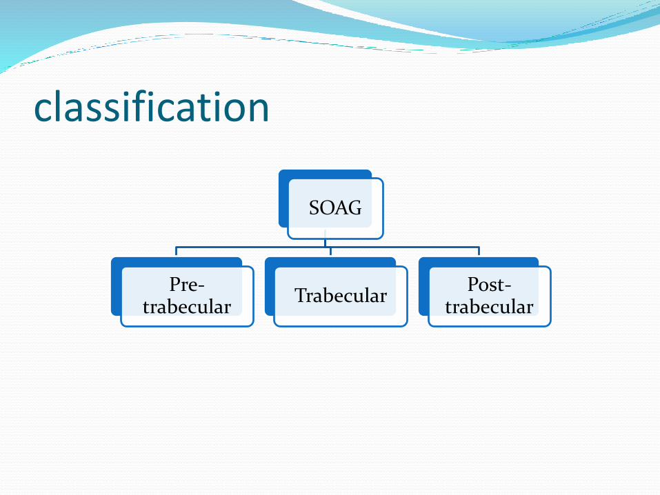
classification
SOAG
Pre-trabecular
TrabecularPost-
trabecular

pretrabecular Aq. Flow obstructed by a membrane covering TM
Consists of –
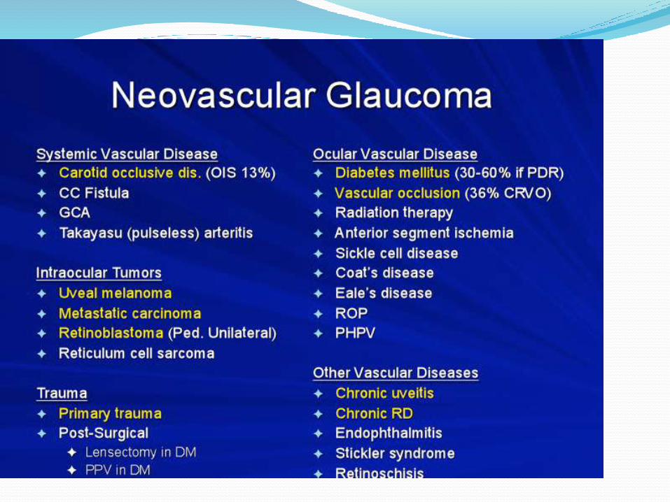
6. Fibrovascular memb.--neovascular glaucoma
7. Descement like memb.—IEC, trauma
8. Epithelial downgrowth
9. Fibrous downgrowth
10. Inflammatory memb– Fuch,s cyclitis

Neovascular glaucomaNeovacular tissue arbourise in the angle
Form a fibrovascular memb.
Blocks TM
SOAG

Stages of neovascular glaucoma. (A)Pre-glaucoma stage with new vessels appearing at pupillary margin
and in angle.(B) Open-angle glaucoma stage with new vessels spreading and
fibrovascular tissue covering angle.(C) Heavy neovascularization and extensive peripheral anterior
synechiae.(D) Regression stage with angle sealed and vessels less visible.
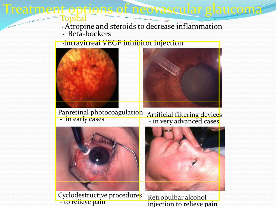
Treatment options of neovascular glaucoma• Atropine and steroids to decrease inflammation• Beta-bockers•intravitreal VEGF inhibitor injection
Panretinal photocoagulation- in early cases
Artificial filtering devices- in very advanced cases
Cyclodestructive procedures- to relieve pain
Retrobulbar alcohol injection to relieve pain
Topical


(A)Membrane forms in one area of angle.(B) Additional areas of angle are involved, and contraction of
membrane displaces pupil.
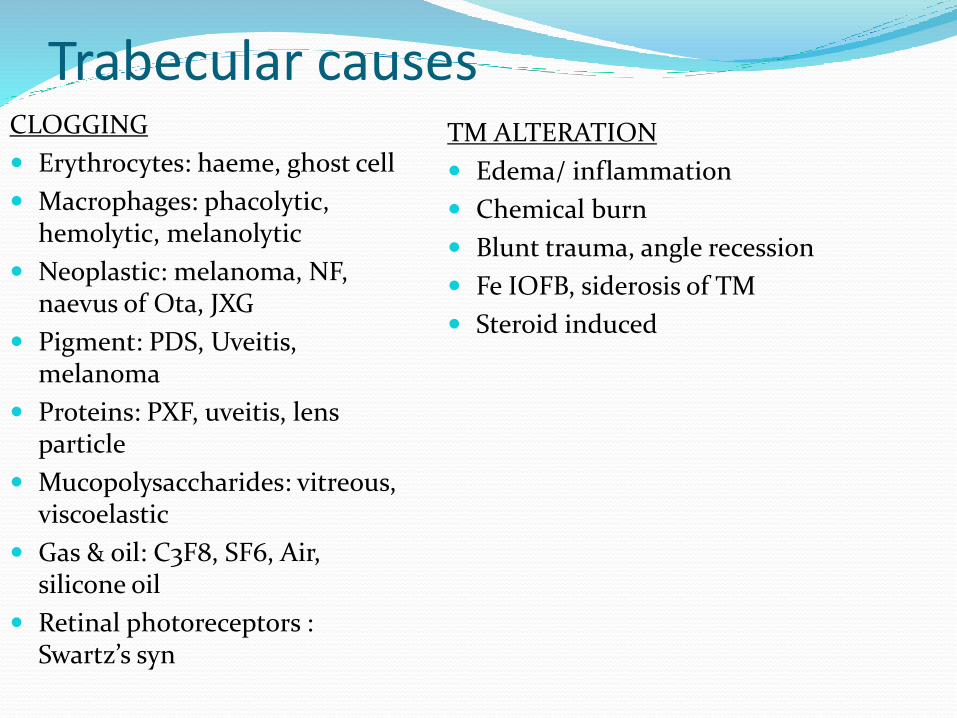
Trabecular causesCLOGGING
Erythrocytes: haeme, ghost cell
Macrophages: phacolytic, hemolytic, melanolytic
Neoplastic: melanoma, NF, naevus of Ota, JXG
Pigment: PDS, Uveitis, melanoma
Proteins: PXF, uveitis, lens particle
Mucopolysaccharides: vitreous, viscoelastic
Gas & oil: C3F8, SF6, Air, silicone oil
Retinal photoreceptors : Swartz’s syn
TM ALTERATION
Edema/ inflammation
Chemical burn
Blunt trauma, angle recession
Fe IOFB, siderosis of TM
Steroid induced
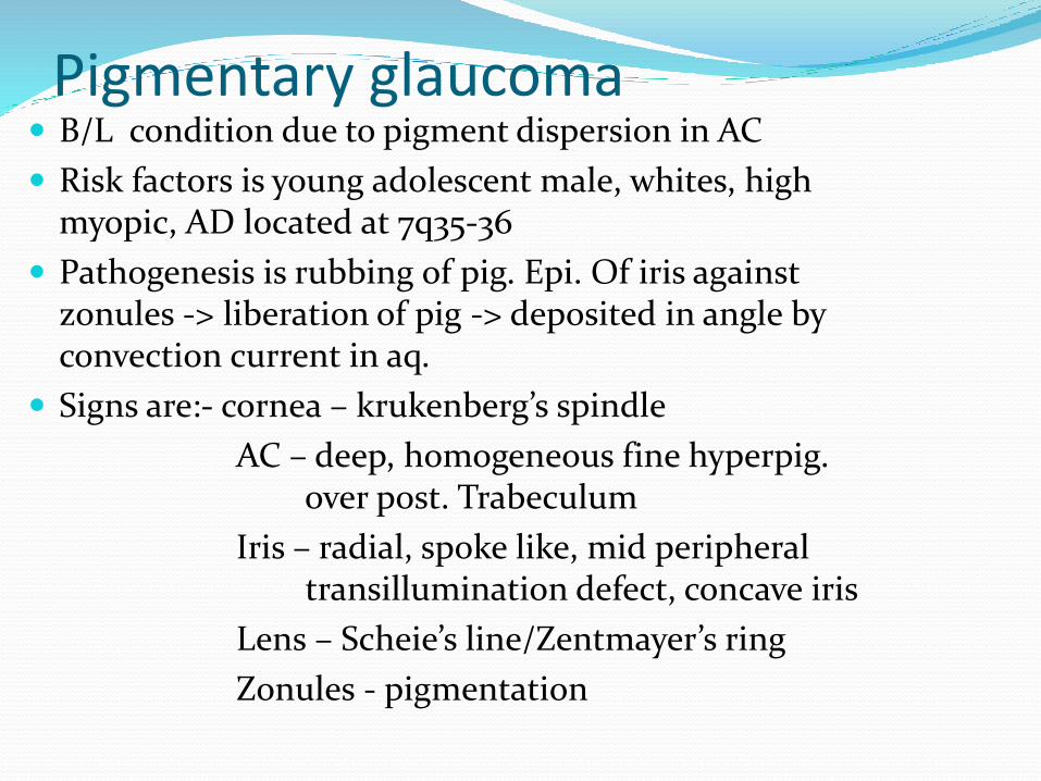
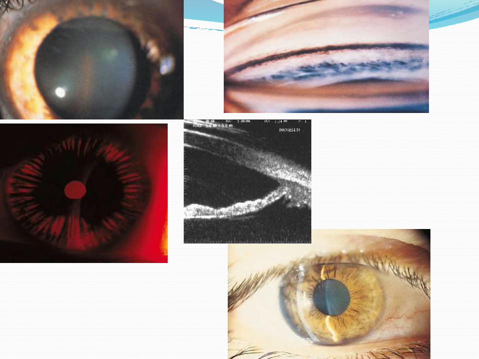
Pigmentary glaucoma B/L condition due to pigment dispersion in AC
Risk factors is young adolescent male, whites, high myopic, AD located at 7q35-36
Pathogenesis is rubbing of pig. Epi. Of iris against zonules -> liberation of pig -> deposited in angle by convection current in aq.
Signs are:- cornea – krukenberg’s spindle
AC – deep, homogeneous fine hyperpig. over post. Trabeculum
Iris – radial, spoke like, mid peripheral transillumination defect, concave iris
Lens – Scheie’s line/Zentmayer’s ring
Zonules - pigmentation

Contd… Management ->
1) medical—beta blockers, adrenaline, dipivefin, CA inhibitor, miotic
2) Laser – ALT, PI, Peri. Iridoplasty
3) Surg - trabeculectomy

Exfoliation glaucoma TM clogging up by PXF material &/or pig. From iris
Risk factors are –fem, in Scandinavia, mutation in LOXL1 gene at 15q22 locus
Patho ---grey-white fibrillary extracellular material composed of protein core surrounded by GAGs produced by abnormal BM of ageing epi. Cells of TM, equatorial lens capsule & CB.
Signs –cornea -> dandruff like deposi, in endo.
AC -> Sampolesi’s line
iris -> absence of pupi. Ruff, moth-eaten trans illumination defect
lens -> cataract, exfo. Deposi. As central disk with peri. Band & a clear zone in middle

Pseudoexfoliation glaucoma
Pseudoexfoliative material Iris sphincter atrophy Gonioscopy
Central disc with peripheral band
Trabecular hyperpigmentation- may extend anteriorly(Sampaolesi line)
On retroillumination
Contd.. T/T ---
1) Medical - beta blockers, epinephrine, miotics
2) Laser – ALT
3) Surg – trabeculectomy
4) Trabecular aspiration
Lens induced glaucoma1) PHACOLYTIC:-
. Due to protein leakage from mat./hypermat. cat
. mechanism is –a) high molecular wt. soluble protein directly block
b)macrophages engulf the proteins & block
. c/f – seen in elderly with h/o poor vision for mons
- acute onset of uniocular pain, redness & watering
- grossly decres. Vision & raised IOP
- signs of uveitis
-hypermat/ morgagnian cat
- AC shows heavy flare a/w hyper refringent crystals which are ca++ oxalate / cholesterol crystals
. t/t – medical (hyperosmotic, CA inhi.,topical b-blockers & steroids
- surg ( ECCE with PCIOL)
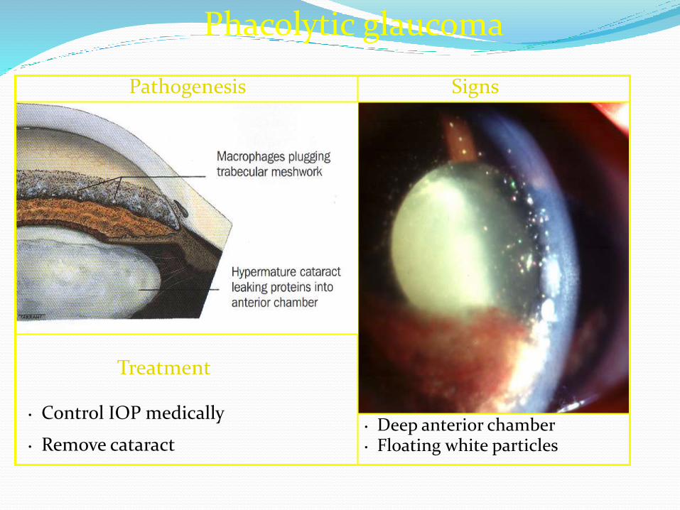
Phacolytic glaucoma
Pathogenesis Signs
• Deep anterior chamber• Control IOP medically
• Remove cataract • Floating white particles
Treatment

Calcium oxalate crystal in the lens of a patient with glaucoma associated with hypermature cataract. (Hematoxylin and eosin stain.)
Phacolytic glaucoma with bloated macrophages and lens material obstructing the trabecularmeshwork.

Contd…2) LENS PARTICLE:-. k/a phacotoxic uveitis
. Mechanism is – due to trauma/ surg.retained lens material
disruption of lens capsule
lens material liberation raised IOP
inflammatory response
raised IOP
. c/f – features of uveitis, rai IOP, chunky white particles in AC, hypopyon
. Diag. by paracentesis
. t/t – medical as phacolytic
- surg. ( removal of lens material)

Contd…3)PHACOANAPHYLAXIS :-
. occur when patients become sensitized to their own lens protein k/a endophthalmitis anaphylactica
. typically develops after penetrating trauma or extracapsular cataract extraction
. granulomatous inflammation of the lens with polymorphonuclear leukocytes, lymphocytes, epithelioid
cells, and giantcells.
. t/t is surg. Removal of residual lens material
Glaucoma after trauma1) CHEMICAL :-
. Alkali>acid
. caused by scleral shrinkage and release of active substances, including prostaglandins
. IOP measured more accurately with the pneumatic or MacKay-Marg tonometers
.managed by – medical ( topical and systemic medications)
2) ELECTRIC :-
. pressure rise to venous dilation, contraction of the extraocular muscles, and pigment dispersion
. No therapy due to transient rise of IOP
Contd…3) RADIATION :-
. Mechanism are neovascularization/ ghost-cell glaucoma associated with radiation retinopathy and vitreous hemorrhage.
4) PENETRATING :-
. Due to retained organic material / FB / severe inflammation / TM damage
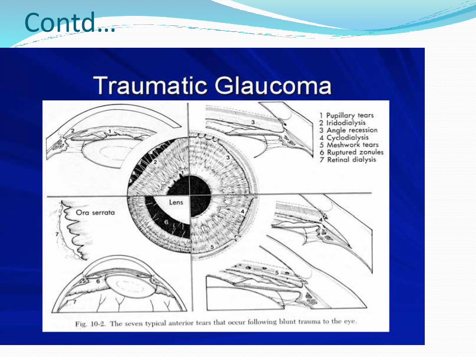
5) CONTUSION :-
. can cause hyphema, iridocyclitis, iris sphincter tears, iridodialysis, cyclodialysis, lens subluxation, retinal tear or dialysis, retinal detachment, vitreous hemorrhage, choroidal rupture, and glaucoma.
Contd…
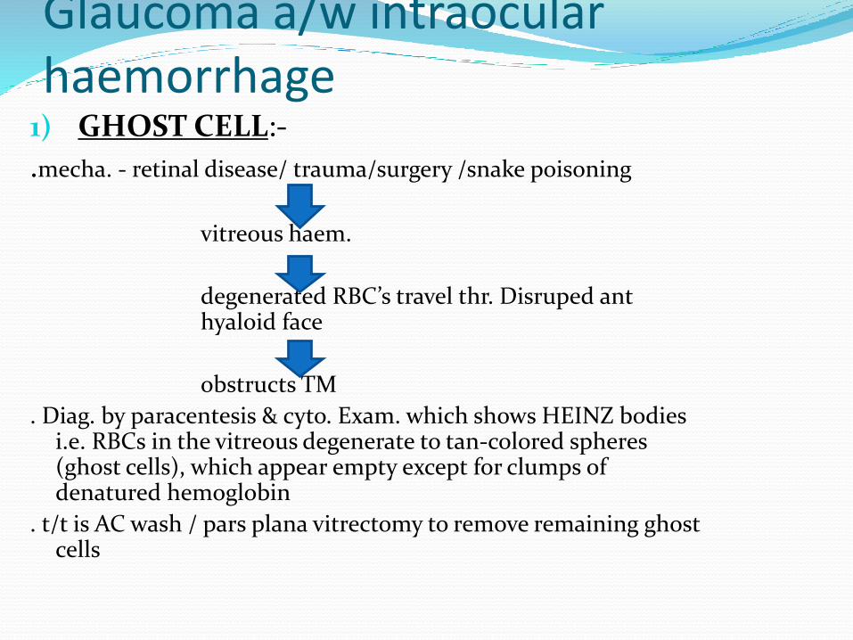
Glaucoma a/w intraocular haemorrhage
1) GHOST CELL:-
.mecha. - retinal disease/ trauma/surgery /snake poisoning
vitreous haem.
degenerated RBC’s travel thr. Disruped ant hyaloid face
obstructs TM
. Diag. by paracentesis & cyto. Exam. which shows HEINZ bodies i.e. RBCs in the vitreous degenerate to tan-colored spheres (ghost cells), which appear empty except for clumps of denatured hemoglobin
. t/t is AC wash / pars plana vitrectomy to remove remaining ghost cells

Contd…2) HEMOLYTIC :-
. Due to macrophages laden with pigments, RBC’s & debris
3) HEMOSIDEROSIS :-
. Due to iron liberted from Hb causing siderosis of TM
4) HYPHEMA :-
. Due to blood & blood products
. Total hyphema changing color from red to black (black-ball or eightball hyphema)
. t/t – topical anti glaucoma
- surgical removal of hyphema
Glaucoma a/w uveitis. Mechanism are-
(1) increased viscosity of aqueous humor;
(2) obstruction of the trabecular meshwork by inflammatory cells and debris
(3) swelling and dysfunction of the trabecularmeshwork;
(4) liberation of active substances such as prostaglandins;
(5)scarring of the outflow channels;
(6) development of a cuticular endothelial membrane over the angle;
(7) neovascularization;
(8) elevation of episcleral venous pressure;
Contd…Causes are:-
1) FUCH’S HETEROCHROMIC IRIDOCYCLITIS
. Mild form of anterior uveitis associated with cataract and glaucoma.
.c/f – mild uveitis, fine filaments on the endothelium between the keratic precipitates, a patchy loss of the iris pigment epithelium, hypochromia, grey-white nodules on the anterior iris, a few opacities in the anterior vitreous, and chorioretinal scars
. Gonioscopy reveals fine vessels that bridge the angle
Contd…2) GLAUCOMATOCYCLITIC CRISIS:-
. k/a POSNER – SCHLOSSMAN SYNDROME
. young to middle-aged adults and consists of recurrent episodes of mild anterior uveitis and marked elevations of IOP
. mild ciliary flush, a dilated or sluggishly reactive pupil, corneal epithelial edema, IOP in the range of 40–60 mmHg, decreased outflow facility, open angles, faint flare, and 1–20 fine
keratic precipitates
Contd…3) JRA:-
. most common in young girls with iridocyclitis and monoarticular or pauciarticular involvement
. Due to inflammation of the trabecular meshwork
4) HERPES :-
. Due to trabeculitis , inflamm. Of TM
. Commonly in disciform & necrotising sromal type
5) SYPHILIS:-
. Due to acute interstitial keratitis
Contd…6) SARCOIDOSIS:-
. swelling and dysfunction of the trabecular meshwork, obstruction of the trabecular meshwork by inflammatory cells and debris
7) PRECIPITATES IN THE TM
Diag. by lab. & imaging technique
t/t- medical ( topical, sys. & periocular steroids, cycloplegics, NSAIDS, immunomodulators, anti glaucoma)
- surg (ALT, filtering procedure, tube shunts, cycloablative, Nd:YAG laser cyclophotocoag.)

Glaucoma a/w intraocular tumors
Mechanisms are:-
(1) direct extension of the tumor into the trabecularmeshwork
(2) seeding of tumor cells into the outflow channels
(3) pigment dispersion
(4) inflammation
(5) Hemorrhage,inducing hemolytic glaucoma, and suprachoroidal hemorrhage
(6) neovascularization of the angle
(7) obstruction of the trabecular meshwork by macrophages containing melanin released by a necrotic tumor (melanomalytic glaucoma)
Contd… Tumors causing rai. IOP:-
1) Iris- neavus, melanocytoma, malignentmelanoma, metastatic
2) CB- medullocytoma, melanocytoma, malignentmelanoma
3) Choroid- malignent melanoma, metastatic
4) ON- melanocytoma, metastatic
5) Retina – retinoblastoma
6) Metastatic CA
7) Others- leukemia, lymphoma, multiple myeloma, juvenile xanthogranuloma

Contd… Diag. by- gonio., indirect & direct ophthalmoscope, USG,
MRI, FNAC, Aq. Aspiration, excisional biopsy
t/t –medical (anti glaucoma with surg./radiation/ chemo/a combination)
- surg—
. smaller tumor ( observation till growth)
. Iridectomy / iridocyclectomy for smaller tumor
. Ant. Tumor ( argon laser trabeculopexy)
. Post. Tumor ( local resection/ photocoagulation/ episceralradiopaque therapy / enucleation/exenteration)

Corticosteroid glaucoma Seen in steroid responders who use topical, creams, oientment
on eyelids, systemic, periocular, inhaled steroids
Steroid responders are those who showed rai. IOP to topical steroids in 4-6 wks as compared to general population
3 groups- low (manifest with no change in IOP)
intermediate ( mod. Elevation of IOP to 22-30)
high ( marked rise to >30)
Patho.:- stabilize lysosomal memb. Prevents polymerization of GAG GAG accumulation in TM
t/t :- dicontinuation of drug
- anti glaucoma medication
- trabeculectomy / seton implantation in uncontrolled
Glaucoma after cataract surgery Mechanism are:-
1. Inflammation with the release of active substances, including prostaglandins and the formation of secondary aqueous humor.
2. A watertight wound closure with multiple fine sutures limiting the ‘safety valve’ leak of aqueous humor.
3. Deformation of the limbal area, reducing trabecular outflow.
4. Obstruction of the trabecular meshwork by pigment, blood,lens particles, inflammatory cells, and viscoelastic substances.

Contd…A. Early onset (within first postoperative week) 1. Pre-existing chronic open-angle glaucoma 2. alpha- Chymotrypsin-induced glaucoma 3. Hyphema/debris 4. Viscoelastic material 5. Idiopathic pressure elevationB. Intermediate onset (after first postoperative week) 1. Pre-existing chronic open-angle glaucoma 2. Vitreous in the anterior chamber 3. Hyphema 4. Inflammation 5. Lens particle glaucoma 6. Corticosteroid-induced glaucoma 7. Ghost-cell glaucomaC. Late onset (more than 2 months postoperatively) 1. Pre-existing chronic open-angle glaucoma 2. Ghost-cell glaucoma 3. Neodymium:yttrium-aluminum-garnet (Nd:YAG) laser capsulotomy 4. Vitreous in the anterior chamber 5. Late-occurring hemorrhage 6. Chronic inflammation
1.alpha-Chymotrypsin Glaucoma
used widely to facilitate intracapsular cataract extraction.
mechanism for alpha-chymotrypsin glaucoma
is that zonular fragments obstruct the outflow channels
t/t by using a lesser concentration of the drug (1:10 000 instead of 1:5000) in a lower volume(0.25–0.5 ml instead of 2 ml)
anterior chamber should be irrigated before lens extraction to remove zonular fragments
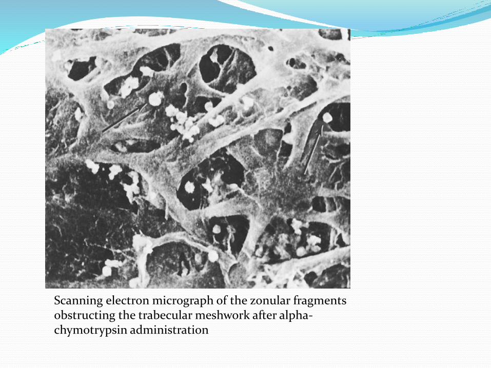
Scanning electron micrograph of the zonular fragmentsobstructing the trabecular meshwork after alpha-chymotrypsin administration

2. Glaucoma From ViscoelasticSubstances
Sodium hyaluronate blocks TM
tiny ruby-like globs of hemorrhage on the iris surface or suspended in the anterior chamber

3. Glaucoma with Pigment Dispersion from Intraocular Lenses
Mechanism:-
Decentered, tilted, excessively mobile, too small, or reversed in position
excess friction between the optic or haptic and the iris pigment epithelium
Pigment particles block TM
c/f :- geographical loss of iris pigment

4. Uveitis-Glaucoma-HyphemaSyndrome Seen in iris supported ACIOL / PCIOL
c/f:- rai.IOP, iridocyclitis & recurrent hyphema for wks to mons after surg.
Due to excessive chafing of the iris by the pseudophakics because the lenses are too mobile
5.Glaucoma After nD: yag LaserPosterior Capsulotomy
occurs within 2–4 hours of the laser treatment and then abates spontaneously over the next 24 hours
usually associated with particulate debris clogging the trabecular meshwork
Pretreatment with apraclonidine 1% 1 hour prior to surgery and one drop 1 hour after surgery has been shown to decrease the number and severity of postoperative pressure spikes

6. Glaucoma from Vitreous in the Anterior Chamber
Common in aphakic eyes
after a spontaneous rupture of the hyaloid face or after an extensive posterior vitreous detachment.
Retinal detachment and glaucoma Due to vitreous loss
Schwartz Syndrome- RD + Rai. IOP + decrea. Outflow + open angles + cells & flare in AC
Mechanism:- a)angle recession,inflammation, pigment granules released by the retinal pigment epithelium, and glycosaminoglycans synthesized by the photoreceptors blocks TM
- b) photoreceptor outer segments migrate through the retinal hole and obstruct the trabecular meshwork
Glaucoma after vitrectomyPre-existing glaucoma Angle recession Ghost cell Primary open-angle glaucoma Pigmentary glaucomaAssociated with intraocular hemorrhage Hyphema Ghost cell Hemolytic HemosiderosisRelated to lens material Phacolytic Lens particle PhacoanaphylacticNeovascularInflammatoryCorticosteroid inducedIntraocular gas or liquid Air Viscoelastic substances Perfluorocarbons Silicone
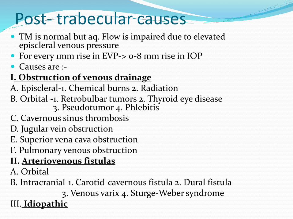
Post- trabecular causes TM is normal but aq. Flow is impaired due to elevated
episcleral venous pressure For every 1mm rise in EVP-> 0-8 mm rise in IOP Causes are :-I. Obstruction of venous drainageA. Episcleral-1. Chemical burns 2. RadiationB. Orbital -1. Retrobulbar tumors 2. Thyroid eye disease
3. Pseudotumor 4. PhlebitisC. Cavernous sinus thrombosisD. Jugular vein obstructionE. Superior vena cava obstructionF. Pulmonary venous obstructionII. Arteriovenous fistulasA. OrbitalB. Intracranial-1. Carotid-cavernous fistula 2. Dural fistula
3. Venous varix 4. Sturge-Weber syndromeIII. Idiopathic
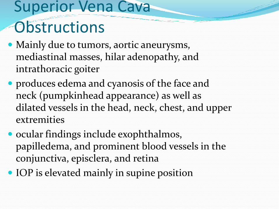
Superior Vena Cava Obstructions
Mainly due to tumors, aortic aneurysms, mediastinal masses, hilar adenopathy, and intrathoracic goiter
produces edema and cyanosis of the face and neck (pumpkinhead appearance) as well as dilated vessels in the head, neck, chest, and upper extremities
ocular findings include exophthalmos, papilledema, and prominent blood vessels in the conjunctiva, episclera, and retina
IOP is elevated mainly in supine position
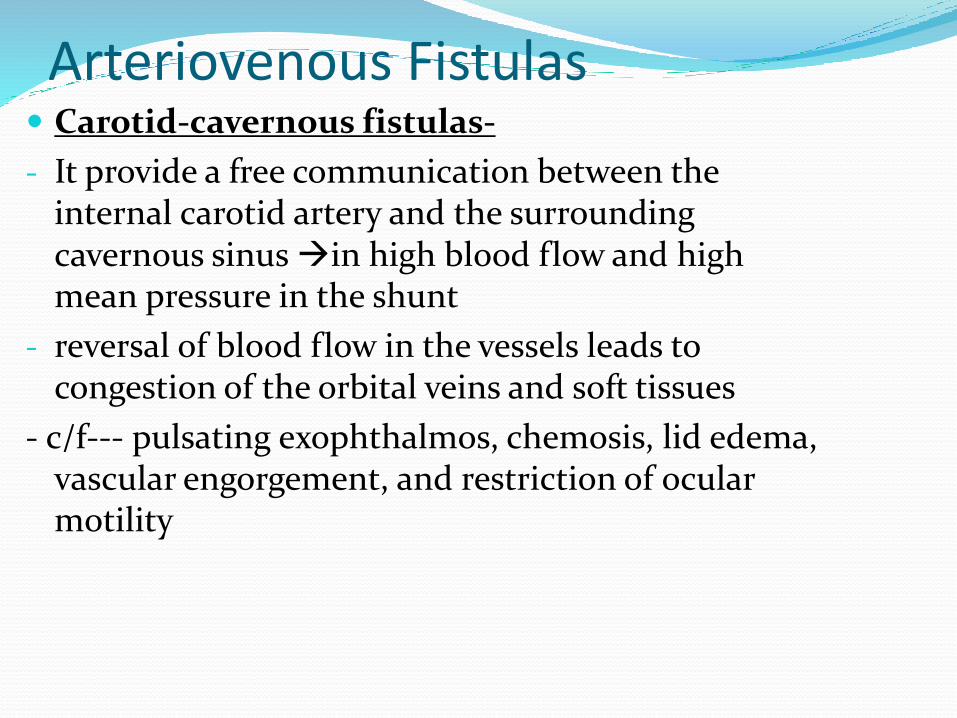
Arteriovenous Fistulas Carotid-cavernous fistulas-
- It provide a free communication between the internal carotid artery and the surrounding cavernous sinus in high blood flow and high mean pressure in the shunt
- reversal of blood flow in the vessels leads to congestion of the orbital veins and soft tissues
- c/f--- pulsating exophthalmos, chemosis, lid edema, vascular engorgement, and restriction of ocular motility
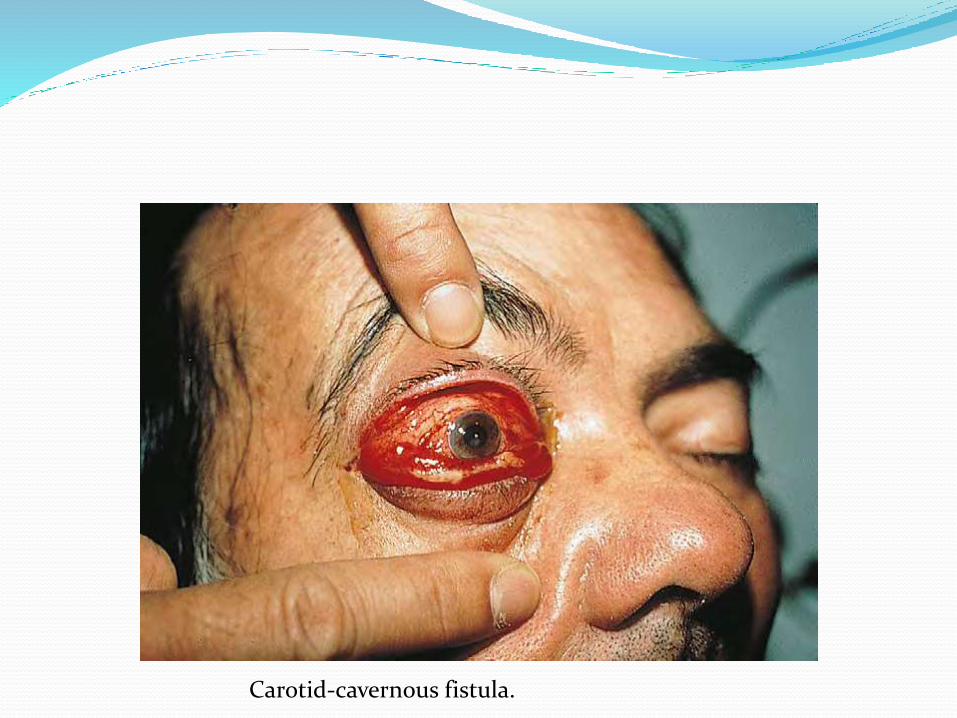
Carotid-cavernous fistula.

Contd… Sturge-Weber Syndrome
- k/a enchephalotrigeminal angiomatosis
- Congenital, sporadic oculocutaneous disorder
- Glaucoma is seen in 30% of pt’s having ipsilateral facial hemangiomata during first 2yrs of life
- Patho isolated trabeculogenesis
-> raised episcleral venous pressure
- t/tmedical (topical PG’s)
goniotomy
combined trabeculotomy & trabeculectomy

THE END