secondary hlh precipitated by tb infection - … cytokines, such as il-1, il-6 and tnf-α , that are...
TRANSCRIPT

Secondary HLH occurs mostly in adults and is usually associated with bacterial or viral infection. Familial HLH, that normallyoccurs in early childhood, has 5 defective genes identified, whereas, no genetic abnormalities are known to cause secondaryHLH. However, there may be subtle genetic defects that only manifest during overwhelming infections. Dysfunctional cytotoxiccells (CD8+ cytotoxic T cells and natural killer cells), unable to control infection, such as Mycobacterium tuberculosis, whichis an intracellular pathogen, promotes excessive production of pro-inflammatory cytokines. High systemic cytokine levelshyperstimulate tissue macrophages (histiocytes) which in severe cases may phagocytose haematopoietic cells in bone marrow(also spleen and lymph node) causing cytopaenia. Pro-inflammatory cytokines secreted by activated macrophages mediatefever, rash (increased vascular permeability) and multiple organ infiltration by immune cells.
Secondary HLH precipitated by TB infection
Lungs
Bonemarrow
Phagocytosis
Alveolarmacrophage
Mycobacteriumtuberculosis
CD8+cytotoxic
T cell
Naturalkillercell
Infectedmacrophage
Pro-inflammatorycytokines
Haemophagocytosis
Haematopoieticcells
Activatedtissue
macrophage(histiocyte)

Monocyte
Macrophage
Activatedtissue
macrophage(histiocyte)
Haematopoieticcells
CD8+cytotoxic
T lymphocyte
Naturalkillercell
IL-1α IL-1β IL-6
INF-γ
INF-γ
TNF-α
TNF-α
M-CSFGM-CSF
TNF-α IL-2
Defectivekilling
mechanismCytokines
Infection
IL-2
Haemophagocytosis
Haemophagocytic Lymphohistiocytosis mechanism
Gene defects are not known for secondary HLH, however, in familial HLH, several genes involved in the cell cytotoxicitymechanism of CD8+ cytotoxic T cells and natural killer cells have been identified. The failure of cytotoxic cells to resolve infectioncoupled to increased systemic levels of pro-inflammatory cytokines, particularly INF-γ and TNF-α, promotes excessive activationof macrophages. The enhanced phagocytic ability of activated macrophages can lead to destruction of haematopoietic cellsor their precursors in bone marrow (also spleen and lymph node) causing cytopaenia. Activated macrophages also secretepro-inflammatory cytokines, such as IL-1, IL-6 and TNF-α , that are associated with some of the symptoms of HLH such asfever, rash (increased vascular permeability) and multiple organ infiltration by immune cells.
Bonemarrow
Cytokines

CD8+ cytotoxic T cell killing mechanism
CD8+ cytotoxic T cells are activated by engagement of the T cell receptor with antigenic peptide-loaded HLA class I receptorson the surface of the target cell. Signal transduction mediated by the intracellular domains of the T cell receptor initiates acascade of cytoplasmic events that promotes the formation and degranulation of exocytic vesicles containing perforin andgranzymes at the contact area between the cytotoxic T cell and the target cell (immunological synapse). Perforin forms apermeable pore in the target cell membrane which allows entry of granzymes (that initiates apoptosis) and loss of water(causes osmotic shock) leading to cell death.
Peptide
CD8 HLA I
TCR
Targetcell
membrane
CytotoxicT cell
membraneSignal
transduction
Extracellularspace
TargetCell
Kill
CD8+cytotoxic
T lymphocyte Pro-inflammatorycytokines
Perforin
Granzymes
Inhibitoryreceptor
(KIR)
Inhibitoryligand(HLA I)
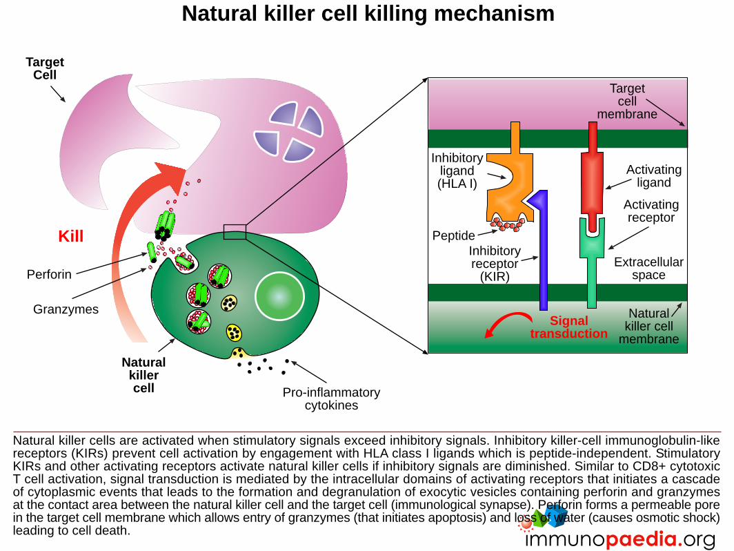
Natural killer cell killing mechanism
TargetCell
Kill
Naturalkillercell
Perforin
Granzymes
Peptide
Targetcell
membrane
Naturalkiller cell
membraneSignal
transduction
Extracellularspace
Activatingligand
Activatingreceptor
Pro-inflammatorycytokines
Natural killer cells are activated when stimulatory signals exceed inhibitory signals. Inhibitory killer-cell immunoglobulin-likereceptors (KIRs) prevent cell activation by engagement with HLA class I ligands which is peptide-independent. StimulatoryKIRs and other activating receptors activate natural killer cells if inhibitory signals are diminished. Similar to CD8+ cytotoxicT cell activation, signal transduction is mediated by the intracellular domains of activating receptors that initiates a cascadeof cytoplasmic events that leads to the formation and degranulation of exocytic vesicles containing perforin and granzymesat the contact area between the natural killer cell and the target cell (immunological synapse). Perforin forms a permeable porein the target cell membrane which allows entry of granzymes (that initiates apoptosis) and loss of water (causes osmotic shock)leading to cell death.

Degranulation mechanism: signal transduction
The first event necessary to initiate the degranulation process of cytotoxic cells, such as CD8+ cytotoxic T cells and naturalkiller cells, is activation of the cell via receptor-ligand signal transduction which takes place at the contact area between thecytotoxic cell and the target cell (immunological synapse). In CD8+ cytotoxic T cells, this is mediated by engagement of theT cell receptor with HLA class I ligands bearing immunogenic peptides. In natural killer cells, activation occurs when stimulatorysignals exceed inhibitory signals.
Cellcytoplasm
Golgibody
Signaltransduction
Targetcell
membraneExtracellularspace
Cytotoxiccell
membrane
1

Degranulation mechanism: Perforin biogenesis
Cellcytoplasm
2Perforin
biogenesis
*Perforin
Lysosome
Exocyticvesicle
Rab27a*Munc13-4 SLP1
or -2
Granzymes
Endosome
Recyclingendosome
Activation of cytotoxic cells via signal transduction initiates the formation of exocytic vesicles containing perforin and granzymes.These proteins are synthesised in the Golgi body and sorted to vesicles. Perforin is sorted into a lysosome which fuses witha late endosome containing granzymes to form an exocytic vesicle destined for membrane fusion at the immunological synapse.Importantly the granzyme-containing endosome is initially formed by sorting of granzymes into an early endosome that fuseswith a recycled endosome returning from the cell membrane. This brings two important membrane proteins, Munc13-4 andRab27a, together which is necessary for pre-fusion docking of the exocytic vesicle with the cytotoxic cell membrane. In familialHLH type 2, which is the most common form of inherited HLH, mutations are observed in the gene encoding perforin protein,while defects in Munc13-4 are known for familial HLH type 3.
Targetcell
membraneExtracellularspace
Cytotoxiccell
membrane
Golgibody

Degranulation mechanism: polarisation
The exocytic vesicle containing perforin and granzymes is destined for fusion with the cell membrane of the cytotoxic cell.Importantly, fusion must take place at the contact area between the cytotoxic cell and the target cell (immunological synapse).The process that mobilises and directs the exocytic vesicle to the correct position inside the cell is called polarisation andmakes use of the microtubule cytoskeleton as well as proteins in the vesicle membrane.
Cellcytoplasm
Polarisation
VAMP
Microtubule
3
Targetcell
membraneExtracellularspace
Cytotoxiccell
membrane

Degranulation mechanism: docking
Once the exocytic vesicle has reached the cell membrane, protein interactions immobilise the vesicle and prepare it formembrane fusion called docking. In familial HLH type 3, 4 and 5, defects in Munc13-4, Syntaxin II and Munc18-2 proteins,respectively, cause failure of the exocytic vesicle to dock at the cell membrane, thus preventing membrane fusion from takingplace.
Cellcytoplasm
Docking
*Syntaxin II*Munc18-2*Munc13-4
4
Targetcell
membraneExtracellularspace
Cytotoxiccell
membrane

Degranulation mechanism: fusion
Fusion of the cell and exocytic vesicle membranes facilitates the release of perforin and granzymes into the extracellular spacebetween the cytotoxic cell and the target cell at the immunological synapse.
Cellcytoplasm
Fusion
5
Targetcell
membraneExtracellularspace
Cytotoxiccell
membrane

Degranulation mechanism: target cell lysis
Cellcytoplasm
Targetcell
lysis
In a calcium-dependent mechanism, perforin monomers complex into a permeable pore in the cell membrane of the targetcell. Granzymes penetrate the target cell by passing through the pore and initiate programmed cell death (apoptosis). Lossof water through the pore also contributes to cell death by osmotic shock.
6Target
cellmembraneExtracellular
space
Cytotoxiccell
membrane

Summary: Defective proteins in familial HLH
In familial HLH a number of defective proteins are known to affect the formation and degranulation of exocytic vesicles containingperforin and granzymes in cytotoxic cells such as CD8+ cytotoxic T cells and natural killer cells. In familial HLH type 1 anunknown gene on chromosome 9 is affected. The most common form of familial HLH is type 2 with defects in the perforinprotein. In familial HLH type 3, 4 and 5, defective proteins Munc13-4, Syntaxin II and Munc18-2, respectively, are all involvedin pre-fusion docking of the exocytic vesicle at the cell membrane. Failure of cytotoxic cells to control intracellular infectionleads to excessive cytokine stimulation of phagocytes.
*Syntaxin II*Munc18-2*Munc13-4
*Perforin
*Munc13-4
*WASp
*AP3B1
*AP3B1
*LYST
*MyoVa
*Rab27a
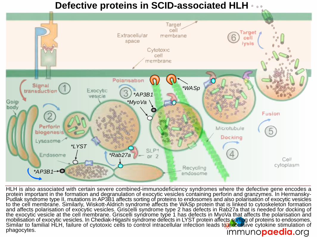
Defective proteins in SCID-associated HLH
HLH is also associated with certain severe combined-immunodeficiency syndromes where the defective gene encodes aprotein important in the formation and degranulation of exocytic vesicles containing perforin and granzymes. In Hermansky-Pudlak syndrome type II, mutations in AP3B1 affects sorting of proteins to endosomes and also polarisation of exocytic vesiclesto the cell membrane. Similarly, Wiskott-Aldrich syndrome affects the WASp protein that is linked to cytoskeleton formationand affects polarisation of exocytic vesicles. Griscelli syndrome type 2 has defects in Rab27a that is needed for docking ofthe exocytic vesicle at the cell membrane. Griscelli syndrome type 1 has defects in MyoVa that affects the polarisation andmobilisation of exocytic vesicles. In Chediak-Higashi syndrome defects in LYST protein affects sorting of proteins to endosomes.Similar to familial HLH, failure of cytotoxic cells to control intracellular infection leads to excessive cytokine stimulation ofphagocytes.