scripta scientifica medica -...
TRANSCRIPT

Medical UniversityProf. Dr. Paraskev Stoyanov
VARNA, Bulgaria
ScriptaScientifica
Medica
Vol. 44 (1), 2012Supplement 1

SCRIPTA SCIENTIFICA MEDICAAn official publication of Medical University "Prof. Dr. Paraskev Stoyanov", Varna
Editor-in-Chief:
Prof. Anelia Klissarova, MD, PhD, DSc
Rector of Medical University of Varna
e-mail: [email protected]
Co-Editor-in-Chief:
Prof. Rossen Madjov, MD, PhD, DSc
Vice Rector of Medical University of Varna
e-mail: [email protected]
Editorial Board:
Assoc Prof. Peter Genev, MD, PhD Assoc. Prof. Boriana Varbanova, MD, PhD
Department of Pathoanatomy Department of Pediatrics and Medical Genetics
E-mail: [email protected] e-mail: [email protected]
Assoc. Prof. Minko Minkov, MD, PhD Assoc. Prof. Zhaneta Georgieva, MD, PhD
Head, Department of Anatomy, Histology and Vice Rector University Hospital Coordination and
Embryology Postgraduate Education
e-mail: [email protected] e-mail: [email protected]
Prof. Krasimir Ivanov, MD, PhD, DSc Assoc. Prof. Svetoslav Georgiev, MD, PhD
Head, Department of Surgery Department of Internal Medicine
e-mail: [email protected] e-mail: [email protected]
Prof. Iskren Kotsev, MD, PhD, DSc Assoc. Prof. Negrin Negrev, MD, PhD
Department of Hepato - Gastroenterology Vice Rector for Students Affair
e-mail: [email protected] e-mail: [email protected]
Assoc. Prof. Marinka Peneva, MD, PhD Assoc. Prof. Stoyanka Popova, MD, PhD
Dean, Faculty of Medicine Dean, Faculty of Public Health
e-mail: [email protected] e-mail: [email protected]
Assoc. Prof. Vasil Svechtarov, DMD, PhD Assoc. Prof. Violeta Tacheva, PhD
Dean, Faculty of Dental Medicine Head, Department of Language Teaching,
e-mail: [email protected] Communications and Sports
e-mail: [email protected]
Assoc. Prof. Diana Ivanova, MD, PhD Assoc. Prof. Valentina Madjova, MD, PhD
Head, Department of biochemistry molecular medicine Department of Family Medicine
and nutragenomics e-mail: [email protected]
e-mail: [email protected]
Assoc. Prof. Emanuela Mutafova, PhD Assoc. Prof. Radoslav Radev, MD, PhD
Head, Department of Economics Head, Department of Surgery
and Healthcare Management e-mail: [email protected]
e-mail: [email protected]
Secretary:
Nikola Kolev, MD, PhD Emilia Jordanova
Department of Surgery IT Center
University Hospital "St. Marina" e-mail: [email protected]

MEDICAL UNIVERSITY “PROF. DR. PARASKEVSTOYANOV” VARNA
BULGARIAN SOCIETY FOR PHYSIOLOGICAL SCIENCES
PROCEEDINGS
OF THE X NATIONAL CONGRESS
OF BULGARIAN SOCIETY FOR PHYSIOLOGICAL SCIENCES
6 - 9 OCTOBER 2011
V A R N A
UNDER THE AUSPICES OF
Prof. Anelia Klissarova, MD, PhD, DSc Rector, Medical University “Prof. Dr. Paraskev Stoyanov”
Varna
and
Prof. Alexander Stoynev, MD, PhD, DSc President, Bulgarian Society for Physiological Sciences
Volume 44(1) Supplement 1, 2012


CONTENTS
Kossev A. - SENSORIMOTOR INTEGRATION: TMS STUDIES OF PROCESSING
OF PROPRIOCEPTIVE INFORMATION IN HEALTH AND DISEASE . . . . . . . . . . . . . . . . . . . . 5
Genova B., N. Bocheva, M. Stefanova, S. Stefanov - EFFECTS OF ADAPTATION
IN VISUAL PERCEPTION OF GLOBAL SPEED . . . . . . . . . . . . . . . . . . . . . . . . . . . . . . . 11
Dushanova J., D. Mitov - VISUAL EVENT-RELATED POTENTIALS BY
AN IDENTIFICATION OF GRATING ORIENTATION DIFFERENCE . . . . . . . . . . . . . . . . . . . . 15
Tancheva L., E. Encheva, M. Novoselski, V. Petkov, R. Klisurov -
SEX-DEPENDENT EFFECT OF A NEW PEPTIDOMIMETIC ON COGNITIVE FUNCTION
OF ISOLATED RATS AFTER MATERNAL DEPRIVATION . . . . . . . . . . . . . . . . . . . . . . . . . 19
Popova E., P. Kupenova, I. Ivanov - EFFECTS OF D1 RECEPTOR BLOCKADE
ON THE INTENSITY-RESPONSE FUNCTION OF FROG DARK ADAPTED ELECTRORETINOGRAM . . 23
Pavlova E., G. Uzunova, P. Somlev - DYNAMICS OF HEART RATE RECOVERY
AFTER CYCLE ERGOmetric PWC170 TEST IN SOCCER PLAYERS. . . . . . . . . . . . . . . . . . . . . 27
Bekyarova G., M. Hristova - URIC ACID, THIOLS AND BURN-INDUCED OXIDATIVE
INJURY OF GASTRIC MUCOSA IN RATS . . . . . . . . . . . . . . . . . . . . . . . . . . . . . . . . . 31
Bekyarova G., M. Hristova, M. Tsaneva - CYTOPROTECTIVE EFFECT
OF MELATONIN AND ANTIOXIDANT DEFENSE AFTER THERMAL SKIN INJURY . . . . . . . . . . . 35
Sarov G. - UNDERSTANDING PATHOGENESIS OF RISKY BEHAVIOR: A HOLISTIC APPROACH . . 39
Sarov G. - COMPARISON OF FUNCTIONAL AND STRUCTURAL CLASSIFICATION IN
PATHOPHYSIOLOGICAL THEORY AND TEACHING . . . . . . . . . . . . . . . . . . . . . . . . . . . 43
Tchekalarova J., D. Pechlivanova, Zl. Petkova, Al. Stoynev - ÅFFECT OF CHRONIC
TREATMENT WITH MELATONIN ON DIURNAL VARIATIONS OF SPONATENOUS MOTOR
SEIZURES AND BEHAVIOR OF WISTAR AND SPONTANEOUSLY HYPERTENSIVE RATS
IN KAINATE MODEL OF TEMPORAL LOBE EPILEPSY . . . . . . . . . . . . . . . . . . . . . . . . . . 47
Georgiev K., E. Miladinova, T. Pajpanova - DESIGN, SYNTHESIS AND ANALYSIS OF NOVEL
ENDOMORPHIN-2 AND MORPHICEPTIN ANALOGUES. . . . . . . . . . . . . . . . . . . . . . . . . . 51
Petrov L., P. Atanasov, N. Zaekov, A. Alexandrova, Z. Zsheliaskova-Koynova,
I. Achkakanov - PHYSIOLOGICAL AND NON-INVASIVE BIOCHEMICAL INDEXES
IN A MODEL OF EMOTIONAL STRESS IN SHOOTERS . . . . . . . . . . . . . . . . . . . . . . . . . . 55
Ivanova M., S. Belcheva, I. Belcheva, N. Negrev, R. Tashev - OLFACTORY BULBECTOMY
INDUCES SHIFTS IN LATERALIZATION OF LOCOMOTOR RESPONSES TO VASOACTIVE
INTESTINAL PEPTIDE . . . . . . . . . . . . . . . . . . . . . . . . . . . . . . . . . . . . . . . . . . . . 59
Danovska M., M. Alexandrova - LOW GRADE INFLAMMATION IN DIABETIC
HYPERTENSIVE INDIVIDUALS . . . . . . . . . . . . . . . . . . . . . . . . . . . . . . . . . . . . . . . 63
Mihaylova M. , I. Hristov, K. Racheva, Ts. Totev, D. Mitov - EARLY VEP WAVES
ARE AFFECTED STRONGER BY GRATING LENGTH THAN WIDTH . . . . . . . . . . . . . . . . . . . 67
Stefanova M., N. Bocheva, O. Georgieva - EVALUATION OF INDIVIDUAL, GENDER
AND AGE DIFFERENCES IN VISUAL MOTION PERCEPTION . . . . . . . . . . . . . . . . . . . . . . . 73
Kolev N., L. Halacheva - SEX DIFFERENCES IN FUNCTIONAL ORGANIZATION OF BIMANUAL
SHOT-LIKE ISOMETRIC HANDGRIP . . . . . . . . . . . . . . . . . . . . . . . . . . . . . . . . . . . . 77
Angelova P., N. Boyadjiev, A. Ivanova, S. Muletarov - CHANGES IN THE PHYSICAL
WORKING CAPACITY, BODY COMPOSITION AND FAT METABOLISM OF WOMEN ON A
THREE-MONTH SPECIALIZED TRAINING AND DIETARY PROGRAM. . . . . . . . . . . . . . . . . . 81
Somlev P. - THE EFFECTS OF PACED BREATHING ON SPECTRAL PARAMETERS
OF HEART RATE VARIABILITY IN ATHLETES AND UNTRAINED CONTROLS . . . . . . . . . . . . . 85
Somlev P., G. Uzunova, E. Pavlova - HEART RATE VARIABILITY AT REST IN ELITE AND FORMER
SOCCER PLAYERS. . . . . . . . . . . . . . . . . . . . . . . . . . . . . . . . . . . . . . . . . . . . . . 89
3

Georgiev Ts., P. Hadzhibozheva, A. Tolekova - CONTRACTILE RESPONSES OF THE RAT
UTERINE SMOOTH MUSCLE TO INFLUENCES WITH ANGIOTENSIN II AND VASOPRESSIN . . . . . 93
Kolarova R. - CARDIOVASCULAR STRESS AND MYOCARDIAL PROTECTION (I) . . . . . . . . . . 97
Kolarova R., Z. Gendzhev - ANTIOXIDANT PREVENTION IN EXPERIMENTAL
HYPERTENSION: HISTOMORPHOLOGICAL CHANGES (II) . . . . . . . . . . . . . . . . . . . . . . . 101
Kolarova R. - EXPERIMENTAL HEMODINAMIC STRESS AND ITS INFLUENCE FROM
PHYSICAL EXERCISE, HYPOKINESIA AND ANTIOXIDANTS . . . . . . . . . . . . . . . . . . . . . . 105
Stefanov S., K. Alexiev, N. Bocheva, B. Genova - AGE-RELATED EFFECTS
ON THE SENSITIVITY TO GLOBAL MOTION DIRECTION DETERMINED BY THE METHOD
OF CLASSIFICATION IMAGES . . . . . . . . . . . . . . . . . . . . . . . . . . . . . . . . . . . . . . . 109
Tzvetkov S. - SPECIFICITY IN BREATHING REGULATION DURING PHYSICAL EXERCISE
IN TAEKWONDO NATIONAL ATHLETES. . . . . . . . . . . . . . . . . . . . . . . . . . . . . . . . . 113
Krustev S. M., N. Negrev, D. I. Stephanova - THE STRENGTH-DURATION PROPERTIES
IN SIMULATED DEMYELINATING NEUROPATHIES DEPEND ON THE MYELIN SHEATH
AQUEOUS LAYERS. . . . . . . . . . . . . . . . . . . . . . . . . . . . . . . . . . . . . . . . . . . . . 117
Minchev Zl., P. Gatev - PSYCHOPHYSIOLOGICAL EVALUATION OF EMOTIONS DUE
TO THE COMMUNICATION IN SOCIAL NETWORKS. . . . . . . . . . . . . . . . . . . . . . . . . . . 125
Decheva L., I. Pashalieva, E. Stancheva, Y. Nyagolov, N. Negrev - MELATONIN
AND THE FIBRINOLYTIC SYSTEM IN THE RAT: EFFECTS ON THROMBIN ACTIVATABLE
FIBRILOLYSIS INHIBITOR (TAFI) . . . . . . . . . . . . . . . . . . . . . . . . . . . . . . . . . . . . . 129
Nikolova J., M. Orbezova, P. Atanasova, P. Nikolov, F. Nikolov, P. Hrischev -
PREHYPERTENSION IN WOMEN WITH METABOLIC SYNDROME . . . . . . . . . . . . . . . . . . . 133
Nyagolov Y., I. Pashaliewa, N. Doncheva, N. Negrev - SOMATOTROPIN, SOMATOSTATIN
AND PROLACTIN EFFECTS ON VITAMIN K-DEPENDENT PLASMA CLOTTING FACTOR
(FII, FVII, FIX, FX) ANTIGEN CONCENTRATIONS IN RATS . . . . . . . . . . . . . . . . . . . . . . . 137
Hristov K. - RESEARCH ON VARIATIONS IN SOME ELECTROPHYSIOLOGICAL
CHARECTERISTIC DATA OF ACUPUNCTURE POINTS IN EXPERIMENTAL ANIMALS PUT
UNDER DIFFERENT ATMOSPHERE PRESSURE . . . . . . . . . . . . . . . . . . . . . . . . . . . . . 141
Daskalov Ì. - LONG-TERM SURVIVAL OF A SERIES OF CANCER PATIENTS WITH PURULENT
SEPTIC POSTOPERATIVE COMPLICATIONS . . . . . . . . . . . . . . . . . . . . . . . . . . . . . . . 145
AUTOR'S INDEX . . . . . . . . . . . . . . . . . . . . . . . . . . . . . . . . . . . . . . . . . . . . . . 149
PERMUTERM SUBJECT INDEX . . . . . . . . . . . . . . . . . . . . . . . . . . . . . . . . . . . . . . 151
INSTRUCTIONS TO AUTHORS. . . . . . . . . . . . . . . . . . . . . . . . . . . . . . . . . . . . . . . 155
4

SENSORIMOTOR INTEGRATION: TMS STUDIES OF PROCESSING
OF PROPRIOCEPTIVE INFORMATION IN HEALTH AND DISEASE
Kossev A.
ABSTRACT
Sensorimotor integration was investigated by transcranial magnetic stimulation (TMS) and muscle vibration
(MV) in healthy subjects and patients with cervical dystonia (CD), idiopathic Parkinson’s disease (IPD), clini-
cally probable Parkinsonian multiple system atrophy (MSA-P) and amyotrophic lateral sclerosis (ALS). MV
(80 Hz, 0.5 mm) was applied to the right extensor carpi radialis muscle (ECR). Single and paired TMS pulses
were applied 3 s after onset of the MV. Motor evoked potentials (MEPs) were recorded from the vibrated ECR
and its antagonist, the flexor carpi radialis muscle using conventional surface electromyographic techniques.
In healthy subjects a vibratory related facilitation of the motor cortex was described. In patients with CD MV
induced facilitation was significantly reduced. During MV the level of intracortical inhibition (ICI) was also
reduced in CD patients in contrast to increased level of intracortical facilitation. The impaired sensorimotor
integration in CD patients is probably due to the alterations of basal ganglial function. We found that process-
ing of proprioceptive information is differently affected in IPD and MSA-P patients. The impaired ICI and
missing MV induced facilitation of MEP in IPD p-atients can be partially normalized by the Levodopa. Our
results suggested an impairment of ICI and processing of proprioception in ALS patients. Inhibition of excit-
atory neurotransmitters by riluzol might explain the recovery of ICI in ALS patients.
Key words: transcranial magnetic stimulation (TMS), muscle vibration (MV), motor evoked potential (MEP),
intracortical inhibition (ICI), intracortical facilitation (ICF), silent period (SP)
INTRODUCTION
Sensorimotor integration is the process whereby sensory in-
put is integrated by the central nervous system and used for
assisting motor program execution. Motor evoked potentials
(MEPs) in response to transcranial magnetic stimulation
(TMS) are subject to various influences. A powerful means
to modify MEPs is muscle vibration (MV) (4). The vibration
stimuli mainly excite the muscle spindle afferents and the
motor cortex is primarily responsible for the processing of
muscle spindle signals and limb movement perception.
METHODS
We have studied healthy volunteers and patients with cer-
vical dystonia (CD), idiopathic Parkinson”s disease (IPD),
clinically probable Parkinsonian multiple system atrophy
(MSA-P) and amyotrophic lateral sclerosis (ALS). The re-
sults from patient’s groups were compared to those of
age-matched control subjects. The study was approved by
the local Ethics Committee and subjects and patients were
studied after giving written informed consent. For the clini-
cal details see the original papers (5,16,18,19).
Subjects and patients were seated with the right arm gently
fixed in slight abduction from the trunk (20o) and flexion in
the elbow (110 o). MV was used with a frequency of 80 Hz
and an amplitude of 0.5 mm which avoided generation of the
tonic vibration reflex (TVR). MV was applied to the right
extensor carpi radialis muscle (ECR) by means of an electro-
magnetic mechanical stimulator (Ling Dynamic Systems,
Model V100) with a disk surface (2 cmin diameter). The du-
ration of the MV trains was 4 s with random intertrial inter-
vals between 12 and 22 s. In a group of healthy subjects we
used different MV frequencies – 80, 120 and 160 Hz.
TMSwasappliedassingleorpairedpulsesusing twoMagStim
200 stimulators (Magstim Company, Ltd., UK) connected to a
stimulating coil through a BiStim module. The circular coil
(mean diameter 9 cm; current flow anticlockwise when viewed
from above) was adjusted over the vertex to evoke optimal
muscle responses in the muscles of the right side.
Motor evoked potentials were recorded from the vibrated
ECR and its antagonist, the flexor carpi radialis muscle
(FCR) using conventional surface electromyographic
(EMG) techniques. Signals were amplified (band pass 10
Hz - 5 kHz) and digitized (sampling rate 10 kHz). Periods
of 400 ms duration (100 ms prior to the stimulus and 300
ms after it) were stored on the disk. The EMG activity was
continuously monitored, and patients and subjects had au-
dio-visual feedback to ensure the absence of voluntary
background activity as well as of TVR. In the group of
healthy subjects we recorded also MEPs of the muscles
contralateral to the vibrated arm.
Motor threshold (MT) was determined at rest as the lowest
stimulus intensity eliciting three responses of at least 50 µV
peak to peak amplitude in four stimuli.
5
Scripta Scientifica Medica, 2012; vol. 44 (1), Supplement 1 Copyright © Medical University, Varna
Address for correspondence:
A. Kossev, Bulgarian Academy of Sciences, Sofia 1113,Acad. G.
Bontchev Str., Bl.21.,
e-mail: [email protected]

Single pulse TMS (s-TMS) was applied using an intensity
of 120 % of MT. Paired stimuli (p-TMS) were performed
with intensities of 70% of MT for the conditioning first
pulse and 120 % of MT for the following test pulse at
interstimulus intervals (ISI) of 3 ms to evaluate intracortical
inhibition (ICI) and 13 ms for intracortical facilitation (ICF)
as originally described by Kujirai et al. (10,15).
TMS was applied first without MV. Five single stimuli were
followed by 10 paired stimuli (five of each ISI presented in
random order) with random intertrial intervals. Thereafter,
the procedure was repeated with MV of the ECR. Single and
paired TMS pulses were applied 3 s after the onset of MV.
In the group of healthy subjects we used both TMS and
transcranial electrical stimulation (TES), which allows for a
differentiation between the cortical and spinal actions. TES
was carried out using a Digitimer (Model D180, Digitimer
Ltd., Welwyn Garden City, UK) as the cathode placed over
the vertex, and the anode - about 7 cm lateral to the left.
Cortical silent period (SP) was investigated at 30 % of maxi-
mal isometric voluntary contraction force (MVIC) of ECR. A
single TMSpulse (intensity120 % of MT) was applied during
voluntary contraction of the ECR. Five trials were obtained
and the procedure was repeated with vibration of the ECR.
The MEP parameters of interest was MEP size as assessed by
the total voltage time integral (area) or peak-to- peak amplitude
and MEP latency. In each subject, MEP values were normal-
ized to themeanvaluesobtainedwithsingleTMSwithoutMV.
The resulting values were pooled for the groups and were ex-
pressed as mean ± standard deviation.
Paired t-test was used to assess the effects of paired pulse TMS.
Differences between controls and patients and the effect of MV
were assessed using analysis of variance (ANOVA). Probabil-
ity (p) values of < 0.05 were considered significant.
RESULTS AND DISCUSSION
MV in healthy subjects.
In healthy subjects MEPs in the vibrated muscle are aug-
mented when TMS is applied more than 120 ms after MV
onset (Fig.1). Simultaneously MEPs recorded from the an-
tagonist of the vibrated muscle are suppressed (20). The
augmentation of the MEPs in the vibrated muscle was ob-
served at MV frequencies 80 and 120 Hz but not at 160 Hz
and was increasing with ongoing MV. In contrast, the inhi-
bition in the antagonist revealed a reverse time course and
was also induced by vibration frequency 160 Hz.
When TES was used, MEPs were not augmented in the vi-
brated muscles while those in the antagonist were de-
creased (9) (Fig.2). This dissociation between the effects of
TMS and TES as well as the different frequency dependen-
cies and different time courses suggest involvement of dif-
ferent neuronal circuits and possibly different receptor in-
puts in the MEP augmentation. It seems that MEP augmen-
tation in the vibrated muscle is caused by cortical activation
while MEP depression in the antagonist seems to involve
both spinal and cortical circuits and probably other dy-
namic mechanoreceptors (8).
MV induced facilitation was stronger at 80 Hz than at
higher frequency stimulation. Using lower vibration fre-
quency Steyvers et al. (21) confirmed that the optimal fre-
quency is in the range around 75 Hz, which is the optimal
for stimulation of the primary endings (14). All this find-
ings suggest that the primary muscle spindle messages (Ia
fibers) are the major sensory input underlying the vibratory
related facilitation of the motor cortex.
During MV the homonymous contralateral muscle revealed
a slight non-significant augmentation of MEPs but its antag-
onist showed a gradual depression of MEPs with ongoing
MV reaching significant levels (7). These findings suggest
transcallosal MEP modulation which involve the Ia
afferents.
MV in patients with cervical dystonia.
Eleven patients with cervical dystonia and 11 age-matched
healthy control subjects were enrolled in the study (5,19).
Motor thresholds (MT) as well as MEP areas after single
TMS without MV in ECR and FCR for CD patients were
fairly similar to those in the control subjects. With condi-
tioning TMS, ICI with short ISIs (3 msec) and ICF with
6
Sensorimotor integration: TMS studies of processing of ...
Fig. 1: Area of motorevoked potentials (MEPs)
recorded from vibrated extensor carpi radialis muscle
(ECR) and its antagonist flexor carpi radialis (FCR).
The parameters are normalized to the mean control
values and pooled for all subjects (mean ± standard
error; n = 10). Motor evoked potentials were recorded
without muscle vibration - MV (control – c.), 0.5 s and
3 s after onset of MV, and 1 s after offset of MV (pv.).
Three different vibration frequencies were used: 80 Hz,
120 Hz, and 160 Hz. Asterisks indicate significant
differences to control values.

long ISIs (13 msec) were significantly pronounced simi-
larly to the control subjects (see Fig.3).
With single-pulse TMS there was a significant MV induced
MEP augmentation in the vibrated ECR, although it was
much less pronounced than in control subjects (see Fig. 3).
Simultaneously, the MEPs recorded from the antagonist
showed a tendency to be reduced compared with single
TMS without MV. Comparison of the two groups (control
subjects and patients) revealed significantly smaller MEP ar-
eas recorded from both muscles during MV in the patients.
The MV-induced changes of the conditioned MEPs were
also different from those in the control subjects. The MEP
reduction at ISIs of 3 ms was smaller than in control sub-
jects and did not reach significant level. In contrast to con-
trol subjects, there was a significant ICF with ISIs of 13 ms.
The MV-induced augmentation in patients was suppressed
significantly, resembling previous findings in patients with
musician’s cramp (Rosenkranz et al., 2000). The essential
difference between the two studies was that the vibrated
forearm flexors were localized in the affected body segment
in musician’s cramp, whereas this was clearly not the case in
cervical dystonia. Therefore, this similarity suggests that the
changes of cortical function in focal dystonia are more gen-
eralized and extend beyond the projection areas of the clini-
cally involved muscle groups. The comparison of MV-in-
duced effects on MEPs in response to TMS and TES
strongly suggest that MV-induced augmentation results from
the cortical rather than spinal activation (9), supporting the
hypothesis that abnormal MEP augmentation in our patients
is caused by an impairment of the cortical mechanisms.
The mechanisms underlying abnormal MV induced effects
in patients, however, are not clear. Because the MEPs in the
patients were abnormal only during MV, it seems most
likely that the sensorimotor integration is impaired. Proba-
bly, the gain of proprioceptive input to the cortex is
changed, or focusing to the target sensorimotor areas is im-
paired as a consequence of the alterations of the basal
ganglial function in dystonia (22). In fact, several studies
have suggested an abnormal central processing of the sen-
sory information in dystonia (1).
7
Kossev A.
Fig. 3: Unconditioned (single TMS) recorded during
MV and conditioned (paired TMS) MEP areas (mean ±
standard error; n = 11) recorded from ECR and FCR
without MV and 3 s after onset of MV applied on the
ECR. The unconditioned values during MV are
normalized to the corresponding mean value of MEP
areas recorded without MV. The conditioned MEP
areas at interstimulus intervals of 3 msec and 13 msec
are normalized to the corresponding mean value of
MEP areas recorded in response to single
(unconditioned) TMS. Open bars - healthy control
subjects; shaded bars - patients with cervical dystonia.
Asterixes indicate a significant effect of MV-induced
augmentation in the vibrated muscle, significant effect
of intracortical inhibition (ISI 3-msec) and intracortical
facilitation (3-msec ISI) and significant differences
between patients and the control group.
Fig. 2: MEPs in response to transcranial electric (TES)
(A) and transcranial magnetic stimulation (TMS) (B)
(both with intensity 120% of motor threshold). MEPs
were recorded from vibrated ECR muscle and its
antagonist FCR without MV and 3 s after the onset of
MV. The responses on the figure are average of 5
stimulations.

MV in patients with idiopathic
Parkinson´s disease (IPD) and
Parkinsonian multiple system atrophy
(MSA-P).
Ten patients with IPD, 10 patients with clinically probable
MSA-P criteria and 10 healthy control subjects were studied.
All patients were examined in a defined off-state, i.e. at least
12 hours after last intake of anti-Parkinson medication (16).
Motor thresholds at rest did not differ significantly between
healthy controls, IPD and MSA-P. Without MV MEP sizes
in both patient groups compare to controls were smaller
(not significant), ICI was missing in ECR in IPD patients
and missing in both muscles in MSA patients (Fig.4).
During MV MEPs of the vibrated ECR in response to sin-
gle and paired TMS were significantly augmented. The
MEPs recorded from the non-vibrated antagonist muscle
(FCR) were only slightly, but not significantly increased
(see Fig.4). In IPD, MEPs were not augmented by MV. In
MSA, however, MV induced significant MEP augmenta-
tion in both muscles, i.e. in the vibrated ECR and in the
non-vibrated FCR.
The duration of SP without MV did not differ significantly
between controls and both patient groups although it
tended to be shorter in IPD (p < 0.08) (Fig.5). The MV did
not change SP in controls and in IPD while it caused signif-
icant SP prolongation in MSA.
Without MV ICI in resting forearm muscles is reduced in
IPD in the off-state (13,2). It was proposed that this deficit
may reflect a reduction of excitability of GABAA depend-
ent inhibitory pathways (23) as a consequence of changed
basal ganglia output (13). In MSA we found the same defi-
cit and the impaired ICI seems to be a common
neurophysiological feature of IPD and MSA-P.
Concerning MV, it has been shown in IPD that the vibra-
tory input has less effect on the accuracy in positioning
tasks than in the healthy subjects (12,6). Thus we had ex-
pected that MV would have a less facilitatory effect on
MEPs which was basically confirmed by our results. The
MV induced MEP augmentation in IPD was completely
absent. We agree with several other authors that the
changed modulation of cortical excitability by sensory in-
formation in IPD is caused by the impaired sensory pro-
cessing. Although the level of these changes within the
CNS is not clear it is generally assumed that the dysfunc-
tion of the basal ganglia plays an important role (11).
In contrast to IPD, our MSA-P patients showed a signifi-
cant MEP augmentation which was, however, weaker than
in controls. Additionally this augmentation was not focal
and was similar in the vibrated muscle and in the non-vi-
brated antagonist.
The duration of cortical SP without MV was not signifi-
cantly different between healthy subjects and both groups
of patients with a tendency to be shorter in IPD. These re-
sults confirm previous findings in IPD and MSA (3). The
MV did not change SP significantly in control group or in
IPD patients while it prolonged the duration of SP signifi-
cantly in the MSA patients. The reason for this change and
8
Sensorimotor integration: TMS studies of processing of ...
Fig. 5: Cortical silent period (SP) (mean + standard
error, n = 10) of healthy controls, patients with
idiopathic Parkinson’s disease (IPD) and multiple
system atrophy (MSA) patients without muscle vibration
(MV) (white bars) and during MV (grey bars). The
asterisk indicates a significant prolongation of SP
Fig. 4: MEP areas (mean + standard error, n = 10)
normalized to unconditioned MEPs recorded in
response to single pulse TMS (s-TMS, S) without muscle
vibration (MV) (white bars) and during MV (grey bars).
The responses to s-TMS are marked by “S” and
responses to paired pulse TMS with 3 and 13 ms
interstimulus interval (ISI) interval are marked by “3
ms” and “13 ms”, respectively. The significance (p <
0.05) of inhibition due to ICI is marked over the white
bars (without MV) by asterisks. The asterisks between
the grey and the white bars indicate the significance of
facilitation due to MV comparing the area of the MEP

its functional significance is not clear, but obviously MSA
shows more widespread MV evoked changes. Differences
in the influence on inhibitory intracortical circuits between
IPD and MSA-P may reflect the different underlying pa-
thology.
Additionally, the processing of proprioceptive information
in IPD patients group was studied also in the on state (1
hour after the take of Levodopa) (17). The impaired ICI and
missing MV induced facilitation of MEP can be partially
normalized by Levodopa (Fig.6)
MV in patients with amyotrophic lateral
sclerosis (ALS).
Thirteen ALS patients (7 with Riluzol, 6 without Riluzol)
and 10 age-matched healthy controls were enrolled into this
study (18). The MV facilitated MEPs significantly in
healthy subjects, but not among ALS patients (Fig.7).
ALS patients without Riluzol showed an impairment of
ICI, while those on riluzole presented with normal inhibi-
tion of MEPs (Fig.8). In contrast to healthy controls the
ICF in ALS patients was significant neither in patients on
Riluzole nor in those without Riluzole although treated pa-
tients revealed a tendency to ICF in both muscles (p=0.08
and p=0.09). The results suggest an impairment of ICI and
processing of proprioception in ALS. The inhibition of ex-
citatory neurotransmitters by Riluzole might explain the re-
covery of ICI in ALS patients.
9
Kossev A.
Fig. 7: MEP areas (mean + standard error) (patients
with amyotrophic lateral sclerosis) in response to single
pulse TMS applied 3 s after mV onset, normalized to the
MEPs recorded in response to single pulse TMS without
muscle vibration (MV). The asterisks indicate
significant effect of MV.
Fig. 8: MEP areas (mean + standard error) (patients
with amyotrophic lateral sclerosis) in response to
paired pulse TMS (ISI - 3 and 13 ms) without MV,
normalized to the corresponding MEP in response to
single pulse TMS. The asterisks indicate significant
effect of conditioned stimulation.
Fig. 6: MEP areas (mean + standard error, n = 10)
normalized to unconditioned MEPs recorded in
response to single pulse TMS without muscle vibration
(MV). White bars – without MV and grey bars 3 s after
MV onset. The responses to s-TMS are marked by “S”
and responses to paired pulse TMS with 3 and 13 ms
ISI interval are marked by “3 ms” and “13 ms”,
respectively. IPD–off and IPD–on - patients with
idiopathic Parkinson’s disease studied in off state and
on state (1 hour after the take of Levodopa). The
asterisks between the grey and the white bars indicate
the significance of facilitation due to MV comparing the
area of the MEP during MV to the corresponding data
without MV.

REFERENCES
1. Abbruzzese, G. , R. Marchese, A.
Buccol ier i , e t al . Abnormalities of sensorimotor
integration in focal dystonia: a transcranial magnetic
stimulation study.- Brain, 2001, 124, 537–545.
2. Bares , M., P. Kanovsky, H. Klajblova, I .
Rektor . Intracortical inhibition and facilitation are
impaired in patients with early Parkinson’s disease: a
paired TMS study.- Eur. J. Neurol., 2003, 10,
385-389.
3. Cantel lo , R. , R. Tarlet t i , C. Civardi .
Transcranial magnetic stimulation and Parkinson’s
disease.- Brain Res. Rev., 2002, 38, 309-327.
4. Claus, D. , K. R. Mil ls , N. M. F. Murray. Fa-
cilitation of muscle responses to magnetic brain stim-
ulation by mechanical stimuli in man.- Exp. Brain
Res., 1988, 71, 273–278.
5. Dengler , R. , S. Siggelkow, J. D. Rollnik, J .
Duper , C. Moll , A. Kossev. Changes of motor
cortical function in dystonia.- In: Sensorimotor Con-
trol. R. Dengler, A. Kossev, eds. NATO Science Se-
ries, Series 1.- Life and Behavioural Sciences, 2001,
326, 150-158.
6. Khudados, E., F. W. Cody, D. J. O’Boyle.
Proprioceptive regulation of voluntary ankle move-
ments, demonstrated using muscle vibration, is im-
paired by Parkinson’s disease.- J. Neurol. Neurosurg.
Psychiatry, 1999, 67, 504-510.
7. Kossev, A., S. Siggelkow, H.-H. Kappels, R. Dengler,
J. D. Rollnik. Crossed effects of muscle vibration on
motor-evoked potentials.- Clin. Neurophysiol., 2001,
112, 453-456.
8. Kossev, A., S. Siggelkow, J. D. Rollnik, J. Däuper, R.
Dengler. Modulation of corticospinal excitability and
intracortical mechanisms during muscle vibration: a
study using transcranial magnetic stimulation.- In:
Sensorimotor Control. R. Dengler, A. Kossev, eds.
NATO Science Series, Series 1.- Life and Behav-
ioural Sciences, 2001, 326, 19-28.
9. Kossev, A., S. Siggelkow, M. Schubert, K.
Wohlfarth, R. Dengler. Muscle vibraation: different
effects on transcranial magnetic and electrical stimu-
lation.- Muscle Nerve, 1999, 22, 946-948.
10. Kujirai, T., M. D. Caramia, J. C. Rothwell, et al.
Corticocortical inhibition in human motor cortex.- J.
Physiol. (Lond.), 1993, 471, 501–519.
11. Maschke,M., C. M. Gomez, P. J. Tuite, J. Konczak.
Dysfunction of the basal ganglia, but not the cerebel-
lum, impairs kinaesthesia.- Brain, 2003, 126,
2312-2322.
12. Rickards, C., F. W. Cody. Proprioceptive control of
wrist movements in Parkinson’s disease. Reduced
muscle vibration-induced errors.- Brain, 1997, 120,
977-990.
13. Ridding, M. C., R. Inzelberg, J. C. Rothwell. Changes
in excitability of motor cortical circuitry in patients
with Parkinson’s disease.- Ann. Neurol., 1995, 37,
181-188.
14. Roll, J. P., J. P. Vedel, E. Ribot. Alteration of
proprioceptive messages induced by tendon vibration
in man: a microneurographic study.- Exp. Brain Res.,
1989, 76, 213–222.
15. Rollnik, J .D. , S. Siggelkow, J. Däuper , C.
Moll , A. R. Kossev, R. Dengler . Die
transkranielle magnetische Doppelstimulation zur
Beurteilung kortikokortikaler Inhibition und
Fazilitierung.- Klin. Neurophysiol., 2001, 32: 26-29.
16. Schrader, C., T. Peschel, J. Däuper, J. D. Rollnik, R.
Dengler, A. Kossev. () Changes in processing of
proprioceptiv information in Parkinson’s disease and
Multiple System Atrophy. Clin. Neurophysiol., 2008,
119, 1139-1146.
17. Schrader, C., T.. Peschel, A. R. Kossev. Verarbeitung
von propriozeptiver Information beim idiopathischem
Parkinson-Syndrom und der Einfluss von Levodopa.-
Klin. Neurophysiol., 2008, 39, 194-200.
18. Schrader , C. , S. Siggelkow, J. D. Roll ing,
A. R. Kossev. Impaired proprioception in
amyotrophic lateral sclerosis - A study using muscle
vibration and transcranial magnetic stimulation.- Klin.
Neurophysiol., 2008, 39, 262-266.
19. Siggelkow, S. , A. Kossev, C. Moll , J .
Däuper , R. Dengler , J . D. Rollnik. Impaired
sensorimotor integration in cervical dystonia - a study
using transcranial magnetic stimulation and muscle
vibration.- J. Clin. Neurophysiol., 2002, 19, 232-239.
20. Siggelkow, S., A. Kossev, M. Schubert,H.-H.
Kappels, W. Wolf, R. Dengler. Modulation of motor
evoked potentials by muscle vibration: the role of vi-
bration frequency.- Muscle Nerve, 1999, 22,
1544-1548.
21. Steyvers et al. (2003b
22. Wichmann, T., M. R. DeLong. Functional and
pathophysiological models of the basal ganglia.-
Curr. Opin. Neurobiol., 1996, 6, 751–758.
23. Ziemann, U., J. C. Rothwell, M. C. Ridding. Interac-
tion between intracortical inhibition and facilitation in
human motor cortex.- J. Physiol. (Lond.), 1996, 496,
873-881.
10
Sensorimotor integration: TMS studies of processing of ...

EFFECTS OF ADAPTATION IN VISUAL PERCEPTION
OF GLOBAL SPEED
Genova B., N. Bocheva, M. Stefanova, S. Stefanov
Department of Sensory Neurobiology, Institute of Neurobiology, BAS, Sofia
Our aim was to investigate how adaptation to the mean speed of motion affects global speed perception. The
stimuli were composed of 50-frame sequences of moving elements presented in two circular apertures to the
left and to the right of a fixation point. Each element moved in random direction and with speed taken from
uniform distribution with different range. We presented two different standard speeds and several speed
ranges. Each block of trials began with an adaptation phase with duration of 32 s. Top-up stimuli with dura-
tion of 2 s were shown 500 ms before each stimulus pair. The standard stimulus was presented always at the
position of the adaptor. The task of the observer was to determine which of two stimuli in the pair, left or right,
was faster. Our data show that the bias and the sensitivity to differences in global speed perception are differ-
ently affected by the adapting speed and the speed range. The results are discussed in relation to tuning char-
acteristics of MT area neurons.
Key words: Vision, Perception, Motion, Speed, Adaptation, Sensitivity
INTRODUCTION
Visual system adapts to the recently viewed stimuli. After
prolonged exposure to moving stimuli sensitivity to speed
differences is increased (2) but absolute speed sensitivity is
reduced and the stimulus may appear to move at a different
speed (10). Adaptation is a process that occurs at different
levels in the visual pathway, and thus tracing its effects
would significantly clarify the organization of the visual
system. Furthermore, motion perception is also affected by
different parameters of stimulation, such as spatial fre-
quency, contrast, etc. For example, the sensitivity to speed
differences is impaired by the presence of noise (1). Motion
perception also declines during normal aging (e.g. (7)).
Our aim is to investigate the effects of adaptation to the
mean speed of motion on sensitivity to global speed differ-
ences and to evaluate whether these effects vary with age.
To achieve this goal we evaluated the changes in the pa-
rameters of the psychometric function with and without ad-
aptation to the mean motion speed.
MATERIAL AND METHODS
Ten younger subjects (mean age 20 yrs., range 17–24 yrs.,
five male) and 10 older subjects (mean age 75 yrs., range
65–85 yrs, four male) took part in the experiments. All ob-
servers were naive to the purposes of the experiment and
reported having normal or corrected to normal vision. The
older observers passed a routine ophthalmologic examina-
tion that excluded ocular diseases, particularly lens opaci-
ties, glaucoma, macular degeneration.
The motion stimuli consisted of movie sequences of 50
dots moving in directions randomly selected in the range of
0o-360o. The stimuli were generated and presented with
Dell computer running MATLAB with the help of
PsychToolbox. The subject sat at a distance of 114 cm from
the computer screen. The monitor operated in a 1280x1024
resolution mode with a refresh rate of 60 Hz.
Two experiments were performed. In a two-alternative
forced choice procedure (2AFC) using the method of con-
stant stimuli the subjects had to discriminate the global speed
of motion of two patterns. In Experiment 1 the stimuli were
presented simultaneously in two circular apertures with di-
ameter of 6 deg positioned to the left and to the right of the
central fixation point. The closest edges of the apertures were
separated by a distance of 3 deg. All elements of the standard
moved at speed Vst. Two speeds of standard motion were
used: Vst1 = 6.2 and Vst2=9.2 deg/s. The dots in the test stim-
uli moved with speeds randomly selected from a uniform
distribution with a predefined range centered on their mean
speed Vt. This range determines the speed noise level. For
each standard speed we presented five speed noise levels: for
Vst1 they were: 0%, 14%, 21%, 29% and 36% and for Vst2 -
0%, 10%, 19%, 29% and 38%, where the noise levels are
expressed as percentage of Vst. The mean speed Vt of the
test stimuli could be faster or slower than Vst. Seven differ-
ent values of Vt were used for each standard.
The same stimuli were presented in Experiment 2. The fol-
lowing adaptation protocol was used: an initial adapting
stimulus lasting 32 s was presented before each block of tri-
als at the position of the standard; a top-up adapting stimu-
lus with duration of 2 s was presented 500 ms before each
pair of standard-test stimuli. The adapting stimuli were the
same as the standard stimuli.
11
Scripta Scientifica Medica, 2012; vol. 44 (1), Supplement 1 Copyright © Medical University, Varna
Address for correspondence:
B. Genova, Dept. of sensory neurobiology, Institute of Neurobiology,
BAS, 23 Acad. G. Bonchev St. Sofia Bulgaria 1113
e-mail: [email protected]
Inbothexperiments theobserverhad to reportwhichof themo-
tions – the left or the right appeared as moving faster. The stan-
dardstimuluswasalwayspresented to the left sideof thescreen.
Each experiment was divided in two experimental sessions
that differ by the value of the standard speed. Half of the
subjects started with the slower, the other half – with the
higher speed. Experiment 1 (without adaptation) and Ex-
periment 2 (with adaptation) were performed at different
days. Each combination of the experimental variables –
magnitude of the standard and of the test speed and noise
level was presented 16 times in random order in each
experiment.
RESULTS
The proportion of responses “test speed faster than the stan-
dard” was use to evaluate the parameters of the psychometric
function: the point of subjective equality (PSE) and the dif-
ferential threshold. Each individual psychometric function
was fitted by a normal cumulative distribution function. To
compare the data for the different standards we transformed
the PSE and the differential thresholds in relative measures:
Weber fractions and relative constant errors. The Weber
fraction is determined as (differential threshold/ standard);
the relative constant error is (PSE-standard)/standard. Fig-
ures 1 and 2 show the effect of the experimental factors on
these characteristic of performance.
Amixed model ANOVAwith factorsage (asbetween-groups
factor) and standard speed, noise level and adaptation (as
within-groups factors) was applied separately to the Weber
fractions and to the relative constant error (CE). A multivariate
approach was applied to the mixed model ANOVA.
The results show that the main effect of age is not signifi-
cant (F(1,18)<1; p=0.05) both for Weber fractions and CE.
The adaptation to the global speed significantly changes the
performance: it reduces the Weber fraction (F(1,18)=5.6;
p<0.05) and increases the CE (F(1,18)=10.6; p<0.01). The
interaction of age and speed noise level is significant
(F(4,15)=6.3; p<0.01) revealing that CE decreases with
noise level for the older observers while for the younger
ones it increases (Fig. 2). As a result, at higher levels of
noise the older observers become more accurate than youn-
ger. The other main factors and their interactions are
insignificant at p=0.05.
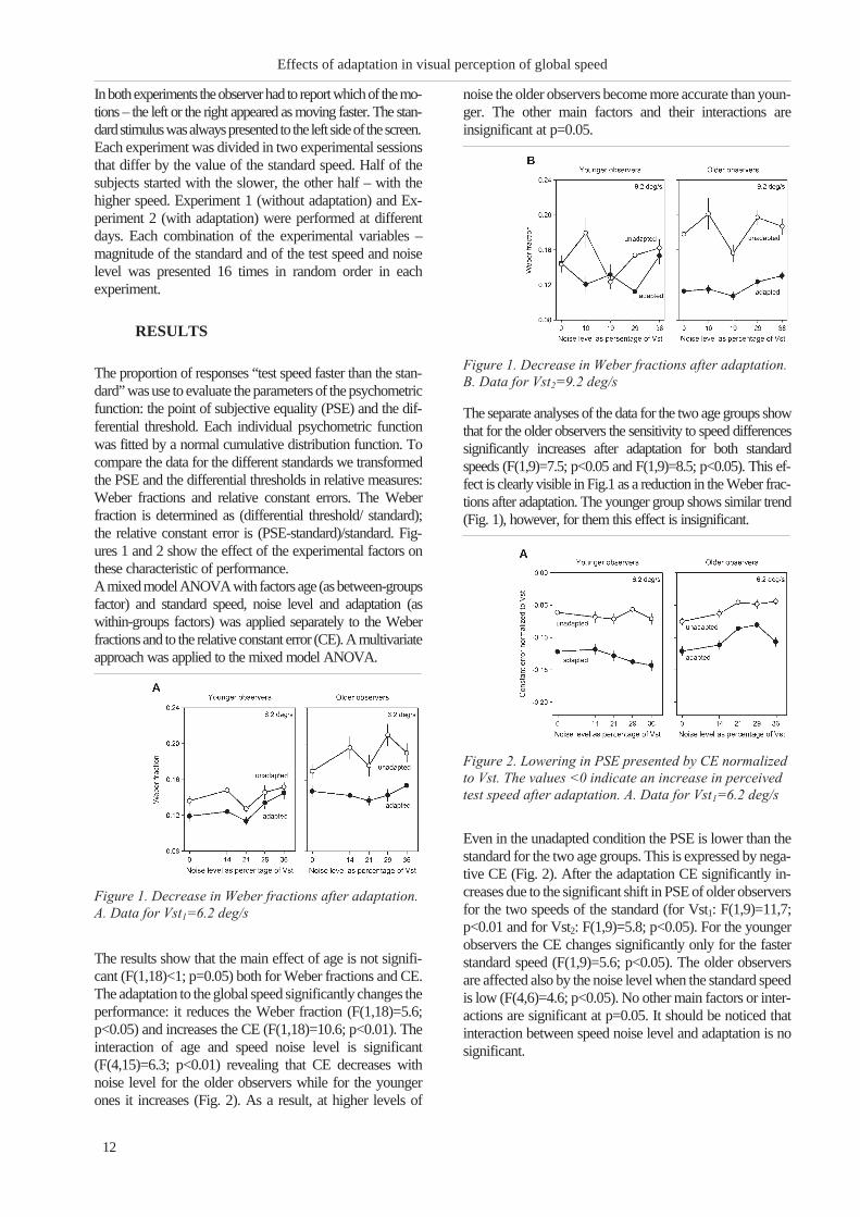
The separate analyses of the data for the two age groups show
that for the older observers the sensitivity to speed differences
significantly increases after adaptation for both standard
speeds (F(1,9)=7.5; p<0.05 and F(1,9)=8.5; p<0.05). This ef-
fect is clearly visible in Fig.1 as a reduction in the Weber frac-
tions after adaptation. The younger group shows similar trend
(Fig. 1), however, for them this effect is insignificant.
Even in the unadapted condition the PSE is lower than the
standard for the two age groups. This is expressed by nega-
tive CE (Fig. 2). After the adaptation CE significantly in-
creases due to the significant shift in PSE of older observers
for the two speeds of the standard (for Vst1: F(1,9)=11,7;
p<0.01 and for Vst2: F(1,9)=5.8; p<0.05). For the younger
observers the CE changes significantly only for the faster
standard speed (F(1,9)=5.6; p<0.05). The older observers
are affected also by the noise level when the standard speed
is low (F(4,6)=4.6; p<0.05). No other main factors or inter-
actions are significant at p=0.05. It should be noticed that
interaction between speed noise level and adaptation is no
significant.
12
Effects of adaptation in visual perception of global speed
Figure 1. Decrease in Weber fractions after adaptation.
A. Data for Vst1=6.2 deg/s
Figure 1. Decrease in Weber fractions after adaptation.
B. Data for Vst2=9.2 deg/s
Figure 2. Lowering in PSE presented by CE normalized
to Vst. The values <0 indicate an increase in perceived
test speed after adaptation. A. Data for Vst1=6.2 deg/s

The observed changes in PSE and in the sensitivity to speed
differences after adaptation modify the position and steepness
of the psychometric curve. It shifts toward lower speeds, and
becomes steeper reflecting a reduction in absolute speed sensi-
tivity and improvement in relative speed sensitivity (Fig. 3).
Our findings for influence of adaptation to global speed per-
ception are similar to the known results obtained in local-mo-
tion perception. In the present study we adapted to a single
speed and compared the perceived speed of the adaptor to
stimuli with different range of speeds. In this way we used as a
measuring device the test stimulus and the performance was
determined not only by the effect of adaptation but also by the
ability of the observers to pool the local velocity in a global es-
timate. Two questions arise: how much the observed effects
are related to the perception of global speed and whether the
adaptation spreads to other parts of the visual field. In our pre-
vious study (3) we obtained no effect of the noise level on the
Weber fractions in global speed discrimination, smaller
Weber fractions for higher standard speed and an overestima-
tion of global speed for high levels of speed noise for the older
group. This implies that the sensitivity to differences in the
global speed of motion is independent of the range of speeds.
The older observers, however, seem to disregard the slowest
speeds which leads to a misperception of the global speed at
high noise levels. Extrapolating to the present study we would
expect an overestimation of the test speed for high noise lev-
els. Since the adaptation reduces the speed of the standard (CE
is negative at noise level =0%), the bias in the perceived speed
should increase, not decrease with the noise level.
DISCUSSION
Our results show that adaptation affects more the sensitivity to
differences in speed of the older observers – the Weber
fractions in the adapted and in the un-adapted condition differ
more than those of the younger observers. This finding could
not be explained by differences in the abilities of the two age
groups to integrate the speeds of the local motions in a global
estimate. It suggests that the effect of adaptation has spread to
un-adapted regions affecting the perceived speed of the test
stimulus. McGraw and Roach (6) have shown that adaptation
to motion propagates across space. When the adaptor is
presented close to the fixation point the adaptation effect
spreads centrifugally. These finding suggest that neural
populations from non-overlapping spatial regions interact.
The larger effect of the adaptation on the performance of the
older observers might be related to the lower inter-cortical
inhibition with age (e.g. 8, 9) that would allow the spread of
the adaptation over larger area.
If adaptation affects the local speeds in the test stimulus in
proportion to their difference with the adapting speed, no
differences in sensitivity or in bias would be expected due to
the symmetry of the speed range with respect to the mean
speed. The different effect of the noise level on the bias for the
two age groups reveals specific adaptation effects. Hietanen et
al (4) showed that the PSE is affected by the adapt/test speed
ratio. When the test speed is equal or slower than the adaptor,
the perceived speed is reduced; however, when the test speeds
are faster than the adaptor, the perceived speed may even
increase. Such effect would imply small negative bias in the
perceived speed in our experimental conditions. When the
range of speed increases, the variability of the global speed
percept might increase due to random deviations of the local
speeds from the uniform distribution, but there are no reasons
to expect that this would change the trend in the bias.
The different effects of the noise level on the bias for the two age
groups might be related to changes in the neural populations
coding speed. Aging shifts the preferred speeds of motion of the
MT neurons to lower speed (11). As adaptation affects more the
neurons that are not optimally tuned to the adapting speed (e.g.
(5)), its effect might be larger for the faster speeds in the test
stimulus for the older observers. This would reduce the perceived
global speedandwouldbemoreevidentathigh levelsofnoise.
CONCLUSIONS
In summary, the results of the experiments indicate that: The
adaptation to mean speed increases the sensitivity to differences in
global speed and reduces the accuracy in speed estimation; The
effects of adaptation are stronger for the older people; The accuracy
of speed estimation is affected differently by the noise level for the
two age groups; The effect of noise on the sensitivity to speed does
notdependonadaptation;Theeffectsofadaptationontheperception
of global speed are similar to the findings in the available literature
about the effects of adaptation on the perception of local velocity.
13
Genova B., N. Bocheva, M. Stefanova ...
Figure 3. After adaptation psychometric curve is
steeper and shifted on the left
Figure 2. Lowering in PSE presented by CE normalized
to Vst. The values <0 indicate an increase in perceived
test speed after adaptation. B. Data for Vst2=9.2 deg/s

ACKNOWLEDGEMENTS
This research is supported by Grant TK01/200-2009 of the
National Science Fund, Bulgaria.
REFERENCES
1. Bravo, M. J. , S. N. J . Watamaniuk. Evidence
for two speed signals: a coarse local signal for segre-
gation and a precise global signal for discrimination.
Vis. Res., 1995, 35, 1691–1697.
2. Cli f ford, C. W. G. , K. Langley .
Psychophysics of motion adaptation parallels insect
electrophysiology. Curr. Biol. 1996, 6, 1340–1342.
3. Genova, B. Z. , N. B. Bocheva, S. Stefanov.
Effects of aging on speed discrimination in the pres-
ence of noise. Perception, 2011, 40, ECVP Abstract
Suppl., p. 91
4. Hietanen, M. A. , N. A. Crowder , M.R.
Ibbotson. Differential changes in human perception
of speed due to motion adaptation. J. Vision, 2008, 8,
1–10.
5. Krekelberg, B. , R.J . van Wezel , T.D.
Albr ight . Adaptation in macaque MT reduces per-
ceived speed and improves speed discrimination. J.
Neurophysiol, 2006, 95, 255–270.
6. McGraw, P. V. , N. W. Roach. Centrifugal
propagation of motion adaptation effects across visual
space. J. Vision, 2008, 8 (11), 1-11.
7. Norman, J . F. , H. E. Ross, L.M. Hawkes, J .
R. Long. Aging and the perception of speed. Per-
ception, 2003, 32, 85–96.
8. Schmolesky, M. T. , Y. Wang, M. Pu, A.G.
Leventhal . Degradation of stimulus selectivity of
visual cortical cells in senescent rhesus monkeys. Nat.
Neurosci., 2000, 3, 384–390.
9. Tadin D. , R. Blake. Motion perception getting
better with age? Neuron, 2005, 45, 325–327
10. Thompson, P. Velocity aftereffects: the effects of
adaptation to moving stimuli on the perception of sub-
sequently seen moving stimuli. Vis. Res., 1981, 21,
337–345.
14
Effects of adaptation in visual perception of global speed

VISUAL EVENT-RELATED POTENTIALS BY AN IDENTIFICATION
OF GRATING ORIENTATION DIFFERENCE
Dushanova J., D. Mitov
Institute of Neurobiology, Bulgarian Academy of Sciences, Sofia, Bulgaria
SUMMARY
Visual event-related potentials (VERP) to sinusoidal gratings employed in orientation discrimination
task were recorded. Stimuli were with spatial frequency of 2.9 cycles deg-1 and contrast of 5%, presented
in a Gaussian window with spatial constant of 0.483 deg. In each trial stimulus orientation varied ran-
domly between two values – 900 and 00, 900 and 750 as well as 900 and 850. The subject task was to press
one of the two keys by the left or the right forefinger according to the stimulus orientation (binary motor
task). It was found that reducing of orientation difference between the test stimuli decreased the ampli-
tude of N1 and N2 waves and increased the amplitude of P2 and in less extent – the amplitude of P3 com-
ponents. The latency of N1, N2 and P2 decreased with decrease of the orientation difference. The latency
of P3 wave prolonged at the smallest orientation difference. The effect of orientation difference on VERP
was negligible when the difference was decreased from 900 to 150, but it becomes substantial when the ori-
entation difference was decreased from 150 to 50.Thus the changes in ERP evaluated the selectivity of ori-
entation-specific channels and as a sign for a transition from detector to computational mode of
operation in orientation perception.
Key words: visual event-related potentials, sinusoidal gratings, orientation discrimination
INTRODUCTION
Many experimental data have given reasons to consider that
two main mechanisms are involved in the identification of
grating orientation – a detector mechanisms at large orienta-
tion differences and a computational mechanismat small ori-
entation differences between the stimuli (1,2,3,5).
In the present study the hypothesis was tested in experiments
based on detection of visual event-related potentials
(VERPs). The purpose of this study was to find a correlation
between the changes of VERP’s parameters, observed by
different orientation differences between the stimuli and the
selectivity of orientation-specific channels based mainly on
previous psycho-physiological investigations (2, 5,8,9).
It was ascertained appropriateness the changes of VERP
components as a sign for a transition from detector to com-
putational mode of operation in orientation perception.
MATHERIALS AND METODS
The EEG was recorded from 12 positions (10/20 system)
Fz, Cz, Pz, Oz, C3, C4, T3, T4, P3, P4, O1, O2 and EOG
for detection of blink artifacts. 20 subjects (11 females, 9
males, 31±7 years) had normal or corrected to normal vi-
sual acuity and no known ophthalmological or neurological
diseases. Handedness was assessed by a questionnaire
adapted from the Edinburgh Handedness Inventory.
Visual event-related potentials (VERP) to sinusoidal gratings
employed in orientation discrimination task were recorded.
Stimuli were with spatial frequency of 2.9 cycles/deg and
contrast of 5%, presented in a Gaussian window with spatial
constant of 0.483 deg for 100 ms in the centre of the visual
field. In each trial stimulus block orientation varied randomly
between two values – 900 and 00, 900 and 750 as well as 900
and 850, i. e. the orientation difference was 900, 150 and 50.
The subject task was to press one of the two keys by the left
or the right forefinger according to the stimulus orientation
(binary motor task, in all tasks by the right forefinger to
stimuli with orientation different from 900).
The VERPs were computed by averaging the artifact-free
trials for each stimulus/task combination and mean interval
across stimulus/task combination for each wave was N1
(80,140), P2 (130,200), N2 (190,298), P3 (290,550). The
statistical analyses of the parameters (amplitudes and laten-
cies of the waves) of VERPs were performed during the
above mentioned post-stimulus intervals and the statistical
difference between the groups (900/850 vs. 900/00, 900/750
vs. 900/00, 900/750 vs. 900/850) was assessed for each task
and interval by means of nonparametric procedure
(Kruskal-Wallis test) for pairs comparison of the scalp
leads between stimulus datasets.
15
Scripta Scientifica Medica, 2012; vol. 44 (1), Supplement 1 Copyright © Medical University, Varna
Address for correspondence:
K. Trifonova, Dept. of ophthalmology and general practiceStara
Zagora, Faculty of medicine, 47 Gen. Stoletov str, entr A, app 19,
e-mail: [email protected]

RESULTS
When binary sensorimotor task was employed, the reduc-
ing of orientation difference between the test’s stimuli de-
creased the amplitudes of N1 and N2, as well as increased
the amplitude of P2 components and in less extent – the
amplitude of P3 components (Fig. 1).
The latency of N1, N2 and P2 decreased and the latency of
P3 wave prolonged at the smallest orientation difference
(Fig. 2).
DISCUSION
The interest to the spatio-temporal organization of the vi-
sual system and to the manner of coding different aspects of
the visual objects is not due to theoretical reasons only.
However, the results of this research could find some prac-
tical application. It is widely accepted in the literature that
the early N1, P2 components are related to the early infor-
mation processing, automatic or task induced, related to the
sensory analysis after the stimulus, while late N2 and P3
components reflect the late cognitive processes. The com-
ponents latency reflects the time for information processing
whereas the amplitude reflects the level of activation of the
brain structures (4). It is well known that perception is im-
paired when some neurological and other diseases exist (6,
7). The investigation of the characteristics of VERPs, regis-
tered under discrimination tasks conditions could be ap-
plied as an adequate and non-invasive method for studying
the sensory and motor information processing at central
brain level in healthy persons and patients with neurologi-
cal and other diseases (7). The research of the visual and
cognitive information processing for instance in diabetics is
of substantial importance since the retina cells are ex-
tremely susceptible to the change of the blood glucose level
and the disease often affects the periphery as well as central
nervous systems (6).
CONCLUSION
The effect of orientation difference on VERPs was negligi-
ble when the difference was decreased from 900 to 150, but
it becomes substantial when the orientation difference was
decreased from 150 to 50.
Thus the changes in VERPs evaluated the selectivity of
orientation-specific channels and as a sign for a transition
from detector to computational mode of operation in orien-
tation perception.
ACKNOWLEDGMENTS
The study was supported by NSF, Bulgaria, contract
0475/2008
REFERENCES
1. Blakemore C. and R. W. Campbel l , “On the
existence of neurones in the visual system selectively
sensitive to the orientation and size of retinal images,”
J. Physiol., 1969, 203, 237–260
2. Campbel l F. W., Robson J. G. , Application of
Fourier analysis to the visibility of gratings. Journal
of Physiology, 1968, 197, 551 - 556
3. Hubbel l D. H. , Wesel T. N. Receptive fields of
single neurons in the cat’s striate cortex. Journal of
Physiology, 1959, 148, 574 - 591.
16
Visual event-related potentials by an identification of ...
Figure 1. Statistical comparisons between theamplitudes of N1, P2, N2, P3 waves (rows) forpairs grating orientation differences duringsensory-motor task (columns).
Figure 2. Statistical comparisons between thelatencies of N1, P2, N2, P3 waves (rows) for pairsgrating orientation differences during

4. Êîê À. Event-related-potential (ÅRP) reflections of
mental resources: a review and synthesis. Biol.
Psychol., 1997, 45, 19 - 56.
5. Legge G. E. , “Sustained and transient mechanisms
in human vision. Temporal and spatial properties,”
Vision Res., 1978, 18, 69–81
6. Mc Crimon R. J . , Deary I .J . , Huntly l . J .
P. , MacLeod K.J. , Fr ier B.M. Visual informa-
tion processing during controlled hypoglycemia in
humans. Brain, 1996, 119 (4), 1277 – 1287.
7. Pol ich J, Herbst K L. , P300 as a clinical assay:
rationale, evaluation, and findings. Int J
Psychophysiol., 2000, 38 (1), 3 - 19.
8. Vassi lev A. , Simeonova B. , Zlatkova M.
Recognition of line’s orientation at the detection
threshold. In V.D. Glezer (Ed.). Pererabotka
informacii v zritelnoj sisteme, 1982, 35 – 40.
17
Dushanova J., D. Mitov

SEX-DEPENDENT EFFECT OF A NEW PEPTIDOMIMETIC ON
COGNITIVE FUNCTION OF ISOLATED RATS AFTER MATERNAL
DEPRIVATION
Tancheva L.1, E. Encheva
2, M. Novoselski
2, V. Petkov
1, R. Klisurov
2
1Institute of Neurobiology, BAS; 2Medical University, Sofia, Dept. of Physiology
ABSTRACT
INTRODUCTION: Maternal deprivation and social isolation lead to stress-related cognitive changes in rats.
The aim of the study was to test the effect of the peptidomimetic M6 on postnatal stress in female and male rat
offspring. MATERIAL AND METHODS: - Wistar pups were separated from their mothers on the 21st
day,
followed by 5-week isolation. Control rats were grouped in 2 cages: 6 male and 6 female. - Half the animals
(grouped and isolated) received M6, 150 mg/kg/d (i.p.) for 3 days. - Exploratory activity, and learning and
memory were tested with hole-board and step-through tests. - Data analysis was performed with SPSS,
ANOVA (mixed design). RESULTS: The isolated rat pups were more exploratory active; in the same time
they had better learning and memory than grouped animals. Cognitive functions were significantly better in
male than in female isolated rats. Exploratory activity was less in male than in female grouped rats. The M6
effect on cognitive functions was sex-dependent: improved memory in male grouped animals and also in iso-
lated female rats, but decreased memory in grouped female rats. CONCLUSION: Combined postnatal stress
exposure (maternal deprivation, followed by social isolation) affects cognitive functions differently, depend-
ing specifically on gender and stress exposure.
Key words: maternal deprivation, social isolation, peptidomimetic, cognitive functions
INTRODUCTION
The importance of early life stress for mental health is well
known, even though the underlying neurobiological mech-
anisms are not completely clear. Postnatal stress exposure
in rodents affects critical periods of brain development that
persistently alter structural, emotional and neuroendocrine
parameters in the adult offspring [1,2,5].
Maternal deprivation followed by social isolation is used as
an animal model for damaged cognitive function. On this
model we study the possibility for pharmacological modu-
lation and protection of cognition with the application of an
amino acid derivative.
In our earlier studies some newly synthesized aminoacids
(L-valine derivatives) demonstrated a significant
neuroprotective effect on adult rodents, especially the com-
pound M6 [4].
We suggest that this preventive effect may be even stronger
in young animals.
The purpose of the study was to examine the ability of M6
for pharmacological modulation and prevention of early
life postnatal stress in female and male rat offspring with
maternal deprivation.
MATERIALS AND METHOD
Experimental model and methods: Wistar male and female
rats were deprived from their mothers on the 21st day after
birth. Immediately after that, the animals were hosted in iso-
lated cages for 5 weeks (social isolation model according to
Valzelli (1978), and modified by Petkov [3]).
The control groups (after mother deprivation) were hosted in
commoncages-6rats incage,maleandfemaleseparatecages.
Half of the animals (grouped and isolated) received à newly
synthesized compound (L-valine derivative, M6) for 3 days
at the end of the isolation period, in a daily dose of 150
mg/kg intraperitoneally (i.p.).
The control groups of rats (male and female) received the
solvent Oleum Helianthi (in the same volume).
Comparative study on their cognitive functions was per-
formed on the 24th hour after the last treatment – namely:
1. Exploratory activity (Hole board test, on the 1st, 2nd and
3rd minute).
2. Learning and memory (on the 1st and on the 24th hour)
with the step-through test.
Statistical evaluation. Experimental data were analyzed
by statistical software SPSS 11.0 for Windows, ANOVA
(mixed design).
19
Scripta Scientifica Medica, 2012; vol. 44 (1), Supplement 1 Copyright © Medical University, Varna
Address for correspondence:
Eleonora N. Encheva, Department of Physiology, Medical Univer-
sity of Sofia 2, Zdrave Str., 1431 Sofia, Bulgaria
Tel: +359 885 089 175
e-mail: [email protected]

RE SULTS AND DIS CUS SION:
The com bi na tion of 2 kinds of stress fac tors- ma ter nal de -
pri va tion (5 weeks) with so cial iso la tion - pro duced dif fer -
ent cog ni tive changes in male and female rats.
Our data show very in ter est ing re sults, like other re ports in
this field, which used dif fer ent mod els of early stress (swim
test and ex po sure to ad verse novel sit u a tion [1,2]).
Re gard ing the ef fect of so cial iso la tion, we found out that
so cially iso lated rats had:
- in creased ex plor atory ac tiv ity (Fig. 1)
- in creased pro cesses of learn ing and mem ory
(step-through test) in com par i son to the grouped an i mals
(on both the 1st and the 24th hour – Fig. 2 and 3)
The role of sex:
We found that the ef fect of the so cial iso la tion in young an i -
mals (56-57 days) on the cog ni tive func tion in rats is
sex-de pend ent. Ef fect of iso la tion on the learn ing and
long-term mem ory (on the 24th hour) is more sig nif i cant in
male than in fe male animals (Fig. 3).
Ex plor atory ac tiv ity is big ger in fe male than in male grouped
an i mals, but in iso lated the dif fer ence is in sig nif i cant (Fig. 4).
The ef fect of M6 treat ment:
Com pound M6 dem on strates pre ven tive ef fect on the cog -
ni tive func tion in rats - better in iso lated than in grouped an -
i mals. (Fig. 5)
We found also that in grouped an i mals the ef fect of M6 on
learn ing and mem ory is dif fer en tial, sex-de pend ent: in male
rats it is pre ven tive on the 1st hour, but in fe male rats, it is
the op po site- there is a mem ory de cline ob served dur ing the
step-through testing.(Fig.6)
Treat ment with M6 in grouped an i mals in creased the learn -
ing and short-term mem ory in male, but not in fe male (de -
creased it).The ten dency is quite the op po site in iso lated an -
20
Sex-dependent effect of a new peptidomimetic on cognitive function of ...
Fig ure 1
Fig ure 2
Fig ure 3
Fig ure 4
Fig ure 5

i mals- M6 de creased short-term mem ory in males and in -
creased it in fe males (Fig. 6)
M6 does not ex hibit any ef fect on long-term mem ory, ac -
cord ing to the step-through test (on the 24th hour af ter M6
treat ment) (Fig.7).
CON CLU SIONS
The com bined postnatal stress ex po sure (ma ter nal de pri va -
tion fol lowed by so cial iso la tion) af fects in dif fer ent way
the cog ni tive func tion of male and fe male ro dents and
prob a bly has a crit i cal in flu ence on brain de vel op ment and
neu ral plas tic ity.
The pro tec tive ef fect of M6 is also sex-de pend ent:
In male grouped an i mals the mem ory was im proved, but it
de creased in iso lated rats. The op po site was ob served in fe -
males- the pep tide mi metic M6 im proved mem ory in the
iso lated rats, but de creased it in grouped animals.
REF ER ENCES
1. Oomen CA, Soeters H, Audureau N,
Vermunt L, van Hassel t FN, Manders EM,
Joels M, Lucassen PJ, Krugers H. Se vere
early life stress ham pers spa tial learn ing and
neurogenesis, but im proves hippocampal syn ap tic
plas tic ity and emo tional learn ing un der high-stress
con di tions in adult hood. J Neurosci 2010; 30:
6635–6645.
2. Oomen CA, Soeters H, Audureau N,
Vermunt L, van Hassel t FN, Manders
EMM, Joels M, Krugers H, Lucassen PJ.
Early ma ter nal de pri va tion af fects dentate gyrus
struc ture and emo tional learn ing in adult fe males.
Psychopharmacology 2011; 214: 249–260.
3. Petkov, V.V. , S. Stancheva, Acta Physiol.
Pharmacol. Bulg., 1984, 10, 3-8
4. Stancheva S. L. Alova, L. Tancheva, V. V.
Petkov, E. Encheva, D. Tsekova, M.
Novoselski , Learn ing, Mem ory and Biogenic
Amine Lev els in Rat Hip po cam pus af ter Treat ment
with New L-Valine De riv a tives, J. Med.CBR I, vol 8,
Feb. 2010, 293-297, ISBN 978-954-9993-91-2
5. Yashmin J .G. Kar ten, Ana Olar iu, Heather
A. Cameron, Stress in early life in hib its
neurogenesis in adult hood, Trends in Neurosciences,
Vol ume 28, Is sue 4, April 2005, Pages 171-172,
(http://www.sciencedirect.com/sci ence/ar ti -
cle/pii/S0166223605000299)
21
Tancheva L., E. Encheva, M. Novoselski ...
Fig ure 7
Fig ure 6


EFFECTS OF D1 RECEPTOR BLOCKADE ON THE
INTENSITY-RESPONSE FUNCTION OF FROG DARK ADAPTED
ELECTRORETINOGRAM
Popova E.1, P. Kupenova
1, I. Ivanov
2
1Department of Physiology, Medical Faculty, Medical University - Sofia;2Specialized Hospital for Infectious and Parasitic Diseases “Prof. Ivan Kirov” - Sofia
ABSTRACT
The effects of dopamine D1 receptor blockade by SCH 23390 on the V – log I function of the ERG b- and
d-waves were investigated in dark adapted frog eyes. An enhancement of the b- and d-wave amplitude was ob-
tained during the blocker application. The effect was more pronounced on the rod- than cone-dominated re-
sponses. A clear ON-OFF asymmetry of the blocker’s action on the absolute and relative sensitivity of the
responses was demonstrated. The b-wave V – log I function had a steeper slope and narrower dynamic range.
The implicit time of the ERG waves was lengthened especially at lower stimulus intensities.
Key words: dark adaptation, dopamine, electroretinogram, frog, retina
INTRODUCTION
Dopamine is the predominant catecholamine in the verte-
brate retina, which is released by a unique set of
dopaminergic amacrine/interplexiform neurons (for review
16). Its participation in the overall retinal sensitivity control
can be readily evaluated by blocking the dopaminergic
transmission and following up the changes of the
electroretinogram (ERG). The most prominent ERG com-
ponents are the b-wave (in response to stimulus onset) and
the d-wave (in response to stimulus offset). These compo-
nents are usually used for assessment of the retinal ON and
OFF channel activity. Most of the authors reported that de-
pletion of retinal dopamine (with 6-OHDA treatment) or
application of the nonselective dopamine antagonist
haloperidol had no effect on the threshold dark adapted
ERG (6,7,10,17). But there is no agreement about the ef-
fects of the dopaminergic blockade on the supratheshold
dark adapted ERG, obtained with different stimulus inten-
sities, which evoked pure rod, mixed rod-cone or pure cone
mediated responses. Some authors failed to observe any ef-
fect on the b-wave amplitude (6,7,17), while other authors
reported its decrease (1,5,15). Still other authors obtained
an increase of the b-wave amplitude (2,10,13) and a left
shift of its V - log I curve, indicating increased relative sen-
sitivity of the response (13). The discrepancy in the results
cited might be due to species differences including involve-
ment of different kinds of dopamine receptors. It is known
that dopamine acts through five subtypes of dopamine re-
ceptors, which are grouped into two subfamilies: the
D1-like receptors (D1 and D5) and the D2-like receptors (D2,
D3 and D4). A few ERG studies were performed with appli-
cation of selective D1 receptor antagonist SCH 23390 and
the results obtained are contradictory. Some authors re-
ported diminution of the b-wave amplitude (9), while other
authors observed no effect on it (8). Changes of the d-wave
amplitude were not followed up in these studies.
In the present study we investigated the effects of SCH
23390 on the intensity-response function of the ERG b- and
d-waves, obtained in dark adapted frog retina.
MATERIAL AND METHODS
The experiments were carried out on 43 eyecup prepara-
tions of frog (Rana ridibunda), continuously superfused
with Ringer solution and supplied with moistened O2. The
dopamine D1 receptors were blocked using 10 mM SCH
23390 (Sigma). Eyecups were stimulated by diffuse white
light stimuli with 5 s duration and interstimulus interval of
25 s, presented in the dark. The test stimulus intensity (It)
was changed in an ascending manner over a range of 11 log
units by means of neutral density filters. The maximal in-
tensity (denoted by 0) was 6 x10⁸ quanta. s-1. ìm-2 at the
plane of retina. These light stimulation conditions allowed
us to obtain rod-dominated responses (using low It), mixed
rod-cone (using middle It) and cone-dominated responses
(using high It). The ERG was recorded by means of non
polarized Ag/AgCl electrodes at bandpass of 0.1 - 1000 Hz
and digitized at 1 kHz. The absolute sensitivity of the ERG
responses was assessed by their thresholds (5 mV criterion
amplitude) and the relative sensitivity – by stimulus inten-
sity (I�) required to produce half-maximum amplitude. The
23
Scripta Scientifica Medica, 2012; vol. 44 (1), Supplement 1 Copyright © Medical University, Varna
Address for correspondence:
E. Popova, Dept. of Physiology, Medical University,
1 “G. Sofiiski” st., 1431 Sofia
e-mail: [email protected]

dynamic range of the b-wave was estimated as intensity
span of the responses with 5 - 95% of the maximal ampli-
tude. The dynamic range of the d-wave could not be deter-
mined because of the more complex character of its V - log
I function (for details see 12). The V - log I function of the
ERG waves was obtained twice in one and the same eye-
cup – first in a control period with Ringer solution perfu-
sion (first series) and then in a test period (second series)
during perfusion with SCH 23390 (test experiments) or
Ringer solution (control experiments). For statistical evalu-
ation of the data, Student’s t-test, One- and Two-Way
ANOVA with Bonferroni test (alpha = 0.05) were used.
RESULTS
In a preliminary group of experiments the effects of SCH
23390 were followed for a period of 26 min (using It = -6.0)
in order to evaluate the time course of the effects. SCH
23390 caused marked increase of the b- and d-wave ampli-
tude, which reached a plateau at the 12th minute from the
beginning of the application (Fig. 1 a). The SCH 23390 ef-
fects on the ERG waves were relatively stable until the end
of the perfusion period. The b- and d-wave amplitudes re-
covered to a great degree during reperfusion with Ringer
solution (Fig. 1 b).
In the control experiments the V - log I function of the b-
and d-waves showed no significant differences between the
first and second intensity series in one and the same eyecup
(Fig. 1 c). This allowed us to evaluate the effect of SCH
23390 on the V- log I function using the first series of the
test experiments as a control one.
In the test experiments SCH 23390 caused significant in-
crease of the b- and d-wave amplitude at all stimulus inten-
sities (Two-way ANOVA p<0.000001) except for the low-
est ones for the b-wave ( It = -11.5, -11, -10.5) and the mid-
dle ones for the d-wave (It = -8, -7.5, -7) (Fig. 1 d). The ab-
solute sensitivity of the b-wave was not significantly al-
tered, but that of the d-wave was increased (Table 1). The
stimulating effect of SCH 23390 on the b- and d-wave am-
plitude showed clear intensity dependence. It was greatest
in the lower part of the intensity range, where the responses
were mediated by rods and was less pronounced at higher
intensities, where the responses were mediated by cones
(One-Way ANOVA with Bonferroni test P<0.05; Fig. 2 a).
The V - log I curve of the b-wave had steeper slope and nar-
24
Effects of D1 receptor blockade on the intensity-response function of ...
Figure 1. (a) Time-course of the SCH 23390 effects on
the amplitude of the b- and d-wave, obtained with It =
-6.0. Results of both control experiments (open symbols;
n = 11) and test experiments (filled symbols; n = 10)
are represented. The amplitudes of the ERG waves are
normalized to the values obtained just prior to blocker
application. The time, when the perfusion was switched
to SCH 23390, is indicated by an arrow. The period,
when the V-log I function was tested in the main group
of experiments, is indicated by a rectangular above the
curves. Mean values ± SEM are shown. (b): Original
ERG records, obtained during the perfusion with
Ringer solution in the control period (upper row), SCH
23390 (middle row) and Ringer solution in the recovery
period (lower row). Calibration: time – 200 ms;
amplitude – 100 �V. (c) and (d): Effects of SCH 23390
on the V - log I function of the b- and d-wave. Results of
both control experiments (c; n = 10) and test
experiments (d; n = 12) are represented. The
amplitudes of the ERG waves are normalized to Vmax of
the responses obtained during the first series of the
experiments. Mean values � SEM are shown.
Figure 2. (a): Relative change of the ERG b- and d-wave
amplitude in the control (open symbols) and test
experiments (filled symbols). The amplitudes of the ERG
waves, obtained at each It during the second stimulus
intensity series were normalized to those, obtained
during the first series. Means � SEM are represented.
(b): Changes in the b/d amplitude ratio during the first
(R) and second (SCH) series in the test experiments. (c):
Normalized V - log I curves obtained during the first (R)
and the second (SCH) series of the test experiments. The
values in each series are normalized to the Vmax obtained
in the same series. This manner of representation
demonstrates the changes of the position of the V - log I
function along the intensity axis and its slope under the
influence of SCH 23390. (d): Original ERG records,
obtained with different stimulus intensities during the
control period (grey lines) and during the treatment with
SCH 23390 (black lines). The numbers on the left side
indicate stimulus intensity (lg It).

rowed dynamic range during the D1 receptor blockade (Ta-
ble 1). The curve was shifted to the left along the intensity
axis, indicating increased relative sensitivity of the ON re-
sponse (Fig. 2 c; Table 1). However, the relative sensitivity
of the d-wave remained unchanged. The b/d amplitude ra-
tio was significantly lowered in the rod-dominated part of
the intensity curve (Two-way ANOVA p<0.01), but it was
significantly increased (p<0.03) in the intensity range (It
=-8; It = -7.5; It = -7), where transition from rod to cone
dominated responses occurred. The b/d amplitude ratio re-
mained unchanged in the higher intensity range, where the
responses were mediated by cones (Fig. 2 b). The perfusion
with SCH 23390 did not change the latency of the ERG
waves, but it increased their implicit time especially at
lower stimulus intensities (Fig. 2 d, Table 1).
DISCUSSION
Our results clearly demonstrate that the blockade of dopa-
mine D1 receptors by SCH 23390 enhances the amplitude of
the suprathreshold ERG b- and d-waves, obtained over a
wide range of stimulus intensities in dark adapted frog retina.
This indicates that endogenous dopamine, acting through D1
receptors, has inhibitory action on the mechanisms, responsi-
ble for generation of these ERG waves regardless of the type
of the photoreceptor input. However, the relative strength of
the dopamine action seems to be greater on the rod- than
cone-mediated responses. It is generally assumed that the
ERG b- and d-waves depend mainly on the activity of ON
and OFF bipolar cells, respectively, with minor direct contri-
bution of proximal retinal activity. Our results are in agree-
ment with the results obtained in amphibian retina showing
that dopamine, acting through D1 receptors, increases the ra-
tio of amplitudes of the cone-driven to rod-driven compo-
nents of the bipolar cell responses (4). Inhibitory action of
dopamine on the rod ON bipolar cells, concomitant with a
decrease in the amplitude of the b-wave, has been demon-
strated also in fish retina (14).
Our results show a clear ON-OFF asymmetry of SCH
23390 effects on the absolute and relative sensitivity of the
ERG responses. The absolute sensitivity of the b-wave is
not changed, but that of the d-wave is significantly in-
creased. This result supports the suggestion that endoge-
nous dopamine, acting through D1 receptors, contributes to
the lower absolute sensitivity of the OFF as compared to
the ON response in dark adapted frog ERG (3). On the
other hand, the relative sensitivity of the b-wave is in-
creased, while that of the d-wave is not significantly
changed. This result is consistent with the greater enhance-
ment of the b-wave amplitude at lower stimulus intensities
during the dopaminergic blockade reported by other au-
thors (11, 13). The increased relative sensitivity of the
b-wave in our present study is accompanied by a steeper
slope and narrowed dynamic range of the response. This re-
sult suggests that endogenous dopamine, acting through D1
receptors, is involved in widening of the intensity range,
where the b-wave amplitude is a linear function of log It.
This might be the contribution of this neurotransmitter to
the retinal sensitivity control in frog retina.
It is well established that the rod- and the cone-dominated
suprathreshold electroretinograms differ markedly in re-
spect to their b/d amplitude ratio and implicit time of the re-
sponses. They have much higher values in the rod- than the
cone-dominated ERG. Our present results indicate that do-
pamine action through D1 receptors may contribute to this
phenomenon as regards to b/d amplitude ratio. We demon-
strate that the initial difference between the b/d ratio values
obtained in the rod- and cone-dominated ERG becomes
smaller under the influence of SCH 23390. The opposite is
true for the dopamine effect on the time course of the re-
sponses, where the initial difference between the implicit
times of the rod- and cone-dominated ERG waves is aug-
mented during the D1 receptor blockade.
CONCLUSION
The results of our study clearly show that D1 receptor
blockade has similar enhancing effect on the
suprathreshold b- and d-wave amplitude irrespective of the
type of the photoreceptor input, although the rod-mediated
responses are affected to a greater degree. Endogenous do-
pamine acting through D1 receptors changes in a specific
manner the intensity-response function of both the ERG b-
and d-waves. A clear ON-OFF asymmetry of the dopamine
25
Popova E., P. Kupenova, I. Ivanov
ERGwaveThreshold (lgIt)
Ringer SCH 23390
I � (lgIt)
Ringer SCH 23390
Dynamic range (log units)
Ringer SCH 23390
Implicit time (ms)
Ringer SCH 23390
b-wave
d-wave
-11.10 ± 0.08 -11.02 ± 0.05
-9.99 ± 0.27 -10.57 ± 0.21
p<0.0004
-7.66 � 0.16 -8.28 � 0.1
p<0.000005
-5.61 � 0.07 -5.72 � 0.04
6.86 � 0.28 6.07 � 0.34
p<0.04
It -9.5 777 � 40.16 883 � 35.57
p<0.025
It-4 296 � 13.36 283 � 21.08
It -9.5 850 � 34.08 1020 �58.94
p<0.005
It -4 213 � 16.69 235 � 19.92
p<0.002
Table 1. Effects of SCH 23390 on the threshold, I�, dynamic range and implicit time of the ERG b- and d-wave

action on the absolute and relative sensitivity of the ERG
responses is demonstrated.
ACKNOWLEDGMENTS
This work was supported by grant ¹ 6/2010 from the
Council for Medical Science, Medical University Sofia.
REFERENCES
1. Adachi-Usami, E. , H. Ikeda, H. Satoh.
Haloperidol delays visually evoked cortical potentials
but not electroretinograms in mice. -J. Ocul.
Pharmacol., 6, 1990, 203-210.
2. Bodis-Wollner , I . , M. S. Marx, M. F.
Ghilardi . Systemic haloperidol administration in-
creases the amplitude of the light- and dark-adapted
ERG in the monkey. -Clin. Vision Sci., 4, 1989,
19-26.
3. Grani t , R. In: The eye. H. Davson, ed. New York,
Academic Press, 1962, 578-582.
4. Hare, W.A. , W.G. Owen. Similar effects of
carbachol and dopamine on neurons in the distal ret-
ina in tiger salamander. -Vis. Neurosci., 12, 1995,
443-55.
5. Kupenova, P. , S. Belcheva. Effects of
haloperidol, methylergometrine and phentolamine on
the frog ERG. -Experientia, 37, 1981, 852-4.
6. Li , L. , J .E. Dowling. Effects of dopamine deple-
tion on visual sensitivity of zebrafish. -J. Neurosci.,
20, 2000, 1893-903.
7. Lin, Z.S., S. Yazulla. Depletion of retinal dopamine
does not affect the ERG b-wave increment threshold
function in goldfish in vivo. -Vis. Neurosci., 11, 1994,
695-702.
8. Manglapus, M.K. , P.M. Iuvone, H. Under-
wood, M.E. Pierce, R.B. Barlow. Dopamine
mediated circadian rhythms of rod-cone dominance in
the Japanese quail retina. -J. Neurosci., 19, 1999,
4132-4141.
9. Marmor, M.F. , P. Hock, G. Schechter , A.
Pfefferbaum, P.A. Berger , R. Maurice. Os-
cillatory potentials as a marker for dopaminergic dis-
ease. -Doc. Ophthalmol., 69, 1988, 255-61.
10. Naarendorp, R. , P.F. Hitchock, P.A.Siev-
ing. Dopaminergic modulation of rod pathway sig-
nals does not affect the scotopic ERG of cat at
dark-adapted threshold. -J. Neurophysiol., 70, 1993,
1681-91.
11. Oliver , P. , F.B. Jol icoeur , G. Lafond, A.
Drumheller , J .R. Brunet te . Effects of retinal
dopamine depletion on the rabbit electroretinogram.
-Doc. Ophthalmol., 66, 1987, 359-71.
12. Popova, E. , P. Kupenova, L. Vitanova, L.
Mitova. ERG OFF response in frog retina : light ad-
aptation and the effect of
2-amino-4-phosphonobutyrate. -Acta Physiol. Scand.,
154, 1995, 377-386.
13. Schneider , T. , E. Zrenner . Effects of D-1 and
D-2 dopamine antagonists on ERG and optic nerve
response of the cat. -Exp. Eye Res., 52, 1991,
425-430.
14. Shiel ls , R.A. , G. Falk. Dopamine hyperpolarizes
and reduces the light responses of rod ON-centre bi-
polar cells in the retina of the dogfish, Scyliorhinus
canicula. -Neurosci. Lett., 55, 1985, 331-6.
15. Wachmeister , L. Further studies of the chemical
sensitivity of the oscillatory potentials of the
electroretinogram (ERG). II. Glutamate-aspartate-and
dopamine antagonists. -Acta Ophthalmol. (Copenh),
59, 1981, 247-58.
16. Witkovsky, P. Dopamine and retinal function.
-Doc. Ophthalmol., 108, 2004, 17-40.
17. Yazul la , S. , Z.S. Lin, K.M.Studholme.
Dopaminergic control of light-adaptive synaptic plas-
ticity and role in goldfish visual behavior. -Vision
Res., 36, 1996, 4045-57.
26
Effects of D1 receptor blockade on the intensity-response function of ...
DYNAMICS OF HEART RATE RECOVERY AFTER CYCLE
ERGOMETRIC PWC170 TEST IN SOCCER PLAYERS
Pavlova E., G. Uzunova, P. Somlev
Department of Physiology and Biochemistry, National Sports Academy “Vassil Levski”- Sofia
ABSTRACT
Relation of heart rate (HR) recovery dynamics with physical work capacity and maximal oxygen uptake
(VO2max) is important to understand more clearly adaptation to PWC170 test. The present aim is to study
correlations between PWC170, indirect measured VO2max and Dobrev’s recovery coefficients through cycle
ergometer PWC170 test and 5-minute recovery period. METHODS: Fifteen soccer players (age 21.5±1.43
years; weight 74.40±1.43 kg) performed two-load cycle ergometer PWC170 test and they have been examined
in 5-minute recovery period after the test. HR was monitored by Suunto t6c. PWC170, indirect VO2max (by
Karpman et al.), Dobdev’s coefficients, recovery HR sum, and BMI were calculated by equations. RESULTS:
It was found significant correlations between parameters (absolute and relative) of physical work capacity
and maximal oxygen uptake. PWC170 and VO2max depend significantly on BMI. There are not correlations
of HR recovery parameters (coefficients, sum of HR in 5-minute recovery) with working HR, VO2max and
PWC170. CONCLUSIONS: These findings support the suggestion that exercise cardiorespiratory parame-
ters and HR recovery dynamics after PWC170 test have independent diagnostics value.
Key words: HR recovery, PWC170, VO2 max, coefficient of recovery, soccer players
INTRODUCTION
Physical work capacity is the maximum amount of work a
person can perform. Increasing physical work capacity is a
basic element of any athletic training program. It is usually
related to a specific heart rate and is used as a measure of
aerobic fitness. Physical work capacity is primarily deter-
mined by the coordinated functioning of the neuro-muscu-
lar, cardiorespiratory, and energy producing systems. The
most common test of physical work capacity is called the
PWC170, which relates to the maximum amount of work
that can be done at a heart rate of 170 beats per minute.
Since originally designed, PWC170 tests (18,19) measur-
ing the power of physical activity required subjects to com-
plete five to seven stages of exertion lasting six minutes
each till nowadays various modified PWC170 tests have
been worked out and applied in functional laboratories
(1,3,4,14,) Their physiological basis is a linear relationship
between heart rate, oxygen uptake and physical activity for
the range between approximately 105-110 bpm and 170
bpm. Later studies by Karpman and others resulted in de-
velopment of the Sjostrand method that reduced testing
time considerably by utilizing only two sessions of load at
moderate power (6,7,8,11,16,17). PWC170 has been uti-
lized in predicting VO2max as well (3,5,9,11,13). In our
previous studies Margaria’s aerobic step-test has been
adapted for estimating physical work capacity and maximal
oxygen uptake (15). Studies about recovery heart rate after
PWC170 tests in athletes are scanty. The recovery heart
rate concept is mostly for people who are out of shape. In
our previous work we reported some data about dynamics
of HR recovery in athletes subjected to PWC170 tests (7). It
is important to clarify the interrelations between heart rate
(HR) recovery dynamics, physical work capacity and max-
imal oxygen uptake (VO2max). The present aim is to study
correlations between PWC170, indirect measured
VO2max and Dobrev’s recovery coefficients through cycle
ergometer PWC170 test and 5-minute recovery period.
METHODS
Fifteen soccer players (age 21.5±1.43 years; weight
74.40±1.43 kg) performed two-load cycle ergometer
PWC170 test (3) and they have been also examined in
5-minute recovery period after the test. The power output
for the load is set based on the subject’s weight and level of
fitness. Experimental design: The duration of the first load
is five minutes which is sufficient time for the heart to reach
a steady state of work. Then there is a three minute rest pe-
riod after completion of the first load. Then there is a sec-
ond five minute period of exertion, and 5-minute recovery
period after stop exercising. Two loads are with a constant
power output and a constant speed (rpm).The tests were
implemented on Monark 828E cycle ergometer. Heart rate
27
Scripta Scientifica Medica, 2012; vol. 44 (1), Supplement 1 Copyright © Medical University, Varna
Address for correspondence:
E. Pavlova. Dept. of Physiology and Biochemistry, National
Sports Academy”V.Levski”
Studentski Grad, Sofia 1710, Bulgaria
e-mail: [email protected]

(HR) monitoring has been done by Suunto t6c. Studied pa-
rameters are absolute and relative physical work capacity
(PWC170, PWC170/kg), absolute and relative maximal
oxygen uptake (VO2max, VO2max/kg), pre-exercise HR,
submaximal HR, Dobdev’s coefficients, recovery HR sum
and BMI. Physical work capacity and indirect measured
VO2max are computed by following equations (3):
1. Absolute PWC170
PWC N N NHR
HR HR170 1 2 1
170 1
2 1� � �
�
�( )
PWC170 [kgm.min-1] - physical working capacity at a
heart rate of 170 beats per minute
N1 [kgm.min-1] - power of 1st step load; N2 [kgm.min-1] -
power of 2nd step load
HR1 [bpm] - steady state heart rate at 1st step load
HR2 [bpm] - steady state heart rate at 2nd step load
2. Relative PWC170/kg
PWC kgPWC
kg170
170/ �
PWC170/kg [kgm.min-1.kg-1] - PWC170 per kg body
mass
3. Absolute VO2max
VO2max = 2.2XPWC170 + 1070
4. Relative VO2max/kg
VO kgVO
kg2
2max/
max�
VO2max/kg [ml.min-1.kg-1] - VO2max per kg body mass
For HR recovery dynamics after PWC170 test are used
Dobdev’s coefficients, calculated by equations (2):
1. HRcoeff.1
HRcoeffHR HR
HR.
.( )�
� �
�100 6 10 60
2. HRcoeff.2
HRcoeffHR HR
HR
HR HRV
HR.
.( ) .( )�
� ��
� �
�100 6 10 60 100 6 10 10
I V��
HR10 – HR for first 10 s in 1st minute of recovery
HR60 –HR for last 10s in 1st minute of recovery
HRV10 – average HR for 10s of 5th minute
�HR – HR for 1st minute
�HRI-V – HR for five minutes of recovery
�HRI-V = HR1 + HR2 + HR3 + HR4 HR5Body mass index (BMI) is computed as follows:
BMIweight kg
height m�
( )
( )2
Statistics analysis has been done through Descriptive Sta-
tistics, Two-Related Samples Tests and Nonparametric
Correlations (Spearman’s rho).
RESULTS
Descriptive statistics (Mean±SD) of variables PWC170,
PWC170/kg, VO2max, VO2max/kg, pre-exercise HR,
submaximal HR, Dobdev’s coefficients, recovery HR sum
and BMI are shown in Table 1.
Submaximal HR reaches the work level before ceasing lin-
ear relationship between HR, physical activity and VO2
and namely 170 bpm. Pre-exercise mean values of HR
have been determined as normocardia. In the end of 5-min-
ute recovery period HR decreases and gets values below
the pre-test ones but the comparison between HR values
shows insignificant difference (p0.05).
The PWC170, PWC170/kg, VO2max, weight and BMI
significant correlations (NPar Spearman’s Rho) have been
considered (Fig.1).
The variable physical work capacity correlates positively
with relative physical work capacity, maximal oxygen up-
take, weight and BMI. PWC170 and VO2max depend sig-
nificantly on BMI. There are insignificant correlations of
28
Dynamics of heart rate recovery after cycle ergometric ...
Variable Mean ±SD
PWC170 [kgm. min-1] 1166 ± 224.97
PWC170 / kg [kgm. min-1. kg-1] 15.65 ± 2.17
VO2 max [ml. min-1] 3493 ± 549.39
VO2 max / kg [ml. min-1. kg-1] 47.04 ± 5.24
HR pre-exercise 61.6 ± 9.26
Submaximal HR 164.8 ± 5.95
K1 0.93 ± 0.11
K2 1.3 ± 0.13
HR Sum 551.4 ± 33.46
BMI 22.82 ± 2.33
Table 1. Descriptive statistics (Mean±SD) of variables
Figure 1. PWC170 correlations with PWC170/kg,
VO2max, weight and BMI

HR recovery parameters (coefficients, sum of HR in 5-min-
ute recovery) with working HR, VO2max and PWC170.
DISCUSSION
The obtained data from the investigation give the reason to
accept that the subjects demonstrate adequate cardiovascu-
lar response to the applied PWC170 test (see Tabl.1). The
workload HR really marks the beginning of the optimal
zone for the cardiorespiratory function in response to physi-
cal activity. According to published data the demands dur-
ing a soccer match may become so high that they lead to fa-
tigue, with impairments of the physical performance poten-
tial and the technical performance even at submaximal ex-
ercise intensities (12). It has been reported, the mean rate of
aerobic energy production for elite players is estimated to
be � 70% VO2max during match-play. Nevertheless, in-
vestigation of aerobic capacity of soccer players is essential
for those athletes to assess basic aspect of their specific
sports abilities. It is well-known the increase in heart rate
that accompanies exercise is due in part to a reduction in
vagal tone. Recovery of the heart rate immediately after ex-
ercise is a function of vagal reactivation. The results enable
us to analyze the relations between workload HR and HR
recovery after PWC170 test. Therefore, an objective quan-
titative assessment of physical work capacity can be an im-
portant tool in measuring the effectiveness of training.
Physical work capacity and maximal oxygen uptake of the
soccer players estimated from the PWC170 test confronted
our previous results and other authors’ research could de-
fine their level of physical and aerobic capacities.
Comparison of PWC170 mean values of examined soccer
players with the published results of physical work capacity
by Karpman (3) and our previous data (7,17) illustrated in
Figure 2 indicate lower level of PWC170 for our contin-
gent than the level of the cited data. One possible cause for
such physical capacity is that the investigated subjects are
students-athletes and they do not train so regularly like the
soccer players from elite teams. The measured indirect
VO2 max from PWC170 test shows lower aerobic capacity
in students toward the maximal oxygen uptake in soccer
players (3,10) using the same procedure (Fig. 3).
Relation of heart rate (HR) recovery dynamics with physi-
cal work capacity and maximal oxygen uptake (VO2max)
is important to understand more clearly adaptation to
PWC170 test. The relationship between PWC170 and re-
covery parameters (k1, k2) are similar to obtained correla-
tions between PWC170 and Dobrev’s coefficients in ath-
letes of different kind of sports in other research (7). Ac-
cording to the authors of this study the cases of high
PWC170 and greater value of coefficients are associated
with better cardiac adaptation to exercise.
CONCLUSION
This study, investigating physical work capacity, aerobic
capacity and HR recovery parameters through PWC170
test and reevaluation older data in light of current knowl-
edge, indicates that such approach assesses more complex
adaptive reactions. Therefore, this objective quantitative
assessment of physical work capacity can be an important
tool in measuring the effectiveness of training. Moreover,
these findings support the suggestion that exercise
cardiorespiratory parameters and HR recovery dynamics
after PWC170 test have independent diagnostics value.
REFERENCES
1. Àðòîìîíîâà, Ë. Ë. , Î. Ï. Ïàíôèëîâ.
Ñïîðòèâíàÿ ìåäèöèí. Ó÷åáíî-ìåòîäè÷åñêîå
ïîñîáèå. ÒóëÃÏÓ, Òóëà, 2002.
2. Äîáðåâ, Ä. Â: Ëàáîðàòîðíè óïðàæíåíèÿ ïî
ôèçèîëîãèÿ, ïîä ðåä. íà Ä. Äîáðåâ, Ñîôèÿ,
Ìåäèöèíà è ôèçêóëòóðà, 1976, 294-299.
3. Êàðïìàí Â., Ç . Áåëîöåðêîâñêèé, È.
Ãóäêîâ. Èññëåäîâàíèå ôèçè÷åñêîé
ðàáîñïîñîáíîñòè ó ñïîðòñìåíîâ. Ôèçêóëüòóðà è
ñïîðò, Ìîñêâà, 1974.
4. Êðúñòåâ, Ê. Â: Ëàáîðàòîðíè óïðàæíåíèÿ ïî
ôèçèîëîãèÿ, ïîä ðåä. íà Ä. Äîáðåâ, Ñîôèÿ,
Ìåäèöèíà è ôèçêóëòóðà, 1983, 232-234.
5. Ìèíåâà, Ì., Ã. Óçóíîâà, Å. Ïàâëîâà.
Àåðîáåí è åðãîìåòðè÷åí êàïàöèòåò íà
ñúñòåçàòåëêè ïî ñïîðòíà àåðîáèêà.- Ïåòè
ìåæäóíàðîäåí íàó÷åí êîíãðåñ „Ñïîðò, ñòðåñ,
àäàïòàöèÿ”, Ñîôèÿ, 23-25 àïðèë, 2010, Ñïîðò è
íàóêà, Èçâ. áð., 2010, ×àñò II, 456-459.
6. Ïàâëîâà, Å. , Ï. Êóòèí÷åâà, Á.
Òðåíäàôèëîâ. Ïàðàëåëíî ïðèëàãàíå íà òåñòà
VC170 è îðòîñòàòè÷íà ïðîáà.- Òðåíüîðñêà ìèñúë,
2, 1988, 20 – 26.
29
Pavlova E., G. Uzunova, P. Somlev
Figure 3 Comparison of VO2max values of examined
soccer players with published data
Figure 2 Comparison of PWC170 values of examined
soccer players with published data

7. Ïàâëîâà, Å. , Ì. Ìàâðîäèåâà, Ä. Äîáðåâ.
Ïðîó÷âàíå íà íÿêîè îñîáåíîñòè ïðè ïðèëàãàíå íà
ïðîáàòà PWC170 çà ôóíêöèîíàëíà îöåíêà íà
ñïîðòèñòèòå.- Âúïðîñè íà ôèçè÷åñêàòà êóëòóðà,
5, 1981, 314 – 319.
8. Ïàâëîâà, Å. , Ì. Ìèíåâà, Ã. Óçóíîâà.
Ôèçè÷åñêè ðàáîòåí êàïàöèòåò íà ñúñòåçàòåëêè ïî
ñïîðòíà àåðîáèêà.- Ïåòè ìåæäóíàðîäåí íàó÷åí
êîíãðåñ „Ñïîðò, ñòðåñ, àäàïòàöèÿ”, Ñîôèÿ, 23-25
àïðèë, 2010, Ñïîðò è íàóêà, Èçâ. áð., 2010, ×àñò
II, 441-445.
9. Ïåòêîâ, Ñò. , Ì. Òîòåâà, È. Ìàçíåâ, Ä.
Äèìèòðîâà. Ïðàêòè÷åñêè óïðàæíåíèÿ ïî
ñïîðòíà ìåäèöèíà, ÍÑÀ, Ñîôèÿ, 2006.
10. Ñîìëåâ Ï. , Ïàâëîâà, Ã. Óçóíîâà.
Èçñëåäâàíå íà àåðîáíèòå âúçìîæíîñòè íà
ôóòáîëèñòè ÷ðåç ñóáìàêñèìàëåí
âåëîåðãîìåòðè÷åí òåñò. VII Ìåæäóíàðîäíà íàó÷íà
êîíôåðåíöèÿ íà êàòåäðà „Ôóòáîë è òåíèñ” ÍÑÀ,
Ñîôèÿ, 2010, Ñá. äîêëàäè, 122-125.
11. Óçóíîâà, Ã. , Å. Ïàâëîâà, Ï. Ñîìëåâ.
Ôèçè÷åñêè ðàáîòåí êàïàöèòåò è ðåñïèðàòîðíè
ïîêàçàòåëè ïðè äâóñòúïàëåí âåëîåðãîìåòðè÷åí
òåñò.- Íàó÷íà êîíôåðåíöèÿ ñ ìåæäóíàðîäíî
ó÷àñòèå -25 ãîäèíè Ïåäàãîãè÷åñêè ôàêóëòåò,
Âåëèêî Òúðíîâî, 6-7 íîåìâðè 2009, Ñá. äîêëàäè,
553-557.
12. Bangsbo, J . Energy demands in competitive soc-
cer. - Journal of sports sciences, 12, 1994, SI, S5 –
S12.
13. Boraczynski , T. , J . Urniaz. Changes in Aerobic
and Anaerobic Power Indices in Elite handball Play-
ers Following a 4-Week General Fitness Mesocycle.-
Journal of Human Kinetics, 19, 2008, 131-140.
14. Oja, P. , B. Tuxworth, eds. Eurofit for Adults:
Assessment of Health-Related Fitness. Finland: Coun-
cil of Europe Publishing; 1995, 46-51.
15. Pavlova, E. , G. Uzunova, Z. Arakchi iski .
Margaria step test for estimating PWC170.- 4th FIEP
European Congress Physical Education and Sports
(Teachers’ Preparation and Their Employability in
Europe), August 29th-31st, 2007, Bratislava, Slovakia,
160-166, ISBN: 978-80-89324-00-2.
16. Pavlova, E. , G. Uzunova, Z. Arakchi iski .
Comparison between PWC170 Estimated by Equation
and Krastev’s Table in Step Test.- World Congress of
Performance Analysis of Sport VIII “Current Trends
in Performance Analysis, Magdeburg, 3-6 September
2008., Shaker Verlag Aachen, 2009, Physiology/In-
jury Risk, Magdeburg, 386-389.
17. Pavlova, E. , G. Uzunova, P. Somlev. Suunto
t6c heart rate monitor for estimating PWC170 and
ventilatory equivalent.- 5th FIEP European Congress,
2nd Serbian Congress of P.E. Teachers, Nish, 23-26
September, 2009, Proc., 174-181. ISBN:
978-86-83811-16-8.
18. Sj `st rand, T. Changes in the respiratory organs of
workmen at an ore smelting works.- Acta Med.
Scand., 196, 1947, Suppl., 687-699.
19. Wahlund, H. Determination of physical working
capacity. A physiological and clinical study with ref-
erence to standardization of cardio-pulmonary func-
tion tests.- Acta Med. Scand., 132, 1948, Suppl., 215,
1-78.
30
Dynamics of heart rate recovery after cycle ergometric ...

URIC ACID, THIOLS AND BURN-INDUCED OXIDATIVE INJURY
OF GASTRIC MUCOSA IN RATS
Bekyarova G., M. Hristova
Department of Physiology and Pathophysiology, Medical University -Varna, Bulgaria
ABSTRACT
Thermal skin injury causes oxidative stress and damage of organs distant from original burns wound. The
mechanisms of burn-induced oxidative damage of gastric mucosa remain unclear. This study was designed to
investigate the gastric mucosal injury after burns in rats using gastric mucosal concentration of
malondialdehyde (MDA), uric acid and thiol groups as biomarkers of the oxidative status. In order to accom-
plish 30 % third degree burn hot boiling water (90oC) as applied on the back of the animals during a period of
ten seconds under thiopental anesthesia. In the present study we found that the concentrations of MDA and
uric acid in gastric mucosa were significantly increased after burns. The decrease of mucosal thiols impairs
the redox balance and stimulates the oxidative injury of gastric mucosa induced by thermal trauma. In con-
clusion, the thermal trauma causes oxidative injury of gastric mucosa evidenced by significant increased con-
centrations of MDA and uric acid as well as a significant reduction of the levels of thiols and uric acid
occupying the front line of the antioxidant defense. Based on our findings we could assume that the introduc-
tion of antioxidants that maintain the concentration of thiol groups in gastric mucosa represents a promising
therapeutic approach for restoring the redox balance in gastric mucosa and protecting it from burns-induced
oxidative injury.
Key words: uric acid, thiol groups, malondialdehyde, gastric mucosa, burns
INTRODUCTION
Ischemia due to transitory and selective vasospasm in the
splanchnic area after major burns causes oxidative damage
in organs of gastro-intestinal system followed by
reperfusion injury (12,22). Increased evidence has been
presented connecting with hepatic oxidative damage after
thermal trauma (13,14). Data about oxidative damage of
gastric mucosa are little. The mechanisms of burn-induced
oxidative mucosal injury are not completely clarified yet.
The changes of the activity of pro-oxidantive enzymes such
as xanthine oxidase (XO) and myeloperoxidase and
antioxidantive enzyme such as superoxide dismutase in
burns were studied (13). The changes of the other end prod-
uct of XO such as uric acid, remain insufficiently investi-
gated yet, too. Certain authors mention uric acid as 'oxida-
tive stress marker' for tissue damage (19). According to
other authors, it has an antioxidant action in the plasma (7).
Being a main non-protein thiol component, glutathione
plays an important role of a cellular antioxidant and
protector of the mucosa from oxidative injury (9).
Experimental and clinical data demonstrate dual role of the
uric acid as a marker of the oxidative damage in tissues as
well as plasma antioxidant (7,19). Both uric acid and pro-
tein thiols accomplish to a great extent the antioxidant
power of the plasma. However, the results about the
changes of the plasma level of uric acid after burns are very
contradictory, indeed (10,21).
This study was designed to investigate the burn-induced gastric
mucosal injury using malondialdehyde (MDA), uric acid and
thiols in gastric mucosa as biomarkers of the oxidative status.
MATERIAL AND METHODS
Experimental procedures
The experimental procedures was approved by the Home
Office for Care and Use of Laboratory Animals and per-
formed with a strong consideration for ethics of animal ex-
perimentation according to the International Guiding Prin-
ciples for Animal Research approved in Bulgaria.
Same-age male rats weighing between 220 and 250g fasted
for 12 h were allowed free access to water before injury
were used. Animals were housed in a 20oC room and of-
fered rat chow and water ad libitum. They were kept in the
individual wire-bottomed cages and fed standard rat chow.
After light ether inhalation, general anesthesia was per-
formed using thiopental 30 mg/kg intraperitoneally (i.p.). In
order to accomplish 30 % third degree burn hot boiling wa-
ter (90oC) as applied on the back of the animals during a pe-
riod of ten seconds. For those rats which were subjected to
31
Scripta Scientifica Medica, 2012; vol. 44 (1), Supplement 1 Copyright © Medical University, Varna
Address for correspondence:
G. Bekyarova, Ph.D, MD Department of Pathophysiology
Medical University of Varna, 55 Marin Drinov st.
Varna 9002, Bulgaria
E-mail: [email protected]

burn injury 4 ml physiological saline was applied i.p. for
immediate resuscitation following burn injury. No animals
died within the first 24 h post-burn period. Twenty nine
male rats were randomly assigned to two groups: 1.con-
trols-non burned rats (C) (n =10); 2.burned rats (B) (n =
19). All animal received buprenorphine 0,30 mg /kg. body
weight, i.p., twice daily for pain control post-burn. Animals
were reanesthetized with thiopental and scarified 24 h after
burns and stomachs were sampled.
Biochemical analysis
Gastric mucosa was gently separated from the underlying
tissue homogenized in 1:5 w/v 50 mM phosphate buffer (pH
7,4) containing 0,1mM EDTA, at 4000rpm for 10 min. The
homogenate was centrufugated at 800 x g rpm/15 min to dis-
card the sediment and supernatant was frozen until analysis.
All manipulations were performed at 4-8oC. Analysis was
performed immediately after thawing of the samples.
Membrane lipid peroxidation was assayed by MDA mea-
sured by its thiobarbituric acid (TBA) reactivity in gastric
mucosa hemogenates using the method of Porter et al (15).
Results were expressed as nmol MDA/g tissue were deter-
mined using the extinction coefficient of MDA-TBA com-
plex at 532 nm =1, 56 x 10-5 cm-1 M-1 solution.
Uric acid in gastric mucosal homogenate was determined by
the method of Bergmeyer (6), based on the ability of urate to
reduce phosphowolframic acid in alkaline solution to a blue
colored product. Standart solutions of uric acid were used to
calculate the extent of gastric mucosal homogenates.
Gastric mucosal thiol (SH) groups were determined by the
method of Hu (11), based on the absorbtion of the color
complex between SH groups and DTNB at 412 nm. Stan-
dard solutions of reduced glutathione were used to calcu-
late the concentration of SH groups.
Statistical analysis
Data were analyzed statistically by one-way analysis of
variance (ANOVA) and expressed as mean + SEM. A
value of p < 0,05 was considered statistically significant.
The statistical procedure was performed with
GraphPadInStat software.
RESULTS
Thermal skin injury causes significant elevation of gastric
mucosal MDA levels by 42 % (p<0,05) (Fig1.), mucosal uric
acid ones by 29% (p<0,05) (Fig.2) while mucosal SH level
was decreased by 26% (p<0,05) Fig.3 in burned rats compared
with those of the controls.
DISCUSSION
The results demonstrate that mucosal MDA level as a
marker of the oxidative damage related by most authors to
the intensive production of free radicals predominantly in
ischemia /reperfusion by activated leukocytes in the acute
phase post-burn (12). MDA accumulation is accompanied
by increasing of uric acid levels in gastric mucosa which al-
lows us to assume that the activation of the lipid
peroxidation is, probably, due either to the XO activation,
or to the accumulation of the substrate hypoxanthine, or
even to both of them. It is known that the uric acid is a natu-
ral product of purine metabolism that influences upon XO
activity. Some authors establish XO activation already dur-
ing the initial minutes after the thermal injury (12) as a re-
sult from ischemia /reperfusion particularly well-mani-
fested in the gastric mucosa after the thermal skin injury,
well as an increased hypoxanthine level (destruction of
32
Uric acid, thiols and burn-induced oxidative injury ...
Fig.1. Gastric mucosal MDA levels at 24 th hour
post-burn in rats. *P<0,05 vs controls; C- controls (non
burned rats); B - burned rats. Results are given as
mean + SEM.
Fig.2. Gastric mucosal uric acid at 24 th hour
post-burn in rats. *P<0,05 vs controls. C- controls (non
burned rats); B - burned rats. Results are given as
mean + SEM.
Fig.3. Gastric mucosal thiol groups at 24 th hour
post-burn in rats. *P<0,05 vs controls; C- controls (non
burned rats); B - burned rats. Results are given as
mean + SEM.

ATP down to AMP) that is a substrate for XO (20).
Hypoxantine conversion to xanthine and finally to uric acid
produces hydrogen peroxide and superoxide, deleterious
oxygen derived free radicals. Several studies suggested that
the burst of superoxide radicals and hydrogen peroxide
overwhelm the scavenging capacity of protective endoge-
nous enzymes (12,13). Superoxide radicals formed during
these reactions produce even more toxic radicals such as
hydroxyl radicals (OH.), and peroxynitrite (ONOO.) by in-
teracting with other radicals such as NO thus activating the
lipid peroxidation and inflammatory response.
Peroxynitrite production increases during the acute period
after burns (16). This suggests that activation of xanthine
oxidase (XO) pathway under conditions of ischemia/
reperfusion in gastric mucosa might be responsible for
superoxide radical production and more toxic radicals such
as OH. ànd ONOO. during the early phase after thermal
skin injury. Lipid peroxidation mediated by these reactive
oxygen- and nitrogen-based species is believed to be the
important factor for blood cells and tissue oxidative
damage after thermal trauma, including gastric mucosa.
Antioxidants block lipid peroxidation and protect gastric
mucosa from oxidative injury in burns (1,3,5,8,23).
As a basic non-protein thiol component, glutathione plays
an essential role of a cellular antioxidant and protector of
the mucosa from oxidative damages (9). It is known that
during tissue hypoxia and burns an exhaustion of the
intracellular glutathione sets in and this is, most probably,
due to its increased consumption during the oxidative stress
(2,17). Our data indicating a diminished level of mucosal
thiols and elevated mucosal MDA are in conformity with
reduced glutathione (GSH) depletion reported by other au-
thors finding that lipid peroxidation is activated after burns
(18). It may be supposed that the diminishion of thiol levels
impairs the redox balance and stimulates the oxidative
injury of gastric mucosa induced by thermal trauma.
In conclusion, the increased levels of MDA and urates
along with the decreased level of thiols could serve as a
marker of oxidative injury in gastric mucosa after thermal
injury. The thermal trauma causes a significant reduction of
the thiol levels occupying the front line of the antioxidant
defense. Based on our findings we could assume that the
introduction of antioxidants that maintain the concentration
of thiol groups in the gastric mucosa represents a promising
therapeutic approach for restoring the redox balance in gas-
tric mucosa and protecting it from oxidative injury induced
by thermal trauma.
REFERENCES
1. Bekyarova G, Tantcheva T, Yankova T. Ef-
fect of alpha-tocopherol on lipid peroxidation,
hemolysis and serum ceruloplasmin in the early post
burn period. Anal Lab1994; 4: 275-277.
2. Bekyarova G,Yankova T. Alpha-tocopherol and
glutathione deficiency and decreased deformability
after burn skin injury. Acta physiol pharmacol bulg,
23, 1998, 55-59.
3. Bekyarova G, Yankova T, Galunska B.Increased anti-
oxidant capacity, suppression of free radical damage
and erythrocyte aggregability after combined applica-
tion of alpha-tocopherol and FC-43 perfluorocarbon
emulsion in early postburn period in rats. Artif Cells,
Blood Subst Immobil Biotech 1996; 24:629-642.
4. Bekyarova G,Yankova T, KozarevI ,Yankov
D. Reduced erythrocyte deformability related to acti-
vated lipid peroxidation during the early postburn pe-
riod. Burns 1996; 22:291-294.
5. Bekyarova, G. ,Yankova T, Marinov M.
Lipofuscin product accumulation, insufficient antioxi-
dant defencein erythrocytes and plasma and enhanced
susceptibility to oxidative haemolysis after thermal
trauma. Acta Chir Plast 1997; 39: 61-64.
6. Bergmeyer HU. Methods of enzymatic analysis.
Breatfield Beach, FL:Verlag Chemie Int.,1981
7. Glantzounis GK, Tsimoyiannis EC, Kappas
AM, Galar is DA..Uric acid and oxidative stress.
Curr Pharm Des 2005;11:4145-4151.
8. Gurbuz V, Corak A, Yegen BC, Kurtel H, Alican I.
Oxidative îrgan damage in a rat model of thermal in-
jury: the effect of cyclosporin A. Burns 1997;
23:37-42.
9. Hayes JD, Mc Lellan LI. Glutathione and
glutathione dependent enzymes represent a
coordinately regulated deffense against oxidative
stress. Free Radic Res 1999;31: 273-280.
10. Haycock JW, Ralston DR, Morris B,
Freedlander E, MacNeil S. Oxidative damage
to protein and alterations to antioxidant levels in hu-
man cutaneous thermal injury. Burns1997;23:
533-540
11. Hu M. Measurment of protein thiol groups and
glutathione in plasma. Methods Enzymol 1994;
53:380-385
12. Latha B, Babu M. The involvement of free radi-
cals in burn injury: a review. Burns 2001; 27:
309-317
13. Naziroglu M., I . Kokcam, S. Yilmaz. Benefi-
cial effects of intraperitoneally administered al-
pha-tocopheryl acetate on the levels of lipid peroxide
and activity of glutathione peroxidase and superoxide
dismutase in skin, blood and liver of thermally injured
guinea pigs. Skin Pharmacol Appl Skin Physiol.
2003;16 : 36-45.
14. Pintaudi AM, Tesoriete LD, Arpa N,
D'Amelio L, D'Arpa D, Bongiorno A, et al.
Oxidative stress moderate to extensive burning in hu-
mans. Free Radic Res 2000; 33: 139-136.
15. Porter NA, Nixon J, Isaac J. Cyclic peroxidase
and thiobarbituric assay. Biochem Biophys Acta 1976;
441: 596-599.
16. Rawlingson A, Greenacre SA, Brain SD.
Generation of peroxynitrite in localised, moderate
temperature burns.Burns 2000; 26: 223-227.
17. Sakarsan A, Serer l i O, Vel ioglu-Ovunc A,
Erkan F, Erkanl G. Ginko Biloba extract im-
proves Oxidative organ damage in rat model of ther-
mal trauma. J Burn Care Rehabil 2005; 26: 515-524.
18. Sener G, Sehir l i O, Vel ioglu Ogune A,
Erean F, Erkainl i G, Gedik N, Yagen BC.
Propylthiouracil (PTU)-induced hypothyroidism alle-
33
Bekyarova G., M. Hristova

viates burn-induced multiple organ injury. Burns
2006;32: 728-736.
19. Siems WG, van-Kuijk FJ, Maass R, Brenke
R. Uric acid and glutatione levels during short term
whole body exposure. Free Radic Biol Med 1994;16:
299-305.
20. Woll iscrof t JO, Prasad JK, Thomson P, Til l
GO, Fox IH. Metabolic alterations in burn patients
and detection of adenosine triphosphate degradation
products and lipid peroxies. Burns 1990; 16: 92-96.
21. Yamazaki T, Ueda, T. Secondary hyperuricemia
in burn and trauma. Nippon Rinsho 2003; 61:
313-317.
22. Yoshida M, Wakabayashi G, Ishikawa H,
Kitahora T, Otani Y, Shimazu M, Miura S,
Ishi i H, Kitaj ima M. Rebamipide attenuates gas-
tric microcirculatory disturbances in the early period
after thermal injury in rats. Dig Dis Sci 1998;
43:148S-153S.
23. Yoshikawa Ò, Naito Y, Ueda S , Oyamada
H, Takemura T, Yashida N, Sugino S,
Kondo M. Role of oxygen derived free radicals in
the pathogenesis of gastric mucosal lesions in rats J
Clin Gastroenterol 1990;12:65-71.
34
Uric acid, thiols and burn-induced oxidative injury ...

CYTOPROTECTIVE EFFECT OF MELATONIN AND ANTIOXIDANT
DEFENSE AFTER THERMAL SKIN INJURY
Bekyarova G.1, M. Hristova
1, M. Tsaneva
2
1 Department of Physiology and Pathophysiology, Medical University -Varna, Bulgaria;2Department of General and Clinical Pathology, Medical University-Varna, Bulgaria
ABSTRACT
Melatonin protects against hepatic injury and this effect has mainly attributed to the antioxidant properties of
the indoleamine. The aim of this study is to investigate the expression of antioxidant enzyme CuZn superoxide
dismutase (CuZnSOD), total thiol levels in liver and their relationship with melatonin-mediated hepatic pro-
tection after burns. Under anesthesia, 30% total body surface area (TBSA), third degree burn was applied on
the back of animals. Melatonin (N-acetyl-5-methoxytriptamine, Merck, Germany 10 mg/kg(-1)) was i.p. ad-
ministered immediately and 12 hours after thermal skin injury. The hepatic CuZnSOD expression was exam-
ined by using light immunohistochemistry. The hepatic malondialdehyde (MDA) and total thiols, as oxidative
markers, were investigated too. Hepatic MDA levels were increased while total thiol in liver did not changed
early post burns. An increased expression of hepatic CuZnSOD in burned group was found. Melatonin re-
stores the expression of increased CuZnSOD activity to the control levels, resulting in normal protection
against burn-induced oxidative damage. In conclusion, thermal skin injury causes lipid peroxidation activa-
tion and compensatory increase in CuZnSOD expression in liver. We suggest that melatonin as a free radical
scavenger improves oxidant /antioxidant balance and restricts burn-mediated injury in the liver.
Key words: superoxide dismutase, light immunohistochemistry, malondialdehyde, liver, burns, melatonin
INTRODUCTION
Thermal skin trauma leads to free radical overproduction
following ischemia reperfusion resulting in elevated risk of
damage to organs distant from the original burn wound, in-
cluding liver (1). Although the mechanisms of burn-in-
duced injury remain unclear increasing evidences indicate
depletion of glutathione peroxidase (GSH-Px), glutathione
and other antioxidants under condition of activated free
radical processes as factor for hepatic injury (15,16,18).
Data about hepatic superoxide dismutase (CuZnSOD) ac-
tivity early post burn are converse. Some authors reported
that hepatic CuZnSOD activity is increased while others
find a decreased activity of this enzymes early post burn (1,
19). Administration of antioxidants (18) and exogenous
SOD (10, 21) restricts burn-induced oxidative damage in
liver and other organs.
Melatonin, a basic secretory product of the pineal gland, is
a powerful scavenger and antioxidant. It scavenges reactive
oxygen species (ROS) and reactive nitrogen species (RNS)
(12). Melatonin has been shown to stimulate CuZnSOD
and GSH-Px expression (14). Melatonin has been found to
attenuate burn-induced oxidative damage evidenced by
lipid peroxidation and antioxidant enzymes such as
glutathione peroxidase (GSH-Px), glutathione in liver and
intestine (3). It also exerts a therapeutic effect on liver dam-
age induced by ischemia/reperfusion (9). There are not data
about melatonin’s effect on hepatic CuZnSOD expression
early post burn.
The aim of this study is to investigate the expression of
hepatic CuZnSOD using light histochemistry, total thiols in
liver and their relationship with melatonin-mediated
hepatic protection after burns.
MATERIAL AND METHODS
Experimental burns and melatonin treatment
The experimental procedures were approved by the Home
Office for Care and Use of Laboratory Animals and per-
formed with a strong consideration for ethics of animal ex-
perimentation according to the International Guiding Prin-
ciples for Animal Research approved in Bulgaria.
Same-age male rats weighing between 220 and 250 g were
fasted for 12 hours, but were allowed free access to water
before injury. Animals were housed in a 20oC room and of-
fered rat chow and water ad libitum. They were kept on a
12-h dark/12-h light cycle, with lights turned off at 8:00
p.m. and turned on at 8:00 a.m. to achieve a satisfactory
photoperiod. After light ether inhalation, general anesthe-
sia was performed using thiopental 30 mg/kg
35
Scripta Scientifica Medica, 2012; vol. 44 (1), Supplement 1 Copyright © Medical University, Varna
Address for correspondence:
G.Bekyarova, Dept. of Pathophysiology
Medical University of Varna, 55 Marin Drinov st
Varna 9002, Bulgaria
e-mail: [email protected]

intraperitoneally (i.p.).In order to accomplish 30% third-de-
gree burns, hot boiling water (90oC) was applied to the
back of the animals for a period of 10 s. For those rats
which were subjected to burn injury, 4 mL physiological
saline was applied i.p. for immediate resuscitation follow-
ing burn injury. No animals died within the first 24-hour
post burn period. Twenty-four male rats were randomly as-
signed to three groups of 8 animals each: 1. non-burned,
non- treated (N-B) (control), 2.burned, non- treated group
(B), 3.burned group, treated with melatonin (B+M).
Melatonin (Merck, Germany) at a dose of 10 mL/kg solved
in vehicle (2% ethyl alcohol diluted in physiological saline
in doses up to 5 mL/kg) was given i.p. to the treated animals
in the morning between 8:00 a.m. and 9:00 a.m. immedi-
ately after burns. Solvent vehicle was administered i.p. in
the burned and control rats (sham groups) immediately af-
ter burns in the morning. All animals received
buprenorphine in a dose of 0.3 mg/kg i.p. twice daily for
pain control post burn. The animals were re anesthetized
with thiopental and sacrificed 24 hours after burns, and
livers were sampled.
Each liver was gently separated from the underlying tissue
homogenized in 1:5 w/v 50mM phosphate buffer (pH =
7.4) containing 0.1 mM ethylene-diaminetetraacetic acid at
4,000 rpm for 10 min. The homogenate was centrifuged at
800 x g every 15 min to discard the sediment, and
supernatant was frozen until analysis. All manipulations
were performed at 4-8oC. Analysis was performed imme-
diately after thawing of the samples.
Biochemical analysis
Membrane lipid peroxidation was assayed by MDA mea-
sured by its thiobarbituric acid (TBA) reactivity in hepatic
homogenates using the method of Porter et al.(13). Results
were expressed as nmol MDA/g tissue and were deter-
mined using the extinction coefficient of MDA–TBA com-
plex at 532 nm = 1.56 x 10⁻⁵ cm–1 M–1 solution.
Hepatic total thiol level were determined by the method of
Hu (7), based on the absorbtion of the color complex be-
tween SH groups and DTNB at 412 nm. Standard solutions
of reduced glutathione were used to calculate the concen-
tration of SH groups.
Immunohistochemistry
Rat liver specimens were fixed in 10% neutral buffered for-
malin and embedded in paraffin. The deparaffinized and
rehydrated sections (5 ìm thick) were treated in 1% hydro-
gen peroxide for peroxidase activity inhibition for 5 min.
Then they were rinsed in 0.1 M phosphate buffered saline
(PBS) pH 7.4 and treated in normal goat serum for 20 min.
Subsequently the sections were incubated with polyclonal
primary antibody for 24 hrs at room temperature. The used
antibody was rabbit anti-SOD (DAKO, USA). After rins-
ing with PBS the sections were incubated for 20 min in goat
anti-rabbit immunoglobulins at room temperature. Then
they were rinsed in PBS again and treated with rabbit
peroxidase-anti-peroxidase complex for 20 min at room
temperature, after which, rinsed in PBS. Finally, peroxidase
activity was developed by the diaminobenzidine-tetrachlo-
ride H2O2- method.
Negative controls. Controls were incubated with non im-
mune sera instead of primary antibody.
Statistics
The liver enzyme data were log transformed to satisfy the
assumptions required to perform parametric tests and are
therefore presented as geometric mean and 95% confi-
dence intervals of the mean. Orthogonal contrasts in
ANOVA were then used to analyze statistically the differ-
ence between any two specified groups
RESULTS
The levels of hepatic MDA were found to be higher by
48% (p<0,05) in the burned group than that of the control
group (fig. 1). Treatment with melatonin signi?cantly in-
36
Cytoprotective effect of melatonin and antioxidant defense ...
Fig. 2 – Effect of melatonin on total thiol levels in liver
after burns. Results are given as mean + SEM. *p <
0.05 vs. control rats; p < 0.05 vs. burned, non-treated
rats. C, Controls; B, burned rats; B + M, burned rats,
treated with melatonin.
Fig. 1 Effect of melatonin on liver MDA levels after
burns. Results are given as mean + SEM. *p < 0.05 vs.
control rats; p < 0.05 vs. burned, non-treated rats. C,
Controls; B, burned rats; B + M, burned rats, treated
with melatonin.

hibited the elevation in hepatic MDA level, maintaining the
level close to the control values. The total thiol levels did
not changed in liver after burns. In the melatonin treated
rats, the thiol level was similar that of the controls (fig. 2)
CuZnSOD expression was found in sinusoidal cells and
sometimes in hepatocytes. The staining intensity of the
SOD-positive cells was weak or moderate in the control
group (Fig. 3A). The expression in hepatocytes and sinu-
soidal cells of SOD was increased in the burned group (Fig.
3B). It was moderate to strong in the individually cells and
their mean content was significantly higher than this of the
control rats. The content of CuZnSOD varied from nega-
tive to weak or moderate in the burned group treated with
melatonin (Fig. 3C). The mean cell content was near to the
control one. (Fig. 3C).
DISCUSSION
Our data demonstrated that levels MDA (as (marker of oxi-
dative stress) increased, whereas the expression of
CuZnSOD increased in liver after burns, possibly to reduce
oxidative stress. The biological effect of free radicals is
controlled by wide range of antioxidant and antioxidant en-
zymes. CuZnSOD reacts with superoxide radicals and is on
the first line of antioxidant defense. We speculate that the
increased expression of CuZnSOD in liver may be acute
compensatory reaction and defense against overproduction
of superoxide radicals. Systemic oxidative stress was de-
fined as persistent imbalance between the formation of
highly reactive molecular species and organism’s antioxi-
dant defenses (11). The activation of oxidative stress path-
way in liver is more likely due to increased production of
free radical by burned skin, activated leukocytes activated
and xanthine/xanthine oxidase system (2).
Accumulating associative data suggest these reactive species
may act directly through oxidative damage on
macromolecules or indirectly activating single transduction
pathway sensible to stress mechanisms namely: nuclear fac-
tor kappa B (NF-kB), protein kinase B or Akt (PKB/Akt) (6).
The activation of these stress sensitive signal pathway is be-
lieved to be responsible for activation of antioxidant en-
zymes including CuZnSOD as adaptation response.
Our data about the increased hepatic CuZnSOD expression in
liver are in accordance with previous studies for increased ac-
tivity of this enzyme as compensatory reaction early post burn
when oxidative stress is increased (16,19). Agay et al (2005)
suggested that increased hepatic CuZnSOD activity is associ-
ated with mobilization of Cu and Zn in hepatic cells early post
burn. Among the major enzymes containing Cu and Zn, SOD
play a key role to counteract the oxidative stress-induced by
thermal injury. The increased CuZnSOD activity is known to
limit the diffusion of oxygen free radicals released by injured
tissues and therefore to avoid the activation of oxidative stress
(11). The expression of hepatic SOD increased while the total
thiol levels remained unchanged on the 24th hours. Previous
study showed an increased protein (SH) oxidation and oxida-
tive stress in the animals with Zn depletion diet (4). Others au-
thors did not find also any changes in total thiols in liver on the
first day (16). There are increased thiol oxidation and oxida-
tive damage of liver in the next days. The increased
CuZnSOD activity and reduced GSH-Px activity in liver to-
gether with revealed unbalanced hepatic antioxidant defenses
capacity contributing to liver oxidative injury (1).
We demonstrated that melatonin restores the expression of
increased CuZnSOD activity to the control levels, resulting
in normal protection against burn-induced oxidative dam-
age. Thus, it is likely that the decreased of hepatic MDA
levels in melatonin-treated rats results from an antioxidant
effect and inhibitory effect of this peptide on oxygen free
radical formation. Melatonin eliminates oxygen free radi-
cals and attenuates lipid peroxidation measured by serum
aspartate amino transaminase (AST) and alanine amino
transaminase (ALT) and MDA, which levels are decreased
and, in this way, restricts burn-induced oxidative hepatic
injury (3). In additional melatonin enters cell membranes, it
mainly localizes in the superficial position in lipid bi layer
near the polar heads of membrane while in this position it is
obviously capable of functioning as a free radical scaven-
ger and may also provide an indirect means by which the
membranes resist oxidative damage. Other possible expla-
nation is that melatonin stimulates production of
glutathione (20). Unlike other antioxidants melatonin don’t
consume cellular glutathione in redox cycling after scav-
enging free radicals (17). Time course studies are needed to
37
Bekyarova G., M. Hristova, M. Tsaneva
Fig. 3 (A) SOD expression in liver of non-burned (control) group (peroxidase-antiperoxidase complex; original
magnification x 100). (B) SOD expression in liver and sinusoidal cells in the burned group (peroxidase-antiperoxidase
complex; original magnification x 100). (C) SOD immunoreactivity in the liver of melatonin-treated burned group

determine the effect of melatonin on antioxidant activities
in this experimental model. The antioxidant effect of
melatonin was shown in different organs such as liver, in-
testine, lung after burns (5). In the previous studies and the
present study, burn-induced tissue damage was shown to
be reduced with melatonin administration (2,3).
In conclusion, thermal skin injury causes activation of
hepatic lipid peroxidation and compensatory increase in
CuZnSOD expression in liver. We suggest that melatonin
as free radical scavenger improves oxidant /antioxidant bal-
ance and restricts burn-mediated injury in liver.
REFERENCES
1. Agay D., Anderson RA, Sandre C. , Alonso,
A. , Roussel , A.M., Chancerel le , Y. Alter-
ations of antioxidant trace elements (Zn, Se, Cu) and
related metallo-enzymes in plasma and tissues follow-
ing burn injury in rats. Burns 2005, 31 (3): 366-71
2. Bekyarova G, Galunska B, Ivanova D,
Yankova T. Effect of melatonin on burn-induced
gastric mucosal injury in rats. Burns. 2009;35(6):
863-8.
3. Bekyarova G, Tancheva S, Hris tova M. Pro-
tective effect of melatonin against oxidative hepatic
injury after experimental thermal trauma. Methods
Find Exp Clin Pharmacol. 2009;31(1): 11-4.
4. Claeyssen R, Andriol lo-Sanchez M, Arnaud
J, Touvard L, Alonso A, Chancerel le Y,
Roussel AM, Agay D. Burn-induced oxidative
stress is altered by a low zinc status: kinetic study in
burned rats fed a low zinc diet. Biol Trace Elem
Res.2008;126 Suppl 1: S80-96.
5. De Fil ippis , D. , Iuvone, T. , Esposi to , G. et
al . Melatonin reverses lipopolysaccharide-induced
gastro-intestinal motility disturbances through the in-
hibition of oxidative stress. J Pineal Res 2008, 44(1):
45-51.
6. Horton JW. Free radicals and lipid peroxidation
mediated injury in burn trauma: the role of antioxi-
dant therapy. Toxicology. 2003;189(1-2): 75-88.
7. Hu M. Measurment of protein thiol groups and
glutathione in plasma. Methods Enzymol 1994;53:
380–5.
8. Hula NM, Chumak AA, Berdyshev AH,
Mehed’ OF, Horid’ko TM, Kindruk NL,
Kosiakova HV, Zhukov OD. Anti-inflamma-
tory effect of N-stearoylethanolamine in experimental
burn injury in rats. Ukr Biokhim Zh.
2009;81(2):107-16.
9. Kesik V, Guven A, Vurucu S, Tunc T,
Uysal B, Gundogdu G, Oztas E, Korkmaz
A.Melatonin and 1400 W ameliorate both intestinal
and remote organ injury following mesenteric
ischemia/reperfusion. J Surg Res. 2009;157 (1):
97-105.
10. Kumar R, Seth RK, Sekhon MS, Bhargava
JS. Serum lipid peroxide and other enzyme levels of
patients suffering from thermal injury. Burns.
1995;21(2): 96-7
11. Latha, B, Babu M. The involvement of free radi-
cals in burn injury: A review. Burns 2001, 27(4):
309-17
12. Mayo, J .C. , Tan, D.X. , Sainz, R.M.,
Lopez-Buri l lo , S. , Rei ter , R.J . Oxidative dam-
age to catalase induced by peroxil radicals: Functional
protection by melatonin and other antioxidants. Free
Radic Res 2003, 37(5): 543-53.
13. Porter , N.A. , Nixon, J .R. Isaac, R. Cyclic
peroxidase and thiobarbituric assay. Biochem Biophys
Acta 1976, 441(3): 596-9.
14. Rei ter , R.J . Melatonin.Lowering the high price of
free radicals. News Physiol Sci 2000, 15(5): 246-50.
15. Sakarcan A, Sehir l i O, Vel ioglu-Ovünç A,
Ercan F, Erkanl G, Gedik N, Sener G.
Ginkgo biloba extract improves oxidative organ dam-
age in a rat model of thermal trauma. J Burn Care
Rehabil. 2005;26(6): 515-24
16. Sandre C, Agay D, Ducros V, Van Uye A,
Cruz C, Chancerel le Y, Roussel AM. Early
evolution of selenium status and oxidative stress pa-
rameters in rat models of thermal injury. J Trace
Elem Med Biol. 2004;17(4): 313-8.
17. Tan DX, Manchester LC, Terron MP,
Flores LJ, Rei ter RJ. One molecule, many deriv-
atives: a never-ending interaction of melatonin with
reactive oxygen and nitrogen species? J Pineal Res
2007; 42(1): 28-42.
18. Toklu HZ, Sener G, Jahovic N, Uslu B,
Arbak S, Yeðen BC. beta-glucan protects against
burn-induced oxidative organ damage in rats. Int
Immunopharmacol. 2006;6(2): 156-69.
19. Wang BH, Yu XJ, Wang D, Qi XM, Wang
HP, Yang TT, Xu XH. Alterations of
traceelements (Zn, Se, Cu, Fe) and related
metalloenzymes in rabbit blood after severetrauma. J
Trace Elem Med Biol. 2007;21(2): 102-7
20. Winiarska K, Fraczyk T, Malinska D,
Drozak J, Bryla J. Melatonin attenuates diabe-
tes-induced oxidative stress in rabbits. J Pineal Res.
2006; 40(2): 168-76.
21. Xia ZF, Hollyoak M, Barrow RE, He F,
Muller MJ, Herndon DN. Superoxide dismutase
and leupeptin prevent delayed reperfusion injury in
the rat small intestine during burn shock. J Burn Care
Rehabil. 1995;16(2 Pt 1): 111-7.
22. Zhou ZB, Ding HQ, Qin FJ, Liu L, Cheng
S. Effect of Zn7-metallothionein on oxidative stress
in liver of rats with severe thermal injury. Acta
Pharmacol Sin.2003; 24(8): 764-70
38
Cytoprotective effect of melatonin and antioxidant defense ...

UNDERSTANDING PATHOGENESIS OF RISKY BEHAVIOR:
A HOLISTIC APPROACH
Sarov G.
Department of Pathophysiology, Trakia University, Medical Faculty, Stara Zagora
ABSTRACT
Epidemiological investigations of risk factors associates some particular form of risk behavior with the high
probability of disease appearance. However, the pathogenic nature of such behaviors remains to be under-
stood. Using a holistic approach I offer a theoretical model that defines risk factors as pathogenic
constelations that initially change body only functionally. However long-lasting disfunctions cause
microstructural damages that summarizes over the time and then disease becomes detectable by medical ex-
aminations. From holistic point of view risky constelations change body identity from healthy into pathogenic
one and this deformed body identity is a real cause for chronic diseases.
INTRODUCTION
Despite the huge number of empirical data on the patho-
genic role of risk factors, the mechanism of action remains
unclear. The origin of the term risk factor is statistical in na-
ture and its pathogenic logic is still not well understood.
This is so may be because insufficient development of ho-
listic understanding about living and disease. Holism is in-
sufficiently defined althought increasing understanding of
the need for a synthetic approach in many areas of medicine
(1-7).Here we offer a holistic explanation of the causation
of diseases and risk factors in particular.
METHOD
One can confirme the truth in three ways: by confirming the
description through observation, by successful prediction
of the future, proven by the practice, and by logical consis-
tency of the theory. There is a huge number of observations
and experiments, confirming the role of risk factors, but still
no consistent theory. Here we use logical reasoning as a re-
search method. The main proposition in our logical re-
search is that every whole has three dimensions: substance,
function and coordination. Starting with this basic holistic
proposition, we will look for a new meaning to physiology,
pathophysiology and causality in diseases.
Physiological extension of the main
propositions
In the field of physiology the three dimensions of the hole
are respectively structures, functions and bioidentity.
Structures. The substance exists in the forms of substrate,
energy (ATP) and structures. Substrate is converted into
ATP and structures, and structures use ATP to function.
Normally consumption of ATP can not exceed the rate of
its synthesis and ATP deficiency inhibits functioning to
prevent cells desintegration. Damage to the structure also
makes functioning impossible.
Function: Life as a function has three dimensions: con-
sumption, production and regulation.
Consumption provides substrates, thus determines both
ATP production and synthesis of the structures. Insufficient
consumption prevents restoration of cellular structures and
down-regulates cellular functioning.
Production. Every cellular population impact some
homeostatic variable (HV). The maximum amount of HV,
produced by the cell population per unit time is its func-
tional capacity. It depends on the number of the cells in the
population and their capacity. The real HV quantity pro-
duced by the cell population at any moment is its functional
volume. The difference between the functional capacity and
the functional volume is functional reserve - additional
quantity of HV, produced by cellular population in
responce of extra stimulation.
Regulation defines the intensity of the cell function and
could be feedback, anticipatory and residual.
Feedback regulation is spontaneous and self-regulated:
HV level activated the sensitive structures that operate
function of the HV producing cells. HV consumption down
regulates HV level and HV production up-regulates it.
Anticipatory regulation determines the HV level above the
physiological values facing increasing needs in the future.
Residual regulation starts with a release of waste products (re-
sidual homeostatic variables - RHVs) and triggers feedback
control by activating cellular populations that decrease RHVs
to minimal levels, otherwise RHVs damage cell structures.
Bioidentity: Bioidentity is the organization of the body
functions in specific situations. The body periodically
changes its bioidentity, but always retains functionally and
structurally integrated. Besides the condition of structures
and functions, bioidentity is determined by the situation to
which the individual must adapt. Relationship between or-
ganism and environment is defined in two ways - through
internal and external locus of control.
39
Scripta Scientifica Medica, 2012; vol. 44 (1), Supplement 1 Copyright © Medical University, Varna

The logic of external locus of control is: situation> evalua-
tion> preparation> action> homeostasis. If the individual
evaluates the situation as important, anticipatory regulation
and decision making are activated. Only after decision taking
individual can start actions, thus duration of decision making
determines the duration of the anticipatory control. If deci-
sion making prolongs, anticipatory control increase progres-
sively HVs and activates simultaneous feed-back regulation.
The logic of the internal locus of control is: homeostasis>
evaluation > preparation> action> homeostasis. It starts
with homeostatic problem that can need behaviour. The
need trigger anticipatory control and decision making. The
anticipatory control increases HVs in proportion with the
duration and intensity of decision making. After taking de-
cision, the actions decrease elevated HVs and increase
RHVs. These homeostatic changes activate feedback
regulations that restores homeostasis.
Holistic explanation of pathophysiology
The pathophysiologic transformation can start with disor-
ders in structures, functions and bioidentity. For that reason
three main pathogenic mechanisms exist: distruction,
disfunction, and disregulation.
Distruction: Every structure corresponds to a specifuc
function, so the magnitude of destruction determined the
severity of dysfunction. This is the law of structural func-
tionalism. Small structural damages can not produce
alarming symptoms, but every structural damage reduces
the functional capacity, and thus the functional reserve. Ir-
reversible destructions accumulate over time and functional
failure progresses irreversibly.
Dysfunction: When HVs deficiency increases, the likeli-
hood of structural damage (dystrophy and necrosis) in-
creases too. According to the law of functional
structuralism function can actively modulate structures and
severe dysfunction cause dystrophy and necrosis
(distruction).
Disregulation (pathogenic identity). Disregulation is a
process that creates pathogenic identity (PI) by two main
mechanisms.
1. Distructions and dysfunctions decrease initially HVs
production and all respective HVs consumers suffer in pro-
portion to their dependence on the HVs. This in turn leads
to secondary reduction in the production of HVs. This is the
law of dysfunctional expansion. Every pathological process
that triggered this low differently, which manifests as dif-
ferent diseases. The diversity and intensity of dysfunctional
expansion is proportional to the severity of the disease.
2. When individual is unable to solve an important adaptive
problem, anticipatory control is activated strongly and
protractedly. The more protracted and stronger is the anticipa-
tory regulation, the bigger is the corresponding feed-back coun-
ter regulation and bigger is the probability for dystrophy and/or
functional exhaustion. This is the law of dysadaptation.
Holistic explanation of causality
Pathogenicity is ability of the pathogenic factor (Fi) to in-
duce the three D (distructions (Ds), disfunctions (Df), and
disregulations (Dr)) in the body. Pathogenic power (PP) is
the intensity of the pathogenicity measured as:
PPFi=DsFi+DfFi+DrFi. Because of the dysfunctional expan-
sion, secondary changeo of the three D is superimposed to
the Fi action. So the pathogenic identity acquires cumula-
tive pathogenic power (CPP), which is calculated as the
sum of changes in the three D: CPPi=�Dsi +�Dfi+�Dri.
CPP varies over the time. When CCP>0, the identity is
pathogenic. Despite the absence of symptoms, the doctor
must imply the existence of an active pathological process
and to seek actively its pathogenesis. When CCP decreases
over the time (CCPt1>CCPt2) the body is in a process of res-
toration. When CCPt1<CCPt2, the dysfunctional expansion
increases the health risk and the patient needs intensive
care.
Structures, functions and identity participate in the
pathogenesis as constitutional factors. The constitutional
complex of genetic and phenotypic factors (CsC) deter-
mines the level of resistance and reactivity and promotes
health. The three ‘’D’’ depends on the contradiction be-
tween defence power (DP) of the constitutional complex
and PP. To be effective Fi should have destructive power
that is able to exceed physiological resistance and to gener-
ate Ds and/or Fi should have physiological activity that is
able to produce Df and/or Fi should put individual into Dr.
All the factors that affect the PP of Fi form the complex of
conditions (CdC). The disease prevention does not neces-
sarily require removal of the pathogen (Fi). It is enough to
reduce the magnitude of PP below the value of DP.
When the first sign of the disease is Dr> 0, the disease is in
preclinical stage and may involve asymptomatic
dysfunctions and structural damages.
When the first sign of the disease is Df> 0, the individual
suffers from a distinct symptoms (clinical stage).
When the first sign of the disease is Ds> 0, the magnitude of
symptoms and adaptive disorders is proportional to the lim-
itation of functional capacity and reserve of the damaged
cell population.
Holistic logic of pathogenesis
The magnitude and composition of PP, Ds, Df, Dr, and
CPP are central for the understanding pathogenesis. Model
proposed here is universal and allows explaining different
types of diseases.
The causal diseases starts with Ds>0. Because of the struc-
tural functionalism after some time (latent period) Df>0,
and PP=Ds+Df (clinical stage). In its turn Df activates dys-
functional expansion and secondary Df-s are activated. Ev-
ery additional Df starst its own chain of pathogenic or coun-
ter-pathogenic logics by changing regulations and structures
(functional structuralism and) and CCP
(CPPi=�Dsi+�Dfi+�Dri) increases (CCPt1<CCPt2) or de-
creases (CCPt1>CCPt2) depending on the contradictions be-
tween pathogenic and healing forces. Complex of conditions
and constitutional complex give specificity and individuality
of the pathogenetic processes. CdC determines the strength
of PP. CsC determines the strength of DP. CCP arises as a re-
sult from the contradiction between the PP and DP.
40
Understanding pathogenesis of risky behavior: a holistic approach
Risk-dependent diseases start with disregulation mainly be-
cause of inability of the individual to take right decisions.
The healthy decision is sometimes really difficult. It requires
knowledge, but also the ability to control. When individual
know the risk, but practice it, the problem is lack of self-con-
trol or ability to control circumstances. This part of the pa-
thology is most closely associated with humanity, since the
right and ability for making choices determine whether the
organism will be in state of regulation or disregulation. If the
choice is wrong disregulation occurs and this activates the
pathogenesis of risk-dependent disease.
The prolonged disregulation leads to dysfunctions
(Dr>Df). The prolonged dysfunction activates mechanisms
of functional structuralism and creates microstructural
damages, that increase with time (Df>Ds). When Ds-s re-
duce enough the functional reserve of the damaged struc-
ture, another set of dysfunctions appears following the logic
of dysfunctional expansion and disease enter into its clini-
cal stage (Ds>�Df’’). Then disease follows both
risk-and-causal logic. The logic of the risk stimulates the
permanent increase of the magnitude of the initial Ds-s by
the association Dr>Df>Ds. The causal logic activates the
mechanisms of dysfunctional expansion (Ds>�Df’’) and
secondary distructioins. The CCP increases progressively
(CCPt1<CCPt2) up to end-stage.
Dysfunctional diseases are three groups according to their
pathogenesis:
Genetic diseases arise due to synthesis of abnormal struc-
tures. During the intrauterine period Ds occurs, which then
manifests as Ds and Dr. Individuality and specificity of the
pathological process is determined by the ability to achieve
adaptation of the maladaptive function by an appropriate
modification of the environment.
Family diseases are caused by habits that create Dr or be-
cause of living in an unhealthy environment. Theses dis-
eases have social and economical background and cultural
specificity. Their individuality depends on the strength of
CsC that determines DP against Dr.
Constitutional diseases arise due to altered reactivity (Df),
which motivates the individual to react abnormally to nor-
mal circumstances and thus creates disregulation (Dr).
They have a psychological basis and relate mainly to the in-
dividual experience. Genetic and social factors contribute
to formation of this experience but not determine it.
CONCLUSION
The holistic model of diseases is universal, but each disease
has specific dimensions. The biggest advantage of the model
is its ability to explain risk-dependent diseases that are unex-
plained by the ‘’causal’’ logic. It also divides diseases into
three groups according to main mechanism: destructive
(causal), dysfunctional (hereditary and constitutional) and
risk-dependent (with disregulation as main mechanism).
REFFERENCES
1. Friboulet A. , D. Thomas. Systems biology-an
interdisciplinary approach. Biosens Bioelectron.
2005; 20(12): 2404-7.
2. Joyner M.J. Giant sucking sound: can physiology
fill the intellectual void left by the reductionists? J
Appl Physiol. 2011; 111(2): 335-42.
3. Joyner M.J. , B.K. Pedersen. Ten questions
about systems biology. J Physiol. 2011; 589(Pt 5):
1017-30.
4. Mesarovic M.D. , S.N. Sreenath, J .D.
Keene. Search for organising principles: understand-
ing in systems biology. Syst Biol (Stevenage).
2004;1(1): 19-27.
5. Neugebauer E.A. , C. Wil ly , S. Sauerland.
Complexity and non-linearity in shock research:
reductionism or synthesis? Shock. 2001; 16(4): 252-8.
6. Pezard L. , J .L. Nandrino. Dynamic paradigm in
psychopathology: “chaos theory”, from physics to
psychiatry. Encephale. 2001; 27(3): 260-8.
7. Pi t t B.R. , J .W. Chris tman, S.J . Gunst , M.A.
Matthay, T. Stevens, L.B. Ware. Physiology,
reductionism, and translational medicine: the right
mix. Am J Physiol Lung Cell Mol Physiol. 2011;
301(4): L389-90.
41
Sarov G.


COMPARISON OF FUNCTIONAL AND STRUCTURAL
CLASSIFICATION IN PATHOPHYSIOLOGICAL THEORY
AND TEACHING
Sarov G.
Department of Pathophysiology, Trakia University, Medical Faculty, Stara Zagora
Nowadays teaching pathophysilogy is based on structural functionalism, although disease is a functional mat-
ter. This inconsistency makes understanding of the subject matter difficult for students. Functional concepts
demand classification of diseases by the similarities in the body disfunctions while the structural aproach mo-
tivates classification by the similarities in the initially damaged body structures. Both classifications have
their reasons but expecially for the pathophysiology and internal medicine functional approach seems to be
better. Since 2003 I have applied a combination of functional and structural approaches into my teaching.
The functional approach resulted in better and faster understanding of the pathophysiological matter and
students improved their performance on examinations.
Key words: holistic, structural, functional, pathophysiology
INTRODUCTION
Every science becomes complete by organizing its content
into consistent system of notions. Otherwise its fundament
concepts are incomplete or even wrong. Since Aristotle one
believe that the scientific systematization should be con-
structed by genus (general) and species (unique variations
of the genus). According to this concept the knowledge
about the body could be systematically organized by
structural or functional point of view.
The first one makes the structure genus and its functions –
species, as it believes that structure determines the function.
This is now the dominant approach in physiology (7,13),
pathophysiology (1,2,4,5,8,10,11), and clinical medicine
(6,9). In the available literature, we saw only one attempt
(14) to a holistic organization of content, which is still dom-
inated by structural approach in classification.
Functional viewpoint is that the function organizes struc-
tures and organises the knowledge just in opposite way (3).
As the science could not be hole without consistent system
of notions, the obvious question is which one concept is
more consistent and reliable.
METHOD
Every classification is questionable if is internally incon-
sistent. The inconsistency becomes obvious by finding log-
ical contradictions. When use this rule we analyse some in-
consistency in the structural classification of diseases and
offer an alternative viewpoint (3), that is more consistent in
understanding and teaching pathophysiology and internal
medicine. As consistency is natural for thinking, more con-
sistent science means more effective teaching. We carry out
a pilot investigation on succesfull taking examinations by
medical students trained in conventional and functional
thinking in the pathophysiology. First experimental group
(n=6) was trained both conventional and functional way of
thinking on the basic pathologic processes. The second one
(n=13) – was trained in the same way in diseases’ mecha-
nisms, and the control group (n=35) was trained only con-
ventionally. The three groups were tested by independent
examiner. Statistics were comparison of percent distribu-
tion of successfully tested students in three consequent ses-
sions and Fisher exact test for insignificance.
RESULTS
About consistency of structuralistic classification. Table 1
illustrates the comparison of six diseases classified both in
structural and functional manner. As one can see first three
rows are functionally incompatible althought all they be-
long to the structural genus ‘’blood diseases’’. The basic
function of the thrombocytes is associated with the
impermeability of the blood vessels. Leucocytes participate
in immunity and leucosis functionally belongs to tissue
homeostatic disorders. Erythrocytes functionally belong to
the system of oxidation. This means that the structural clas-
sification is inconsistent with functional logic in these par-
ticular points. And vise versa, the last three rows belong to
one functional genus and different structural species . They
all have as common syndrom (functional disorder) the in-
sufficiency of oxygenation.
The same contradictions one can find in endocrine disorders.
Some of the hormones belong functionally to homeostasis
(aldosteron, insulin, glucagon,ect.), other could be determined
as stress hormones (epinephrine, corticosterone, ect), third
play important role in the physiological effect of the social in-
teractions (sex hormones, oxitocine, ect) and could be classi-
fied as social hormones. In the structural classification all they
43
Scripta Scientifica Medica, 2012; vol. 44 (1), Supplement 1 Copyright © Medical University, Varna
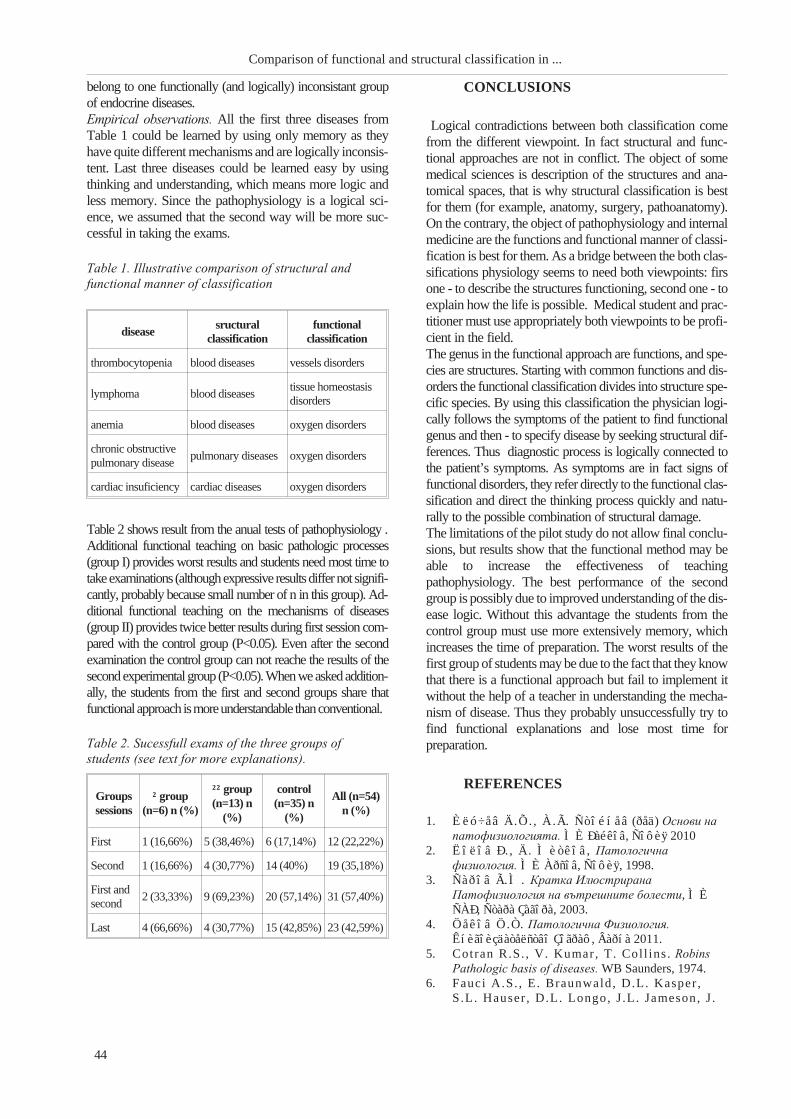
belong to one functionally (and logically) inconsistant group
of endocrine diseases.
Empirical observations. All the first three diseases from
Table 1 could be learned by using only memory as they
have quite different mechanisms and are logically inconsis-
tent. Last three diseases could be learned easy by using
thinking and understanding, which means more logic and
less memory. Since the pathophysiology is a logical sci-
ence, we assumed that the second way will be more suc-
cessful in taking the exams.
Table 2 shows result from the anual tests of pathophysiology .
Additional functional teaching on basic pathologic processes
(group I) provides worst results and students need most time to
takeexaminations (althoughexpressive resultsdiffernot signifi-
cantly, probably because small number of n in this group). Ad-
ditional functional teaching on the mechanisms of diseases
(group II) provides twice better results during first session com-
pared with the control group (P<0.05). Even after the second
examination the control group can not reache the results of the
second experimental group (P<0.05). When we asked addition-
ally, the students from the first and second groups share that
functionalapproach ismoreunderstandable thanconventional.
CONCLUSIONS
Logical contradictions between both classification come
from the different viewpoint. In fact structural and func-
tional approaches are not in conflict. The object of some
medical sciences is description of the structures and ana-
tomical spaces, that is why structural classification is best
for them (for example, anatomy, surgery, pathoanatomy).
On the contrary, the object of pathophysiology and internal
medicine are the functions and functional manner of classi-
fication is best for them. As a bridge between the both clas-
sifications physiology seems to need both viewpoints: firs
one - to describe the structures functioning, second one - to
explain how the life is possible. Medical student and prac-
titioner must use appropriately both viewpoints to be profi-
cient in the field.
The genus in the functional approach are functions, and spe-
cies are structures. Starting with common functions and dis-
orders the functional classification divides into structure spe-
cific species. By using this classification the physician logi-
cally follows the symptoms of the patient to find functional
genus and then - to specify disease by seeking structural dif-
ferences. Thus diagnostic process is logically connected to
the patient’s symptoms. As symptoms are in fact signs of
functional disorders, they refer directly to the functional clas-
sification and direct the thinking process quickly and natu-
rally to the possible combination of structural damage.
The limitations of the pilot study do not allow final conclu-
sions, but results show that the functional method may be
able to increase the effectiveness of teaching
pathophysiology. The best performance of the second
group is possibly due to improved understanding of the dis-
ease logic. Without this advantage the students from the
control group must use more extensively memory, which
increases the time of preparation. The worst results of the
first group of students may be due to the fact that they know
that there is a functional approach but fail to implement it
without the help of a teacher in understanding the mecha-
nism of disease. Thus they probably unsuccessfully try to
find functional explanations and lose most time for
preparation.
REFERENCES
1. Èëó÷åâ Ä.Õ. , À.Ã. Ñòîéíåâ (ðåä) Îñíîâè íà
ïàòîôèçèîëîãèÿòà. ÌÈ Ðàéêîâ, Ñîôèÿ 2010
2. Ëîëîâ Ð. , Ä. Ìèòêîâ, Ïàòîëîãè÷íà
ôèçèîëîãèÿ. ÌÈ Àðñîâ, Ñîôèÿ, 1998.
3. Ñàðîâ Ã.Ì. Êðàòêà Èëþñòðèðàíà
Ïàòîôèçèîëîãèÿ íà âúòðåøíèòå áîëåñòè, ÌÈ
ÑÀÐ, Ñòàðà Çàãîðà, 2003.
4. Öåêîâ Ö.Ò. Ïàòîëîãè÷íà Ôèçèîëîãèÿ.
Êíèãîèçäàòåëñòâî Çîãðàô, Âàðíà 2011.
5. Cotran R.S. , V. Kumar, T. Coll ins . Robins
Pathologic basis of diseases. WB Saunders, 1974.
6. Fauci A.S. , E. Braunwald, D.L. Kasper ,
S.L. Hauser , D.L. Longo, J .L. Jameson, J .
44
Comparison of functional and structural classification in ...
diseasesructural
classification
functional
classification
thrombocytopenia blood diseases vessels disorders
lymphoma blood diseasestissue homeostasis
disorders
anemia blood diseases oxygen disorders
chronic obstructive
pulmonary diseasepulmonary diseases oxygen disorders
cardiac insuficiency cardiac diseases oxygen disorders
Table 1. Illustrative comparison of structural and
functional manner of classification
Groups
sessions
² group
(n=6) n (%)
²² group
(n=13) n
(%)
control
(n=35) n
(%)
All (n=54)
n (%)
First 1 (16,66%) 5 (38,46%) 6 (17,14%) 12 (22,22%)
Second 1 (16,66%) 4 (30,77%) 14 (40%) 19 (35,18%)
First and
second2 (33,33%) 9 (69,23%) 20 (57,14%) 31 (57,40%)
Last 4 (66,66%) 4 (30,77%) 15 (42,85%) 23 (42,59%)
Table 2. Sucessfull exams of the three groups of
students (see text for more explanations).

Loscalzo Harrison’s principles of internal medi-
cine, 17th edition. The MacGraw Hill Co. Inc. , 2008.
7. Guyton A.C. , J .E. Hall Textbook of medical phys-
iology. WB Saunders, 2000.
8. Huether S.E. Understanding Pathophysiology.
Mosby Inc., 2008.
9. Kumar P. , M. Clark. Clinical medicine. WB
Saunders, 2002.
10. Lazenby R.B. , Handbook of Pathophysiology.
Wolters Kluwer Health, Lippincott Wil-
liams&Wilkins, 2011.
11. McCann J.A.S. Professional guide to
pathophysiology 3th edition. Wolters Kluwer Health,
Lippincott Williams&Wilkins, 2011.
12. McPhee S.J . , G.D. Hammer. Pathophysiology
of Disease: An introduction to Clinical Medicine. The
MacGraw Hill Co. Inc., 2006.
13. Pocock G., C.D. Richards, Human Physiology.
The basis of Medicine. Oxford University Press,
2001.
14. Tortora GF, S.R. Grabovski . Principles of
anatomy and physiology. John Wiley&Sons Inc,
2000.
45
Sarov G.


ÅFFECT OF CHRONIC TREATMENT WITH MELATONIN ON
DIURNAL VARIATIONS OF SPONATENOUS MOTOR SEIZURES
AND BEHAVIOR OF WISTAR AND SPONTANEOUSLY
HYPERTENSIVE RATS IN KAINATE MODEL
OF TEMPORAL LOBE EPILEPSY
Tchekalarova J.1, D. Pechlivanova
1, Zl. Petkova
1, Al. Stoynev
2
1Institute of Neurobiology, Bulg. Acad. Sci.;2Department of Pathophysiology, MU - Sofia
ABSTRACT
The aim of the present study was to analyze the effect of long-term melatonin treatment during
epileptogenesis on diurnal rhythms of spontaneous recurrent seizures (SRS) during the chronic phase in
Wistar (WIS) and spontaneously hypertensive rats (SHRs) in kainate (KA) model of temporal lobe epilepsy
(TLE). Melatonin treatment (10 mg/kg diluted in drinking water, 8 wk) increased the latency for appearance
of SRS in WIS rats. Whereas epileptic WIS and SHRs treated with saline exhibited circadian fluctuations in
the frequency of the SRS with a prevalence of seizures during the light phase, melatonin-treated rats did not
show circadian rhythms of seizures during the 3th
and 4th
month after status epilepticus (SE). Taken together,
our data suggest that chronic treatment with melatonin after SE may have potential to aid in the prevention
and development of epilepsy particularly accompanied with high arterial blood pressure.
Key words: Wistar rats, spontaneous hypertensive rats (SHRs), kainate model of temporal lobe epilepsy;
melatonin; diurnal rhythms
INTRODUCTION
The hormone melatonin, which is the main secretory prod-
uct of the pineal gland, plays a modulatory role in numer-
ous physiological functions. Accumulating clinical and ex-
perimental data raise the point of potential therapeutic role
of melatonin in epilepsy. Clinical evidence revealed that
melatonin posses low toxicity and may be used in seizure
control in conjunction with anti-seizure medications (10).
On the other side, experimental data revealed that
melatonin act as an anticonvulsant against chemically-in-
duced seizures (7,15), maximal electroshock (3), and elec-
trically kindled stimulation of amygdala (8).
Spontaneously hypertensive rats (SHRs) are widely ac-
cepted as an experimental model of essential hypertension
and has extensively been explored in chronobiological as-
pect (5,11). In these rats cerebrovascular changes, brain at-
rophy, loss of nerve cells in cerebrocortical areas, and glial
reaction were documented (reviewed in:1). In vivo imaging
studies suggest that hypertension-induced brain aberrations
detected in SHRs are similar to those found in patients with
essential hypertension. Clinical data showed disturbances
of melatonin biosynthesis in hypertensive patients while
experimental studies reported antihypertensive effect of
melatonin in SHRs (9). Taken together, these data are con-
sonant with the idea that melatonin may hold potential for
preventing neuronal damage after brain insults such as
brain trauma, status epilepticus (SE) and
hypertension-evoked brain damages.
Recently, we have demonstrated a circadian rhythm of ap-
pearance of SRS in epileptic state and strain-dependent di-
urnal variations in behavioral changes of normotensive
WIS and SHRs in KA model of TLE (13,14). Considering
the important function of melatonin in the circadian timing
system and numerous evidence supporting melatonin role
in epileptic phenomena, in the present study we aimed to
analyze whether the chronic melatonin treatment during
epileptogenesis would be able to attenuate the frequency of
spontaneous recurrent seizures (SRS) during the chronic
epileptic state in kainate (KA) model of TLE.
METHODS
1. Subjects
Male normotensive Wistar (WIS) rats and SHRs were indi-
vidually housed under standardized conditions (12/12-h
light/dark cycle and 20±1 oC, 50-60% humidity). Food and
water were available ad libitum. The experimental design
was approved by governmental authorities fully in accor-
dance with the European Communities Council Directives
of 24 November 1986 (86/609/EEC).
47
Scripta Scientifica Medica, 2012; vol. 44 (1), Supplement 1 Copyright © Medical University, Varna

2. Induction of status epilepticus with
kainic acid
Details of methodology were recently published (13,14). In
brief, SE was induced by a repetitive kainic acid (KA,
Sigma-Aldrich, Bulgaria) injection (5 mg/kg/h,
intraperitoneally). The first melatonin or saline
intraperitoneal (i.p.) injection was started 3 h after the onset
of SE. After recovery melatonin (Sigma-Aldrich, Bulgaria)
was administered chronically for a period of two months
(10 mg/kg in drinking water). Matched controls were
treated with an equivalent volume and number of injections
of sterile saline.
3.Video monitoring of spontaneous
reccurent seizures
The circadian distribution and the frequency of spontane-
ous recurrent seizures were ascertained by continuous (24
h/3 months) video-monitoring with a light-sensitive black
and white camera (S- 2016, AVTECH, Taivan, No
AVC307R).
Statistical analysis
ANOVA analysis of variance was used followed by a
post-hoc Bonferroni t-test to examine individual group dif-
ferences. P<0.05 was accepted as an index of statistically
significant differences.
RESULTS
Chronic melatonin treatment increased the latency for onset
of the first SRS in WIS rats (*p<0.05) (Fig. 1).
The distribution of SRS during the 24-h light-dark cycle at
the 3rd month after KA-induced SE is demonstrated in
Fig.2. Similarly to our previous studies (13,14) we have
found circadian distribution in the appearance of SRS with
a prevalence of seizures during the light phase
(08:00-20:00) (82%). At the 3rd month after SE both WIS
and SHRs exhibited higher seizure frequency during the
light phase (82 %).
However, melatonin treatment abolished circadian rhythms
of SRS in WIS and SHRs (Fig. 2 and Fig.3).
Although more SRS appeared during the light phase com-
pared to the dark one, significant difference was found for
the 3rd month (WIS: op<0.047; SHRs: op<0.013) and the
4th month (SHRs op<0.001), respectively. The total number
of SRS was statistically different between WIS and SHRs
at the 3rd month (11:00 p.m.), the 4th month (08:00;
10:00-16:00 p.m.) and the 5th month (12:00; 14:00 p.m.),
respectively, op<0.05 (Fig. 3). In 63 % of WIS rats and 100
% of SHRs, respectively, seizures appeared in clusters (i.e.
>3 seizures per day) (38). In 100 % of WIS rats and 63 % of
48
Sex differences in functional organization of ...
Fig. 1. The effect of melatonin on the latency for onset
of the first spontaneous recurrent seizure after kainite
(KA)-induced status epilepticus (SE) in Wistar and
spontaneous hypertensive rats (SHRs). Data represent
the mean ± SEM (n=10). *p<0.05 vs Wistar KA group.
Fig. 3. Total seizure frequency (mean ± SEM) during
the IIIrd, IVth and Vth month of KA-induced SE in Wistar
and SHRs. *p<0.05 light vs dark phase; op<0.05 vs
WIS with the same treatment.
Fig. 2. Dynamics of spontaneous reccurent seizures
(SRS) during the chronic phase of epilepsy counted for
12 weeks (starting three months after KA-induced SE in
Wistar and SHRs. Data shows the mean ± SEM of
weekly seizure frequency during the light (WISd, SHRd)
and dark (WISn, SHRn) phase of the twenty-four-hour
period of observation. *p<0.05 light vs dark phase;op<0.05 vs WIS with the same treatment.

SHRs the appearance of clusters was followed by a sei-
zure-free period of 2-5 days.
DISCUSSION AND CONCLUSION
Our results support previous reports demonstrating that
acute administration of melatonin in rats before and during
SE induced by either KA or pilocarpine displays
neuroprotective effects by reducing neuronal death,
supragranular mossy fiber sprouting, lipid peroxidation,
and microglial activation. In this line, Guisti et al. (1996) re-
ported that the cumulative dose of 10 mg/kg melatonin pre-
vented KA-induced neuronal death as well as behavioral
and biochemical disturbances in WIS rats. The hormone is
also able to reduce the spiking activity and seizure fre-
quency in patients with intractable epilepsy (2). In patients
with TLE high levels of salivary melatonin were observed
during the postictal period (4). The hormone is also able to
reduce the spiking activity and seizure frequency in patients
with intractable epilepsy (2). In patients with TLE high lev-
els of salivary melatonin were observed during the postictal
period (4). We have found that melatonin modified the de-
velopment, the frequency and the severity of SRS in a
strain-dependent manner. It could be speculated that
melatonin exerts its neuroprotective effect in KA model
through different mechanism in WIS and SHRs. Previous
clinical and experimental findings, including ours, suggest
a circadian rhythm of appearance of SRS in epileptic state
(12-14). However, we have fount that the hormone was
able to diminish the circadian rhythm of SHRs in both WIS
and SHRs. This effect might be a part of the
neuroprotective mechanism of melatonin action in KA
model of TLE.
In conclusion, the major findings of this study are that
melatonin exerts a potent neuroprotective effect on seizure
susceptibility in WIS and SHRs during the chronic epilep-
tic phase. While the hormone treatment increased the la-
tency for onset of the first SRS in KA-treated WIS rats it at-
tenuated the seizure frequency of SRS in SHRs.
REFERENCES
1. Amenta, F. , M.A. Di Tull io , D. Tomassoni .
Arterial hypertension and brain damage--evidence
from animal models (review). Clin. Exp. Hypertens.,
25, 2003, No 6, 359-380.
2. Anton-Tay, F. Melatonin: effects on brain func-
tion. Adv. Biochem. Psychopharmacol., 11, 1974, No
4, 315- 324.
3. Bazi l C.W., D. Short , D. Crispin, W.
Zheng. Patients with intractable epilepsy have low
melatonin, which increases following seizures. Neu-
rology, 55, 2000, No 2, 1746-1748.
4. Borowicz, K.K. , R. Kaminski , M. Gasior ,
Z. Kleinrok, S.J . Czuczwar . Influence of
melatonin upon the protective action of conventional
anti-epileptic drugs against maximal electroshock in
mice. Eur. Neuropharmacol., 9, 1999, No 3, 185-190.
5. El-Mas, MM, AA. Abdel-Rahman. Longitudi-
nal studies on the effect of hypertension on circadian
hemodynamic and autonomic rhythms in telemetered
rats. Life Sci., 76, 2005, No 8, 901-915.
6. Guist i P. , M. Lipart i t i , D. Franceschini , N.
Schiavo, M. Floream, H. Manev.
Neuroprotection by melatonin from kainate-induced
excitotoxicity in rats. FASEB J., 10, 1996, No 1,
891-896.
7. Lapin, I .P. , S.M. Mirzaev, I .V. Ryzon,
G.F. Oxenkrug. Anticonvulsant activity of
melatonin against seizures induced by quinolinate,
kainate, glutamate, NMDA, and pentilenotetrazole in
mice. J. Pineal Res., 24, 1998, No 4, 215-218.
8. Mevissen, M. , U. Ebert . Anticonvulsivant ef-
fects of melatonin in amygdala-kindled rats. Neurosci.
Lett., 257, 1998, No 1, 13- 16.
9. Oxenkrug, G.F. , P. Summergrad. Ramelteon
attenuates age-associated hypertension and weight
gain in spontaneously hypertensive rats. Ann. N Y
Acad Sci., 1199, 2010, 114-20.
10. Rufo-Campos, M. Melatonin and epi lepsy .
Rev. Neurol. Suppl. 1, 2002, S51-58.
11. Stoynev, A.G. , O.C. Ikonomov, N.K.
Minkova, S.Z. Zacharieva, V.G.
Stoyanovsky. Circadian rhythms of arterial pres-
sure: basic regulatory mechanisms and clinical value.
Acta Physiol. Pharmacol. Bulg., 24, 1999, No 3,
43-51.
12. Stewart , L.S. , L.S. Leung. Temporal lobe sei-
zures alter the amplitude and timing of rat behavioral
rhythms. Epilepsy Behav., 4, 2003, No 4, 153-160.
13. Tchekalarova J. , D. Pechl ivanova, D. I tzev,
N. Lazarov, P. Markova, A. Stoynev. Diur-
nal rhythms of spontaneous recurrent seizures and be-
havioural alterations of Wistar and spontaneously
hypertensive rats in kainate model of epilepsy. Epilep.
Behav. 17, 2010, No 1, 23-32.
14. Tchekalarova J. , D. Pechl ivanova, T.
Atanasova, P. Markova, V. Lozanov, Al .
Stoynev. Diurnal variations of depressive-like be-
havior of Wistar and spontaneously hypertensive rats
in kainate model of temporal lobe epilepsy. Epilep.
Behav. 20, 2011, 2, 277-285.
15. Yamamoto, H.A. , H.W. Tang. Melatonin at-
tenuates l-cysteine-induced seizures and peroxidation
lipid in the brain of mice. J. Pineal Res., 21, 1996, No
6, 108- 113.
49
Tchekalarova J., D. Pechlivanova, Zl. Petkova ...


DESIGN, SYNTHESIS AND ANALYSIS OF NOVEL
ENDOMORPHIN-2 AND MORPHICEPTIN ANALOGUES
Georgiev K.1, E. Miladinova
2, T. Pajpanova
2
1Medical University of Varna, 2Institute of Molecular Biology "Roumen Tsanev",
Bulgarian Academy of Sciences
Endomorphins (endomorphin-1, H-Tyr-Pro-Trp-Phe-NH2, endomorphin-2, Tyr-Pro-Phe-Phe-NH2) are po-
tent and selective �-opioid receptor agonists. Morphiceptin (Tyr-Pro-Phe-Pro-NH2), a tetrapeptide present in
the enzymatic digest of bovine �-casein, is a selective ligand of the �-opioid receptor. New endomorphin-2 and
morphiceptin analogues incorporating halogenated phenylalanines in place of the native phenylalanine are
reported. In order to improve the membrane permeability of the parent molecule, deoxycholic acid was at-
tached to the N-terminus of the peptide chain.
Key words: endomorphins, morphiceptin, opioid receptors, halogenated phenylalanines, peptide mimetics
INTRODUCTION
The three opioid receptors (�, and �) belong to the G-pro-
tein coupled receptors (GPCRs), which are characterized
structurally with a core of seven helical transmembrane do-
mains connected by three extracellular and intracellular
loops (1) These receptors are extremely important clinical
target in the treatment of pain and additionally have pro-
found effects on the neuroendocrine system immune re-
sponse (2). The � opioid receptor (MOR) is proven to be the
major target of analgesics, (2) because of the particular phys-
iological characteristics of this receptor, development of the
synthetic MOR-specific ligand is of great importance.
µ-Opioid agonists, such as morphine and heroin, produce
potent antinociception through µ-opioid receptors. However,
they also produce serious side-effects such as respiratory de-
pression and physical and psychological dependence.
The endogenous peptide ligands for �-opioid receptor,
endomorphin-1 (Tyr-Pro-Trp-Phe-NH2, EM-1) and
endomorphin-2 (Tyr-Pro-Phe-Phe-NH2, EM-2) were dis-
covered in 1997 by Zadina et al. (3). Both endomorphins
(EMs) exhibited the highest affinity for �-opioid receptor
and extraordinarily high selectivity relative to - and
�-opioid receptor systems of all known opioid substances
(3). These tetrapeptides are involved in the regulation of
several behavioral, emotional (4), cardiovascular (5), gas-
trointestinal (6,7) and respiratory functions (8). Further-
more, both EMs display a strong antinociceptive effect on
acute pain (9), similar to that of morphine. They are more
effective than the majority of the opioid peptides in reliev-
ing neuropathic pain even at very low dose, opening up the
possibility of pharmaceutical uses of these peptides (10).
They are also important model peptides to determine the
structure-activity relationship based on their typical charac-
teristics of backbones and aromatic side chains (11,12).
Morphiceptin (Tyr-Pro-Phe-Pro-NH2), which was origi-
nally synthesized as an analog possessing the N-terminal
tetrapeptide fragment of �-casomorphin (13) and was later
isolated from the enzymatic digest of bovine �-casein (14)
is a selective ligand of the �-opioid receptor. Its structure
differs from endomorphin-2 only by the amino acids in the
fourth position (Pro and Phe, respectively).
Morphiceptin, among other opioid peptides, has been
shown to elicit strong supraspinal antinociception when
given intracerebroventicularly (i.c.v.), which means di-
rectly to its site of action, which is the central nervous sys-
tem (CNS). This effect is not observed after peripheral ad-
ministration due to relatively rapid degradation of
morphiceptin, resulting in a short duration of action, and
limited delivery to the CNS.
A close structural similarity of endomorphin-2 and another
atypical opioid peptide, morphiceptin, which both have a
Phe residue in the third position, encouraged us to study
antinociceptive activity of these two peptides and their ana-
logues.
In order to improve the affinity and chemical stability of these
opioid peptides, we have designed, synthesized, and analyzed
9 novel analogues incorporating halogenated phenylalanines
in position 3 or 4 as surrogates of the native phenylalanine.
MATERIAL AND METHODS
Peptide synthesis
Peptides EM-2 and analogue 1 were synthesized by an ''in
solution, step-by-step'' method, starting with proline or
phenylalanine amides, respectively. To C-terminal frag-
ments respective Boc-amino acids were coupled using
dicyclohexylcarbodiimide and N-hydroxybenzotriazole.
Deprotection of the Boc-group was provided by 2 N HCl in
dioxane.
Peptides 2-9 in this study were synthesized by manual
solid-phase procedures using techniques for Fmoc-pro-
tected amino acids on Wang or MBHA Rink-Amide pep-
51
Georgiev K., E. Miladinova, T. Pajpanova
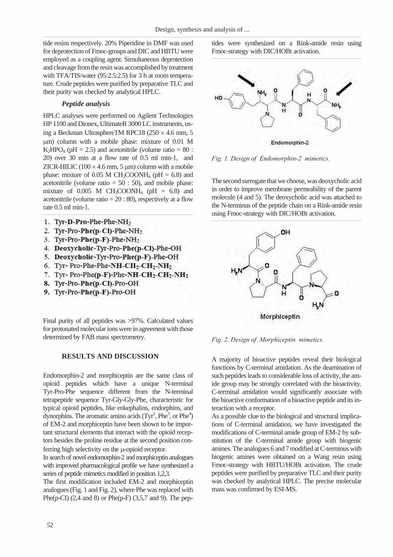
tide resins respectively. 20% Piperidine in DMF was used
for deprotection of Fmoc-groups and DIC and HBTU were
employed as a coupling agent. Simultaneous deprotection
and cleavage from the resin was accomplished by treatment
with TFA/TIS/water (95:2.5:2.5) for 3 h at room tempera-
ture. Crude peptides were purified by preparative TLC and
their purity was checked by analytical HPLC.
Peptide analysis
HPLC analyses were performed on Agilent Technologies
HP 1100 and Dionex, UltimateR 3000 LC instruments, us-
ing a Beckman UltrasphereTM RPC18 (250 � 4.6 mm, 5
�m) column with a mobile phase: mixture of 0.01 M
K2HPO4 (pH = 2.5) and acetonitrile (volume ratio = 80 :
20) over 30 min at a flow rate of 0.5 ml min-1, and
ZICR-HILIC (100 � 4.6 mm, 5 �m) column with a mobile
phase: mixture of 0.05 M CH3COONH4 (pH = 6.8) and
acetonitrile (volume ratio = 50 : 50), and mobile phase:
mixture of 0.005 M CH3COONH4 (pH = 6.8) and
acetonitrile (volume ratio = 20 : 80), respectively at a flow
rate 0.5 ml min-1.
Final purity of all peptides was >97%. Calculated values
for protonated molecular ions were in agreement with those
determined by FAB mass spectrometry.
RESULTS AND DISCUSSION
Endomorphin-2 and morphiceptin are the same class of
opioid peptides which have a unique N-terminal
Tyr-Pro-Phe sequence different from the N-terminal
tetrapeptide sequence Tyr-Gly-Gly-Phe, characteristic for
typical opioid peptides, like enkephalins, endorphins, and
dynorphins. The aromatic amino acids (Tyr1, Phe3, or Phe4)
of EM-2 and morphiceptin have been shown to be impor-
tant structural elements that interact with the opioid recep-
tors besides the proline residue at the second position con-
ferring high selectivity on the �-opioid receptor.
In search of novel endomorphin-2 and morphiceptin analogues
with improved pharmacological profile we have synthesized a
series of peptide mimetics modified in position 1,2,3.
The first modification included EM-2 and morphiceptin
analogues (Fig. 1 and Fig. 2), where Phe was replaced with
Phe(p-CI) (2,4 and 8) or Phe(p-F) (3,5,7 and 9). The pep-
tides were synthesized on a Rink-amide resin using
Fmoc-strategy with DIC/HOBt activation.
The second surrogate that we choose, was deoxycholic acid
in order to improve membrane permeability of the parent
molecule (4 and 5). The deoxycholic acid was attached to
the N-terminus of the peptide chain on a Rink-amide resin
using Fmoc-strategy with DIC/HOBt activation.
A majority of bioactive peptides reveal their biological
functions by C-terminal amidation. As the deamination of
such peptides leads to considerable loss of activity, the am-
ide group may be strongly correlated with the bioactivity.
C-terminal amidation would significantly associate with
the bioactive conformation of a bioactive peptide and its in-
teraction with a receptor.
As a possible clue to the biological and structural implica-
tions of C-terminal amidation, we have investigated the
modifications of C-terminal amide group of EM-2 by sub-
stitution of the C-terminal amide group with biogenic
amines. The analogues 6 and 7 modified at C-terminus with
biogenic amines were obtained on a Wang resin using
Fmoc-strategy with HBTU/HOBt activation. The crude
peptides were purified by preparative TLC and their purity
was checked by analytical HPLC. The precise molecular
mass was confirmed by ESI-MS.
52
Design, synthesis and analysis of ...
Fig. 2. Design of Morphiceptin mimetics.
Fig. 1. Design of Endomorphin-2 mimetics.

CONCLUSIONS
In summary, we succeeded to incorporate the halogenated
amino acids into EM-2 and morphiceptine peptide moiety.
We are able to obtain the peptide mimetics with satisfactory
purities (typically > 97%), as assessed by analytical HPLC).
The investigation of the biological activities of the new an-
alogues is in progress also. They are object of pharmaco-
logical investigation, and this data will de presented latter.
REFERENCES
1. Chen, Y. , A. Mestek, J . Liu, J , L. Yu. Mo-
lecular cloning of a rat kappa opioid receptor reveals
sequence similarities to the mu and delta opioid re-
ceptors. - Biochem. J., 295,1993, 625-628.
2. Waldhoer , M., S. E. Bart le t t , J . L. Whis-
t ler . Opioid receptors.- Annu. Rev. Biochem., 73,
2004, 953-990.
3. Zadina, J .E. , L-J . Ge, L. Hackler , A.J .
Kast in . A potent and selective endogenous agonist
for the mu-opiate receptor. - Nature ,
386,1997,499-502.
4. McLay, R.N. , W. Pan , A.J . Kast in . Effects
of peptides on animal and human behavior: a review
of studies published in the first twenty years of the
journal Peptides. - Peptides , 22, 2001, 218-255.
5. Wang, C.L. , Y. Yu, L.H. Lai , Y. Cui , X.
Wang, R. Wang. Cardiovascular responses to
intrathecal administration of endomorphins in anes-
thetized rats. - Peptides, 28, 2007; 871-877.
6. Wang, C.L. , X. Wang , Y. Yu, Y. Cui ,
H.M. Liu, L.H. Lai , e t al . Type 1 diabetes at-
tenuates the modulatory effects of endomorphins on
mouse colonic motility. - Neuropeptides, 42, 2008,
69-77.
7. Yu, Y. , Y. Cui , X. Wang, L.H. Lai , C.L.
Wang, Y.Z. Fan, et al . In vitro characterization
of the effects of endomorphin 1 and 2, endogenous
ligands for mu-opioid receptors, on mouse colonic
motility. - Biochem . Pharmacol., 73, 2007,
1384-1393.
8. Yu, Y. , Y. Cui , X. Wang, Y.Z. Fan, J .
Liu, X. Yan, et al . Endomorphin-1 and
endomorphin-2, endogenous potent inhibitors of elec-
trical field stimulation (EFS)-induced cholinergic
contractions of rat isolated bronchus. - Peptides, 27,
2006,1846-1851.
9. Tseng, L.F. , M. Nari ta , C. Suganuma, H.
Mizoguchi , M. Ohsawa, H. Nagase, et al .
Differential antinociceptive effects of endomorphin-1
and endomorphin-2 in the mouse. -J. Pharmacol. Exp.
Ther., 292, 2000, 576-583.
10. Przew �ocki , R. , D. Labuz , J . Mika,B.
Przew �ocka, C. Tomboly , G. Toth. Pain inhi-
bition by endomorphins. - Ann. N. Y. Acad. Sci., 897,
1999, 154-164.
11. Okada, Y. , Y. Fuj i ta , T. Motoyama, Y.
Tsuda, T. Yokoi , T. Li , e t a l . Structural stud-
ies of [2',6'-dimethyl-L-tyrosine1]endomorphin-2 ana-
logues: enhanced activity and cis orientation of the
Dmt-Pro amide bond. - Bioorg. Med. Chem., 11,
2003, 1983-1994.
12. Szatm�r i , I . , D. Biyashev, C. T�mb� ly , G.
T� th , M. M�csai , G. Szab� , e t a l . Influence of
degradation on binding properties and biological ac-
tivity of endomorphin-1. - Biochem. Biophys. Res.
Commun., 284, 2001, 771-776.
13. Chang, K.J . , A. Li l l ian, E. Hazum, P.
Cuatrecasas , J .K. Chang. Morphiceptin: a po-
tent and specific agonist for morphine (mu) receptors.
- Science, 212, 1981, 75-77.
14. Chang , K.J., Y.F. Su, D.A. Brent, J.K. Chang. Isola-
tion of a specific mu-opiate receptor peptide,
morphiceptin, from an enzymatic digest of milk pro-
teins. - J. Biol. Chem., 260, 1985, 9706-9712.
53
Georgiev K., E. Miladinova, T. Pajpanova


PHYSIOLOGICAL AND NON-INVASIVE BIOCHEMICAL INDEXES
IN A MODEL OF EMOTIONAL STRESS IN SHOOTERS
Petrov L.1, P. Atanasov
1, N. Zaekov
1, A. Alexandrova
2,
Z. Zsheliaskova-Koynova1, I. Achkakanov
1
1National Sports Academy "Vassil Levski", Sofia, 2Institute of Neurobiology,
Bulgarian Academy of Sciences
ABSTRACT
The use of noninvasive methods for assessment of stress under real and experimental conditions in people has
obvious advantages. Invasive methods cause stress and make difficult the interpretation of results. In sports
shooting psycho-emotional stress is severe and is associated with both the shooting performance and the reac-
tion to the reported results. The aim of this study was to evaluate the informative value of some stress bio-
chemical indexes, determined in saliva, and to compare them with suitable noninvasive physiological
parameters such as heart rate (HR) and heart rate variability. Twelve sports shooters (age from 14 to 19
years), divided into two groups: 5 in discipline air rifle and 7 in air pistol were tested. Two tournaments in
both disciplines were held. Each shooter performed 20 shots within 30 minutes. Samples of saliva were taken
by salivettes and the concentration of Na+, K
+, protein and salivary alpha-amylase activity (sAA) were mea-
sured. HR and physical activity were recorded, using GSM device in communication with a server. After the
tournament, in saliva the concentration of K+
and protein were increased significantly in all shooters. The
sAA activity demonstrated the same dynamics with two exceptions that are discussed. The concentration of
Na+
did not change. HR showed a typical increase in the middle and a gradual decrease to the end of the tour-
nament. rMSSD index showed a mirror mode dynamics. So we suggest that rMSSD represents the most stable
cardio vascular index of the emotional stress level in the model used.
Key words: heart rate, heart rate variability, saliva, salivary alpha-amylase activity, salivary potassium
INTRODUCTION
The use of noninvasive methods for assessment of stress
under real and experimental conditions in people has obvi-
ous advantages. Invasive methods cause stress and make
difficult the interpretation of results. In recent years more
often to evaluate the stress, changes of certain parameters,
measured in saliva are used. A parameter that has been sug-
gested to reflect the stress-related changes in the body is the
salivary enzyme alpha-amylase (sAA) (1,2,6,9). The sAA
release is known to be elicited by activation of the auto-
nomic nervous system (ANS) which controls the salivary
glands (7). Other relevant parameters are the concentration
of protein and some electrolytes in saliva. Their typical
changes in stress correlate with blood adrenaline and
cortisol levels. In particular, many researchers have re-
ported salivary K+ and protein increase in stress. While in
same conditions, the salivary Na+ did not change or even
has shown a reduction (5,8,10).
In sports shooting the psycho-emotional stress is severe and
is associated with both the shooting performance and the
reaction to the reported result - especially when the series of
successive shots are successful or unsuccessful. In turn, the
physiological changes in stress affect the shooting
performance, having in mind that the good players pull the
trigger in exact phase of breath holding and even at exact
time point of the cardiac cycle (3,4).
The aim of this study was to evaluate the informative value
of some biochemical indexes determined in saliva,
assessing the level of stress in shooters and to compare
them with suitable noninvasive physiological parameters
such as heart rate (HR) and heart rate variability (HRV).
MATERIALS AND METHODS
Twelve sports shooters, divided into two groups: 5 (2
women and 3 men) in the discipline air rifle and 7 (1
woman and 6 men) in the discipline air pistol were tested.
The age of the shooters varied between 14 and 19 years
(average age of 16 years). Two tournaments including the
both disciplines were held. Each shooter performed 20
shots from a distance of 10 m within 30 minutes. For better
motivation a small prize - 60 BGN for the first place and 40
BGN for the second place were provided. One day before
competition, the players were instructed not to use alcohol,
cigarettes and coffee, and to abstain from plenty eating just
before the tournament. Before and after the tournament,
samples of saliva were taken by salivettes. The samples
were centrifuged at 8000g and were stored at -20°C. On the
55
Scripta Scientifica Medica, 2012; vol. 44 (1), Supplement 1 Copyright © Medical University, Varna

next day the concentration of Na+, K+, protein and activity
of alpha-amylase in the saliva were measured on biochemi-
cal analyzer Human80. Reagents were purchased from Hu-
man Ltd (France). The HR was recorded (as R-R intervals
with 1 ms accuracy) during the entire period of the investi-
gation, using GSM device in communication with a re-
search server. The device, produced by Security Solutions
Institute Ltd. (Bulgaria), registered the physical activity ev-
ery second by built-in accelerometer, too. The instanta-
neous HR and the index of HRV - the square root of the
mean squared difference of successive R-R intervals
(rMSSD), were calculated automatically.
Significance of the differences was tested by Student test
for paired data.
RESULTS
The changes in the concentration of saliva potassium of any
one of the tested persons before and after tournaments is
presented on Figure 1. On Figure 2 the average values of
saliva K+ concentration of both disciplines separately and
jointly for all athletes are presented.
The average concentrations of Na+ in the saliva, collected
before and after both tournaments are shown in Table 1. In
terms of this index, statistically significant differences be-
fore and after the tournaments are not registered.
The changes in protein content in saliva of any one of the
tested persons before and after tournaments are presented
on Figure 3. On Figure 4 the average values of saliva
protein content of both disciplines separately and jointly for
all athletes are shown.
The individual sAA activity before and after the air rifle
and air pistol tournaments are presented on Figure 5 and on
Figure 6 the average sAA activity before and after both
tournaments separately and jointly for all shooters are
summarized.
56
Physiological and non-invasive biochemical indexes in a model of ...
Figure 1. Individual saliva K+ concentration before and
after pneumatic rifle (A) and air pistol (B) tournament.
Figure 2. Average saliva K+ concentration before and
after the tournament for both disciplines separately and
jointly for all athletes.
Befor competition
mmol/l ± SE
After competition
mmol/l ± SE
Air rifle 51.15 ±1.74 50.54 ±2.33
Air pistol 52.30±2.30 55.0 ±4.41
Both 50.31±2.58 47.33 ±1.91
Table 1. Average concentrations of Na+ in the saliva of
player shooters collected before and after both
tournaments separately and jointly for all athletes.
Figure 3. Individual saliva protein concentration before
and after the air rifle (A) and air pistol (B) tournaments.
Figure 4. Average saliva protein concentration before
and after the tournaments separately for both
Figure 5. Individual sAA activity before and after the
air rifle (A) and air pistol (B) tournaments.
Figure 6. Average sAA activity before and after both
tournaments separately and jointly for all shooters.

The records of HR during the tournaments showed large
fluctuations with a period covering the shooting
performance (about 60 seconds). It seems that these
fluctuations coincided with the phases of shooter's
breathing. The differences in minimal and maximal
instantaneous HR in these periods reached 25-30 bpm
(Figure 7). Therefore as indexes of the sympathetic activity
of the ANS we accepted the rMSSD, maximal, average and
minimal HR, calculated within 5 min intervals. Heart rate
showed a characteristic increase till the middle of the
tournament and a gradual decrease to the end of the
tournament. The rMSSD index showed a mirror mode
dynamics (Figures 8 and 9). So we suggest that rMSSD
represents the most stable cardio vascular index of the
emotional stress level in the model used.
DISCUSSION
The concentrations of both saliva K+ and protein were in-
creased in all shooters. The sAA activity showed the same
dynamics with two exceptions only. The baseline of the
winner of the air rifle tournament (shooter number 4) is
lower than that of the other competitors and remains un-
changed after the tournament. This probably reflects the
good mental strength and conscious self-confidence of the
player. The sAA from the shooter ranked last (shooter
number 5) showed higher baseline in comparison to other
competitors and decreased after the tournament. Perhaps
this reflects the greater emotional stress in the begging of
the tournament, which decline rapidly after the initial poor
performance.
The changes in the biochemical parameters measured in
saliva in this experiment are in accordance with the
observations in emotional stress conditions, described in
the literature. This is an argument in favor of the idea that
such improvised tournaments could be used to study the
emotional stress in different sports.
HR showed a characteristic increase in the middle of the
tournament and a gradual decrease to the end. The rMSSD
index showed a mirror mode dynamics and we believe that
it is the most stabile HRV index of the level of emotional
stress in the model used (Figures 8 and 9). The analysis of
the dynamics of these parameters shows that the emotional
stress level in this model reaches the maximum most often
on the 20th min.
CONCLUSIONS
In this emotional stress model, the protein concentration
and amylase activity in saliva are sensitive markers. The
concentration of K+ in contrast to Na+ is a reliable marker
for assessing the level of emotional stress. HR fluctuations
during the shooting performance (about 60 seconds) make
difficult the use of HR, calculated for short periods of time
(less than 2 minutes) as indicators for ANS activation. As
indexes of stress during shooting competition we recom-
mend registration of following indexes: the minimal, aver-
age and maximal HR and rMSSD for 5-min periods.
REFERENCES
1. C h a t t e r t o n , R . T . , J r . , V o g e l s o n g , K .
M . , L u , Y . C . , E l l m a n , A . B . ,
H u d g e n s , G . A . Salivary alpha-amylase as a
measure of endogenous adrenergic activity.- Clin
Physiol, 16, 1996, 4, 433-448.
2. G r a n g e r , D . A . , K i v l i g h a n , K . T . ,
e l - S h e i k h , M . , G o r d i s , E . B . , S t r o u d ,
L . R . Salivary alpha-amylase in biobehavioral re-
search: recent developments and applications.- Ann N
Y Acad Sci, 1098, 2007, 122-144.
3. H e l i n , P . , S i h v o n e n , T . , H a n n i n e n , O .
Timing of the triggering action of shooting in relation
to the cardiac cycle.- Br J Sports Med, 21, 1987, 1,
33-36.
4. K o n t t i n e n , N . , M e t s , T . , L y y t i n e n ,
H . , P a a n a n e n , M . Timing of triggering in rela-
57
Petrov L., P. Atanasov, N. Zaekov ...
Figure 7. Five minutes (from 15th to 20th minute)
recording of instantaneous HR of the air rifle
Figure 8. rMSSD, minimal, average and maximal HR in
intervals of 5 minutes of the air rifle tournament winner
(shooter number 4).
Figure 9. rMSSD, minimal, average and maximal HR in
intervals of 5 minutes of the third ranked shooter in the
air rifle tournament (shooter number 2).

tion to the cardiac cycle in nonelite rifle shooters.-
Res Q Exerc Sport, 74, 2003, 4, 395-400.
5. M i n a s i a n , S . M . , G e v o r k i a n , E . S . ,
D a i a n , A . V . , G r i g o r i a n , G . G . ,
G r i g o r i a n , G . S . Influence of mental and emo-
tional stress on the levels of electrolytes in the saliva
of senior schoolchildren.- Gig Sanit, 2004, 4, 46-48.
6. N a t e r , U . M . The role of salivary alpha-amylase
in stress research. 1. G�ttingen, Cuvillier Verlag,
2004.
7. Nater, U. M., Rohleder, N. Salivary alpha-amylase
as a non-invasive biomarker for the sympathetic ner-
vous system: current state of research.-
Psychoneuroendocrinology, 34, 2009, 4, 486-496.
8. R i c h t e r , P . , H i n t o n , J . W . , R e i n h o l d ,
S . Effectiveness in learning complex problem solving
and salivary ion indices of psychological stress and
activation.- Int J Psychophysiol, 30, 1998, 3, 329-337.
9. R o h l e d e r , N . , N a t e r , U . M . , W o l f , J .
M . , E h l e r t , U . , K i r s c h b a u m , C.
Psychosocial stress-induced activation of salivary al-
pha-amylase: an indicator of sympathetic activity?-
Ann N Y Acad Sci, 1032, 2004, 258-263.
10. U l l m a n n , Y . , K l e i n , Y . , S a v u l e s c u ,
D . , B o r o v o i , I . , E g o z i , D . , G a v i s h ,
M . , N a g l e r , R . Salivary monitoring related to
major surgery.- Eur J Clin Invest, 40, 2010, 12,
1074-1080.
58
Physiological and non-invasive biochemical indexes in a model of ...

OLFACTORY BULBECTOMY INDUCES SHIFTS IN
LATERALIZATION OF LOCOMOTOR RESPONSES TO
VASOACTIVE INTESTINAL PEPTIDE
Ivanova M.2, S. Belcheva
1,3, I. Belcheva
1, N. Negrev
2, R. Tashev
1
1Institute of Neurobiology, Bulgarian Academy of Sciences,2 Department of Physiology and Pathophysiology, Medical University - Varna, 55,
3Faculty of Pre-School and Primary School Education, SU "Sv. Kl. Ohridsky" - Sofia
SUMMARY
The olfactory bulbectomy (OBX) is an animal model of depression that produces behavioral, physiological,
and neurochemical alterations resembling clinical depression. OBX rats typically exhibit hyperactivity in an
open field test. The effects of vasoactive intestinal peptide (VIP) in a dose of 100 ng microinjected unilaterally
into the hippocampal CA1 area on the locomoptor activity of sham-operated rats and bulbectomized rats
were examined. VIP injected into the left CA1 area suppressed the locomotor activity of sham-operated rats,
while the injection into the right CA1 area stimulated it. In the rats with OBX model of depression VIP de-
creased the number of movements when injected into the right CA1 area, while left-side administration fur-
ther stimulated locomotion. Our results suggest a shift in the lateralization of the locomotor effects of VIP
following bulbectomy which could be due to a reorganization of neuronal connections in the brain or to a
hemispheric asymmetry in the distribution of VIP or VIP receptors.
Key words: Vasoactive intestinal polypeptide, Olfactory bulbectomy, Hippocampus, Locomotor activity,
Lateralization, Rat
INTRODUCTION
Vasoactive Intestinal Peptide (VIP) is a 28 amino acids
polypeptide, which was first isolated from porcine small in-
testine (1). The neuropeptide is widely distributed in the
central and peripheral nervous system. VIP modulates
hippocampal synaptic transmission in a complex manner
involving multiple mechanisms (2). VIP has been sug-
gested to participate in the pathophysiology of neurological
and psychiatric disorders such as Parkinson's and Alzhei-
mer's diseases and depression. The reports on the lowered
plasma concentration of VIP in patients with anxiety and
depressive disorders as well as the lowered levels of VIP in
the cerebrospinal fluid of patients with depression suggest a
possible role of the peptide in the neurobiological
mechanisms of affective disorders (3,4,5).
The olfactory bulbectomy in rats is widely accepted as an an-
imal model of depression. The bilateral removal of bulbi
olfactorii produces a constellation of behavioral,
neurochemical and physiological alterations that resembles
human depressive disorders. It is considered that bulbectomy
causes a major dysfunction of the cortical-
hippocampal-amygdala circuit that underlies the behavioral
changes (6,7). The hippocampus is a brain structure with
high concentrations of VIP and VIP receptors. Together with
other structures of the limbic system it has been implicated in
the pathogenesis of major depressive disorder. Data are pres-
ent about the involvement of VIP in locomotion of rodents
but the role of the neuropertide in the hyperactivity exhibited
by OBX rats has not been examined.
MATERIALS AND METHODS
The experiments were carried out on 36 male Wistar rats.
Experimental model of depression - bilateral olfactory
bulbectomy (OBX). Bilateral OBX was performed accord-
ing to the method described by Kelly et al., (6). Animals
were anesthetized with Calypsol (50 mg/kg i.p.) and placed
in a stereotaxic apparatus (Stoelting Co., USA). The surgi-
cal procedure involved drilling two burr holes 2 mm in di-
ameter at the points 8 mm anterior to bregma and 2 mm
from the midline on it's both sides (coordinates according to
the stereotaxic atlas of Pellegrino and Cushman (8). The
bulbs were aspirated with a stainless needle attached to a
water pump. After the surgery the rats were housed in
groups of two and were handled and weighed daily during
a 15 day period. The sham operation was performed in the
same way as in the case of olfactory bulbectomy without
the removal of the olfactory bulbs.
59
Scripta Scientifica Medica, 2012; vol. 44 (1), Supplement 1 Copyright © Medical University, Varna
Address for correspondence:
M. Ivanova, Dept. of Physiology and Pathophysiology,
Faculty of Medicine, Medical University of Varna
55 Marin Drinov St., Varna 9002, Bulgaria
e-mail: [email protected]

Stereotaxic implantation and drug injection into
hippocampal CA1 area of OBX rats. After anesthesia
(Calypsol 50 mg/kg i.p.) the OBX rats were placed in the
stereotaxic apparatus and guide cannulae (right and left)
were implanted into CA1 hippocampal area according co-
ordinates to the stereotaxic atlas of Pellegrino and Cushman
(8). After surgery the animals were allowed 7 days to re-
cover before the behavioral studies. During the recovery
period the rats were handled daily.
Rats were microinjected into left or right hippocampal CA1
areas with VIP (100 ng) or with saline (1 ml). VIP (Sigma)
was dissolved ex tempore in saline and 1 ml of VIP solu-
tion (pH 7.4) was infused 5 minutes before the test for loco-
motor activity. Immediately prior to sacrifice rats were in-
jected with 1 ml 2% fast green dye through the injection
cannula. Brains were removed, and bulbectomy was veri-
fied macroscopically. Injection sites were verified anatomi-
cally in 25 mm coronal brain sections cut through the hip-
pocampus.
Test for locomotor activity. Locomotor activity was re-
corded in an Opto Varimex apparatus (Columbus Instru-
ments, USA) according to the method of Kohler and
Lorens (9). The apparatus records the number of photo
beam interruptions during the movements of the animal in
the experimental chamber. It provides selective counting of
the number of horizontal (ambulation) and vertical move-
ments (rearing) in arbitrary units (AU). The information
was recorded automatically for a 5 minute period of obser-
vation. The rats were placed in the central quadrant of the
activity monitor 5 min after the microinjection of VIP.
Statistical analysis. Data were processed by analysis of
variance (ANOVA). Separate ANOVA was used to pro-
cess the data obtained for the total number of movements
for a 5 minute period of VIP-treated sham-operated rats and
OBX rats. Where appropriate, ANOVA data were further
analysed by the t-test for post hoc group comparisons.
RESULTS
VIP (100 ng) administered into the left CA1 area of
sham-operated rats reduced the total number of horizontal
movements (t=4.63; P�0.001), while the microinjections
into the right CA1 area increased the total number of hori-
zontal movements (t=3.29; P�0.01) compared to the sa-
line-treated controls (Fig.1). VIP injected into the left CA1
area did not exert significant effect on the total number of
vertical movements; the microinjections into the right CA1
area significantly increased the total number of vertical
movements (t=6.57; P�0.001) compared to the saline
microinjections (Fig.2). VIP injected into the right CA1
area produced higher total number of both horizontal
movements (t=5.36; P�0,001) and vertical movements
(t=5.74; P�0.001) as compared to the left-side
microinjections (Fig.1, 2).
After microinjection of VIP into the right CA1 area OBX rats
showed adecrease in the total numberofhorizontal movements
(t = 3.03; P �0.01), as compared to the OBX saline-treated rats
(Fig.1). Microinjection of VIP into the left CA1 area did not af-
fect significantly horizontal activity of OBX rats (t=1.02; P -
N.S) (Fig.1).VIPinjected into the leftCA1areaofOBXrats in-
creased the total number of vertical movements (t=3.70; P�0.01) while upon administration into right CA1 area a decrease
in the number of vertical movements was observed (t=2.82; P�0.05) compared to the saline-treated OBX controls (Fig.2).
Right-sideVIPadministration inOBXratsproduced lower total
number of both horizontal movements (t=7.95; P�0.001) and
vertical movements (t=4.89; P�0.001) compared to the left
CA1 area (Fig.1,2).
60
Olfactory bulbectomy induces shifts in lateralization of ...
Fig 1. Effects of VIP (100 ng) injected unilaterally into
the hippocampal ÑÀ1 area of sham-operated rats and
OBX rats on the total number of horizontal movements
for a 5 minute period of observation. AU - arbitrary
units, **Ð £ 0.01; ***Ð £ 0.001-comparison to the
respective saline-injected controls. 000Ð ?
0.001-comparison of the number of movements after left
VIP injections vs. right VIP injections. Means (±S.E.M.)
are presented.
Fig 2. Effects of VIP (100 ng) injected unilaterally into
the hippocampal ÑÀ1 area of sham-operated rats and
OBX rats on the total number of vertical movements for
a 5 minute period of observation. AU - arbitrary units,
* Ð £ 0.05; ** Ð £ 0.01; ***Ð £ 0.001- comparison to
the respective saline-injected OBX controls.000Ð?
0.001-comparison of the number of movements after left
VIP injections vs. right VIP injections. Means (±
S.E.M.) are presented.

DISCUSSION
Òhe present study showed that olfactory bulbectomy in-
duced a shift in the lateralization of the locomotor effects of
VIP. Unilateral microinjection of VIP into hippocampal
CA1 area affected in an opposite way the locomotor activ-
ity of sham-operated rats and OBX rats, depending on the
side of injection. Left-side injection inhibited locomotion of
sham-operated rats while injection into the right CA1 stim-
ulated it. Hyperactivity (increased locomotor activity) in an
enclosed arena is a typical behavioural reaction of the OBX
rat; it is also accepted as an index of depressive-like behav-
ior in this animal model. VIP administration into the right
CA1 area inhibited the locomotor activity of OBX rats,
while VIP microinjection into the left CA1 area further
augmented the hyperactivity and aggravated the
depression-like state.
Literature data about central administration of VIP also
show opposite effects depending on the initial state of the
animal. VIP injected i.c.v. in rodents induced hyperactivity
(10,11). After lobectomy in rats, VIP (i.c.v.) showed a reg-
ulatory effect on locomotor activity and completely re-
stored the impairments in locomotion following the injury
(12). A correlation between locomotion and hippocampal
VIP was reported by Eilam et al. (13). Expression of VIP in
hippocampal rat neurons was increased after locomotor ac-
tivity, associated with an increase of VIP mRNA in both
the hippocampus and motor cortex.
The lateralized effect of VIP on locomotor activity of OBX
rats could be attributed to the hemispheric asymmetry in the
number of VIP interneurons or the density of the VIP re-
ceptors in the hippocampus; to the asymmetry in the pro-
jections from the hippocampus to other brain structures, in-
volved in the learning processes; as well as to asymmetry in
the neurotransmitter systems, with which VIP interacts.
Changes in the expression of neuropeptides in some brain
areas have been found following bulbectomy (7). We sup-
pose that the neurodegenerative changes in the hippocam-
pus after bulbectomy (14) and the following compensatory
neuronal reorganization could contribute to the opposite ef-
fects of VIP on locomotion. It is possible the asymmetry in
the expression of VIP or VIP receptors in the hippocampal
neurons to account for the observed shifts in the locomotor
effects of VIP after its administration into CA1 area.
CONCLUSION
VIP (100 ng) infused unilaterally into CA1 hippocampal
area of OBX rats produced opposite lateralized effects on the
locomotion of OBX rats and sham-operated rats. The shift in
the lateralization of the behavioral effect of VIP following
the olfactory bulbectomy could be due to a reorganization of
neuronal connections in the brain or to hemispheric asym-
metry in the distribution of VIP or VIP receptors.
REFERENCES
1. S a i d S I , M u t t V . Polypeptide with broad bio-
logical activity:isolation from small intestine. Sci-
ence. 1970; 169:1217–8.
2. C u n h a - R e i s D , S e b a s t i ã o A , W i r k n e r
K , I l l e s P , R i b e i r o . VIP enhances both pre-
and post-synaptic GABAergic transmission to
hippocampal interneurones leading to increased excit-
atory synaptic transmission to CA1 pyramidal cells.Br
J Pharmacol.2004;143:733-44.
3. G j e r r i s , A . , R a f a e l s e n , O J ,
V e n d s b o r , G P , F a h r e n k r u g , J ,
R e h f e l d , J F . Vasoactive intestinal polypeptide
decreased in cerebrospinal fluid (CSF) in atypical de-
pression. Vasoactive intestinal polypeptide,
cholecystokinin and gastrin in CSF in psychiatric dis-
orders. J Affect Disord. 1984; 7:325-37.
4. R a f a e l s e n , O . J . , G j e r r i s , A .
Neuropeptides in the cerebrospinal fluid (CSF) in
psychiatric disorders. Prog Neuropsychopharmacol
Biol Psychiatry. 1985; 9:533-8.
5. S a i z R u i z , J , C a r r a s c o P e r e r a , J L ,
H e r n a n z , A . Plasma neuropeptides in affective
and anxiety disorders. Arch Neurobiol (Madr). 1992;
55:1-5.
6. K e l l y J P , W r y n n A . , L e o n a r d B E . The
olfactory bulbectomized rat as a model of depression:
an update, Pharmacol Ther. 1997; 74:299–316.
7. S o n g C , L e o n a r d B E . The olfactory
bulbectomized rat as model of depression. Neurosci
Biobehav Rev. 2005; 29 :627–647.
8. P e l l e g r i n o L J , C u s h m a n A J . 1967 A
stereotaxic atlas of the rat brain. New York.
9. K o h l e r C , L o r e n s S A . Open field activity
and avoidance behavior following serotonin deple-
tion: A comparison of the effects of
parachlorpheylalanin and electrolitic midbrain raphe
lesions. Pharmacol Biochem Behav. 1978; 8:223-233.
10. I t o h S , K a t s u u r a G , Y o s h i k a w a K .
Hypermotility induced by vasoactive intestinal pep-
tide in the rat: its reciprocal action to cholecystokinin
octapeptide. Peptides. 1985; 6:53–7.
11. K u l k o s k y P J , D o y l e J S , C o o k V I ,
G l a z n e r G W , F o d e r a r o M A . Vasoactive
intestinal peptide: behavioral effects in the rat and
hamster. Pharmacol Biochem Behav. 1989;
34:387–93.
12. R o m a n o v a G A , K a r g a n o v M I u . The regu-
latory effect of VIP on exploratory behavior of the rat
and on the neuromediator level in the normal brain
and in frontal lobectomy. Fiziol Zh SSSR Im I M
Sechenova. 1989; 75:670–6.
13. E i l a m R , D a v i d s o n A , G o z e s I , S e g a l
M . Locomotor activity causes a rapid up-regulation
of vasoactive intestinal peptide in the rat hippocam-
pus. Hippocampus 1999; 9:534–41.
14. C a r l s e n J , D e O l m o s J , H e i m e r L .
Tracing of two-neuron pathways in the olfactory sys-
tem by the aid of transneuronal degeneration: projec-
tions to the amygdaloid body and hippocampal
formation. J Comp Neurol. 1982; 208:196–208.
61
Ivanova M., S. Belcheva, I. Belcheva ...


LOW GRADE INFLAMMATION IN DIABETIC HYPERTENSIVE
INDIVIDUALS
Danovska M.1, M. Alexandrova
2
1Neurology Clinic, University Hospital "Dr G. Stranski" - Pleven,2Department of Biophysics, Medical University - Pleven
ABSTRACT
Individuals with hypertension and diabetes mellitus are at high risk of cerebrovascular and cardiovascular
morbidity and mortality. Such a combination of vascular risk factors has a multifactorial pathophysiology
but they are both recognized as pro-inflammatory conditions. The objective of the study was to test the hy-
pothesis that low grade systemic inflammation is present in hypertensive diabetic individuals. We examined 19
(8 male and 11 female) diabetic hypertensive individuals, median age 61 years, and 18 (7 male and 11 female)
healthy age-matched controls. Serum hs-CRP level (p=.000) and peripheral white blood cell count (p=0.049) were
found to be significantly higher in hypertensive diabetics than in control subjects. There is increasing evidence
that some markers of low-grade inflammation are associated with future risk of atherosclerotic complications
and may serve as predictors of stroke and myocardial infarction. Knowing the complex functional interrela-
tionship between traditional vascular risk factors and low-grade inflammation may help achieve primary
prophylaxis of cerebrovascular and cardiovascular events. Future studies should address the therapeutic po-
tential of inflammatory markers reduction for successful vascular protection in high-risk patients.
Key words: diabetes, hypertension, stroke, inflammation, CRP
INTRODUCTION
Arterial hypertension and diabetes mellitus play crucial role
in the etiology of atherosclerosis and atherosclerotic compli-
cations. The combination of arterial hypertension and ath-
erosclerosis as vascular risk factors accounts for a large pro-
portion of cardiovascular and cerebrovascular morbidity and
mortality. Recent advances on the role of inflammation in
mediating all stages of atherosclerosis provide important in-
formation about the complex interrelations between the vas-
cular risk factors and the complex mechanisms of
atherogenesis (5). Low grade inflammation indicated by the
levels of some inflammatory markers is an important predic-
tor of future coronary or cerebral vascular events, especially
in patients with arterial hypertension and diabetes mellitus,
both recognized as pro-inflammatory conditions (2). Despite
increasing experimental evidence that hs-CRP (high-sensi-
tivity C-reactive protein) level is a useful marker in the as-
sessment of vascular risk, its routine measurement in clinical
practice has not gained enough popularity yet.
The objective of the present study was to measure the levels of
some inflammatory markers indicating low grade inflamma-
tion in diabetic hypertensive individuals and to compare the
resultswith thoseobtained fromage-matchedcontrol subjects.
MATERIALS AND METHODS
We studied 19 (8 male and 11 female) diabetic hyperten-
sive individuals, median age 63 (59-67) years (mean value
(25-th - 75-th percentile)) è 18 (7 male and 11 female),
age-matched healthy controls. In the study were not in-
cluded patients with acute or chronic infection, coexisting
liver, kidney or cancer diseases and those who had surgical
intervention. A fasting venous blood was collected and leu-
kocyte count was measured using standard biochemical
analysis. Concentrations of hs-CRP were measured by
immunoturbidimetric assay on Cobas Integra 400 with
CRP LT 300T (Roche Diagnostic).
All experiments were conducted in accordance with the
rules and regulations approved by the University Research
Ethics Committee. Inform consent was obtained by all the
patients or by their authorized relatives.
The statistical analysis was performed with the Statistical
Package of Social Sciences 19.0 (SPSS Inc., Chicago, IL).
As data were not normally distributed the significance of
differences between the patient groups was assessed with
the Mann-Whitney test. A value of p<0.05 was considered
statistically significant.
RESULTS
We found increased white blood cell count (Z=-1.963,
p=0.049) (Fig 1) and significantly higher levels of hs-ÑRP
63
Scripta Scientifica Medica, 2012; vol. 44 (1), Supplement 1 Copyright © Medical University, Varna

(Z=-3.723, p=0.000) (Fig.2) in patients with arterial hyper-
tension and diabetes mellitus than in age-matched control
subjects (without history of diabetes and arterial
hypertension).
DISCUSSION
Inflammation plays a fundamental role in atherogenesis (5).
Evidence from experimental and clinical studies implicates
inflammatory mechanisms in the initiation and progression
of atherosclerosis associated with stroke and myocardial in-
farction (4). Furthermore, data exists that some inflamma-
tory parameters even modify the risk of atherosclerotic
complications regardless of presence of traditional risk fac-
tors (8). According to Saito et al (10) markers of inflamma-
tion such as CRP and leukocyte count are strongly associ-
ated with an increased risk of atherosclerotic complications
and might predict future myocardial infarction or stroke. At
the moment the complex interrelations between inflamma-
tory markers and some vascular risk factors like arterial hy-
pertension and diabetes are not completely understood (1),
and the sources of persistent low grade inflammation are
not well defined.
For example, Angiotensin II could be responsible in part
for the mediation of vascular inflammation, triggering oxi-
dative stress, up-regulation of pro-inflammatory transcrip-
tion factors that regulate the generation of inflammatory
mediators and lead to endothelial dysfunction and vascular
damage (11). Recently, the so-called acute phase reactants
have been studied as potential markers of persisting or
ongoing subclinical systemic vascular alterations (2).
Our findings suggest the existence of increased immune sta-
tus in diabetic hypertensive individuals as judged from en-
hanced white blood cell count (p=0.049) and significantly
higher serum hs-CRP levels (ð=0,000) as compared with the
control group. The obtained data are in accordance with the
results from experimental and clinical studies that have also
established the presence of low grade inflammation in pa-
tients with arterial hypertension and diabetes (7,11).
Vascular risk factors impair endothelium causing endothelial
dysfunction and trigger inflammation resulting in elevated
concentrations of CRP, chemokines and adhesion mole-
cules. However, it is still unknown whether these raised in-
flammatory parameters are only markers of low grade in-
flammation or active contributors to development and pro-
gression of atherosclerotic complications (3). Data have been
published that the nonspecific acute phase reactant CRP is
directly involved in the progression of vascular damage by
connecting and activating complement, inducing adhesion
molecules expression, mediating LDL capture by endothe-
lial macrophages and inducing accumulation of monocytes
in the arterial wall (3,9).
According to some published data, increased inflammatory
marker levels in diabetic hypertensive individuals are asso-
ciated with ongoing vascular pathology and predict in-
creased risk of vascular events (1,11).
Future prospective large population-based studies should
confirm the presence of increased immune state in hyper-
tensive diabetics. Conducting such studies will improve the
basic knowledge on the complex interrelations between
vascular risk factors and low grade inflammation responsi-
ble for progressive vascular damage. New scientific
achievements will answer to the question of how the in-
creased immune state could serve as additional marker for
cardiovascular and cerebrovascular risk prediction. Fur-
thermore, the potential involvement of inflammatory mark-
ers in the pathogenesis of vascular complications calls for
debate over use of anti-inflammatory treatment in the com-
plex therapeutic approach for primary prophylaxis of future
coronary and cerebral vascular events in high-risk patients
as diabetic hypertensive individuals.
CONCLUSIONS
Our study results show increased immune response in pa-
tients with arterial hypertension and diabetes mellitus. Future
studies should confirm the role of inflammatory markers in
64
Olfactory bulbectomy induces shifts in lateralization of ...
Fig.1. White blood cell count of hypertensive diabetics
and control subjects
Fig.2. Serum hs-CRP in hypertensive diabetics and
control subjects

vascular risk prediction and prevention of cerebrovascular
and cardiovascular morbidity and mortality.
REFERENCE
1. Anand A, Manikandan R, Begum T, Kumar
K, Akilandeswari G, Jer l ine M, Kumar P.
Hypertension, diabetes, metabolic syndrome, inflam-
mation and the risk of stroke. Int J Biol Med Res.
2011; 2(1): 429-432
2. Danesh J, Whincup P, Walker M, Lennon
L, Thomson A, Appleby P, Gal l imore R,
Pepys M. Low grade inflammation and coronary
heart disease: prospective study and updated
meta-analyses. BMJ 2000; vol 321:199-204.
3. Di Napoli M, Papa F. Inflammation, hemostatic
factors and antithrombotic agents in relation to
long-term risk of new cardiovascular events in
first-ever ischemic stroke patients. Stroke 2002;
33:1763-71.
4. Elkind M. Inflammatory mechanisms of stroke.
Stroke 2010; 41: S3-S8.
5. Libby P, Ridker P. Inflammation and atheroscle-
rosis: role of C-reactive protein in risk assessment.
The American Journal of Medicine 2004; 116, Suppl
1: 9-16
6. Libby P, Ridker P, Maser i A. Inflammation
and atherosclerosis. Circulation 2002; 105:1135.
7. Lind L. Circulating markers of inflammation and
atherosclerosis. Atherosclerosis 2003; 169 (2):
203-14.
8. Lindsberg P, Grau A. Inflammation and infec-
tions as risk factors for ischemic stroke. Stroke 2003;
34: 2518-2532
9. Ridker PM, Silver town JD. Inflammation, ñ-re-
active protein, and atherothrombosis. J Periodontol.
2008; 79 (8 Suppl);1544-51.
10. Sai to M, Ishimit tsu T, Minami J, Ono H,
Ohrui M, Matsuoka H. Relations of plasma
high-sensitivity C-reactive protein to traditional risk
factors. Atherosclerosis 2003; 167 (1) :73-79.
11. Savoia C, Schiffr in E. Vascular inflammation in
hypertension and diabetes: molecular mechanisms and
therapeutic interventions. Clinical Science 2007; 112:
375-384; Doi; 10.1042/CS20060247.
65
Danovska M., M. Alexandrova


EARLY VEP WAVES ARE AFFECTED STRONGER
BY GRATING LENGTH THAN WIDTH
Mihaylova M. , I. Hristov, K. Racheva, Ts. Totev, D. Mitov
Institute of Neurobiology, Bulgarian Academy of Sciences
SUMMARY
Visually evoked potentials (VEPs) were recorded at 3 spatial frequencies (SFs), 1.45, 2.9 and 5.8 c/deg while
grating lengths and widths varied. Stimulus contrast was 3 times above the detection threshold at each SF. In
most of cases the amplitude of the first negative VEP wave, N1, increased to a greater extends with increasing
stimulus length than with increasing stimulus width. However, such a difference between the effects of the stim-
ulus length and width on the amplitude of the first positive VEP-wave, P1, was not observed. The results ob-
tained are neurophysiological correlate of the psychophysically revealed difference in effect of increasing
grating length and width (Foley et. al, 2007, Vision Research, 47, 85-107), indicating that the underlying mecha-
nisms are arrays of slightly elongated receptive fields with lengths and widths inversely proportional to the SF.
Key words: Contrast, VEP, Spatial frequency, Receptive fields
INTRODUCTION
Many attempts have been applied to determine
psychophysically receptive fields form and size of human grat-
ing detecting mechanisms. To this aim Gabor patterns of vary-
ing size and shape were used and the effect of stimulus size on
the detection threshold was studied (for a review see 1).
Our psychophysical results (3) as well as the data obtained
by Foley et. al., 2007 (1) showed greater effect of gratings
length on the contrast detection threshold in comparison
with the effect of the width. Moreover, our data revealed
that the difference in the effects is stronger at higher than at
lower spatial frequencies.
The aim of the present study was to examine the effects of
stimulus size on the cortical visually evoked potentials
(VEPs) at various stimulus spatial frequencies (SFs) and to
search for a neurophysiological correlate of
psychophysically revealed difference in the effect of the
grating length and width on the detection threshold.
METHODS
Stimulation Vertical Gabor patterns of varying lengths and
widths were presented at 3 SFs, 1.45, 2.9 and 5.8 cycles deg-1.
At SFs of 1.45 and 2.9 cycles deg-1 the stimulus standard devi-
ation (�x and �y) varied between 0.146 and 2.33 deg. At the
highestSF,used in theexperiment,5.8cyclesdeg-1 , �x and �y
varied between 0.0833 and 2.33 deg. Stimulus contrast was 3
times above the detection threshold, measured for each subject
at each SF and at the smallest values of the grating length and
width. Stimulus duration was 100 ms. Stimuli were generated
on a black & white monitor (phosphor P4) by electronics de-
signed in our laboratory. The frame rate was 60 Hz, the spatial
resolution was 640x480 pxls, and the mean luminance - 100
cd/m2. Viewing was binocular, with natural pupils, from a dis-
tance of 114 cm, at which the screen subtended 11.6/8.7 deg of
visual angleat theeyes.Thestimuli alwaysappeared in thecen-
ter of the screen and small fixation lines were located along the
central horizontal line at the distance of 3x� from the centre.
VEP recording Stimuli of a given combination of SF, length
and width were presented in a block in which the
interstimulus interval was randomly varied between 1900
and 2700 ms. VEPs were recorded fromOz, O7 and O8. The
reference electrodes were positioned on both mastoids and
an Fp electrode served as a ground. The signal was ampli-
fied, band-pass filtered (0.3-70 Hz) and fed into a computer
at a sampling interval of 2 ms. Sweeps started 500 ms before
grating onset and lasted for 1500 ms. Four emmetropic
right-handed (5) observers participated in the experiment.
RESULTS AND DISCUSSION
The three groups of traces were recorded at 1.45, 2.9 and
5.8 cycles deg-1 at various combinations of lengths and
widths. Early VEPs consist of negative-positive complex
which is characteristic for the responses to grating stimuli,
central fixation and registration at Oz (e.g. 2,4,6). It could
be seen that the N1 amplitude increases with increasing of
the stimulus size.
It might be seen at Fig. 2 that the stimulus length affects
VEP amplitude at Oz in a greater extent than the width.
When SF was 5.8 cycles deg-1, the VEP amplitude function
increased with length increase up to values of 0.29-0.58
67
Scripta Scientifica Medica, 2012; vol. 44 (1), Supplement 1 Copyright © Medical University, Varna
Address for correspondence:
M. Mihailova, Institute of Neurobiology, Bulgarian Academy of Sci-
ences, Acad. G. Bonchev Street, bl. 23, 1113 Sofia, Bulgaria,
e-mail: [email protected]

deg. It might be also seen that increasing of the stimulus
width initially decreases N1 amplitude and than increases
it. At medium SF, 2.9 cycles deg-1, N1 amplitude in re-
sponse to small patterns increases with both stimulus length
and width Increase up to 0.58-1.17 deg. Contrary, for the
largest patterns N1 amplitude only slightly varies and even
decreases with increasing stimulus length and width. When
stimulus SF is low, 1.45 cycles deg-1, the response ampli-
68
Early vep waves are affected stronger by grating length than width
Fig. 1. Onset VEPs, recorded from Oz, at three SFs: 1.45 (left panel), 2.9 (middle panel) and 5.8 (right panel) cycles
deg-1 and various lengths and widths. Grating size is shown on the right of each trace. Subject LS.

69
Mihaylova M. , I. Hristov, K. Racheva ...
Fig. 2. VEP amplitudes of the earliest VEP wave recorded from Oz, N1, as functions of stimulus length, �y (the blue
lines and symbols) and stimulus width, �x (the red lines and symbols) at three SFs: 1.45 (left panel), 2.9 (middle panel)
and 5.8 (right panel) cycles deg-1. Subject LS.

tudes increase steadily up to the largest stimulus size avail-
able and show no signs of saturation.
VEP responses recorded at lateral occipital positions O7
and O8 (shown on Fig. 3) do not show significant differ-
70
Early vep waves are affected stronger by grating length than width
Fig. 3. VEP amplitudes of N1, recorded at O8, as functions of stimulus length, �y (the blue lines and symbols) and
stimulus width, �x (the red lines and symbols) at three SFs: 1.45 (left panel), 2.9 (middle panel) and 5.8 (right panel)
cycles deg-1. Subject LS.

ence in effects of grating length and width on the VEP am-
plitude, except at 1.45 c/deg. This observation may imply
that the mechanism contributing to the different effect of
stimulus length and width is located rather in the striate cor-
tex than in the extrastiate visual cortex.
Contrary to N1, the second VEP wave, which is of positive po-
larity, P1, do not show significantly different effects of stimulus
length and width on its amplitude, as could be seen at Fig.4.
This observation may imply that the underlying mechanism is
involved predominantly in the initial part of the response.
71
Mihaylova M. , I. Hristov, K. Racheva ...
Fig. 4. VEP amplitudes of the second VEP wave, P1, recorded at Oz as functions of stimulus length, �y (the blue lines
and symbols) and stimulus width, �x (the red lines and symbols) at three SFs: 1.45 (left panel), 2.9 (middle panel) and
5.8 (right panel) cycles deg-1. Subject LS.

The stronger effect of stimulus length and width on the early
VEP wave amplitude obtained in the present experiment
support assumption (1) that the underlying mechanisms are
arrays of slightly elongated receptive fields. They are proba-
bly situated in the primary visual cortex, V1, and contribute
predominantly to the initial part of the VEP response.
The fact that the effect of the stimulus length and width on the
VEP amplitude is limited to different critical size at different
SFs might be interpreted as a consequence of the difference in
the size of the receptive fields tuned to different SFs.
REFERENCES
1. Foley JM, Varadharajan S, Koh ChC,
Far ias MCQ. Detection of Gabor patterns of differ-
ent sizes, shapes, phases and eccentricities. Vision Re-
search, 2007; 47: 85-107.
2. Jones, R. , & Keck, M. J. (1978). Visual evoked
response as a function of grating spatial frequency.
In6estigati6e Ophthalmology & Visual Science, 17,
652-659.
3. Mitov D, Totev Ts. Effects of grating width and
length on detection threshold. IX National Congress
of Bulgarian Society of physiological sciences, 9-11
November 2007, Blagoevgrad.
4. Musselwhi te , M. J. , & Jeffreys, D. A.
(1985). The influence of spatial frequency on the re-
action times and evoked potentials recorded to grating
pattern stimuli. Vision Research, 25, 1545-1555.
5. Oldf ield, R. C. (1971). The assessment and analy-
sis of handedness: The Edinburgh Inventory.
Neuropsychologia, 9, 97-113.
6. Vassi lev, A. , Stomonyakov, V. , &
Manahi lov, V. (1994). Spatial-frequency specific
contrast gain and flicker masking of human transient
VEP. Vision Research, 34, 863-872.
72
Early vep waves are affected stronger by grating length than width

EVALUATION OF INDIVIDUAL, GENDER AND AGE DIFFERENCES
IN VISUAL MOTION PERCEPTION
Stefanova M.1, N. Bocheva
1, O. Georgieva
2
1Institute of Neurobiology, Bulgarian Academy of Sciences, Sofia;2 Faculty of Mathematics and Informatics, Sofia University "St. Kl. Ohridski"
The aim of the present study is to explore the possibility to differentiate the age-related and the individual dif-
ferences in visual motion processing. Psychophysical experiments of motion direction discrimination using
the method of equivalent noise were performed. We applied two different analytical techniques: fuzzy cluster-
ing and mixed ANOVA to the data. The results suggest that the clustering methods allow better separation of
the gender and individual effects. Lowest sensitivity is observed for a group predominantly of older females,
while the highest sensitivity group contains mainly younger participants and few older males. The most dis-
tinct separation of the subjects in different groups based on their sensitivity to differences in motion direction
is observed in demanding tasks and difficult experimental conditions. The ANOVA provides a good descrip-
tion of the common trends in performance and the age-related effects. The combination of these analytical
methods offers new opportunities to use the data from the psychophysical studies to better describe the
changes in the cognitive processes in different experiment conditions and could be used as a potential diagnos-
tic tool for deviant behavior.
Key words: Motion discrimination, Ageing, Fuzzy clustering, Mixed ANOVA
INTRODUCTION
Many cognitive abilities decline with age, but ageing is ac-
companied by great variability within older population. Most
studies investigate the age-related changes in cognitive pro-
cesses by comparing the performance of participants from
two age groups (1,5) or by the correlation between different
behavioral measures and age (3). These analyses provide
valuable information about the factors that affect the age-re-
lated changes, but the individual differences among the sub-
jects are undermined. For this reason it is important to seek
for methods to evaluate the individual differences in order to
determine behavioral norms and to distinguish normal from
pathological ageing. The psychophysical studies have the
potential not only to describe human performance but also,
when combined with other data, to be used as an indicator
for degenerative processes.
The aims of the present study are to suggest new data ana-
lytic strategies to assess the individual differences between
the participants in psychophysical experiments and to de-
termine the most appropriate conditions for evaluation of
these differences. In order to achieve this goal we selected
four experiments from our dataset of studies on visual per-
ception of dynamic information. The experiments focus on
the age-related changes in the sensitivity to motion direc-
tion of dynamic noisy stimuli. This study is a first attempt to
apply clustering analysis to differentiate effects of age, gen-
der and individual differences on performance of
behavioral tasks.
METHODS
Subjects: Twelve younger subjects (mean age 19.5 yrs.,
range 16-24 yrs., six male) and 12 older subjects (mean age
73.9 yrs., range 66-82 yrs, four male) participated in the ex-
periments.
Stimuli: The stimuli consisted of 50 frame movie sequences
with spatially band-pass elements. They moved in circular
aperture with radius of 7.0 deg, positioned at the middle of
the computer screen. In Experiments 1 and 3 the moving el-
ements were 32, whereas in Experiments 2 and 4 they were
128. If any element left the aperture during its motion, it
re-appeared from the opposite side of the aperture in order
to keep the number constant. In Experiments 1 and 2 the
mean speed of motion was 6.64 deg/s, while in Experi-
ments 3 and the 4 the mean speed was 18.6 deg/sec. The
motion direction of each element was taken randomly from
a normal distribution of different spread. Six different val-
ues of the spread were used: 2°, 5°, 10°, 15°, 25°, and 35°.
They determined the level of external directional noise.
Procedure: The task of the observers was to indicate
whether the mean direction of motion appeared to the left
or to the right of vertical downward motion. An adaptive
QUEST (6) algorithm estimated the angular deviation of
the mean direction from the vertical to be presented on the
next trial.
Statistical analyses: A between-within subjects ANOVA
(mixed ANOVA) was applied to the log transformed
73
Scripta Scientifica Medica, 2012; vol. 44 (1), Supplement 1 Copyright © Medical University, Varna
Address for correspondence:
M. Stefanova, Sofia-1113, st. "Acad. G. Bonchev", bl.23, Institute of
Neurobiology
e-mail: [email protected]

thresholds obtained in each experiment. A post-hoc Tukey
HSD test was applied to the results of ANOVA with fac-
tors: noise and subjects to divide the participants in
homogeneous groups.
Clustering algorithm: A fuzzy clustering technique that
finds not well separated data groups with vague and uncer-
tain boundaries was used. The clusters are described by
their centre. In the simplest case, this is a point in the data
space that is most representative for the cluster in probabil-
istic sense. Every point of the data space belongs to the dis-
tinct clusters with different degree of membership - a value
between 0 and 1, such that if the data is close to the cluster
centre, the membership degree is closer to 1.
The widespread Fuzzy-C-Means (FCM) algorithm (2) was
applied. It is an objective function-based algorithm with
clustering criteria defined by the Euclidean distance of the
data point from a cluster center, by a predefined number of
clusters (c) and degree of their overlap. As the number of
data N=24 in our study is relatively small, meaningful parti-
tion could be expected for clustering in no more than two or
three clusters.
RESULTS AND DISCUSSION
A mixed model ANOVA with age and gender as be-
tween-group factors and dot number, speed and noise level
as within-group factors was applied to the data. The results
show significant effect of age (F(1,20)=6.84, p<0.05) due
to the higher discrimination thresholds and thus, to the
lower sensitivity to motion direction of older subjects. The
noise level had significant effect on the performance; the
thresholds increased with the increase in the noise levels
(F(5,16) = 20.92; p<0.05). The dot number also had signifi-
cant effect on the performance- (F(1,20)=12.5; p<0.05).
The sensitivity to the global direction of the moving stimu-
lus improves when the number of dots increases. The inter-
action between the speed of motion and the dot number
was significant (F(1,20)=9.19; p<0.05) due to the improved
sensitivity when the speed and the number of dots in-
creased. These results suggest that the sensitivity to motion
direction depends in different way on the spatial and the
temporal characteristics of the motion sequences, espe-
cially on the magnitude of speed. The interaction between
dot number and noise was also significant (F(5,16)=3.93;
p<0.05).
The lack of interaction between the age group and any of
the other experimental factors implies that the temporal and
the spatial characteristics of the moving patterns affect the
two age groups in a similar way, the only difference be-
tween the two groups being the lower sensitivity of the
older group. The insignificant interaction between the noise
level and the age group suggests that the reduced sensitivity
of the older observers is caused by an increase in the multi-
plicative noise in the visual system i.e. the noise that de-
pends on the strength of the signal (4). These results suggest
little effect of the gender on the sensitivity to motion direc-
tion, implying that based on their sensitivity to motion di-
rection, the subjects could be divided in two groups deter-
mined by their age. Figure 1 illustrates the effect of the
noise level on the mean thresholds in each experiment.
In the mixed ANOVA each subject serves as his/hers own
control under the assumption of equal variance of the ex-
perimental groups at all factor levels and therefore, the vari-
ance should be independent of the magnitude of the noise
level. To achieve this, we have transformed the discrimina-
tion thresholds by a logarithmic transformation. In the clus-
ter analysis such restrictions are not obligatory and we seek
groupings based on the performance of the subjects using
the untransformed data.
The diversity within the trivial groups based on age and the
discrimination thresholds of the subjects in the different ex-
perimental conditions could be investigated by partition in
three clusters.
The common trend in the data, observed in most of the ex-
perimental conditions, is the splitting of the older group
(Figure 2). This corresponds to the complexity of the age-
ing process and its dependence on multitude of factors that
leads to larger individual differences at older age. However
this tendency is not equally expressed and depends on the
spatial and motion parameters of the stimulation. When the
speed of motion is high (i.e. in Experiments 3 and 4) the
younger observers also show a tendency to split in two
groups in some conditions. The splitting of the data for ei-
ther age group, however, does not seem systematically
related to the noise level.
Characterization of the individual differences: To evaluate the
individual differences in task performance an ANOVA with
factors:noiseandsubject (regardedasa randomfactor),wasap-
plied to the discrimination thresholds of the subjects. A post hoc
Tukey HSD test was used to divide the observers in homoge-
neous groups based on their sensitivity to motion direction.
These groups include subjects of different age and gender.
However, the groups strongly overlap and we have no good
measure to affiliate a subject to a specific group. The minimal
number of homogenous groups we have obtained by the
post-hoc Tukey HSD test is seven - a large number for a group
of only 24 subjects that took part in our experiments. Hence, the
74
Evaluation of individual, gender and age differences in ...
Fig.1. Effect of the noise level (on the abscissa) on
differential threshold (tm). The error-bars represent one
standard error.

possibility to describe the individual differences between the
subjects by applying post-hoc tests is limited. Moreover, the
measurements for each subject are few, and the violation of the
ANOVA assumptions will have greater impact on the results.
For this reason, we have applied cluster analysis to the data.
Information about the individual differences between the
subjects is searched by clustering in a higher dimensional
data space, defined by the noise level values, independently
of the subjects' age and gender. When we have three clus-
ters centers, the participants are divided in a group of high
sensitivity (H) that corresponds to low thresholds values, a
group of medium (Md) sensitivity and a low sensitivity
group (L) that corresponds to high thresholds values. The
results show that:
a) The cluster H of the low tm values contains mainly
younger people and a few older ones;
b) The medium cluster Md associates female and
male older subjects and few young females;
c) The cluster L is formed predominantly by the older
females.
The level of association of a person to every cluster is pre-
sented in Figure 4. It could be seen that cluster H is more
compact than the others, which suggests small variation in
the sensitivity to motion direction of the members of this
cluster. As the others two groups predominantly associate
older people, this again confirms the higher variability of
the older group in comparison with the younger one. The
membership to a cluster is also better defined in Experi-
ments 3 and 4, where the speed of motion is high.
We have explored whether the noise level affects the dis-
tinctness of the clusters. The general observation is that the
clusters overlap more for small noise levels as well as for
Experiments 1 and 2, whereas they are more distinguish-
able for large levels of external noise and high speeds (i.e.
in Experiments 3 and 4). The clustering data suggest that
the level of external noise has little effect on the perfor-
mance for the participants in the high sensitivity group. The
cluster of the low sensitivity group depends strongly on the
external noise. In most experimental conditions single sub-
jects belong to this group, suggesting that they significantly
deviate in their performance from the rest of the subjects.
Experiment 4 allows the most distinct separation of the
subjects in different groups based on their sensitivity to mo-
tion direction. It provides the opportunity to classify reli-
ably new subjects to the existing groups and to treat the
grouping as a norm for characterizing people. The both the
speed of motion and the dot density in this experiment was
higher which imply that clustering data in these experimen-
tal conditions are related to the ability of the subjects to pool
the information about motion direction in space and time.
CONCLUSIONS
Our study is a first attempt to apply clustering algorithms to
differentiate the effects of age, gender and individual differ-
ences on performance of a behavioral task. The interpreta-
tion of the clustering results allows detecting the deviation
level of a subject from the respective age group. Whether
this is related to degenerative processes or not, could not be
determined only by the results of the present study; it re-
quires tracking the changes in the cognitive abilities of the
participants in longitudinal studies or a combination of the
75
Kolev N., L. Halacheva
Fig.2. Clustering results for c=3. Asterisks- cluster
centre; triangles – males; circles – females.
Fig. 3. The distance of the individual discrimination
thresholds from the cluster centres. Each point
represents the data of a single subject. The order of the
subjects in each plot is the same. Triangles – male;
circles – female; dashed line – older participants

data with other neuro-physiological measures of cognitive
aging. However, our approach provides opportunities to
use psychophysical methods for early diagnostics of the de-
terioration in the cognitive abilities of the individual with
age. The results of the cluster analysis suggest also, that it is
easier to detect the individual deviations in demanding
tasks and difficult experimental conditions. This makes
them a useful tool to characterize the process of ageing and
to seek association between the behavioral data and the
physiological changes in the brain.
ACKNOWLEDGEMENTS
This work was supported by grant TK01-200 of the Na-
tional Science Fund, Bulgaria.
REFERENCES
1. Berard, J .R. , Fung, J . , McFadyen, B.J . &
Lamontagne, A. , 2009. Aging affects the ability to
use optic flow in the control of heading during loco-
motion. Experimental Brain Research, 194(2):
183-190.
2. Bezdek, J . C. , 1981. Pattern Recognition with
Fuzzy Objective Function Algorithms. Plenum Press.
New York.
3. Bi l l ino, J . , Bremmer, F. , & Gegenfurtner ,
K. R. , 2008. Differential aging of motion processing
mechanisms: Evidence against general perceptual de-
cline. Vision Research, 48: 1254–1261.
4. Lu, Z.-L. , Dosher , B.A. 2009. Mechanisms of
Perceptual Learning. Learning and Perception, 1:
19–36.
5. Pi lz , K.S, Bennet t . P .J . & Sekuler , A. B. ,
2010. Effects of aging on biological motion discrimi-
nation. Vision Research, 50(2): 211-219
6. Watson, A. B. & Pell i , D. G. , 1983. QUEST: a
Bayesian adaptive psychometric method. Perception
& Psychophysics, 33(2): 113-20.
76
Evaluation of individual, gender and age differences in ...

SEX DIFFERENCES IN FUNCTIONAL ORGANIZATION
OF BIMANUAL SHOT-LIKE ISOMETRIC HANDGRIP
Kolev N., L. Halacheva
Department of Physiology and Pathophysiology, Medical University - Pleven
ABSTRACT
The aim of this study was to examine isometric force production during ballistic isometric handgrip in unilat-
eral (UL) and bilateral (BL) tasks in men and women. Ten right-handed young men and 12 young women
were studied. The subject was instructed after a command to perform a shot-like handgrip, raising maximal
force as fast as possible. Three series were performed as follows: 1) UL - right handgrip; 2) UL - left handgrip;
3) BL- handgrip. We measured peak force, time to peak of force and force rate. The BL/UL ratios for the right
(R) and left (L) hand and L/R ratios for the UL and BL tasks were calculated. The peak force and force rate
were smaller in women compared with men (p<0,01), indicating less forceful and powerful handgrip in
women. The L/R ratios for peak force and force rate ranged across all tasks and conditions from 0.71 to 0,75 in
both groups, suggesting that these measures indicate the handedness. The L/R ratios for time to peak of force
showed a functional symmetry in both groups. The BL/UL ratios for peak force were in men 0,89 and 0,89;
women - 0,91 and 0,92 for the R and L hand, respectively. These data suggested that symmetrical bilateral
strength deficit (BD) is present in men and women. These ratios for time force indicated asymmetrical BD in
women.
Key words: Handgrip, muscle force, bilateral deficit, sex differences
INTRODUCTION
Bilateral hand use occurs at least 24 percent of each day,
every day (5). With 26 muscles and 27 bones in each hand
and wrist structure (4), bimanual grasp force production is a
complex task. Many investigators have reported a reduc-
tion in maximal voluntary strength induced by simulta-
neous bilateral (BL) exertion as compared with the sum of
left and right unilateral (UL) limb actions (9,19). This "bi-
lateral limb deficit" (BD) has been observed in both upper
(17,20) and lower limbs (12,14). It was found to depend on
the type and speed of the muscle contraction (13), joint po-
sition (15), training (10) and age (8). The bilateral deficit of
voluntary force varied from 3 to 25% of maximal voluntary
contraction (2). The mechanism responsible for the bilat-
eral deficit is currently unknown. However, Jakobi and
Cafareli (11) found no bilateral deficit in the group of un-
trained young males which finding suggests that maturity
of the subjects investigated is important as well. Further-
more, a left/right asymmetry of bilateral deficit was first re-
ported (17) and thereafter was not confirmed (18) which
controversy might be due to the sample differences related
to age and training. Unfortunately, a little experimental in-
formation is available on sex differences of bilateral deficit
(16).
Our goal was to test the functional organization of maximal
force production during shot-like isometric handgrip in
young men and women. In addition to the traditionally used
measure peak of force we used as measures also the time to
peak of force and the rate of force.
METHODS
We examined isometric force production during shot-like
handgrip in unilateral and bilateral tasks. Ten healthy
right-handed males and 12 healthy right-handed females
were studied (Tabl.1). Handedness was determined by
questioning the subjects (1). The experimental subject
seated in an armchair with arms along the body, forearms
were flexed (90 deg elbow flexion) and semipronated and
77
Scripta Scientifica Medica, 2012; vol. 44 (1), Supplement 1 Copyright © Medical University, Varna
Address for correspondence:
N. Kolev, Dept. of Physiology and Pathophysiology, Medical Uni-
versity - Pleven, 1, Sv. Kliment Ohridski Str.
5800 Pleven, Bulgaria
e-mail: [email protected]
Group nAge
(years)
Height
(cm)
Weight
(kg)BMI
Females 12 19,7 � 1,4163,2 �
6,857,3 � 7,1
21,49 �2,2
Males 10 20,3 � ���181,7 �
6,3
78,3 �
10,6
23,67 �2,5
Table 1. Group mean values � SD for age, sex, height,
weight and Body Mass Index.

wrists were supported. The subject was instructed after a
command to make a shot-like handgrip, raising maximal
force as fast as possible and to act only with forearm and
hand flexors and not to include elbow flexors and shoulder
muscles. Three series in random order were done as fol-
lows: 1) unilateral (UL) right handgrip; 2) UL left
handgrip; 3) bilateral (BL) handgrip.
We measured peak force and time to peak of force. The
force of rate was calculated (Fig. 1. Shot-like handgrip).
The BL/UL ratios for the right (R) and left (L) hand and the
L/R ratios for the UL and BL tasks were calculated.
An 1 - sample t test against 1 was used to determine if the
average ratio for each task was significantly different than
1.The main effects of the gander (men and women) as well
of hand (R and L) or task (UL and BL) were evaluated by
ANOVA. In each case significance was accepted at p <
0,05.
RESULTS AND DISCUSSION
The group of the women showed significantly smaller peak
of force and force rate (Tabl. 2.) for all tasks and conditions
compared with men (p<0,01). These results indicate less
forceful and powerful handgrip contraction in females com-
pared with males that is in line with the findings that the cross
- sectional area of muscle and single fiber levels were larger
in men than in women (3,6). For time to peak of force, there
was no significant difference between the two sexes.
The L/R ratios (Fig. 2. Handedness - L/R ratios.) for peak
force and force rate ranged across all tasks and conditions
from 0,71 to 0,75 in both groups. They were significantly
lower than 1 (p<0,01), suggesting that these measures indi-
cate the handedness and similar levels of hemisphere domi-
nance in both sexes. The L/R ratios for time to peak of force
ranged across 1,0 to 1,04 in both groups. They did not differ
significantly from 1 suggesting a functional bilateral
symmetry for this measure.
The BL/UL ratios (Fig. 3. Bilateral deficit - BL/UL ratios.)
for peak force were in women - 0,91 and 0,92 and in men -
0,89 and 0,89, respectively for R and L hand. They were
significantly lower than 1 and showed a symmetrical bilat-
eral deficit (BD) in both sexses. There were no significant
differences between the groups. These data supported the
finding that no gander effect has been observed in the BD
during isometric elbow flexion (16). Also, it has been re-
ported that the degree of decrease of the maximal bilateral
grip strength was higher for the dominant hand in both
sexes (19). Our previous study (7) showed a symmetrical
BD in groups of male adults and adolescent oarsmen and
an asymmetrical BD in a group of untrained male adoles-
cents. The finding in the present study confirms our hy-
pothesis that the asymmetry of BD is not established phe-
nomenon (17,18), dependent on age and training. The
BL/UL ratios for time to peak of force in the group of the
women were significantly closer to 1 for L hand - 0,97 (NS)
compared to R hand - 0,94 (p<0,05) suggesting an asym-
metry of bilateral deficit in females. These ratios were in
78
Sex differences in functional organization of ...
GROUPPEAK FORCE (N) TIME TO PEAK FORCE (ms) FORCE RATE (N. s-1)
LUL LBL RUL RBL LUL LBL RUL RBL LUL LBL RUL RBL
Females196,6
� 14,5
180,2
� 13,7
260,8
� 16,6
239,3
� 17,2
197,3
� 7,0
189,3
± 7,1
197,0
� 9,2
184,9
� 8,9
994,3
� 59,1
949,0
� 55,8
1329,5
� 63,8
1290,2
± 54,2
Males267,6
� 18,2
240,7
� 20,0
370,6
� 32,3
331,5
� 32,6
196,9
� 6,4
181,8
� 8,9
190,2
� 9,1
180,2
� 8,3
1350
� 61,8
1309
� 63,1
1922,2
� 93
1810,7
� 106,4
Table 2. Group mean values � SE for Peak force, Time to peak force and Force rate
Fig.1
Fig.2

men significantly lower than 1 (p < 0,05) - 0,92 and 0,95,
respectively for R and L hand indicating a symmetrical BD
for this measure. The BL/UL ratios for the force rate did not
differ significantly from 1 in the group of the women - 0,97
and 0,96, respectively for R and L hand. These ratios in
men were significantly closer to 1 for the L hand - 0,97
(NS) compared to R hand 0,94 (p < 0,05) suggesting BD in
the dominant hand only.
CONCLUSIONS
The main finding of this study was that symmetrical bilateral
deficit for measure peak of force is present in both young
males and females and it is similar for both sexes. For the
time to peak force the bilateral deficit was symmetrical in the
men and asymmetrical in the women. For the rate of force bi-
lateral deficit was observed in the group of the men only, and
it was greater for the dominant hand. The results showed that
young men are stronger and more powerful than young
women for all tasks and conditions. There were no signifi-
cant differences for time to peak of force in both sexes. The
L/R ratios indicated that peak of force and rate of force are
indicative measures for the handedness. They suggested
equal levels of the handedness in both ganders.
REFERENCES
1. Annet M. A classification of hand preference by as-
sociation analisis. Br. J. Physiol., 1970; 61: 545-558.
2. Archont ides C. , J . A. Fazey. Inter-limb interac-
tions and constraints in the expression of maximum
force: A review, some implications and suggested
underlying mechanisms. J. Sports Sc.,1993;
11:145-158.
3. Brown R. E. , D .L. Edwards, J .M. Jakobi .
Sex differences in force steadiness in three positions
of the forearm. Eur. J. Appl. Physiol., 2010;
110:1251-1257.
4. Carmeli E. , H. Pat ish , R. Coleman. The ag-
ing hand. J. Geront., 2003; 58A(2):146-152.
5. Clark M. C. , S. J . Czaka, R. A. Weber .
Older adults and daily living task profiles. Human
Factors,1990; 32(5): 537-549.
6. Frontera W .R. , D. Suh, L. S. Krivickas , V.
A. Hughes, R. Goldstein, R. Roubenoff .
Skeletal muscle fiber quality in older men and
women. Am. J. Physiol., 2000; 279(3): 611-618.
7. Gatev P. , T. Gavri lenko, N. Kolev. Effects of
maturity and training on bimanual ballistic isometrik
handgrip. Acta Physol. & Pharm. Bul., 2001;
26(1/2):97-101.
8. H�kkinen K, W. J. Kraemer, R. U. Newton.
Muscle activation and force production during bilat-
eral and unilateral concentric and isometric contrac-
tions of the knee extensors in men and women at
different ages. Electromyogr. Clin. Neurophysiol.,
1997; 37:131-142.
9. Henry F. M., L.E.Smith. Simultaneous vs. sep-
arate bilateral muscular contractions in relation to
neural overflow theory and neuromotor specificity.
Res. Q. Exerc. Sports, 1961; 32: 42-46.
10. Howard, J . D. , R. M. Enoka. Maximum bilat-
eral contractions are modified by neurally me-
diated interlimb effects. J. Appl. Physiol.,1991; 70:
306-316.
11. Jakobi J . M., E. Cafarel l i . Neuromuscular drive
and force production are not altered during bilateral
contractions. J. Appl. Physiol., 1998; 84(1):200-206.
12. Kawakami Y., D. G. Sale , J . D.
MacDougal l , J . S. Moroz. Bilateral deficit in
plantar flexion: relation to knee joint position, muscle
activation, and reflex excitability. Eur. J. Appl.
Physiol. Occup. Physiol., 1998; 77:212-216.
13. Koh T. J . , M. D. Grabiner , C. A. Clough.
Bilateral deficit is larger for step than for ramp iso-
metric contractions. J. Appl. Physiol., 1993;
74(3):1200-1205.
14. Kurugant i U. , V. Chester . Force production and
Neuromuscular Function in Bilateral Movements
among Young Females at Low and High Speeds.25th
Southern biomedical engineering conference 2009,
15-17 May 2009, Miami, Florida, USA, IFMBE Pro-
ceedings, 2009, vol.24, part 4, 51-54.
15. Kurugant i U. , T. Murphy, T. Pardy. Bilateral
deficit phenomenon and the role of antagonist muscle
activity during maximal isometric knee extensions in
young, athletic men. Eur. J. Appl. Physiol., 2011;
111(7):1533-1539.
16. McLean S.P. , P.F. Vint , A.J . Stember .
Submaximal expression of the bilateral deficit., 2006,
Res. Quarterly for Exercise and Sport. Res. Q. Exerc.
Sports, 2006; 77(3): 340-50.
17. Oda S. , T. Mori tani . Maximal isometric force and
neural activity during bilateral and unilateral elbow flexion
in humans. Eur. J. Appl. Physiol., 1994; 69: 240-243.
18. Oda S. , T. Mori tani . Movement-related cortical
potentials during handgrip contractions with special
reference to force and electromyogram bilateral defi-
cit. Eur. J. Appl. Physiol., 1995; 72:1-5.
19. Ohtsuki T. Decrease in grip strength induced by si-
multaneous bilateral exertion with reference to finger
strength. Ergonomics, 1981; 24(1):37-48.
20. Post M. , H. van Diunen, A. Sheens, R.
Renken, B. Kuipers , N. Mauri ts , I .
Zijdewind. Redusec cortical activity during maximal
bilateral contractions of the index finger.
Neuroimage, 2007; 35:16-27.
79
Kolev N., L. Halacheva
Fig.3


CHANGES IN THE PHYSICAL WORKING CAPACITY,
BODY COMPOSITION AND FAT METABOLISM
OF WOMEN ON A THREE-MONTH SPECIALIZED TRAINING
AND DIETARY PROGRAM
Angelova P.1, N. Boyadjiev
1, A. Ivanova
1, S. Muletarov
2
1 Department of Physiology;2 Department of Anatomy, Histology and Embryology, Medical University Plovdiv
ABSTRACT
A group of 15 healthy women (age 31.36±5.95 years, BMI 22.53±3.94) underwent a 3 month program with
specific training and diet, and took fat burning supplements for improving their physical working capacity
and for optimizing their body weight. A number of anthropometrical measurements (body mass, BMI, fat
mass, muscle mass), functional (VEpeak, Wpeak, VO2peak, HRpeak, O2pulse peak, recovery after
spiroergometry), metabolic (RQ, Wfat), blood (CHOL, HDL, TG) and sports tests were performed at the be-
ginning and at the end of the trial. In the third month the body fats were reduced from 25.76±8.86 to
25.27±8.38% (p<0.05), and the muscle mass was slightly increased. The functional examination established an
increase of the absolute (from 166.36±20.36 to 193.64±2.63 W, p<0.01), and relative (2.83±0.53 to 3.21±0.52
W/kg, p<0.05) work capacity. VO2peak (absolute and relative) and O2pulse peak (absolute and relative) were
also increased but not significantly. There was an improvement in heart rate recovery after step work load
(from grade 2.00±0.82 to grade 2.90±0.30, p<0.01). In the third month of the experiment the energy supply
was shifted to fat catabolism. The optimal loading for maximal adipolysis was decreased from 124.54±18.09 to
109.09±13.00 W (p<0.02). The sports tests determined an improvement in the physical efficiency and flexibil-
ity (from 14.73±4.92 to 16.59±3.95 cm, p<0.01) of the participants. In conclusion, in spite of the short duration
of the program, the specialized diet, supplements used and the training improved the capacity of fat burning,
the physical working capacity and the body composition of the female participants in the study.
Key words: exercise, physical working capacity, fat metabolism
INTRODUCTION
Contemporary people find themselves in a constant strife
for self-perfection and improvement of their lifestyle.
Threatening growth of obesity and related socially signifi-
cant diseases naturally lead to an ambition to optimize one's
body mass and increase the physical working capacity. Ef-
fective methods refer to a sustainable change of lifestyle,
proper diet and optimal physical activity. Aerobic training
programs have proven their efficacy both with young peo-
ple (improvement of working capacity) and with adults (9,
8,7,5). Controlled diet together with training sessions con-
tribute to improvement of anthropometric indices and
increase work capacity.
This study was aimed at investigating changes of certain
functional, anthropometric, clinical-and-laboratory values
and indicators of physical capacity following a three-month
specialized training and dietary program.
MATERIALS AND METHODS
A group of 15 healthy women volunteers aged 31.36±5.95
with BMI 22.53±3.94 underwent a three-month program of
a specific motor and dietary regimen aimed at improving
physical capacity and optimizing body mass.
In the beginning and at the end of the program, anthropometric,
functional and clinical-and-laboratory examinations were per-
formed, as well as tests of aerobic capacity indicators.
TRAINING PROGRAM
Training sessions in Tae Bo (type of intensive aerobics with
martial art components) were carried out thrice-weekly with
a duration of 45-70 min according to the increasing physical
potential of the participants. Choreography consisted of se-
ries of punches and kicks, and aerobic steps set rhythmic mu-
sic. The basic complex at the start of the sessions was made
more and more complicated by adding more complex coor-
dination movements, and increasing music tempo, and dura-
tion, and also by changing the manner of performance from
81
Scripta Scientifica Medica, 2012; vol. 44 (1), Supplement 1 Copyright © Medical University, Varna

Low impact to High impact, number of repetitions of aerobic
combinations and patterns (2, 3,4). Due to the high intensity
of performed combinations, in aerobics a mixed type of en-
ergy supply was predominant including both aerobic and
anaerobic paths for the production of ATP in muscles (6).
DIETARY PROGRAM
The participants were on a four meal food regimen providing
up to 2500 kcal/24 h, distributed in the following way: 1st
breakfast -20 % energy intake; 2nd breakfast -10 % ei.; lunch -
45 % ei.; dinner - 25 % ei. The last meal for the day was taken
at 18:30 h. Intake of salt, refined sugar, and cholesterol (<200
mg/24h)was reduced, and intake of fatswasup to 25-30 %, as
instructions for avoidance of fatty acids were followed. Given
the increased energy expenditure and the desire for enhance-
ment of muscle strength and endurance, a controlled protein
intake was introduced which was provided by protein blocks
and reduced to 1.5 g/êg/24 h.
All participants took L-carnitine (L-carnitine tartrate) and
Fat burner in dose 3x500 mg for both preparations (Neuber,
Austria). (Table 1)
The following anthropometric indices (TANITA BC-418,
Japan) were monitored twice: body mass (kg) and BMI
(kg/m2), percentage of body fats, percentage of water con-
tent, percentage of visceral fats, muscular and bone mass
(kg), 6 skin flaps: of m. triceps brachii, subscapular, iliac,
abdominal, of Õ-rib, and femoral (mm) measured by
caliperometry (Harpenden, Croatia).
Physical working capacity was also evaluated twice using
spiroergometry tests of the system SCHILLER AT104
SpiroErgo (Switzerland). The functional test began with syn-
chronization of measurement - without loading and with nor-
mal breathing for about 40 s, followed by a period of warm-
ing up for 1-3 minutes, and effective loading with a
step-by-step increase of 30W every two minutes.The recov-
ery period proceded with 10% of peak loading for a mini-
mum of five minutes. Spiroergometry tests were carried out
without a training session or heavy physical load during the
preceding day. Indices traced included peak ergometric
working capacity (Wpeak), relative peak working capacity
(Wpeak/kg), peak oxygen consumption (VO2peak), relative
peak oxygen consumption (VO2peak/kg), absolute and rela-
tive peak oxygen pulse (mlO2/bt/kg), maximum heart beat at
Wpeak (bt/min), and recovery of heart rhythm after
step-by-step loading, which was getting higher, from 0 by
30W, every 2 minutes until participant rejection.
RQ at rest and during peak loading was determined. Opti-
mal loading (W) and heart rhythm (bt/min) were also deter-
mined, where work was suported in energy terms mainly at
the expense of fat burning.
Clinical-and-laboratory examinations included initial and
final levels of cholesterol (mmol/l), HDL (mmol/l), triglyc-
erides (mmol/l), and blood sugar (mmol/l). OGTT was pre-
liminary performed for the purpose of monitoring the glu-
cose tolerance of the participants.
In the beginning and at the end of study standard
sports-and-pedagogic tests were carried out (pushups, ab-
dominal presses, rising dynamometry), as well as long
jump, and coordination and flexibility tests (1).
Results were presented as X±SD. A two-way ANOVA
t-test (STATISTICA v. 6.0, StatSoft, U.S.A.) was per-
formed to determine the authenticity of variance between
the value of indices which were measured twice. A vari-
ance at Ð<0.05 was assumed as significant.
RESULTS AND DISCUSSIONS
At the end of the period the percentage of body fats de-
creased from 25.76±8.86 to 25.27±8.38% (Ð<0.05), mus-
cular mass increased from 43.20±3,07 to 43.37±3.05kg
(Ð>0.05). Body mass and BMI also decreased, but with no
statistical significance.
82
Changes in the physical working capacity, body composition ...
Vitamin B1
(Thiaminmononitrate)0.80 mg
Vitamin B2 (Riboflavin) 0.88 mg
Vitamin B6
(Pyridoxinhydrochloride)1.35 mg
Vitamin B12 (Cyancobalamine
0.1%)150.0 µg
Folic acid 132.0 µg
Calcium-D-Pantothenate 3.69 mg
Nicotinamide 9.97 mg NE
Chronicum chelate 2.5% 555 µg
Inositol NF 12 230.77 mg
L-carnitinå tartrate 230.77 mg
L-Lysine 230.77 mg
L-Methionine 92.35 mg
Choline bitartrate 461.54 mg
Chitosan 230.77 mg
Table 1. Composition of daily dose of Fat burner
(Neuber, Austria).
Index BMI (kg/m2)
Muscular
mass (kg)Body fats (%)
Start 22.52±3.95 43.20±3.07 25.76±8.68
End 22.23±4.56 43.37±3.05 25.27±8.38
Variance
authenticityP>0.05 P>0.05 P<0.05
Table 2. Change of body composition prior to and after
completion of the program (X±SD).

Thus, an overall improvement of body composition after a
three-month program was demonstrated (Table 2).
Functional examinations showed an increase of absolute
peak working capacity (from 166.36±20.63 to
193.64±20.63W, Ð<0.01), and of a relative peak working
capacity (from 2.83±0.53 to 3.21±0.52W/kg, Ð<0.05).
Peak oxygen consumption (absolute and relative) also in-
creased, but with no statistical significance. The same ten-
dency was also observed with the absolute (mlO2/bt) and
relative oxygen (mlO2/bt/kg) pulse of the participants.
Maximum heart frequency at Wpeak increased from
167.27±20.03 to 172.27±44.30 (Ð>0.05) b p m. At the end
of the three-month program faster recovery of heart rate
was observed after step-by-step loading untill rejection
from 2.00±0.82 to 2.90±0.30 (Ð<0.01) according to a
three-stage scale. As a whole, functional tests showed an
increase of physical working capacity (Table 3).
Breathing coefficient at rest did not show any substantial
variance prior to and after completion of the period, though
at peak loading it decreased from 0.98±0.09 to 0.89±0.06
(Ð<0.01), which shows that in the third month a kind of ad-
aptation occurs and metabolism shifts to predominant supply
of energy at the expense of fat decomposition under the con-
ditions of aerobic loading. The indirect calorimetry per-
formed on each participant showed that maximum burning
of fat was achieved at a lower stage of loading at the end of
the period (124.54±18.09W up to 109.09±13.00W (Ð<0.02),
which was evidence of adaptation of mass metabolism to the
three-month training and dietary program (Table 4).
Initial average levels of cholesterol, HDL, triglycerides and
blood sugar remained within the reference limits after the
three-month program.
Sports-and-pedagogic tests carried out in the beginning and
at the end of the training period showed improvement of the
indices of physical ability, enhancement of rising strength
from 100.18±12.01 to 107.50±9.50 kgm (Ð<0.05) and better
flexibility from 14.73±4.92 to 16.59±3.95 ñm (Ð<0.01).
CONCLUSION
The three-month training and dietary program had a posi-
tive effect on the indices of aerobic capacity of the body, re-
duced the fat mass and improved the working capacity.
REFERENCES
1. Åâðîôèò. Åâðîïåéñêè òåñòîâå çà ôèçè÷åñêà
ãîäíîñò. Êîìèòåò çà ðàçâèòèå íà ñïîðòà ïðè
Ñúâåòà íà Åâðîïà. Òåñòîâå çà ó÷àùèãå. Ñ. 1990.
2. Èâëåâ Ì. Ï., Àåðîáèêà-òåîðèÿ è ìåòîäèêà
ïðîâåäåííèÿ çàíÿòèé. Ìîñêâà, 2002.
3. Ìèíåâà Ì., Ì. Òðúí÷èêîâà. Ïðàêòè÷åñêî
ðúêîâîäñòâî ïî àåðîáíà ãèìíàñòèêà. Ñîôèÿ, 2006.
4. Íåñòîðîâà Ä. Ðàçðàáîòâàíå ìîäåë íà ó÷åáíè
ïðîãðàìè ïî àåðîáèêà çà ñðåäíàòà ñòåïåí íà
îáùîîáðàçîâàòåëíîòî ó÷èëèùå. Ñîôèÿ, 2006.
5. Ahmed Abdul Allah El Roby. The effect of a tae bo
exercise program on physical fitness and some kines-
thetic perceptions for university level basketball play-
ers in Egypt. World Journal of Sport Sciences 3 (2):
107-112, 2010.
6. Kikuchi H. and H. Sasaki. A study on the physical
characteristics of aerobic gymnastics junior competi-
tors. Bull Hokkaido Assasi Gauken Coll, 2005,
43:21-30.
7. Loretta DiPietro, James Dziura, Catherine W. Yeckel,
and P. Darrell Neufer. Exercise and improved insulin
sensitivity in older woman: evidence of the enduring
benefits of higher intensity training. J Apply Physiol,
100: 142-149, 2006.
8. Martins et al. Effects of aerobic and strength-based
training on metabolic health indicators in older adults.
Lipids in Health and Disease 2010, 9:76.
9. Noakes T. Lore of Running. Fourth Edition. Human
kinetics. Cape Town. 2003.
83
Angelova P., N. Boyadjiev, A. Ivanova ...
IndexVO2peak/kg
(mlO2/min/kg)Wpeak/kg
(W/kg)
Relative peak
oxygen pulse
(mlO2/bt/kg)
Start 31.64±9.37 2.83±0.53 0.91±0.05
End 35.83±6.03 3.21±0.52 0.21±0.03
Variance
authenticityp>0.05 p<0.05 p>0.05
Table 3. Change of physical working capacity prior to
and after the program (X±SD).
Index RQpeak
Optimum
load for fat
burning (W)
Maximum fat
burning
(kcal/h)
Start 0.98±0.09 124.54±18.09 255.46±109.78
End 0.89±0.06 109.09±13.00 326.36±43.42
Variance
authenticityP<0.01 P<0.02 P>0.05
Table 4. Change of indices of fat metabolism prior to
and after the program (X±SD).


THE EFFECTS OF PACED BREATHING ON SPECTRAL
PARAMETERS OF HEART RATE VARIABILITY IN ATHLETES AND
UNTRAINED CONTROLS
Somlev P.
Department of Physiology and Biochemistry, National Sports Academy "Vassil Levski", Sofia
ABSTRACT
Respiration influences the high frequency power (HFP) of heart rate variability. When assessing autonomic
cardiac regulation in athletes with spectral analysis of HRV it is recommended to use controlled breathing
rate. Studies evaluating the changes in HRV induced by paced breathing in trained and untrained men are
scarce. The aim of this investigation was to assess the effects of paced breathing on HRV indices in athletes and
untrained men. METHODS: Ten trained athletes (age=±years) and eleven (age=±years) untrained controls
participated in the study. RR intervals of each subject were recorded for 5 min during spontaneous breathing
and during breathing at rate of 0.25 Hz. From RR intervals the standard spectral HRV parameters were ob-
tained - low frequency (LF) power, high frequency (HF) power, both in absolute (ms2) and normalized units
(n.u.) and LF/HF ratio. RESULTS: In sedentary controls paced breathing induced significant decrease in LF
(ms2), LF (n.u.), and LF/HF and increase in HF (n.u.) in comparison with spontaneous breathing. In athletes
paced breathing induced significant decrease only in LF (ms2) and LF/HF. There was no difference in HRV
between two groups during spontaneous breathing. During breathing at 0.25 Hz LF (n.u.) and LF/HF were
significantly lower, and HF (n.u.) was significantly greater in athletes than in untrained. CONCLUSIONS:
The influence of paced breathing on HRV is less pronounced in athletes. The paced breathing at rate of 0.25
Hz emphasizes the differences in HRV indices between trained and untrained men.
Key words: heart rate variability, paced breathing, athletes
INTRODUCTION
Spectral analysis of heart rate variability (HRV) is a
noninvasive tool for assessing the modulating influence of
the autonomic nervous system (ANS) on the activity of the
sinus node (18). Two main spectral HRV indices are com-
monly used: high frequency (HF) - the range between 0.15
and 0.40 Hz, considered a marker of parasympathetic activ-
ity; low frequency (LF) - the range between 0.04 and 0.15
Hz, witch reflects both sympathetic and parasympathetic
cardiac control (Task, 1996).
In recent years there is an increasing interest in using HRV
to study the effects of training on cardiac autonomic regula-
tion (2,8).
Respiration significantly affects cardiac autonomic regula-
tion through the mechanism of respiratory sinus arrhythmia
(3). The frequency of breathing has particularly pro-
nounced effect on high frequency power (HFP) of HRV (9,
15). If respiratory rate is below 12 breaths/min (i.e. <0.2
Hz) HF and LF can overlap (5,10). In connection with this
phenomenon there is recommendation to standardize
breathing at 15 breaths/min when using HRV to assess au-
tonomic activity in athletes, who may have low respiratory
rate (17). In the available literature of recent years, studies
evaluating the differences in HRV between trained and un-
trained men during controlled breathing are scarce. The aim
of this study is to compare the effects of paced breathing on
the HRV in athletes and untrained individuals.
MATERIAL AND METHODS
Òen athletes (aged 23±3.86 years, weight 77.40±7.59 kg, height
181.35±7.25 cm) and eleven sedentary controls (age
21.55±1.29 years, weight 73.91±11.70 kg, height 179.68±5.14
cm) were included in the study after providing written consent.
The athletes were representatives of sports characterized by en-
durance training (athletics, cross country skiing, orienteering,
biathlon, canoeing). At the the time of the study they all partici-
pated in training sessions. None of the sedentary subjects was
engaged in any form of sporting activity.
After 20 min in supine rest RR intervals of each subject
were recorded with Polar S810i heart rate monitor (Polar
Electro, Finland) with a accuracy of 1 ms (20). For each
subject were made two 5-minute recordings - during spon-
taneous breathing and during paced breathing with fre-
quency 0.25 Hz (i.e. 15 breaths/min). The subjects con-
85
Scripta Scientifica Medica, 2012; vol. 44 (1), Supplement 1 Copyright © Medical University, Varna
Address for correspondence:
P. Somlev, Dept. of Physiology and Biochemistry, National Sports
Academy "Vassil Levski", 1700, Sofia, Studentski grad,
e-mail: [email protected]

trolled their respiratory rate with the help of a computer
generated audio signal.
From RR intervals the standard HRV parameters were ob-
tained with Kubios HRV 2.0 software (Biosignal Analysis
and Medical Imaging Group, Department of Physics, Uni-
versity of Kuopio; Kuopio, Finland). Frequency domain
parameters were determined by spectral analysis using fast
Fourier transform after detrending and interpolation at rate
of 4 Hz of RR series. The power spectrum indices included:
LF (low frequency, 0.04 Hz-0.15 Hz) and HF (high fre-
quency, 0.15-0.40 Hz), both expressed in absolute (ms2)
and in normalized units (n.u.). LF/HF ratio was also calcu-
lated. Mean HR (1/min) was also determined.
Simultaneously RR intervals were recorded with Suunto
t6c heart rate monitor. The data were downloaded from the
monitor via usb interface in the Suunto Training Manager
2.3 software (Suunto, Finland), which calculates respira-
tory rate with an accuracy of 93% through a specific pro-
cessing algorithm (6).
For each subject the mean breathing rate (Mean BR) was
calculated for the same 5-minute period of RR measure-
ment, which is used to determine HRV indices during
spontaneous breathing condition. The software is also used
to check whether subjects really accomplished paced
breathing with required frequency of 0.25 Hz.
Aerobic fitness (VO2max) was determined indirectly with
Astrand cycle ergometer test (1).
Independent t-test was used to compare the HRV parame-
ters between the two groups during spontaneous and paced
breathing. To assess the effect of change in respiratory rate
on HRV, the parameters were compared within each group
by paired t-test. The statistical significance threshold was
set at p<0.05.
RESULTS
Athletes had a significantly higher VO2max than untrained
subjects - 57.55±11.54 ml.kg-1.min-1 vs. 35.80±7.78
ml.kg-1.min-1 (p<0.001). During spontaneous breathing
Mean BR in untrained individuals was 13.5±1.8 breaths/min
and in athletes - 12.5±2.1 breaths/min. There is no statistical
difference in Mean BR between two groups. As summarized
in Table 1 during spontaneous breathing frequency HRV pa-
rameters showed no statistical difference between both group,
but Mean HR was significantly lower in athletes.
During paced breathing with 0.25 Hz frequency the difference
between the Mean HR in trained and untrained remained (Ta-
ble 2). The differences in LF (n.u.), HF (n.u.) and LF/HF be-
tween the two groups are statistically significant (Table 2).
86
The effects of paced breathing on spectral parameters of ...
Athletes Untrained
Mean HR (1/min) 57.08±6.63 70.29±8.33*
LF (ms2) 1233.59±873.18 1105.13±795.30
HF (ms2) 1564.22±754.84 1006.02±770.32
LF (n.u.) 42.73±16.38 53.48±7.90
HF (n.u.) 57.27±16.38 46.52±7.90
LF/HF 0.88±0.52 1.20±0.34
Table 1. Mean HR and spectral indices in athletes and
untrained during spontaneous breathing; * - significant
difference between the compared parameters (p<0.05).
spontaneous
breathingpaced breathing
Mean HR (1/min) 70.29±8.33 72.44±8.31*
LF (ms2) 1105.13±795.30 417.29± 82.15*
HF (ms2) 1006.02±770.32 711.59±559.12
LF (n.u.) 53.48±7.90 38.51±6.57**
HF (n.u.) 46.52±7.90 61.49±6.57**
LF/HF 1.20±0.34 0.64±0.18*
Table 3. Mean HR and spectral indices during
spontaneous and paced breathing in untrained; * -
significant difference between the compared parameters
(p<0.05), ** - (p<0.001).
Athletes Untrained
Mean HR (1/min) 57.59±6.83 72.44±8.31**
LF (ms2) 480.86± 24.85 417.29± 82.15
HF (ms2) 1294.51±964.82 711.59± 559.12
LF (n.u.) 24.79±13.10 38.51±6.57*
HF (n.u.) 75.21±13.10 61.49±6.57*
LF/HF 0.37±0.26 0.64±0.18*
Table 2. Mean HR and spectral indices in athletes and
untrained during paced breathing; * - significant
difference between the compared parameters (p<0.05),
** - (p<0.001).
spontaneous
breathingpaced breathing
Mean HR (1/min) 57.08±6.63 57.59±6.83
LF (ms2) 1233.59±873.18 480.86± 24.85*
HF (ms2) 1564.22±754.84 1294.51±964.82
LF (n.u.) 42.73±16.38 24.79±13.10
HF (n.u.) 57.27±16.38 75.21±13.10
LF/HF 0.88±0.52 0.37±0.26*
Table 4. Mean HR and spectral indices during
spontaneous and paced breathing in athletes;
* - significant difference between the compared
parameters (p<0.05).

In the group of untrained subjects the paced breathing causes
significant increase in Mean HR and HF (n.u.) and signifi-
cant decrease in LF (ms2), LF (n.u.) and LF/HF (Table 3).
In the group of athletes the breathing rate of 0.25 Hz did not
cause a change of Mean HR, as well as in high-frequency spec-
tral indices-HF(ms2)andHF(n.u.) (Table4).There isastatisti-
cally significant decrease in LF (ms2) and LF/HF during paced
breathing in comparison with spontaneous breathing.
DISCUSSION
The marked difference in indirectly determined VO2max
between the two groups showed significantly better aerobic
capacity in athletes, developed under the influence of train-
ing. Training, especially in endurance sports, induces both
structural and functional changes in the athletes' heart (14,
19). Bradycardia at rest is the most common
electrocardiographic change in the activity of the sinus
node, as it is observed in up to 91% of endurance athletes
(7,19). Usually there is inverse relationship between exer-
cise capacity and the heart rate and bradycardia is seen as
an indicator of a certain level of training (4,7). The differ-
ence in heart rate between the two groups underlines the
difference in training status.
During 0.25 Hz breathing the differences in LF (n.u.), HF
(n.u.) and LF/HF ratio between the two groups became statis-
tically significant. LF (n.u.) can be used as an index of sympa-
thetic modulation of the heart rate (11). The greater value of
HF (n.u.) and smaller value of LF (n.u.) in trained subjects
compared with untrained show that athletes have a more pro-
nounced vagal influences on heart rate, combined with de-
crease of sympathetic tone. The difference in LF/HF ratio
highlights the shift in sympathovagal balance in athletes.
These results can be explained by the fact that the paced
breathing concentrates high-frequency rhythms in the area of
0.25 Hz and allows better separation of HF and LF, as found
in other studies with controlled breathing (13). Paced breath-
ing emphasizes the differences in autonomic cardiac regula-
tion between subjects with different level of training.
In sedentary subjects controlled breathing causes significant
changes in spectral characteristics of HRV - the increase in
HF (n.u.) and the reduction of LF (ms2), LF (n.u.) and LF/HF
ratio are signs of the shift of autonomic balance towards en-
hancement of parasympathetic effects on heart rate. This
parasympathetic enhancement is also result of the concentra-
tion of high-frequency oscillations around 0.25 Hz.
The comparisons of HRV indices during spontaneous and
paced breathing in the group of untrained individuals con-
firm the data from the literature that the breathing pattern
affects HRV and especially HF (5,9,12,15,16).
In the group of athletes it is remarkable the lack of signifi-
cant change in HF (ms2) and HF (n.u.). This is an evidence
of adaptation of cardiac autonomic regulation occurred in
trained subjects, expressed in markedly increased parasym-
pathetic influences on heart rate at rest - since the vagal tone
has been already increased it could not be expected to fur-
ther increase under the influence of paced breathing. The
decrease of LF/HF ratio also shows the dominance of para-
sympathetic effects in this group, and a tendency towards
decrease of sympathetic influences expressed in the reduc-
tion of LF (n.u.) during paced breathing. Since LF (ms2) re-
flects both sympathetic and parasympathetic influences on
heart rate, there is a significant reduction of this parameter
during 0.25 Hz breathing.
CONCLUSIONS
The study indicates that frequency indices of HRV show
differences in autonomic cardiac regulation between ath-
letes and untrained subjects, which are more pronounced
during paced breathing at 0.25 Hz. The influence of paced
breathing on HRV is smaller in trained individuals, sug-
gesting the presence of enhanced cardiac parasympathetic
control in comparison with untrained subjects. In cross-sec-
tional studies with HRV, when comparing athletes and sed-
entary men it is preferable to record RR intervals for spec-
tral analysis during both spontaneous and paced breathing.
REFERENCES
1. Adams, G. , Aerobic cycling, In: Exercise Physiol-
ogy Laboratory Manual, McGraw-Hill: New York,
1998, 139-149.
2. Aubert , A.E. , B. Seps, and F. Beckers . Heart
rate variability in athletes. Sports Med., 33, 2003, No.
12, 889-919.
3. Berntson, G.G. , J .T. Cacioppo, and K.S.
Quigley . Respiratory sinus arrhythmia: autonomic
origins, physiological mechanisms, and
psychophysiological implications. Psychophysiology,
30, 1993, No. 2, 183-96.
4. Bi l lman, G.E. Aerobic exercise conditioning: a
nonpharmacological antiarrhythmic intervention. J.
Appl. Physiol., 92, 2002, No. 2, 446-54.
5. Brown, T.E. , L.A. Beightol , J . Koh, and
D.L. Eckberg. Important influence of respiration
on human R-R interval power spectra is largely ig-
nored. J. Appl Physiol., 75, 1993, No. 5, 2310-7.
6. Fletcher , E. Suunto Running Guide. Available at
http://www.fletchersportscience.co.uk. [Accessed
30.08.2009]
7. Frankl in , B.A. , G.F. Fletcher , N.F. Gordon,
T.D. Noakes, P.A. Ades, and G.J . Balady.
Cardiovascular evaluation of the athlete. Issues re-
garding performance, screening and sudden cardiac
death. Sports Med., 24, 1997, No. 2, 97-119.
8. Hautala , A.J . , A.M. Kiviniemi, and M.P.
Tulppo. Individual responses to aerobic exercise:
the role of the autonomic nervous system. Neurosci.
Biobehav. Rev., 33, 2009, No. 2, 107-15.
9. Hayano, J . , S . Mukai , M. Sakakibara, A.
Okada, K. Takata , and T. Fuj inami. Effects
of respiratory interval on vagal modulation of heart
rate. Am. J. Physiol., 267, 1994, No. 1 Pt 2, H33-40.
10. Hirsch, J .A. and B. Bishop. Respiratory sinus
arrhythmia in humans: how breathing pattern modu-
87
Somlev P.

lates heart rate. Am. J. Physiol., 241, 1981, No. 4,
H620-9.
11. Lahir i , M.K. , P.J . Kannankeri l , and J.J .
Goldberger . Assessment of autonomic function in
cardiovascular disease: physiological basis and prog-
nostic implications. J. Am. Coll. Cardiol., 51, 2008,
No. 18, 1725-33.
12. Novak, V. , P. Novak, J . de Champlain,
A.R. Le Blanc, R. Mart in , and R. Nadeau.
Influence of respiration on heart rate and blood pres-
sure fluctuations. J. Appl. Physiol., 74, 1993, No. 2,
617-26.
13. Pagani , M., F. Lombardi , S. Guzzet t i , O.
Rimoldi , R. Furlan, P. Pizzinel l i , G.
Sandrone, G. Malfat to , S. Del l ’Orto, E.
Piccaluga, and et al . Power spectral analysis of
heart rate and arterial pressure variabilities as a
marker of sympatho-vagal interaction in man and
conscious dog. Circ. Res., 59, 1986, No. 2, 178-93.
14. Pluim, B.M., A.H. Zwinderman, A. van der
Laarse, and E.E. van der Wall . The Athlete’s
Heart : A Meta-Analysis of Cardiac Structure and
Function. Circulation, 101, 2000, No. 3, 336-344.
15. Sanderson, J .E. , L.Y. Yeung, D.T. Yeung,
R.L. Kay, B. Tomlinson, J .A. Cri tchley ,
K.S. Woo, and L. Bernardi . Impact of changes
in respiratory frequency and posture on power spec-
tral analysis of heart rate and systolic blood pressure
variability in normal subjects and patients with heart
failure. Clin. Sci., 91, 1996, No. 1, 35-43.
16. Schipke, J .D. , Pelzer , M. , Arnold, G. Effect
of respiration rate on short-term heart rate variability.
J. Clin. Basic. Cardiol., 2, 1999, No. 2, 92-95.
17. Strano, S. , S. Lino, G. Calcagnini , V. Di
Virgi l io , R. Ciardo, S. Cerut t i , and G.
Casel l i . Respiratory sinus arrhythmia and cardio-
vascular neural regulation in athletes. Med. Sci.
Sports Exerc., 30, 1998, No. 2, 215-9.
18. Task Force. Heart rate variability: standards of mea-
surement, physiological interpretation and clinical
use. Circulation, 93, 1996, No. 5, 1043-65.
19. Thompson, P.D. Exercise and the heart: the Good,
the Bad, and the Ugly. Dialogues Cardiovasc. Med.,
7, 2002, No. 3, 143-162.
20. Weippert , M., M. Kumar, S. Kreuzfeld, D.
Arndt , A. Rieger , and R. Stol l . Comparison of
three mobile devices for measuring R-R intervals and
heart rate variability: Polar S810i, Suunto t6 and an
ambulatory ECG system. Eur. J. Appl Physiol., 109,
2010, No. 4, 779-86.
88
The effects of paced breathing on spectral parameters of ...

HEART RATE VARIABILITY AT REST IN ELITE AND FORMER
SOCCER PLAYERS
Somlev P., G. Uzunova, E. Pavlova
Department of Physiology and Biochemistry, National Sports Academy "Vassil Levski", Sofia
ABSTRACT
The aim of this investigation was to assess the differences in HRV parameters between elite soccer players and
soccer players, who have discontinued their training. METHODS: Eleven elite soccer players (age=21.45±3.8
years) from elite professional club and fourteen former soccer players (age=21.79±2.46 years) participated in
the study. RR intervals of each subject were recorded for 5 min in supine rest. From RR intervals the standard
HRV parameters were obtained - SDNN, RMSSD, pNN50% , low frequency (LF) power, high frequency (HF)
power, both in absolute (ms2) and normalized units (n.u.) and LF/HF ratio. Aerobic fitness (VO2max) was de-
termined indirectly with submaximal cycle ergometer test. RESULTS: VO2max was greater in elite soccer
players. SDNN, RMSSD, pNN50% and HF power were significantly higher in elite soccer players than in for-
mer players. LF (n.u.) and LF/HF ratio were significantly lower in elite players than in former players. Elite
soccer players presented also a marked bradycardia. CONCLUSIONS: HRV parameters, which reflect vagal
influences on heart rate, are more pronounced in elite soccer player. The results suggest that there is a para-
sympathetic predominance in elite soccer players in comparison with soccer players, who have discontinued
their training.
Key words: heart rate variability, soccer players, training
INTRODUCTION
Heart rate variability (HRV) is a non-invasive tool to ana-
lyze the role of the autonomic nervous system in the cardiac
regulation, based on the measurement of instantaneous
variations of RR intervals from the electrocardiogram (17).
Changes in the HRV parameters reflect the parasympa-
thetic and sympathetic influences on heart rate.
In the last years HRV is being used to assess the effect of
exercise training on cardiac function and autonomic control
(3). Most of the cross-sectional studies compare sedentary
individuals and endurance-trained athletes (1).
Studies with HRV, evaluating athletes with different train-
ing status from sports with aerobic and anaerobic training
are scarce.
Soccer is an intermittent sport, in which is important the de-
velopment of both aerobic and anaerobic systems (2).
There are only few studies, in which HRV is used to evalu-
ate soccer players e.g. (4,12).
The aim of this investigation was to assess the differences
in HRV parameters between elite soccer players and soccer
players, who have discontinued their training.
MATERIALS AND METHODS
Eleven elite soccer players (age=21.45±3.8 years,
weight=78.55±7.7 kg, height=183.14± 4.18 cm) from elite
professional club and fourteen former soccer players
(age=21.79±2.46 years, weight=73.43±6.66 kg,
height=178.25±6.25 cm) participated in the study. The elite
soccer players were studied in january before training
camp. The former players had stopped their soccer training
for a period of 4 months to 5 years, but played soccer for
educational and/or recreational purposes.
After 20 min in supine rest RR intervals of each subject
were recorded for 5 min with Polar S810i heart rate monitor
(Polar Electro, Finland) with a accuracy of 1 ms (18). From
RR intervals the standard HRV parameters were obtained
with Kubios HRV 2.0 software (Biosignal Analysis and
Medical Imaging Group, Department of Physics, Univer-
sity of Kuopio; Kuopio, Finland).
Time domain parameters included Mean RR (ms) - the
mean of RR intervals, SDNN (ms) - the standard deviation
of normal-to-normal RR intervals, RMSSD (ms) - the
square root of the mean squared differences of successive
RR intervals and pNN50% - the percentage of intervals dif-
fering by more than 50 ms from preceding interval. Mean
HR (1/min) was also determined.
SDNN reflects all the cyclic components responsible for
variability, RMSSD and pNN50% are markers of parasym-
pathetic modulations of RR intervals (6,17).
89
Scripta Scientifica Medica, 2012; vol. 44 (1), Supplement 1 Copyright © Medical University, Varna
Address for correspondence:
P. Somlev, Dept. of Physiology and Biochemistry, National Sports
Academy "Vassil Levski", 1700, Sofia, Studentski grad,
e-mail: [email protected]
Frequency domain parameters were determined by spectral
analysis using fast Fourier transform method after
detrending and interpolation at rate of 4 Hz of RR series.
The power spectrum indices included: LF (low frequency,
0.04 Hz-0.15 Hz) and HF (high frequency, 0.15-0.40 Hz),
both expressed in absolute (ms2) and in normalized units
(n.u.). LF/HF ratio was also calculated.
HF (ms2) and HF (n.u.) are markers of vagal contribution to
heart rate regulation, while LF (ms2) reflects both sympa-
thetic and vagal influences (17). LF/HF ratio is an estimate
of sympathovagal balance.
Aerobic fitness (VO2max) was determined indirectly with
submaximal cycle ergometer test (5).
Statistical analysis included independent t-test and
Mann-Whitney test. The statistical significance threshold
was set at p<0.05. Data are presented as mean±standart de-
viation (Mean±SD).
RESULTS
VO2max in elite soccer players was higher than in former
players (Table 1).
Elite soccer players had a significantly lower resting heart
rate than former players (Table 2). Mean RR, SDNN,
RMSSD and pNN50% were greater in elite soccer players
compared with former players (Table 2).
Analysis of the frequency domain parameters (Table 3) re-
vealed that elite soccer players had a significantly higher
LF (ms2), HF (ms2) and HF (n.u.) than former players. LF
(n.u) and LF/HF ratio were greater in former players than in
elite players.
DISCUSSION
The higher value of VO2max of elite players reflects their
better aerobic capabilities developed under the influence of
training. The resting bradycardia, demonstrated by elite
players, suggests a large degree of cardiac adaptation to
training (13). Accordingly, the duration of RR intervals was
greater in elite players, which corresponds to the pro-
nounced bradycardia in these athletes.
The lower SDNN in the former players shows significantly
reduced overall HRV in comparison with elite athletes.
RMSSD and pNN50% are vagal-related HRV indices. The
higher values of these parameters suggest increased para-
sympathetic tone in elite players. These results are similar
to data obtained by other authors, who compared time do-
main indices in trained and untrained individuals (9,11).
Several studies have shown differences between trained
and untrained individuals in time domain of HRV, but not
in frequency parameters (8,10). Sacknoff et al. (14) re-
ported increased SDNN and pNN50% but reduced spectral
components in athletes compared with control subjects.
In our study HF (ms2) and HF (n.u.) were significantly
higher in elite players than in the former players. These data
indicate more pronounced parasympathetic effects on car-
diac regulation in elite players in comparison with former
players.
Low frequencies in normalized units, LF (nu) show less
marked sympathetic cardiac influences in elite players, be-
cause those parameter can be interpreted as an index of
sympathetic modulation of the heart rate (7).
The higher value of LF (ms2) in elite players probably dem-
onstrates the greater involvement of parasympathetic influ-
ences since this parameter represents both sympathetic and
vagal activities. This is also reflected in the LF/HF ratio,
which is significantly lower in elite players.
The differences in frequency domain parameters between
elite and former players are similar to data found in various
works, which compared trained and untrained subjects and
showed greater HF and lower LF in athletes (15,16).
The results of our study show that both time domain and
frequency domain indices, which reflect parasympathetic
influences in heart rate regulation, are greater in elite, i.e.
well trained soccer players than in former athletes.
90
Heart rate variability at rest in elite and former soccer players
VO2max (ml.min-1.kg-
1)
Elite players 54.12± 5.88
Former players 45.57± 6*
Table 1. VO2max (Mean±SD); *- significant difference
(p<0.05).
Elite players Former players
Mean HR (1/min) 53.28±5.38 68.00±9.38*
Mean RR (ms) 1142.61 ±106.09 900.98±129.05*
SDNN (ms) 80.47±31.30 43.47±7.80*
RMSSD (ms) 76.82±37.14 34.47±8.66*
Table 2. Mean HR and time domain HRV parameters;
*- significant difference (p<0.05).
Elite players Former players
LF (ms2) 1576.32±1117.47 731.19±200.40*
HF (ms2) 2417.47±1791.17 457.25±187.49*
LF (n.u.) 42.56±16.11 61.91±10.21*
HF (n.u.) 57.44±16.11 38.09±10.21*
Table 3. Frequency domain HRV parameters; *-
significant difference (p<0.05).

CONLUSIONS
The study shows that HRV parameters, which reflect vagal
effects on heart rate, are greater in elite players than in for-
mer players. These results are similar to findings of other
authors, comparing trained athletes and sedentary subjects.
The present findings suggest that there is a parasympathetic
predominance at rest in elite soccer players in comparison
with soccer players, who have discontinued their training.
The differences in HRV indices could reflect the influence
of different training status on autonomic cardiac regulation.
REFERENCES
1. Aubert , A.E. , B. Seps, and F. Beckers . Heart
rate variability in athletes. Sports Med., 33, 2003, No.
12, 889-919.
2. B a n g s b o , J . , M . M o h r , a n d P .
K r u s t r u p . Physical and metabolic demands of
training and match-play in the elite football player. J.
Sports Sci., 24, 2006, No. 7, 665-74.
3. Borresen, J . and M.I . Lambert . Autonomic
control of heart rate during and after exercise : mea-
surements and implications for monitoring training
status. Sports Med., 38, 2008, No. 8, 633-46.
4. Bricout , V.A. , S. Dechenaud, and A.
Favre-Juvin. Analyses of heart rate variability in
young soccer players: the effects of sport activity.
Auton. Neurosci., 154, 2010, No. 1-2, 112-6.
5. Karpman, V.L. , Z.B. Belotzerkovski i &
Y.A. Gudkov. Study of physical working capacity
in athletes [in Russian]. Phyzcultura i Sport, Moskva,
1974
6. Kleiger , R.E. , P.K. Stein, and J.T. Bigger ,
Jr . Heart rate variability: measurement and clinical
utility. Ann. Noninvasive. Electrocardiol., 10, 2005,
No. 1, 88-101.
7. Lahir i , M.K. , P.J . Kannankeri l , and J.J .
Goldberger . Assessment of autonomic function in
cardiovascular disease: physiological basis and prog-
nostic implications. J Am. Coll. Cardiol., 51, 2008,
No. 18, 1725-33.
8. Mart inel l i , F .S. , M.P. Chacon-Mikahi l ,
L.E. Mart ins , E.C. Lima-Fi lho, R. Golfet t i ,
M.A. Paschoal , and L. Gallo-Junior . Heart
rate variability in athletes and nonathletes at rest and
during head-up tilt. Braz. J. Med. Biol. Res., 38, 2005,
No. 4, 639-47.
9. Melanson, E.L. Resting heart rate variability in
men varying in habitual physical activity. Med. Sci.
Sports. Exerc., 32, 2000, No. 11, 1894-901.
10. Migl iaro, E.R. , P. Contreras , S. Bech, A.
Etxagibel , M. Castro, R. Ricca, and K.
Vicente . Relative influence of age, resting heart rate
and sedentary life style in short-term analysis of heart
rate variability. Braz. J. Med. Biol. Res., 34, 2001,
No. 4, 493-500.
11. Mourot , L. , M. Bouhaddi , S. Perrey , J .D.
Rouil lon, and J. Regnard. Quantitative Poincare
plot analysis of heart rate variability: effect of endur-
ance training. Eur. J. Appl. Physiol., 91, 2004, No. 1,
79-87.
12. Rebelo, A.N. , O. Costa , A.P. Rocha, J .M.
Soares, and P. Lago. (Is autonomic control of the
heart rate at rest altered by detraining? A study of
heart rate variability in professional soccer players af-
ter the pretraining period and after the preparatory pe-
riod for competitions]. Rev. Port. Cardiol., 16, 1997,
No. 6, 535-41, 508.
13. Rei l ly , T. , Football, In: Physiology of sports, T.
Reilly, Secher, N., Snell, P., Williams, C., ed. E. & F.
N. Spon: London, 1990, 371-426.
14. Sacknoff , D.M., G.W. Gleim, N.
Stachenfeld, and N.L. Coplan. Effect of ath-
letic training on heart rate variability. Am. Heart. J.,
127, 1994, No. 5, 1275-8.
15. Shin, K. , H. Minamitani , S. Onishi , H.
Yamazaki , and M. Lee. Assessment of train-
ing-induced autonomic adaptations in athletes with
spectral analysis of cardiovascular variability signals.
Jpn. J. Physiol., 45, 1995, No. 6, 1053-69.
16. Shin, K. , H. Minamitani , S. Onishi , H.
Yamazaki , and M. Lee. Autonomic differences
between athletes and nonathletes: spectral analysis ap-
proach. Med. Sci. Sports, Exerc., 29, 1997, No. 11,
1482-90.
17. Task Force. Heart rate variability: standards of mea-
surement, physiological interpretation and clinical
use. Circulation., 93, 1996, No. 5, 1043-65.
18. Weippert , M., M. Kumar, S. Kreuzfeld, D.
Arndt , A. Rieger , and R. Stol l . Comparison of
three mobile devices for measuring R-R intervals and
heart rate variability: Polar S810i, Suunto t6 and an
ambulatory ECG system. Eur. J. Appl. Physiol., 109,
2010, No. 4, 779-86.
91
Somlev P., G. Uzunova, E. Pavlova


CONTRACTILE RESPONSES OF THE RAT UTERINE SMOOTH
MUSCLE TO INFLUENCES WITH ANGIOTENSIN II AND
VASOPRESSIN
Georgiev Ts., P. Hadzhibozheva, A. Tolekova
Department of Physiology, pathophysiology and pharmacology, Medical Faculty,
Trakia University - Stara Zagora
ABSTRACT
The purpose of this study was to compare the registered Angiotensin II (Ang II) and Arginine - Vasopressin
(AVP) - provoked contractions in experiments in vitro on muscle strips from rat uterus. Female non-pregnant
mature Wistar rats, weighting 250-300g, were used. Longitudinal strips from uterine horns were prepared
and influenced by Ang II and AVP. The recorded force-vs.-time curves were analyzed including calculation of
amplitudes, area under the curve (AUC) of the smooth muscle contraction, as well as defining of time-parame-
ters. Ang II and AVP applied in a dose of 10-6
M caused powerful tonic contractions with approximately equal
amplitudes. AVP-induced responses were several times greater than those of Ang II. The recording of
AVP-mediated contractions was stopped on the 30th
minute because they showed slow tendency to decrease.
The experiment demonstrates the significance of the investigated peptides for the uterine function and pro-
vides a direction for further research work on AVP- and Ang II - mediated contractions.
Key words: smooth muscle contraction, uterus, peptides, hyperactivity, in vitro experiments
INTRODUCTION
The disorders in uterine contractile activity are in the basis
of a number of conditions such as preterm deliveries, dys-
functional labour, post-partum hemorrhage, primary
dysmenorrhea etc., which affect the normal rhythm and
function of the whole organism, and often lead to fatal
complications for the individual (9). A better understanding
of the mechanisms that lead to myometrial contractility and
the humoral factors responsible for the regulation of the
uterine tone is of an essential importance for the prophylac-
tic and treatment of these contractile uterine disorders.
Ang II and AVP are peptides, which role is generally asso-
ciated with regulation of the smooth muscle tone. They act
in a similar pattern, by activating the inositol-triphosphate
signal pathway.
It has been reported that in the uteri of a number of species,
local production of Ang II and the enzymes for its synthesis
are present. Besides the proven contractile effect of Ang II
on the uterine arteries, research in this area showed that
myometrium is also sensitive to the effect of this
octapeptide (7).
There is substantial evidence for the involvement of AVP
in conditions of uterine hyperactivity. Even more, it has
been shown that the human myometrium is more sensitive
to AVP than to oxytocin (2).
The purpose of this study was to investigate and analyze the
contractile responses of the uterine smooth muscle to influ-
ences with Ang II and AVP.
MATERIAL AND METHODS
Sample preparation
The experiments were performed in the uterus smooth
muscles isolated from adult non pregnant Wistar rats,
weighing 200-250 g. The animals were anesthetized with
Nembutal 50mg/kg i.p. and exsanguinated. The experi-
ments were carried out in accordance with the national reg-
ulations, the European Council Directive - 86/609/EEC,
and Directive 2003/65/EC of the European Parliament,
concerning the protection of animals used for scientific and
other experimental purposes.
Abdominal cavity was opened and the uterus was dissected
out and immediately placed in cold Krebs solution (3 °C),
containing the following composition (in mmol):
NaCl-118.0, KCl-4.74, NaHCO3-25.0, MgSO4-1.2,
CaCl2-2.0, KH2PO4-1.2 and glucose 11.0. The surrounding
tissue was dissected and sections from uterine horns (ap-
proximately 10 mm long) were prepared.
The two ends of each preparation were tied with ligatures.
The distal end was connected to the organ holder; the prox-
imal end was stretched and attached to a mechano-electri-
cal transducer FSG-01 (Experimetria Ltd., Hungary) via a
hook. The preparations were placed in organ baths
TSZ-04/01, containing Krebs solution, pH 7.4, continu-
93
Scripta Scientifica Medica, 2012; vol. 44 (1), Supplement 1 Copyright © Medical University, Varna
Address for correspondence:
e-mail: [email protected]

ously bubbled with Carbogen (95% O2, 5%CO2). The or-
gan baths were mounted in parallel above an enclosed wa-
ter bath, maintaining the solution temperature at 37 °C.
Preparations were placed under an initial tension (preload)
of 1 g and allowed to equilibrate for at least 75 min (three
periods: 15 min, 45 min and 15 min and two washes with
Krebs solution between them). After the equilibration pe-
riod, preparations were influenced by Ang II and AVP in a
dose of 1 mol (10-6 M).
Recording of mechanical activity
Mechanical activity was digitized and recorded by using
S.P.E.L. ISOSYS Advanced Software (Experimetria Ltd.,
Hungary). The conversion of the data for later analysis was
performed with KORELIA-Processing and the analysis
and graphic processing-with KORELIA-Dynamics
computer programs (10).
Chemicals and drugs
Ang II (Sigma-Aldrich) and AVP (Sigma-Aldrich) were
solubilized in bidistillated water. All reagents for prepara-
tion of Krebs solution were purchased from Sigma-Aldrich.
Data analysis and statistical processing
The recorded force-vs.-time curves were analyzed includ-
ing calculation of amplitudes, area under the curve (AUC)
of the smooth muscle contraction, as well as defining of
time-parameters (Fig. 1): half-contraction time (Thc), con-
traction time (Tc), half-relaxation time (Thr), contraction
plus half-relaxation time (Tchr). The duration of interval for
analysis of tonic contraction was defined from the begin-
ning of contraction, until the amplitude fell to 50%.
Fmax - maximal force of the smooth muscle contraction
(SMC)
Fmax/2 - half of maximal force of the SMC
Thc - time interval between the start of the SMC and Fmax/2
Tc - time interval between the start of the SMC and Fmax
Thr - time interval between Fmax and Fmax/2
Tchr - time between the start of the SMC and Fmax/2
Obtained data were processed by the statistical program
Statistica 6.1, StaSoft, Inc. and presented as mean ± stan-
dard deviation. A P-value less than or equal to 0.05 was
considered to be statistically significant.
RESULTS
Ang II at concentration of 1 µmol induced tonic contraction
with maximum amplitude of 6.00 ± 0.22 g (n = 8) and an
integral force of muscle contraction of 1150.00 ± 614.70 gs
(Fig. 2).
AVP applied in the same concentration as Ang II induced
tonic contractions with amplitude of 6.61 ± 0.39 g (n = 8)
which was no significantly different from amplitude of Ang
II - mediated contractions (P>0.05). The integral force of
AVP-provoked muscle response was 7245.00 ±901.00 gs
and there was a statistical significant difference (P<0.05)
with AUC of Ang II - mediated contractions (Fig. 3).
The duration of the AVP-induced responses was several
times greater than those of Ang II (Fig. 4) and the recording
of AVP-mediated contractions was stopped on the 30th
minute without achievement of Tchr parameter.
The calculated time-parameters are given on Fig. 5.
*P<0.05 vs. Thc of AVP-mediated contraction. **P<0.05
vs. Tc of AVP-mediated contraction. #P<0.05 vs. Tchr of
AVP-mediated contraction.
94
Contractile responses of the rat uterine smooth muscle to ...
Fig. 1. Scheme of time-parameters analysis.
Fig. 2. Original recording of Ang II - mediated
contraction of rat uterus
Fig. 3. Original recording of AVP - mediated
contraction of rat uterus
Fig. 4.Graphical comparison of the duration of the
smooth-muscle contractions caused by Ang II and AVP
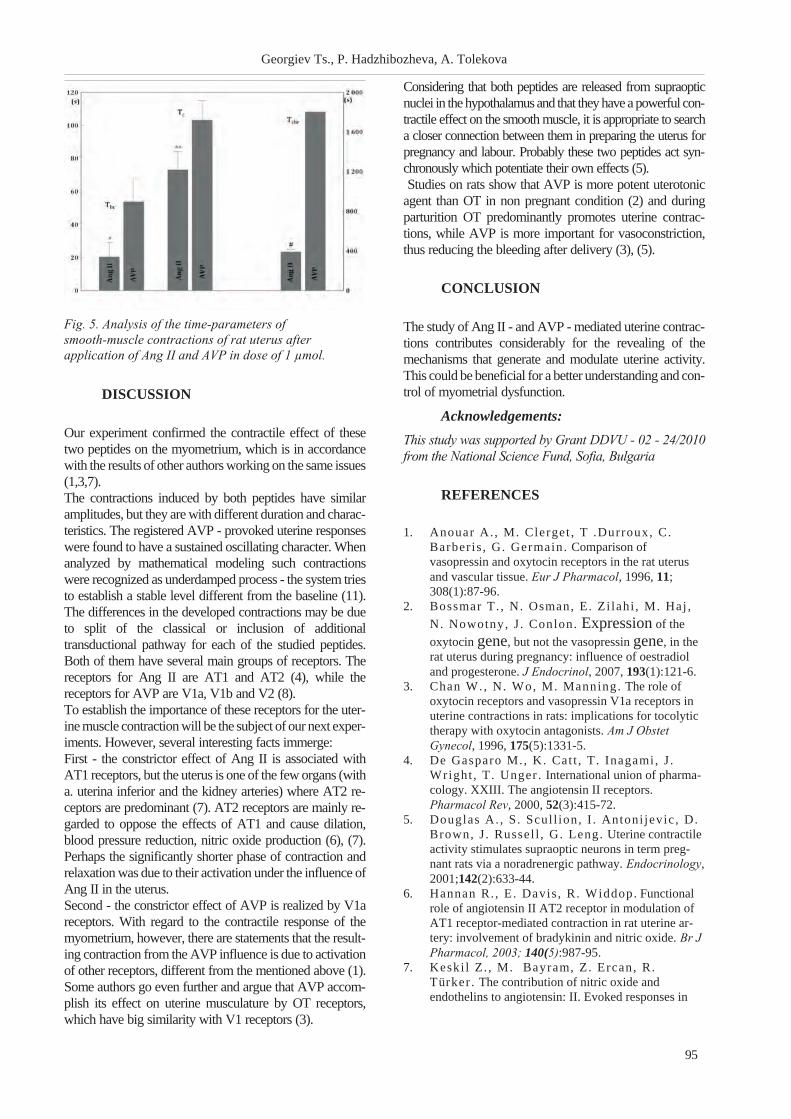
DISCUSSION
Our experiment confirmed the contractile effect of these
two peptides on the myometrium, which is in accordance
with the results of other authors working on the same issues
(1,3,7).
The contractions induced by both peptides have similar
amplitudes, but they are with different duration and charac-
teristics. The registered AVP - provoked uterine responses
were found to have a sustained oscillating character. When
analyzed by mathematical modeling such contractions
were recognized as underdamped process - the system tries
to establish a stable level different from the baseline (11).
The differences in the developed contractions may be due
to split of the classical or inclusion of additional
transductional pathway for each of the studied peptides.
Both of them have several main groups of receptors. The
receptors for Ang II are AT1 and AT2 (4), while the
receptors for AVP are V1a, V1b and V2 (8).
To establish the importance of these receptors for the uter-
ine muscle contraction will be the subject of our next exper-
iments. However, several interesting facts immerge:
First - the constrictor effect of Ang II is associated with
AT1 receptors, but the uterus is one of the few organs (with
a. uterina inferior and the kidney arteries) where AT2 re-
ceptors are predominant (7). AT2 receptors are mainly re-
garded to oppose the effects of AT1 and cause dilation,
blood pressure reduction, nitric oxide production (6), (7).
Perhaps the significantly shorter phase of contraction and
relaxation was due to their activation under the influence of
Ang II in the uterus.
Second - the constrictor effect of AVP is realized by V1a
receptors. With regard to the contractile response of the
myometrium, however, there are statements that the result-
ing contraction from the AVP influence is due to activation
of other receptors, different from the mentioned above (1).
Some authors go even further and argue that AVP accom-
plish its effect on uterine musculature by OT receptors,
which have big similarity with V1 receptors (3).
Considering that both peptides are released from supraoptic
nuclei in the hypothalamus and that they have a powerful con-
tractile effect on the smooth muscle, it is appropriate to search
a closer connection between them in preparing the uterus for
pregnancy and labour. Probably these two peptides act syn-
chronously which potentiate their own effects (5).
Studies on rats show that AVP is more potent uterotonic
agent than OT in non pregnant condition (2) and during
parturition OT predominantly promotes uterine contrac-
tions, while AVP is more important for vasoconstriction,
thus reducing the bleeding after delivery (3), (5).
CONCLUSION
The study of Ang II - and AVP - mediated uterine contrac-
tions contributes considerably for the revealing of the
mechanisms that generate and modulate uterine activity.
This could be beneficial for a better understanding and con-
trol of myometrial dysfunction.
Acknowledgements:
This study was supported by Grant DDVU - 02 - 24/2010
from the National Science Fund, Sofia, Bulgaria
REFERENCES
1. Anouar A. , M. Clerget , T .Durroux, C.
Barber is , G. Germain. Comparison of
vasopressin and oxytocin receptors in the rat uterus
and vascular tissue. Eur J Pharmacol, 1996, 11;
308(1):87-96.
2. Bossmar T. , N. Osman, E. Zi lahi , M. Haj ,
N. Nowotny, J . Conlon. Expression of the
oxytocin gene, but not the vasopressin gene, in the
rat uterus during pregnancy: influence of oestradiol
and progesterone. J Endocrinol, 2007, 193(1):121-6.
3. Chan W., N. Wo, M. Manning. The role of
oxytocin receptors and vasopressin V1a receptors in
uterine contractions in rats: implications for tocolytic
therapy with oxytocin antagonists. Am J Obstet
Gynecol, 1996, 175(5):1331-5.
4. De Gasparo M., K. Cat t , T. Inagami, J .
Wright , T. Unger . International union of pharma-
cology. XXIII. The angiotensin II receptors.
Pharmacol Rev, 2000, 52(3):415-72.
5. Douglas A. , S. Scul l ion, I . Antoni jevic , D.
Brown, J . Russel l , G. Leng. Uterine contractile
activity stimulates supraoptic neurons in term preg-
nant rats via a noradrenergic pathway. Endocrinology,
2001;142(2):633-44.
6. Hannan R. , E. Davis , R. Widdop. Functional
role of angiotensin II AT2 receptor in modulation of
AT1 receptor-mediated contraction in rat uterine ar-
tery: involvement of bradykinin and nitric oxide. Br J
Pharmacol, 2003; 140(5):987-95.
7. Keski l Z. , M. Bayram, Z. Ercan, R.
Türker . The contribution of nitric oxide and
endothelins to angiotensin: II. Evoked responses in
95
Georgiev Ts., P. Hadzhibozheva, A. Tolekova
Fig. 5. Analysis of the time-parameters of
smooth-muscle contractions of rat uterus after
application of Ang II and AVP in dose of 1 µmol.

the rat isolated uterus smooth muscle. Gen
Pharmacol, 1999; 33(4):307-12.
8. Petersen M. The effect of vasopressin and related
compounds at V1a and V2 receptors in animal models
relevant to human disease. Basic Clin Pharmacol
Toxicol, 2006; 99(2):96-103.
9. Wray S. Insights into the uterus. Exp Physiol,
2007; 92(4):621-31.
10. Yankov K. Preprocessing of Experimental Data in
Korelia Software. Trakia Journal of Sciences, 2010;
8, Suppl. 3: 41-48.
11. Yankov K. Recognition and function association of
experimental data. Proc. of the Int. Conference on In-
formation Technologies (InfoTech-2009),
Constantine and Elena resort, sept.17-20, 2009,
Varna, Bulgaria, 131-141.
96
Contractile responses of the rat uterine smooth muscle to ...

CARDIOVASCULAR STRESS AND MYOCARDIAL PROTECTION (I)
Kolarova R.
Department of Physiology and Biochemistry, National Sports Academy "Vassil Levski", Sofia
ABSTRACT
Hemodynamic and oxidative stress injuries are characteristic for cardiovascular diseases and the role of the
endogenous and exogenous antioxidant systems as cardioprotective mechanisms are quite interesting. The dy-
namics of antioxidant action of Vit. E on some functional and metabolic changes in the myocardium of hyper-
tensive rats was studied. The following methods were applied: a model of vasorenal hypertension; treatment
of the experimental animals with the antioxidant Vit. E; tracing of changes in arterial blood pressure, the ratio
of grams of myocardial weight/100 g body weight and activity of some "key enzymes" of cardiac metabolism -
of glycolysis, pentosomonophosphate shunt, cycle of Krebs, as well as enzymes of energetic metabolism.
Throughout the experimental periods (3rd
, 15th
and 30th
day) a progressively developing hypertension was es-
tablished. Glycolytic enzymes showed no essential deviations in their activity, whereas the activity of
pentosomonophosphate shunt was increased significantly at all experimental terms. A considerable increase
in the myocardial weight and values of the enzyme activity close to those of the control animals were estab-
lished in the groups of Vit. E-treated animals with experimental hypertension. The results obtained are char-
acteristic for the protective effects of Vit. E in the course of arterial hypertension regarding some metabolic
changes in the myocardium. The dominating role of the pentose monophosphate shunt as an anti-stress reac-
tion becomes prominent in this kind of hemodynamic injury of the myocardium. The combined treatment of
the experimental animals with Vit. E leads to cardioprotection, probablyby limiting of lipid peroxidation.
Key words: experimental hypertension, myocardial hypertrophy, myocardial enzyme activity, Vit. E
INTRODUCTION
Haemodynamic stress is one of the most common mecha-
nisms of injury not only of heart but also of the vascular
system. The causes for this stress can be different: increased
levels of catecholamines in the blood, treatment with
sympathomimetics, diverse forms of hypertension.
Haemodynamic stress could be acute, chronic, continuous
or interrupted (10). The changes in haemodynamics ulti-
mately lead to both pressure and volume overload to the
myocardium (17,19) with correspondingly severe conse-
quences later (1,4,5,6,11,13,18).
Regarding the role of endogenous antioxidant systems, it is
considered (12) that myocardial hypertrophy in experimen-
tal conditions provokes an increase of the cell antioxidant
reserve, which reduces the oxidative stress. Other authors
(2,3,9) have reported a protective effect of antioxidants in
patients with hypertension and still reversible vascular
damage, pointing out the potential therapeutic value of
long-term treatment with vitamin E in modulating or pre-
venting the pathogenesis of heart failure. It was found (8)
that the supplementation with a combination of vitamin C
and E among basketball players enhanced their body
antioxidant defense system by reducing the free radicals.
Having in mind the data from recent years for possible in-
volvement of free radicals in the pathogenesis of myocar-
dial hypertrophy and hypertension (7,12,14,15,16), the
question how free radical neutralization by the antioxidants
will reflect on the myocardial metabolism arises, since the
myocardial injury ultimately depends on metabolic
disorders.
In view of above data, we aimed to study the effect of anti-
oxidant Vit. E in dynamics on the activity of "key" enzymes
of myocardial metabolism in conditions of experimental
hypertension.
MATERIALS AND METHODS
1. Experimental animals.
Male Wistar rats with an initial body weight of 168 ±9 g
were used.
2. Groups:
a) a control group - healthy, unoperated animals
(n=6)
b) a group with experimental hypertension, untreated
(n=7)
c) a group with experimental hypertension, treated
with the solvent of vitamin E (n=18)
d) a group with experimental hypertension, treated
with vitamin E (n=20)
3. Experimental period.
Studies were performed on the 3rd, 15th and 30th day
after the hypertension creation.
4. Model of experimental hypertension.
97
Scripta Scientifica Medica, 2012; vol. 44 (1), Supplement 1 Copyright © Medical University, Varna
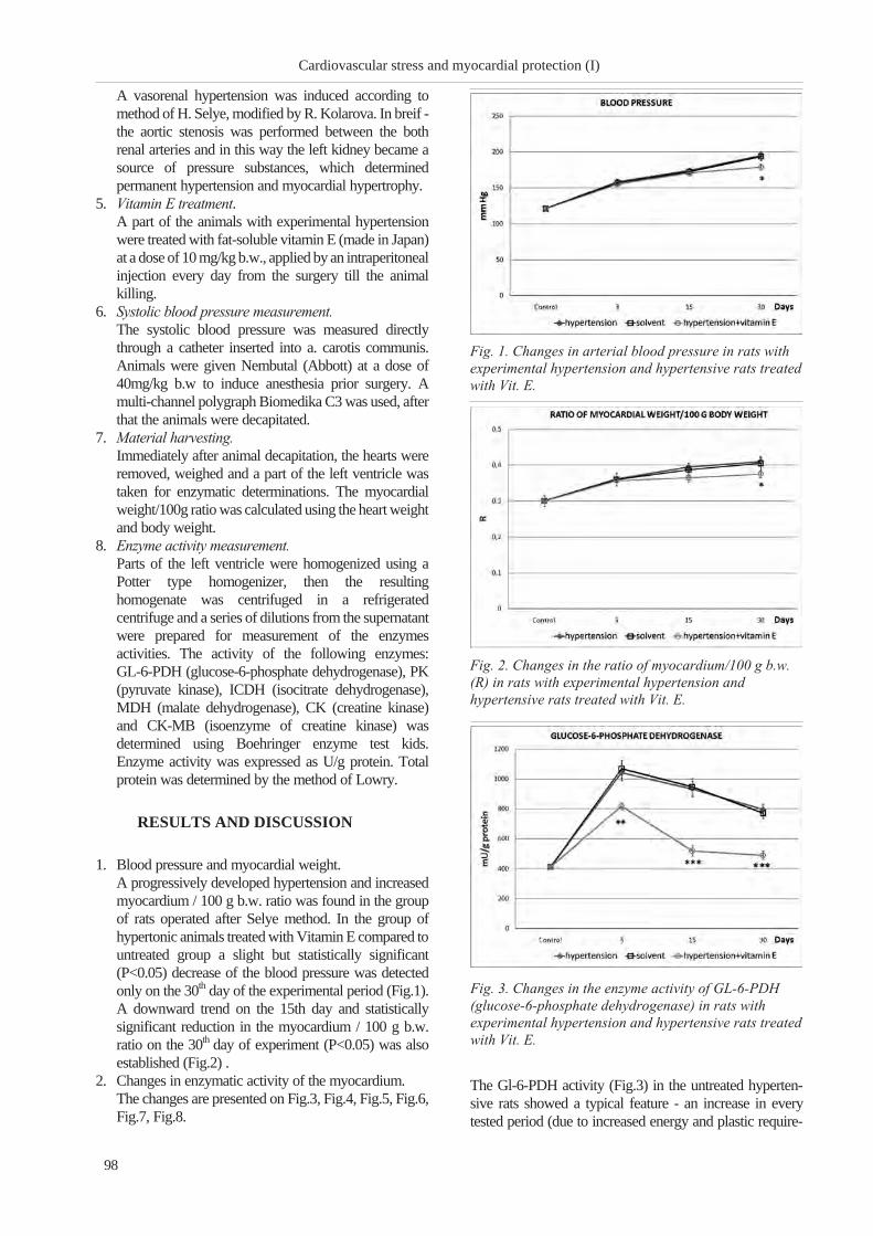
A vasorenal hypertension was induced according to
method of H. Selye, modified by R. Kolarova. In breif -
the aortic stenosis was performed between the both
renal arteries and in this way the left kidney became a
source of pressure substances, which determined
permanent hypertension and myocardial hypertrophy.
5. Vitamin E treatment.
A part of the animals with experimental hypertension
were treated with fat-soluble vitamin E (made in Japan)
at a dose of 10 mg/kg b.w., applied by an intraperitoneal
injection every day from the surgery till the animal
killing.
6. Systolic blood pressure measurement.
The systolic blood pressure was measured directly
through a catheter inserted into a. carotis communis.
Animals were given Nembutal (Abbott) at a dose of
40mg/kg b.w to induce anesthesia prior surgery. A
multi-channel polygraph Biomedika C3 was used, after
that the animals were decapitated.
7. Material harvesting.
Immediately after animal decapitation, the hearts were
removed, weighed and a part of the left ventricle was
taken for enzymatic determinations. The myocardial
weight/100g ratio was calculated using the heart weight
and body weight.
8. Enzyme activity measurement.
Parts of the left ventricle were homogenized using a
Potter type homogenizer, then the resulting
homogenate was centrifuged in a refrigerated
centrifuge and a series of dilutions from the supernatant
were prepared for measurement of the enzymes
activities. The activity of the following enzymes:
GL-6-PDH (glucose-6-phosphate dehydrogenase), PK
(pyruvate kinase), ICDH (isocitrate dehydrogenase),
MDH (malate dehydrogenase), CK (creatine kinase)
and CK-MB (isoenzyme of creatine kinase) was
determined using Boehringer enzyme test kids.
Enzyme activity was expressed as U/g protein. Total
protein was determined by the method of Lowry.
RESULTS AND DISCUSSION
1. Blood pressure and myocardial weight.
A progressively developed hypertension and increased
myocardium / 100 g b.w. ratio was found in the group
of rats operated after Selye method. In the group of
hypertonic animals treated with Vitamin E compared to
untreated group a slight but statistically significant
(P<0.05) decrease of the blood pressure was detected
only on the 30th day of the experimental period (Fig.1).
A downward trend on the 15th day and statistically
significant reduction in the myocardium / 100 g b.w.
ratio on the 30th day of experiment (P<0.05) was also
established (Fig.2) .
2. Changes in enzymatic activity of the myocardium.
The changes are presented on Fig.3, Fig.4, Fig.5, Fig.6,
Fig.7, Fig.8.
The Gl-6-PDH activity (Fig.3) in the untreated hyperten-
sive rats showed a typical feature - an increase in every
tested period (due to increased energy and plastic require-
98
Cardiovascular stress and myocardial protection (I)
Fig. 1. Changes in arterial blood pressure in rats with
experimental hypertension and hypertensive rats treated
with Vit. E.
Fig. 2. Changes in the ratio of myocardium/100 g b.w.
(R) in rats with experimental hypertension and
hypertensive rats treated with Vit. E.
Fig. 3. Changes in the enzyme activity of GL-6-PDH
(glucose-6-phosphate dehydrogenase) in rats with
experimental hypertension and hypertensive rats treated
with Vit. E.

ments in myocardial hypertrophy, found in our previous
studies). Upon Vit. E treatment the enzyme activity signifi-
cantly decreased compared with that of hypertensive rats,
but does not reach the control level. This change corre-
sponds to the reduction of the ratio g myocard/100 g. b.w.
and reflects the reduced energy and plastic requirements by
heart.
Demonstrative changes of PK-activity (Fig.4) are regis-
tered during the experimental period (an expressed increase
in the activity of the enzyme on the 3rd day and a mani-
fested decrease of the activity on the 15th day and again in-
crease of the PK-activity on the 30th day. It should be noted
that Vit. E treatment prevents all these changes.
The activity of Krebs cycle enzymes (ICDH / Fig.5/ and
MDH / Fig.6) - in hypertensive rats showed a dramatic
drop. When hypertensive animals were treated with Vit.E,
this supplementation prevented the decrease significantly
with exception of the 30th day (for both enzymes), which
clearly demonstrated that the oxidation processes in this pe-
riod cannot be stabilized.
The reduced activity of CK (Fig.7) in untreated hyperten-
sive rats on the 3rd and 15th day probably reflected the in-
creased energy consumption of the myocardium subjected
to compensatory hyperfunction, while in regard to the sharp
increase in activity of the enzyme on the 30th day we as-
sumed that it could be compensatory due to reduced activ-
ity of other metabolic pathways. In Vit. E treated hyperten-
99
Kolarova R.
Fig. 4. Changes in the enzyme activity of PK (pyruvate
kinase) in rats with experimental hypertension and
hypertensive rats treated with Vit. E.
Fig. 5. Changes in the enzyme activity of ICDH
(isocitrate dehydrogenase) in rats with experimental
hypertension and hypertensive rats treated with Vit. E.
Fig. 6. Changes in the enzyme activity of MDH (malate
dehydrogenase) in rats with experimental hypertension
and hypertensive rats treated with Vit. E.
Fig. 7. Changes in the enzyme activity of CK (creatine
kinase) in rats with experimental hypertension and
hypertensive rats treated with Vit. E.
Fig. 8. Changes in the enzyme activity of CK-MB
(isoenzyme of creatine kinase) in rats with experimental
hypertension and hypertensive rats treated with Vit. E.

sive rats only a trend towards normalization of enzyme ac-
tivity was observed in all experimental periods.
The changes in the CK-MB-activity, which is highly selec-
tive for the heart muscle, are particularly typical of the un-
treated hypertensive rats. They showed a progressive in-
crease throughout the entire experimental period, which is
indicative of myocardial ischemic damage. In Vit. E treated
hypertensive rats the CK-MB-activity was falling, as on the
15th day reached the control level.
The obtained results about the changes in the activity of
some key enzymes studied in hypertensive rats treated with
Vit. E are evidence for the marked protective effect of the
antioxidant Vit. E on myocardial metabolism.
CONCLUSIONS
The myocardium/100 g b.w. ratio was increased in the rats
with experimental hypertension and it was reduced in hy-
pertensive rats treated with Vit. E at the end of the experi-
mental period (on the 30th day after surgery); in the rats with
experimental hypertension the pentose monophosphate
shunt activity was significantly increased, CPK- and
CK-MB activity was also increased; in hypertensive rats
treated with Vit. E the pentose monophosphate shunt activ-
ity was reduced in all tested periods, the PK-acivity (a rep-
resentative of glycolysis) deviations were reduced also, the
changes in the Krebs cycle enzymes activity were an ex-
pression of improvement in the oxidative processes,
CPK-activity tended to normalize and CK-MB activity in
all tested periods of the experimental period was lower - an
indication for reduction of ischemic heart damage. The
changes in the enzyme activities studied in hypertensive
rats treated with Vit. E are an expression of the cardio-pro-
tective effects of Vit. E.
REFERENCES
1. Chandola T, Bri t ton A, Brunner E, Heming-
way H, Malik M, Kumari M et al , Work Stress
and coronary heart disease: what are the mechanisms?
Eur Heart J 2008; 29, 640-648.
2. Dhal la AK, Hil l MF & Singal PK. Role of ox-
idative stress in transition of hypertrophy to heart fail-
ure. J Am Coll Cardiol 1996; 28, 506-514.
3. Dhal la NS, Elmoselhi AB, Hata T &
Makino N. Status of myocardial antioxidants in
ischemia-reperfusion injury. Cardiovasc Res 2000;
47, 446-456.
4. De Rosa S, Cir i l lo P, Pagl ia A, Sasso L, Di
Palma & Chiar iel lo M. Reactive oxygen species
and antioxidants in the pathophysiology of cardiovas-
cular disease: does the actual knowledge justify a
clinical approach? Curr Vasc Pharmacol 2010; 8,
259-275.
5. Forogos R. Does Stress Really Cause Heart Dis-
ease? 2008.
6. I to H, Tori i M & Suzuki T. Comparative study
on free radical injury in the endothelium of SHR and
WKY aorta. Clin Exp Pharmacol Physiol Suppl 1995;
22, S157-159.
7. Nakamura K, Fushimi K, Kouchi H, Mihara
K, Miyazaki M, Ohe T et al , Inhibitory effects
of antioxidants on neonatal rat cardiac myocyte hy-
pertrophy induced by tumor necrosis factor-alpha and
angiotensin II. Circulation 1998; 98, 794-799.
8. Naziroglu M, Kil inc F, Uguz AC, Cel ik O,
Bal R, Butterworth PJ et al , Oral vitamin C and
E combination modulates blood lipid peroxidation
and antioxidant vitamin levels in maximal exercising
basketball players. Cell Biochem Func 2010; 28,
300-305.
9. Parik T, All ikmets K, Teesalu R & Zilmer
M. Evidence for oxidative stress in essential hyper-
tension: perspective for antioxidant therapy. J
Cardiovasc Risk; 3, 49-54.
10. Perr ino C, Naga Prasad SV, Mao L, Noma
T, Yan Z, Kim HS et al . Intermittent pressure
overload triggers hypertrophy-independent cardiac
dysfunction and vascular rarefaction. J Clin Inves
2006; 116, 1547-1560.
11. Sharma AB, Sun J, Howard LL, Wil l iams
AG, JR & Mallet RT. Oxidative stress reversibly
inactivates myocardial enzymes during cardiac arrest.
Am J Physiol Heart Circ Physiol 2007; 292,
H198-206.
12. Singh N, Dhal la AK, Seneviratne C &
Singal PK. Oxidative stress and heart failure. Mol
Cell Biochem 1995; 147, 77-81.
13. Stopler M. Stress; 2010 [Accessed 1/28/2010].
14. Taddei S, Virdis A, Ghiadoni L & Salvet t i
A. Endothelial dysfunction in hypertension: fact or
fancy? J Cardiovasc Pharmacol 1998; 32 Suppl 3,
S41-7.
15. Taddei S, Virdis A, Ghiadoni L, Sudano I
& Salvet t i A. Endothelial dysfunction in hyperten-
sion. J Cardiovasc Pharmacol 2001; 38 Suppl 2,
S11-14.
16. Taddei S, Virdis A, Ghiadoni L, Versar i D
& Salvet t i A. Endothelium, aging, and hyperten-
sion. Curr Hypertens Rep 2006; 8, 84-89.
17. Touyz RM. Advancement in hypertension
pathogenesis: some new concepts. Curr Opin Nephrol
Hypertens 2011; 20, 105-106.
18. Victor VM., Rocha M, Sola E, Banuls C,
Gardia-Malpart ida K & Hernandez-Mijares
A. Oxidative stress, endothelial dysfunction and ath-
erosclerosis. Curr Pharm Des 2009; 15, 2988-3002.
19. Wojciechowski P, Jur ic D, Louis XL,
Thadapi l ly SJ, Yu L, Taylor C et al .
Resveratrol arrests and regresses the development of
pressure overload- but not volume overload- induced
cardiac hypertrophy in rats. J Nutr 2010; 140,
962-968.
100
Cardiovascular stress and myocardial protection (I)

ANTIOXIDANT PREVENTION IN EXPERIMENTAL
HYPERTENSION: HISTOMORPHOLOGICAL CHANGES (II)
Kolarova R.1, Z. Gendzhev
2
1Department of Physiology and Biochemistry, National Sports Academy "Vassil Levski", Sofia;2Chemical Pharmaceutical Research Institute, Sofia
ABSTRACT
The dynamics of antioxidant action of Vit. E (for 30 days) on some histomorphological changes in the
myocardium of rats with experimental hypertension was studied. The following methods were applied: a
model of coarctation (vasorenal) hypertension after the method of H. Selye, modified by R. Kolarova; treat-
ment of the experimental animals with the antioxidant Vit. E (10 mg/kg) b.w., i.p.; tracing of changes in arte-
rial blood pressure, the ratio of grams of myocardial mass/100 g body mass and histomorphological study. A
progressive rise of arterial blood pressure and a statistically significant increase in the ratio of grams of myo-
cardial mass/100 g body mass were found in the course of the experimental period. In the group of hyperten-
sive rats untreated with the antioxidant typical histomorphological injuries were observed: focal or diffuse
myolysis of cardiomyocytes, without vascular injuries on the 3rd
day, myocardial hypertrophy, accompanied
with focal or diffuse myocardiofibrosis and coronarofibrosis on the 15th
day, which were strongly manifested
on the 30th
day of the experimental period. In the hypertensive rats treated with Vit. E the injuries were signif-
icantly less expressed as compared to the untreated animals in all experimental stages. Mostly necrotic and fi-
brous changes as well as coronarofibrosis and coronarosclerosis in the myocardium at the later term - 30th
day
were reduced. These results indicate the positive prophylactic role of Vit. E on the cardiac tissue in circum-
stances of hemodynamic stress in experimental arterial hyprtension.
Key words: experimental hypertension, myocardial hypertrophy, cardiac histomorphological changes, Vit. E
INTRODUCTION
It is known that in stress provoked heart damage destructive
disorders of cell membrane structures occur - a result of the
free radicals attacks. There are extensive clinical data about
both early (arrhythmias and sudden cardiac death) and late
(mostly coronary) heart damage (1,3,4,13,18). The distur-
bance of normal adrenergic mechanism in a number of
stress conditions gives rise to so-called lipid triad of
biomembranes damage. The activation of lipid
peroxidation (LPO) lead to severe disorders in
myocardiocytes structure (1,12). Many authors provide
data (some of them still hypothetical) about the role of free
radicals in pathogenesis of myocardial hypertrophy
(10,11), in induction of endothelial dysfunction (15,16) as
well as in development of heart failure (6,7). On the other
hand, in recent years, the data (mostly from the clinical
practice) about the cardioprotective effects of antioxidants
(2,5,6,12,14,17) increase. Another group of authors (9) has
ascertained the protective effect of vitamin E on the integ-
rity and function of mitochondria in cross-striated muscle,
whereas in regard to heart, such data are scanty.
Having in mind the data on the involvement of free radicals
in the genesis of hypertension and myocardial hypertrophy,
we aimed to study in dynamics the effect of the antioxidant
vitamin E on histomorphological damage in rat heart, sub-
jected to experimental hypertension.
MATERIALS AND METHODS
1. Experimental animals.
In the experiments 51 male Wistar rats with an initial
body weight of 168± 9g were used.
2. Groups:
a) a control group - healthy, unoperated (n=6)
b) a group with experimental hypertension, untreated
(n=7)
c) a group with experimental hypertension, treated
with the solvent of vitamin E (n=18)
d) a group with experimental hypertension, treated
with vitamin E (n=20)
3. Experimental period.
Studies were conducted on the 3rd, 15th and 30th day
after the hypertension induction.
4. Model of hypertension
A vasorenal hypertension was induced according to the
method of Selye, modified by Kolarova. In breif - the
aortic stenosis was performed between the both renal
arteries and in this way the left kidney became a source
of pressure substances, which determined permanent
hypertension and myocardial hypertrophy.
101
Scripta Scientifica Medica, 2012; vol. 44 (1), Supplement 1 Copyright © Medical University, Varna

5. Treatment with vitamin E.
A part of the animals with experimental hypertension
were treated with fat-soluble vitamin E (made in Japan)
at a dose of 10 mg/kg b.w. applied by an intraperitoneal
injection every day from the day of surgery till the
animal killing.
6. Systolic blood pressure measurement.
The systolic blood pressure was measured directly
through a catheter inserted into a. carotis communis.
Animals were given Nembutal (Abbott) at a dose of 40
mg/kg b.w to induce anesthesia prior surgery. A
multi-channel polygraph Biomedika C3 was used.
After than the animals were decapitated.
7. Material harvesting.
Immediately after animal decapitation, the hearts were
removed, weighed and a part of the left ventricle was
taken for histomorphological analysis. The myocardial
weight/100g b.w. ratio was calculated.
8. Histomorphological analysis.
After fixation with 3% formalin (neutral) the sections
were prepared and stained with hemalaun-eosin.
RESULTS
1. Blood pressure and myocardial mass.
In the rats operated after the method of Selye a
progressively developed hypertension and increased
myocardium/100g b.w. ratio were found. In the
hypertensive animals, treated with vitamin E, compared
to those of the untreated group, a slight but statistically
significant (P <0.05) decrease in blood pressure was
102
Antioxidant prevention in experimental hypertension ...
3rd
day from the begging
of the hypertension
15th
day from the begging
of the hypertension
30th
day from the begging
of the hypertension
Myocardial
damage
Solvent
n=5
Vitamin E
n=5
Solvent
n=8
Vitamin E
n=7
Untreated
n=7
Solvent
n=5
Vitamin E
n=8
Without special
feature
Mild
Severe
2
1
2
-
2
3
3
1
4
2
4
1
3
1
3
1
-
4
2
5
1
Mild - only eosinophilic
myositis and / or 1-3 small foci
of myolysis
Severe - more than 3 foci of
myolysis
Mild - only muscular hypertrophy
and / or 1-3 small foci of myo- or
arteriofibrosis
Severe - more than 3 foci of myo- or
arteriofibrosis or diffuse myofibrosis
Mild - only muscular hypertrophy and / or
1-3 small foci of myo- or arteriofibrosis
Severe - more than 3 foci of myo- or
arteriofibrosis and / or necrosis
Table 1. Degree of myocardial injury in hypertensive rats untreated, treated with the solvent of vitamin E or treated
with vitamin E
Fig. 1. Rat myocardium on the 3rd day of hypertension
induction. Diffuse myolysis of the left ventricular wall.
Activated endothelial cells of capillaries and small
number of fibroblasts were infiltrated the muscle fibers,
underwent myolysis. XE stain, zoom 80x.
Fig. 2. Rat myocardium on the 3rd day of hypertension
induction. Contractile stripes with light fields between
them /possibly due to rupture of protofibers/ in
myocardium area, unaffected directly by the myolysis.
XE stain, zoom 200x.

detected only on the 30th day of the experimental
period. It is also established a downward trend (on the
15th day of the experiment) and statistically significant
reduction (on the 30th day of the experiment) in the
myocardium/100 g b.w. ratio (P <0.05).
2. Histomorphological data - The histomorphological data
were presented on Table 1
On the 3rd day of hypertension, when haemodynamic stress
was particularly great in both groups of animals - untreated
and treated with vitamin E, severe heart damage was ob-
served (Fig.1, 2). On the 15th and 30th day of the experiment
(in contrast to the 3rd day) the histomorphological changes
in the myocardium of the hypertensive rats treated with vi-
tamin E differed significantly from those of untreated rats,
in particular: in the animals, treated with vitamin E there
was a pronounced myocardial hypertrophy, but the other
disabilities were poorly expressed or absent. Above all, the
necrotic and fibrotic changes were reduced - the focal and
diffuse myocardial fibrosis as well as coronary fibrosis and
coronary sclerosis. The histomorphological changes in un-
treated and treated hypertensive animals are presented on
Figures 1,2,3,4,5, and 6.
DISCUSSION
The fact that myocardial hypertrophy, coronary fibrosis and
myocardial fibrosis (as well as some other metabolic pa-
rameters, tested in our previous study, performed in the
same experimental setup) were influenced by the antioxi-
dant vitamin E, suggests its positive effect on the heart.
In regard to the cardioprotective effect of vitamin E we can
assume (based on data of other authors - 6,8,12,14,17), that
through inhibition of free-radical processes their harmful
effects on the myocardium reduce.
Having in mind that the genesis of myocardial hypertrophy
and myocardial and vascular lesions in hypertension, in-
duced after Selye, is a complex process (pressor overload,
catecholamines, RAS), our results give us reason to accept
the possible participation of another factor - the free radical
processes in this model of hypertension.
The possible mechanism of the protective effect of vitamin
E against cardiovascular damage in experimental hyperten-
103
Kolarova R., Z. Gendzhev
Fig. 3. Rat myocardium on the 15th day of hypertension
induction. Numerous foci of myocardial fibrosis covered
by hypertrophied muscle fibers in the left ventricular
wall. XE stain, zoom 80x.
Fig. 4. Rat myocardium on the 15th day of hypertension
induction; the hypertensive rats were treated with
vitamin E. In the left ventricular wall the muscle fibers
were strongly hypertrophied without any other damage.
XE stain, zoom 200x.
Fig. 5. Rat myocardium on the 30th day of hypertension
induction. Necrotic rings spots of the left ventricular
wall. Hypertrophied muscle fibers were found nearby.
XE stain, zoom 80x.
Fig. 6. Rat myocardium on the 30th day of the beginning
of hypertension the hypertensive rats were treated with
vitamin E. Pronounced hypertrophy of myocardial cells
without focal impairment. Part of the coronary artery
with minimal sclerotic changes. XE stain, zoom 80 x 20.

sion, induced according to the method of Selye, in our view
could be presented as follows (Fig. 7).
CONCLUSIONS
1. In the hypertensive rats, treated with vitamin E a slight
decrease in systolic blood pressure of the 30th day of
the experiment was detected.
2. In the hypertensive animals, treated with vitamin E a
reduction of the myocardium/100 g b.w. ratio on the
30th day of surgery was observed.
3. In this experimental model the treatment of the
hypertensive rats with vitamin E significantly protected
the myocardium from severe structural damage
(myocardial fibrosis, coronary fibrosis and coronary
sclerosis).
REFERENCES
1. Ìååðñîí ÔÇ, Ïøåííèêîâà ÌÃ. Àäàïòàöèÿ ê
ñòðåññîðíûì ñèòóàöèÿì è ôèçè÷åñêèì íàãðóçêàì;
Ìîñêâà, Måäèöèíà, 1988.
2. Benderi t ter M, Maingon P, Abadie C,
Assem M, Maupoil V, Briot F et al , Effect of
in vivo heart irradiation on the development of anti-
oxidant defenses and cardiac functions in the rat.
Radiat Res 1995; 144, 64-72.
3. Bonomini F, Tengat t ini S, Fabiano A,
Bianchi R & Rezzani R. Atherosclerosis and ox-
idative stress. Histol Histopathol 2008; 23, 381-390.
4. De Rosa S, Cir i l lo P, Pagl ia A, Sasso L, Di
Palma V & Chiar iel lo M. Reactive oxygen spe-
cies and antioxidants in the pathophysiology of car-
diovascular disease: does the actual knowledge justify
a clinical approach? Curr Vasc Pharmacol 2010; 8,
259-275.
5. Dhal la AK, Hil l MF & Singal PK. Role of ox-
idative stress in transition of hypertrophy to heart fail-
ure. J Am Coll Cardiol 1996; 28, 506-514.
6. Dhal la NS, Elmoselhi AB, Hata T &
Makino N. Status of myocardial antioxidants in
ischemia-reperfusion injury. Cardiovasc Res 2000;
47, 446-456.
7. Feuerstein G, Yue TL, Ma X & Ruffolo
RR. Novel mechanisms in the treatment of heart fail-
ure: inhibition of oxygen radicals and apoptosis by
carvedilol. Prog Cardiovasc Dis 1998; 41, 17-24.
8. Hamilton KL. Antioxidants and cardioprotection. Med
Sci Sports Exerc 2007; 39, 1544-1553.
9. Magalhaes J , Ferreira R, Neuparth MJ,
Oliveira PJ, Marques F & Ascenao A. Vita-
min E prevents hypobaric hypoxia-induced mitochon-
drial dysfunction in skeletal muscle. Clin Sci (Lond)
2007; 113, 459-466.
10. Nakamura K, Fushimi K, Kouchi H, Mihara
K, Miazaki M, Ohe T et al , Inhibitory effects of
antioxidants on neonatal rat cardiac myocyte hyper-
trophy induced by tumor necrosis factor-alpha and an-
giotensin II. Circulation 1998; 98, 794-799.
11. Singh N, Dhal la AK, Seneviratne C &
Singal PK. Oxidative stress and heart failure. Mol
Cell Biochem 1995; 147, 77-81.
12. Stampfer MJ, Hennekens CH, Manson JE,
Coldi tz GA, Rosner B & Willet t WC. Vita-
min E consumption and the risk of coronary disease
in women. N Engl J Med 1993; 328, 1444-1449.
13. Stopler M. Stress 2010; [Accessed 1/28/2010].
14. Schiffr in EL. Antioxidants in hypertension and
cardiovascular disease. Mol Interv 2010; 10, 354-362.
15. Taddei S, Virdis A, Ghiadoni L, Sudano I
& Salvet t i A. Endothelial dysfunction in hyperten-
sion. J Cardiovasc Pharmacol 2001; 38 Suppl 2,
S11-14.
16. Taddei S, Virdis A, Ghiadoni L, Versar i D
& Salvet t i A. Endothelium, aging, and hyperten-
sion. Curr Hypertens Rep 2006; 8, 84-89.
17. Thurich T, Berei ter-Hahn J, Schneider M &
Zimmer G. Cardioprotective effects of
dihydrolipoic acid and tocopherol in right heart hy-
pertrophy during oxidative stress.
Arzneimittelforschung 1998; 48, 13-21.
18. Victor VM, Rocha M, Sola AE, Banuls C,
Garcia-Malpart ida K & Hernandez-Mijares
A. Oxidative stress, endothelial dysfunction and ath-
erosclerosis. Curr Pharm Des 2009; 15, 2988-3002.
104
Antioxidant prevention in experimental hypertension ...
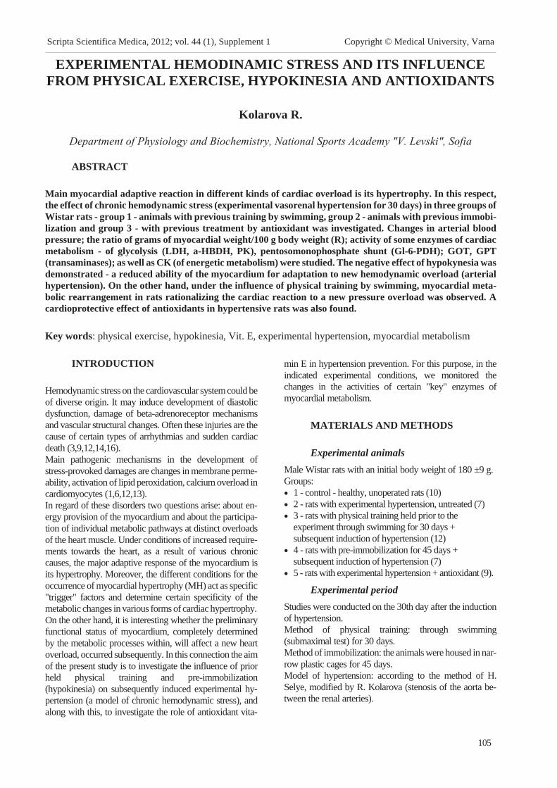
EXPERIMENTAL HEMODINAMIC STRESS AND ITS INFLUENCE
FROM PHYSICAL EXERCISE, HYPOKINESIA AND ANTIOXIDANTS
Kolarova R.
Department of Physiology and Biochemistry, National Sports Academy "V. Levski", Sofia
ABSTRACT
Main myocardial adaptive reaction in different kinds of cardiac overload is its hypertrophy. In this respect,
the effect of chronic hemodynamic stress (experimental vasorenal hypertension for 30 days) in three groups of
Wistar rats - group 1 - animals with previous training by swimming, group 2 - animals with previous immobi-
lization and group 3 - with previous treatment by antioxidant was investigated. Changes in arterial blood
pressure; the ratio of grams of myocardial weight/100 g body weight (R); activity of some enzymes of cardiac
metabolism - of glycolysis (LDH, a-HBDH, PK), pentosomonophosphate shunt (Gl-6-PDH); GOT, GPT
(transaminases); as well as CK (of energetic metabolism) were studied. The negative effect of hypokynesia was
demonstrated - a reduced ability of the myocardium for adaptation to new hemodynamic overload (arterial
hypertension). On the other hand, under the influence of physical training by swimming, myocardial meta-
bolic rearrangement in rats rationalizing the cardiac reaction to a new pressure overload was observed. A
cardioprotective effect of antioxidants in hypertensive rats was also found.
Key words: physical exercise, hypokinesia, Vit. E, experimental hypertension, myocardial metabolism
INTRODUCTION
Hemodynamic stress on the cardiovascular system could be
of diverse origin. It may induce development of diastolic
dysfunction, damage of beta-adrenoreceptor mechanisms
and vascular structural changes. Often these injuries are the
cause of certain types of arrhythmias and sudden cardiac
death (3,9,12,14,16).
Main pathogenic mechanisms in the development of
stress-provoked damages are changes in membrane perme-
ability, activation of lipid peroxidation, calcium overload in
cardiomyocytes (1,6,12,13).
In regard of these disorders two questions arise: about en-
ergy provision of the myocardium and about the participa-
tion of individual metabolic pathways at distinct overloads
of the heart muscle. Under conditions of increased require-
ments towards the heart, as a result of various chronic
causes, the major adaptive response of the myocardium is
its hypertrophy. Moreover, the different conditions for the
occurrence of myocardial hypertrophy (MH) act as specific
"trigger" factors and determine certain specificity of the
metabolic changes in various forms of cardiac hypertrophy.
On the other hand, it is interesting whether the preliminary
functional status of myocardium, completely determined
by the metabolic processes within, will affect a new heart
overload, occurred subsequently. In this connection the aim
of the present study is to investigate the influence of prior
held physical training and pre-immobilization
(hypokinesia) on subsequently induced experimental hy-
pertension (a model of chronic hemodynamic stress), and
along with this, to investigate the role of antioxidant vita-
min E in hypertension prevention. For this purpose, in the
indicated experimental conditions, we monitored the
changes in the activities of certain "key" enzymes of
myocardial metabolism.
MATERIALS AND METHODS
Experimental animals
Male Wistar rats with an initial body weight of 180 ±9 g.
Groups:
� 1 - control - healthy, unoperated rats (10)
� 2 - rats with experimental hypertension, untreated (7)
� 3 - rats with physical training held prior to the
experiment through swimming for 30 days +
subsequent induction of hypertension (12)
� 4 - rats with pre-immobilization for 45 days +
subsequent induction of hypertension (7)
� 5 - rats with experimental hypertension + antioxidant (9).
Experimental period
Studies were conducted on the 30th day after the induction
of hypertension.
Method of physical training: through swimming
(submaximal test) for 30 days.
Method of immobilization: the animals were housed in nar-
row plastic cages for 45 days.
Model of hypertension: according to the method of H.
Selye, modified by R. Kolarova (stenosis of the aorta be-
tween the renal arteries).
105
Scripta Scientifica Medica, 2012; vol. 44 (1), Supplement 1 Copyright © Medical University, Varna

Treatment with the antioxidant vitamin E: the hypertensive ani-
mals receivedvitaminE10mg/kgb.w., i.p.daily, for30days.
Measurement of the enzyme activities
After several steps - material harvesting from the left ventricle,
weighing, determination of the coefficient R, homogenization
of myocardial tissue with subsequent centrifugation and ap-
propriate dilutions, the enzyme activities (U/g protein) were
determined. The activities of the following enzymes
Gl-6-PDH, PK, LDH, a-HBDH, GOT, GPT, CK were mea-
sured. The test reagents were purchased from Boehringer Ltd.
(the biochemical analyses in details are presented in our previ-
ous publications). The g myocard/100g b.w. ratio (R) was cal-
culated. The systolic blood pressure was measured directly us-
ing polygraph "Biomedica" C3. Statistics - The data were ana-
lyzed by the method of variational analysis.
RESULTS AND DISCUSSION
The results are presented on Figures 1-9.
The systolic blood pressure in hypertensive animals (2nd
group), as well as in those combined with subsequent hyper-
tension (3rd, 4th and 5th groups) was statistically significantly
higher than that in control animals / p (1st-2nd) <0.001/, p
(1st-3rd) <0.001, p (1st-4th) <0.001, p (1st-5th) <0.001.
The increased blood pressure in these groups correlated
with growth rate of the coefficient R, suggesting the devel-
opment of myocardial hypertrophy /p (1st- 2nd <0.001 and p
(1st-3rd) <0.001, p (1st-4th) <0.05, p (1st-5th) < 0.01/.
Enzyme activities - more typical were the following
changes:
106
Experimental hemodinamic stress and its influence from ...
Fig. 1. Changes in arterial blood pressure.
Fig. 2. Changes in the ratio of myocardium/100 g b.w. (R).
Fig. 3. Changes in the enzyme activity of PK (pyruvate
kinase).
Fig. 4. Changes in the enzyme activity of LDH (lactate
dehydrogenase).
Fig. 5. Changes in the enzyme activity of alpha-HBDH
(alpha-hydroxi-butirate dehydrogenase).
Fig. 6. Changes in the enzyme activity of GL-6-PDH
(glucose-6-phosphate dehydrogenase).

Glycolytic enzymes - PK (Fig. 3) and LDH (Fig. 4): a
higher activity of PK in group 2 /p (1st-2nd) <0.01/, and
group 3 /p (1st-3rd) <0.001/, but decreased activity of this
enzyme in group 4 /p (1st- 4th) <0.05 / was detected. In con-
trast, in regard to LDH the opposite changes were observed
- a tendency of decreased activity in groups 2 and 3, and
statistically significantly increased activity in group 4 /p
(1st-4th) <0.001/.
Alpha-HBDH (Fig. 5) - its activity did not show statisti-
cally significant changes in the experimental groups com-
pared with the control.
Gl-6-P-DH (Fig. 6) - the most typical change in this en-
zyme was its reduced activity in the hypertensive rats,
treated with vitamin E (group 5) compared with untreated
hypertensive animals (group 2) with clearly expressed sta-
tistical accuracy, p (2nd-5th) <0.001, as this value tended to
approach the control value.
CK (Fig.7) - the changes in creatine kinase activity in all
groups were particularly demonstrative compared with the
control. There was a pronounced decrease in groups 2, 3
and 4, as in the three groups p <0.001 compared to the con-
trol animals, while in rats treated with antioxidant the activ-
ity is also reduced, but still showed some tendency to ap-
proach the control value - p (1st-5th) <0.01.
The transaminases GOT (Figure 8) and GPT (Figure 9)
showed similar changes in their activities in the distinct
groups. It was noteworthy that the highest values of both
enzymes were measured in the immobilized rats with sub-
sequent hypertension-for GOT the p (1st-4th) <0.001, and
for GPT - p (1st-4th) <0.05. In the hypertensive animals
(group 2) the activities of both enzymes were also increased
in comparison with the control values, which was more
pronounced in regard to GOT.
The resulting changes in myocardial enzyme activity in the
groups could be interpreted as a result of the impact of the
additionally induced hemodynamic stress (hypertension)
on the heart of experimental animals under different condi-
tions - previously conducted physical training (by swim-
ming) and pre-immobilization (hypokinesia), and antioxi-
dant treatment of the hypertensive rats. It is possible that
these changes were due to the subsequent severe overload
of the heart muscle thought hypertension moreover on the
background of these preliminary impacts.
The increase in blood pressure and the increase of the coef-
ficient R could be explain with the developing myocardial
hypertrophy (MH), as in the swimming group the factors
for MH development were the following - physical training
plus hypertension. In some our previous studies, we ob-
served in Wistar rats with induced immobilization stress
only, statistically significant changes in both blood pressure
and coefficient R The changes in regard to these two in-
dexes in this group clearly were due to the hypertension,
induced later.
The increased activity of pyruvate kinase (PK-catalyzed re-
action from the 10th step of glycolysis) in groups 2 and 3
suggested activation of glycolytic exchange in
hemodynamic stress, confirming the potential power of
glycolysis as a compensatory mechanism of the
myocardium under conditions of increased demands on
him (1). In these same two groups a tendency of fall in
LDH was observed, which is in favor of aerobic metabo-
lism within them, whereas in group 4 with pre-immobiliza-
tion the LDH activity was strongly increased in comparison
with other groups - most likely an indicator of compensa-
tory activated anaerobic glycolysis. It is noteworthy that
Gl-6-PDH - an enzyme with a powerful compensatory role
in chronic overload of the myocardium particularly, was
with highly increased activity only in the group of hyper-
tensive animals, while in the other groups its activity did
not differ significantly from the control value. The fact, that
in trained animals this enzyme showed only an upward
trend, could be linked with the opinion of other authors
107
Kolarova R.
Fig. 7. Changes in the enzyme activity of CK (creatine
kinase).
Fig. 8. Changes in the enzyme activity of GOT
(glutamate oxalacetic transaminase).
Fig. 9. Changes in the enzyme activity of GPT
(glutamate pyruvate transaminase).

(4,10) for the protective role of physical exercise on the
heart. An important result was that in the hypertensive rats
treated with vitamin E a significant decrease in
Gl-6-PDH-activity was found /p (2nd-5th) <0.001/, which is
consistent with our results obtained in another experimental
design and using a synthetic antioxidant (7). The significant
decline of CK-activity in all groups most likely was due to
increased energy consumption because of rapid overload of
the myocardium. Only in group 5 this enzyme showed a
weak tendency to approach the control level, which still
speaks in favor of the protective effect of vitamin E. In rats
with prior immobilization, except the above results on
LDH, the transaminases were increased significantly,
which was probably a compensatory response to increased
protein synthesis, which correlated with the increase of the
coefficient R - an indicator for the MH development. Ac-
cording to some authors, there are probably other, still hy-
pothetical mechanisms to stimulate protein synthesis and
survival of cells (2,17). There is also evidence that the
hypokinesia, except induction of oxidative stress, induces
also antioxidant response with individual variability (8,11).
CONCLUSION
The results show that the negative effect of hypokinesia
consisted in reducing the level of certain metabolic pro-
cesses in the myocardium, which determinate its reduced
ability to adapt in the subsequent considerable overload -
hypertension. On the other hand, the physical exercise
through impact on important exchange pathways enables
optimal response of the heart under conditions of increased
requirements. The results also speak in favor of the
cardioprotective effects of antioxidants.
REFERENCES
1. Ìååðñîí ÔÇ, Ïøåííèêîâà ÌÃ. Àäàïòàöèÿ ê
ñòðåññîðíûì ñèòóàöèÿì è ôèçè÷åñêèì íàãðóçêàì;
Ìîñêâà, Måäèöèíà, 1988.
2. Cat taruzza M, Hecker M. A new twist to the
complex response of vascular cells to oxidative stress.
Circ Res 2008; 102, 273-274.
3. De Rosa S, Cir i l lo P, Pagl ia A, Sasso L, Di
Palma & Chiar iel lo M. Reactive oxygen species
and antioxidants in the pathophysiology of cardiovas-
cular disease: does the actual knowledge justify a
clinical approach? Curr Vasc Pharmacol 2010; 8,
259-275.
4. Emter CA, McCune SA, Sparagna GC,
Radin MJ & Moore RL. Low-intensity exercise
training delays onset of decompensated heart failure
in spontaneously hypertensive heart failure rats. Am J
Physiol Heart Circ Physiol 2005; 289, H2030-8.
5. Gey KF, Puska P, Jordan P & Moser UK.
Inverse correlation between plasma vitamin E and
mortality from ischemic heart disease in cross-cultural
epidemiology. Am J Clin Nutr 1991; 53, 326S-334S.
6. Hamil ton KL. Antioxidants and cardioprotection.
Med Sci Sports Exerc 2007; 39, 1544-1553.
7. Kolarova R. Myocardial catecholamine stress and
antioxidant protection. Sci. Res. J. of South-West Univ
2008; 1, 33-37.
8. Margari t is I , Rousseau AS, Marini JF &
Chopard A. Does antioxidant system adaptive re-
sponse alleviate related oxidative damage with long
term bed rest? Clin Biochem 2009; 42, 371-379.
9. Perr ino C, Naga Prasad SV, Mao L, Noma
T, Yan Z, Kim HS et al . Intermittent Pressure
overload triggers hypertrophy-independent cardiac
dysfunction and vascular rarefaction. J Clin Inves
2006; 116, 1547-1560.
10. Puffer JC. Exercise and heart disease. Clin Corner-
stone 2001; 3, 1-9.
11. Sahin E & Gumuslu S. Immobilization stress in
rat tissues: alterations in protein, lipid peroxidation
and antioxidant defense system. Comp Biochem
Physiol Toxicol Pharmacol 2007; 144, 342.
12. Sam F, Kerstet ter DL, Pimental DR,
Mulukut la S, Tabaee A, Bris tow MR et al . ,
Increased reactive oxygen species production and
functional alterations in antioxidant enzymes in hu-
man failing myocardium. J Card Fail 2005; 11,
473-480.
13. Schiffr in EL. Antioxidants in hypertension and
cardiovascular disease. Mol Interv 2010; 10, 354-362.
14. Sharma AB, Sun J, Howard LL, Wil l iams
AG, JR & Mallet RT. Oxidative stress reversibly
inactivates myocardial enzymes during cardiac arrest.
Am J Physiol Heart Circ Physiol 2007; 292,
H198-206.
15. Stampfer MJ, Hennekens CH, Manson JE,
Coldi tz GA, Rosner B & Willet t WC. Vita-
min E consumption and the risk of coronary disease
in women. N Engl J Med 1993; 328, 1444-9.
16. Touyz RM. Advancement in hypertension
pathogenesis: some new concepts. Curr Opin Nephrol
Hypertens 2011; 20, 105-106.
17. Wong CM, Cheema AK, Zhang L, Suzuki
YJ. Protein carbonylation as a novel Mechanism in
redox signaling. Circ Res 2008; 102, 310-318.
108
Experimental hemodinamic stress and its influence from ...

AGE-RELATED EFFECTS ON THE SENSITIVITY TO GLOBAL
MOTION DIRECTION DETERMINED BY THE METHOD
OF CLASSIFICATION IMAGES
Stefanov S.1, K. Alexiev
2, N. Bocheva
1, B. Genova
1
1Institute of Neurobiology, Bulgarian Academy of Sciences, Sofia; 2Institute of Information and
Communication Technologies, Bulgarian Academy of Sciences, Sofia
SUMMARY
The method of classification images was used to evaluate whether the perceptual template applied by subjects
in the discrimination of global motion direction changes with ageing. The stimuli were 25-frame motion se-
quences of signal and noise band-limited elements. The motion direction of the signal dots was randomly se-
lected from a uniform distribution with range of 60° and mean ±10° from the vertical direction. The noise dots
moved in random directions taken uniformly in the range 0°-360°. The signal-to-noise ratio was determined in
a preliminary experiment separately for each observer to ensure 75% correct responses. The task of the ob-
servers was to discriminate whether the mean direction of motion was to the left or to the right of the vertical.
Nine younger (mean age 19.8 yrs) and nine older subjects (mean age 72 yrs) participated in the study. Classifi-
cation images were calculated from the directional distribution of all potential dot trajectories in the motion
sequences. The results show that the older observers have higher internal noise and they need higher sig-
nal-to-noise ratio in order to successfully perform the task. The calculated perceptual templates show similar
directional tuning but different temporal evolution for the two age groups. These findings are discussed in re-
lation to the age-related changes in center-surround suppression in dynamic information processing.
Key words: motion direction discrimination, classification images, internal noise, directional tuning, temporal
integration
INTRODUCTION
Motion information is essential for various everyday tasks
like navigation, figure-ground segregation, collision avoid-
ance, determination of spatial layout and the three-dimen-
sional shape of the objects, etc. Several studies have dem-
onstrated visual deficits in motion processing with age (e.g.
13,14,16,3,4). Kavcic et al (7) suggest that age-related
changes in spatio-temporal integration are the main factor
contributing to the impaired perception of optic flow in el-
derly and in Alzheimer patients. Differences in perfor-
mance might occur, however, not only due to perceptual
factors but to changes in attention and the to the impaired
attentional capacities of the elderly people (9), their inabil-
ity to scale attention to the scope of relevant stimuli (5) or to
differences in the decision strategies used in task
performance.
The aim of the present study was to evaluate the age-related
changes in the cognitive processes in a task related to inte-
gration of motion information. The experimental design
used in the study resembles the classification image para-
digm (e.g. 1,2,10 for a recent review). In a typical experi-
ment of this type the observers classify signals in the pres-
ence of additive noise. The characteristics of the noise pat-
terns for the correct and the incorrect trials are analyzed in
order to reveal the perceptual "template" used for solving
the task e.g. to infer the stimulus features that determine the
perceptual decisions. Our stimuli could be described as a
signal modulated by noise in the presence of background
noise. This stimulation increases the perceptual load and al-
lows not only to evaluate the perceptual abilities of the sub-
jects, but also to reveal differences in performance due to
attentional processes and different decision strategies.
MATERIALS AND METHODS
Nine older (mean age 73.9, range 66-82 years) and nine
younger ( mean age 19.5; range 16-24 year) observers took
part in the study. All participants have normal or corrected
to normal vision and have passed eye examination. None of
them reported having any major health problems.
The stimuli were motion sequences of 48 moving elements.
Each motion sequence contained 25 frames. The moving
elements were spots with intensity profile that varied as a
Laplacian of Gaussian. The mean speed of motion was 3.67
deg/s. The moving elements were divided in two groups.
109
Scripta Scientifica Medica, 2012; vol. 44 (1), Supplement 1 Copyright © Medical University, Varna
Address for correspondence:
S. Stefanov, Institute of Neurobiology, Bulgarian Academy of Sci-
ences, 23 Acad. G. Bonchev St. Sofia Bulgaria 1113
e-mail: [email protected]

In the first, the direction of motion was restricted to a range
of 60? and the speed varied within 50-150% from the mean
speed. Each of these elements randomly changes direction
on every frame. The range of directions was centered either
at ±10° from the vertical downward direction. These ele-
ments will be labeled as the main set. The other group of el-
ements moved with constant direction chosen randomly in
the range 0°-360° with a constant speed of 3.67 deg/s. They
will be referred as supplementary set. The number of ele-
ments in each set was determined in preliminary experi-
ment to ensure an accuracy of 75% in motion discriminate
for each observer. The moving elements were presented in
a circular aperture with radius of 7 deg of arc. Table 1 pres-
ents the number of elements in the main set needed to en-
sure similar performance for the two age groups.
The stimuli were presented on a computer screen (21" Dell
Trinitron with Nvidia Quadro 900XGL graphic board) in a
1280x1024 resolution mode. A chin-rest was used to main-
tain a fixed distance to the screen. The refresh rate of the
monitor was 85 Hz. The observation was binocular.
The task of the observers was to indicate whether the mean
direction of motion was to the left or to the right of the verti-
cal. To respond the younger observers used two buttons of
a computer mouse, while the older observers gave an oral
response and it was recorded by the experimenter. Each ob-
server participated in 10 experimental sessions of 500 trials
(5000 trials in total). The random seed for stimulus genera-
tion was recorded in order to reproduce the motion se-
quences for further analysis. The last 500 trials were exact
repetition of the trials used in the previous session and were
used to evaluate the internal noise of the two age groups.
RESULTS AND DISCUSSION
First, we estimated the proportion of cases when the ob-
servers gave the same response to the identical stimuli ses-
sions 9 and 10 i.e. the proportion of coincidence. If the level
of internal noise in the visual system is low or negligible,
the responses to the identical stimuli should be the same.
Fig. 1 shows the effect of age on this measure of internal
noise; Fig. 2 shows the proportion of correct responses ob-
tained for each observer for the whole experimental set. It is
clear that the level of internal noise is higher in the older
groups and that without feedback the performance of the
older observers deteriorates from the initial level.
The classification images methodology is based on present-
ing a signal perturbed by noise. On every trial the noise
characteristics could either help or hinder the signal detec-
tion because they will correlate with the template used by
the observer to perform the task. Therefore, the methodol-
ogy relies on studying the collection of noise patterns clas-
sified by subject's responses. In our stimuli the signal is the
uniform distribution of directions in the main set and in the
supplementary sets as they both has fixed range and mean.
On every trial the distribution of the directions varied de-
pending on the number of elements in the main and the sup-
plementary set and the process of random generation, but
110
Age-related effects on the sensitivity to ...
Fig. 1. The proportion of coincidence of subject’s
responses on equivalent stimuli
Fig. 2. The proportion of correct responses for all
experimental session
Fig. 3. Example of the different decision template for
the stimuli moving left (solid line) and right (dash line)

their distributions remained the same. Thus, we can regard
as noise patterns the random deviations of the motion
directions in the stimulus from the predefined uniform
distributions.
To evaluate the motion direction of an element we deter-
mined its closest neighbor on the next frame. This selection
rule corresponds to the preference of the visual system to-
wards slow motions (e.g. 15). We estimated the distribution
of the directions of the elements and determined the differ-
ence in frequencies of the simulated distributions and their
realization on every frame. For this purpose we divided the
range of 360° in 45 bins of directions. These were the noise
patterns we classified by the subjects' response and by the
stimulus type. All subsequent analyses were performed
separately on the noise patterns obtained for each subject.
Here we present the results related to the directional per-
ceptual template used by the observers to discriminate the
motion directions i.e. the template averaged by time. This
decision was based on the physiological data for the tempo-
ral dynamic of directional selectivity in MT (e.g. 12) that
show that changes in the receptive field structure and pref-
erence of the neurons in MT occur over the first hundred
milliseconds. Moreover, Neri & Levi (11) have shown an
independence of the temporal and spatial parameters in dis-
crimination motion direction using classification images.
We used a generalized additive model (as suggested by 8)
with a probit link to evaluate the contribution of each direc-
tional bin on the discrimination performance. The analysis
was applied to the noise patterns taking into account
whether the stimulus moved on average to the left or to the
right. This approach allows to obtain a smooth classifica-
tion image and to take into account the dependencies
introduced by the observer's choices.
We tested two models -a linear template model that as-
sumes that the decision is determined by the correlation of
the noise pattern and the perceptual template, and a sec-
ond-order model that assumes the existence of
nonlinearities in the decision processes. For this case addi-
tional terms were added by squaring the directional infor-
mation of the noise patterns.
The results show that all subjects used different templates
for the left and for the right stimuli. The non-linear model
describes better the performance and significantly increas-
ing the explained deviance. The significant contribution of
the non-linear terms might be related to center-surround ef-
fects in processing directional information by the neurons
in MT (12), to temporal effects or to the correlation be-
tween the neurons with similar directional preferences (6).
Our task requires comparison between the perceived direc-
tion and an internal standard. The internal standard will
vary in location due to the internal noise. Therefore, the
perceptual template might be the difference between the
distribution of locations of this standard and the distribution
of the signal. This would suggest that the observers were
able to separate the elements from the main set from those
of the supplementary set i.e. that the observers are sensitive
to the motion differences between the two sets. For exam-
ple, the observers might separate the two sets by the differ-
ences in velocity or in temporal dynamics. However, an-
other possible discrimination strategy would be to compare
the mean direction of motion of all moving elements to the
internal standard. These two cases would differ by the posi-
tion of the peaks in the perceptual template. Fig. 3 shows
examples of these strategies from the two age groups. It
should be mentioned that there is a significant variability in
both groups. It is related to the differences in sensitivity and
to differences in strategies used by the observers.
CONCLUSIONS
The present study aims to evaluate whether differences in
the sensitivity to global motion direction affect the decision
strategy of the observers. The performance of the task to
discriminate the motion direction of a noisy motion se-
quence with respect to an internal standard depends both on
the external and the internal noise in the stimulus. The re-
sults confirm the higher level of internal noise of the older
observers. However, no significant differences were ob-
served in the decision strategy used by the two age groups.
The data suggest that to make a decision, the observers pool
all motion directions and compare their mean with an inter-
nal standard. The second-order terms significantly contrib-
ute to the perceptual template used to discriminate the
global motion direction of dynamic patterns. Further
analyses are needed to evaluate the temporal dynamics in
this process.
REFERENCES
1. Ahumada, A. J . , Jr . Perceptual classification im-
ages from Vernier acuity masked by noise. Percep-
tion, 25, (1996). ECVP Abstract Suppl.
2. Ahumada, A. J . , Jr . Classification image weights
and internal noise level estimation. J. Vis.,2, 2002,
121-131
3. Bennet t , P. J . , R. Sekuler , A.B. Sekuler .
The effects of aging on motion detection and direc-
tion identification. Vision Res, 47, 2007, 799-809
4. Bil l ino, J . , F . Bremmer, K. R. Gegenfurtner.
Differential aging of motion processing mechanisms:
Evidence against general perceptual decline. Vision
Res., 48, 2008, 1254-1261
111
Stefanov S., K. Alexiev, N. Bocheva ...
Older participants Younger participants
Signal dots number Signal dots number
2 15 B 8
3 31 C 26
4 20 D 29
7 26 K 24
Table 1. Number of the signal elements needed to
ensure 75% correct motion direction discrimination

5. Greenwood, P.M, R. Parasuraman. The scal-
ing of spatial attention in visual search and its modifi-
cation in healthy aging. Percept. Psychophys.,66,
2004, 3-22
6. Huang, X. , S.G. Lisberger . Noise Correlations
in cortical area MT and their potential impact on
trial-by-trial variation in the direction and speed of
smooth pursuit eye movements. J. Neurophysiol.,
101, 2009, 3012-3030
7. Kavcic , V, W. Vaughn, C.J . Duffy . Distinct
visual motion processing impairments in aging and
Alzheimer's disease. Vision Res., 51, 2011, 386-95.
8. Knoblauch, K. , L.T. Maloney. Estimating
classification images with generalized linear and addi-
tive models. J. Vis., 8, 2008, 1-19
9. Mapstone, M., K. Dickerson, C. J . Duffy .
Distinct mechanisms of impairment in cognitive age-
ing and Alzheimer's disease. Brain, 131, 2008,
1618-1629
10. Murray, R. F. Classification images: a review. J.
Vis., 11(5), 2011,1-25
11. Neri , P. , D. M. Levi . Temporal dynamics of di-
rectional selectivity in human vision. J. Vis., 8(1),
2008, 1-11
12. Perge, J .A. , B. G. Borghuis , R. J . E. Bours ,
M. J . M. Lankheet , R. J . A. van Wezel . Dy-
namics of directional selectivity in MT receptive field
centre and surround. European J. Neurosci., 22, 2005,
2049-2058
13. Scialfa , C. T. , D. W. Kline, B. J . Lyman.
Age-differences in target identification as a function
of retinal location and noise-level-Examination of the
useful field of view. Psych. Aging, 2, 1987, 14-19
14. Snowden R. J. , E. Kavanagh. Motion percep-
tion in the ageing visual system: minimum motion,
motion coherence and speed discrimination thresh-
olds. Perception, 35, 2006, 9-24
15. Weiss , Y. , E.H. Adelson. Slow and smooth: a
Bayesian theory for the combination of local motion
signals in human vision. A.I. Memo No. 1624., 1998
Cambridge, MA: MIT.
16. Wood J. M. , M. A. Bullmore. Changes in the
lower displacement limit for motion with age.
Ophthal. Physiol.l Optics, 15,199, 36.
ACKNOWLEDGEMENT
This research is supported by Grant TK01/200-2009 of the
National Science Fund, Bulgaria
112
Age-related effects on the sensitivity to ...

SPECIFICITY IN BREATHING REGULATION DURING PHYSICAL
EXERCISE IN TAEKWONDO NATIONAL ATHLETES
Tzvetkov S.
NSA - Department of Sports medicine, NSA - Sofia
ABSTRACT
Maximal oxygen consumption (VÎ2max) is a parameter which depends on breathing response, oxygen carry-
ing capacity of blood, cardiac output and intensity of metabolic processes. According to some scientific
sources, up to 11% of the body total VÎ2 during heavy exercise can be used by the respiratory muscles. In
Taekwondo training specific breathing patterns are essential and fundamental factors for successful competi-
tive performance. The purpose of the study was to compare the breathing pattern and maximal respiratory
parameters of national level Taekwondo athletes and of non-athlete students and their effect on VÎ2max regis-
tered during incremental exercise test. The results demonstrated that significantly higher values for
VO2max/kg were registered in the athlete group and these higher values were not accompanied by correspond-
ing differences in maximal speed, maximal heart rate and maximal lactate concentration. It was also found
that Taekwondo athletes reacted to exercise with moderate and high intensity with markedly less expressed
increase of pulmonary ventilation (VE) compared to the student group, this increase being predominantly due
to a significant increase of tidal volume (VT) with less pronounced increase in breathing frequency (BF). As a
conclusion, we can summarize that the use of specific breathing exercises in the training process of
Taekwondo athletes improves the regulation of their external breathing and ensures optimal ventilatory re-
sponse, characterized by increase in VE during exercise with moderate and heavy intensity as a result of opti-
mal increase predominantly of VT with less increase of BF.
Key words: ventilatory response, maximal oxygen consumption, maximal incremental exercise test,
taekwondo athletes
INTRODUCTION
It is widely acknowledged that maximal oxygen consump-
tion (VÎ2max) is a key parameter for assessment of athletes'
maximal functional capacity (6). Researches have also found
that VÎ2max is an integral parameter depending to a large ex-
tent on breathing response, oxygen transport capacity of arte-
rial blood, cardiac output and intensity of cellular metabolic
processes (8). According to J. Wilmore and D. Costill
(2006), up to 11% of the body total VÎ2 and 15% of the car-
diac output during heavy exercise can be used by the respira-
tory muscles. Some authors even comment that the oxygen
cost of the maximal exercise ventilation may reach up to
18% of the maximal oxygen consumption (5). For that rea-
son there is considerable interest within the scientific com-
munity in the effect of specific respiratory muscle training
and specific breathing regulation on the performance of re-
spiratory muscles and on total exercise performance (2). Fur-
thermore, there isn't yet a common scientific consensus on
the problemand discussions on the effect of respiratory train-
ing on exercise performance parameters continue.
It is also well known that in Martial Arts appropriate
breathing and particularly specific breathing patterns are
essential and fundamental factors for concentration and
successful performance. Therefore athletes involved in
Eastern martial arts disciplines (such as Taekwondo) dedi-
cate significant time during training sessions for practicing
specific breathing techniques. Moreover, a leading ap-
proach in Taekwondo training is the belief that during per-
formance of a Taekwondo form, with harmony of breath-
ing and correct breathing pattern, one can feel rhythmical
movement at every moment of execution of the
Taekwondo sport technique. According to the main breath-
ing instructions in the breathing model most commonly
used in Taekwondo "the Taekwondo athletes have to
breath slowly and imagine the air they are breathing as it
fills their entire abdomen, concentrating and when they ex-
hale, they can imagine the energy moves to any part of their
body while the air is being released through their nose" (7).
In accordance with these instructions, athletes practicing
this sport purposely use specific breathing techniques for
improvement of external breathing regulation during
exercises which are typical for this kind of sport.
The purpose of the study was to compare the main charac-
teristics of the exercise breathing pattern and some key
maximal respiratory parameters of national level
113
Scripta Scientifica Medica, 2012; vol. 44 (1), Supplement 1 Copyright © Medical University, Varna
Address for correspondence:
S. Tzvetkov, NSA – Department of Sports medicine,
St. grad, 1700 Sofia, Bulgaria
e-mail: [email protected]

Taekwondo athletes with those of non-athlete students and
their potential effect on maximal oxygen consumption reg-
istered during maximal incremental exercise test.
METHODS
The subjects of the study were 10 high-level national male
athletes (main group - MG) from the Bulgarian national
team of Taekwondo, Olympic version - WTF, and 8 male
non-athlete students from the National Sport Academy
(control group - CG). All subjects were healthy, injury-free
and non-smokers with the following physical characteris-
tics: main group: age - 23.90±5.13 years; height -
178±7.59 cm; weight - 71.54±8.25 kg; BMI - 22.67±1.76
and control group: age - 22.78±1.45 years; height -
179±8.32 cm; weight - 74.10±7.54 kg; BMI - 23.03±1.94.
Before participating in the study, each subject was familiar-
ized with the experimental procedure and informed of the
risks associated with the testing protocol. All participants
gave their written voluntary informed consent. To avoid
external influence on the tested functional parameters, 2
days prior testing all athletes from the main group carried
out their training at low intensity. The subjects from both
groups were tested on separate consecutive days within 1
week. After a 5-minute warm-up at low speed, which al-
lowed the subjects to adapt to exercise, they performed a
maximal incremental test on a treadmill (Quasar 4.0 Med;
HP Cosmos, Germany). An exercise stepwise model was
applied, with initial speed of 6 km/h, increased by 1.2 km/h
every 90 sec, with constant elevation of 2.5%, until objec-
tive voluntary exhaustion was achieved (3). Blood lactate
concentration was measured immediately prior the test and
during the recovery period (2nd; 4th; 7th; 10th; 12th min)
through analysis of arterialized capillary blood. The follow-
ing criteria were used to verify maximal exertion: a plateau
in oxygen consumption dynamics; RERmax>1.10; regis-
tered maximal heart rate close to the maximal heart rate
predicted from the subject's age; physical inability to con-
tinue the test (8). Gas exchange was measured throughout
the test using a metabolic system Oxycon Pro (Erich Jaeger
GmBH & Co Wuerzburg, Germany). Before each test, the
gas-analyzers were calibrated using ambient air (20.9% O2
and 0.04% CO2) and calibration gas (0.00% O2 and 4.83%
CO2). In all tested subjects we registered the following
functional and hematological parameters: maximal
ergometric work capacity (expressed as the maximal speed
achieved on the treadmill, Smax); maximal oxygen con-
sumption (VO2max); maximal pulmonary ventilation
(VÅmax); maximal breathing frequency (BFmax); maximal
tidal volume (VTmax); maximal heart rate (HRmax); maximal
lactate concentration (Lamax). Relative values were calcu-
lated to ignore the influence of body weight in the analysis
of the maximal oxygen consumption. They were expressed
as ratio of absolute values to body weight of the respective
athlete (VO2max/kg). Student's T criterion was used for eval-
uation of the statistical results with standard level of signifi-
cance (SPSS 14.0; p<0.05). The graphical presentation of
results was realized with specialized software
(GraphPadPrism 5.0).
RESULTS
The statistical analysis of the results of the tested subjects from
both groups demonstrated that, despite the lack of a significant
difference in physical characteristics, significantlyhigheraver-
age values for VO2max/kg were registered in national level
Taekwondo athletes compared to the subjects fromthe control
group (VO2max/kg (MG) (56.99±5.94) - VO2max/kg (CG)
(49.23±1.89)= 7.76ml/kgmin, p<0.05). It was also established
that these higher VO2max/kg values were not accompanied by
statistically significant differences in the registered values of
Smax, HRmax and Lamax (Smax (MG) (14.54±1.53) - Smax (CG)
(13.21±0.91) = 1.33 km/h, p>0.05; HRmax (MG) (194.3±8.12)
114
Specificity in breathing regulation during physical exercise in ...
Fig. 1. Dynamic of pulmonary ventilation in subjects
from both experimental groups (main group: n = 10;
control group: n = 8).
Fig. 2. Dynamic of tidal volume in subjects from both
experimental groups (main group: n = 10; control
group: n = 8).
Fig. 3.Dynamic of breathing frequency in subjects from
both experimental groups (main group: n = 10; control
group: n = 8).
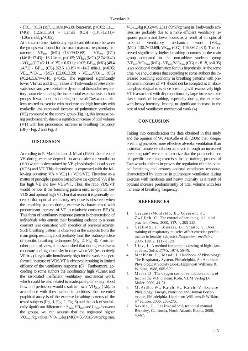
- HRmax (CG) (197.1±10.41)=-2.80 beats/min, p>0.05; Lamax
(MG) (12.61±1.93) - Lamax (CG) (13.87±2.11)=
-1.26mmol/l, p>0.05).
At the same time, statistically significant difference between
the groups was found for the main maximal respiratory pa-
rameters: VEmax (MG) (130.7±13.68) - VEmax (CG)
(146.8±17.43)=-16.1 l/min, p<0.05; VÒmax (MG) (2.74±0.42)
- VÒmax (CG) (2.11 ±0.35) = 0.63 l, p<0.05; BFmax (MG) (48.4
±4.71) - BFmax (CG) (62.6 ±8.19) = -14.2 min-1, p<0.05;
VEmax/VO2max (MG) (32.06±3.28) - VEmax/VO2max (CG)
(40.24±5.67)=-8.18, p<0.05. The registered significantly
lower VEmax and BFmax values in Taekwondo athletes moti-
vatedus toanalyze indetail thedynamicof thestudied respira-
tory parameters during the incremental exercise tests in both
groups. It was found that during the test all Taekwondo ath-
letes reacted to exercise with moderate and high intensity with
markedly less expressed increase of pulmonary ventilation
(VE) compared to the control group (Fig. 1), this increase be-
ing predominantly due to a significant increase of tidal volume
(VT) with less pronounced increase in breathing frequency
(BF) - Fig. 2 and Fig. 3.
DISCUSSION
According to P. Macklem and J. Mead (1988), the effect of
VE during exercise depends on actual alveolar ventilation
(VA) which is determined by VE, physiological dead space
(VDS) and VT. This dependence is expressed with the fol-
lowing equation: VA = VE (1 - VDS/VT). Therefore as a
matter of principle a person can achieve the optimal VA if he
has high VE and low VDS/VT. Thus, the ratio VDS/VT
would be low if the breathing pattern ensures optimal low
VDS and optimal high VT. For that reason it is generally ac-
cepted that optimal ventilatory response is observed when
the breathing pattern during exercise is characterized with
predominant increase of VT in relatively constant BF (6).
This form of ventilatory response pattern is characteristic of
individuals who entrain their breathing cadence to a some
constant unit consistent with specifics of physical activity.
Such breathing pattern is observed in the subjects from the
main group resulting most probably fromthe routine practice
of specific breathing techniques (Fig. 2, Fig. 3). From an-
other point of view, it is established that during exercise at
moderate and high intensity in cases when VE (respectively
VEmax) is typically inordinately high for the work rate per-
formed, increase of VDS/VT is observed resulting in limited
efficacy of the ventilatory response (8). Furthermore, ac-
cording to some authors the inordinately high VEmax and
the associated inefficient ventilatory mechanical work,
which could be also related to inadequate pulmonary blood
flow and perfusion, would result in lower VO2max (1,6). In
accordance with these scientific positions, the presented
graphical analysis of the exercise breathing patterns of the
tested subjects (Fig. 1, Fig. 2, Fig. 3) and the lack of statisti-
cally significant difference in Smax, HRmax and Lamax between
the groups, we can assume that the registered higher
VO2max/kg values (VO2max/kg (MG)= 56.99±5.94ml/kg min,
VO2max/kg (CG)=49.23±1.89ml/kg min) in Taekwondo ath-
letes are probably due to a more efficient ventilatory re-
sponse pattern and lower losses as a result of an optimal
maximal ventilatory mechanical work (VEmax
(MG)=130.7±13.68l; VEmax (CG)=146.8±17.43 l). The ob-
served significantly higher breathing economy in the main
group compared to the non-athlete students group
(VEmax/VO2max (MG) - VEmax/VO2max (CG) = - 8.18, p<0.05)
is an additional confirmation for this hypothesis. At the same
time, we should stress that according to some authors the in-
creased breathing economy in breathing patterns with pre-
dominant increase of VT should not be accepted as an abso-
lute physiological rule, since breathing with excessively high
VT is associated with disproportionately large increase in the
elastic work of breathing, especially during the exercises
with heavy intensity, leading to significant increase in the
cost of total ventilatory mechanical work (4).
CONCLUSION
Taking into consideration the data obtained in this study
and the opinion of W. McArdle et al. (2006) that "deeper
breathing provides more effective alveolar ventilation than
a similar minute ventilation achieved through an increased
breathing rate" we can summarize that the purposeful use
of specific breathing exercises in the training process of
Taekwondo athletes improves the regulation of their exter-
nal breathing and ensures optimal ventilatory response,
characterized by increase in pulmonary ventilation during
exercise with moderate and heavy intensity as a result of
optimal increase predominantly of tidal volume with less
increase of breathing frequency.
REFERENCES
1. Caruana-Montaldo, B. , Gleeson, K. ,
Zwil l ich, C. The control of breathing in clinical
practice. Chest, 2000, 117, 1, 205-225.
2. Gigl iot t i , F . , Binazzi , B. , Scano, G. Does
training of respiratory muscles affect exercise perfor-
mance in healthy subjects? Respiratory medicine,
2006, 100, 2, 1117-1120.
3. I l iev, I . A method for complex testing of high-class
athletes. Sofia, BSFS, 1974, 56-79.
4. Macklem, P. , Mead, J . Handbook of Physiology:
The Respiratory System. Philadelphia, An American
Physiological Society Book, Lippincott Williams &
Wilkins, 1988, 605-629.
5. Marks D. The oxygen cost of ventilation and its ef-
fect on the VO2 plateau. K�ln, VDM Verlag Dr
Muler, 2009, 41-51.
6. McArdle, W., Katch, F. , Katch, V. Exercise
Physiology: Energy, Nutrition and Human Perfor-
mance, Philadelphia, Lippincott Williams & Wilkins,
6th edition, 2006, 260-275.
7. Savoie , G. Taekwondo: A technical manual.
Berkeley, California, North Atlantic Books, 2009,
43-67.
115
Tzvetkov S.

8. Wasserman, K. , Hansen, J . , Sue, D. et al .
Principles of exercise testing and interpretation. Phila-
delphia, Lippincott Williams & Wilkins, 4th ed., 2005,
77-106.
9. Wilmore, J . , Cost i l l , D. Physiology of sport and
exercise. Champaign, Human Kinetics, 3rd ed., 2006,
242-264.
116
Specificity in breathing regulation during physical exercise in ...

THE STRENGTH-DURATION PROPERTIES IN SIMULATED
DEMYELINATING NEUROPATHIES DEPEND ON THE MYELIN
SHEATH AQUEOUS LAYERS
Krustev S. M.1, N. Negrev
1, D. I. Stephanova
2
1Medical University - Varna; 2Institute of Biophysics and Biomedical Engineering,
Bulgarian Academy of Sciences - Sofia
ABSTRACT
BACKGROUND: The strength-duration properties (time constants and rheobases) reflect the membrane
properties in body excitable structures. OBJECTIVE: To expand our studies on the membrane properties,
the effect of the myelin sheath aqueous layers on the strength-duration properties in simulated by us
demyelinating neuropathies is compared. METHODS: Using a multi-layered model of the myelin sheath of
human motor nerve fibre, the strength-duration time constants and rheobases are calculated and compared
in simulated normal case and in three cases of progressively greater demyelinations [such as internodal sys-
tematic demyelinations (ISDs), paranodal systematic demyelinations (PSDs) and paranodal internodal sys-
tematic demyelinations (PISDs)] without/with aqueous layers within the myelin sheath. Two cases of each
demyelinated type are mild and one is severe. RESULTS: When the aqueous layers are taken into account, the
strength-duration properties are improved in the ISDs and in the PSDs, except for the rheobases in the ISDs
where they worsen. However, the effect of the aqueous layers on these properties is neutralized when the
demyelinations are heterogeneous such as in the PISDs. CONCLUSIONS: Previously, it has been proven by
us that the ISDs are specific indicators of Charcot-Marie-Tooth disease type 1A (CMT1A) whereas the PSDs
and the PISDs are specific indicators of chronic inflammatory demyelinating polyneuropathy (CIDP) and its
subtypes. The present study shows that the strength-duration properties in patients with CMT1A and CIDP
depend on the myelin sheath aqueous layers.
Key words: myelin sheath aqueous layers, strength-duration time constant, demyelinating neuropathies,
computational neuroscience
INTRODUCTION
The typical picture which we have from electron micros-
copy of fixed myelin sheath is rather misleading. The my-
elin lamellae are not tightly compacted according to X-ray
and neutron diffraction (5,6), so that up to half the volume
fraction of myelin is taken up by water with salts (2,14).
Recently, it has been observed that the peripheral nerves of
patients with Charcot-Marie-Tooth disease type 1A
(CMT1A) and chronic inflammatory demyelinating
polyneuropathy (CIDP) have significant abnormalities in
their multiple membrane excitability properties such as
threshold electrotonus, strength-duration time constant,
rheobasic current and recovery cycle (4,12,13) measured
by a non-invasive threshold tracking technique (3). The
CMT1A is the most common form of hereditary neuropa-
thy. Its hallmark is uniform demyelination, which results in
slowing of conduction in motor and sensory nerves (1,11).
The CIDP as a peripheral nerve disorder is one of several
chronic demyelinating neuropathies that are believed to
have autoimmune etiology (7). There are subtypes of
chronic demyelinating neuropathies (8,10) that are broadly
classified under the term of chronic inflammatory
demyelinating polyneuropathy.
The present study compare the effect of the aqueous layers
on the previously calculated strength-duration properties,
reflecting the adaptive processes in simulated
demyelinating neuropathies such as CMT1A, CIDP and its
subtypes (9,18,19).
METHODS
The electrogenesis of human motor nerve fibres can now
be studied successfully using a multi-layered model of the
fibres, which is thoroughly described and discussed earlier
(15). The myelin sheath as a series of 150 aqueous and 150
lipid layers was first presented in this model. The same
multi-layered model has been used by us (9,18,19) to simu-
late normal case and three types of progressively greater
117
Scripta Scientifica Medica, 2012; vol. 44 (1), Supplement 1 Copyright © Medical University, Varna
Address for correspondence:
D.I. Stephanova, Institute of Biophysics and Biomedical Engineer-
ing, Bulgarian Academy of Sciences, Acad. G. Bontchev Str., Bl. 21,
Sofia 1113, Bulgaria.
e-mail: [email protected]

internodal, paranodal and paranodal internodal
demyelinations, each of them sys tem atic (Fig.1).
The internodal sys tem atic demyelination (ISD) is as so ci -
ated with a cor re spond ing loss of the my elin lamellae with -
out/with their aque ous lay ers away from the axolemma.
The paranodal sys tem atic demyelination (PSD) is as so ci -
ated with a cor re spond ing loss of the my elin end bulbs
away from the axolemma with out/with my elin sheath
aque ous lay ers. This case is equiv a lent to the case of the re -
duc tion of the paranodal seal re sis tance. The paranodal
internodal demyelination (PISD) is as so ci ated with a cor re -
spond ing loosing or lift ing of the my elin end bulbs and my -
elin lamellae with out/with their aque ous lay ers away from
the axolemma. The char ac ter is tic pa ram e ters de fin ing the
com pared cases are given in Ta ble 1, for the reader's con ve -
nience. Two cases of each demyelinated type are mild
(70%, 80%), and one is se vere. The 70% (ISD1, PSD1,
PISD1) and 80% (ISD2, PSD2, PISD2) re duc tion val ues
(of the my elin lamellae or of the paranodal seal re sis tance,
or of the paranodal seal re sis tance and my elin lamellae, re -
spec tively) uni formly along the fi bre length are not suf fi -
cient to de velop a con duc tion block and such
demyelinations are re garded as mild. The 93% (ISD3),
90% (PSD3) and 82% (PISD3) re duc tion val ues are the
first de gree of the demyelinated sub types for achiev ing the
con duc tion block and such demyelinations are re garded as
se vere.
The strength-du ra tion time con stant is based on the pas sive
(RC) fi bre mem brane pa ram e ters in par al lel, ac cord ing to
the clas si cal the ory. The strength-du ra tion and charge-du -
ra tion curves are ob tained fol low ing a thresh old stim u lus at
dif fer ent du ra tion. The thresh old stim u lus du ra tion is in -
creased in 0.025 ms steps from 0.025 ms to 1 ms, in or der to
ob tain the strength-du ra tion curves. A poly no mial func tion
of de gree 2 (trans fer stan dard pa rab ola), which re lates
thresh old charge (Q) to stim u lus du ra tion (t), pro vides an
ac cu rate fit of the data: Q = a2[t2+(a1/a2).t +a0/a2], where a0,
a1, a2 are the poly no mial co ef fi cients. The strength-du ra tion
time con stant is de fined as the ab so lute value of the small -
est square root of the func tion (i.e. only one of the di rect in -
ter cepts of the re gres sion curve on the du ra tion axis has a
bio phys i cal sense and only this di rect in ter cept is shown on
the given be low fig ures). The rheobasic cur rent is de fined
as the fi nal de creased thresh old value, af ter which the po -
ten tial gen er a tion can not be ob tained with an in crease in the
stim u lus du ra tion.
RESULTS
The strength-du ra tion curves (Fig. 2) and charge-du ra tion
curves (Fig. 3) in the nor mal (n) and ISDs (a), PSDs (b),
PISDs (c) cases of the hu man mo tor nerve fibres with out
(Figs. 2, 3 - left col umns) and with (Figs. 2, 3 - right col -
umns) aque ous lay ers are shown. His to grams are also used
118
The strength-duration properties in ...
Fig. 1. Diagram of human motor nerve fibres from the7th to the 14th nodes in the normal, ISD, PSD and PISDcases. The reduction of the myelin lamellae (ISD) or ofthe myelin end bulbs (PSD) or simultaneously of themyelin lamellae and myelin ends bulbs (PISD)without/with myelin sheath aqueous layers is uniformalong the fibre length.
Table 1. Membrane parameter values characteristic forhuman motor nerve fibres without and with aqueouslayers in the normal and in the ISDs, PSDs, PISDscases, when the demyelinations are mild (ISD1, PSD,PISD1 – 70%; ISD2, PSD2, PISD2 – 80%) and severe(ISD3– 93%, PSD3 – 90%, PISD – 82%), respectively.N (number of myelin lamellae); Rmy (myelin resistance);Cmy (myelin capacitance); Raql (aqueous-layeredlongitudinal resistance), Raqr (aqueous-layered radialresistance), Rpa (periaxonal resistance), Rpn (paranodal seal resistance).

119
Krustev S. M., N. Negrev, D. I. Stephanova
Fig. 2. Comparison between the strength-duration curves of human motor nerve fibres in the normal (n), ISDs (a),
PSDs (b), PISDs (c) cases without (left column) and with (right column) aqueous layers within the myelin sheath.

120
The strength-duration properties in ...
Fig. 3. Comparison between the charge-duration curves of human motor nerve fibres in the normal (n), ISDs (a), PSDs
(b), PISDs (c) cases without (left column) and with (right column) aqueous layers within the myelin sheath.

121
Krustev S. M., N. Negrev, D. I. Stephanova
Fig. 4. Comparison of the strength-duration time constants (left column) and rheobasic currents (right column)
without (hatched bars) and with (unhatched bars) aqueous layers within the myelin sheath.

to provide a better illustration of the strength-duration time
constants and rheobasic currents (Fig. 4). The threshold
currents are significantly higher in all investigated system-
atically demyelinated cases without/with aqueous layers
than in the normal ones in the different strength-duration
curves. There is an inverse relationship between the
strength-duration time constants (longer/shorter) which are
0.583/0.497, 0.757/0.592, 1.149/0.736 ms for the ISD1,
ISD2 and ISD3 cases without/with aqueous layers, respec-
tively. There is also an inverse relationship between the
time constants, however, they are shorter/longer
(0.093/0.099, 0.065/0.068, 0.064/0.068 ms) for the PSD1,
PSD2 and PSD3 cases without/with aqueous layers, re-
spectively. An overlap of the strength-duration time con-
stants is obtained for the PISDs cases without /with aque-
ous layers. They are substantially shorter 0.211, 0.202,
0.201 ms for the PISD1, PISD2 and PISD3, respectively,
than for the normal one. The results show that the aqueous
layers do not modulate the normal strength-duration time
constant and rheobasic current, which are 0.291 ms and
0.388 nA, respectively. For the ISD1, ISD2 and ISD3
cases, the rheobasic currents increase in both cases with-
out/with myelin sheath aqueous layers and are 0.568/0.605,
0.709/0.805, 1.444/1.718 nA, respectively. However, they
are increased/decreased 0.842/0.793, 1.149/1.084,
1.741/1.654 nA for the PSD1, PSD2 and PSD3 cases with-
out/with aqueous layers, respectively. For both cases with-
out/with aqueous layers, the rheobasic currents signifi-
cantly increase in the PISDs cases with the increase of the
degree of demyelination. The rheobases are 1.020, 1.487,
1.616 nA for the PISD1, PISD2 and PISD3, respectively.
DISCUSSION
The present study shows that the aqueous layers within the
internodally and paranodally systematically demyelinated
sheaths modulate the strength-duration properties of the hu-
man motor nerve fibres. However, the aqueous layers do
not modulate these properties, when the systematic
demyelinations are mixed, i.e. paranodal internodal
(PISDs). The time constants are longer/shorter for the
ISDs/PSDs, respectively without aqueous layers, than the
normal one. The addition of the aqueous layers improves
the strength-duration time constants for both demyelinated
types. However, these time constants are changed in totally
opposite directions, because of the different electrogenesis
of both demyelinations. The slopes of the charge-duration
curves in the ISDs and in the PSDs cases are in opposite di-
rections, because of the different changed RC membrane
parameters. According to the classical theory, there is an in-
verse relationship between the time constant and rheobasic
current. Compared to the normal case, the study shows that
this is valid for the PSDs when the aqueous layers are taken
into account. The ISDs are regarded as specific indicators
of CMT1A, whereas the PSDs and PISDs are regarded as
specific indicators of CIDP and its subtypes (16,17). Con-
sequently, our present study confirms that the systematic
demyelinations without/with aqueous layers may provide
important information on the pathophysiological
mechanisms underlying hereditary and chronic
demyelinating neuropathies.
REFERENCES
1. Birouk N, Gouider R, Le Guern E,
Gugenheim M, Tardieu S, Maisonobe T, Le
Forest ier N, Agid Y, Brice A, Bouche P.
Charcot-Marie-Tooth disease type 1A with 17p11.2
duplication. Clinical and electrophysiological pheno-
type study and factors influencing disease severity in
119 cases. Brain 1997, 120:813-823.
2. Blaurock AE. Structure of nerve myelin mem-
brane: proof of the low resolution profile, J Memb
Biol 1971, 56:35-52.
3. Bostock H, Cikurel K, Burke D. Threshold
tracking techniques in the study the human peripheral
nerve, Muscle Nerve 1998, 21:137-158.
4. Cappelen-Smith C, Kuwabara S, Lin CS-Y,
Mogyoros I, Burke D. Membrane properties in
chronic inflammatory demyelinating polyneuropathy.
Brain 2001, 124:2439-2447.
5. Kirschner DA, Caspar DL. Comparative diffraction
studies on myelin membrane. Ann N Y Acad Sci
1972, 195:309?317.
6. Kirschner DA, Ganser AL, Caspar DL. Dif-
fraction studies of molecular organization and mem-
brane interactions in myelin. In: Myelin. Morrel P,
ed., 2nd ed, New York, Plenum Press, 1984, 51-59.
7. K�ller H, Kieseier BC, Jander S, Hartung H-P.
Chronic inflammatory demyelinating polyneuropathy.
Review Article. N Eng J Med 2005, 352:1343-1356.
8. Krarup C, Stewart JD, Sumner AJ, Pestronk
A, Lipton SA. A syndrome of asymmetric limb
weakness with motor conduction block. Neurology
1990, 40:118-127.
9. Krustev S, Daskalova M, Stephanova D.
Myelin sheath aqueous layers do not modulate mem-
brane fibre properties of simulated cases of paranodal
internodal systematic demyelinations. Compt rend
Acad bulg Sci 2010, 63(12):1845-1852.
10. Lewis RA, Sumner AJ, Brown MJ, Asbury
AK. Multifocal demyelinating neuropathy with per-
sistent conduction block. Neurology 1982,
32:958-964.
11. Lupski JR, Montes de Oca-Luna R,
Slaugenhaupt S, Pentao L, Guzzet ta V,
Trask BJ, Saucedo-Cardenas O, Barker DF,
Kil l ian JM, Garcia CA, Chakravart i A,
Patel PI . DNA duplication associated with
Charcot-Marie-Tooth disease type 1A. Cell 1991,
66:219-232.
12. Nodera H, Bostock H, Kuwabara S,
Sakamoto T, Asanuma K, Sung JY,
Ogawara K, Hattor i N, Hirayama M, Sobue
G, Kaj i R. Nerve excitability properties in
Charcot-Marie-Tooth disease type A1. Brain 2004,
27:203-211.
13. Nodera H, Kaj i R. Nerve excitability testing and
its clinical application to neuromuscular diseases (in-
122
The strength-duration properties in ...

vited review). Clin Neurophysiol 2006,
117:1902-1916.
14. Ropte S, Scheidt P, Fr iede RL. The intermedi-
ate dense line of the myelin sheath is preferentially
accessible to cations and is stabilized by cations. J
Neurocytol 1990, 19:242-252.
15. Stephanova DI. Myelin as longitudinal conductor:
a multi-layered model of the myelinated human motor
nerve fibre. Biol Cybern 2001, 84:301-308.
16. Stephanova DI, Alexandrov AS. Simulated
mild systematic and focal demyelinating
neuropathies: membrane property abnormalities J
Integr Neurosci 2006, 5:595-623.
17. Stephanova DI, Daskalova M. Membrane
property abnormalities in simulated cases of mild sys-
tematic and severe focal demyelinating neuropathies.
Eur Biophys J 2008, 37:183-195.
18. Stephanova DI, Krustev SM, Daskalova M.
The aqueous layers within the myelin sheath modu-
late the membrane properties of simulated hereditary
demyelinating neuropathies. J Integr Neurosci 2011,
10(1):89-103.
19. Stephanova DI, Krustev SM, Negrev N,
Daskalova M. The myelin sheath aqueous layers
improve the membrane properties of simulated
chronic demyelinating neuropathies. J Integr
Neurosci 2011, 10(1):105-120.
123
Krustev S. M., N. Negrev, D. I. Stephanova

PSYCHOPHYSIOLOGICAL EVALUATION OF EMOTIONS DUE TO
THE COMMUNICATION IN SOCIAL NETWORKS
Minchev Zl.1, P. Gatev
2
1Joint Training Simulation and Analysis Center, Institute of Information and Communication
Technologies, 2Department of Cognitive Psychophysiology, Institute of Neurobiology,
Bulgarian Academy of Sciences
ABSTRACT
The emerging social networks like Facebook and Twitter with their huge popularity produce a strong impact
to our life. The study of human emotions by means of bioelectrical signals and psychometrics has a long his-
tory, but the data for an influence of Web-based social networks is still scarce and specific methods are lack-
ing. We tried to assemble an ad-hoc methodology for exploring emotions of a Facebook pilot group of users
implementing time-frequency spectral analysis (based on S-transform) and rhythm dynamics characteristics
representation of a complex set of recorded bioelectrical signals with parallel psychometric tests. The spectral
nature of the studied signals suggests correlation with the emotions of the Facebook users. The obtained initial
results have shown that the assembled methodology for bioelectrical signals' recording and analysis might be
a valuable tool for the study of impact of social networks on human emotions and facilitation of IT develop-
ments assessment and control in the information society.
Key words: human emotions, bioelectricity, social networks, spectral analysis, rhythm dynamics
INTRODUCTION
In today's world social interaction is increasingly per-
formed via social networks like Facebook and Twitter re-
lated to the huge popularity of modern information technol-
ogies (15). However, there is a paucity of studies of human
emotions and behavior related to this communication pro-
cess as well as the hidden threats behind it.
There are two kinds of theories for studying human emo-
tions because of the different understandings of the nature
of emotions and their relation to the emotions' stimuli (6):
(i) cognitive based (3,7,13) and (ii) somatic factors based
(4,11,14). Due to the complex nature of the emotions there
are and two approaches for their exploration: (iii) discrete
(assuming the existence of a number of basic emotions that
are universally displayed and recognized, like: happiness,
sadness, anger, fear, disgust and surprise (2), (5), whilst
other emotions are expressed as combination of these basic
ones) and (iv) continuous (assuming a possibility for
multivariate measurement of emotions, like e.g. the
arousal-valence model (12).
The present study uses a cognitive based model and contin-
uous approach to evaluate a methodological framework for
studies of emotions in the context of IT threats identifica-
tion for Facebook social network users.
MATERIAL AND METHODS
We have studied 18 healthy volunteers (15 men and 3
women) aged between 15-18 years. We assembled an
ad-hoc methodology for their evaluation as Facebook us-
ers, which includes: (i) habits and purposes in using the so-
cial network Facebook; (ii) Eysenk's personality question-
naire (both available at web page of TK 02/60:
www.cleverstance.com, "Links" section); (iii) 3 minutes
monitoring of Facebook navigation of two profiles: (a) the
one of the popular singer Shakira and (b) the profile of the
scientific project "Cortical Regulation of the Quiet Stance
during Sensory Conflict", Grant TK 02/60 of the NSF in-
cluding popular 39 photos for the standing balance
problems.
Here it should be noted that (iii) has been performed by an
own software tool "Social Network Activities Recorder" -
SNAR v. 1.0 (developed in Borland Delphi® 2008 soft-
ware environment) for user screen activity recording that
encompasses: mouse clicks and X-Y screen position coor-
dinates together with the background changes snapshots
(the studied subjects have been instructed to operate with
the mouse excluding any keyboard activities for simplifica-
tion of the study). These experiments has been performed
on personal computers during the Cyber security training
course at XI Summer School of Mathematics and Informa-
125
Scripta Scientifica Medica, 2012; vol. 44 (1), Supplement 1 Copyright © Medical University, Varna
Address for correspondence:
Zl. Minchev,
Joint Training Simulation and Analysis Center, Institute of Informa-
tion and Communication Technologies, Bulgarian Academy of Sci-
ences, Sofia 1113, acad. Georgi Bonchev Str., Bl. 25A, room 116,
Tel.: + 359 2 979 6631,
å-mail: [email protected]

tics, August 17-19, 2011 in collaboration with the High
School Institute of Mathematics and Informatics, Institute
of Mathematics and Informatics - Bulgarian Academy of
Sciences.
Additionally, a psychophysiological monitoring of 5 male
volunteer, aged between 26-42 years has been performed
afterward at the laboratory of the Department of Cognitive
Psychophysiology at the Institute of Neurobiology, Bulgar-
ian Academy of Sciences, with Mitsar 202 equipment re-
cording: EEG (following 10-20 system - F3, Fz, F4, C3,
Cz, C4, P3, Pz, P4), ECG (leads from the hands wrists) and
EMG (by bipolar leadings-off from the mimic muscles: m.
orbicularis oculi and m. corrugator superscilii) activities
during the performance of the tasks (i) - (iii). Finally, only
one 42 years experimental subject has been selected as a
stable, extrovert representative Facebook user. All data was
digitized in 500 Hz sampling frequency.
This experimental monitoring has been performed on HP
6730s laptop machine with Intel Pentium Dual-Core Pro-
cessor T4200, 3072 MB RAM, 320 GB SATA HDD, ATI
Radeon HD 3430 video card with 256 MB dedicated video
memory and with WXGA BrightView 15.4" screen. The
mimic of face has been also recorded via the build-in web
camera and WebCam Max®, v.7.5.3.2 software.
The subject's instruction was to operate with the mouse ex-
cluding any keyboard activities for simplicity of the study.
The SNAR v. 1.0 software has been accomplished for the
mouse clicks capturing, by using a short (1 ms duration and
1 kHz frequency) sound signal (produced as a result of left
mouse button click) via the standard audio output of the
experimental setup
All experiments have been performed in the context of vali-
dating the Facebook information flows threats model pub-
lished in (10) and developed in the framework of EU Net-
work of Excellence in Managing Threats and Vulnerabili-
ties for the Future Internet: Europe for the World - SySSec.
This model determines as a critical threat for the Facebook
users "Friends" and as hidden threats: "Photos_Videos",
"Profile", "Wall" and "Other".
The records of the physiological monitoring have been pro-
cessed by using Matlab® R2011a. All records have been
initially filtered with zero-phase shift digital filter using
Chebishev IIR 50 Hz notch filter with slope -18 dB/oct and
passband ripple 0.5 dB, Wn=[47,52] Hz. Additionally,
Butterworth band pass IIR filter with slope -18 dB/oct, and
passband ripple = 0 dB has been also used, as follows:
Wn=[2, 42] Hz for the EEG, Wn=[1, 150] Hz for the EMG
and ECG.
Finally, time-frequency spectral analysis (using S- trans-
form and the approach presented in (9)) was performed.
RESULTS
Regarding the participants' habits and purposes in using the
social network Facebook almost all users (about 90%) are
reporting Facebook usage for communication with real
people and on-line stay for more than 12 hours daily, hav-
ing more than 100 friendships. Only 10% of the studied
participants report Facebook usage for entertainment
and/or other purposes.
The generalized results of the Facebook psychological
analysis and item usage validation according to (i) - (iii)
tasks from the previous paragraph are shown for 18 healthy
volunteers on Figure 1. As it is clear from Figure 1 (left),
the stable and extroverts participants are encompassing al-
most all of the Facebook users stuff. On Figure 1 (right) the
item "Wall" has 90% coincidence between model's pre-
dicted and really used items, whilst item "Friends" and
"Other" have the lowest coincidence.
The pilot results of the polyphysiographic monitoring dem-
onstrate that changes in the heart rate variability coincide
with the S-transform variability of m. corrugator superscilii
and/or m. orbicularis oculi (Figure 2 A). These results con-
126
Psychophysiological evaluation of emotions due to ...
Fig.1. Eysenck’s personality test generalized results expressed in percentages of the sample (left) and Facebook item
usage (right) expressed in percentages of the coincidence between model predicted and really used items.

firmed that heart rate variability can be connected with dif-
ferent kind of emotions (8).
The EEG S-transform of F3, C3 P3 vs F4, C4, P4 leads dur-
ing 7 second epochs showed typical frontal area left-right
asymmetry (1) as well as centro-parietal cortex left-right
asymmetry.
DISCUSSION
The psychometric results showed that extroversion and sta-
bility of the studied Facebook users are correlated posi-
tively with the intensity of social networks usage. The re-
sults of the threats coincidence of Facebook social network
for the "Wall" item and the differences for "Friends" and
"Other" items, suggesting that the obtained differences
could be generators of hidden threats, which was not
initially expected.
The pilot results of the polyphysiographic monitoring have
demonstrated promising initial base for evaluation of the
experts' models threats' foresees and psychometric mea-
surements using bioelectrical signals like: EEG, EMG and
ECG dynamic changes monitoring.
127
Minchev Zl., P. Gatev
Fig.2. A: parallel changes in EMG envelope S-transform of m. corrugator superscilii (top), m. orbicularis oculi
(middle) and hear rate variability (bottom) during 3 minutes gallery items navigation of Facebook profile for standing
balance problems; B: left-right EEG asymmetry in left (F3, C3 P3 - top) vs right (F4, C4, P4 - bottom) during 7 second
epoch evaluated by S-transform.

ACKNOWLEDGEMENTS
The study was partially supported by the National Science
Fund of Bulgaria, Grant TK02/60 (www.cleverstance.com)
and EU FP7 project - A European Network of Excellence in
Managing Threats and Vulnerabilities in the Future Internet:
Europe for the World (www.syssec-project.eu) under the
agreement No 257007. The authors also express their grati-
tude to the organizers and sponsors of XI Summer School of
Mathematics and Informatics, August 17-19, 2011.
REFERENCES
1. Coan, J . A. , J . J .B. Alle , Frontal EEG asymme-
try as a moderator and mediator of emotion, Biologi-
cal Psychology, 67, 2004, 7-49.
2. Darwin, C. The expression of the emotions in man
and animals. Kessinger Publishing, 2005.
3. Fr i jda, N. H. Varieties of affect: Emotions and ep-
isodes, moods, and sentiments. The nature of emo-
tion. New York: Oxford University Press, 59-67,
1994.
4. Ekman, P. Expression and the nature of emotion.
Approaches to emotion. Hillsdale, New Jersey: Law-
rence Erlbaum Associates, 1984, 319-344.
5. Ekman, P. Basic emotions. The handbook of cogni-
tion and emotion. UK: John Wiley & Sons, Ltd.,
1999, 45-60.
6. Lopatovska, I . , I . Arapakis . Theories, Methods
and Current Research on Emotions in Library and In-
formation Science, Information Retrieval and Hu-
man-Computer Interaction, Information Processing
and Management, 2010.
7. Lazarus, R. S. Thoughts on the relations between
emotion and cognition. Approaches to emotion.
Hillsdale, New Jersey: Lawrence Erlbaum Associates,
247-259, 1984.
8. LeDoux, J .E. Emotion circuits in the brain, Annu.
Rev. Neurosci, 2000, 23, 155-184.
9. Minchev Z. , G. Dukov, S. Georgiev. EEG
Spectral Analysis in Serious Gaming: An Ad Hoc Ex-
perimental Application, Int. J. Bioautomation, 13 (4),
2009, 79-88.
10. Minchev, Z. , M. Petkova. Information Pro-
cesses and Threats in Social Networks. A Case Study,
Proceedings of Conjoint Scientific Seminar "Model-
ling and Control of Information Processes", Sofia,
November 20, 2010, 85-93, Available at:
http://www.math.bas.bg/telecom/old_site/seminar201
0/doc9.pdf
11. Plutchik, R. A general psychoevolutionary theory
of emotion. Emotion: Theory, research and experi-
ence. Theories of emotion, New York: Academic, 1,
1980, 3-33.
12. Russel l , J . , A. Weiss , G. Mendelsohn. Affect
grid: a single item scale of pleasure and arousal, Jour-
nal of Personality and Social Psychology, 57(3),
1989, 493-502.
13. Scherer , K. R. What are emotions? And how can
they be measured? Social Science Information, 44(4),
2005, 695-729.
14. Scherer , K.R. , and P. Ekman. (Eds.) Affect
theory. Approaches to emotion. Hillsdale, New Jer-
sey: Lawrence Erlbaum Associates, 1984.
15. The Cisco Connected World Technology Report,
September 21, 2011, Available at:
http://www.cisco.com/en/US/solu-
tions/ns341/ns525/ns537/ns705/ns1120/CCWTR-Cha
pter1-Report.pdf
128
Psychophysiological evaluation of emotions due to ...

MELATONIN AND THE FIBRINOLYTIC SYSTEM IN THE RAT:
EFFECTS ON THROMBIN ACTIVATABLE FIBRILOLYSIS
INHIBITOR (TAFI)
Decheva L.1, I. Pashalieva
1, E. Stancheva
1, Y. Nyagolov
2, N. Negrev
1
1Department of Physiology and Pathophysiology, Medical University "Prof. Paraskev Stoyanov" -
Varna, 2Department of Physiology, Medical University - Sofia
SUMMARY
There is scanty and contradictory data about the effect of the hormone melatonin on fibrinolysis. The purpose
of the present investigation is to study the effects of melatonin and its antagonist luzindolå on TAFI
(Thrombin Activatable Fibrinolysis Inhibitor) activity and plasma antigen level of TAFIa/TAFIai (TAFI ac-
tivated/TAFIa inactivated) complex which are specific markers of plasma fibrinolytic activity. The study cov-
ered 52 male Wistar rats divided into four groups, i. e.: injected with melatonin (in a dose of 0.2 mg/kg b.m.),
with luzindole (in a dose of 0.4 mg/kg b.m.), with melatonin and luzindolå both as luzindole pretreatment was
performed one hour earlier. The control group was injected with the vehicle. All the substances were adminis-
tered two times daily for three consecutive days. It was established that melatonin induced significant alter-
ations in the examined parameters as TAFI activity increased (p<0.001) while the plasma level of
TAFIa/TAFIai complex was strongly reduced (p<0.001). Luzindolå being a non-selective antagonist of
melatonin receptors diminished TAFI activity (ð<0.001) and elevated the plasma concentration of
TAFIa/TAFIai complex (ð<0.001). Luzindolå pretreatment and subsequent melatonin application caused
changes of the examined parameters which, in their essence, repeated the effect of the independent luzindolå
administration at ð<0.001. The reptilase time was significantly decreased in all tested groups (ð<0.001). The
results obtained allowed us to accept that melatonin significantly reduced TAFI activity and elevated
TAFIa/TAFIai. The blockade of MT1 and MT2 melatonin receptors by luzindolå eliminated the effect of this
hormone on TAFI activity and TAFIa/TAFIai.
Key words: melatonin, luzindolå, reptilase time, TAFI, TAFIa/TAFIai
INTRODUCTION
Melatonin is a neurohormone secreted by the pineal gland.
It regulates other hormones and maintains the body's circa-
dian rhythms. In addition to the involvement of the hor-
mone in the transduction and processing of the
photoperiodical information and modulation of various
neuronal and endocrine functions in mammalians, a wide
spectrum of biological effects, including regulation of re-
production, liver protein synthesis control, adipogenesis
and osteogenesis, oncostatic, antiapoptotic, immune-mod-
ulating and antioxydant effects are attributed to melatonin
(1-5). However, data about involvement of the hormone in
the complex mechanisms of haemostasis regulation are
relatively limited and partially controversial (6,7).
Recently a special scientific interest is directed towards
TAFI (thrombin activatable fibrinolysis inhibitor), a novel
molecule which is proposed to play a key role in the inter-
action between procoagulant, anticoagulant and
fibrinolytic systems expressing various additional biologi-
cal effects (8,9). TAFI (Plasma pro-carboxypeptidase B,
carboxypeptidase U) is a single chain glycoprotein
zymogen (Mr=60,000) synthesized in the liver and circu-
lating at plasma concentration of 50 nM/l as a
plasminogen-bound zymogen. Thrombin (plasmin,
trypsin) cleavage of the zymogen releases a 92 amino acid
N-terminal activation peptide containing 4 N-linked
glycosylation sites (N22, N51, N63, N86) and the proposed
plasminogen recognition site. The rate of thrombin cata-
lyzed activation of TAFI is increased 1250 fold by forma-
tion of a ternary complex with thrombomodulin (10). The
309 amino acid C-terminal (Mr=35,783) catalytic domain
(TAFIa, pCPB) displays the properties of a basic
carboxypeptidase, hydrolyzing lysine and arginine from the
C-terminal position of polypeptides. This portion of the
molecule is homologous to tissue carboxypeptidase B and
contains 7 conserved cystine residues
(64,77,136,151,160,165,291), the active site Zn2+ coordi-
nation site (H67, E69, H196) and the basic C-terminal
amino acid substrate binding pocket (D257, G244, S207).
129
Scripta Scientifica Medica, 2012; vol. 44 (1), Supplement 1 Copyright © Medical University, Varna
Address for correspondence:
L. Decheva, Dept. of Physiology and Pathophysiology, Medical
University "Prof. Paraskev Stoyanov" - Varna, 55 Marin Drinov str.,
9002 Varna
e-mail: [email protected]

After its activation by thrombin, the
thrombin-thrombomodulin complex, or plasmin, the acti-
vated form of TAFI (TAFIa) attenuates fibrinolysis by re-
moving the carboxyl-terminal lysine residues from partially
degraded fibrin that mediate positive feedback in the
fibrinolytic cascade. These residues are required for ad-
sorption of tissue-type plasminogen activator (t-PA) and
plasminogen to fibrin. Therefore, TAFIa decreases plasmin
formation and protects the fibrin clot against lysis. An in-
creased of pro-TAFI/TAFIa levels have been reported in
some clinical conditions associated with thrombotic ten-
dency, as type II diabetes mellitus, deep vein thrombosis
and symptomatic artery disease. The thrombin-activatable
fibrinolysis inhibitor pathway defines a novel molecular
connection between blood coagulation and both
fibrinolysis and inflammation. A role for TAFIa in modu-
lating inflammation is suggested by the ability of this en-
zyme to down-regulate pericellular plasminogen activation
and to inactivate the inflammatory peptides bradykinin and
the anaphylatoxins C3a and C5a. TAFIa undergoes a rapid
conformational change at 37°C to an inactive isoform
called TAFIai.
This study was designed to investigate the effects of
melatonin and its nonselective receptor inhibitor luzindole
on TAFI pathway in rats (11).
MATERIAL AND METHODS
The experiments were performed on 52 male Wistar rats
weighing 200-220 g, in accordance with the European
Convention for protection of experimental animals (Protec-
tion of animals used for experimental purposes, Council
Directive 86/609/EEc of November 1986). The rats were
kept with free access to standard food and water at
dark/light regimen 12:12 hours and were divided into four
equal groups, 13 animals each. The daily doses for
melatonin (Ìårck, Germany), 0.2 mg/kg, and luzindole
(Sigma Chemicals/St. Louis, MO, USA), 0.4 mg/kg, were
applied twice daily s.c. for three consecutive days. The rats
from one of the groups were injected with melatonin; an-
other group of animals were treated with luzindole; and a
third group with luzindole, followed after 1 hour by
melatonin. The control group was treated with the vehicle
by the same dose and schema. Melatonin and luzindole
doses were determined by literature data and by experi-
mental study on dose/effect dependence. The necessary
blood volume from a rat was acquired by cardiac puncture
under urethane narcosis. Sodium citrate (0.11 mol/l) was
used as an anticoagulant (blood/citrate ratio 9:1). The citrat-
ed blood was centrifuged for 10 min at 3000 revolutions
per minute. The citrated plasma (the supernatant) was sepa-
rated and stored at 4 0C in haemostatic test-tubes. The pa-
rameters studied TAFI activity and plasma antigen level of
the complex TAFIa/TAFIai, were determined not later than
2 hours. TAFI activity was estimated by the commercial
enzyme kit Stachrom® Diagnostica Stago (Franñå) and
measured on STA® analyzer. TAFIa/TAFIai antigen con-
centration was determined by Asserachrom® en-
zyme-linked immunosorbent assay (ELISA). The reptilase
time was estimated by a routine coagulation method. The
data were analysed by variation analysis using
Student-Fisher's t-test and values of p<0.05 were
considered statistically significant.
RESULTS AND DISCUSSION
The effects of melatonin and luzindole on TAFI activity are
presented in figure 1. Melatonin elevated this parameter
(presented in % of activity from the baseline) from
115.20±4.10 for the control group to 180.70±9.97
(p<0.001), while both luzindole applied separately, and
luzindole applied 1 hour before melatonin decreased TAFI
activity to 50.09±4.81 (p<0.001) and to 40.00±4.82
(p<0.001), respectively. The other parameter,
TAFIa/TAFIai antigen concentration (ng/ml) was de-
creased by melatonin from 13.00±0.82 for the control
group to 5.02±0.49 (p<0.001) and was increased signifi-
cantly to 24.09±0.84 (p<0.001) and to 23.72±0.82
(p<0.001) both by the separate luzindole and combined
luzindole/melatonin treatment (figure 2). The reptilase time
130
Melatonin and the fibrinolytic system in the rat: effects on ...
Fig. 1. Melatonin and luzindole effects on TAFI activity.
M – melatonin; L – luzindole; C – control * - ð<0,001
Fig. 2. Melatonin and luzindole effects on TAFIa/
TAFIai complex.
M – melatonin; L – luzindole; C – control; * - ð<0,001

(figure 3) was shortened significantly from 11.77±0.57 sec
for the control group to, respectively 5.11±0.45 (p<0.001),
8.09±0.33 (p<0.001) and 7.00±0.45 sec (p<0.001) after
treatment with melatonin, luzindole alone and combined
luzindole/melatonin.
Striking is the observation, the three day application of
melatonin in this study in rats (24 hour dose 0.2 mg/kg
body mass) significantly increased the activity of and de-
creased TAFIa/TAFIai antigen concentration. Similar
findings indicating opposite effects on plasma activity and
concentration are abundant in literature and are not new in
relation to various clotting factors. The most tempting hy-
pothesis for explanation of this phenomenon is the suppres-
sion of the biosynthesis of TAFI by melatonin expressed by
TAFIa/TAFIai decrease, as well as suppression of its inac-
tivation presented by the stimulated TAFI activity. Having
in mind TAFI is synthesized and released in the liver (12),
and the various melatonin liver effects related to protein
synthesis reported, this hypothesis might be acceptable
(13,14). We should hardly use the upper facts to specify the
mechanism of the effect of melatonin, but it is quite proba-
ble the stimulating effect of the hormone to be accom-
plished at the level of the liver. Having in mind the abun-
dant expression of melatonin receptors in various areas of
the central nervous system (15), we might consider other
possible central effects of the hormone via other
mechanisms of haemocoagulation regulation.
Luzindole is the first competitive nonselective melatonin
receptor antagonist (16). Separate luzindole treatment (24
hour dose - 0.4 mg/kg) in the course of three days decreased
significantly the levels of activity of TAFI and increased
TAFIa/TAFIai concentration. These findings are exactly
opposite to melatonin effects discussed above and may be
attributed to one of the basic luzindole effects resulting
from its capability to block nonselectively melatonin MT1
and MT2 receptors. This is may be an evidence of a direct
participation of MT1and MT2receptors in the implementa-
tion of the melatonin effects in haemocoagulation via
TAFI.
Melatonin treatment of the rats one hour after luzindole
pre-treatment by the same doses and schema of application,
significantly decreased TAFI activity and increased
TAFIa/TAFIai concentration. The data indicate luzindole
pre-treatment effectively inverses the effect of melatonin on
the parameters studied. In addition, melatonin applied after
luzindole pre-treatment almost completely reproduces the
shifts of the parameters observed after separate luzindole
application. These results are in support of the presumtion
that luzindole eliminates the exogenous melatonin effect. In
addition the non-selective MT1 and MT2 receptor antago-
nist luzindole to a certain extent may inhibit the endoge-
nous melatonin too.
This study followed up the melatonin effect on reptilase
time (RT). RT is a basic integral haemocoagulation test to
estimate the functions of the factors involved in the trans-
formation of fibrinogen to fibrin. This parameter was sup-
pressed by melatonin, luzindole and melatonin applied af-
ter luzindole pretreatment. The registerred RT shifts are not
in a complete coherence with the effects on TAFI observed
and represent another pattern of the effects of melatonin
and luzindole compared to the effects on TAFI. This find-
ing may indicate a different mechanism of melatonin and
luzindole involved in transformation of fibrinogen to fibrin.
CONCLUSIONS
In conclision, the results of the study indicate: 1). Three day
treatment of male Wistar breed rats with melatonin in-
creases the activity of TAFI and may lead to activation of
fibrinolysis via TAFI pathway. 2). Separate luzindole ap-
plication for three consecutive days leads to a suppression
of TAFI activity. 3). TAFIa/TAFIai antigen concentration
is influenced in the opposite direction compared to TAFI
activity; decreased by melatonin and increased by
luzindole. 4). The treatment of the rats with melatonin after
luzindole pre-treatment completely reproduces the effect of
separate luzindole application. The data evidence the
nonselective melatonin receptor antagonist luzindole
blocks the effects both of endogenous and exogenous
melatonin on TAFI. 5). RT is shortened in all tested groups
versus the controls via mechanism(s) different from the
effect on TAFI.
REFERENCES
1. Boutin J . A. , Audinot V. , Ferry G. ,
Delagrange P. (2005). "Molecular tools to study
melatonin pathways and actions". Trends in Pharma-
cological Sciences 26 (8): 412-9.
2. Borj igin J. , Li X. , Snyder S.H. (1999) The pi-
neal gland and melatonin: Molecular and pharmaco-
logic regulation. Annu Rev Pharmacol
Toxicol.;39:53-65.
3. Chal let E. (2007). "Minireview: Entrainment of the
Suprachiasmatic Clockwork in Diurnal and Nocturnal
Mammals". Endocrinology 148 (12): 5648-55.
4. A l t u n A . , U g u r - A l t u n B . (2007).
"Melatonin: therapeutic and clinical utilization". In-
ternational Journal of Clinical Practice 61 (5):
835-45.
131
Decheva L., I. Pashalieva, E. Stancheva ...
Fig. 3. Melatonin and luzindole effects on reptilase time (RT).
M - melatonin; L - luzindole; C - control; * - ð<0,001

5. Wolden-Hanson T; Mitton, D. R., McCants
R. L., Yellon S. M., Wilkinson C. W.,
Matsumoto A. M., Rasmussen D. D. (2000).
"Daily melatonin administration to middle-aged male
rats suppresses body weight, intraabdominal adipos-
ity, and plasma leptin and insulin independent of food
intake and total body fat". Endocrinology 141 (2):
487-97.
6. Claustrat B. , J . Brun, Chazot G. (2005) The
basic physiology and pathophysiology of melatonin
Sleep Medicine Reviews 9, 11-24.
7. Wirtz P. H., Spillmann M., B�rtschi C., Ehlert
U., von K�nel R. (2008) Oral melatonin reduces
blood coagulation activity: a placebo-controlled study
in healthy young men. J Pineal Res. Mar;
44(2):127-33.
8. Wang W., Hendriks D. F. , Scharpe S. S.
(1994) Carboxypeptidase U, a plasma
carboxypeptidase with high affinity for plasminogen.
J Biol Chem. 269:15937-15944.
9. Bajzar L. , Manuel R. , Nesheim M. E. (1995)
Purification and characterization of TAFI, a
thrombin-activatable fibrinolysis inhibitor. J Biol
Chem. 270:14477-14484.
10. Mosnier L. O. , Meijers J . C. M., Bouma B.
N. (2001) Regulation of fibrinolysis in plasma by
TAFI and protein C is dependent on the concentration
of thrombomodulin. Thromb Haemost.85:5-11.
11. Bouma B. N. , Mosnier L. O. (2005).
"Thrombin activatable fibrinolysis inhibitor (TAFI) at
the interface between coagulation and fibrinolysis."
Pathophysiol. Haemost. Thromb. 33 (5-6): 375-81.
12. Kerr R . (2003) New insights into haemostasis in
liver failure. Blood Coagul Fibrinolysis 14(Suppl 1):
43- 45.
13. Brodsky V. Y. , Zvezdina N. D. (2010)
Melatonin as the most effective organizer of the
rhythm of protein synthesis in hepatocytes in vitro
and in vivo. Cell Biol Int. Dec; 34 (12) :1199-204.
14. Brodsky V. Y. , Dubovaya N. D. , Zvezdina
N. D. , Fateeva V. I . , Malchenko L. A.
(2010) Melatonin modifies the rhythm of protein syn-
thesis. Bull Exp Biol Med Jul ; 149 (1) : 40-3.
15. Wit t -Enderby P. A. , Bennet t J . , Jarzynka
M. J. , Firest ine S. , Melan M. A. (2003)
Melatonin receptors and their regulation: biochemical
and structural mechanisms. Life Sciences 72 :
2183-2198.
16. Dubocovich M. L. (1988) Luzindole (N-0774): a
novel melatonin receptor antagonist. J Pharmacol
Exp Ther Sep; 246 (3) :902-10.
132
Melatonin and the fibrinolytic system in the rat: effects on ...

PREHYPERTENSION IN WOMEN WITH METABOLIC SYNDROME
Nikolova J.1, M. Orbezova
2, P. Atanasova
3, P. Nikolov
4, F. Nikolov
4, P. Hrischev
3
1Department of Physiology, 2Clinic of Embriology, 3Department of Anatomy, Histology and
Embriology, 4Clinic of Cardiology, Medical university - Plovdiv
INTRODUCTION
There is a continuous relationship between the level of
blood pressure (BP) and cardiovascular (CV) risk. The
2003 European Society of Hypertension - European Soci-
ety of Cardiology (ESH-ESC) guidelines for the manage-
ment of arterial hypertension referred the BP range
130-139/85-89 mmHg to as "borderline" hypertension ac-
cording JNC6 (Joint National Committee on Prevention,
Detection, Evaluation and Treatment of High Blood Pres-
sure - USA) and "high- normal" according ESH-ESC
guidelines. JNC 7 established new classification of BP,
coining the term "prehypertension" for those with blood
pressure ranging from 120 to 139 mmHg systolic and/or
from 80 to 89 mmHg diastolic. Prehypertension is not a dis-
ease. But increasing the BP with 20/10 mmHg for systole
and diastole respectively doubles the CV risk. Thus higher
CV risk in prehypertensive patients is an argument the cate-
gory "prehypertension" to be introduced. In one third of
181 followed up hypertensive people, jepaneese authors
register metabolic syndrome (MS) and morning
hypertention/MS ratio is significant. Prehypertension and
hypertension are components of MS, characterized by a
number of metabolic abnormalities including central obe-
sity, elevated triglycerides (TG) with decreased levels of
high-density lipoprotein - cholesterol(HDL-C), raised
blood pressure³135/80mmHg and impaired glucose toler-
ance. The MS predicts cardiovascular mortality in both
men and women. In the DECODE study, non-diabetic men
and women with MS had an increased risk of death from all
causes as well as CV disease. Leptin, one of the basic mast
tissue products, plays a major role as in carbohydrates' and
lipids' metabolism, as in appetite control and energy bal-
ance. Leptine/ insuline resistance correlation is an object of
discussion and intense examination.
The goal of our study was to follow up the frequency of
prehypertension in women with MS and the leptine's mast
tissue expression.
MATERIAL AND METHODS
46 women of 36.68±6.48 years age with proven MS and 32
clinically healthy women of 35.54±5.39 years age were ex-
amined. The parameters: body mass index (BMI), arterial
blood pressure (ABP), based on multiple measurements
taken on separate occasions, plasma blood sugar (RA 1000
Technicon, USA), insulin and HOMA - index (MEIA,
ÀÂÂÎÒÒ, USA, AxSYM) and plasma lipid profile (Op-
tima KONE) were followed up. Imune histochemic investi-
gation of biopsy material for leptine was done. The material
was taken from gluteal under skin mast tissue region. The
immune histochemical reaction was followed on 5 m par-
affin slides by avidin biotidin peroxydase method (ABC)
using primary antibodies for leptin(Santa Cruz, USA), dis-
solved in PBS in 1:200 ratio.
RESULTS
Measuring ABP, it was established that in 37% of the
women with proven MS, the values are ³135/80 mmHg.
These values were registered in 20% of clinically healthy
women (p<0.05).
To value the degree of obesity, BMI was used, according to
the world health organization (WHO) criteria. In women
with proven MS, BMI was 35.43±3.18 kg/m2 and in clini-
cally healthy women - 23.43±5.11 kg/m2 (p<0.05) respec-
tively.
HOMA index in women with proven MS was 4.15±1.10
v/s 1.15±0.80 (p<0.05) in clinically healthy women.
Plasma lipid profile data were: total cholesterol (TC) - 4,1
0,9 mmol.L-1 in MS women v/s 6,3±0,28 mmol.L-1
(p<0,001) in clinically healthy women women; TG: 1,5 0,3
mmol.L-1 in clinically healthy women v/s 2,1 0,73
mmol.L-1 (p<0,05) in MS women.
Plasma glucose level (GLU) in MS women was 6,51 1,04
mmol.L-1 (p<0,05) v/s 4,46±1,53 mmol. L-1 in clinically
healthy women.
133
Scripta Scientifica Medica, 2012; vol. 44 (1), Supplement 1 Copyright © Medical University, Varna
Èìóíîõèñòîõèìè÷íà åêñïðåñèÿ íà ëåïòèí ó çäðàâ.
Óâ. X400.
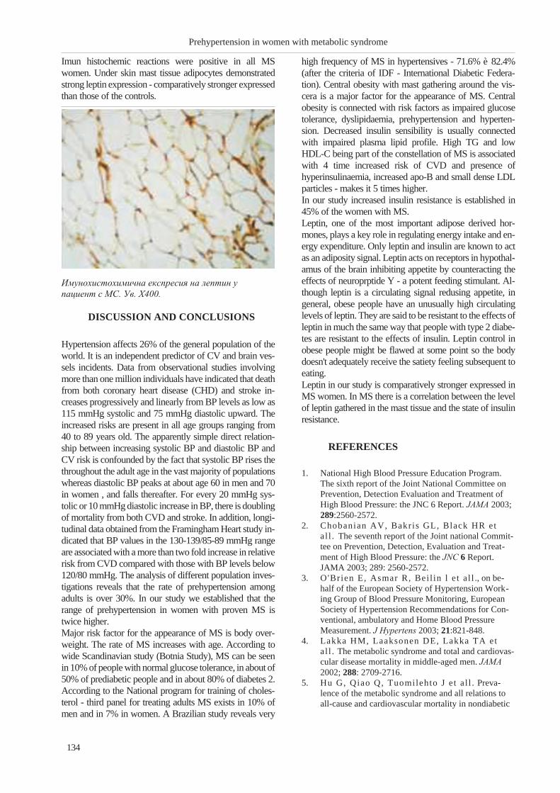
Imun histochemic reactions were positive in all MS
women. Under skin mast tissue adipocytes demonstrated
strong leptin expression - comparatively stronger expressed
than those of the controls.
DISCUSSION AND CONCLUSIONS
Hypertension affects 26% of the general population of the
world. It is an independent predictor of CV and brain ves-
sels incidents. Data from observational studies involving
more than one million individuals have indicated that death
from both coronary heart disease (CHD) and stroke in-
creases progressively and linearly from BP levels as low as
115 mmHg systolic and 75 mmHg diastolic upward. The
increased risks are present in all age groups ranging from
40 to 89 years old. The apparently simple direct relation-
ship between increasing systolic BP and diastolic BP and
CV risk is confounded by the fact that systolic BP rises the
throughout the adult age in the vast majority of populations
whereas diastolic BP peaks at about age 60 in men and 70
in women , and falls thereafter. For every 20 mmHg sys-
tolic or 10 mmHg diastolic increase in BP, there is doubling
of mortality from both CVD and stroke. In addition, longi-
tudinal data obtained from the Framingham Heart study in-
dicated that BP values in the 130-139/85-89 mmHg range
are associated with a more than two fold increase in relative
risk from CVD compared with those with BP levels below
120/80 mmHg. The analysis of different population inves-
tigations reveals that the rate of prehypertension among
adults is over 30%. In our study we established that the
range of prehypertension in women with proven MS is
twice higher.
Major risk factor for the appearance of MS is body over-
weight. The rate of MS increases with age. According to
wide Scandinavian study (Botnia Study), MS can be seen
in 10% of people with normal glucose tolerance, in about of
50% of prediabetic people and in about 80% of diabetes 2.
According to the National program for training of choles-
terol - third panel for treating adults MS exists in 10% of
men and in 7% in women. A Brazilian study reveals very
high frequency of MS in hypertensives - 71.6% è 82.4%
(after the criteria of IDF - International Diabetic Federa-
tion). Central obesity with mast gathering around the vis-
cera is a major factor for the appearance of MS. Central
obesity is connected with risk factors as impaired glucose
tolerance, dyslipidaemia, prehypertension and hyperten-
sion. Decreased insulin sensibility is usually connected
with impaired plasma lipid profile. High TG and low
HDL-C being part of the constellation of MS is associated
with 4 time increased risk of CVD and presence of
hyperinsulinaemia, increased apo-B and small dense LDL
particles - makes it 5 times higher.
In our study increased insulin resistance is established in
45% of the women with MS.
Leptin, one of the most important adipose derived hor-
mones, plays a key role in regulating energy intake and en-
ergy expenditure. Only leptin and insulin are known to act
as an adiposity signal. Leptin acts on receptors in hypothal-
amus of the brain inhibiting appetite by counteracting the
effects of neuroprptide Y - a potent feeding stimulant. Al-
though leptin is a circulating signal redusing appetite, in
general, obese people have an unusually high circulating
levels of leptin. They are said to be resistant to the effects of
leptin in much the same way that people with type 2 diabe-
tes are resistant to the effects of insulin. Leptin control in
obese people might be flawed at some point so the body
doesn't adequately receive the satiety feeling subsequent to
eating.
Leptin in our study is comparatively stronger expressed in
MS women. In MS there is a correlation between the level
of leptin gathered in the mast tissue and the state of insulin
resistance.
REFERENCES
1. National High Blood Pressure Education Program.
The sixth report of the Joint National Committee on
Prevention, Detection Evaluation and Treatment of
High Blood Pressure: the JNC 6 Report. JAMA 2003;
289:2560-2572.
2. Chobanian AV, Bakris GL, Black HR et
al l . The seventh report of the Joint national Commit-
tee on Prevention, Detection, Evaluation and Treat-
ment of High Blood Pressure: the JNC 6 Report.
JAMA 2003; 289: 2560-2572.
3. O'Brien E, Asmar R, Bei l in l et al l ., on be-
half of the European Society of Hypertension Work-
ing Group of Blood Pressure Monitoring, European
Society of Hypertension Recommendations for Con-
ventional, ambulatory and Home Blood Pressure
Measurement. J Hypertens 2003; 21:821-848.
4. Lakka HM, Laaksonen DE, Lakka TA et
al l . The metabolic syndrome and total and cardiovas-
cular disease mortality in middle-aged men. JAMA
2002; 288: 2709-2716.
5. Hu G, Qiao Q, Tuomilehto J et al l . Preva-
lence of the metabolic syndrome and all relations to
all-cause and cardiovascular mortality in nondiabetic
134
Prehypertension in women with metabolic syndrome
Èìóíîõèñòîõèìè÷íà åêñïðåñèÿ íà ëåïòèí ó
ïàöèåíò ñ ÌÑ. Óâ. X400.

European men and women. Arch Inter Med 2004;
164: 1066-1076.
6. Hunt KJ, Resendez RG, Wil l iams K,
Haffner SM, Stern MP. National Cholesterol Ed-
ucation Program versus World Health Organization
metabolic syndrome in relation to all-cause and car-
diovascular mortality in the San Antonio Heart Study.
Circulation 2004, 110: 1251-1257.
7. Malik S, Wong ND, Frankl in SS et al l . Im-
pact of the metabolic syndrome on mortality from
coronary heart disease, cardiovascular disease, and all
causes in United States adults. Circulation 2004; 110:
1245-1250.
8. Dekker JM, Girman C, Rhodes T et al l . Met-
abolic syndrome and 10-year cardiovascular disease
risk in the Hoorn study. Circulation 2005; 112:
666-673.
9. Qiao Q, Jousi laht i P, Erikson J,
Tuomilehto J. Predicted properties of impaired
glucose tolerance for cardiovascular risk are not ex-
plained with by the development of overt diabetes
during follow-up. Diabetes care 2003; 26:
2910-2914.
10. Third report of the National Cholesterol Education
Program (NCEP) Expert Panel on Detection, Evalua-
tion and Treatment of High Blood Cholesterol in
Adults (Adult Treatment Panel III) final report. Cir-
culation 2002; 106: 3143-3421.
11. McKenney JM, Davidson MH, Jacobson
TA, Guyton JR. Final conclusions and recommen-
dations of the National Lipid Association Statin
Safety ssessment Task Force. Am J Cardiol 2006;
97:89C-97C.
135
Nikolova J., M. Orbezova, P. Atanasova ...


SOMATOTROPIN, SOMATOSTATIN AND PROLACTIN EFFECTS
ON VITAMIN K-DEPENDENT PLASMA CLOTTING FACTOR
(FII, FVII, FIX, FX) ANTIGEN CONCENTRATIONS IN RATS
Nyagolov Y.1, I. Pashaliewa
2, N. Doncheva
3, N. Negrev
2
1Medical University - Sofia, Department of Physiology; 2Medical University - Varna, Department
of Physiology; 3Medical Institute, Ministry of Interior, Department of Clinical Laboratory
ABSTRACT
Somatotropin, somatostatin and prolactin hormonal disorders in clinical practice are often associated with
modulated haemostasis. However, the precise role of those hormones in haemocoagulation is not yet clearly
defined. The present survey was designed to measure the effects of somatotropin, somatostatin and prolactin
on plasma concentrations of vitamin K-dependent clotting factors FII, FVII, FIX and FX in rats. Experi-
ments were performed on male Wistar rats. Hormones enrolled in the study were applied in three consecutive
days as follows: somatotropin (0.2 mg/kg body mass, s.c.); somatostatin (0.1 mg/kg, s.c.); somatostatin ap-
plied 3 h before somatotropin in the same doses and scheme; and prolactin (2 õ 0.1 mg/kg, i.p.). Blood samples
were gathered by cardiac puncture under ether narcosis. Plasma clotting factor concentrations were
estimated by ELISA methods. Somatotropin reduced significantly plasma antigen concentration (p<0.001) of
FII, FVII, and FX, but did not affect FIX. Somatostatin, as well as somatostatin applied 3 h before somatotro-
pin and prolactin increased (p<0.001) FII, FVII, and FX, and reduced FIX (p<0.001). Prolactin elevated all
parameters studied (p<0.001). These results evidence somatotropin, somatostatin and prolactin are substan-
tially involved in regulation of plasma levels of the factors studied, as somatotropin reduces FII, FVII, and FX,
while somatostatin and prolactin provide the opposite effects. The completely different pattern of the effects
of the hormones on F IX suggest different mechanism(s) of actions related to this clotting factor.
Key words: vitamin K-dependent clotting factors, FII, FV, FIX, FX, somatotropin, somatostain, prolactin
INTRODUCTION
Hemostatic system is a well balanced mechanism acting to
protect organism of hemorrhages and thrombotic events.
The complex hemostasis regulation is a precisely tuned
machinery maintaining the functions of coagulation,
anticoagulation and fibrinolytic systems in harmony and
synchrony with platelets, endothelium, hepatocytes and
leukocytes (1). Endocrine system is undisputedly substan-
tially involved in the regulation of those processes. Addi-
tionally to the well known effects of sex hormones, insulin,
thyroid hormones, oral contraceptives, hormone replace-
ment therapy in various hormonal disorders on multiple
hemostasis parameters, literature data for direct influence of
almost all hormones are on primary or secondary
hemostasis are available (2-4). A lot of endocrine diseases
are related to larger or smaller anomalies of laboratory co-
agulation tests or manifestations of clinical tendencies of
thromboses or hemorrhages (5).
As far as Somatotropin, somatostatin and prolactin are con-
cerned, literature data evidence the hormonal disorders in
clinical practice related to these hormones are often associ-
ated with modulated haemostasis (6-8). However, the pre-
cise role of those hormones in haemocoagulation is not yet
clearly defined. We have previously reported effects of
growth hormone (somatotropin) and somatostatin on
plasma activity of clotting factors FII, FVII, FIX and FX in
rats (9). A point of specific scientific and clinical interest is
whether those hormones exert similar effects on plasma an-
tigen concentrations of the same clotting factors.
The present survey was designed to estimate the effects of
somatotropin, somatostatin and prolactin on plasma antigen
concentrations of vitamin K-dependent clotting factors FII,
FVII, FIX and FX in rats.
MATERIAL AND METHODS
Experiments were performed on 78 male Wistar rats
weighing 200-220 grams. The animals were bred under
standard laboratory conditions with free access to standard
food and water. The experiments were performed follow-
ing the requirements of the European Convention for the
Protection of Experimental Animals (Protection of animals
used for experimental purposes, Council Directive
86/609/EEC of November 1986). The rats were received
from the internationally accredited vivarium of the Medical
University, Varna, Bulgaria and the experiments were
approved by the ethical committee of the University.
137
Scripta Scientifica Medica, 2012; vol. 44 (1), Supplement 1 Copyright © Medical University, Varna

In order to avoid possible diurnal effects the animals were
exposed to 12/12 hours dark/light cycle and hormone ap-
plication, as well as, blood acquisition were performed at
defined times of the day.
Six groups of rats, 13 animals in a group, including two
control groups were used for determination of plasma anti-
gen concentrations of clotting factors F II, F VII, F IX and
F X. The hormones enrolled in the study were applied in
three consecutive days as follows: somatotropin (Somato-
tropin, human, Boehringer Mannheim, activity 1.8
units/ml) - 0.2 mg/kg body mass, s.c.; somatostatin
(Tyr-somatostatin, Sigma, activity minimum 97%) - 0.1
mg/kg, s.c.; somatostatin applied 3 h before somatotropin
in the same doses and scheme; and prolactin (prolactin ace-
tate, Ferring) - 2 õ 0.1 mg/kg, i.p.), and the animals in the
control groups were treated by the solvent. The doses used
were determined, on the bases of literature data, and
through preliminary analysis of logarithmic dose-effect de-
pendence. Blood samples were gathered by a cardiac punc-
ture under ether narcosis. Sodium citrate (0.11 mol/l) was
used as an anticoagulant (blood/citrate ratio 9:1). The citrat-
ed blood was centrifuged for 10 minutes at 3000 revolu-
tions per minute. The citrated plasma (the supernatant) was
separated and stored at 40Ñ in haemostatic test-tubes. The
coagulation parameters studied were determined not later
than 2 hours.
Plasma clotting factor concentrations were estimated by
ELISA methods using tests of Diagnostica Stago, France.
ELISA assays were done in duplicate and each sample was
measured twice. Optical densities were recorded on
Organon Teknika ELISA reader and the corresponding pa-
rameters (concentrations) were calculated by 4 parameter
fit logarithmic curves by the means the software of the
ELISA reader. Histological screening and macroscopic in-
spection for microhemorrhages and intravascular
thromboses were performed. Slices from internal organs
(liver, kidney and intestine) were prepared and stained by
hematoxylin-eosin and by Weigert for fibrin for detection
of micro-hemorrhages and/or intravascular coagulation.
All data were analyzed by variation analysis by GraphPad
Prism 4 software package, using the t-test of Student-Fisher
and the normality hypothesis was checked. Values of p less
than 0.05 were considered significant.
138
Somatotropin, somatostatin and prolactin effects on ...
Fig.1. Somatotropin effects on plasma clotting factors
II, VII, IX and X.
Data are presented as MEAN ± SEM; * - p<0.001, N.S.
Fig. 2 Somatostatin effects on plasma clotting factors II,
VII, IX and X.
Data are presented as MEAN ± SEM; * - p<0.001
Fig. 3. Somatotropin applied after somatostatin
pretreatment. Effects on plasma clotting factors II, VII,
IX and X.
Data are presented as MEAN ± SEM; * - p<0.001
Fig. 4. Prolactin effects on plasma clotting factors II,
VII, IX and X.
Data are presented as MEAN ± SEM; * - p<0.001

RESULTS AND CONCLUSIONS
Somatotropin reduced significantly plasma antigen con-
centration (p<0.001) of FII, FVII, and FX, respectively
from 130.40±6.00 to 50.08±5.22, 59.32±5.14 and
38.97±5.59 versus control group, but did not affect FIX.
(Fig. 1) Somatostatin, applied separately increased
(p<0.001) FII, FVII, and FX from 100.10±6.32 in the con-
trol group to, respectively 190.0±8.81, 169.30±6.99 and
189.30±13.65, while F IX was decreased to 47.85±5.80
(Fig. 2). Somatostatin applied 3 h before somatotropin in-
creased (p<0.001) FII, FVII, and FX from 100.00±3.62 in
the control group to respectively, 158.5±6.66, 162.40±9.49
and 175.00±10.85, and decreased F IX (p<0.001) to
50.83±8.02. (Fig. 3) Prolactin elevated all parameters stud-
ied (p<0.001) from 69.26±5.44 to 120.10±9.15,
155.50±8.31, 115.10±5.55, 125.10±9.67, respectively for F
II, F VII, F IX and F X versus controls (Fig. 4).
These results evidence:
� Somatotropin, somatostatin and prolactin are
substantially involved in regulation of plasma levels of
the factors studied, as somatotropin reduces FII, FVII,
and FX , while somatostatin and prolactin provide the
opposite effects.
� The completely different pattern of the effects of the
hormones on F IX suggest different mechanism(s) of
the hormones applied related to this clotting factor.
� Somatostatin application 3 h before somatotropin
completely abolishes the effect of the exogenous
somatotropin and this combination of the hormones
reproduces the effect of somatostatin applied separately.
� The results of the effects of somatotropin, somatostatin
and prolactin on vitamin K-dependent plasma clotting
factor (F II, F VII, F X and F IX) antigen concentrations
reproduce the effects of the same hormones on F II, F
VII and F X plasma activities, but not F IX reported
previously (9).
� Having in mind the basic site of biosynthesis of the
vitamin K-dependant clotting factors enrolled in the
study, as well as the site of their gamma-carboxylation,
the liver may be considered the place where the
hormonal effects are exerted. However, other
mechanisms of the actions of the hormones applied
should not be excluded.
REFERENCES:
1. Hoffman M., Monroe D. M. III . (2001) A
cell-based model of hemostasis. Thromb Haemost
85:958-65.
2. Rosendaal F. R. , van Hylckama V. A.,
Tanis B. C. , Helmerhorst F. M. (2003)
Estrogens, progestogens and thrombosis. J Thromb
Haemost 1:1371-1380.
3. Orwoll E. S. , Orwoll R. L. (1987) Hematologic
abnormalities in patients with endocrine and meta-
bolic disorders. Hematol Oncol Clin North Am
1:261-279.
4. Banga J. D. (2002) Coagulation and fibrinolysis in
diabetes. Semin Vasc Med. Feb;2(1):75-86.
5. Targher G. , Pichir i I . , Zoppini G. , Bonora
E. , Chonchol M. (2009) Hemostatic and
fibrinolytic abnormalities in endocrine diseases: a
narrative review. Semin Thromb Hemost.
Oct;35(7):605-12.
6. Scarpignato C. , Pelosini I . (1999) Somatostatin
for Upper Gastrointestinal Hemorrhage and Pancre-
atic Surgery A Review of Its Pharmacology and
Safety. Digestion 60:1-16.
7. S�vendahl L.S., Grankvist K., Engstr�m K. G.
(1995) Growth hormone deficiency impairs blood
clotting and reduces factor VII coagulant activity in
rat. Thromb Haemost. Apr;73(4):626-9.
8. Kerst in Landin-Wilhelmsen. (1999) Effects of
growth hormone on blood coagulation and
fibrinolysis. In Growth hormone. Ed. Bengt-Àke
Bengtsson. Klewer Academic Publishers. Dordrecht.
The Netherlands.
9. Negrev N., Nyagolov Y., Stanchewa E. .
(1995) Somatotropin and somatostatin effects on vita-
min K-dependent plasma coagulation factors.Euro-
pean Journal of Pharmacology Volume: 277, Issue:
2-3, Pages: 145-149.
139
Nyagolov Y., I. Pashaliewa, N. Doncheva ...

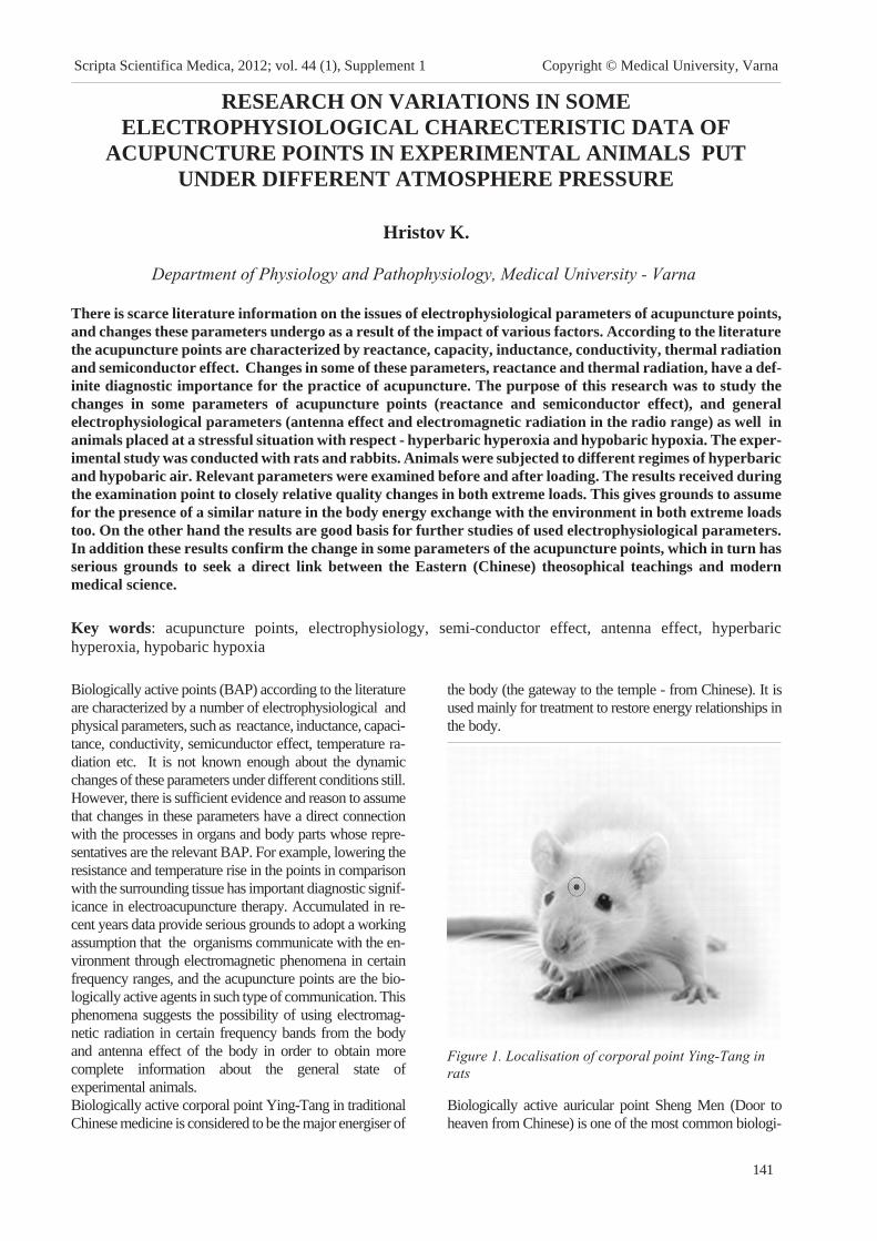
RESEARCH ON VARIATIONS IN SOME
ELECTROPHYSIOLOGICAL CHARECTERISTIC DATA OF
ACUPUNCTURE POINTS IN EXPERIMENTAL ANIMALS PUT
UNDER DIFFERENT ATMOSPHERE PRESSURE
Hristov K.
Department of Physiology and Pathophysiology, Medical University - Varna
There is scarce literature information on the issues of electrophysiological parameters of acupuncture points,
and changes these parameters undergo as a result of the impact of various factors. According to the literature
the acupuncture points are characterized by reactance, capacity, inductance, conductivity, thermal radiation
and semiconductor effect. Changes in some of these parameters, reactance and thermal radiation, have a def-
inite diagnostic importance for the practice of acupuncture. The purpose of this research was to study the
changes in some parameters of acupuncture points (reactance and semiconductor effect), and general
electrophysiological parameters (antenna effect and electromagnetic radiation in the radio range) as well in
animals placed at a stressful situation with respect - hyperbaric hyperoxia and hypobaric hypoxia. The exper-
imental study was conducted with rats and rabbits. Animals were subjected to different regimes of hyperbaric
and hypobaric air. Relevant parameters were examined before and after loading. The results received during
the examination point to closely relative quality changes in both extreme loads. This gives grounds to assume
for the presence of a similar nature in the body energy exchange with the environment in both extreme loads
too. On the other hand the results are good basis for further studies of used electrophysiological parameters.
In addition these results confirm the change in some parameters of the acupuncture points, which in turn has
serious grounds to seek a direct link between the Eastern (Chinese) theosophical teachings and modern
medical science.
Key words: acupuncture points, electrophysiology, semi-conductor effect, antenna effect, hyperbaric
hyperoxia, hypobaric hypoxia
Biologically active points (BAP) according to the literature
are characterized by a number of electrophysiological and
physical parameters, such as reactance, inductance, capaci-
tance, conductivity, semicunductor effect, temperature ra-
diation etc. It is not known enough about the dynamic
changes of these parameters under different conditions still.
However, there is sufficient evidence and reason to assume
that changes in these parameters have a direct connection
with the processes in organs and body parts whose repre-
sentatives are the relevant BAP. For example, lowering the
resistance and temperature rise in the points in comparison
with the surrounding tissue has important diagnostic signif-
icance in electroacupuncture therapy. Accumulated in re-
cent years data provide serious grounds to adopt a working
assumption that the organisms communicate with the en-
vironment through electromagnetic phenomena in certain
frequency ranges, and the acupuncture points are the bio-
logically active agents in such type of communication. This
phenomena suggests the possibility of using electromag-
netic radiation in certain frequency bands from the body
and antenna effect of the body in order to obtain more
complete information about the general state of
experimental animals.
Biologically active corporal point Ying-Tang in traditional
Chinese medicine is considered to be the major energiser of
the body (the gateway to the temple - from Chinese). It is
used mainly for treatment to restore energy relationships in
the body.
Biologically active auricular point Sheng Men (Door to
heaven from Chinese) is one of the most common biologi-
141
Scripta Scientifica Medica, 2012; vol. 44 (1), Supplement 1 Copyright © Medical University, Varna
Figure 1. Localisation of corporal point Ying-Tang in
rats

cally active points. It is believed that this point has a serious
impact in the treatment of various pathological abnormali-
ties, including respiratory and cardiac diseases, pain, in-
flammation, dysfunctional states, general stimulating effect
and others.
Biologically active auricular point Sheng Men (Door to
heaven from Chinese) is one of the most common biologi-
cally active points. It is believed that this point has a serious
impact in the treatment of various pathological abnormali-
ties, including respiratory and cardiac diseases, pain, in-
flammation, dysfunctional states, general stimulating effect
and others.
In the second experimental setup research was conducted
with 30 white nonbreed rats of both sexes with an average
body weight 170 g. Animals were divided into 5 groups
that undermine the response to hypobaric hypoxia: 830 hPa
equal to 1500 m above sea level, 710 hPa - 3000 m above
sea level, 480 hPa - 6000 m, 320 hPa - 8850 m and 220 hPa
- 12,000 m. Speed of decompression and recompression in
the chamber for all groups - 50 hPa / min, exposure time -
15 minutes. Before and after loading in all animals the
electrophysiological parameters of antenna effect were
traced.
The obtained results were processed by variance analysis.
RESULTS AND DISCUSSION
The results of studies of animals from the first experimental
setup with hyperbaric load are set as follows:
The changes of resistance the in straight and reverse direc-
tion and the resulting changes in the semiconductor effect
under hyperbaric pressure in our opinion are evidence for
increased getting into contact of the BAP with the energy
sources of the environment. Increasing of the antenna effect
142
Research on variations in some electrophysiological charecteristic data of ...
Figure 2. Location of auricular point Sheng-Men in rats
Figure 3. Biosensoral detector for electromagnetic
radiation
Figure 4. Hyperbaric chamber MKBK – 42
Figure 5. Resistance in a straight and reverse direction
under hyperbaric pressure.
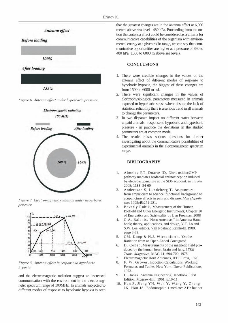
and the electromagnetic radiation suggest an increased
communication with the environment in the electromag-
netic spectrum range of 100MHz. In animals subjected to
different modes of response to hypobaric hypoxia is seen
that the greatest changes are in the antenna effect at 6,000
meters above sea level - 480 hPa. Proceeding from the no-
tion that antenna effect could be considered as a criteria for
communicative capabilities of the organism with environ-
mental energy at a given radio range, we can say that com-
municative opportunities are higher at a pressure of 830 to
480 hPa (1500 to 6000 m above sea level).
CONCLUSIONS
1. There were credible changes in the values of the
antenna effect of different modes of response to
hypobaric hypoxia, the biggest of these changes are
from 1500 to 6000 m asl.
2. There were significant changes in the values of
electrophysiological parameters measured in animals
exposed to hyperbaric stress where despite the lack of
statistical reliability there is a serious trend in all animals
to change the parameters.
3. In two disparate impact on different states between
unpaid animals - response to hypobaric and hyperbaric
pressure - in practice the deviations in the studied
parameters are at common mode.
4. The results raises serious questions for further
investigating about the communicative possibilities of
experimental animals in the electromagnetic spectrum
range.
BIBLIOGRAPHY
1. Almeida RT, Duarte ID. Nitric oxide/cGMP
pathway mediates orofacial antinociception induced
by electroacupuncture at the St36 acupoint. Brain Res
2008; 1188: 54-60
2. Andersson S, Lundeberg T. Acupuncture -
from empiricism to science: functional background to
acupuncture effects in pain and disease. Med Hypoth-
eses 1995;45:271-281.
3. Bever ly Rubik, Measurement of the Human
Biofield and Other Energetic Instruments, Chapter 20
of Energetics and Spirituality by Lyn Freeman, 2008
4. C.A. Balanis , "Horn Antennas," in Antenna Hand-
book; theory, applications, and design, Y.T. Lo and
S.W. Lee, edilors, Van Nostrand Reinhold, 1988,
page 8-39.
5. CM. Knop & H.J. Wiesenfor th. "On the
Ratiation from an Open-Ended Corrugated
6. D. Cohen, Measurements of the magnetic field pro-
duced by the human heart, brain and lung, IEEE
Trans. Magnetics, MAG-11, 694-700, 1975.
7. Electromagnetic Horn Antennas, IEEE Press, 1976.
8. F. W. Grover , Induction Calculations. Working
Formulas and Tables, New York: Dover Publications,
1973.
9. H. Jasik, Antenna Engineering Handbook, First
Edition, Mcgraw-Hill, 1961, p.10-11.
10. Han Z, Jiang YH, Wan Y, Wang Y, Chang
JK, Han JS. Endomorphin-1 mediates 2 Hz but not
143
Hristov K.
Figure 6. Antenna effect under hyperbaric pressure.
Figure 7. Electromagnetic radiation under hyperbaric
pressure.
Figure 8. Antenna effect in response to hypobaric
hypoxia

100 Hz electroacupuncture analgesia in the rat.
Neurosci Lett 1999; 274: 75-78.
11. J . P. Hauser , A 20-Hz to 200-kHz magnetic flux
probe for EMI surveys, IEEE Trans. Electromagnetic
Compatibility, 32, 67-69, 1990.
12. Kir i lov J. Needle antenna properties in acupunc-
ture. Needle inducing mechanism on biological active
points - IV Ñèìïîçèóì "Íîâîå â ïðàêòèêå è òåîðèè
ÃÁÎ, 1989, Ìîñêâà.
13. K.Par tr idge. Associating Users and Devices using
Attention-Correlated Communication. PhD thesis,
University of Washington, 2005.
14. L.Cho, L.Chan, J .Bae, and H.Yoo. A 60 kb/s
- 10 mb/s adaptive frequency hopping transciever for
interferance-resilient body channel communication.
IEEE Journal of Solid state circuits, 44(3), March
2009.
15. Mayer DJ. Biological mechanisms of acupuncture.
Prog Brain Res 2000;122:457-477.
16. M. Shinagawa, K.Ochiai , H.Sakamoto, and
T.Asahi . Human area networking technology:
Redtacton. NTT Review, 3(5), May 2005.
17. Oleson T, Kroening RJ, Bresler DE. An ex-
perimental evaluation of auricular diagnosis: the
somatotopic mapping or musculoskeletal pain at ear
acupuncture points. 1980.
144
Research on variations in some electrophysiological charecteristic data of ...

LONG-TERM SURVIVAL OF A SERIES OF CANCER PATIENTS
WITH PURULENT SEPTIC POSTOPERATIVE COMPLICATIONS
Daskalov Ì.
Medical University of Sofia
ABSTRACT
The author analyzed the results from the postoperative management of the septic complications of eight pa-
tients, six males and two females, with advanced colorectal cancer. These patients have undergone radical and
enlarged operations. All of them survived longer than 10 years. A paradrainage transwound economic
laparostomy after Daskalov was also performed. All the patients were postoperatively treated with chemo-
therapy, immune stimulation with decaris and large amounts of vitamin C. The probable reason for the long
survival was the stimulation of cell immunity. T-lymphocytes activated during inflammation could destroy
the etiological agents of inflammation and single cells having undergone carcinomatous modifications. The fa-
vourable survival was promoted by hyperthermia and stimulation of the cellular immune functions during
the septic process.
Key words: advanced colorectal cancer, septic postoperative complications, hyperthermia, survival, cell
immunity
INTRODUCTION
Term of survival represents the main criterion for success
of cancer treatment. Thus it is a parameter for a well-per-
formed surgical intervention favourably combined with
chemo-, x-ray, immunotherapy and other treatment modal-
ities as well (3-9,11,13,14). During the advanced stage of
colorectal cancer presenting with lymph node metastases
and local progress up to T3-T4, the survival rate over 5
years is very low and there are almost no cases survived
longer than 10, 12, 15 and more years. In such cases of
serously proliferating cancer and operative metastases
some authors induce hyperthermia with a liquid that con-
tains cytostatic drugs such as mitomycin and cistplatine
(1,2,6-11,13,14). Hyperthermia combined with x-ray ther-
apy is administered, too. When the temperature of 410C is
reached, cell proliferation remains unaffected, at 42-430C
and during perfusion hyperthermic hyperpyrexia there oc-
curs apoptosis while at 440C cell necrosis and fibrous
histiocytosis can be seen. Hyperthermia influence varies in
different cytostatic combinations, however, as a whole, it is
considered positive.
The patients with serously proliferating gastric cancer pre-
senting with metastases are favourably influenced by
hyperthermia combined with cytostatic drugs (6,9,14). This
occurs also in cases with peritoneal metastases of ovary
cancer (10,14) and colorectal one as well (14).
In such cases, some authors apply, similarly to S. Hadzhiev
(cited after 1), a variety of vaccines, e.g. BCG, herpes virus
and other vaccines as well as other forms of immunologic
management (1).
MATERIAL AND METHODS
Our study covers six male and two female patients with
colorectal cancer. The local progression consists in the af-
fection of the three layers of the organ, or in proliferation
into adjacent organs along with first- and second-range
lymph node metastases. It deals with carcinoma at stage Ò3
and Ò4, with or without lymph node metastases (N1 or N2).
These patients have undergone radical and enlarged opera-
tion. Due to current reasons, purulent septic complications
develop which are successfully treated. All the patients sur-
vive longer than 10 years as two of them survive even 16
and 21 years after the operation.
Case reports
The first patient was 60 years old. He underwent
hemicolectomy with lymph node dissection and resection
of 5th and 6th liver segments. A postoperative subhepatic ab-
scess in the right abdominal part occurred. It healed follow-
ing drainage aspiration and opening of the dermal and
fascial sutures along the bundle of tube drainages
(paradrainage transwound economic laparostomy after
Daskalov, 1). Septic fever lasted more than two weeks.
Healing resulted in the occurrence of a postoperative repo-
sition hernia that did not hamper his physical activity. He
survived longer than 20 years without any relapses or
metastases at all.
The second patient presented with rectal cancer of T3N1/2
as well as with empyema vesicae felleae, diabetes mellitus
and obesity of second degree. We performed
cholecystectomy, lymphadenectomy, abdomino-perineal
rectum amputation, and definite iliac anus praeter. Postop-
145
Scripta Scientifica Medica, 2012; vol. 44 (1), Supplement 1 Copyright © Medical University, Varna

erative sepsis lasted longer than two weeks and consisted in
a purulent secretion originating from the bundle of tube
drainages located in the pelvis and from those draining the
operative wound itself. A paradrainage laparostomy after
Daskalov was carried out as both adjacent skin and fascial
sutures were removed (1). Healing was achieved through
active reanimation and antibiotic therapy. At the site of
laparotomy, subsequently, a postoperative hernia devel-
oped which relapsed after the plastic correction done sev-
eral years later on. His work capacity was restored and he
survived longer than 17 years without any relapses. He died
of adhesion ileus following surgical intervention in another
health institution. He suffered from diabetes mellitus
controlled by maninil.
The third patient was 59 years old and underwent an
abdominoperineal rectum amputation on the occasion of a
carcinoma at stage Ò4 N1/2 which removal caused
parenchymatous bleeding and necessitated tamponade with
local Mickulicz compress. A suppuration and sepsis devel-
oped at the background of diabetes mellitus. The patient
survived 21 years long.
The fourth patient presented with rectum amputation as a
consequence of carcinoma at T3N1/2 and accompanying
pelvic peritonitis. Sepsis occurred and lasted more than one
week. It was managed with drainage aspiration and
paradrainage transwound economic laparostomy (1) as
well as by antibiotic therapy and plasty with an
antimicrobial cusp for the emerged postoperative hernia
was performed resulting in a durable healing lasting more
than 20 years.
The fifth patient was 60 years old and presented with rec-
tum amputation for a carcinoma of Ò4N1/2. Postopera-
tively, septic fever occurred and lasted more than one week
favourably managed by the aspiration of the purulent con-
tent from the pelvic space through drainages as well as by
the accompanying antibiotic treatment. Two years later on,
the patient was operated for a localized carcinoma of the
urinary bladder and healed. The patient survived longer
than 20 years under good health conditions and with a pre-
served work capacity. He died of another disease.
The sixth patient was 60 years old and presented with a rec-
tal cancer at T3 stage. Postoperatively, he developed a pu-
rulent sepsis. He restored and reached up the age of 82
years.
The seventh patient was a 60-year-old female. She was op-
erated on the occasion of a genital neoplasm and developed
a purulent collection. After the successful operation she re-
mained under a good condition and reached up the age of
90 years without any relapses.
The eighth patient was a 45-year old female and presented
with gastric lymphosarcoma. She developed a postopera-
tive peritonitis and a purulent fistula. She was re-operated
after Daskalov's method (1) and enjoyed a good health sta-
tus longer than 21 years.
All the patients were postoperatively treated with chemo-
therapy according to a scheme approved by the Clinic of
Chemotherapy, Tsaritsa Giovanna University Hospital of
Sofia. Besides, an immune stimulation with decaris was
performed. Additionally, large amounts of vitamin C were
administered.
The long survival of these patients presenting with carcino-
mas of the size of children's hand and fist and affecting the
entire wall of the colon and rectum can't be explained nei-
ther by the radical intervention, nor by the cytostatic ther-
apy only. Probably, hyperthermia caused by the purulent
septic condition experienced by the patients is of impor-
tance, too. This coincides with the opinions of numerous
authors about the significance of the hyperthermia (11,13).
The involvement of the autoimmunization, similarly to the
influence of the fixation abscesses during the rheumatic
conditions can't be definitely considered. Suppuration and
fever in four of our cases occurred along with diabetes
mellitus which was routinely treated.
The probable reason for the long survival of these patients
who experienced sepsis and suppuration is the stimulation
of cell immunity. T-lymphocytes activated during inflam-
mation are capable of destroying not only the etiological
agents of inflammation but also single cells having under-
gone carcinomatous modifications. This reduces the proba-
bility of metastases during the postoperative period.
CONCLUSION
Herewith we report the unusually long-lasting survival of a
group of patients with advanced colorectal cancer who
have undergone radical interventions, with postoperative
hyperthermia based on the emerged suppuration and mi-
crobial septic process. The principle of radical operation
was observed in an optimal volume. However, the favour-
able survival was, probably, promoted by hyperthermia and
stimulation of the cellular immune functions during the
septic process.
REFERENCES
1. Äàñêàëîâ, Ì. Ïàòîãåíåòè÷íî öåëåñúîáðàçåíè
îïåðàöèè, ñòóäèè è âúïðîñè ïî êîðåìíà õèðóðãèÿ.
Ñîôèÿ, 1996.
2. Bonenkamp, J. J., C. J. van de Velde, G. H.
Kampsch�er, J. Hermans, P. Hermanek, M.
Bemelmans, et al. Comparison of factors influenc-
ing the prognosis of Japanese, German, and Dutch
gastric cancer patients.- World J. Surg., 17, 1993, No
3, 410-414; discussion 415.
3. Fuj imoto, S. , M. Takahashi , F. Endoh, R.
D. Shrestha, M. Kokubun, M. Takai , e t a l .
A clinical pilot study combining surgery with
intraoperative pelvic hyperthermochemotherapy to
prevent the local recurrence of rectal cancer.- Ann.
Surg., 213, 1991, No 1, 43-47.
4. Fuj imoto, S. , M. Takahashi , K. Kobayashi ,
M. Kure, T. Mutou, H. Masaoka, et al . Rela-
tion between clinical and histologic outcome of
intraperitoneal hyperthermic perfusion for patients
with gastric cancer and peritoneal metastasis.- Oncol-
ogy, 50, 1993, No 5, 338-343.
146
Long-term survival of a series of cancer patients with ...

5. Furukawa, H. , M. Hiratsuka, T. Iwanaga. A
rational technique for surgical operation on Borrmann
type 4 gastric carcinoma: left upper abdominal evis-
ceration plus Appleby's method.- Br. J. Surg., 75,
1988, No 2, 116-119.
6. Mosser , D. D. , L. H. Mart in . Induced
thermotolerance to apoptosis in a human T lympho-
cyte cell line.- J. Cell Physiol., 151, 1992, No 3,
561-570.
7. Panteix, G. , M. Guil laumont , L. Cherpin, J .
Cuichard, F. N. Gil ly , P. Y. Carry , et al .
Study of the pharmacokinetics of mitomycin C in hu-
mans during intraperitoneal chemohyperthermia with
special mention of the concentration in local tissues.-
Oncology, 50, 1993, No 5, 366-370.
8. Sayag, A. C. , F. N. Gil ly , P. Y. Carry , J . P.
Perdr ix , G. Panteix, A. Brachet , e t al .
Intraoperative chemohyperthermia in the management
of digestive cancers. A general review of literature.-
Oncology, 50, 1993, No 5, 333-337.
9. Sel l ins , K. S. , J . J . Cohen. Hyperthermia in-
duces apoptosis in thymocytes.- Radiat. Res., 126,
1991, No 1, 88-95.
10. Takano, Y. S. , B. V. Harmon, J . F. Kerr .
Apoptosis induced by mild hyperthermia in human
and murine tumour cell lines: a study using electron
microscopy and DNA gel electrophoresis.- J. Pathol.,
163, 1991, No 4, 329-336.
11. Yonemura, Y. , T. Fuj imura, S. Fushida, S.
Takegawa, T. Kamata, K. Katayama, et al .
Hyperthermo-chemotherapy combined with
cytoreductive surgery for the treatment of gastric can-
cer with peritoneal dissemination.- World J. Surg., 15,
1991, No 4, 530-535; discussion 535-536.
12. Yonemura, Y. , K. Katayama, T. Kamata, S.
Fushida, M. Segawa, S. Ooyama, et al . Sur-
gical treatment of advanced gastric cancer with me-
tastasis in para-aortic lymph node.- Int. Surg., 76,
1991, No 4, 222-225.
13. Yonemura, Y. , I . Ninomiya, M. Kaj i , K.
Sugiyama, K. Fuj imura, T. Sawa, et al . Pro-
phylaxis with intraoperative chemohyperthermia
against peritoneal recurrence of serosal invasion-posi-
tive gastric cancer.- World J. Surg., 19, 1995, No 3,
450-454; discussion 455.
14. Yonezawa, M., T. Otsuka, N. Matsui , H.
Tsuj i , K. H. Kato, A. Moriyama, et al .
Hyperthermia induces apoptosis in malignant fibrous
histiocytoma cells in vitro.- Int. J. Cancer, 66, 1996,
No 3, 347-351.
147
Daskalov Ì.
During the recent year, the eminent Bulgarian surgeon, professorMarko Daskalov, MD, celebrated his 80th anniversary. He was bornon June 19, 1931, in the village of Smilets, Pazardzhik district. In1955, he graduated with excellent scores from the Higher Institute ofMedicine of Plovdiv. Then he was occupied as surgeon and gynae-cologist in the towns of Ivailovgrad and Rakovski. In 1959-1960, hemade the specialities of 'surgery' and 'orthopaedics and traumatology'and was occupied in the corresponding clinics in the city of Plovdiv.In 1961, he became an assistant in the Clinic of Surgery and Ortho-paedics in Alexandrovska Hospital of Sofia. Later on he made this spe-ciality in Paris, France. He consecutively became associate professorand then full professor in the Second Department of Surgery of theMedical Academy of Sofia where he delivered the courses of lecturesto the medical students and the postgraduate students as well.Professor Marko Daskalov has authored more than 280 scientific pub-lications, including 4 monographs. He was the author of numerousown unique operative techniques as well as of new methods for simul-
taneous operation of several organs, the so-called 'en-bloc' operations. Besides we elaborated surgi-cal techniques for the 'creation' of new organs such as neogaster, ampula recti, etc. Additionally, heintroduced a series of modifications of already existing operative methods into the clinical practice.Professor Marko Daskalov performed the first successful experiments in dogs in order to transplantthe spleen and pancreas which were filmed and broadcasted by the national TV.He is an active member of the Bulgarian Society of Surgery. He is our first board member in the Coun-cil of Eurosurgery where he represented our country during a lot of years. Professor Marko Daskalovwas awarded the Medal of Cyril and Methodius of first degree for his active creative, scientific, teach-ing and surgical work.
Happy Birthday to You, our Teacher! And Happy Anniversary!


AUTOR'S INDEX
Achkakanov I. ........................... 55
Alexandrova A. ......................... 55
Alexandrova M. ........................ 63
Alexiev K. ................................. 109
Angelova P. ............................... 81
Atanasov P. ............................... 55
Atanasova P. ............................. 133
Bekyarova G. ............................ 31, 35
Belcheva I. ................................ 59
Belcheva S. ............................... 59
Bocheva N. ............................... 11, 73, 109
Boyadjiev N. ............................ 81
Danovska M. ............................ 63, 145
Decheva L. ............................... 129
Doncheva N. ............................. 137
Dushanova J. ............................ 15
Encheva E. ................................ 19
Gatev P. .................................... 125
Gendzhev Z. ............................. 101
Genova B. ................................. 11, 109
Georgiev K. .............................. 51
Georgiev Ts. ............................. 93
Georgieva O. ............................ 73
Hadzhibozheva P. ..................... 93
Halacheva L. ............................. 77
Hrischev P. ............................... 133
Hristov I. .................................. 67
Hristov K. ................................. 141
Hristova M. .............................. 31
Hristova M. ............................... 35
Ivanov I. .................................... 23
Ivanova A. ................................. 81
Ivanova M. ................................ 59
Klisurov R. ................................ 19
Kolarova R.tragvam .................. 97, 101, 105
Kolev N. .................................... 77
Kossev A. .................................. 5
Kupenova P. .............................. 23
M. Krustev S. ............................ 117
Mihaylova M. ............................ 67
Miladinova E. ............................ 51
Minchev Zl. ............................... 125
Mitov D. .................................... 15, 67
Muletarov S. .............................. 81
Negrev N. .................................. 59, 117, 129,137
Nikolov F. ................................. 133
Nikolov P. ................................. 133
Nikolova J. ................................ 133
Novoselski M. ........................... 19
Nyagolov Y. .............................. 129, 137
Orbezova M............................... 133
Pajpanova T............................... 51
Pashalieva I. .............................. 129
Pashaliewa I............................... 137
Pavlova E. ................................. 27, 89
Pechlivanova D. ........................ 47
Petkov V.................................... 19
Petkova Zl. ................................ 47
Petrov L. .................................... 55
Popova E. .................................. 23
Racheva K. ................................ 67
Sarov G...................................... 39, 43
Somlev P. .................................. 27, 85,89
Stancheva E. .............................. 129
Stefanov S. ................................ 11, 109
Stefanova M. ............................. 11, 73
Stephanova D. I. ........................ 117
Stoynev Al................................. 47
Tancheva L................................ 19
Tashev R.................................... 59
Tchekalarova J........................... 47
Tolekova A................................ 93
Totev Ts. ................................... 67
Tsaneva M. ................................ 35
Tzvetkov S. ............................... 113
Uzunova G. ............................... 27, 89
Zaekov N. .................................. 55
Zsheliaskova-Koynova Z. ......... 55
149
Scripta Scientifica Medica, 2012; vol. 44 (1), Supplement 1 Copyright © Medical University, Varna


PERMUTERM SUBJECT INDEX
acupuncture points, electrophysiology,
semi-conductor effect, antenna effect,
hyperbaric hyperoxia, hypobaric hypoxia_______141
advanced colorectal cancer, septic
postoperative complications, hyperthermia,
survival, cell immunity ____________________145
Ageing, Motion discrimination, Fuzzy
clustering, Mixed ANOVA___________________73
bioelectricity, human emotions, social
networks, spectral analysis, rhythm
dynamics ________________________________125
classification images, motion direction
discrimination, internal noise, directional
tuning, temporal integration _________________109
Contrast, VEP, Spatial frequency,
Receptive fields____________________________67
dark adaptation, dopamine,
electroretinogram, frog, retina _______________23
diabetes, hypertension, stroke,
inflammation, CRP _________________________63
dopamine, dark adaptation,
electroretinogram, frog, retina _______________23
electrophysiology, acupuncture points,
semi-conductor effect, antenna effect,
hyperbaric hyperoxia, hypobaric hypoxia_______141
endomorphins, morphiceptin, opioid
receptors, halogenated phenylalanines,
peptide mimetics ___________________________51
exercise, physical working capacity, fat
metabolism _______________________________81
experimental hypertension, myocardial
hypertrophy, cardiac histomorphological
changes, Vit. E ___________________________101
experimental hypertension, myocardial
hypertrophy, myocardial enzyme activity,
Vit. E____________________________________97
FII, vitamin K-dependent clotting factors,
FV, FIX, FX, somatotropin, somatostain,
prolactin ________________________________137
Handgrip, muscle force, bilateral deficit,
sex differences ____________________________77
heart rate variability, heart rate, saliva,
salivary alpha-amylase activity, salivary
potassium ________________________________55
heart rate variability, paced breathing,
athletes __________________________________85
heart rate variability, soccer players,
training __________________________________89
heart rate, heart rate variability, saliva,
salivary alpha-amylase activity, salivary
potassium ________________________________55
holistic, structural, functional,
pathophysiology ___________________________43
HR recovery, PWC170, VO2 max,
coefficient of recovery, soccer players __________27
human emotions, bioelectricity, social
networks, spectral analysis, rhythm
dynamics ________________________________125
hypertension, diabetes, stroke,
inflammation, CRP _________________________63
hypokinesia, physical exercise, Vit. E,
experimental hypertension, myocardial
metabolism ______________________________105
light immunohistochemistry, superoxide
dismutase, malondialdehyde, liver, burns,
melatonin ________________________________35
luzindolå, melatonin, reptilase time, TAFI,
TAFIa/TAFIai____________________________129
maximal oxygen consumption, ventilatory
response, maximal incremental exercise
test, taekwondo athletes ____________________113
melatonin, luzindolå, reptilase time, TAFI,
TAFIa/TAFIai____________________________129
morphiceptin, endomorphins, opioid
receptors, halogenated phenylalanines,
peptide mimetics ___________________________51
motion direction discrimination,
classification images, internal noise,
directional tuning, temporal integration ________109
Motion discrimination, Ageing, Fuzzy
clustering, Mixed ANOVA___________________73
muscle force, Handgrip, bilateral deficit,
sex differences ____________________________77
muscle vibration (MV), transcranial
magnetic stimulation (TMS), motor evoked
potential (MEP), intracortical inhibition
(ICI), intracortical facilitation (ICF), silent
period (SP) ________________________________5
myelin sheath aqueous layers,
strength-duration time constant,
demyelinating neuropathies,
computational neuroscience _________________117
myocardial hypertrophy, experimental
hypertension, cardiac histomorphological
changes, Vit. E ___________________________101
myocardial hypertrophy, experimental
hypertension, myocardial enzyme activity,
Vit. E____________________________________97
Olfactory bulbectomy, Vasoactive
intestinal polypeptide, Hippocampus,
Locomotor activity, Lateralization, Rat _________59
paced breathing, heart rate variability,
athletes __________________________________85
Perception, Vision, Motion, Speed,
Adaptation, Sensitivity ______________________11
physical exercise, hypokinesia, Vit. E,
experimental hypertension, myocardial
metabolism ______________________________105
physical working capacity, exercise, fat
metabolism _______________________________81
151
Scripta Scientifica Medica, 2012; vol. 44 (1), Supplement 1 Copyright © Medical University, Varna
PWC170, HR recovery, VO2 max,
coefficient of recovery, soccer players __________27
septic postoperative complications,
advanced colorectal cancer, hyperthermia,
survival, cell immunity ____________________145
sinusoidal gratings, visual event-related
potentials, orientation discrimination ___________15
smooth muscle contraction, uterus,
peptides, hyperactivity, in vitro
experiments_______________________________93
soccer players, heart rate variability,
training __________________________________89
spontaneous hypertensive rats (SHRs),
Wistar rats, kainate model of temporal
lobeepilepsy; melatonin; diurnal rhythms________47
strength-duration time constant, myelin
sheath aqueous layers, demyelinating
neuropathies, computational neuroscience ______117
structural, holistic, functional,
pathophysiology ___________________________43
superoxide dismutase, light
immunohistochemistry, malondialdehyde,
liver, burns, melatonin ______________________35
thiol groups, uric acid, malondialdehyde,
gastric mucosa, burns ______________________31
transcranial magnetic stimulation (TMS),
muscle vibration (MV), motor evoked
potential (MEP), intracortical inhibition
(ICI), intracortical facilitation (ICF), silent
period (SP) ________________________________5
uric acid, thiol groups, malondialdehyde,
gastric mucosa, burns ______________________31
uterus, smooth muscle contraction,
peptides, hyperactivity, in vitro
experiments_______________________________93
Vasoactive intestinal polypeptide,
Olfactory bulbectomy, Hippocampus,
Locomotor activity, Lateralization, Rat _________59
ventilatory response, maximal oxygen
consumption, maximal incremental
exercise test, taekwondo athletes _____________113
VEP, Contrast, Spatial frequency,
Receptive fields____________________________67
Vision, Perception, Motion, Speed,
Adaptation, Sensitivity ______________________11
visual event-related potentials, sinusoidal
gratings, orientation discrimination ____________15
vitamin K-dependent clotting factors, FII,
FV, FIX, FX, somatotropin, somatostain,
prolactin ________________________________137
Wistar rats, spontaneous hypertensive rats
(SHRs), kainate model of temporal lobe
epilepsy; melatonin; diurnal rhythms ___________47
152

INSTRUCTIONS TO AUTHORS
Scripta Scientifica Medica is the official publication of Medical University Prof. Dr. Paraskev Stoyanov, Varna, Bulgaria. It is
currently disseminated among medical university libraries from all over the world on exchange basis. This peer-reviewed annual
accepts for publication original articles, unpublished papers recently presented at national and international congress proceedings, and
book reviews from Bulgarian and foreign authors. The contributions should be devoted to actual topics in contemporary biomedicine,
clinical medicine and interdisciplinary fields as well. They should not have been submitted or accepted for publication elsewhere. The
journal publication is offered to the national and international readership in English only.
The manuscript signed by all the authors has to be submitted in duplicate to the Editor-in-Chief of Scripta Scientifica Medica.
Prof. Anelia Klisarova, MD, PhD, DSc
Medical University Prof. Dr. Paraskev Stoyanov, Varna
55 Marin Drinov Street
BG-9002 Varna
Bulgaria
Phone: +359-52- 650 057
Fax: +359-52- 650 019
E-mail: [email protected]
The contributors are encouraged to submit the files of the text and figures of their revised manuscripts via e-mail to
[email protected] or via postal service on CD/DVD with a printing proof on A4 paper.
The authors must strictly follow some main instructions listed below.
The manuscript of an original paper should not exceed 4 single-spaced pages. The total volume of the text, tables, and references
should not exceed 15000 characters. Printed and electronic copies should be on Times New Roman, size 12.
The structure of the article should include the following sections: Introduction, Material and Methods, Results, Discussion and
Conclusion(s). Additionally, there should be: a structured abstract of 200-250 words; key-words (5 to 6 words or non-verbal phrases);
a reference list (up to 20 references); a complete address of the author for correspondence (postal and e-mail address), and an
appropriate number of tables and figures. The figures should be provided on separate files in TIFF, EPS or JPEG format. All photos
should be scanned as grayscale at 300 dpi. The tables should be provided on separate sheets and each table should have a title. Data
sheets of diagrams should be obligatorily provided. No data reiteration in the text, tables, and figures is permitted. Photographs and
microphotographs should be sufficiently contrasted and with size of 8 cm in width (1 column) and 17 cm in width (2 columns).
Black-and-white drawings and diagrams are accepted only. Cited authors are ordered alphabetically in the reference list starting with
those in the Cyrillic alphabet. Most commonly, these authors should be identified in the text of the article by Arabic numerals in
parentheses - for example (1). Please, do not make use of Caps Lock option at all.
Preparation of references
Numbering of all the publications cited in the text should correspond to that in the list of references. Bibliographic citations of
articles in journals should contain initials and names of all the authors (or at least the first six ones), article title, abbreviated title
of the journal according to the style used in Index Medicus (National Library of Medicine, Bethesda, MD, USA), volume, year of
publication, issue number (absolutely obligatory for Bulgarian and Russian journals), and page numbers (from-till). The citations of
books should contain initials and names of the authors (up to three), book title, number of edition (if any), editor(s) (if any), location of
publishing, publishing house and year of publication. Book chapters should contain initials and names of the authors of the chapter,
title of the whole book, editor(s) (if any), location of publishing, publishing house, year of publication and page numbers (from-till).
Congress proceedings should contain along with data as for book chapters, location and date of the corresponding meeting, kind of
materials (abstracts or full papers), and page numbers. Author’s name of the dissertation, title, location, institution, and year of defence
should be indicated. With patents and licences, author’s names (if any), registration number, and year of publication should be shown.
Personal communications containing the name of the author cited and the date should be accompanied by his (her) permission in
written for the corresponding statement.
Let us give some examples.1. Biderman, I., S. Somien, Z. Shimshoni. In: Tissue Nutrition and Viability. A. R. Hargens, ed. New York, etc., Springer-Verlag, 1986, 121-134.
2. Goute, A. M., A. R. Haynes, M. J. Owen. New aspects of psychotic drug usage.- J. Clin. Psychopharmacol., 8, 1988, No 4, 315-317.
3. Youmans, G. P., A. N. Lewin. Tuberculosis. 3rd
ed. Philadelphia, etc., W. W. Saunders, 1979.
The authors will receive 25 reprints of their articles along with a sample copy of the issue free of charge.
153
Scripta Scientifica Medica, 2012; vol. 44 (1), Supplement 1 Copyright © Medical University, Varna