screen-printed electrodes | encyclopedia
TRANSCRIPT
Screen-Printed ElectrodesSubjects: Engineering, ChemicalContributors: Julio Bastos Arrieta , Karina Torres-Rivero Submitted by: Julio Bastos Arrieta
Definition
1. IntroductionElectrochemical methods are advantageous regarding miscellanies instrumental analysis due to their lowcost, simplicity, high sensitivity, ease of operation, rapid analysis, portability, and applicability formonitoring different samples in the environmental and pharmaceutical field. In recent years, research inthe field of electrochemical sensors has evolved towards the simultaneous analysis of species andminiaturization of electrodes based on new materials and their strategic surface functionalization. Screenprinting is a well-established method to produce thick-film electrochemical transducers . Thistechnology is highly reproducible and used for the preparation of single-use screen-printed electrodes(SPEs). SPEs composed of carbon nanoallotropes (e.g., carbon nanotubes, nanofibers, or graphene)represent a versatile sensing tool due to their suitability for incorporation in portable instrumentation
. Additionally, it has been reported how modifying their surface with metal nanoparticles (MNPs)leads to the enhancement of the electrochemical reactivity and sensitivity for specific analytes .
The use of nanoparticles (NPs) to modify Screen Printed Electrodes (SPEs) offers significant advantages inenhancing the mass transference rate and the electrocatalytic activity of the electrode . NPs exhibit ahigher reactive surface, directly influenced by exposed atoms disposition, which results in moreelectrocatalytically active sites (edge and corner sites) . This fact provides NPs of different sizes andshapes, preferential reactivity, and selectivity towards electrocatalytically detection of specific analytes,due to different charge distribution or polarization of the shaped entity, during the electrochemicaldetermination . Particularly, some studies have reported using SPEs modified with different MNPs toenhance the sensitivity towards the determination of different toxins in different samples; goldnanoparticles and graphene oxide modified screen printed carbon electrode to detect carbofuran ,reduced graphene oxide/gold nanoparticles/boronic acid nanocomposite modified screen printedelectrode to determine glycoside in food samples , Prussian blue nanoparticles-based screen printedelectrodes to detect mustard agents , a nanocomposite based on gold nanoparticles and grapheneoxide quantum to modify screen printed electrodes for the voltammetric determination of Aflatoxin B
, and dendritic platinum nanoparticles and gold nanoparticles on screen printed electrode todetermine bisphenol A on tap water samples electrochemically .
2. SPEs Modification by Physical ApproachesSome of the modification techniques rely on the physical incorporation of nanomaterials on SPEs surfacewithout changing their chemical integrity to improve the electrode’s electrocatalytic performance byincreasing the effective surface area. Hence, low detection limits, fast responses, high sensitivity, andreproducibility can be obtained with the resulting nano-enabled sensor.
2.1. Drop-Casting Method
Several investigations have been reported using this methodology to modify screen-printed electrodes,
Screen printed electrodes(SPE) are disposable, low-cost and portable devices that include aminiaturized display of the working, reference and counter electrodes; based on conductive substrateslike carbon nano allotropes (e.g. graphite, graphene) and metals (e.g. Au, Ag, Pt). This configuration isoptimized to work with micro-volumes of samples. Their range of applications includeselectrochemical analysis in environmental, clinical and agri-food fields.
[1]
[1][2][3][4][5][6]
[7][8]
[9]
[10]
[11]
[12][13]
1[14]
[15]
[16] [17] [18][19]
using different NPs solutions: copper , bismuth , gold , silver.
It is a simple technique where the screen-printed electrode surface is modified by using a solution thatcan be composed of particles such as nanotubes or nanoparticles .
Usually, the NPs solution is placed onto the sensor surface. The solvent evaporation can be performed byintroducing the sensor in an oven at a temperature at which the electrode is not damaged. The drop castamount onto the electrode is the only parameter to be considered for this modification technique .
Nevertheless, other investigations have reported drawbacks regarding this methodology: the so-called“ring coffee effect” , where the ring’s periphery concentrates the non-volatile particles in contrastto the center. Marangoni effect, anisotropic nanoparticles, or even surfactants can reduce this effect,creating a uniform distribution of the drop cast nanoparticles on the sensor surface.
2.2. Spin Coating
The spin coating method allows producing a uniform distribution of the nanoparticles on the screen-printed electrode surface. This methodology is performed in four steps, (1) deposition in which thematerial (NPs solution) is deposited onto the sensor’s surface; (2) spin up (acceleration); (3) spin off(deceleration), the applied solution is distributed via centrifugal force, the high spinning speed results in auniformed layer; and (4) evaporation of the solvent is possible because of rapid rotation . Thecoating solution viscosity and the rotation speed controlled the thickness of the deposited layer .
2.3. Spray Coating
Chomoucka et al. and Mayousse et al. have reported using this method to modify screen printedelectrodes. Nanoparticles dispersed in an appropriate solvent (e.g., alcohol) are sprayed onto a substratethrough jet/nozzle equipment. The liquid evaporates, allowing the NPs to settle on the surface .
2.4. Sputter Coating
Other investigations have reported the use of this technique to modify the sensor’s surface . Thesputter coating is referred to the use of the energy of a partially ionized gas (usually argon) on thesurface of a target (cathode) to pull out the atoms of the material one by one, and deposit them on thesubstrate .
2.5. Electrospray
This method relies on propelling nanoparticles using voltage . The NPs are dispersed into droplets byan existing electrical field between a nozzle and the targeted substrate. The solvent is evaporated beforereaching the surface, where the NPs are deposited . Mettakoonpitak et al. reported usingelectrospray for the deposition of silver nanoparticles on SPEs.
2.6. Chemical and Electrochemical Deposition
Chemical deposition takes place when reacting volatile precursors in the gas phase to form a layer thatdeposits on the desired surface, like SPEs. This approach includes homogeneous reactions occurring in thegas phase and heterogeneous chemical reactions which occur close to a heated surface, forming(nano)powders or (nano)coatings. For example, this strategy was used for the customized preparation ofa nanocomposite modified SPE, that consisted of carbon black-Prussian blue NPs .
Similarly, electrochemical disposition is a modifying technique that can produce nanoparticles withcontrolled characteristics, size, morphology, and composition on an electrode’s surface . Typically, theoxidized species (metallic salts) are reduced at a fixed potential or current to obtain the metal particlesgrown on substrates. Using this methodology, it is possible to optimize the precursor solution parameters
[16] [17] [18][19]
[20]
[3]
[20][21]
[22][23][24][22]
[25] [26]
[27][28]
[29][30]
[31]
[27]
[32] [33]
[34][35]
[36]
(e.g., salt type and concentration) and those regarding the electrochemical deposition. Even thoughbigger particles can be obtained using higher precursor concentrations, two parameters are critical incontrolling NPs’ size and shape: the deposition time and the applied potential or current .
Many studies have been performed using the electrodeposition and various salts precursor to modifyscreen-printed electrodes; Ag-NPs and Au-NPs were electrodeposited using AgClO and HAuCl ,respectively. Pt-NPs using K PtCl , Cu-NPs via CuSO , and NiO-NPs employing Ni(NO ) ·6H O.
2.7. Ink Mixing and Printing Method
This technique incorporates into the ink preparation three components: conductive particles usually madeof carbonous material, a binder paste mixture, such as resins or cellulose acetate, and even solvents suchas terpineol, ethylene glycol, or cyclohexanone that allow the particulate matter to transfer onto theelectrode substrate, and the modifying agent, in this particular case, NPs . Sometimes, screen-printedelectrodes composed of graphite particles are electrochemically activated to enhance theirelectrochemical performance . Additionally, a pre-treatment could be performed in carbon-basedelectrodes to improve the electron transfer rates between the electrode surface and the compounds insolution .
It is possible to mention some studies reporting the utilization of this modifying technique: nanocompositeconsisting of bismuth nanoparticles and amorphous carbon , silver and carbon nanoparticlesconductive inks , silver nanoparticles ink , and gold nanoparticles ink .
3. SPEs Modification with Morphologically Different NPs Systems
3.1. Spherical Nanoparticles
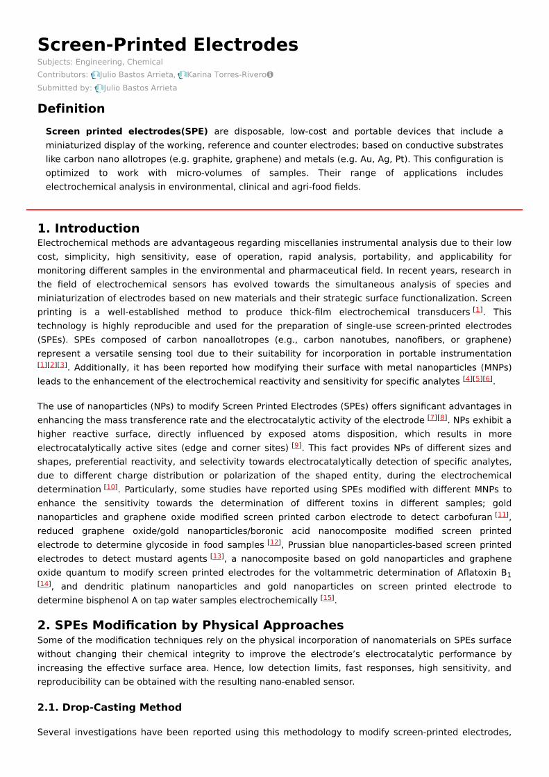
Several investigations include the synthesis of nanoparticles with a spherical shape. These NPs have beenused to modify SPEs with different applicability in numerous fields. Singh et al. prepared a grapheneoxide-cyclodextrin composite with platinum nanoparticles (GR/CD/Pt). This nanocomposite wasincorporated into the SPEs by printing it upon the working electrode’s top, obtaining the GR/CD/Pt/SPE,further used for cysteine determination. The modified SPE were characterized by employing scanningelectron microscopy (SEM), transmission electron microscopy (TEM), atomic force microscopy (AFM),Fourier transform infrared (FT-IR), and thermogravimetric analysis (TGA). Figure 2A shows a TEM image ofthe GR/CD/Pt with spherical-shaped particle structure. A coating of platinum NPs over GR/CD compositewith an average diameter of 15 nm can be observed.
Figure 2. (A) TEM micrograph of GR/CD/Pt composite. (B) CV response of cysteine at the SPE modifiedwith the GR/CD/Pt in a concentration range of 0.5–170 µM in 0.1 M PBS buffer pH 7.4. Reproduced withpermission of Singh et al., Journal of Electroanalytical Chemistry; published by Elsevier, 2018 .
Moreover, an electrochemical characterization of the GR/CD/Pt/SPE was performed using cyclicvoltammetry (CV), differential pulse voltammetry (DPV), and electrochemical impedance spectroscopy
[3]
[37] 4 4[38] 2 6 [39] 4 [40] 3 2 2
[41][3]
[42]
[41][43]
[44][45] [46][47] [48]
[49]
[49]
(EIS). DPV studies (Figure 2B) exhibited two ranges in which current and cysteine concentrations had alinear correlation from 0.5 to 40 µM and from 40 to 170 µM with a limit of detection (LOD) of 0.12 µM.
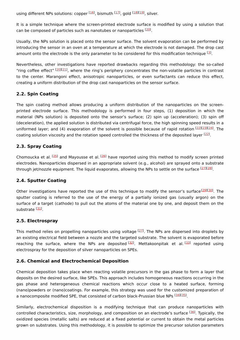
Other studies carried out by Cunha-Silva and Arcos-Martinez functionalized a SPE with rhodiumnanoparticles (Rh-NPs) using the chronoamperometric technique. The obtained sensor was used forbromide anion determination in seawater, surfactant, and pharmaceutical samples. Figure 3A shows thatthe SPEs surface modified with the electrodeposited rhodium nanoparticles at −0.25 V for 480 s. Figure3B exhibited the voltammograms obtained using the modified electrodes in 0.05 M phosphate-bufferedsaline (PBS) with 0.05 M of NaCl as supporting electrolyte. The bromide concentration ranged from 0 to40 mM.
Figure 3. (A) SEM micrograph of Rh-NPs deposited on SPEs. (B) Cathodic stripping voltammograms ofmodified electrodes recorded using 150 μ L drop of supporting electrolyte with bromide. The cathodiclinear sweep voltammetry scan was from 1.11 to −0.25 V at a scan rate of 0.10 Vs . Reproduced withpermission of Cunha-Silva and Arcos-Martínez, Sensors and Actuators B: Chemical; published by Elsevier,2019 .
3.2. Triangle Shaped Nanoparticles
Baradoke et al. developed triangular ruthenium nanoplates (Ru-NPLs) to modify graphene screen-printed electrodes to determine ß-Nicotinamide adenine nucleotide in its reduced form (NADH), which isrelated to depression, neurodegenerative diseases (Parkinson and Alzheimer), and even cancer.
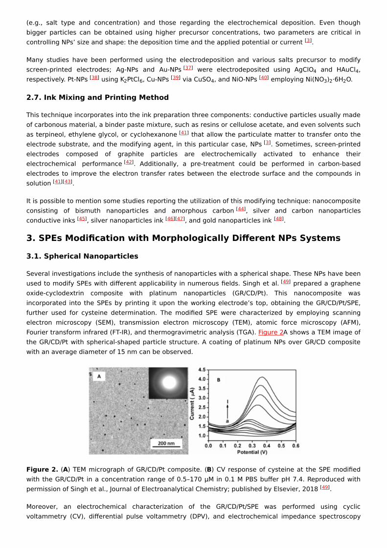
The authors synthesized these Ru nanoplates through a hydrothermal reduction of a ruthenium salt(RuCl ·xH O) with formaldehyde in the presence of polyvinylpyrrolidone (PVP). TEM micrographs showvery thin triangular nanoplates with an edge length of 18 ± 3 nm (Figure 4A).
Figure 4. (A) TEM micrograph of Ru-NPLs obtained through the hydrothermal reduction of Ru salt byformaldehyde in the presence of PVP. SEM micrographs of Ru-NPLs in H O, (B) in ethanol, (C) modifiedscreen-printed graphene electrode. Reproduced with permission of Baradoke, Pastoriza-Santos yGonzález-Romero, Electrochimica Acta; published by Elsevier, 2019 .
After the synthesis, the nanoplates were incorporated, using the drop-casting method, to graphenescreen-printed electrodes (Ru-NPLs-SPEGPH). Water (Figure 4B) and ethanol (Figure 4C) were used ascasting solvents to deposit the Ru-NPLs onto SPEGPH; the first led to the formation of large aggregates of
[50]
−1
[50]
[51]
3 2
2
[51]
nanoparticles. The second permitted a more homogeneous distribution of the Ru-NPLs. The authorsperformed a polymerization at pH 7.2 to incorporate the Ru-NPLs on screen-printed carbon electrodesusing a poly(o-phenylenediamine) (PoPD) film. Finally, the study of the NADH oxidation on modifiedSPEGPH was performed. Analytical determination showed that the highest NADH oxidation current wasobtained when NADH had direct contact with Ru-NPLs, while the SPEGPH modified with the Ru-NPLs andthe PoPD film offered an improved and stable electrocatalytic activity toward the NADH oxidation,exhibiting a very low detection limit (LOD) (4.0 ± 0.9 µM), wide linear range, and good reproducibility.
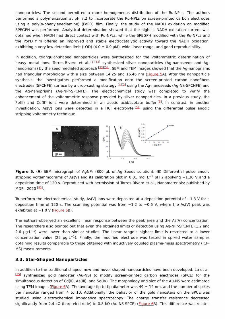
In addition, triangular-shaped nanoparticles were synthesized for the voltammetric determination ofheavy metal ions. Torres-Rivero et al. synthesized silver nanoparticles (Ag-nanoseeds and Ag-nanoprisms) by the seed mediated approach . SEM and TEM images showed that the Ag-nanoprismshad triangular morphology with a size between 14.25 and 16.46 nm (Figure 5A). After the nanoparticlesynthesis, the investigators performed a modification onto the screen-printed carbon nanofiberselectrodes (SPCNFE) surface by a drop-casting strategy using the Ag-nanoseeds (Ag-NS-SPCNFE) andthe Ag-nanoprisms (Ag-NPr-SPCNFE). The electrochemical study was completed to verify theenhancement of the voltammetric response provided by silver nanoparticles. In a previous study, thePb(II) and Cd(II) ions were determined in an acetic acid/acetate buffer . In contrast, in anotherinvestigation, As(V) ions were detected in a HCl electrolyte using the differential pulse anodicstripping voltammetry technique.
Figure 5. (A) SEM micrograph of AgNPr (800 µL of Ag Seeds solution). (B) Differential pulse anodicstripping voltammograms of As(V) and its calibration plot in 0.01 mol L pH 2 applying −1.30 V and adeposition time of 120 s. Reproduced with permission of Torres-Rivero et al., Nanomaterials; published byMDPI, 2020 .
To perform the electrochemical study, As(V) ions were deposited at a deposition potential of −1.3 V for adeposition time of 120 s. The scanning potential was from −1.2 to −0.6 V, where the As(V) peak wasexhibited at −1.0 V (Figure 5B).
The authors observed an excellent linear response between the peak area and the As(V) concentration.The researchers also pointed out that even the obtained limits of detection using Ag-NPr-SPCNFE (1.2 and2.6 µg·L ) were lower than similar studies. The linear range’s highest limit is restricted to a lowerconcentration value (25 µg·L ). Finally, the modified electrode was tested in spiked water samplesobtaining results comparable to those obtained with inductively coupled plasma-mass spectrometry (ICP-MS) measurements.
3.3. Star-Shaped Nanoparticles
In addition to the traditional shapes, new and novel shaped nanoparticles have been developed. Lu et al. synthesized gold nanostar (Au-NS) to modify screen-printed carbon electrodes (SPCE) for the
simultaneous detection of Cd(II), As(III), and Se(IV). The morphology and size of the Au-NS were estimatedusing TEM images (Figure 6A). The average tip-to-tip diameter was 49 ± 14 nm, and the number of spikesper nanostar ranged from 4 to 10. Additionally, the behavior of the gold nanostars on the SPCE wasstudied using electrochemical impedance spectroscopy. The charge transfer resistance decreasedsignificantly from 2.4 kΩ (bare electrode) to 0.8 kΩ (Au-NS-SPCE) (Figure 6B). This difference was related
[5][52][53][54]
[4][5]
[5][52]
−1
[52]
−1−1
[55]
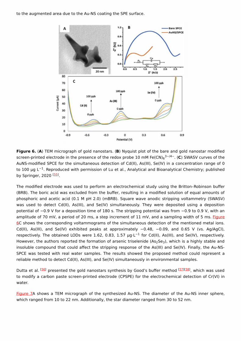
to the augmented area due to the Au-NS coating the SPE surface.
Figure 6. (A) TEM micrograph of gold nanostars. (B) Nyquist plot of the bare and gold nanostar modifiedscreen-printed electrode in the presence of the redox probe 10 mM Fe(CN) . (C) SWASV curves of theAuNS-modified SPCE for the simultaneous detection of Cd(II), As(III), Se(IV) in a concentration range of 0to 100 μg L . Reproduced with permission of Lu et al., Analytical and Bioanalytical Chemistry; publishedby Springer, 2020 .
The modified electrode was used to perform an electrochemical study using the Britton–Robinson buffer(BRB). The boric acid was excluded from the buffer, resulting in a modified solution of equal amounts ofphosphoric and acetic acid (0.1 M pH 2.0) (mBRB). Square wave anodic stripping voltammetry (SWASV)was used to detect Cd(II), As(III), and Se(IV) simultaneously. They were deposited using a depositionpotential of −0.9 V for a deposition time of 180 s. The stripping potential was from −0.9 to 0.9 V, with anamplitude of 70 mV, a period of 20 ms, a step increment of 11 mV, and a sampling width of 5 ms. Figure6C shows the corresponding voltammograms of the simultaneous detection of the mentioned metal ions.Cd(II), As(III), and Se(IV) exhibited peaks at approximately −0.48, −0.09, and 0.65 V (vs. Ag/AgCl),respectively. The obtained LODs were 1.62, 0.83, 1.57 µg·L for Cd(II), As(III), and Se(IV), respectively.However, the authors reported the formation of arsenic triselenide (As Se ), which is a highly stable andinsoluble compound that could affect the stripping response of the As(III) and Se(IV). Finally, the Au-NS-SPCE was tested with real water samples. The results showed the proposed method could represent areliable method to detect Cd(II), As(III), and Se(IV) simultaneously in environmental samples.
Dutta et al. presented the gold nanostars synthesis by Good’s buffer method , which was usedto modify a carbon paste screen-printed electrode (CPSPE) for the electrochemical detection of Cr(VI) inwater.
Figure 7A shows a TEM micrograph of the synthesized Au-NS. The diameter of the Au-NS inner sphere,which ranged from 10 to 22 nm. Additionally, the star diameter ranged from 30 to 52 nm.
63−/4−
−1[55]
−1
2 3
[56] [57][58]
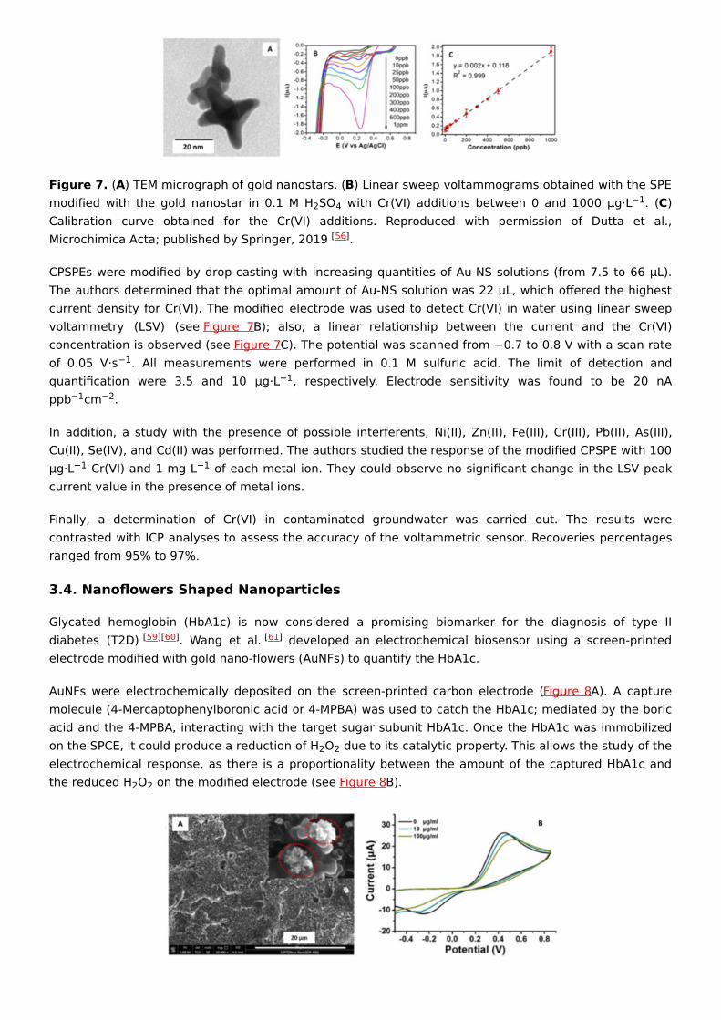
Figure 7. (A) TEM micrograph of gold nanostars. (B) Linear sweep voltammograms obtained with the SPEmodified with the gold nanostar in 0.1 M H SO with Cr(VI) additions between 0 and 1000 μg·L . (C)Calibration curve obtained for the Cr(VI) additions. Reproduced with permission of Dutta et al.,Microchimica Acta; published by Springer, 2019 .
CPSPEs were modified by drop-casting with increasing quantities of Au-NS solutions (from 7.5 to 66 µL).The authors determined that the optimal amount of Au-NS solution was 22 µL, which offered the highestcurrent density for Cr(VI). The modified electrode was used to detect Cr(VI) in water using linear sweepvoltammetry (LSV) (see Figure 7B); also, a linear relationship between the current and the Cr(VI)concentration is observed (see Figure 7C). The potential was scanned from −0.7 to 0.8 V with a scan rateof 0.05 V·s . All measurements were performed in 0.1 M sulfuric acid. The limit of detection andquantification were 3.5 and 10 µg·L , respectively. Electrode sensitivity was found to be 20 nAppb cm .
In addition, a study with the presence of possible interferents, Ni(II), Zn(II), Fe(III), Cr(III), Pb(II), As(III),Cu(II), Se(IV), and Cd(II) was performed. The authors studied the response of the modified CPSPE with 100µg·L Cr(VI) and 1 mg L of each metal ion. They could observe no significant change in the LSV peakcurrent value in the presence of metal ions.
Finally, a determination of Cr(VI) in contaminated groundwater was carried out. The results werecontrasted with ICP analyses to assess the accuracy of the voltammetric sensor. Recoveries percentagesranged from 95% to 97%.
3.4. Nanoflowers Shaped Nanoparticles
Glycated hemoglobin (HbA1c) is now considered a promising biomarker for the diagnosis of type IIdiabetes (T2D) . Wang et al. developed an electrochemical biosensor using a screen-printedelectrode modified with gold nano-flowers (AuNFs) to quantify the HbA1c.
AuNFs were electrochemically deposited on the screen-printed carbon electrode (Figure 8A). A capturemolecule (4-Mercaptophenylboronic acid or 4-MPBA) was used to catch the HbA1c; mediated by the boricacid and the 4-MPBA, interacting with the target sugar subunit HbA1c. Once the HbA1c was immobilizedon the SPCE, it could produce a reduction of H O due to its catalytic property. This allows the study of theelectrochemical response, as there is a proportionality between the amount of the captured HbA1c andthe reduced H O on the modified electrode (see Figure 8B).
2 4 −1
[56]
−1−1
−1 −2
−1 −1
[59][60] [61]
2 2
2 2
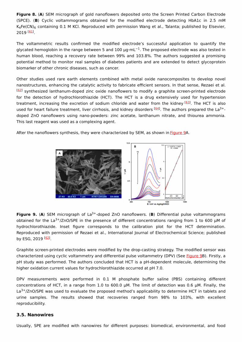
Figure 8. (A) SEM micrograph of gold nanoflowers deposited onto the Screen Printed Carbon Electrode(SPCE). (B) Cyclic voltammograms obtained for the modified electrode detecting HbA1c in 2.5 mMK Fe(CN) containing 0.1 M KCl. Reproduced with permission Wang et al., Talanta; published by Elsevier,2019 .
The voltammetric results confirmed the modified electrode’s successful application to quantify theglycated hemoglobin in the range between 5 and 100 µg·mL . The proposed electrode was also tested inhuman blood, reaching a recovery rate between 99% and 103.8%. The authors suggested a promisingpotential method to monitor real samples of diabetes patients and are extended to detect glycoproteinbiomarker of other chronic diseases, such as cancer.
Other studies used rare earth elements combined with metal oxide nanocomposites to develop novelnanostructures, enhancing the catalytic activity to fabricate efficient sensors. In that sense, Rezaei et al.
synthesized lanthanum-doped zinc oxide nanoflowers to modify a graphite screen-printed electrodefor the detection of hydrochlorothiazide (HCT). The HCT is a drug extensively used for hypertensiontreatment, increasing the excretion of sodium chloride and water from the kidney . The HCT is alsoused for heart failure treatment, liver cirrhosis, and kidney disorders . The authors prepared the La -doped ZnO nanoflowers using nano-powders: zinc acetate, lanthanum nitrate, and thiourea ammonia.This last reagent was used as a complexing agent.
After the nanoflowers synthesis, they were characterized by SEM, as shown in Figure 9A.
Figure 9. (A) SEM micrograph of La -doped ZnO nanoflowers. (B) Differential pulse voltammogramsobtained for the La /ZnO/SPE in the presence of different concentrations ranging from 1 to 600 µM ofhydrochlorothiazide. Inset figure corresponds to the calibration plot for the HCT determination.Reproduced with permission of Rezaei et al., International Journal of Electrochemical Science; publishedby ESG, 2019 .
Graphite screen-printed electrodes were modified by the drop-casting strategy. The modified sensor wascharacterized using cyclic voltammetry and differential pulse voltammetry (DPV) (See Figure 9B). Firstly, apH study was performed. The authors concluded that HCT is a pH-dependent molecule, determining thehigher oxidation current values for hydrochlorothiazide occurred at pH 7.0.
DPV measurements were performed in 0.1 M phosphate buffer saline (PBS) containing differentconcentrations of HCT, in a range from 1.0 to 600.0 µM. The limit of detection was 0.6 µM. Finally, theLa /ZnO/SPE was used to evaluate the proposed method’s applicability to determine HCT in tablets andurine samples. The results showed that recoveries ranged from 98% to 103%, with excellentreproducibility.
3.5. Nanowires
Usually, SPE are modified with nanowires for different purposes: biomedical, environmental, and food
4 6[61]
−1
[62]
[63][64] 3+
3+3+
[62]
3+
industry. In particular, nanowires are capable of interfacing with other nano-micro scale systems. Due tothe long axial morphology, nanowires have a higher surface-to-volume ratio making them similar tobiological macromolecules to create excellent nano-bio devices . Kabir et al. developed anelectrochemical sensor to detect phosphate using novel ammonium molybdate tetrahydrate/silvernanowires (AMT/AgNWs) modified SPE.
The authors prepared the AgNWs following the procedure developed by Korte et al. . AgNWs weresynthesized using silver nitrate as a precursor and polyol as a reducing agent. Additionally, CuCl or CuClwere added to reduce the remaining free Ag ions during the initial phase of AgNWs formation.
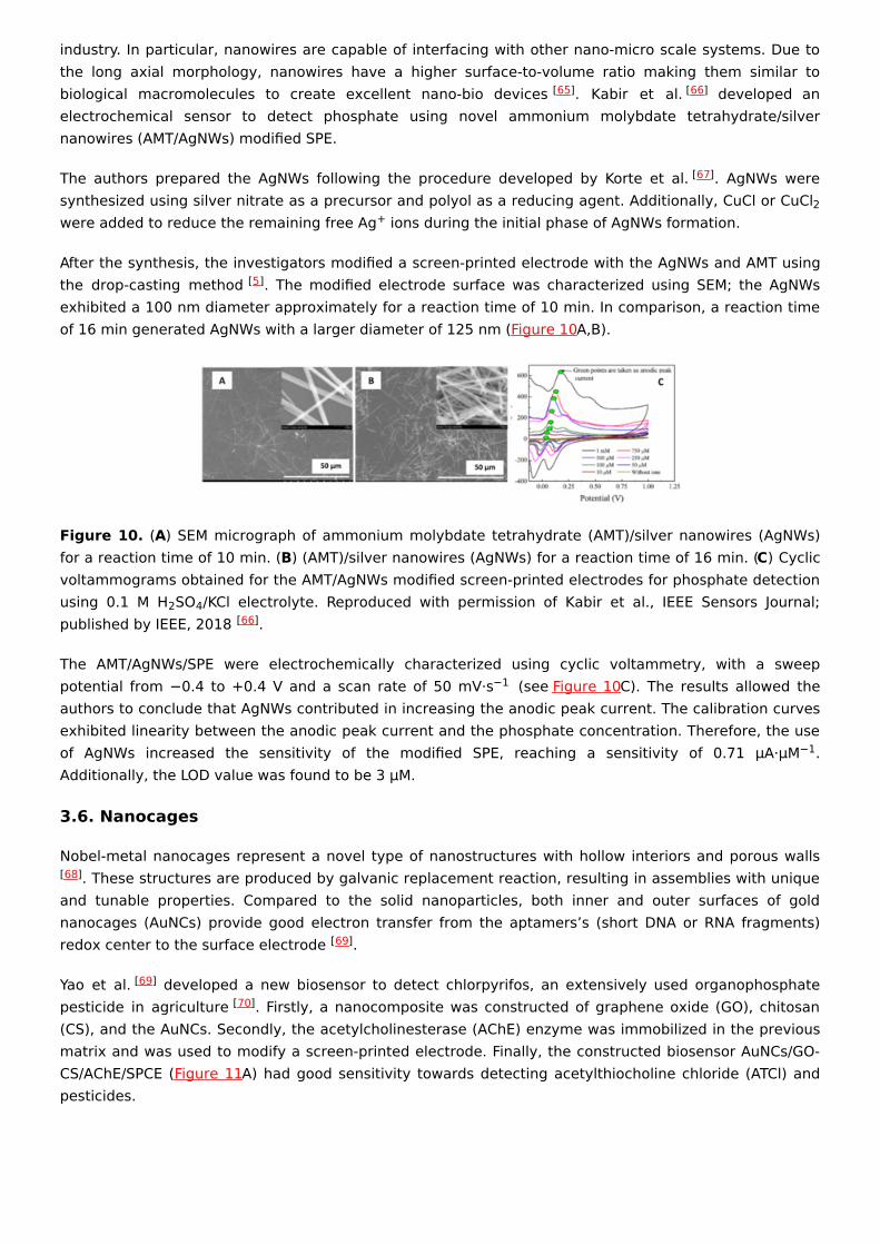
After the synthesis, the investigators modified a screen-printed electrode with the AgNWs and AMT usingthe drop-casting method . The modified electrode surface was characterized using SEM; the AgNWsexhibited a 100 nm diameter approximately for a reaction time of 10 min. In comparison, a reaction timeof 16 min generated AgNWs with a larger diameter of 125 nm (Figure 10A,B).
Figure 10. (A) SEM micrograph of ammonium molybdate tetrahydrate (AMT)/silver nanowires (AgNWs)for a reaction time of 10 min. (B) (AMT)/silver nanowires (AgNWs) for a reaction time of 16 min. (C) Cyclicvoltammograms obtained for the AMT/AgNWs modified screen-printed electrodes for phosphate detectionusing 0.1 M H SO /KCl electrolyte. Reproduced with permission of Kabir et al., IEEE Sensors Journal;published by IEEE, 2018 .
The AMT/AgNWs/SPE were electrochemically characterized using cyclic voltammetry, with a sweeppotential from −0.4 to +0.4 V and a scan rate of 50 mV·s (see Figure 10C). The results allowed theauthors to conclude that AgNWs contributed in increasing the anodic peak current. The calibration curvesexhibited linearity between the anodic peak current and the phosphate concentration. Therefore, the useof AgNWs increased the sensitivity of the modified SPE, reaching a sensitivity of 0.71 µA·µM .Additionally, the LOD value was found to be 3 µM.
3.6. Nanocages
Nobel-metal nanocages represent a novel type of nanostructures with hollow interiors and porous walls. These structures are produced by galvanic replacement reaction, resulting in assemblies with unique
and tunable properties. Compared to the solid nanoparticles, both inner and outer surfaces of goldnanocages (AuNCs) provide good electron transfer from the aptamers’s (short DNA or RNA fragments)redox center to the surface electrode .
Yao et al. developed a new biosensor to detect chlorpyrifos, an extensively used organophosphatepesticide in agriculture . Firstly, a nanocomposite was constructed of graphene oxide (GO), chitosan(CS), and the AuNCs. Secondly, the acetylcholinesterase (AChE) enzyme was immobilized in the previousmatrix and was used to modify a screen-printed electrode. Finally, the constructed biosensor AuNCs/GO-CS/AChE/SPCE (Figure 11A) had good sensitivity towards detecting acetylthiocholine chloride (ATCl) andpesticides.
[65] [66]
[67]
2+
[5]
2 4[66]
−1
−1
[68]
[69]
[69][70]
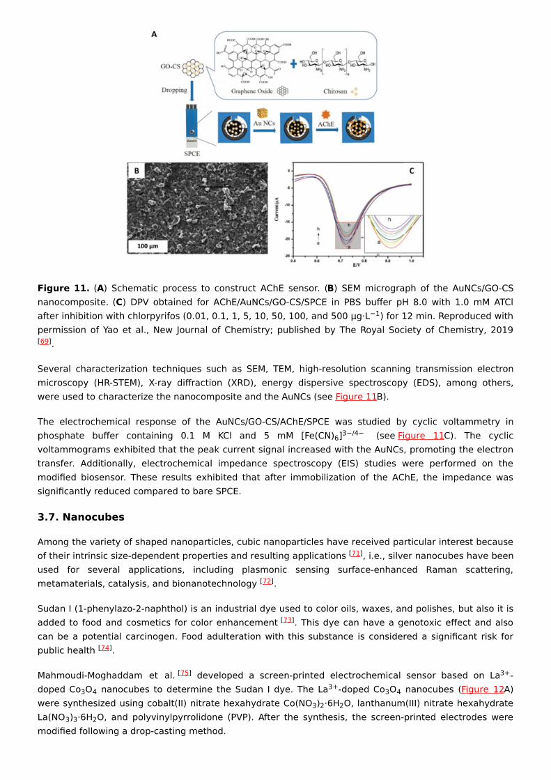
Figure 11. (A) Schematic process to construct AChE sensor. (B) SEM micrograph of the AuNCs/GO-CSnanocomposite. (C) DPV obtained for AChE/AuNCs/GO-CS/SPCE in PBS buffer pH 8.0 with 1.0 mM ATClafter inhibition with chlorpyrifos (0.01, 0.1, 1, 5, 10, 50, 100, and 500 µg·L ) for 12 min. Reproduced withpermission of Yao et al., New Journal of Chemistry; published by The Royal Society of Chemistry, 2019
.
Several characterization techniques such as SEM, TEM, high-resolution scanning transmission electronmicroscopy (HR-STEM), X-ray diffraction (XRD), energy dispersive spectroscopy (EDS), among others,were used to characterize the nanocomposite and the AuNCs (see Figure 11B).
The electrochemical response of the AuNCs/GO-CS/AChE/SPCE was studied by cyclic voltammetry inphosphate buffer containing 0.1 M KCl and 5 mM [Fe(CN) ] (see Figure 11C). The cyclicvoltammograms exhibited that the peak current signal increased with the AuNCs, promoting the electrontransfer. Additionally, electrochemical impedance spectroscopy (EIS) studies were performed on themodified biosensor. These results exhibited that after immobilization of the AChE, the impedance wassignificantly reduced compared to bare SPCE.
3.7. Nanocubes
Among the variety of shaped nanoparticles, cubic nanoparticles have received particular interest becauseof their intrinsic size-dependent properties and resulting applications , i.e., silver nanocubes have beenused for several applications, including plasmonic sensing surface-enhanced Raman scattering,metamaterials, catalysis, and bionanotechnology .
Sudan I (1-phenylazo-2-naphthol) is an industrial dye used to color oils, waxes, and polishes, but also it isadded to food and cosmetics for color enhancement . This dye can have a genotoxic effect and alsocan be a potential carcinogen. Food adulteration with this substance is considered a significant risk forpublic health .
Mahmoudi-Moghaddam et al. developed a screen-printed electrochemical sensor based on La -doped Co O nanocubes to determine the Sudan I dye. The La -doped Co O nanocubes (Figure 12A)were synthesized using cobalt(II) nitrate hexahydrate Co(NO ) ·6H O, lanthanum(III) nitrate hexahydrateLa(NO ) ·6H O, and polyvinylpyrrolidone (PVP). After the synthesis, the screen-printed electrodes weremodified following a drop-casting method.
−1
[69]
6 3−/4−
[71]
[72]
[73]
[74]
[75] 3+
3 4 3+ 3 43 2 2
3 3 2
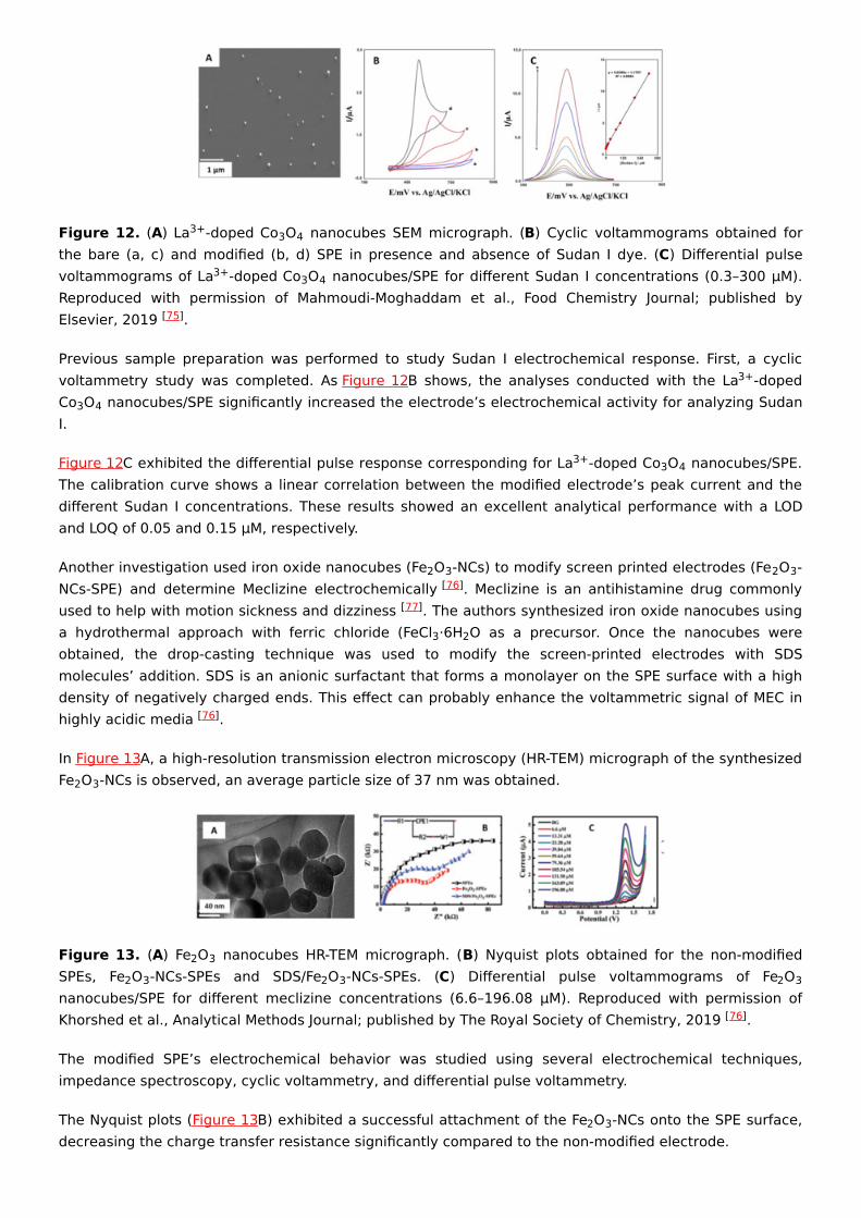
Figure 12. (A) La -doped Co O nanocubes SEM micrograph. (B) Cyclic voltammograms obtained forthe bare (a, c) and modified (b, d) SPE in presence and absence of Sudan I dye. (C) Differential pulsevoltammograms of La -doped Co O nanocubes/SPE for different Sudan I concentrations (0.3–300 µM).Reproduced with permission of Mahmoudi-Moghaddam et al., Food Chemistry Journal; published byElsevier, 2019 .
Previous sample preparation was performed to study Sudan I electrochemical response. First, a cyclicvoltammetry study was completed. As Figure 12B shows, the analyses conducted with the La -dopedCo O nanocubes/SPE significantly increased the electrode’s electrochemical activity for analyzing SudanI.
Figure 12C exhibited the differential pulse response corresponding for La -doped Co O nanocubes/SPE.The calibration curve shows a linear correlation between the modified electrode’s peak current and thedifferent Sudan I concentrations. These results showed an excellent analytical performance with a LODand LOQ of 0.05 and 0.15 µM, respectively.
Another investigation used iron oxide nanocubes (Fe O -NCs) to modify screen printed electrodes (Fe O -NCs-SPE) and determine Meclizine electrochemically . Meclizine is an antihistamine drug commonlyused to help with motion sickness and dizziness . The authors synthesized iron oxide nanocubes usinga hydrothermal approach with ferric chloride (FeCl ·6H O as a precursor. Once the nanocubes wereobtained, the drop-casting technique was used to modify the screen-printed electrodes with SDSmolecules’ addition. SDS is an anionic surfactant that forms a monolayer on the SPE surface with a highdensity of negatively charged ends. This effect can probably enhance the voltammetric signal of MEC inhighly acidic media .
In Figure 13A, a high-resolution transmission electron microscopy (HR-TEM) micrograph of the synthesizedFe O -NCs is observed, an average particle size of 37 nm was obtained.
Figure 13. (A) Fe O nanocubes HR-TEM micrograph. (B) Nyquist plots obtained for the non-modifiedSPEs, Fe O -NCs-SPEs and SDS/Fe O -NCs-SPEs. (C) Differential pulse voltammograms of Fe Onanocubes/SPE for different meclizine concentrations (6.6–196.08 µM). Reproduced with permission ofKhorshed et al., Analytical Methods Journal; published by The Royal Society of Chemistry, 2019 .
The modified SPE’s electrochemical behavior was studied using several electrochemical techniques,impedance spectroscopy, cyclic voltammetry, and differential pulse voltammetry.
The Nyquist plots (Figure 13B) exhibited a successful attachment of the Fe O -NCs onto the SPE surface,decreasing the charge transfer resistance significantly compared to the non-modified electrode.
3+ 3 4
3+ 3 4
[75]
3+
3 4
3+ 3 4
2 3 2 3[76]
[77]
3 2
[76]
2 3
2 32 3 2 3 2 3
[76]
2 3
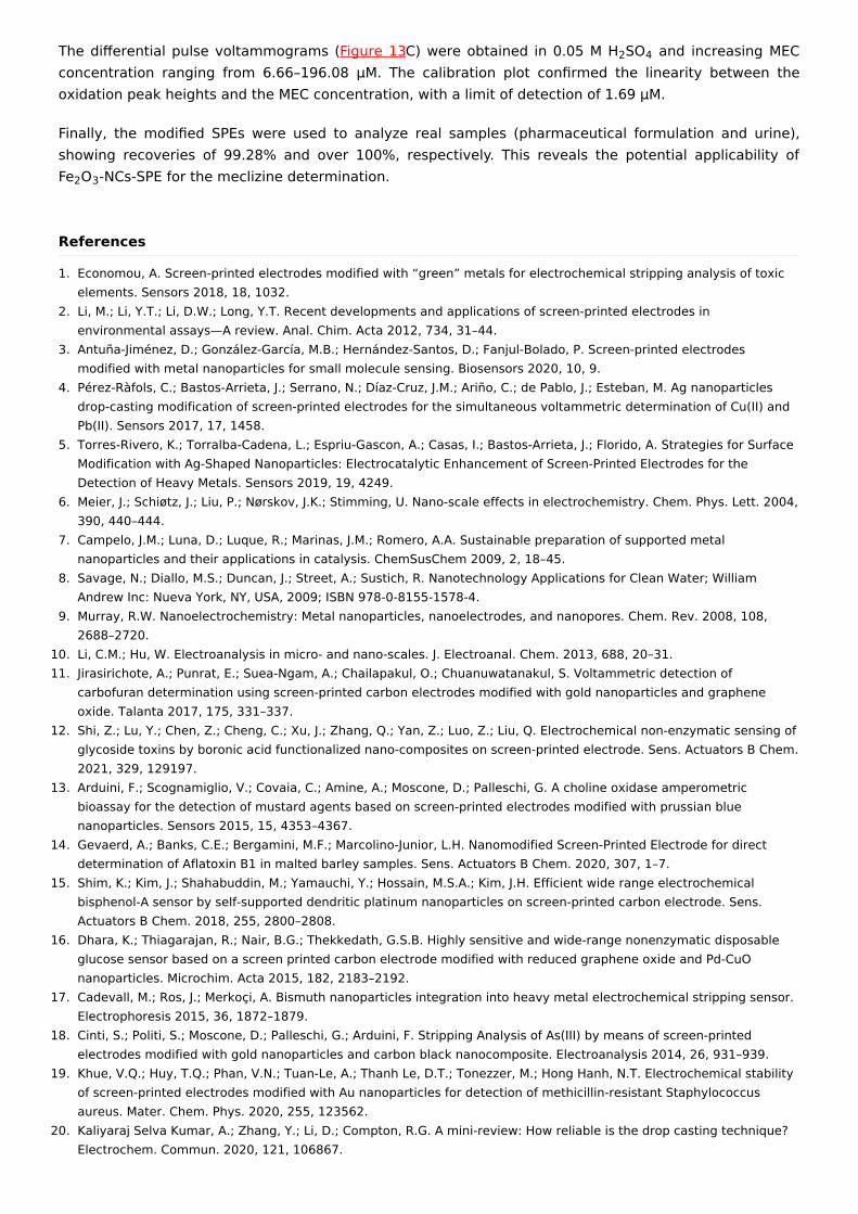
The differential pulse voltammograms (Figure 13C) were obtained in 0.05 M H SO and increasing MECconcentration ranging from 6.66–196.08 µM. The calibration plot confirmed the linearity between theoxidation peak heights and the MEC concentration, with a limit of detection of 1.69 µM.
Finally, the modified SPEs were used to analyze real samples (pharmaceutical formulation and urine),showing recoveries of 99.28% and over 100%, respectively. This reveals the potential applicability ofFe O -NCs-SPE for the meclizine determination.
2 4
2 3
References
1. Economou, A. Screen-printed electrodes modified with “green” metals for electrochemical stripping analysis of toxicelements. Sensors 2018, 18, 1032.
2. Li, M.; Li, Y.T.; Li, D.W.; Long, Y.T. Recent developments and applications of screen-printed electrodes inenvironmental assays—A review. Anal. Chim. Acta 2012, 734, 31–44.
3. Antuña-Jiménez, D.; González-García, M.B.; Hernández-Santos, D.; Fanjul-Bolado, P. Screen-printed electrodesmodified with metal nanoparticles for small molecule sensing. Biosensors 2020, 10, 9.
4. Pérez-Ràfols, C.; Bastos-Arrieta, J.; Serrano, N.; Díaz-Cruz, J.M.; Ariño, C.; de Pablo, J.; Esteban, M. Ag nanoparticlesdrop-casting modification of screen-printed electrodes for the simultaneous voltammetric determination of Cu(II) andPb(II). Sensors 2017, 17, 1458.
5. Torres-Rivero, K.; Torralba-Cadena, L.; Espriu-Gascon, A.; Casas, I.; Bastos-Arrieta, J.; Florido, A. Strategies for SurfaceModification with Ag-Shaped Nanoparticles: Electrocatalytic Enhancement of Screen-Printed Electrodes for theDetection of Heavy Metals. Sensors 2019, 19, 4249.
6. Meier, J.; Schiøtz, J.; Liu, P.; Nørskov, J.K.; Stimming, U. Nano-scale effects in electrochemistry. Chem. Phys. Lett. 2004,390, 440–444.
7. Campelo, J.M.; Luna, D.; Luque, R.; Marinas, J.M.; Romero, A.A. Sustainable preparation of supported metalnanoparticles and their applications in catalysis. ChemSusChem 2009, 2, 18–45.
8. Savage, N.; Diallo, M.S.; Duncan, J.; Street, A.; Sustich, R. Nanotechnology Applications for Clean Water; WilliamAndrew Inc: Nueva York, NY, USA, 2009; ISBN 978-0-8155-1578-4.
9. Murray, R.W. Nanoelectrochemistry: Metal nanoparticles, nanoelectrodes, and nanopores. Chem. Rev. 2008, 108,2688–2720.
10. Li, C.M.; Hu, W. Electroanalysis in micro- and nano-scales. J. Electroanal. Chem. 2013, 688, 20–31.11. Jirasirichote, A.; Punrat, E.; Suea-Ngam, A.; Chailapakul, O.; Chuanuwatanakul, S. Voltammetric detection of
carbofuran determination using screen-printed carbon electrodes modified with gold nanoparticles and grapheneoxide. Talanta 2017, 175, 331–337.
12. Shi, Z.; Lu, Y.; Chen, Z.; Cheng, C.; Xu, J.; Zhang, Q.; Yan, Z.; Luo, Z.; Liu, Q. Electrochemical non-enzymatic sensing ofglycoside toxins by boronic acid functionalized nano-composites on screen-printed electrode. Sens. Actuators B Chem.2021, 329, 129197.
13. Arduini, F.; Scognamiglio, V.; Covaia, C.; Amine, A.; Moscone, D.; Palleschi, G. A choline oxidase amperometricbioassay for the detection of mustard agents based on screen-printed electrodes modified with prussian bluenanoparticles. Sensors 2015, 15, 4353–4367.
14. Gevaerd, A.; Banks, C.E.; Bergamini, M.F.; Marcolino-Junior, L.H. Nanomodified Screen-Printed Electrode for directdetermination of Aflatoxin B1 in malted barley samples. Sens. Actuators B Chem. 2020, 307, 1–7.
15. Shim, K.; Kim, J.; Shahabuddin, M.; Yamauchi, Y.; Hossain, M.S.A.; Kim, J.H. Efficient wide range electrochemicalbisphenol-A sensor by self-supported dendritic platinum nanoparticles on screen-printed carbon electrode. Sens.Actuators B Chem. 2018, 255, 2800–2808.
16. Dhara, K.; Thiagarajan, R.; Nair, B.G.; Thekkedath, G.S.B. Highly sensitive and wide-range nonenzymatic disposableglucose sensor based on a screen printed carbon electrode modified with reduced graphene oxide and Pd-CuOnanoparticles. Microchim. Acta 2015, 182, 2183–2192.
17. Cadevall, M.; Ros, J.; Merkoçi, A. Bismuth nanoparticles integration into heavy metal electrochemical stripping sensor.Electrophoresis 2015, 36, 1872–1879.
18. Cinti, S.; Politi, S.; Moscone, D.; Palleschi, G.; Arduini, F. Stripping Analysis of As(III) by means of screen-printedelectrodes modified with gold nanoparticles and carbon black nanocomposite. Electroanalysis 2014, 26, 931–939.
19. Khue, V.Q.; Huy, T.Q.; Phan, V.N.; Tuan-Le, A.; Thanh Le, D.T.; Tonezzer, M.; Hong Hanh, N.T. Electrochemical stabilityof screen-printed electrodes modified with Au nanoparticles for detection of methicillin-resistant Staphylococcusaureus. Mater. Chem. Phys. 2020, 255, 123562.
20. Kaliyaraj Selva Kumar, A.; Zhang, Y.; Li, D.; Compton, R.G. A mini-review: How reliable is the drop casting technique?Electrochem. Commun. 2020, 121, 106867.
21. Deegan, R.D.; Bakajin, O.; Dupont, T.F.; Huber, G.; Nagel, S.R.; Witten, T.A. Capillary flow as the cause of ring stainsfrom dried liquid drops. Nature 1997, 389, 827–829.
22. Sami Yilbas, B.; Al-Sharafi, A.; Ali, H. Surfaces for Self-Cleaning. In Self-Cleaning of Surfaces and Water DropletMobility; Sami Yilbas, B., Al-Sharafi, A., Ali, H., Eds.; Elsevier: Amsterdam, The Netherlands, 2019; pp. 45–98. ISBN9780128147764.
23. Mishra, A.; Batt, N.; Bajpai, A.K. Nanostructured superhydrophobic coatings for solar panel applications. InNanomaterials-Based Coatings; Nguyen Tri, P., Rtimi, S., Ouellet Plamondon, C.M., Eds.; Elsevier: Amsterdam, TheNetherlands, 2019; pp. 397–424. ISBN 978-0-12-815884-5.
24. Zhang, J.X.J.; Hoshino, K. Fundamentals of nano/microfabrication and scale effect. In Molecular Sensors andNanodevices; Zhang, J.X.J., Hoshino, K., Eds.; Elsevier: Amsterdam, The Netherlands, 2019; pp. 43–111. ISBN 978-0-12-814862-4.
25. Chomoucka, J.; Prasek, J.; Businova, P.; Trnkova, L.; Drbohlavova, J.; Pekarek, J.; Hrdy, R.; Hubalek, J. NovelElectrochemical Biosensor for Simultaneous Detection of Adenine and Guanine Based on Cu2O Nanoparticles.Procedia Eng. 2012, 47, 702–705.
26. Mayousse, C.; Celle, C.; Moreau, E.; Mainguet, J.-F.; Carella, A.; Simonato, J.-P. Improvements in purification of silvernanowires by decantation and fabrication of flexible transparent electrodes. Application to capacitive touch sensors.Nanotechnology 2013, 24, 215501.
27. Crystals, W. Delivery of Nanoparticles on Surfaces. In Fundamentals and Applications of Nano Silicon in Plasmonicsand Fullerines; Elsevier: Amsterdam, The Netherlands, 2018; pp. 341–362. ISBN 9780323480574.
28. Girotto, C.; Rand, B.P.; Steudel, S.; Genoe, J.; Heremans, P. Nanoparticle-based, spray-coated silver top contacts forefficient polymer solar cells. Org. Electron. 2009, 10, 735–740.
29. Akhtar, M.A.; Batool, R.; Hayat, A.; Han, D.; Riaz, S.; Khan, S.U.; Nasir, M.; Nawaz, M.H.; Niu, L. FunctionalizedGraphene Oxide Bridging between Enzyme and Au-Sputtered Screen-Printed Interface for Glucose Detection. ACSAppl. Nano Mater. 2019, 2, 1589–1596.
30. Gasparotto, G.; Costa, J.P.C.; Costa, P.I.; Zaghete, M.A.; Mazon, T. Electrochemical immunosensor based on ZnOnanorods-Au nanoparticles nanohybrids for ovarian cancer antigen CA-125 detection. Mater. Sci. Eng. C 2017, 76,1240–1247.
31. Simon, A.H. Sputter Processing. In Handbook of Thin Film Deposition; Elsevier: Amsterdam, The Netherlands, 2018; pp.195–230. ISBN 9781437778731.
32. Zhang, S.; Kawakami, K. One-step preparation of chitosan solid nanoparticles by electrospray deposition. Int. J. Pharm.2010, 397, 211–217.
33. Mettakoonpitak, J.; Mehaffy, J.; Volckens, J.; Henry, C.S. AgNP/Bi/Nafion-modified Disposable Electrodes for SensitiveZn(II), Cd(II), and Pb(II) Detection in Aerosol Samples. Electroanalysis 2017, 29, 880–889.
34. Zhao, X.; Wei, C.; Gai, Z.; Yu, S.; Ren, X. Chemical vapor deposition and its application in surface modification ofnanoparticles. Chem. Pap. 2020, 74, 767–778.
35. Cinti, S.; Arduini, F.; Vellucci, G.; Cacciotti, I.; Nanni, F.; Moscone, D. Carbon black assisted tailoring of Prussian Bluenanoparticles to tune sensitivity and detection limit towards H2O2 by using screen-printed electrode. Electrochem.Commun. 2014, 47, 63–66.
36. Mohanty, U.S. Electrodeposition: A versatile and inexpensive tool for the synthesis of nanoparticles, nanorods,nanowires, and nanoclusters of metals. J. Appl. Electrochem. 2011, 41, 257–270.
37. Dominguez Renedo, O.; Ruiz Espelt, L.; García Astorgano, N.; Arcos Martinez, M.J. Electrochemical determination ofchromium(VI) using metallic nanoparticle-modified carbon screen-printed electrodes. Talanta 2008, 76, 854–858.
38. Sanllorente-Méndez, S.; Domínguez-Renedo, O.; Arcos-Martínez, M.J. Determination of arsenic(III) using platinumnanoparticle-modified screen-printed carbon-based electrodes. Electroanalysis 2009, 21, 635–639.
39. Pérez-Fernández, B.; Martín-Yerga, D.; Costa-García, A. Galvanostatic electrodeposition of copper nanoparticles onscreen-printed carbon electrodes and their application for reducing sugars determination. Talanta 2017, 175, 108–113.
40. Rafiee, B.; Fakhari, A.R. Electrocatalytic oxidation and determination of insulin at nickel oxide nanoparticles-multiwalled carbon nanotube modified screen printed electrode. Biosens. Bioelectron. 2013, 46, 130–135.
41. González-Sánchez, M.I.; Gómez-Monedero, B.; Agrisuelas, J.; Iniesta, J.; Valero, E. Electrochemical performance ofactivated screen printed carbon electrodes for hydrogen peroxide and phenol derivatives sensing. J. Electroanal.Chem. 2019, 839, 75–82.
42. Kubendhiran, S.; Sakthinathan, S.; Chen, S.M.; Lee, C.M.; Lou, B.S.; Sireesha, P.; Su, C.C. Electrochemically ActivatedScreen Printed Carbon Electrode Decorated with Nickel Nano Particles for the Detection of Glucose in Human Serumand Human Urine Sample. Int. J. Electrochem. Sci. 2016, 11, 7934–7946.
43. Wang, J.; Pedrero, M.; Sakslund, H.; Hammerich, O.; Pingarron, J. Electrochemical activation of screen-printed carbonstrips. Analyst 1996, 121, 345.
44. Niu, P.; Fernández-Sánchez, C.; Gich, M.; Navarro-Hernández, C.; Fanjul-Bolado, P.; Roig, A. Screen-printed electrodesmade of a bismuth nanoparticle porous carbon nanocomposite applied to the determination of heavy metal ions.
Microchim. Acta 2016, 183, 617–623.45. Jadav, J.K.; Umrania, V.V.; Rathod, K.J.; Golakiya, B.A. Development of silver/carbon screen-printed electrode for rapid
determination of vitamin C from fruit juices. LWT 2018, 88, 152–158.46. Ghosale, A.; Shrivas, K.; Deb, M.K.; Ganesan, V.; Karbhal, I.; Bajpai, P.K.; Shankar, R. A low-cost screen printed glass
electrode with silver nano-ink for electrochemical detection of H2O2. Anal. Methods 2018, 10, 3248–3255.47. Ali, T.A.; Mohamed, G.G. Potentiometric determination of La(III) in polluted water samples using modified screen-
printed electrode by self-assembled mercapto compound on silver nanoparticles. Sens. Actuators B Chem. 2015, 216,542–550.
48. Deng, M.; Zhang, X.; Zhang, Z.; Xin, Z.; Song, Y. A Gold Nanoparticle Ink Suitable for the Fabrication ofElectrochemical Electrode by Inkjet Printing. J. Nanosci. Nanotechnol. 2014, 14, 5114–5119.
49. Singh, M.; Jaiswal, N.; Tiwari, I.; Foster, C.W.; Banks, C.E. A reduced graphene oxide-cyclodextrin-platinumnanocomposite modified screen printed electrode for the detection of cysteine. J. Electroanal. Chem. 2018, 829, 230–240.
50. Cunha-Silva, H.; Arcos-Martinez, M.J. A disposable rhodium nanoparticle-modified screen-printed sensor for directdetermination of bromide anions. Sens. Actuators B Chem. 2019, 282, 603–608.
51. Baradoke, A.; Pastoriza-Santos, I.; González-Romero, E. Screen-printed GPH electrode modified with Ru nanoplatesand PoPD polymer film for NADH sensing: Design and characterization. Electrochim. Acta 2019, 300, 316–323.
52. Torres-Rivero, K.; Pérez-Ràfols, C.; Bastos-Arrieta, J.; Florido, A.; Martí, V.; Serrano, N. Direct As(V) determination usingscreen-printed electrodes modified with silver nanoparticles. Nanomaterials 2020, 10, 1280.
53. Aherne, D.; Ledwith, D.M.; Gara, M.; Kelly, J.M. Optical properties and growth aspects of silver nanoprisms producedby a highly reproducible and rapid synthesis at room temperature. Adv. Funct. Mater. 2008, 18, 2005–2016.
54. Aherne, D.; Gara, M.; Kelly, J.M.; Gun’Ko, Y.K. From Ag Nanoprisms to Triangular AuAg Nanoboxes. Adv. Funct. Mater.2010, 20, 1329–1338.
55. Lu, D.; Sullivan, C.; Brack, E.M.; Drew, C.P.; Kurup, P. Simultaneous voltammetric detection of cadmium(II), arsenic(III),and selenium(IV) using gold nanostar–modified screen-printed carbon electrodes and modified Britton-Robinsonbuffer. Anal. Bioanal. Chem. 2020, 412, 4113–4125.
56. Dutta, S.; Strack, G.; Kurup, P. Gold nanostar-based voltammetric sensor for chromium(VI). Microchim. Acta 2019,186, 1–7.
57. Habib, A.; Tabata, M.; Wu, Y.G. Formation of Gold Nanoparticles by Good’s Buffers. Bull. Chem. Soc. Jpn. 2005, 78,262–269.
58. Dutta, S.; Strack, G.; Kurup, P. Gold nanostar electrodes for heavy metal detection. Sens. Actuators B Chem. 2019,281, 383–391.
59. Song, S.Y.; Han, Y.D.; Park, Y.M.; Jeong, C.Y.; Yang, Y.J.; Kim, M.S.; Ku, Y.; Yoon, H.C. Bioelectrocatalytic detection ofglycated hemoglobin (HbA1c) based on the competitive binding of target and signaling glycoproteins to a boronate-modified surface. Biosens. Bioelectron. 2012, 35, 355–362.
60. Cohen, R.M.; Haggerty, S.; Herman, W.H. HbA1c for the Diagnosis of Diabetes and Prediabetes: Is It Time for a Mid-Course Correction? J. Clin. Endocrinol. Metab. 2010, 95, 5203–5206.
61. Wang, X.; Su, J.; Zeng, D.; Liu, G.; Liu, L.; Xu, Y.; Wang, C.; Liu, X.; Wang, L.; Mi, X. Gold nano-flowers (Au NFs) modifiedscreen-printed carbon electrode electrochemical biosensor for label-free and quantitative detection of glycatedhemoglobin. Talanta 2019, 201, 119–125.
62. Rezaei, R.; Foroughi, M.M.; Beitollahi, H.; Tajik, S.; Jahani, S. Synthesis of lanthanium-doped ZnO nanoflowers:Supported on graphite screen printed electrode for selective and sensitive detection of hydrochlorothiazide. Int. J.Electrochem. Sci. 2019, 14, 2038–2048.
63. González-Vargas, C.; Serrano, N.; Ariño, C.; Salazar, R.; Esteban, M.; Díaz-Cruz, J.M. Voltammetric Determination ofAnti-Hypertensive Drug Hydrochlorothiazide Using Screen-Printed Electrodes Modified with L-Glutamic Acid.Chemosensors 2017, 5, 25.
64. Heli, H.; Pishahang, J.; Amiri, H.B.; Sattarahmady, N. Synthesis of nickel nanowrinkles and its application for theelectrocatalytic oxidation and sensitive detection of hydrochlorothiazide. Microchem. J. 2017, 130, 205–212.
65. Jansi Rani, B.; Babu, E.S.; Praveenkumar, M.; Ravichandran, S.; Ravi, G.; Yuvakkumar, R. Morphology-DependentPhotoelectrochemical and Photocatalytic Performance of γ-Bi2O3 Nanostructures. J. Nanosci. Nanotechnol. 2019, 20,143–154.
66. Kabir, M.F.; Rahman, M.T.; Gurung, A.; Qiao, Q. Electrochemical Phosphate Sensors Using Silver Nanowires TreatedScreen Printed Electrodes. IEEE Sens. J. 2018, 18, 3480–3485.
67. Korte, K.E.; Skrabalak, S.E.; Xia, Y. Rapid synthesis of silver nanowires through a CuCl− or CuCl2−mediated polyolprocess. J. Mater. Chem. 2008, 18, 437–441.
68. Skrabalak, S.E.; Chen, J.; Sun, Y.; Lu, X.; Au, L.; Cobley, C.M.; Xia, Y. Gold Nanocages: Synthesis, Properties, andApplications. Acc. Chem. Res. 2008, 41, 1587–1595.
69. Yao, Y.; Wang, G.; Chu, G.; An, X.; Guo, Y.; Sun, X. The development of a novel biosensor based on goldnanocages/graphene oxide–chitosan modified acetylcholinesterase for organophosphorus pesticide detection. New J.
Retrieved from https://encyclopedia.pub/9767
Chem. 2019, 43, 13816–13826.70. Hodgson, E. Biotransformation of Individual Pesticides. In Pesticide Biotransformation and Disposition; Elsevier:
Amsterdam, The Netherlands, 2012; pp. 195–208. ISBN 9780123854810.71. Kato, K.; Dang, F.; Mimura, K.; Kinemuchi, Y.; Imai, H.; Wada, S.; Osada, M.; Haneda, H.; Kuwabara, M. Nano-sized
cube-shaped single crystalline oxides and their potentials; composition, assembly and functions. Adv. PowderTechnol. 2014, 25, 1401–1414.
72. Nano Composix Silver Nanocubes. Available online: (accessed on 9 February 2021).73. Patra, S.; Roy, E.; Madhuri, R.; Sharma, P.K. A technique comes to life for security of life: The food contaminant
sensors. In Nanobiosensors: Nanotechnology in the Agri-Food Industry; Grumezescu, A., Ed.; Academic Press:Cambridge, MA, USA, 2017; pp. 713–772. ISBN 9780128043721.
74. Agència Catalana de Seguretat Alimentària Colorants Sudan. Available online: (accessed on 9 February 2021).75. Mahmoudi-Moghaddam, H.; Tajik, S.; Beitollahi, H. Highly sensitive electrochemical sensor based on La3+ doped
Co3O4 nanocubes for determination of sudan I content in food samples. Food Chem. 2019, 286, 191–196.76. Khorshed, A.A.; Khairy, M.; Elsafty, S.A.; Banks, C.E. Disposable screen-printed electrodes modified with uniform iron
oxide nanocubes for the simple electrochemical determination of meclizine, an antihistamine drug. Anal. Methods2019, 11, 282–287.
77. Center Memorial Sloan Kettering Cancer Meclizine. Available online: (accessed on 17 March 2021).
Keywords
nanoparticles;nanomaterials;shape;screen-printed electrodes;electrochemistry;surface-modification