scott silvers, md, facep optimizing headache management in the ed: a focus on subarachnoid...
TRANSCRIPT
Scott Silvers, MD, FACEP
Optimizing Headache Optimizing Headache Management in the ED: Management in the ED:
A Focus on Subarachnoid A Focus on Subarachnoid HemorrhageHemorrhage
Scott Silvers, MD, FACEP
Scott Silvers, MD, FACEPScott Silvers, MD, FACEPAssistantAssistant ProfessorProfessor
Co-Director Primary Stroke Center Co-Director Primary Stroke Center Department of Emergency MedicineDepartment of Emergency Medicine
Mayo Clinic College of MedicineMayo Clinic College of MedicineJacksonville, FloridaJacksonville, Florida
Scott Silvers, MD, FACEP
ObjectivesObjectives
• Improve evaluation of patients for SAH
• Learn key points in diagnosis, treatment disposition, documentation
• Improve outcome of patients with SAH
• Further Emergency Medicine practice as it relates to SAH
Scott Silvers, MD, FACEP
MethodsMethods
• Case presentation
• Discussion of critical questions
• State current recommendations
• Review how we diagnose SAH
• Evaluate patient outcome
• Review ED documentation
Scott Silvers, MD, FACEP
A Clinical CaseA Clinical Case
Scott Silvers, MD, FACEP
Patient Clinical HistoryPatient Clinical History• 47 yo female
• Shopping with her husband
• Severe, sudden onset of headache
• Sat down passed out for 3-5 minutes
• Hx of HTN on diuretic
Scott Silvers, MD, FACEP
ED PresentationED Presentation• Vitals: 99.5F, 105, 16, 190/95, 98% RA
• Lying still on stretcher with eyes closed
• NCAT, Heart, lungs, abdomen normal
• “Sore” neck, no clear meningismus
• Alert, mild confusion
• CN intact, strength 5/5 all 4 ext, sensory intact, DTRs normal, FTN normal
Scott Silvers, MD, FACEP
Clinical QuestionsClinical Questions• Who is at risk for SAH?
• What symptoms suggest SAH?
• How can we best diagnose SAH?
• Who requires CT? LP? Angiography?
• When should an LP be deferred?
• When is “traumatic tap” the likely diagnosis?
• When does symptom resolution suggest a benign headache etiology?
Scott Silvers, MD, FACEP
Headache in the ED:Headache in the ED:Evidence-based Evidence-based
RecommendationsRecommendations
Scott Silvers, MD, FACEP
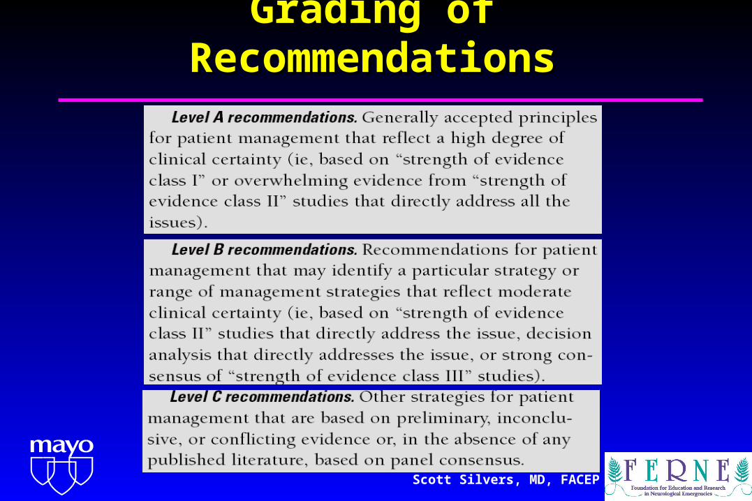
Grading of RecommendationsGrading of Recommendations

Scott Silvers, MD, FACEPAnn Emerg Med, Jan 2002; 39:108-122
ACEP Policy: Acute HeadacheACEP Policy: Acute Headache
• Does a response to therapy predict the etiology of an acute headache?– Level C: •Pain response to therapy should not be used as the sole diagnostic criteria in determining the underlying etiology of an acute headache.
Scott Silvers, MD, FACEPAnn Emerg Med, Jan 2002; 39:108-122
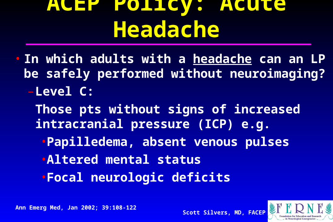
ACEP Policy: Acute HeadacheACEP Policy: Acute Headache• In which adults with a headache can an LP be
safely performed without neuroimaging?–Level C:
Those pts without signs of increased intracranial pressure (ICP) e.g.•Papilledema, absent venous pulses•Altered mental status•Focal neurologic deficits
Scott Silvers, MD, FACEPAnn Emerg Med, Jan 2002; 39:108-122
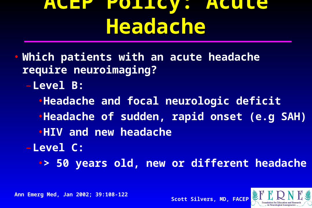
ACEP Policy: Acute HeadacheACEP Policy: Acute Headache• Which patients with an acute headache require
neuroimaging?
–Level B:
•Headache and focal neurologic deficit
•Headache of sudden, rapid onset (e.g SAH)
•HIV and new headache
–Level C:
• > 50 years old, new or different headache
Scott Silvers, MD, FACEPAnn Emerg Med, Jan 2002; 39:108-122
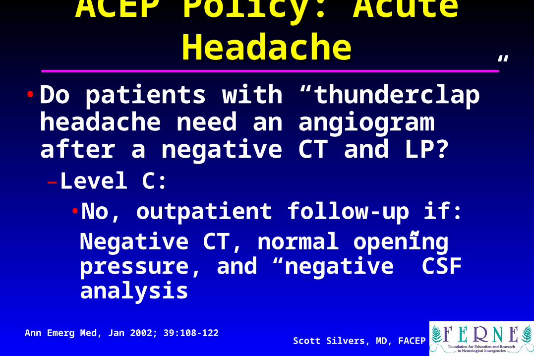
ACEP Policy: Acute HeadacheACEP Policy: Acute Headache• Do patients with “thunderclap”
headache need an angiogram after a negative CT and LP?–Level C: •No, outpatient follow-up if:Negative CT, normal opening pressure, and “negative” CSF analysis
Scott Silvers, MD, FACEPNeurol Sci (2004) 25:S 215-217
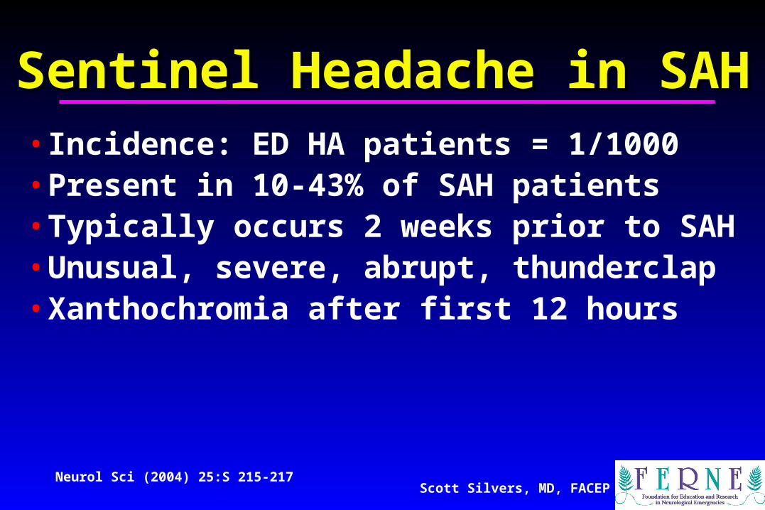
Sentinel Headache in SAHSentinel Headache in SAH• Incidence: ED HA patients = 1/1000• Present in 10-43% of SAH patients• Typically occurs 2 weeks prior to SAH• Unusual, severe, abrupt, thunderclap• Xanthochromia after first 12 hours
Scott Silvers, MD, FACEPNeurol Sci (2004) 25:S 215-217
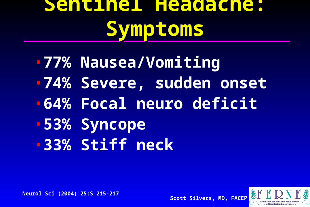
Sentinel Headache: SymptomsSentinel Headache: Symptoms
• 77% Nausea/Vomiting• 74% Severe, sudden onset• 64% Focal neuro deficit• 53% Syncope• 33% Stiff neck
Scott Silvers, MD, FACEPAnn Emer Med, Sept 1998; 32: 297-304
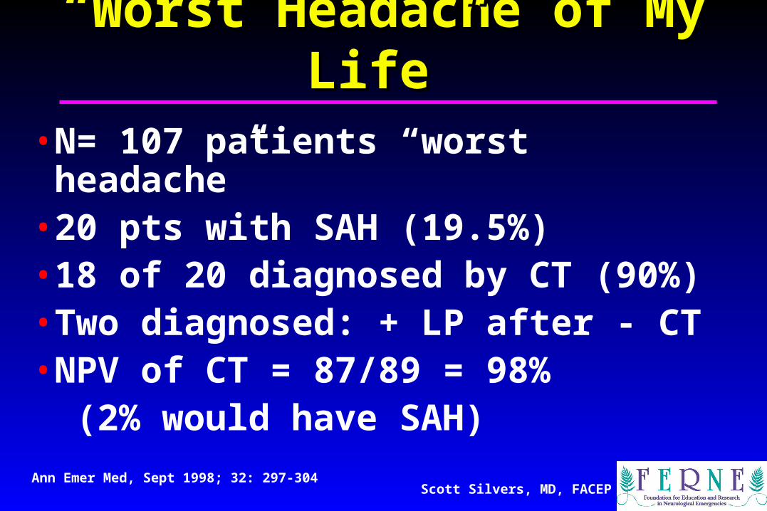
““Worst Headache of My Life”Worst Headache of My Life”• N= 107 patients “worst headache”• 20 pts with SAH (19.5%)• 18 of 20 diagnosed by CT (90%)• Two diagnosed: + LP after - CT• NPV of CT = 87/89 = 98% (2% would have SAH)

Scott Silvers, MD, FACEPAnn Emer Med, Sept 1998; 32: 297-304
““Worst Headache” LP ResultsWorst Headache” LP Results• Positive LP, Negative CT (n=2)–Tube 1 RBCs: 163,000 median
–Tube 4 RBCs: 221,000 median
• Negative LP, Negative CT (N = 77)–Tube 1 RBCs: 19 median
–Tube 4 RBCs: 0 median
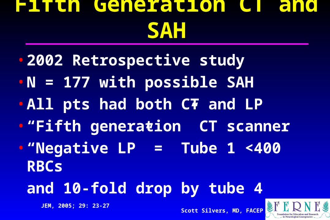
Scott Silvers, MD, FACEPJEM, 2005; 29: 23-27
Fifth Generation CT and SAHFifth Generation CT and SAH• 2002 Retrospective study • N = 177 with possible SAH• All pts had both CT and LP• “Fifth generation” CT scanner• “Negative LP” = Tube 1 <400 RBCs
and 10-fold drop by tube 4
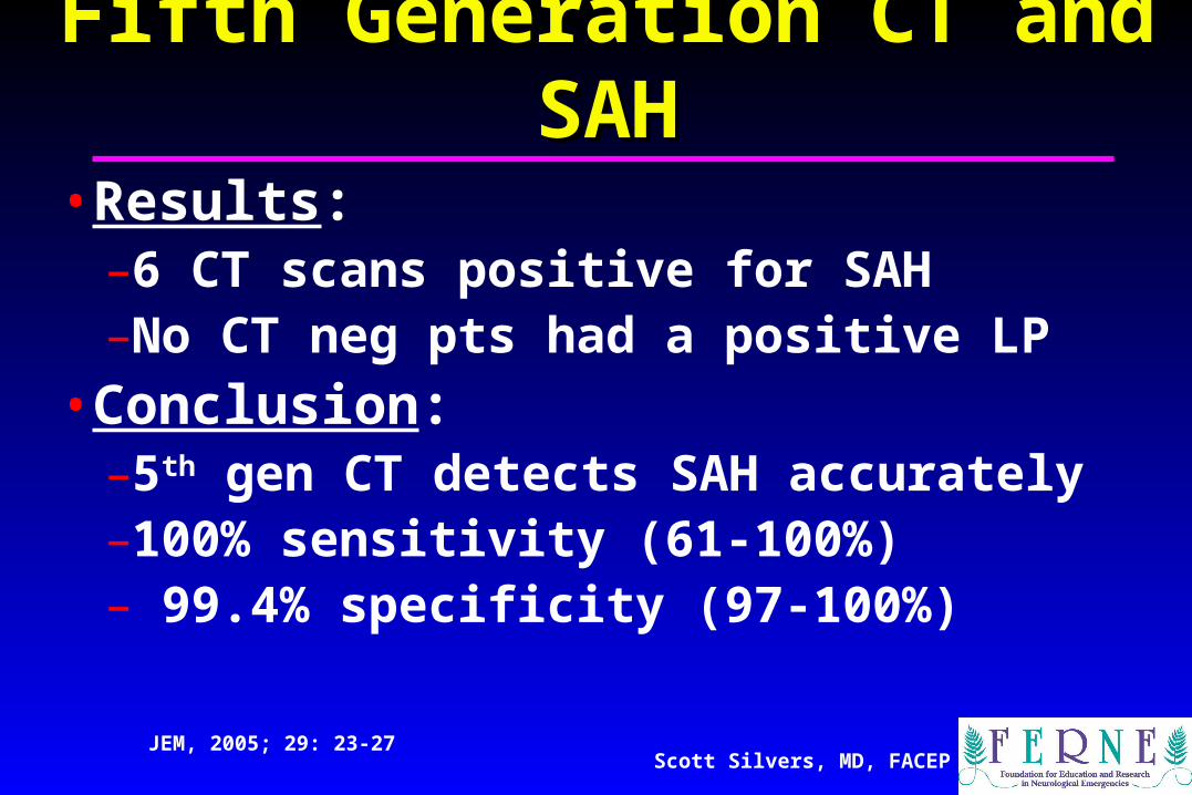
Scott Silvers, MD, FACEPJEM, 2005; 29: 23-27
Fifth Generation CT and SAHFifth Generation CT and SAH• Results:–6 CT scans positive for SAH–No CT neg pts had a positive LP
• Conclusion: –5th gen CT detects SAH accurately–100% sensitivity (61-100%)– 99.4% specificity (97-100%)
Scott Silvers, MD, FACEP
SAH: SAH: The EvaluationThe Evaluation
Scott Silvers, MD, FACEP
SAH: The EvaluationSAH: The Evaluation
• Evaluate ABCs, altered mental status
Scott Silvers, MD, FACEP
• Evaluate ABCs, altered mental status
• Know SAH risk factors:
–Hypertension, DM, prior aneurysm/SAH
–Thunderclap headache
–Maximum severity in minutes
–Focal neurological deficit
SAH: The EvaluationSAH: The Evaluation
Scott Silvers, MD, FACEP
Non-contrast CT HeadNon-contrast CT Head
• Inform radiologist to rule out SAH
• CT should be performed with sufficiently thin cuts (3 – 5 mm cuts)
• Unlikely to miss SAH on CT if performed and interpreted well
Scott Silvers, MD, FACEP
• How do we evaluate a CT for SAH?
SAH: The EvaluationSAH: The Evaluation
Scott Silvers, MD, FACEP
SAH: CT InterpretationSAH: CT Interpretation
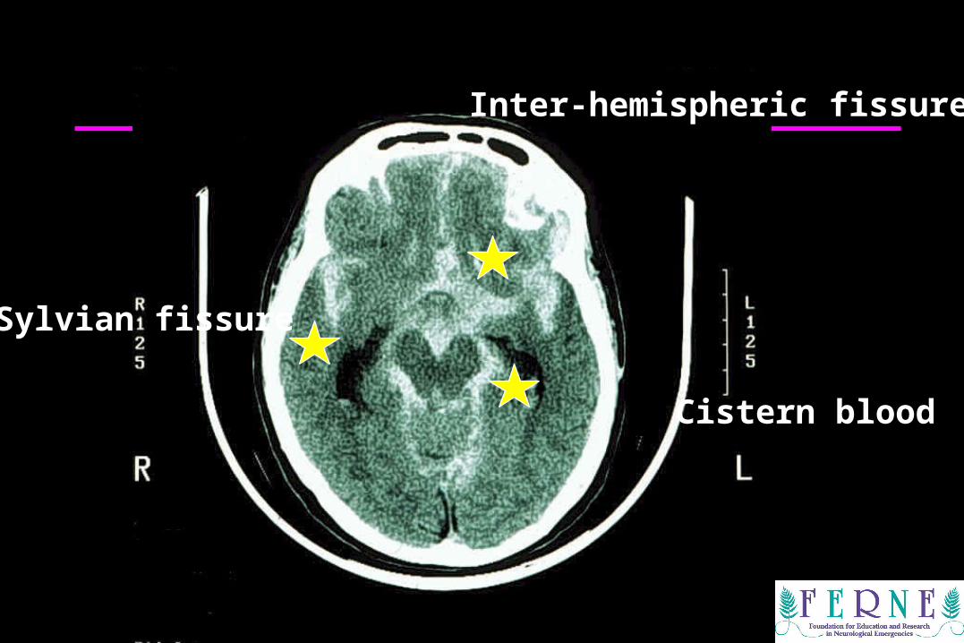
• CT evaluation for subarachnoid blood– 1) Inter-hemispheric fissure– 2) Inferior frontal sulci– 3) Third ventricle – 4) Ambient cistern – 5) Sylvian fissure
Scott Silvers, MD, FACEP
Sylvian fissure
Inter-hemispheric fissure
Cistern blood
Scott Silvers, MD, FACEP
CT Interpretation: Elevated ICPCT Interpretation: Elevated ICP
• CT findings that exclude elevated ICP–Normal cisterns–No obliteration of cistern space–No edema, mass effect, or midline shift–No hydrocephalus
Scott Silvers, MD, FACEP
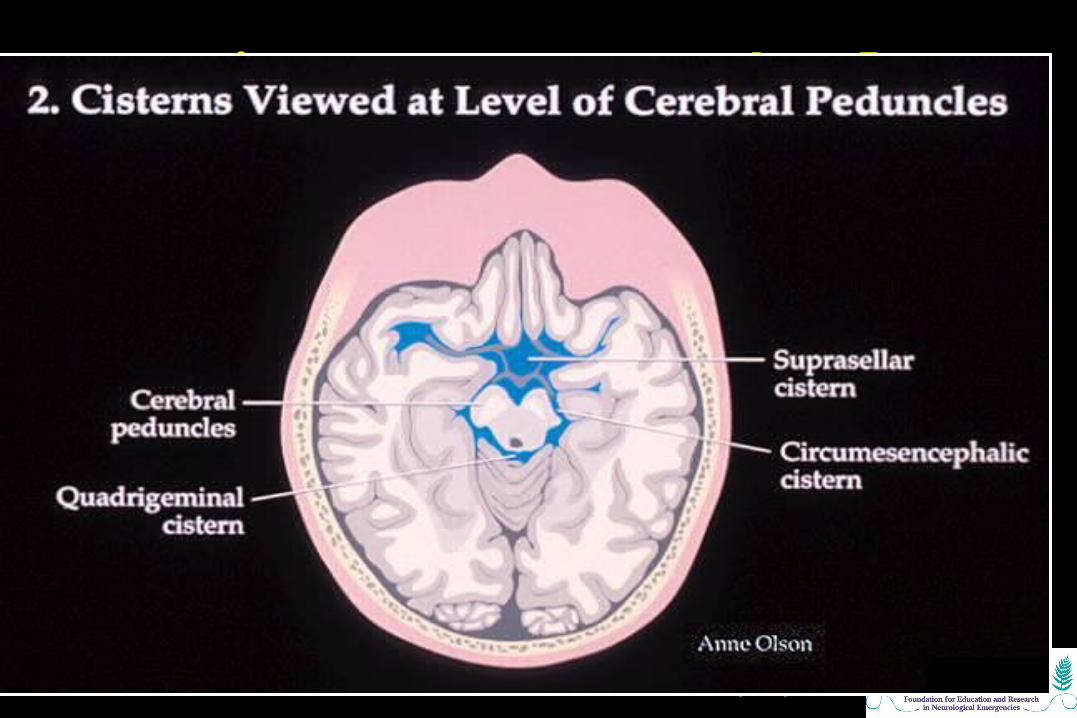
Cisterns at Cerebral Peduncles Cisterns at Cerebral Peduncles LevelLevel
Scott Silvers, MD, FACEP
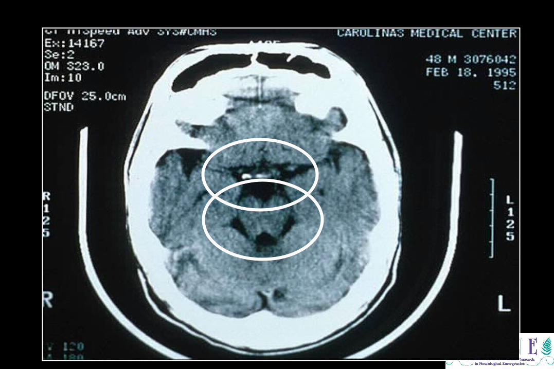
CT ScanCT Scan
Scott Silvers, MD, FACEP
Symptom ResolutionSymptom Resolution
• Can headache resolution be used to exclude SAH?
• Brings to mind another question….
In a patient who presents to the ED with a headache, can you rule out SAH by clinical evaluation alone?
Scott Silvers, MD, FACEP
Symptom ResolutionSymptom Resolution
Consider headaches likely benign if:
• Low risk SAH patient
• No focal neurological findings
• Complete symptom resolution with meds that effectively treat migraine and muscle- tension headache (i.e. non-narcotic)
Scott Silvers, MD, FACEP
Lumbar Puncture NeedLumbar Puncture Need
Which patients should have a lumbar puncture?
Scott Silvers, MD, FACEP
Lumbar Puncture IndicationsLumbar Puncture Indications
• Moderate to high risk SAH patients following negative CT
• Severe, abrupt, thunderclap headache• Focal neurological findings• Unknown CT protocol / interpretive quality• Minimal symptom resolution with meds
that effectively treat migraine and muscle- tension headache
Scott Silvers, MD, FACEP
Deferred Lumbar PunctureDeferred Lumbar Puncture
• Is it sometimes reasonable to not perform a lumbar puncture on patients suspected of SAH?
Scott Silvers, MD, FACEP
Deferred Lumbar PunctureDeferred Lumbar Puncture
• Positive CT–Evidence of elevated ICP, edema, mass
effect, midline shift, ICH, hydrocephalus
• Technically difficult procedure
• Critically ill or unstable patient
• Coagulopathy
Scott Silvers, MD, FACEP
Measuring Opening PressureMeasuring Opening Pressure• Is it necessary to measure opening
pressure when performing an LP?
Scott Silvers, MD, FACEP
Measuring Opening PressureMeasuring Opening Pressure• Variable practice….–Measure if CSF flowing rapidly
–Consider measuring with every LP

Scott Silvers, MD, FACEP
SAH: The EvaluationSAH: The Evaluation
• How should we interpret CSF results?
Scott Silvers, MD, FACEP
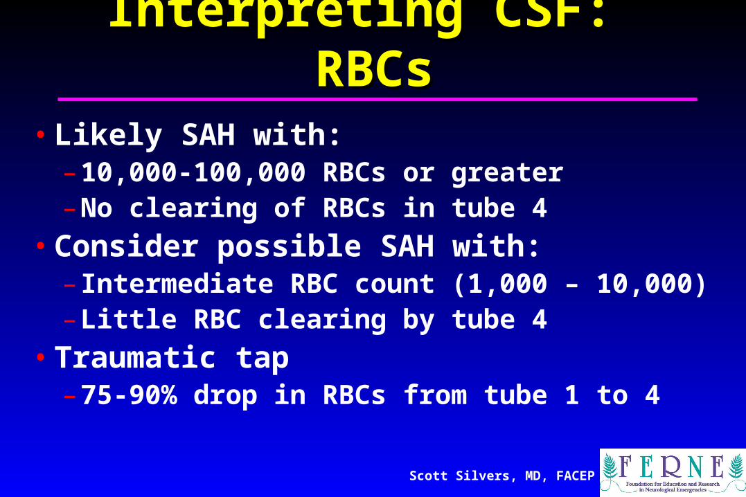
Interpreting CSF: RBCsInterpreting CSF: RBCs• Likely SAH with: –10,000-100,000 RBCs or greater–No clearing of RBCs in tube 4
• Consider possible SAH with:– Intermediate RBC count (1,000 – 10,000)–Little RBC clearing by tube 4
• Traumatic tap–75-90% drop in RBCs from tube 1 to 4

Scott Silvers, MD, FACEP
CSF XanthochromiaCSF Xanthochromia
• Xanthochromia characteristics–Typically > 12 hours from headache onset
–Quanitative and qualitative measurements
“Read news print test” most often used
–Clears after weeks
Scott Silvers, MD, FACEP
SAH: The EvaluationSAH: The Evaluation
• When is angiography indicated?
Scott Silvers, MD, FACEP
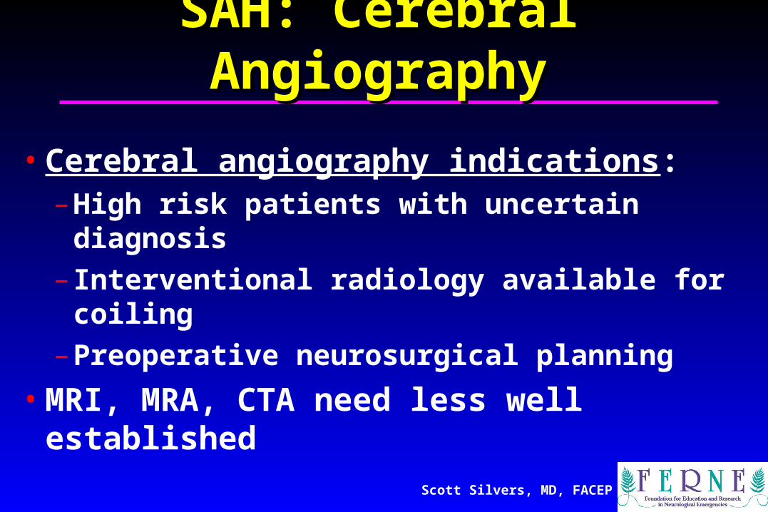
SAH: Cerebral AngiographySAH: Cerebral Angiography
• Cerebral angiography indications:–High risk patients with uncertain diagnosis
– Interventional radiology available for coiling
–Preoperative neurosurgical planning
• MRI, MRA, CTA need less well established
Scott Silvers, MD, FACEP
SAH: TreatmentSAH: Treatment
• How should be treat patients with SAH?
Scott Silvers, MD, FACEP
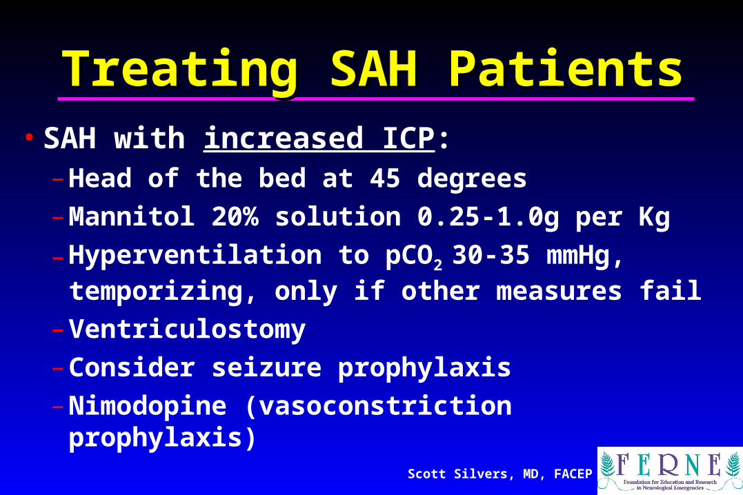
Treating SAH PatientsTreating SAH Patients• SAH with increased ICP:–Head of the bed at 45 degrees
–Mannitol 20% solution 0.25-1.0g per Kg
–Hyperventilation to pCO2 30-35 mmHg, temporizing, only if other measures fail
–Ventriculostomy
–Consider seizure prophylaxis
–Nimodopine (vasoconstriction prophylaxis)
Scott Silvers, MD, FACEP
ED Case ED Case Patient OutcomePatient Outcome
Scott Silvers, MD, FACEP
ED Patient ManagementED Patient Management
• Pt had a generalized tonic-clonic seizure• Responded to benzodiazapines• Return to normal mental status
Scott Silvers, MD, FACEP
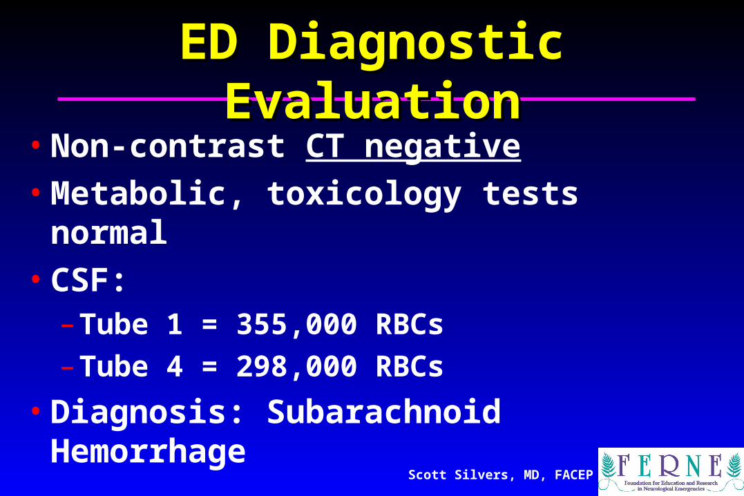
ED Diagnostic EvaluationED Diagnostic Evaluation• Non-contrast CT negative
• Metabolic, toxicology tests normal
• CSF:–Tube 1 = 355,000 RBCs
–Tube 4 = 298,000 RBCs
• Diagnosis: Subarachnoid Hemorrhage
Scott Silvers, MD, FACEP
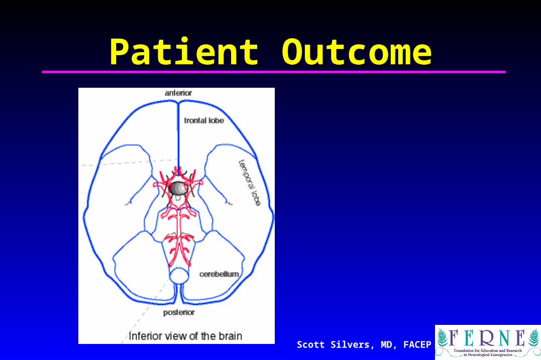
Patient OutcomePatient Outcome• Cerebral angiogram performed
• Saccular aneurysm in the posterior communicating artery
• Neurosurgical aneurysm clipping
• Pt was discharged in one week
• No residual neurological deficit
Scott Silvers, MD, FACEP
Patient OutcomePatient Outcome
Scott Silvers, MD, FACEPferne_2005_acep_sa_silvers_BIC_SAH_fshow
Questions??Questions??
[email protected]@ferne.org
Scott Silvers, MD, FACEPScott Silvers, MD, [email protected]@mayo.edu
(904) 296 - 5741(904) 296 - 5741